non-invasive discrimnation between diabetic states...
TRANSCRIPT

NON-INVASIVE DISCRIMNATION BETWEEN DIABETIC STATES (HBA1C<8% AND HBA1C>10%) USING
PHOTOPLETHYSMOGRAPHY
SAHNIUS BT USMAN
THESIS SUBMITTED IN FULFILMENT FOR THE DEGREE OF DOCTOR OF PHILOSOPHY
FACULTY OF ENGINEERING AND BUILT ENVIRONMENT UNIVERSITI KEBANGSAAN MALAYSIA
BANGI
2015

DISKRIMINASI TIDAK INVASIF ANTARA KEADAAN DIABETIK (HBA1C<8% DAN HBA1C>10%) MENGGUNAKAN
FOTOPLETISMOGRAFI
SAHNIUS BT USMAN
TESIS YANG DIKEMUKAKAN UNTUK MEMPEROLEH IJAZAH DOKTOR FALSAFAH
FAKULTI KEJURUTERAAN DAN ALAM BINA UNIVERSITI KEBANGSAAN MALAYSIA
BANGI
2015

iv
ACKNOWLEDGEMENT
First and foremost I express my utmost gratitude to ALLAH, the Almighty, without whose mercy and blessing, this work would ever be possible. Next, I would like to express my sincere gratitude to my supervisors Prof. Dr. Md. Mamun Bin Ibne Reaz and Prof. Dr. Mohd Alauddin Mohd Ali for their excellent supervisions especially in giving important technical advice, invaluable guidance, many fruitful discussions and providing constant support and cooperation towards making this research progressive over the years. Additional thankful is extended to my employer, Universiti Teknologi Malaysia for allowing me to take full study leave to concentrate on the research as well as Kementerian Pengajian Tinggi (KPT) for sponsoring the study leave.
I would like to thank Universiti Kebangsaan Malaysia for sponsoring this work under Research University Grant: UKM-AP-TKP-07-2009. I am most grateful to all personnel in the Endocrine Clinic KPP2 at Pusat Perubatan Universiti Kebangsaan Malaysia for their kind contribution and cooperation.
Last but not least, this special appreciation is extended to my life-companion and beloved husband for his caring, understanding, patience, motivation, and never-ending support and encouragement. Not to forget, I also extend my exceptional appreciation towards my young and lovely children whom are the most precious in my life. They are my motivations and inspirations to continuously educate and furnish myself with sufficient knowledge and the completion of this study is part of it.
Sincerely, thank you to all!

v
ABSTRACT Diabetes mellitus is a group of metabolic diseases associated with the production and/or reaction of insulin leading to hyperglycemia. Glycated hemoglobin (HbA1c) level is generally measured for hyperglycemia. The risk of developing complications depends on both the duration of diabetes and hyperglycemia. A trend of increasing arterial stiffness has been identified in type 2 diabetes. Photoplethysmographic (PPG) pulse wave provides a ‘window’ into the properties of small arteries whereas stiffening of these arteries will alter the PPG waveform. In this research, the potential of PPG in discriminating between type 2 diabetic patients at risk of having HbA1c level > 10% has been investigated. To this end, PPG signals recorded from diabetic patients with different levels of HbA1c (HbA1c level < 8% and HbA1c level > 10%) were acquired from the index finger of the right arm of 101 subjects (53 subjects with HbA1c level < 8% and 48 subjects with HbA1c level > 10%) at a sampling rate of 275 Hz. The area under the curve of PPG (auc-PPG) was proposed in analyzing the PPG pulse contour. Results of t-test analysis show that auc-PPG is significantly larger in diabetic patients with HbA1c level < 8% than in those with HbA1c level > 10% (p-value <0.001). Repeated measurement of PPG using paired t-test on 30 diabetic patients with HbA1c level < 8% and 26 diabetic patients with HbA1c level > 10% (total 56 subjects) show that there is no significant difference in the mean value of auc-PPG between the first measurement and repeated measurement for both groups. Finally, a logistic regression model for estimating the risk of having HbA1c level > 10% among diabetic patients was estimated using data from 51 female diabetic patients. The model shows that the auc-PPG is an independent predictor for estimating the risk of having HbA1c level > 10% (p-value = 0.005) among female diabetic patients.

vi
ABSTRAK Diabetes mellitus adalah sekumpulan penyakit metabolik yang yang dikaitkan dengan pengeluaran dan/atau tindak balas insulin yang menyebabkan hiperglisemia. Paras ‘glycated hemoglobin’(HbA1c) merupakan salah satu ukuran bagi hiperglisemia. Risiko komplikasi bergantung kepada tempoh menghidap diabetes dan hiperglisemia. Suatu trend peningkatan ketegangan arteri telah dikenalpasti pada pesakit diabetes mellitus jenis 2. Gelombang denyut photoplethysmographic (PPG) menyediakan suatu 'jendela' kepada sifat-sifat arteri dan ketegangan arteri-arteri tersebut boleh mengubah bentuk gelombang PPG. Dalam kajian ini, potensi PPG dalam membezakan antara pesakit diabetes mellitus jenis 2 yang berisiko mempunyai HbA1c >10% telah dikaji. Kemudian, isyarat PPG yang direkodkan daripada pesakit diabetes dengan paras HbA1c yang berbeza (paras HbA1c < 8% dan paras HbA1c > 10%) telah diperolehi daripada jari telunjuk tangan kanan 101 orang subjek (53 orang subjek dengan paras HbA1c < 8% dan 48 orang subjek dengan paras HbA1c > 10%) pada kadar persampelan 275 Hz. Kawasan bawah lengkung PPG (auc-PPG) adalah teknik yang dicadangkan untuk membuat analisis kontur isyarat PPG. Keputusan analisis ‘independent t-test’ menunjukkan bahawa auc-PPG secara signifikannya lebih besar pada pesakit diabetes dengan paras HbA1c < 8% daripada mereka yang mempunyai paras HbA1c >10% (nilai p < 0.001). Pengukuran ulangan PPG menggunakan ‘paired t-test’ pada 30 orang pesakit diabetes mellitus dengan paras HbA1c < 8% dan 26 orang pesakit diabetes mellitus dengan HbA1c > 10% (berjumlah 56 orang subjek) menunjukkan bahawa tiada terdapat perbezaan yang signifikan dalam nilai min auc-PPG antara pengukuran pertama dan pengukuran ulangan untuk kedua-dua kumpulan. Akhirnya, model regresi logistik jangkaan risiko mempunyai paras HbA1c > 10% di kalangan pesakit diabetes mellitus telah dianggarkan dengan menggunakan data daripada 51 orang pesakit diabetes wanita. Model tersebut menunjukkan bahawa auc-PPG adalah suatu pembolehubah tak bersandar bagi menjangkakan risiko mempunyai paras HbA1c > 10% (nilai p = 0.005) di kalangan pesakit diabetes wanita.
vii
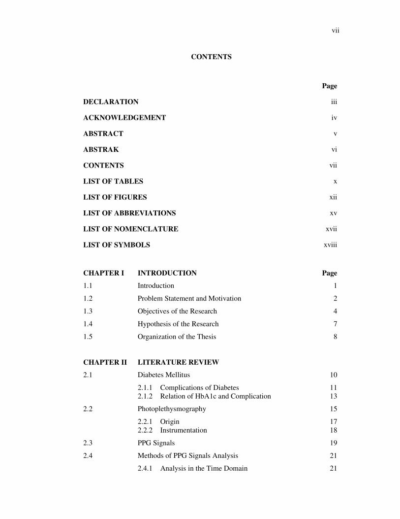
CONTENTS
Page
DECLARATION iii
ACKNOWLEDGEMENT iv
ABSTRACT v
ABSTRAK vi
CONTENTS vii
LIST OF TABLES x
LIST OF FIGURES xii
LIST OF ABBREVIATIONS xv
LIST OF NOMENCLATURE xvii
LIST OF SYMBOLS xviii
CHAPTER I INTRODUCTION Page
1.1 Introduction 1
1.2 Problem Statement and Motivation 2
1.3 Objectives of the Research 4
1.4 Hypothesis of the Research 7
1.5 Organization of the Thesis 8
CHAPTER II LITERATURE REVIEW
2.1 Diabetes Mellitus 10
2.1.1 Complications of Diabetes 11 2.1.2 Relation of HbA1c and Complication 13
2.2 Photoplethysmography 15
2.2.1 Origin 17 2.2.2 Instrumentation 18
2.3 PPG Signals 19
2.4 Methods of PPG Signals Analysis 21
2.4.1 Analysis in the Time Domain 21

viii
2.4.2 Analysis in the Frequency Domain 25 2.4.3 Bilateral PPG Analysis 28
2.5 Chapter Summary 29
CHAPTER III METHODS
3.1 Introduction 30
3.2 PPG Measurement System and Setup 30
3.3 Clinical Data Acquisition 33
3.3.1 Clinical Research Ethical Issues 33 3.3.2 Descriptions of Sample Population 34 3.3.3 Protocols for Data Acquisition 34
3.4 PPG Data Analysis 38
3.5 Statistical Analysis 48
3.5.1 Statistical Terminology 48 3.5.2 Sample Size Estimation 56 3.5.3 Statistical Tests 58
3.6 Chapter Summary 60
CHAPTER IV RESULTS AND DISCUSSION
4.1 Analysis of PPG Results 62
4.1.1 Experimental Results and Discussion 62 4.1.2 Conclusion 68
4.2 Statistical Analysis of Data 68
4.2.1 Descriptive Statistics of Data Population 69 4.2.2 Tests of Normal Distribution 70 4.2.3 Comparing Variables Between Two Independent
Groups 72
4.2.4 Comparing the Effect of Age on Auc-PPG 79 4.2.5 Conclusion 80
4.3 Study of Repeatability 81
4.3.1 Experimental Results and Discussion 82 4.3.2 Conclusion 89
4.4 Statistical Modeling of Auc-PPG 89
4.4.1 Experimental Results and Discussions 89 4.4.2 Conclusion 95
4.5 Chapter Summary 95

ix
CHAPTER V CONCLUSION AND SUGGESTIONS FOR FUTURE WORK
5.1 Conclusion 97
5.2 Suggestions for Future Work 98
5.2.1 PPG Recording System and Data Acquisition 98 5.2.2 Data Analysis and Sample Population 99
REFERENCES 100
APPENDIX
A List of Publications 115
B Proposal to Conduct Study 116
C Research & Ethics Committee PPUKM Study Approval 127
D Results of Additional Figures 129
E Results of Statistical Test 134
F Listing of MATLAB Program Codes 141

x
LIST OF TABLES
Table No. Page
2.1 Comparison of several arterial stiffness studies 14
3.1 Example of tabulated data after completion of Step 3 and Step 4 44
3.2 Example of tabulated data after completion of Step 6 45
3.3 Contingency table 51
3.4 Relationships among statistical terms 53
4.1 Auc-PPG for 15 patients 67
4.2 Distributions of the studied subjects by ethnicity 69
4.3 List of skewness and kurtosis for diabetic patients with HbA1c<8% 71
4.4 List of skewness and kurtosis for diabetic patients with HbA1c>10% 71
4.5 Levene’s test for equality of variances 73
4.6 Characteristics of the studied subjects 74
4.7 Listing of effect size 75
4.8 Mann-Whitney U test 75
4.9 Mean and standard deviation of auc-PPG 76
4.10 Mean and standard deviation of auc-PPG 79
4.11 Analysis of covariance for auc-PPG 79
4.12 Mean and standard deviation of auc-PPG for diabetic patients with HbA1c<8% and those with HbA1c>10%
80
4.13 Characteristics of the studied subjects in the first measurement 85
4.14 Listing of effect size 85
4.15 Characteristics of the studied subjects in the second measurement 86
4.16 Listing of effect size 87

xi
4.17 Mean and standard deviation of auc-PPG for both measurements 88
4.18 Mean and standard deviation of HbA1c for both measurements 88
4.19 Correlations between pairs of variables 90
4.20 Classification of HbA1c level for the developed model 90
4.21 Classification results for all index parameter from PPG pulse 91
4.22 Variables used in performing logistic regression 91
4.23 Classification results for group of female aged within 50 years to 59 years
93
4.24 Classification results for group of female aged within 60 years to 69 years
93
4.25 Variables used in performing logistic regression 94

xii
LIST OF FIGURES
Figure No. Page
1.1 The structure of the research 5
1.2 The structure of the measurement and instrumentation systems 5
1.3 The structure of the data collection 6
1.4 The structure of the data analysis 6
1.5 The structure of the technique validation 7
2.1 Arrangement of LED and photodetector in (a) transmission and (b) reflection mode PPG sensors
19
2.2 PPG waveforms generated from the variations of absorbed and transmitted light in living tissue
20
2.3 The absorption spectrum of HbO2 and Hb 21
2.4 Characteristic of a single pulse PPG signal 22
2.5 The systolic and diastolic component of the PPG signal 24
2.6 Second derivative of the PPG waveform 25
2.7 FFT spectra and estimates of PSD 27
2.8 HRV spectrums 28
3.1 Snapshot of the Dolphin ONE software GUI screen display 31
3.2 Dolphin ONE sensor with two extra mounted red LEDs 31
3.3 Experimental setup for PPG measurement 32
3.4 Recording the PPG signal 32
3.5 Close-up of PPG signal 33
3.6 Socio-demographic and medical records detail 35
3.7 Snapshot of the SPSS software GUI screen display for ‘complete data.sav’
36
xiii
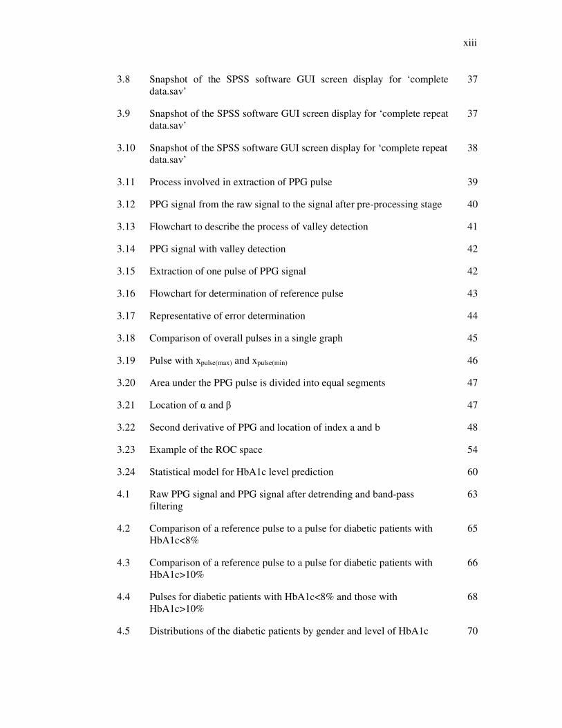
3.8 Snapshot of the SPSS software GUI screen display for ‘complete data.sav’
37
3.9 Snapshot of the SPSS software GUI screen display for ‘complete repeat data.sav’
37
3.10 Snapshot of the SPSS software GUI screen display for ‘complete repeat data.sav’
38
3.11 Process involved in extraction of PPG pulse 39
3.12 PPG signal from the raw signal to the signal after pre-processing stage 40
3.13 Flowchart to describe the process of valley detection 41
3.14 PPG signal with valley detection 42
3.15 Extraction of one pulse of PPG signal 42
3.16 Flowchart for determination of reference pulse 43
3.17 Representative of error determination 44
3.18 Comparison of overall pulses in a single graph 45
3.19 Pulse with xpulse(max) and xpulse(min) 46
3.20 Area under the PPG pulse is divided into equal segments 47
3.21 Location of α and β 47
3.22 Second derivative of PPG and location of index a and b 48
3.23 Example of the ROC space 54
3.24 Statistical model for HbA1c level prediction 60
4.1 Raw PPG signal and PPG signal after detrending and band-pass filtering
63
4.2 Comparison of a reference pulse to a pulse for diabetic patients with HbA1c<8%
65
4.3 Comparison of a reference pulse to a pulse for diabetic patients with HbA1c>10%
66
4.4 Pulses for diabetic patients with HbA1c<8% and those with HbA1c>10%
68
4.5 Distributions of the diabetic patients by gender and level of HbA1c 70

xiv
4.6 Distributions of the auc-PPG by histogram for diabetic patients with (a) HbA1c<8% (b) HbA1>10%
70
4.7 Gender distribution ofdiabetic patients with a) HbA1c<8% and b) HbA1c>10%
72
4.8 Distributions of the diabetic patients by level of HbA1c and gender 72
4.9 Boxplot of auc-PPG for diabetic patients with different level of HbA1c 74
4.10 Relationship of auc-PPG with a) BP diastolic, b) LDL, c) age and d) HbA1c
77
4.11 Interaction between level of HbA1c, hypertension and auc-PPG 78
4.12 Interaction between hypertension, level of HbA1c and auc-PPG 78
4.13 Gender distribution for each group of diabetic patients 83
4.14 Distributions of the diabetic patients by level of HbA1c and gender 83
4.15 Boxplot of age for diabetic patients with different levels of HbA1c 84
4.16 Boxplot of duration of diabetes for diabetic patients with different levels of HbA1c
84
4.17 Boxplot of auc-PPG for diabetic patients with different level of HbA1c 86
4.18 Boxplot of auc-PPG for diabetic patients with different level of HbA1c 87
4.19 Accuracy graph for different cut-off points 91
4.20 The ROC curve to estimate the HbA1c level >10% 92
4.21 Accuracy graph for different cut-off points for (a) group of female aged within 50 years to 59 years and (b) group of female aged within 60 years to 69 years
94
4.22 The ROC curve to estimate the HbA1c level >10% 95

xv
LIST OF ABBREVIATIONS
ADA American Diabetes Association
AIx Aortic augmentation index
AGEs Advanced glycation end products
AM Amplitude
ANOVA Analysis of variance
ANCOVA Analysis of covariance
APSS Air pressure sensing system
ASCII American Standard Code for Information Interchange
Auc-PPG Area under the curve of PPG
Ba-PWV Brachial-ankle pulse wave velocity
BL Baseline
BMI Body mass index
BP Blood pressure
CAD Coronary artery disease
cf-PWV Carotid to femoral PWV
CHD Coronary heart disease
CI Confidence interval
CR Coefficient of correlation
CRP C-reactive protein
CV Coefficient of variation
CVD Cardiovascular diseases
ECG Electrocardiograph/Electrocardiogram
FFT Fast Fourier Transform
FN False negative
FP False positive
GCP Good clinical practice
GDM Gestational diabetes
GUI Graphical user interface
HDL High-density lipoprotein
HF High frequency
HRV Heart rate variability
xvi
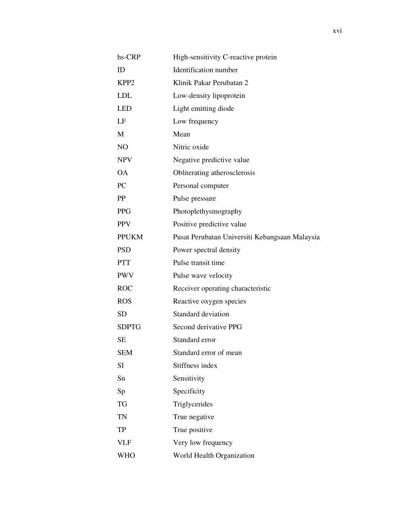
hs-CRP High-sensitivity C-reactive protein
ID Identification number
KPP2 Klinik Pakar Perubatan 2
LDL Low-density lipoprotein
LED Light emitting diode
LF Low frequency
M Mean
NO Nitric oxide
NPV Negative predictive value
OA Obliterating atherosclerosis
PC Personal computer
PP Pulse pressure
PPG Photoplethysmography
PPV Positive predictive value
PPUKM Pusat Perubatan Universiti Kebangsaan Malaysia
PSD Power spectral density
PTT Pulse transit time
PWV Pulse wave velocity
ROC Receiver operating characteristic
ROS Reactive oxygen species
SD Standard deviation
SDPTG Second derivative PPG
SE Standard error
SEM Standard error of mean
SI Stiffness index
Sn Sensitivity
Sp Specificity
TG Triglycerides
TN True negative
TP True positive
VLF Very low frequency
WHO World Health Organization

xvii
LIST OF NOMENCLATURES
Hb Deoxygenated hemoglobin
HbA1c Glycated hemoglobin
HbO2 Oxygenated hemoglobin
SpO2 Blood oxygen saturation

xviii
LIST OF SYMBOLS
π Probability of the outcome of interest or ‘event’
a Y intercept
B Regression coefficient
ln Natural logarithm
∆T Time delay
Ho Null hypothesis
p-value Significance level (probability of rejecting the Ho)
R Correlation coefficient
Z Probability distribution

CHAPTER I
INTRODUCTION
1.1 INTRODUCTION
Diabetes mellitus also known as diabetes is defined as abnormally high blood glucose
with disturbances of protein metabolism and altered fat (Iftikhar & Waqar 2011).
Diabetes has been a silent killer all over the world. Hence, the primary goal of any
diabetes treatment is to maintain the glucose level and the risk of developing late-
stage diabetic complications. A stable blood glucose level would significantly reduce
any cardiovascular disease (CVD) among diabetic patients.
There are two major types of diabetes which are Type 1 diabetes and Type 2
diabetes. The absence (Type 1) or insufficient production (Type 2) of insulin can
elevate the blood glucose levels, leading to a condition known as hyperglycemia. The
risk of developing complications of diabetes depends on both the duration and the
severity of hyperglycemia (Fowler 2008; ADA 2002).
Hyperglycemia is the main metabolic abnormality of diabetes (Iftikhar &
Waqar 2011; ADA 2010) and can be measured as glycated haemoglobin (HbA1c).
HbA1c, also known as long-term glucose, shows the amount of glycation of
haemoglobin, a condition where haemoglobin cell in blood had been bound to a
glucose molecule. The HbA1c is measure of long-term glucose level is approximately
the same for the period of six to eight weeks. The HbA1c level is given in percentage
value.

2
The target level of HbA1c is 7% as recommended by the American Diabetes
Association (ADA) in all patients with diabetes mellitus (Iftikhar & Waqar 2011).
Besides, diabetic patients should be maintained their HbA1c level less than 6%
without causing significant hypoglycemia (Iftikhar & Waqar 2011). Therefore, the
determination of HbA1c level may assist in the initial diagnosis of diabetes and can be
used to indicate the degree of long-term diabetic control among diabetic patients.
Impaired glycemic control is another possible factor, instead of aging, for
increased arterial stiffness among Type 2 diabetic patients (Schram et al. 2004;
Woodman & Watt 2003). Arterial stiffness reflects the rigidity of arterial wall, and
other terms used to characterize the properties of the arterial wall are compliance,
elasticity (or elastic modulus), distensibility and vascular impedance (Mackenzie et al.
2002). Various different techniques were applied for measuring arterial stiffness
which includes pulse pressure, pulse wave velocity (PWV), ultrasound-derived
indices, magnetic resonance imaging (MRI)-derived indices and waveform analysis.
Technique proposed by researchers for measuring arterial stiffness using waveform
analysis is photoplethysmography (PPG) (Huotari et al. 2011; Pilt et al. 2013,
Qawqzeh et al. 2014). PPG is an optical non-invasive technique that detects and
measures blood volume changes in the peripheral vessels at any location on skin
surface (fingers, earlobes, toes, etc.) and it is often used in clinical research (Allen
2007). The blood volume pulsations, produced by heart, propagate through the arterial
tree and are affected by reflected waves from the arterial branching sites (Rubins et al.
2008).The relation of PWV and PPG has been reported in previously published works
(Alty et al. 2007; Padilla et al. 2006; Loukogeorgakis et al. 2002).
1.2 PROBLEM STATEMENT AND MOTIVATION
Previous studies have shown the relation between HbA1c level and increased risk of
microvascular complications (UKPDS 33 1998; Shichiri et al. 2000), myocardial
infarction, macrovascular mortality and all causes of death among Type 2 diabetes
(Stratton et al. 2000; Craig et al. 2010). Selvin et al. (2005) reported that the risk of
coronary heart disease (CHD) is associated with HbA1c level and thus is increased
throughout the range of HbA1c level.

3
The ability to detect alterations in the vascular structure and function in
hypertension, diabetes, and atherosclerosis such as by the assessment of vascular
compliance has potential advantages for improving risk stratification (Verma &
Anderson 2002). Several studies have demonstrated an increased trend in arterial
stiffness for patients with Type 2 diabetes (Tamminen et al. 2002; Henry et al. 2003;
Schram et al. 2004). Although previous studies reported about the cardiovascular
events related to arterial stiffness for diabetic patients, these available publications did
not report about the relationship of vascular condition and HbA1c level among
diabetic patients. Therefore, the aim of this research is to investigate and study the
relationship between vascular condition and HbA1c level.
Analysis of HbA1c usually in blood sample can be obtained by venipunture or
finger prick. Such technique has the drawbacks of pain and also delay before the result
is available. For this reason, it is desirable that noninvasive technique in estimation of
HbA1c should be developed. The existing noninvasive PPG could be a suitable
technique. Contour analysis of the PPG pulse wave is a promising method to obtain
the information of vascular condition (Shi 2009). The PPG pulse wave provides a
‘window’ into the properties of small arteries and stiffening of the small arteries alters
the magnitude and timing of reflected waves (Duprez et al. 2004).
Previously, analysis of PPG pulse contour using second derivative technique
has been developed in several studies (Takazawa et al. 1993; Takazawa et al. 1998;
Miyai et al. 2001; Otsuka et al. 2006; Otsuka et al. 2007). Most of these studies were
focused mainly on peripheral pressure pulse that including the evaluation of ageing
effect in the cardiovascular system and the screening of arteriosclerotic disease
(Bortolotto et al. 2000; Takazawa et al. 1998; Takada et al. 1996; Imanaga et al.
1998). Besides artificial neural networks (Allen & Murray 1995; Salih et al. 2012),
nonlinear dynamic analysis (Bhattacharya et al. 2001) and the extraction of periodic
components using frequency analysis (Sherebrin & Sherebrin 1990) were also used to
recognize features or patterns of pulse wave.
4
A study by Spigulis et al. (2002) has shown that diabetes can damp the PPG
pulses and cause the dicrotic notch to become less prominent. Furthermore, Allen and
Murray (2003) has reported that it is difficult to locate the dicrotic notch in subjects
who are more than 50 years old as well as in some other diseases, thereby increasing
the uncertainty in the timing measurements related to the reflected wave. Hence, it is
meaningful to investigate the vascular condition of diabetic patients through contour
analysis of PPG pulse wave without locating dicrotic notch.
The simplicity of the PPG measurements and the valuable ‘global’
cardiovascular information contained would make the PPG a useful clinical
investigation tool (Allen 2007). Non-invasive measuring technique which is cost-
effective is the reason for utilising PPG in this project. The advantages of the HbA1c
level assessment among diabetic patients based on PPG signal analysis are the
simplicity of the instrumentation system, manageable data size and digital signal
processing techniques by computer programming that offers prospective advancement
of the technique. Besides, this research is motivated by the ease in the setting-up of
PPG measurement with easy application on the index finger and the measurement is
operator-independence.
1.3 OBJECTIVES OF THE RESEARCH
The main objective of this research is to develop a non-invasive technique to
discriminate between diabetic patients at risk of having HbA1c level>10% by utilizing
the PPG system. Through this research, the PPG signals acquired from index finger
are analyzed, and thus the area under the curve of PPG (auc-PPG) has been selected as
a parameter to predict the HbA1c level of diabetic patients. The relation of the auc-
PPG index and HbA1c level is investigated. This research has involved
experimentation, data acquisition and data analysis on diabetic patients using PPG
measurement. The specific objectives of the research are as follows:
• To determine the relationship between HbA1c and auc-PPG by statistical analysis.
• To generate the possible statistical model to estimate the risk of having HbA1c
level>10%.

5
• To validate the PPG technique using repeated measurement.
The overall structure of the research work is shown in Figures1.1 to 1.5.
FIGURE 1.1 The structure of the research
FIGURE 1.2 The structure of the measurement and instrumentation systems
MEASUREMENT &
INSTRUMENTATION
SYSTEM
DEMOGRAPHIC DATA - Height - Weight
- Diastolic blood pressure - Systolic blood pressure
- Age - Gender
BLOOD TEST - HbA1c
- Lipid profile (HDL, LDL, total cholesterol,
triglycerides)
PPG SYSTEM (DOLPHIN MEDICAL)
Adult finger probe - 660nm LED data - 275 Hz sampling
MEASUREMENT & INSTRUMENTATION
SYSTEM
DATA COLLECTION
DATA ANALYSIS
TECHNIQUE VALIDATION
START
END

6
FIGURE 1.3 The structure of the data collection
FIGURE 1.4 The structure of the data analysis
DATA ANALYSIS
SIGNAL ANALYSIS STATISTICAL ANALYSIS
PRE-PROCESSING STAGE - Signal detrending
- Signal bandpass filtering
COMPARISON MEANS
- Independent t-test
ANCOVA
LOGISTIC REGRESSION
REFERENCE PULSE - Identify the appropriate
reference pulse
AUC CALCULATION
DATA COLLECTION
RECORD DATA PROTOCOLS FOR DATA COLLECTION
FIRST MEASUREMENT
REPEATED MEASUREMENT
DIABETIC PATIENTS -with HbA1c<8% -with HbA1c>10%
CONSENT FORM - Obtain written
informed consent
POSITION - Sitting with hand at
heart level

7
FIGURE 1.5 The structure of the technique validation
1.4 HYPOTHESIS OF THE RESEARCH
Well controlled diabetic patients is defined as those with Hba1c less than 7 (Iftikhar &
Waqar 2011) and those with having HbA1c greater than 9 (Juarez et al. 2012; Shani et
al. 2008) is categorized as poorly controlled diabetic patients. Besides, Wu et al.
(1997) reported that HbA1c levels <10% were considered as indicatives of
satisfactory glycemic control.
The value of HbA1c greater than 10% was chosen to make sure that only
diabetic patients with very poorly controlled diabetes would be included in this study.
HbA1c of less than 8% was chosen to enlarge the well-controlled group. The
intermediate group was excluded from the study in advance to sharpen the differences.
The hypothesis of this research is the analysis of PPG signals of the diabetic
patients with HbA1c level less than 8% and among those with HbA1c level greater
than 10% will reveal significant trends, which will characterize their vascular
condition based on HbA1c level.
TECHNIQUE VALIDATION
FIRST MEASUREMENT REPEATED MEASUREMENT
REPEATABILITY STUDY

8
1.5 ORGANIZATION OF THE THESIS
This thesis is organized to describe fundamentals of the research and activities
involved in the study. The framework of this thesis dissertation is associated with the
research objectives and the research structure mentioned earlier.
Chapter I begins with background information on all matters pertaining to this
research as well as the motivation behind the research. The research objectives,
structure of the research work, research hypothesis and the framework of the
dissertation are also described in this chapter.
Chapter II begins with a comprehensive introduction regarding diabetes
mellitus and reviews some literatures about the relation of diabetes and vascular
condition. Subsequent to the fundamentals of PPG, descriptions of the various
methods of PPG signal analysis are elaborated in this chapter. The clinical
applications, advantages and limitations of the PPG technique are discussed towards
the end of the chapter.
Chapter III describes the instrumentation systems utilized in this research,
including their specifications for the hardware and software for PPG systems. This
chapter also covers data collection, data organization and protocols for data
acquisition. The primary steps involved in the data processing in preparation for
further analysis for PPG are described in this chapter as well.
Chapter IV covers the results and discussion of the overall research finding.
This chapter begins with discussion of the results obtained from the pre-processing of
raw PPG data. This is followed with the discussion on the results after comparing the
mean of area under curve of PPG (auc-PPG) between the different levels of HbA1c.
Then, the result of the comparison of the mean and age is discussed. The proposed
method is verified by the study of the measurement repeatability. Finally, statistical
modelling is identified to estimate the risk of having HbA1c level>10%.

9
Chapter V summarizes the research, describes some limitations of the research,
recommends necessary future improvements, and proposes future research directions
initiated from this study.

REFERENCES Abramowitz, H.B., Queral, L.A., Finn, W.R., Nora, P.F. Jr., Peterson, L.K., Bergan,
J.J. & Yao, J.S. 1979. The Use of Photoplethysmography in the Assessment of Venous Insufficiency: A Comparison to Venous Pressure Measurements. Surgery. 86(3): 434-441.
Accetto, R., Salobir, B., Brguljan, J. & Dolenc, P. 2011. Comparison of two
techniques for measuring pulse wave velocity and central blood pressure. Artery Research. 5: 97-100.
Allen, J. 2007. Photoplethysmography and Its Application in Clinical Physiological
Measurement. Physiological Measurement 28: R1-R39. Allen, J. & Murray, A. 1993. Development of a Neural Network Screening Aid for
Diagnosing Lower Limb Peripheral Vascular Disease from Photoelectric Plethysmography Pulse Waveforms. Physiol Meas. 14(1): 13-22.
Allen, J. & Murray, A. 1995. Prospective Assessment of an Artificial Neural Network
for the Detection of Peripheral Vascular Disease from Lower Limb Pulse Waveforms. Physiological Measurement 16: 29-38.
Allen, J. & Murray, A. 1999. Modelling the Relationship between Peripheral Blood
Pressure and Blood Volume Pulses Using Linear and Neural Network System Identification Techniques. Physiological Measurement 20: 287-301.
Allen, J. & Murray, A. 2000a. Similarity in Bilateral Photoplethysmographic
Peripheral Pulse Wave Characteristics at the Ears, Thumbs and Toes. Physiological Measurement 21: 369–377.
Allen, J. & Murray, A. 2000b. Variability of Photoplethysmography Peripheral Pulse
Measurements at the Ears, Thumbs and Toes. IEE Proceedings-Science
Measurement Technology, 147: 403-407. Allen, J. & Murray, A. 2002. Age Related Changes in Peripheral Pulse Timing
Characteristics at the Ears, Fingers and Toes. Journal of Human Hypertension 16: 711-717.
Allen, J. & Murray, A. 2003. Age-Related Changes in the Characteristics of the
Photoplethysmographic Pulse Shape at Various Body Sites. Physiological
Measurement 24: 297-307. Allen, J., Oates, C., Lees, T. & Murray, A. 2005. Photopletysmography Detection of
Lower Limb Peripheral Arterial Occlusive Disease: A Comparison of Pulse Timing, Amplitude and Shape Characteristics. Physiological Measurement 26: 811-821.

101
Alty, S.R., Jaimes, N.A., Millasseau, S.C. & Chowienczyk, P.J. 2007. Predicting Arterial Stiffness from The Digital Volume Pulse Waveform. IEEE
Transactions On Biomedical Engineering 54(12): 2268-2275. American Diabetes Association (ADA). 2002. Implications of the United Kingdom
Prospective Diabetes Study. Diabetes Care 25(Suppl. 1): S28-S32. American Diabetes Association (ADA). 2004. Nephropathy in Diabetes. Diabetes
Care 27(Suppl. 1): S79-S83. American Diabetes Association (ADA). 2010. Diagnosis and Classification of
Diabetes Mellitus. Diabetes Care 33(Suppl. 1): S62-S69. Anan, F., Masaki, T., Umeno, Y., Iwao, T., Yonemochi, H., Eshima, N., Saikawa, T.
Yoshimatsu, H. 2007. Correlations of high-sensitivity C-reactive protein and artherosclerosis in Japanese type 2 diabetic patients. European Journal of
Endocrinology. 157: 311-317. Anderson, R.R. & Parrish, J.A. 1981. The Optics of Human Skin. J Invest Dermatol
77(1): 13-19. Arnett, D.K., Evans, G.W. & Riley, W.A. 1994. Arterial Stiffness: A New
Cardiovascular Risk Factor? Am J Epidemiol.140(8): 669-682. Aronson, D. & Rayfield, E.J. 2002. How Hyperglycemia Promotes Atherosclerosis:
Molecular Mechanisms-Review. Cardiovascular Diabetology. 1(1): 1-10. Asmar, R., Benetos, A., Topouchian, J., Laurent, P., Pannier, B., Brisac, A.-M.,
Target, R. & Levy, B.I. 1995. Assessment of Arterial Distensibility by Automatic Pulse Wave Velocity Measurement. Hypertension 26: 485-490.
Aso, K., Miyata, M., Kubo, T., Hashiguchi, H., Fukudome, M., Fukushige, E.,
Koriyama, N., Nakazaki, M., Minagoe, S. & Tei, C. 2003. Brachial-ankle Pulse Wave Velocity is Useful for Evaluation of Complication in Type 2 Diabetic Patients. Hypertension Res 26(10): 807-813.
Avnon, Y., Nitzan, M., Sprecher, E., Rogowski, Z. & Yarnitsky, D. 2003. Different
Patterns of Parasympathetic Activation in Uni- and Bilateral Migraineurs. Brain. 126 (Pt 7): 1660-1670.
Avnon, Y., Nitzan, M., Sprecher, E., Rogowski, Z. & Yarnitsky, D. 2004. Autonomic
Asymmetry in Migraine: Augmented Parasympathetic Activation in Left Unilateral Migraineurs. Brain. 127(Pt 9): 2099-2108.
Barschdorff, D. & Zhang, W. 1994. Respiratory Rhythm Detection with
Photoplethysmographic Methods. Proceeding of the 16th Annual International
Conference of the IEEE: 912-913.

102
Beckman, J.A., Creager, M.A. & Libby, P. 2002. Diabetes and Atherosclerosis: Epidemiology, Pathophysiology and Management. JAMA. 287(19): 2570-2581.
Bernardi, L., Radaelli, A., Solda, P.L., Coats, A.J., Reeder, M., Calciati, A., Garrard,
C.S. & Sleight, P. 1996. Autonomic Control of Skin Microvessels: Assessment by Power Spectrum of Photoplethysmographic Waves. Clinical
Science 90(5): 345-355. Bhattacharya, J., Kanjilal, P.P. & Muralidhar, V. 2001. Analysis and Characterization
of Photo-Plethysmographic Signal. IEEE Trans Biomed Eng 48(1): 5-11. Bortolotto, L.A., Blacher, J., Kondo, T., Takazawa, K. & Safar M.E. 2000.
Assessment of Vascular Aging and Atherosclerosis in Hypertensive Subjects: Second Derivative of Photoplethysmogram versus Pulse Wave Velocity. Am J
Hypertens 13(2): 165-171. Brooks, B. A., Molyneaux, L. M. & Yue, D. K. 2001. Augmentation of Central
Arterial Pressure in Type 2 Diabetes. Diabetic Med. 18: 374-380. Brumfield, A.M. & Andrew, M.E. 2005. Digital Pulse Contour Analysis: Investigating
Age-Dependent Indices of Arterial Compliance. Physiol Meas.26(5): 599-608. Buchs, A., Slovik, Y., Rapoport, M., Rosenfeld, C., Khanokh, B. & Nitzan, M. 2005.
Right-left correlation of the sympathetically induced fluctuations of photoplethysmographic signal in diabetic and non-diabetic subjects. Med.
Biol. Eng. Comput. 43: 252-257. Cade, W.T. 2008. Diabetes-related microvascular and macrovascular diseases in the
physical therapy setting. Phys. Ther. 88(11): 1322-1335. Chan, Y.H. 2003. Biostatistics 101: Data Presentation. Singapore Medical Journal
44(6): 280-285. Cheang, P.Y.S. & Smith, P.R. 2003. An Overview of Non-Contact
Photoplethysmography. Electronics Systems and Control Division Research -
Internal Circulation of Dept of EE Engineering, Loughborough University: 57-59.
Chellappan, K., Ali, M. A. M. & Zahedi, E. 2008. An Age Index for Vascular System
Based on Photoplethysmogram Pulse Contour Analysis. IFMBE Proceedings,
4th Kuala Lumpur International Conference on Biomedical Engineering. 21: 125-128.
Chen, W., Kobayashi, T., Ichikawa, S., Takeuchi, Y. & Togawa, T. 2000. Continuous
Estimation of Systolic Blood Pressure Using the Pulse Arrival Time and Intermittent Calibration. Med Biol Eng Comput. 38(5): 569-574.

103
Chen, Y., Huang, Y., Li, X., Xu, M., Bi, Y., Zhang, Y., Gu, W. & Ning, G. 2009. Association of Arterial Stiffness with Hba1c in 1,000 Type 2 Diabetic Patients with or without Hypertension. Endocrine. 36(2): 262-267.
Choi, B.C., Lee, H.J., Ye, S.Y., Jung, D.K., Kim, G.R., Kim, K.N. & RJeon, G. 2004.
Evaluation of Arterial Compliance on Pulse Transit Time Using Photo-Plethysmography. IECON Proceedings (Industrial Electronics Conference 3: 3219-3222.
Choi, S.W., Shin, M.H., Yun, W.J., Kim, H.Y., Lee, Y.H., Kweon, S.S., Rhee, J.A. &
Choi, J.S. 2011. Association Between Hemoglobin A1c, Carotid Atherosclerosis, Arterial Stiffness, and Peripheral Arterial Disease in Korean Type 2 Diabetic Patients. J. Diabetes Complications. 25(1): 7-13.
Chowienczyk, P.J., Kelly, R.P. & McCallum, H. 1999. Photoplethysmographic
Assessment of Pulse Wave Reflection. Blunted Response to Endothelium-Dependent Beta2-Adrenergic Vasodilation in Type II Diabetes Mellitus. Journal of the American College of Cardiology 34: 2007-2014.
Chowienczyk, P.J., Watts, G.F., Cockcroft, J.R. & Ritter, J.M. 1992. Impaired
Endothelium-Dependent Vasodilation of Forearm Resistance Vessels in Hypercholesterolaemia. The Lancet 340(8833): 1430-1432.
Cohn, J.N. 2006. Arterial Stiffness, Vascular Disease, and Risk of Cardiovascular
Events. Circulation. 113(5): 601-603. Cooke, E.D., Bowcock, S.A. & Smith A.T. 1985. Photoplethysmography of the Distal
Pulp in the Assessment of the Vasospastic Hand. Angiology. 36(1): 33-40. Couceiro, R., Carvalho, P., Paiva, R.P., Henriques, J. & Muehlsteff, J. 2014a.
Detection of Motion Artifacts in Photoplethysmographic Signals based on Time and Period Domain Analysis. Physiol. Meas. 35: 2369-2388. http://dx.doi.org/10.1088/0967-3334/35/12/2369.
Couceiro, R., Carvalho, P., Paiva, R.P., Henriques, J., Quintal, I. & Muehlsteff, J.
2014b. Detection of Motion Artifact in Photoplethysmographic Signals: Algorithms Comparison. IFMBE Proceedings 42: 327-330.
Craig, J.C., Peters, J.R., Tynan, A., Evans, M., Heine, R.J., Bracco, O.L., Zagar, T. &
Poole, C.D. 2010. Survival as a Function of HbA1c in People with Type 2 Diabetes: A Retrospective Cohort Study. The Lancet 375: 481-489.
Davis, T.M., Stratton, I.M., Fox, C.J., Holman, R.R. & Turner, R.C. 1997. U.K.
Prospective Diabetes Study 22. Effect of age at diagnosis on diabetic tissue damage during the first 6 years of NIDDM. Diabetes Care. 20 (9): 1435-1441
Dawber, T.R., Thomas, H.E. Jr. & Mcnamara, P.M. 1973. Characteristics of the
Dicrotic Notch of the Arterial Pulse Wave in Coronary Heart Disease. Angiology 24(4): 244-255.

104
Department of Statistics Malaysia. http://www.statistics.gov.my/portal/. [2 May 2013] Deveci, E., Yesil, M., Akinci, B., Yesil, S., Postaci, N., Arikan, E. & Koseoglu, M.
2009. Evaluation of insulin resistance in normoglycemic patients with coronary artery disease. Clin. Cardiol. 32(1): 32-36.
Dorlas, J.C. & Nijboer, J.A. 1985. Photo-Electric Plethysmography as a Monitoring
Device in Anaesthesia. Application and Interpretation. Br J Anaesth. 57(5): 524-530.
Drummond, G.B. & Park, G.R. 1984. Arterial Oxygen-Saturation before Intubation of
the Trachea - An Assessment of Oxygenation Techniques. Br J Anaesth. 56(9): 987-993.
Duprez, D.A., Kaiser, D.R., Whitwam, W., Finkelstein, S., Belalcazar, A., Patterson,
R., Glasser, S. & Cohn J.N. 2004. Determinants of Radial Artery Pulse Wave Analysis in Asymptomatic Individuals. Am J Hypertens. 17(8): 647-653.
Elwell, C. & Hebden, J. 1999. Near-Infrared Spectroscopy, Biomedical Optics
Research Laboratory. http://www.medphys.ucl.ac.uk/research/borl/research/ NIR_topic s/nirs.htm (31 March 2008).
Engelgau, M.M., Narayan, K.M. & Herman, W.H. 2000. Screening for Type 2
Diabetes. Diabetes Care. 23(10): 1563-1580. Erts, R., Spigulis, J., Kukulis, I. & Ozols, M. 2005. Bilateral Photoplethysmography
Studies of the Leg Arterial Stenosis. Physiological Measurement 26: 865-874. Fowler, M.J. 2008. Microvascular and Macrovascular Complications of Diabetes.
Clinical Diabetes 26(2): 77-82. Glasser, S.P., Arnett, D.K., McVeigh, G.E., Finkelstein, S.M., Bank, A.K., Morgan,
D.J. & Cohn, J.N. 1998. The Importance of Arterial Compliance in Cardiovascular Drug Therapy. J Clin Pharmacol. 38(3): 202-212.
Gonzales, R., Manzo, A., Delgado, J., Padilla, J.M., Trenor, B. & Saiz, J. 2008. A
computer based photoplethysmographic vascular analyzer through derivatives. Computers in Cardiology. 35: 177-180.
Gopaul, N.K., Manraj, M.D., Hébé, A., Yan, S.L.K., Johnston, A., Carrier, M.J. &
Änggård, E.E. 2001. Oxidative Stress Could Precede Endothelial Dysfunction and Insulin Resistance in Indian Mauritians with Impaired Glucose Metabolism. Diabetologica 44: 706-712.
Gu, Y.Y., Zhang, Y. & Zhang, Y.T. 2003. A Novel Biometric Approach in Human
Verification by Photoplethysmography Signals. Proceeding of the 4th IEEE
Conf on Information Technology Application in Biomedicine. 13-14.
105
Hashimoto, J., Chonan, K., Aoki, Y., Nishimura, T., Ohkubo, T., Hozawa, A., Suzuki, M., Matsubara, M., Michimata, M., Araki, T. & Imai, Y. 2002. Pulse Wave Velocity and the Second Derivative of the Finger Photoplethysmogram in Treated Hypertensive Patients: Their Relationship and Associating Factors. J
Hypertens.20(12): 2415-2422. Heck, A.F. & Hall, V.R. 1975. An On-Line System for Measurement of Opacity Pulse
Propagation Times in Atraumatic Screening of Patients for Occlusive Vascular Disease. Med Instrum. 9(2): 88-92.
Henry, R.M., Kostense, P.J., Spijkerman, A.M., Dekker, J.M., Nijpels, G., Heine, R.J.,
Kamp, O., Westerhof, N., Bouter, L.M. & Stehouwer, C.D. 2003. Arterial Stiffness Increases with Deteriorating Glucose Tolerance Status. Circulation. 107(16): 2089-2095.
Hertzman, A.B. 1937. Photoelectric Plethysmography of the Fingers and Toes in Man.
Proceeding of Society for Experimental Biologial Medicine, 37: 529-542. Hlimonenko, I., Meigas, K. & Vahisalu, R. 2003. Waveform Analysis of Peripheral
Pulse Wave Detected in the Fingertip with Photoplethysmograph. Measurement Science Review. 3: 49-52.
Hovagim, A.R., Vitkun, S.A., Manecke, G.R. & Reiner, R. 1989. Arterial Oxygen
Desaturation in Adult Dental Patients Receiving Conscious Sedation. J Oral
Maxillofac Surg. 47(9): 936-939. Huotari, M., Vehkaoja, A., Määttä, K. & Kostamovaara, J. 2011.
Photoplethysmography and Its Detailed Pulse Waveform Analysis for Arterial Stiffness. Journal of Sructural Mechanics. 44(4): 345-362.
Iftikhar, A.A.S. & Waqar, A.K. 2011. Glycated Haemoglobin - A Marker and
Predictor of Cardiovascular Disease. Journal Of Pakistan Medical
Association. 61(17): 690-695. Iketani, Y., Iketani, T., Takazawa, K. & Murata, M. 2000. Second Derivative of
Photoplethysmogram in Children and Young People. Jpn Circ J. 64: 110-116. Imanaga, I., Hara, H., Koyanagi, S. & Tanaka, K. 1998. Correlation Between Wave
Components of the Second Derivative of Plethysmogram and Arterial Distensibility. Jpn Heart J. 39(6): 775-784.
Jaafar, R., Zahedi, E., Ali, M.A.M., Mohamed, A.L. & Maskon, O. 2007.
Photoplethysmographic Pulse Amplitude Response to Flow Mediated Dilation In IFMBE Proceedings Biomed06, Edited by Ibrahim, F., Osman, N.A.A., Usman, J. & Kadri, N.A. 15: 423-426.
Jaafar, R. 2008. Development of a Noninvasive Technique for Endothelial
Dysfunction Assessment Using Photoplethysmography. Ph.D. Thesis. Universiti Kebangsaan Malaysia.
106
Jawa, A., Kcomt, J. &Fonseca, V.A. 2004. Diabetic Nephropathy and Retinopathy. Med Clin N Am 88: 1001-1036.
Jayasree, V.K., Sandhya, T.V. & Radhakrishnan, P. 2008. Non-invasive studies on
age related parameters using a blood volume pulse sensor. Measurement
Science Review. 8(2): 82-86. Johansson, A. & Oberg, P.A. 1999. Estimation of Respiratory Volumes from the
Photoplethysmographic Signal. Part 2: A Model Study. Med Biol Eng Comput. 37(1): 48-53.
Johansson, A. 2003. Neural Network for Photoplethysmographic Respiratory Rate
Monitoring. Med Biol Eng Comput. 41(3): 242-248. Johansson, A., Ahlstrom, C., Lanne, T. & Ask, P. 2006. Pulse Wave Transit Time for
Monitoring Respiration Rate. Medical Biological Engineering Computing 44: 471-478.
Johnson, N., Johnson, V.A., Fisher, J., Jobbings, B., Bannister, J. & Lilford, R.J. 1991.
Fetal Monitoring with Pulse Oximetry. Br J Obstet Gynaecol. 98(1): 36-41. Juarez, D.T., Sentell, T., Tokumaru, S. Goo, R. Davis, J.W. & Mau M.M. 2012.
Factors Associated with Poor Glycemic Control or Wide Glycemic Variability among Diabetes Patients in Hawaii, 2006-2009. Prev Chronic Dis. 9:1-10. http://dx.doi.org/10.5888/pcd9.120065 [5 Jan 2014]
Kamal, A.A.R., Harness, J.B., Irving, G. & Mearns, A.J. 1989. Skin
Photoplethysmography: A Review. Computer Methods and Programs in
Biomedicine 28: 257-269. Keikhosravi, A., Aghajani, H. & Zahedi, E. 2013. Discrimination of Bilateral Finger
Photoplethysmogram Responses to Reactive Hyperemia in Diabetic and Healthy Subjects using a Differential Vascular Model Framework. Physiol.
Meas. 34: 513-525. Kelly, R., Hayward, C., Avolio, A. & O'Rourke, M. 1989. Noninvasive Determination
of Age-Related Changes in the Human Arterial Pulse. Circulation 80(6): 1652-1659.
Kenfack, M.A., Lador, F., Licker, M., Moia, C., Tam, E., Capelli, C., Morel, D. &
Ferretti, G. 2004. Cardiac Output by Modelflow Method from Intra-Arterial and Fingertip Pulse Pressure Profiles. Clin Sci (Lond). 106(4): 365-369.
Khanokh, B., Slovik, Y., Landau, D. & Nitzan, M. 2004. Sympathetically Induced
Spontaneous Fluctuations of the Photo-Plethysmographic Signal. Medical &
Biological Engineering & Computing 42: 80-85. Khanolkar, M.P., Ban, S.C. & Stephens, J.W. 2008. Review: The Diabetic Foot. Q J
Med 101: 685-695.

107
Kim, J.S., Chee, Y.J., Park, J.W., Choi, J.W. & Park, K.S. 2006. A New Approach for Non-Intrusive Monitoring of Blood Pressure on a Toilet Seat. Physiol Meas.
27(2): 203-211. Komatsu, K., Fukutake, T. & Hattori, T. 2003. Fingertip Photoplethysmography and
Migraine. J Neurol Sci. 216(1): 17-21. Kooijman, M., Poelkens, F., Rongen, G.A., Smits, P. & Hopman, M.T.E. 2007. Leg
Blood Flow Measurements Using Venous Occlusion Plethysmography During Head-up Tilt. Clinical Autonomic Research 17: 106–111.
Kvernebo, K., Megerman, J., Hamilton, G. & Abbott, W.M. 1989. Response of Skin
Photoplethysmography, Laser Doppler Flowmetry and Transcutaneous Oxygen Tensiometry to Stenosis-Induced Reductions in Limb Blood Flow. Eur J Vasc Surg. 3(2): 113-120.
Leech, N.L., Barret, K.C. & Morgan, G.A. 2008. SPSS for intermediate statistics: Use
and Interpretation. 3rd
edition. Taylor & Francis Group. Lindberg, L.G., Ugnell, H. & Oberg, P.A. 1992. Monitoring of Respiratory and Heart
Rates Using a Fiberoptic Sensor. Med Biol Eng Comput. 30(5): 533-537. Lipsky, B.A., Weigelt, J.A., Sun, X., Johannes, R.S., Derby, K.G. & Tabak, Y.P.
2011. Developing and Validating a Risk Score for Lower-Extremity Amputation in Patients Hospitalized for a Diabetic Foot Infection. Diabetes
Care. 34: 1695-1700. Loukogeorgakis, S., Dawson, R., Phillips, N., Martyn, C.N. & Greenwald, S.E. 2002.
Validation of a Device to Measure Arterial Pulse Wave Velocity by a Photoplethysmographic Method. Physiol Meas 23(3): 581-596.
Mackenzie, I.S., Wilkinson, I.B. & Cockroft, J.R. 2002. Assessment of Arterial
Stiffness in Clinical Practice. QJ Med 95: 67-74. Madhavan, G. 2005. Plethysmography. Biomedical Instrumentation & Technology.
39(5): 367- 371. Maltz, J.S. & Budinger, T.F. 2005. Evaluation of Arterial Endothelial Function Using
Transit Times of Artificially Induced Pulses. Physiological Measurement 26: 293-307.
McVeigh, G.E., Bratteli, C.W., Morgan, D.J., Alinder, C.M., Glasser, S.P.,
Finkelstein, S.M. & Cohn, J.N. 1999. Age-Related Abnormalities in Arterial Compliance Identified by Pressure Pulse Contour Analysis: Aging and Arterial Compliance. Hypertension 33: 1392-1398.
Mealey, B.L. 2006. Periodontal disease and diabetes: A two-way street. JADA.
137(10): 26S-31S.

108
Meierstauss, P., Bucher, H.U., Hurlimann, R., Konig, V. & Huch, R. 1990. Pulse Oximetry Used for Documenting Oxygen-Saturation and Right-to-Left Shunting Immediately After Birth. Eur J Pediatr. 149(12): 851-855.
Millasseau, S.C., Guigui, F.G., Kelly, R.P., Prasad, K., Cockcroft, J.R., Ritter, J.M. &
Chowiencyzk, P.J. 2000. Noninasive Assessment of the Digital Volume Pulse: Comparison with the Peripheral Pressure Pulse. Hypertension 36: 952-956.
Millasseau, S.C., Kelly, R.P., Ritter, J.M. & Chowienczyk, P.J. 2002. Determination
of Age-Related Increases in Large Artery Stiffness by Digital Pulse Contour Analysis. Clinical Science 103: 371-377.
Millasseau, S.C., Ritter, J.M., Takazawa, K. & Chowienczyk, P.J. 2006. Contour
Analysis of the Photoplethysmographic Pulse Measured at the Finger. J.
Hypertens. 24( 8): 1449-1456. Miyai, N., Miyashita, K., Arita, M., Morioka, I., Kamiya, K. & Takeda, S. 2001.
Noninvasive Assessment of Arterial Distensibility in Adolescents using the Second Derivative of Photoplethysmogram Waveform. Eur J Appl Physiol. 86(2): 119-124.
Montgomery, D.C., Runger, G.C. & Hubele, N.F. 2003. Engineering Statistics. John
Wiley & Sons, Inc. Murray, W.B. & Foster, P.A. 1996. The Peripheral Pulse Wave: Information
Overlooked. J Clin Monit. 12(5): 365-377. Murthy, V.S., Ramamoorthy, S., Srinivasan, N., Rajagopal, S. & Rao, M.M. 2001.
Analysis of Photoplethysmographic Signals of Cardiovascular Patients. Proceeding of the 23rd Annual EMBS International Conference. 2204-2207.
Mustafa, N., Kamarudin, N.A., Ismail, A.A., Khir, A.S., Ismail, I.S., Musa, K.I. Kadir,
K.A., Yaacob, N.A. Ali, O., Isa, S.H.M., Bebakar W.M.W. & Mohamud, W.N.W. 2011. Prevalence of abnormal glucose tolerance and risk factors in urban and rural Malaysia. Diabetes Care. 34: 1362-1364.
Nakajima, K., Tamura, T. & Miike, H. 1996. Monitoring of Heart and Respiratory
Rates by Photoplethysmography Using a Digital Filtering Technique. Medical
Engineering & Physics 18(5): 365-372. Naschitz, J.E., Bezobchuk, S., Mussafia-Priselac, R., Sundick, S., Dreyfuss, D.,
Khorshidi, I., Karidis, A., Manor, H., Nagar, M., Peck, E.R., Peck, S., Storch, S., Rosner, I. & Gaitini, L. 2004. Pulse Transit Time by R-Wave-Gated Infrared Photoplethysmography: Review of the Literature and Personal Experience. J Clin Monit Comput. 18(5-6): 333-342.
Nasimi, S.G., Harness, J.B., Marjanovic, D.Z., Knight, T. & Mearns, A.J. 1992.
Periodic Posture Stimulation of the Baroreceptors and the Local Vasomotor Reflexes. J Biomed Eng.14(4): 307-312.

109
Nasimi, S.G., Mearns, A.J., Harness, J.B. & Heath, I. 1991. Quantitative Measurement
of Sympathetic Neuropathy in Patients with Diabetes Mellitus. J Biomed Eng. 13(3): 203-208.
Njoum, H. & Kyriacou, P.A. 2013. Investigation of finger reflectance
photoplethysmography in volunteers undergoing a local sympathethic stimulation. Journal of Physics: Conference Series 450: 1-6.
Nilsson, L., Johansson, A. & Kalman, S. 2000. Monitoring of Respiratory Rate in
Postoperative Care Using a New Photoplethysmographic Technique. J Clin
Monit Comput. 16(4): 309-315. Nitzan, M., Babchenko, A. & Khonokh, B. 1999. Very Low Frequency Variability in
Arterial Blood Pressure and Blood Volume Pulse. Medical and Biological
Engineering and Computing 37: 54-58. Nitzan, M., Babchenko, A., Khanokh, B. & Landau, D. 1998. The Variability of the
Photoplethysmographic Signal a Potential Method for the Evaluation of the Autonomic Nervous System. Physiological Measurement. 19: 93-102.
Nitzan, M., Khanokh, B. & Slovik, Y. 2002. The Difference in Pulse Transit Time to
the Toe and Finger Measured by Photoplethysmography. Physiological
Measurement. 23: 85-93. Nketia, P. & Reisman, S. 1997. The relationship between thermoregulatory and
haemodynamic responses of the skin to relaxation and stress. Proceedings of
the IEEE 1997 23rd Northeast Bioengineering Conference. 27-28. Oliva, I. & Roztoĉil, K. 1983. Toe pulse wave analysis in obliterating atherosclerosis.
Angiology. 34: 610-619. Orimo, H., Ito, H., Suzuki, T., Araki, A., Hosoi, T. & Sawabe, M. 2006. Reviewing
the definition of “elderly”. Geriatr Gerontol Int. 6: 149-158. O'Rourke, M.F & Hashimoto, J. 2007. Mechanical factors in Arterial Aging : A
Clinical Perspective. Journal of the American College of Cardiology 50(1): 1-13.
Osmundson, P.J., O'Fallon, W.M., Clements, I.P., Kazmier, F.J., Zimmerman, B.R. &
Palumbo, P.J. 1985. Reproducibility of Noninvasive Tests of Peripheral Occlusive Arterial Disease. J Vasc Surg. 2(5): 678-683.
Otsuka, T., Kawada, T., Katsumata, M. & Ibuki, C. 2006. Utility of second derivative
of the finger photoplethysmogram for the estimation of the risk of coronary heart disease in the general population. Circulation Journal. 70: 304-310.
110
Otsuka, T., Kawada, T., Katsumata, M., Ibuki, C. & Kusama, Y. 2007. Independent determinants of second derivative of the finger photoplethysmogram among various cardiovascular risk factors in middle-aged men. Hypertens Res. 30: 1211-1218.
Padilla, J.M., Berjano, E.J., Sáiz, J., Fácila, L., Díaz, P. & Mercé, S. 2006. Assessment
Of Relationship Between Blood Pressure, Pulse Wave Velocity And Digital Volume Pulse. Computers In Cardiology 33: 893-896.
Peňaz, J. 1973. Photoelectric Measurement of Blood Pressure, Volume, and Flow in
the Finger. Digest 10th Int Conf Med Biol Eng. Dresden, Germany. 104. Peskin, B.S. & Rowen, R.J. 2010. Breakthrough in Clinical cardiology: In-office
assessment with pulse wave velocity (PWV) and digital pulse analysis (DPA). Townsend Letter. 80-86.
Pilt, K., Ferenets, R., Meigas, K., Lindberg, L.G., Temitski, K. & Viigimaa, M. 2013.
New photoplethysmographic signal analysis algorithm for arterial stiffness estimation. The Scientific World Journal. 1-9. http://dx.doi.org/10.1155/2013/169035. [5 Jan 2014]
Qawqzeh, Y.K., Ali, M.A.M., Reaz, M. & Maskon, O. 2010. Photoplethysmography
Analysis of Artery Properties in Patients Presenting with Established Erectile Dysfunction. Proceedings of the 2010 2nd International Conference on
Electronic Computer Technology. 165-168. Qawqzeh, Y.K., Reaz, M B. I., Maskon, O., Chellappan, K. & Ali, M. A.M. 2011a.
Photoplethysmogram Reflection Index and Aging. Proc. SPIE 8285,
International Conference on Graphic and Image Processing (ICGIP 2011). 82852: R1-R6. http://dx.doi.org/10.1117/12.913587
Qawqzeh, Y.K., Reaz, M.B.I., Maskon, O., Chellappan, K., Islam, M.T., & Ali,
M.A.M. 2011b. The Investigation of the Effect of Aging Through Photoplethysmogram Signal Analysis of Erectile Dysfunction Subjects. Proceedings of the 10th WSEAS international conference on
Telecommunications and informatics and microelectronics, nanoelectronics,
optoelectronics, and WSEAS international conference on Signal processing. 53-58
Qawqzeh, Y., Reaz, M.B.I., & Ali, M.A.M. 2012. The Analysis of PPG Morphology:
Investigating the Effects of Aging on Arterial Compliance. Measurement
Science Review. 12(6): 266-271. Qawqzeh, Y., Reaz, M.B.I., & Ali, M.A.M. 2013. Determination of the Presence of
Early Sub-clinical Atherosclerosis in Erectile Dysfunction Patients by Measuring CIMT. Advances in Control Engineering (ACE-2013). 239-242.

111
Qawqzeh, Y., Reaz, M.B.I., & Ali, M.A.M. 2014. Sub-clinical Prediction of High-risk of Atherosclerosis by Measuring CIMT. International Journal of Conceptions
on Computing and Information Technology. 2(2): 5-8. Reisner, A., Shaltis, P.A., McCombie, D. & Asada, H.H. 2008. Utility of the
photoplethysmogram in circulatory monitoring. Anesthesiology. 108(5): 950-958.
Roberts, V.C. 1982. Photoplethysmography—Fundamental Aspects of the Optical
Properties of Blood in Motion. Trans Inst Meas Control 4(2): 101-106. Roebuck J. When does old age begin?: the evolution of the English definition. 1979.
Journal of Social History. 12(3): 416-428. Rubins, U., Grabovskis, A., Grube, J. & Kukulis, I. 2008. Photoplethysmography
Analysis of Artery Properties in Patients with Cardiovascular Diseases. IFMBE Proceedings. 20: 319-322.
Sackett, D.L., Rosenberg, W.M., Gray, J.A., Haynes, R.B. & Richardson, W.S. 1996.
Evidence Based Medicine: What It Is and What It Isn’t. BMJ 312(7023): 71-72.
Salih, F., Hameed, L., Kamil, A. & Bolz, A. 2012a. Arterial stiffness detection
depending on neural network classification of the multi-input parameters. World Academy of Science, engineering and Technology. 70: 1204-1207.
Salih, F.M., Abdallah, O., Qananwah, Q. & Bolz, A. 2012b. Normalized Area Under
Catacrotic Phase of the Photoplethysmogram Pulse for Estimating Vascular Aging. Proceedings of the IASTED International Conference Biomedical
Engineering, 166-170. Saville, B.P. 1998. Physical Testing of Textiles. Boca Raton, CRC Press. Schram, M.T., Henry, R.M., Van Dijk, R.A., Kostense, P.J., Dekker, J.M., Nijpels, G.,
Heine, R.J., Bouter, L.M., Westerhof, N. & Stehouwer, C.D. 2004. Increased Central Artery Stiffness in Impaired Glucose Metabolism and Type 2 Diabetes. Hypertension. 43(2): 176-181.
Selvin, E., Coresh, J., Golde, S.H., Brancati, F.L., Folsom, A.R. & Steffes, M.W.
2005. Glycemic Control and Coronary Heart Disease Risk in Persons with and without Diabetes. Arch Intern Med 165: 1910-1916.
Shani, M., Taylor, T.R., Vinker, S., Lustman, A. Erez, R., Elhayany, A. & Lahad, A.
2008. Characteristics of Diabetics with Poor Glycemic Control Who Achieve Good Control. J Am Board Fam Med 21(6): 490-496. http://dx.doi.org/ 10.3122/jabfm.2008.06.070267

112
Shariati, N.H., Zahedi, E. & Jajai, H.M. 2008. Classification of Vascular Function in Upper Limb Using Bilateral Photoplethysmographic Signals. Physiological
Measurement 29(3): 365–374. Shaw, J.E., Sicree, R.A. & Zimmet, P.Z. 2010. Global estimates of the prevalence of
diabetes for 2010 and 2030. Diabetes Research and Clinical Practice. 87: 4-14.
Sherebrin, M.H. & Sherebrin R.Z. 1990. Frequency Analysis of the Peripheral Pulse
Wave Detected in the Finger with a Photoplethysmograph. IEEE Trans
Biomed Eng 37(3): 313-317. Shi, P. 2009. Photoplethysmography in noninvasive cardiovascular assessment.
Ph.D.Thesis. Loughborough University Shichiri, M., Kishikawa, H., Ohkubo, Y. & Wake, N. 2000. Long-Term Results of the
Kumamoto Study on Optimal Diabetes Control in Type 2 Diabetic Patients. Diabetes Care 23(Suppl. 2): B21-B29.
Short, L., Hecker, R.B., Middaugh, R.E. & Menk, E.J. 1989. A Comparison of Pulse
Oximeters During Helicopter Flight. J Emerg Med. 7(6): 639-643. Spigulis, J. 2005. Optical Non-Invasive Monitoring of Skin Blood Pulsations. Applied
Optics 44(10): 1850-1857. Spigulis, J., Erts, R. & Ozols, M. 2003. A portable two-channel PPG cardiovascular
sensor device. Proceedings SPIE. 5138: 65-71. Spigulis, J., Kukulis, I., Fridenberga, E. & Venckus, G. 2002. Potential of Advanced
Photoplethysmography Sensing for Non-Invasive Diagnostics and Early Screening. Proceeding SPIE. 38-43.
Spigulis, J., Venckus, G. & Ozols, M. 2000. Optical Sensing for Early Cardiovascular
Diagnostics. Proceeding SPIE, 3911: 27-31. Spigulis, J., Venckus, G. & Ozols, M. 2004. Optical Multi-Channel Monitoring of
Skin Blood Pulsations for Cardiovascular Assessment. Proceeding SPIE
(Advanced Biomedical and Clinical Diagnostic Systems - BIOS’04), 5318: 133-139.
Stevens, J. 2002. Applied multivariate statistics for the social sciences. 4
th edition.
Lawrence Erlbaum Associates, Inc. Stratton, I.M., Adler, A.I., Neil, H.A.W., Matthews, D.R., Manley, S.E., Cull, C.A.,
Hadden, D., Turner, R.C. & Holman, R.R. 2000. Association of Glycaemia with Macrovascular and Microvascular Complications of Type 2 Diabetes (UKPDS 35): Prospective Observational Study. BMJ. 321(7258): 405-412.

113
Takada, H., Washino, K., Harrell, J.S. & Iwata, H. 1996. Acceleration Plethysmography to Evaluate Aging Effect in Cardiovascular System. Using New Criteria of Four Wave Patterns. Med Prog Technol.21(4): 205-210.
Takazawa, K., Fujita, M., Yabe, K., Sakai, T., Kobayashi, T., Maeda, K., Yamashita,
Y., Hase, M. & Ibukiyama, C. 1993. Clinical Usefulness of the Second Derivative of a Plethysmogram (Acceleration Plethysmogram). J Cardiol. 23(suppl 37): 207-217.
Takazawa, K., Tanaka, N., Fujita, M., Matsuoka, O., Saiki, T., Aikawa, M., Tamura,
S. & Ibukiyama, C. 1998. Assessment of Vasoactive Agents and Vascular Aging by the Second Derivative of Photoplethysmogram Waveform. Hypertension 32: 365-370.
Tamminen, M., Westerbacka, J., Vehkavaara, S. & Yki-Järvinen, H. 2002. Insulin-
Induced Decreases in Aortic Wave Reflection and Central Systolic Pressure are Impaired in Type 2 Diabetes. Diabetes Care. 25(12): 2314-2319.
Taylor, B.N. & Kuyatt, C.E. 1993. Guidelines for Evaluating and Expressing the
Uncertainty of Nist Measurement Results. NIST Technical Note 1297, prepared under the auspices of the NIST Ad Hoc Committee on Uncertainty Statements (U.S. Government Printing Office, Washington, DC).
Trang, H., Boureghda, S. & Leske, V. 2004. Sleep Desaturation: Comparison of Two
Oximeters. Pediatric Pulmonology. 37(1): 76-80. UK Prospective Diabetes Study (UKPDS) Group. 1998. Intensive Blood-Glucose
Control with Sulphonylureas or Insulin Compared with Conventional Treatment and Risk of Complications in Patients with Type 2 Diabetes (UKPDS 33). The Lancet 352: 837-853.
Verma, S. & Anderson, T.J. 2002. Fundamentals of Endothelial Function for the
Clinical Cardiologist. Circulation 105: 546-549. Webster, J.G. 1997. Design of Pulse Oximeters. Bristol: Institute of Physics
Publishing Ltd. Wei, C., Sheng, L., Lihua, G., Yuquan, C. & Min. P. 2011. Study on Conditioning and
Feature Extraction Algorithm of Photoplethysmography Signal for Physiological Parameters Detection. 4th
International Congress on Image and
Signal Processing. 2194-2197. Wild, S., Roglic, G., Green, A., Sicree, R. & King, H. 2004. Global Prevalence of
Diabetes: Estimates for the year 2000 and projections for 2030. Diabetes
Care. 27: 1047-1053.

114
Wilkinson, I.B., Hall, I.R., MacCallum, H., Mackenzie, I.S., McEniery, C.N., Arend, B.J.V.d., Shu, Y.E., Mac-Kay, L.S., Webb, D.J. & Cockcroft, J.R. 2002. Pulse-Walve Analysis Clinical Evaluation of a Noninvasive, Widely Applicable Method for Assessing Endothelial Function. Arteriosclerosis
Thrombosis Vascular Biology 22: 147-152. Woodman, R.J. & Watts, G.F. 2003. Measurement and Application of Arterial
Stiffness in Clinical Research: Focus on New Methodologies and Diabetes Mellitus. Med. Sci. Monit.9(5): RA81-RA89.
World Health Organization (WHO). 1999. Definition, Diagnosis and Classification of
Diabetes Mellitus and Its Complications. Part 1: Diagnosis and Classification of Diabetes Mellitus. Geneva.
World Health Organization (WHO). 2006. Definition and Diagnosis of Diabetes
Mellitus and Intermediate Hyperglycemia. Geneva. World Health Organization (WHO). 2009. Global Health Risks. Mortality and Burden
of Disease Attributable to Selected Major Risks. Geneva. World Health Organization (WHO). 2011. Global Status Report on
Noncommunicable Diseases 2010. Geneva. World Health Organization (WHO). 2012. Global Data on Visual Impairments 2010.
Geneva. Wu, M.S., Yu, C.C., Yang, C.W., Wu, C.H., Haung, J.Y., Hong, J.J., Chiang, C.Y.F.,
Huang, C.C. & Leu, M.L. 1997. Poor Pre-Dialysis Glycaemic Control is a Predictor of Mortality in Type II Diabetic Patients on Maintenance Haemodialysis. Nephrol. Dial. Transplant 12: 2105-2110.
Wu, H.T., Lee, C.H., Liu, A.B., Chung, W.S., Tang, C.J., Sun, C.K. & Yip, H.K.
2011. Arterial Stiffness Using Radial Arterial Waveforms Measured at the Wrist as an Indicator of Diabetic Control in the Elderly. IEEE Trans Biomed
Eng. 58(2): 243-252. Zahedi, E. & Mohd Ali, M.A. 2004. Parametric Differential Approach for Modeling
the Upper Limb Human Vasculature. Proceedings of the 26th Annual
International Conference of the IEEE EMBS. 742-745. Zahedi, E., Chellappan, K., Ali, M.A.M. & Singh, H. 2007. Analysis of the Effect of
Ageing on Rising Edge Characteristics of the Photoplethysmogram Using a Modified Windkessel Model. Cardiovascular Engineering 7: 172–181.
Zahedi, E., Jaafar, R. & Ali, M.A.M. 2008. Finger Photoplethysmogram Pulse
Amplitude Changes Induced by Flow Mediated Dilation. Physiological
Measurement 29: 625-637.