non responsive coeliac disease: next steps for investigation · pdf filenon‐responsive...
TRANSCRIPT
Non‐responsive coeliac disease: next steps for investigation
Dr Peter Mooney Clinical Research Fellow
Royal Hallamshire
Hospital, Sheffield, UK

Outline
• Cases• Non‐Responsive Coeliac Disease
– Causes– Investigation– Treatment
• Refractory Coeliac Disease– Investigation– Management
Case 1
• 27 year old female
• GP referral– Lifelong history of GI upset– Intermittent diarrhoea and constipation– Bloating abdominal discomfort– More symptomatic on eating wheat– Positive coeliac serology please see and advise

Case 1
• Bloods– Anti TTG 36 U/ml– FBC/U+E/LFT/CRP/bone profile normal
• OGD – “Poorly tolerated”
but no mucosal abnormality
duodenal biopsies taken as requested
• Histology– Raised IELs
and “blunted”
villi
please correlate with
serology and clinical picture
Case 1
• Follow up– Diagnosed coeliac disease and referred to dietician started
on GFD– Good adherence
with GFD
– DEXA – normal BMD –
lifestyle advice only required– 1 year later – after initial improvement ongoing bloating
abdominal discomfort GP suggests codeine/loperamide for diarrhoea and fybogel
for diarrhoea
– No weight loss– Normal biochemistry

Case 1
What would you do next?1.Repeat Serology?2.Repeat Biopsy?3.Steroids?4.Other tests?5.Discharge back to GP for follow up?

Case 2
• 64 year old female– Diagnosed with coeliac disease many years previously
in different area but lost to follow up– CD diagnosed on background diarrhoea and iron
deficiency anaemia– Apparent improvement on GFD and managed CD
herself so not turned up to follow up

Case 2
• GP referral on 2 week wait– Weight loss, diarrhoea, iron deficiency anaemia– Seen in surgical clinic gastroscopy
and colonoscopy
arranged• Bloods
– HB 9.7 MCV 73 Ferritin
4– Albumin 27 ALT 64 Alk
P 189 Bilirubin
18
– U+Es
normal– Corrected calcium 2.01
• OGD/Colonoscopy normal – D2 biopsies taken

Case 2
• Histology– Raised IELs
and total villous atrophy “suggest referral
to gastroenterology for further advice”• Represents to A+E
– Tetany
and positive chvostek’s
sign– Corrected ca – 1.45– Hb7.4– Albumin 17– Continued weight loss and diarrhoea– Apparently good adherence with GFD
Case 2
What would you do next?1.Prescribe calcium supplements and ask to stick
to GFD and f/up in clinic?2.Repeat Serology?3.Repeat Biopsy?4.Steroids?5.Other tests?

Definitions
• Non‐Responsive coeliac disease (7‐30%)Ryan BN et al Gastroenterology
2000;119:243‐51
– Failure of symptomatic or histological improvement with a presumed GFD– Primary or secondary
• Refractory coeliac disease– Persistent malabsorptive
symptoms and villous
atrophy despite strict adherence to a gluten free diet (GFD) with negative serology for anti‐TTG or EMA
Grey Cases
NRCD
Refractory Coeliac Disease
Adherence?

Non‐Responsive Coeliac Disease (NRCD)
• Has the correct initial diagnosis been made
– Review supporting evidence –
serology, FHx,
hyposplenism
etc– Review biopsies – Consider alternative causes
of villous atrophy– Initial symptomatic
response to GFD not necessarily a marker of
coeliac– HLA DQ2/DQ8

Serology
• Anti‐tTG
alone– 15 U/ml cut off (n=2000)
• Sensitivity 90.9%• Specificity 90.9%• Positive predictive value 28.6%• Negative predictive value 99.6%.• Prevalence of tTG
negative coeliac
disease – 0.4%– False positive tTG
antibody results may
occur in chronic liver disease, myeloma, monoclonal gammopathy, and type 1
diabetes among others
Hopper AD et al. BMJ 2007;335:558‐562Hopper AD et al. Clin
Gastro Hep
2008;6:314‐320

Marsh classification
• Marsh stage 0: normal mucosa
• Marsh stage 1: increased number of intra‐epithelial lymphocytes, usually
exceeding 20 per 100 enterocytes
• Marsh stage 2: proliferation of the crypts of lieberkuhn
• Marsh stage 3: partial or complete villous atrophy
• Marsh stage 4: hypoplasia
of the small bowel architecture

Causes of small bowel villous atrophy
• Agammaglobulinaemmia
or hypogammaglobulinaemia
–
Check immunoglobulins
• AIDS enteropathy
–
HIV status
• Amyloidosis• Autoimmune enteropathy
–
anti enterocyte
ABs• Bacterial Overgrowth –
SB
aspirate/?H2 breath test• Collagenous
sprue
• Crohn’s
disease• Eosinophilic
enteritis
Mooney PD et al. JGLD 2012;21(2):197‐203
•Giardiasis
–
Stool OCP/SB biopsy for PCR
•Graft versus host disease•Intestinal lymphangiectasia•Intestinal lymphoma•Ischaemia
–
CTA/MRA
•Mastocytosis•Tropical sprue•Tuberculosis•Radiation enteritis•Whipple’s disease –
SB biopsy
for PCR•Zollinger
Ellison Syndrome

Adherence to GFD
• No adherence most common cause of NRCD• Estimated adherence 42‐91%• Check serology – marker of gluten exposure not
villous atrophy• Food diaries• Dietetics input• ?Oats
O’Leary C, et al. Am J Gastro 2004;99:2437‐2441Leffler
DA, et al. Dig Dis
Sci
2008;53:1573‐1581Hall NJ et al Ali Phar Ther
2009;30:315‐330.

Other causes for symptoms
• Linked with coeliac disease
• Microscopic colitis• Lactose/fructose
malabsorption• Small bowel bacterial
overgrowth• Pancreatic exocrine
insufficiency
•Other co‐existing conditions
•IBS•IBD•Anal sphincter
dysfunction•Protein losing
enteropathies•Hyperthyroidism•Giardia

Exocrine Pancreatic Insufficiency using FEL‐1
p=<0.0001

Response to therapy
p<0.0001

Summary of data on exocrine pancreatic insufficiency
• N=259 (50 controls)• 20/66 CD with diarrhoea
had low FPE (30%)
• Stool frequency reduced but no changes in weight
• Creon
initially at 10,000 units tds
then titrated
Leeds JS et al Aliment Pharmacol Therap 2007;25:265-71.

What happened with time? Evans KE at al Dig Dis
Sci
2010;55(10):2999‐3004

Irritable Bowel Syndrome in patients with coeliac disease
• IBS prevalence: coeliac disease 22%, (n=225)
• Concomitant IBS was associated with reduced SF‐36 scores in
patients (P=<0.0001).
O’Leary C et al Am J Gastroenterol
2002;97:1463‐67Barratt SM et al Eur
J Gastroenterol
Hepatol
2011;23:159‐165
• Adult coeliac patients on GFD (n=51) still have more GI
symptoms than healthy controls (n=182)
Midhagen
G et al Am J Gastroenterol
2003;98:2023‐6
Barratt SM et al Gut 2010;59:suppl1 A94
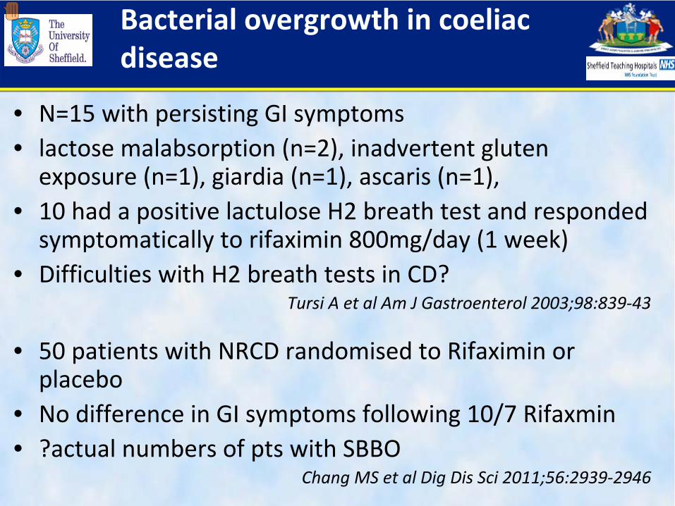
Bacterial overgrowth in coeliac disease
• N=15 with persisting GI symptoms• lactose malabsorption
(n=2), inadvertent gluten
exposure (n=1), giardia
(n=1), ascaris
(n=1), • 10 had a positive lactulose
H2 breath test and responded
symptomatically to rifaximin
800mg/day (1 week)• Difficulties with H2 breath tests in CD?
Tursi
A et al Am J Gastroenterol
2003;98:839‐43
• 50 patients with NRCD randomised to Rifaximin
or placebo
• No difference in GI symptoms following 10/7 Rifaxmin• ?actual numbers of pts with SBBO
Chang MS et al Dig Dis
Sci
2011;56:2939‐2946

NRCD
• Repeat gastroscopy
with biopsy and aspirate• Colonoscopy and biopsy• Faecal elastase• Stool culture• Bloods inc inflammatory markers, thyroid function
• Microscopic colitis• Exocrine pancreatic
insufficiency• Giardiasis• Hyperthyroidism
Dietary review Gluten contamination
Exclude other causes:• SBBO• PLE• Fructose intolerance
Consider RCD?
Review original diagnosis: biopsy, HLA, serology, FHx No coeliac disease
• Lactose intolerance• Consider FODMAP's
Investigation NRCD Algorithm

Refractory coeliac disease
• Rare cause of NRCD unknown true incidence ? 1.5%• Diagnosis of exclusion• Persistent changes of CD despite strict adherence to
GFD• Pre‐malignant condition• Type 1 – polyclonal expansion of IELs
and villous
atrophy• Type 2 –
includes ulcerative jejunitis,
clonal
expansion of abberant
IELs
(CD8+ TCR γδ cells)
• Enteropathy
Associated T‐cell Lymphoma (EATL)

Refractory coeliac disease
• Symptoms– Persistent
malabsorptive
symptoms should prompt re‐
evaluation– Diarrhoea/steatorrhoea– Iron deficiency– Weight loss– Micronutrient loss ‐
Zn,
Cu, Se etc

Investigation
• Rule out malignancy – EATL/adenocarcinoma/other
– ‘B’
symptoms?– Abdominal pain– GI bleeding– Obstructive symptoms– EATL most commonly affects
proximal jejunum• Consider CT/PET‐CT, small
bowel imaging (MR/Ba/capsule etc)
• DBE

Investigation
• OGD – multiple bx
strategy ‐
discussion with tertiary centre –
Histology for IEL population analysis
(PCR/immunohistochemistry)– Caveat 1 : Study of asymptomatic coeliac patients only
17.5% had achieved complete histological response at 2 years (Bardella
et al. Histopath. 2007;50:465‐471)
– Caveat 2 : Changes are well recognised to be patchy in some patients (Hopper et al. Endoscopy 2007;39:219‐224)
– Caveat 3 : Recent evidence has shown that the presence of an aberrant immunophenotype
and monoclonality
do
not definitively confer a diagnosis of RCD – can be seen in uncomplicated coeliac disease (Liu et al. Gut 2010;59:452‐460)
Management
• Nutrition, nutrition, nutrition• Gluten free diet • Enteral
vs
PN
• Micronutrients• Re‐feeding
Management
• Steroids –
budesonide/prednisolone?• 5‐ASA?• Azathioprine?• Cladribine?• Stem cell transplant?• Inliximab? Why not…• IL‐15? Enhances anti‐tumour immunity in CD8+ T‐
cells

Prognosis
• Type 1– 90‐100% 5 year survival
• Type 2– 50% 5 year survival
• Ulcerative jejunitis– Dismal
• EATL– 20‐30% 2 year survival– 50 times more common in someone with coeliac disease, the annual
incidence is low (0.5‐1 per million people)
• Not necessarily a linear progression

Cases

Case 1
• 27 year old female with persistent GI symptoms despite apparent adherence to GFD
What would you do next?1.
Repeat Serology?
2.
Repeat Biopsy?3.
Steroids?
4.
Other tests?5.
Discharge back to GP for follow up?

Case 2
• 64 year old female with symptoms of severe malabsorption
and weight loss on background of
coeliac disease• What would you do next?
1.
Prescribe calcium supplements and ask to stick to GFD and f/up in clinic?
2.
Repeat Serology?3.
Repeat Biopsy?
4.
Steroids?5.
Other tests?

Case 2
• Given IV calcium, IV iron• Senior dietician review – apparent adherence to GFD,
BMI dangerously low – NG feeding commenced• Anti TTG – normal• Immunoglobulins
–
IgA
low
• CT chest abdo
pelvis – oedematous small bowel, scattered lymphadenopathy
but not significant by
size criteria• Referred to surgeons for laparoscopy/LN biopsy – no
evidence of lymphoma

Case 2
• Unable to tolerate NG feeding and TPN commenced
• Commenced on prednisolone
and slowly starts to put on weight and taken off PN
• Starts to tolerate oral intake and meeting nutritional requirements
• Unfortunately develops Hospital Acquired Pneumonia and rapidly deteriorates – Despite ICU
input dies 3 days later

Thanks Thanks –– Any Questions?Any Questions?
OnOn--going Research at the Royal going Research at the Royal HallamshireHallamshire HospitalHospital