nonarteritic anterior ischemic optic neuropathy
TRANSCRIPT
THE JOURNAL OF CLINICAL HYPERTENSION VOL. 7 NO. 2 FEBRUARY 2005130
Nonarteritic anterior ischemic optic neuropathy is a common cause of sudden, painless loss of vision present commonly on awakening from sleep. It most commonly affects middle-aged and elderly Caucasian men and women. Involvement of the opposite eye occurs within 3 years in less than 43% of patients. Hypertension, diabetes, and noc-turnal hypotension are risk factors. A congenital small cup-to-disk ratio also predisposes to the optic nerve ischemia. There is no effective therapy to treat patients acutely or to prevent recurrence. After 6 months of careful follow-up, 57.3% of patients will have no significant change or wors-ening of their vision in the involved eye. (J Clin Hypertens. 2005;7:130–133) ©2005 Le Jacq Ltd.
From an epidemiologic point of view, the lower the blood pressure, the fewer the cardiovas-
cular events observed; however, there is a sug-gestion that strokes and blindness may increase uncommonly in elderly people who experience an excessive decline in nocturnal blood pressure.1–3 Nonarteritic anterior ischemic optic neuropathy (NAION) is a leading cause of sudden vision loss and the second most common form of optic neu-ropathy in people over 50 years of age, affecting over 6000 Americans each year.4 NAION is an ischemic insult that affects the short posterior cili-ary vessels that supply the optic nerve head.
CLINICAL PRESENTATIONThe classic presentation is a sudden, painless loss of vision in one eye present on awakening with-out premonitory symptoms. In one prospective study, 41% of 418 patients did not experience the visual loss within 2 hours of awakening, and 17% could not remember when it occurred.5 In contrast, Hayreh et al.6 reported that in 544 episodes of NAION, 51.8% were symptomatic on awakening, 21.5% in the early morning and 26.7% later during the day. Episodes were more likely to occur in the summer months rather than the winter months.6
The visual field defect of NAION may worsen over several days. The visual loss may be central, peripheral, or both. An afferent pupillary defect (Marcus Gunn pupil), an indication of optic nerve damage, is present when only one eye is involved or with asymmetric bilateral disease. An afferent pupillary defect is detected by alternating a bright light in one eye and then the other eye and by observing pupillary dilatation rather than constric-tion in the affected eye. A Marcus Gunn pupil may not be present in bilateral NAION as seen in patients with postoperative hypotension.
Giant cell arteritis, a cause of arteritic anterior ischemic optic neuropathy, should be excluded in all cases since corticosteroids are required in these cases. Bilateral simultaneous or sequential involve-ment can be seen if untreated.
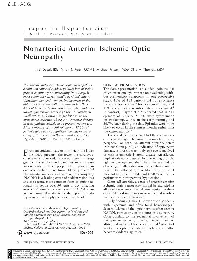
Early findings (Figure 1) show optic disc edema with hyperemia and often focal hemorrhages.7 Sectoral edema of the optic nerve is often seen in NAION, particularly of the superior disc margin. Corresponding to this segmental involvement of the optic nerve head, arcuate, wedge-shaped or altitudinal visual field defects are noted.8 After 4–8 weeks, the optic disc edema resolves and pallor becomes evident (Figure 2).
Nonarteritic Anterior Ischemic Optic Neuropathy
Niraj Desai, BS;1 Milan R. Patel, MD;2 L. Michael Prisant, MD;3 Dilip A. Thomas, MD2
I m a g e s i n H y p e r t e n s i o nL . M i c h a e l P r i s a n t , M D , S e c t i o n E d i t o r
www.lejacq.com ID: 4095
From the School of Medicine,1 Department of Ophthalmology,2 and Department of Medicine and Clinical Pharmacology Unit,3 Medical College of Georgia, Augusta, GAAddress for correspondence: L. Michael Prisant, MD, 1120 15th Street, HB-2010, Medical College of Georgia, Augusta, GA 30912
The Journal of Clinical Hypertension (ISSN 1524-6175) is published monthly by Le Jacq Ltd., Three Parklands Drive, Darien, CT 06820-3652. Copyright ©2005 by Le Jacq Ltd., All rights reserved. No part of this publication may be reproduced or transmitted in any form or by any means, electronic or mechanical, including photocopy, recording, or any information storage and retrieval system, without permission in writing from the publishers. The opinions and ideas expressed in this publication are those of the authors and do not necessarily reflect those of the Editors or Publisher. For copies in excess of 25 or for commercial purposes, please contact Sarah Howell at [email protected] or 203.656.1711 x106.

VOL. 7 NO. 2 FEBRUARY 2005 THE JOURNAL OF CLINICAL HYPERTENSION 131
Visual deterioration may occur during the first 2 weeks. Recurrence in the same eye is unusu-al; however, recurrence in the other eye varies between 15% and 40% on average after 2.9 years (Figure 3).9–12 In one prospective study of 418 patients, 19% had bilateral NAION at baseline and 14.7% developed NAION in the contralateral eye after a median followup of 5.1 years.9 The
median occurrence of new NAION was 1.2 years. Recurrence of NAION was more common among patients with diabetes (24% vs. 12%, p=0.02).
INCIDENCE AND DEMOGRAPHICSThe yearly incidence of NAION is between 2.3 and 10.2 per 100,000 patients 50 years or older.7 While NAION shows no gender bias (Figure 4), African Americans, Asians, and Hispanics are rarely affected.13 The mean age of onset was 60.1±13.6 years among 406 patients, but it can occur in younger patients.11
Established risk factors for NAION are large-ly vasculopathic and include increasing age, acute and chronic hypertension, nocturnal hypotension,
Figure 1. A 45-year-old hypertensive white male, on awakening from sleep, experienced painless loss of vision in his left eye. His right eye was unaffected. An intensification of antihypertensive medication had been recently initiated to the point of causing postural symptoms. What is the explanation for this patient’s symptoms? The arrows show optic nerve head edema manifested by blurring of the disc margins. Also note multiple disc hemorrhages (see block arrow).
Figure 2. Anterior ischemic optic neuropathy: chronic changes. The arrow shows the pallor of the optic disc in the left eye that developed months after the changes observed in Figure 1.
Figure 3. Panel A: Right eye before anterior ischemic optic neuropathy. Panel B: Right eye after anterior isch-emic optic neuropathy. These are from the same patient as in Figures 1 and 2. Note the development of optic nerve head pallor/atrophy (arrow) indicating axonal loss. Retinal arteriolar attenuation is noted also.
The Journal of Clinical Hypertension (ISSN 1524-6175) is published monthly by Le Jacq Ltd., Three Parklands Drive, Darien, CT 06820-3652. Copyright ©2005 by Le Jacq Ltd., All rights reserved. No part of this publication may be reproduced or transmitted in any form or by any means, electronic or mechanical, including photocopy, recording, or any information storage and retrieval system, without permission in writing from the publishers. The opinions and ideas expressed in this publication are those of the authors and do not necessarily reflect those of the Editors or Publisher. For copies in excess of 25 or for commercial purposes, please contact Sarah Howell at [email protected] or 203.656.1711 x106.

THE JOURNAL OF CLINICAL HYPERTENSION VOL. 7 NO. 2 FEBRUARY 2005132
diabetes, and hyperlipidemia.2,11,13,14 A lower noctur-nal systolic blood pressure was present with deterio-rating visual fields in hypertensive patients compared with subjects without deterioration (113 mm Hg vs. 130 mm Hg, p=0.006).15 Most commonly, however, a congenitally anomalous optic disc is a risk factor that has been associated with NAION.11,13,16–20 Several case reports have suggested that sildenafil may cause NAION when associated with a decreased cup-to-disc ratio and possible arterial hypotension.21
PATHOPHYSIOLOGYThe pathophysiology behind NAION is an idio-pathic ischemic process. Previous research has suggested occlusion of the short posterior ciliary arteries as a possible mechanism.8 In the past, researchers have successfully induced a clinical state compatible with NAION in primates by ligating their posterior ciliary arteries.22 Still, other research points toward a congenitally anomalous optic disc as the etiology. Clinically, a small optic disc is often noted without physiologic cupping or, when cupping is present, a small cup-to-disc ratio has been seen. These congenitally anomalous optic discs have been associated with a higher risk for developing NAION.16–18,20 One theory proposes that a small cup-to-disk ratio might function to cause mechanical crowding of optic nerve axons that further cause adjacent vessels to be compro-mised through a cascade effect.17
Since hypertension and diabetes are associated with NAION, research has focused on vasculopath-ic processes.10,13 Clinically, loss of blood flow in the optic nerve initially results in swelling of the optic nerve head, which in turn induces ischemia second-ary to compression. Research suggests a generalized disturbance of blood flow to the nerve head possibly due to a variety of predisposing factors.16–20
TREATMENTCurrently, there is no effective treatment for NAION. In the past, surgical decompression of the optic nerve was performed until the Ischemic Optic Neuropathy Decompression Trial13 showed no benefit of sur-gery. This study randomized 125 patients to follow-up and 119 patients to surgery, which consisted of placing slits or a window in the optic nerve sheath to reduce pressure by allowing cerebrospinal fluid to drain. Vision acuity tended to worsen in the surgical arm compared with the observational control group (23.9% vs. 12.4%, p=0.04). Furthermore, there was a greater improvement of three or more lines of visual acuity in the nonsurgical arm (42.7%) than in the surgical arm (32.6%).
Although still used by some physicians, ste-roid therapy has also shown no benefit in these patients.23 In a retrospective cohort study, aspirin was observed to be beneficial in decreasing NAION in the fellow eye over 2 years, but not at 5 years.12 However, although treatment was not random-ized, aspirin use was not associated with a lower rate of recurrence in the contralateral eye in the Ischemic Optic Neuropathy Decompression Trial.9 Control of hypertension, diabetes, and hyperlipid-emia may slow progression or reduce the incidence of NAION, but is not thought to be effective in any regression of NAION-induced visual losses.
REFERENCES 1 Kario K, Matsuo T, Kobayashi H, et al. Nocturnal fall
of blood pressure and silent cerebrovascular damage in elderly hypertensive patients. Advanced silent cere-brovascular damage in extreme dippers. Hypertension. 1996;27:130–135.
2 Hayreh SS. Role of nocturnal arterial hypotension in the development of ocular manifestations of systemic arterial hypertension. Curr Opin Ophthalmol. 1999;10:474–482.
3 Prisant LM. Hypertension and chronotherapy: shifting the treatment paradigm. Am J Hypertens. 2001;14:277S–279S.
4 Hattenhauer MG, Leavitt JA, Hodge DO, et al. Incidence of nonarteritic anterior ischemic optic neuropathy. Am J Ophthalmol. 1997;123:103–107.
5 Characteristics of patients with nonarteritic anterior isch-emic optic neuropathy eligible for the Ischemic Optic Neuropathy Decompression Trial. Arch Ophthalmol. 1996;114:1366–1374.
6 Hayreh SS, Podhajsky PA, Zimmerman B. Nonarteritic anterior ischemic optic neuropathy: time of onset of visual loss. Am J Ophthalmol. 1997;124:641–647.
7 Buono LM, Foroozan R, Sergott RC, et al. Nonarteritic anterior ischemic optic neuropathy. Curr Opin Ophthalmol. 2002;13:357–361.
8 Shults WT. Ischemic optic neuropathy. Still the ophthalmol-ogist’s dilemma. Ophthalmology. 1984;91:1338–1341.
56%
65%
55%
44%
35%
45%
0
20
40
60
80
100
<45(n=43)
45–64(n=199)
≥65(n=164)
Age group (yr)
Prev
alen
ce (
%)
Male Female
Figure 4. Prevalence of anterior ischemic optic neuropa-thy by age and gender. Derived from Am J Ophthalmol. 1994;118:766–780.11
The Journal of Clinical Hypertension (ISSN 1524-6175) is published monthly by Le Jacq Ltd., Three Parklands Drive, Darien, CT 06820-3652. Copyright ©2005 by Le Jacq Ltd., All rights reserved. No part of this publication may be reproduced or transmitted in any form or by any means, electronic or mechanical, including photocopy, recording, or any information storage and retrieval system, without permission in writing from the publishers. The opinions and ideas expressed in this publication are those of the authors and do not necessarily reflect those of the Editors or Publisher. For copies in excess of 25 or for commercial purposes, please contact Sarah Howell at [email protected] or 203.656.1711 x106.

VOL. 7 NO. 2 FEBRUARY 2005 THE JOURNAL OF CLINICAL HYPERTENSION 133
9 Newman NJ, Scherer R, Langenberg P, et al. The fellow eye in NAION: report from the ischemic optic neuropathy decompression trial follow-up study. Am J Ophthalmol. 2002;134:317–328.
10 Repka MX, Savino PJ, Schatz NJ, et al. Clinical profile and long-term implications of anterior ischemic optic neuropa-thy. Am J Ophthalmol. 1983;96:478–483.
11 Hayreh SS, Joos KM, Podhajsky PA, et al. Systemic diseases associated with nonarteritic anterior ischemic optic neu-ropathy. Am J Ophthalmol. 1994;118:766–780.
12 Beck RW, Hayreh SS, Podhajsky PA, et al. Aspirin therapy in nonarteritic anterior ischemic optic neuropathy. Am J Ophthalmol. 1997;123:212–217.
13 Optic nerve decompression surgery for nonarteritic anterior ischemic optic neuropathy (NAION) is not effective and may be harmful. The Ischemic Optic Neuropathy Decompression Trial Research Group. JAMA. 1995;273:625–632.
14 Hayreh SS, Zimmerman MB, Podhajsky P, et al. Nocturnal arterial hypotension and its role in optic nerve head and ocular ischemic disorders. Am J Ophthalmol. 1994;117:603–624.
15 Hayreh SS, Podhajsky P, Zimmerman MB. Beta-block-er eyedrops and nocturnal arterial hypotension. Am J Ophthalmol. 1999;128:301–309.
16 Beck RW, Savino PJ, Repka MX, et al. Optic disc structure in anterior ischemic optic neuropathy. Ophthalmology. 1984;91:1334–1337.
17 Feit RH, Tomsak RL, Ellenberger C Jr. Structural factors in the pathogenesis of ischemic optic neuropathy. Am J Ophthalmol. 1984;98:105–108.
18 Doro S, Lessell S. Cup-disc ratio and ischemic optic neu-ropathy. Arch Ophthalmol. 1985;103:1143–1144.
19 Beck RW, Servais GE, Hayreh SS. Anterior ischemic optic neuropathy. IX. Cup-to-disc ratio and its role in pathogen-esis. Ophthalmology. 1987;94:1503–1508.
20 Jonas JB, Gusek GC, Naumann GO. Anterior ischemic optic neuropathy: nonarteritic form in small and giant cell arteritis in normal sized optic discs. Int Ophthalmol. 1988;12:119–125.
21 Pomeranz HD, Smith KH, Hart WM Jr, et al. Sildenafil-associated nonarteritic anterior ischemic optic neuropathy. Ophthalmology. 2002;109:584–587.
22 McLeod D, Marshall J, Kohner EM. Role of axoplasmic transport in the pathophysiology of ischaemic disc swelling. Br J Ophthalmol. 1980;64:247–261.
23 Boghen DR, Glaser JS. Ischaemic optic neuropathy. The clinical profile and history. Brain. 1975;98:689–708.
The Journal of Clinical Hypertension (ISSN 1524-6175) is published monthly by Le Jacq Ltd., Three Parklands Drive, Darien, CT 06820-3652. Copyright ©2005 by Le Jacq Ltd., All rights reserved. No part of this publication may be reproduced or transmitted in any form or by any means, electronic or mechanical, including photocopy, recording, or any information storage and retrieval system, without permission in writing from the publishers. The opinions and ideas expressed in this publication are those of the authors and do not necessarily reflect those of the Editors or Publisher. For copies in excess of 25 or for commercial purposes, please contact Sarah Howell at [email protected] or 203.656.1711 x106.