noninvasive cardiac imaging with computed tomography
TRANSCRIPT
Review
Noninvasive Cardiac Imaging withComputed Tomography
Gregory T. Wilson, DO; Prabhakaran Gopalakrishnan, MD; and Tahir Tak, MD, Ph.D
Despite major improvements in the treatment of heart disease, it remains a major source ofmorbidity and mortality on a global scale. Currently, invasive coronary angiography remains thegold standard for identification of obstructive coronary artery disease. However, recentadvances in computerized tomographic (CT) techniques of the heart allow for accurate,non-invasive characterization of atherosclerotic coronary disease and other cardiacabnormalities. The calculation of coronary artery calcium scores with electron beam CT haslargely been supplanted by high-resolution CT angiography using multi-slice detectors (MSCT)which can provide detailed multidimensional visualization of cardiac structures. Althoughevaluation of obstructive coronary disease is the primary use of MSCT, its use in identifyingcongenital defects, planning thoracic procedures and characterizing cardiac function continues togrow. Accordingly, appropriate incorporation of MSCT/CT angiography into clinical practicecontinues to be defined. Several limitations to MSCT remain which reduce its accuracy, such asin patients with arrhythmia and in patients with either coronary stents or heavily calcifiedcoronaries. Despite its current limitations, MSCT remains a rapidly advancing field and anincreasingly valuable tool for the non-invasive evaluation of cardiac pathology.
Reprint Requests: Tahir Tak, MD, PhD, Division of Cardiovascular Diseases,Mayo Clinic, 200 First Street SW, Rochester. MN 55905, Tel: 507-284-2941,Fax: 507-266-7929, Email: [email protected]
Received: January 5, 2007 Revised: April 20, 2007 Accepted: May 2, 2007
doi:10.3121/cmr.2007.747
Keywords: Computed tomographic angiography; CT; CTA; Computed tomography, multi-detector row;Coronary artery calcification; Coronary vessels; Stenosis
Cardiovascular diseases remain the leading cause ofmorbidity and mortality in industrialized nations. In spite ofadvances in medicine, cardiovascular mortality in the UnitedStates continues to be high at 332.9 deaths per 100,000.More than half of these deaths are related to coronary arterydisease (CAD). The estimated direct and indirect cost ofCAD for 2006 is approximately 142.5 billion US dollars.1
Invasive coronary angiography (ICA) has been the standardof reference for diagnosis of CAD. Approximately 1.4million diagnostic catheterizations were performedthroughout the world in 2003.1 Incidence of normalangiograms has varied from 20% to 27% according to datafrom the Society for Cardiac Angiography andInterventions.2,3 Even though ICA is safe with an in-hospitalmortality rate of <1%, it still carries a potential risk bothfrom iodinated contrast agents used in ICA and the risks ofarterial catheterization itself. Diagnostic catheterizationswhich are not done in conjunction with an interventionalprocedure, especially the ones resulting in normalangiograms, could potentially be replaced by alternate
methods of assessing CAD. Several alternate methods ofassessing CAD have been studied, such as treadmill stresstesting, single photon emission computed tomography(SPECT), positron emission tomography (PET), and stressechocardiography. Non-invasive diagnostic tests have theirinherent limitations and do not provide morphological detailsof the coronary occlusion. The availability of reliablenoninvasive coronary imaging could potentially decrease theneed for ICA in some patients.
Noninvasive coronary imaging techniques include electronbeam computed tomography (EBCT), multi-slice computedtomography (MSCT) and cardiac magnetic resonanceimaging (MRI). With MSCT, the addition of multipledetector rows allowing simultaneous acquisition improvesperformance significantly. While single and dual detectorrow spiral computed tomography (CT) systems wereavailable in the mid 1990s, 4-, 8-, 10-, 16-, 32-, 40- and 64-detector row CT scanners have since been developed,improving the spatial resolution while reducing scan time.While EBCT technology has been plagued by less
165
Clinical Medicine & ResearchVolume 5, Number 3: 165-171©2007 Marshfield Clinic http://www.clinmedres.org
Cardiac imaging with computed tomography
widespread availability and the technology of cardiac MRI isstill evolving, MSCT angiography appears to be developing ata rapid pace.
While non-invasive coronary imaging continues to evolve, itsrole in current clinical practice is still being debated.Numerous studies have been done to validate the differenttechniques in comparison to ICA, which is still considered thegold standard. In this review, we give an overview of CTcoronary angiography, the technique, applications, pitfallsand future directions.
Coronary Artery CalciumThe presence and extent of coronary artery calcification hasbeen shown to correlate with the degree of atherosclerosis.4
Studies evaluating the clinical utility of EBCT-detectedcoronary artery calcification report a high negative predictivevalue in negative scans. This finding makes coronary arterycalcification detection with CT a valuable test when rulingout obstructive atherosclerotic coronary disease.5-8 Due to itslow specificity, calcium scoring is not suitable for thedetection of significant CAD and should not be regarded as asole indication for coronary angiography.
Various methods of quantifying coronary artery calcificationhave been proposed in order to estimate a patient’scardiovascular risk. One of the more widely used methodswas originally proposed by Agatston et al9 in 1990 where alesion’s area, peak density and mean density were measuredfor each segmental lesion. Poor inter-scan reproducibilitywith Agatston scoring prompted the development of thevolumetric calcium scoring algorithm by which the volume ofa plaque is measured irrespective of its area or density.10
Although the volumetric score has been shown to improvereproducibility in serial scans, the Agatston scoring system isstill frequently used in clinical practice.
Despite initial hopes that coronary calcium characterizationoffers a novel, non-invasive tool to identify patients at highrisk for future major cardiovascular events (i.e., myocardialinfarction, unstable angina, death), several meta-analysesreport only a modest prognostic correlation between coronarycalcification and significant cardiovascular outcome.11,12
One possible explanation for this observation is thatvulnerable coronary plaques are often composed of non-calcified lipomatous or fibrous tissue, while heavily calcifiedplaques are typically less likely to rupture.13 Additionally,when compared to other well-validated diagnostic tests suchas stress testing and perfusion imaging, EBCT coronarycalcium scoring does not provide clearly superior diagnosticor prognostic information.11 Still, the exclusion of coronarycalcifications is associated with a significantly reduced rateof future cardiovascular events.14 Overall, dedicated EBCTscanners are becoming obsolete.
However, current MSCT scanners also provide calciumscores equivalent to that of EBCT. According to the American
College of Cardiology/American Heart Association expertconsensus,11 a low coronary calcium scoring makes thepresence of atherosclerosis very unlikely and occurs in themajority of patients with normal coronary arteries. Therefore,CT coronary artery calcification measurements may remain auseful test to rule out significant atherosclerotic coronarydisease.11 A negative predictive value of more than 95% canbe achieved using the existence of any calcium as athreshold.15 The recently published cardiac CTappropriateness criteria suggest that coronary calciumscoring be reserved for patients with an intermediate or highrisk of coronary disease (Framingham risk criteria), whererisk stratification may aid providers in selecting treatmentstrategies, i.e., initiating statin therapy.16 Repeatedexaminations within 5 years are not recommended.16
CT AngiographyDue to the uncertain value of coronary artery calcificationscores, recent emphasis has been on improving contrastenhanced CT angiography of the coronary arteries usingMSCT. Addition of multiple detectors and improved temporaland spatial resolution on current CT scanners allow forreliable, non-invasive detection and characterization ofcoronary plaques.17-23 Early CT scanners using either 4 or 8rows of detectors were able to provide accurate data onproximal coronary trunks, while newer generation scannersdemonstrate reliable visualization of all coronary segmentsincluding distal branches.23
Patient PreparationIn order to obtain optimal and nearly motion-free images, thepatient’s heart rate should be regular and preferably <65 beatsper minute, which is often accomplished with a pre-scan betablocker.24 Calcium channel blockers can be given in patientswith contraindications to beta blockers. The patient shouldavoid caffeine the day of the exam. Although no studies haveevaluated its use in CT angiography, short-acting sublingualnitroglycerin (2 x 0.4 mg tablets) can be given at the time ofscanning to improve visualization of the coronary lumen.25
Hypersensitivity to iodinated contrast and pregnancy remainthe only true absolute contraindications for CTangiography.26 Relative contraindications for CT angiographyscanning include the inability to hold one’s breath forsufficient scan time, irregular or rapid (>90) heart rhythm,significant renal insufficiency and inability to cooperateduring scanning. Other relative contraindications are similarto those for standard CT scanning.25
Data AcquisitionUsing a spiral ECG-gated image acquisition protocol, threecommon steps are employed.25 The first step utilizes a lowenergy, non-contrast topogram of the chest to define thevolume and boundaries of the scan. The second step involveseither a bolus timing or tracking technique to ensureappropriate coronary enhancement during scanning. The thirdstep is the coronary CT angiography itself which covers the
CM&R 2007 : 3 (October)166
entire heart from proximal ascending aorta to thediaphragmatic surface in a single breath hold. Total volume ofcontrast is typically 80 cc to 100 cc given at a rate of 4 cc/s to5 cc/s.27-29 The average scan time for 64-slice MSCT is 5 seconds to 11 seconds, depending on the vendor.
Image Reconstruction and InterpretationUsing current generation scanners, scans are obtained withcontinuous, spiral image acquisition. Typical reconstructionslice thickness is approximately 0.5 mm to 0.6 mm with 50%overlap between images, depending on the scanner.25 Using atechnique termed retrospective image reconstruction, imagescan be reconstructed at any point in the cardiac cycle.29,30
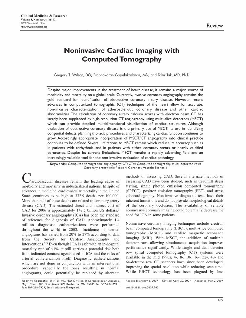
Although several types of reconstruction algorithms exist, aproposed optimal reconstruction window for motion-freevisualization of the coronary arteries is in mid diastole at 60%to 70% of the R-R interval.30 Three dimensional volumerendered color images show surface anatomy (figure 1).Three dimensional maximum intensity projection and twodimensional multiplanar reformation are used to see insidethe heart and visualize the coronary arteries. Furthermore,available ECG gating software allows for editing of thetemporal windows to eliminate ECG irregularities, i.e.,premature ventricular beats.29 The entire dataset obtainedfrom a single multi-slice CT angiography study is extensiveand can include up to 3000 axial images and 1.5 gigabytes ofdata, making timely assessment and data storagechallenging.25
Clinical ApplicationsCurrent generation 64-slice scanners, boasting improvedtemporal and spatial resolution, demonstrate excellentdiagnostic accuracy for both proximal coronary vessels and
smaller distal vessels.25,28,31-38 MSCT allows an accurate,non-invasive evaluation of the coronary vessels in those atintermediate risk for CAD. As stated previously, MSCTscanners also provide a coronary artery calcium score.
Currently, general screening of asymptomatic populations forobstructive coronary disease, who are at low or moderatecardiac risk as determined by Framingham criteria, is notrecommended.16 Concern arises regarding uncontrolledusage of such testing and the potential for unnecessaryinvasive work-up.16 Similarly, MSCT is currently notrecommended in patients in whom other diagnostic testsalready suggest high risk.16 Current American HeartAssociation/American College of Cardiologyrecommendations suggest MSCT angiography insymptomatic patients with low to intermediate pretestprobability.39
In addition to visualizing the coronary lumen, MSCT allowscharacterization of the coronary atherosclerotic plaque andhas shown a good correlation with intravascularultrasound.31,32 A major goal of non-invasive imaging is toidentify vulnerable plaques with the potential to rupture.25,32
The accuracy of plaque characterization seems to vary withplaque size, as larger plaques tend to be overestimated andsmaller plaques may be either underestimated or notdetected.32 Further technical improvements will likelyenhance the ability of MSCT to characterize coronaryplaques.
MSCT can also provide a measure of left ventricular functionand regional wall motion, information that may prove usefulwhen assessing patients overall cardiovascular risk.40,16
Recent comparisons have shown that left ventricular functionas determined by MSCT correlates well with data obtainedwith echocardiography.40-42
MSCT has also been shown to be a useful tool in evaluatingcoronary anomalies and for planning coronary bypassgrafts.16 Thoracic surgeons are able to visualize the course ofvessels, as well as their relationship to adjacent structureswhen planning. Similarly, MSCT has been applied tocoronary venous mapping for planning device implantation(i.e., left ventricular pacing leads) and for evaluatinganatomic variation. Pulmonary venous mapping with CTangiography has also been useful in planning arrhythmiaablation procedures.43
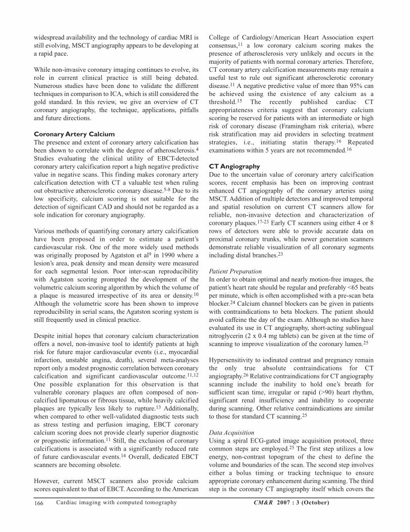
MSCT remains a potential tool to determine the patency ofboth venous and arterial coronary bypass grafts, an asset thatwas considered early in the development of non-invasiveimaging (figure 2).44,45 According to the recently publishedAmerican College of Cardiology Foundation appropriatenesscriteria for cardiac CT/cardiac MRI, CT angiography ofbypass grafts should be reserved for symptomatic patients.16
However, it is generally thought that MSCT cannotconsistently visualize distal graft anastomosis and fails toprovide a functional assessment of bypass graft flow.33
Wilson et al. CM&R 2007 : 3 (October) 167
Figure 1. Three-dimensional volume rendered image of CT angiogram.
Cardiac imaging with computed tomography
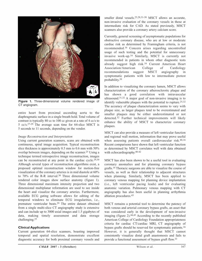
Recently, MSCT has been evaluated for its potential role indefining in-stent restenosis. Using 4- and 16-slice MSCT, amajor limitation of MSCT has been its inability to adequatelycharacterize stenosis within a stented segment due tosignificant artifact (figure 3).8,27,35 Large diameter stents (>3 mm) and those with thinner struts improve the accuracyfor detecting and quantifying in-stent restenosis withMSCT.37 Furthermore, use of high-resolution “kernels”, orspecific reconstruction algorithms, can improve in-stentevaluation by fine-tuning image resolution.37,38 Still, onlyevaluation of stent patency is possible with an adequatediagnostic accuracy. A reliable assessment of stent stenosiscannot be expected. Therefore, conventional coronaryangiography should be performed in symptomatic patientsafter coronary stent implantation.46
Artifacts and LimitationsAccurate image interpretation can be compromised by severaltypes of artifacts. One of the more common problems ismotion artifact induced by irregular cardiac rhythms,breathing, or other patient movement. High resolution MSCTscanners require nearly motion-free imaging to provideaccurate data; consequently, MSCT is indicated only forcooperative patients with regular heart rhythms.25 Anothercommon problem is “blooming artifact” where high-attenuation structures such as metal clips, stents, andcalcified plaques may obscure adjacent tissue.25 Thisphenomenon can significantly limit the accuracy of MSCT indetermining coronary stenosis in a heavily calcified vessel.Similarly, dense structures or calcification may causestreaking through an image, a phenomenon termed “beamhardening” artifact.25 Furthermore, if the timing of the
contrast bolus is not optimal, inadequate enhancement of thecoronary tree results.29
With MSCT, some concern has been raised regarding theradiation dose administered to patients.47 The effectiveradiation dose using a 64-slice MSCT is approximately 6.5 mSv to 15 mSv.48 This value is approximately two to threetimes the patient dose during a diagnostic coronaryangiogram.48,49 Techniques have been developed to reducethe radiation dose with newer generation scanners, such aslimiting x-ray emission during certain phases of imageacquisition.48
Conclusion and Future PerspectivesMSCT is a promising technique within the realms ofnoninvasive coronary imaging. With the advent of 64-sliceMSCT, the diagnostic accuracy of CT coronary angiographyhas improved significantly and fares better in comparison toother currently available methods of noninvasive coronaryimaging. With recent studies validating this, the role ofMSCT in the evaluation of CAD is likely to grow in thefuture.
The major development resulting in improved diagnosticaccuracy of MSCT has been the increase in the detector rows.In continuing this trend, 256-slice MSCT is being currentlystudied with the promise of increased accuracy and decreasedscan time.50 In addition to developments in CT technology,studies looking at combining MSCT with other modalities ofnoninvasive cardiac imaging are being done. Protocols arebeing developed for combined evaluation of coronaryanatomy and cardiac function with the development of hybrid
CM&R 2007 : 3 (October)168
Figure 3. CT axial image demonstrating intracoronary stent inthe left circumflex (LCX) artery and the presence of plaquedistal to the stent (LCX plaque).
Figure 2. Three-dimensional volume rendered image ofangiogram demonstrating coronary artery bypass grafts. VG,vein graft; RCA, right coronary artery; LIMA, left internalmammary artery; LAD, left anterior descending artery; R, right.
scanners that combine MSCT with PET or SPECT, allowingsimultaneous imaging of coronary tree, perfusion scan,cardiac metabolism and function.51 Co-registered MRImyocardial viability mapping and MSCT angiography arebeing studied for optimizing surgical revascularizationplanning.52 Furthermore, MSCT has been well studied fordetection of pulmonary embolism and aortic dissection. Thepossibility of a wider use of MSCT in emergency roomevaluations of chest pain is currently being considered as atest to achieve the so-called “triple rule out” in patients withchest pain, essentially excluding aortic dissection, pulmonaryembolism and CAD.25,53
AcknowledgmentsThe authors would like to acknowledge Spencer Smith, MDof Radiology Associates, Tarrant Medical Imaging, FortWorth, TX for reviewing this manuscript, providing MSCTfigures and for technical advice.
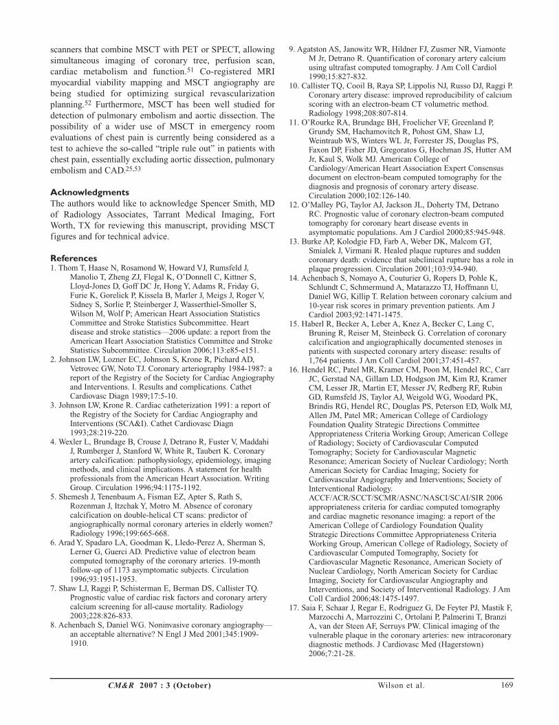
References1. Thom T, Haase N, Rosamond W, Howard VJ, Rumsfeld J,
Manolio T, Zheng ZJ, Flegal K, O’Donnell C, Kittner S,Lloyd-Jones D, Goff DC Jr, Hong Y, Adams R, Friday G,Furie K, Gorelick P, Kissela B, Marler J, Meigs J, Roger V,Sidney S, Sorlie P, Steinberger J, Wasserthiel-Smoller S,Wilson M, Wolf P; American Heart Association StatisticsCommittee and Stroke Statistics Subcommittee. Heartdisease and stroke statistics—2006 update: a report from theAmerican Heart Association Statistics Committee and StrokeStatistics Subcommittee. Circulation 2006;113:e85-e151.
2. Johnson LW, Lozner EC, Johnson S, Krone R, Pichard AD,Vetrovec GW, Noto TJ. Coronary arteriography 1984-1987: areport of the Registry of the Society for Cardiac Angiographyand Interventions. I. Results and complications. CathetCardiovasc Diagn 1989;17:5-10.
3. Johnson LW, Krone R. Cardiac catheterization 1991: a report ofthe Registry of the Society for Cardiac Angiography andInterventions (SCA&I). Cathet Cardiovasc Diagn1993;28:219-220.
4. Wexler L, Brundage B, Crouse J, Detrano R, Fuster V, MaddahiJ, Rumberger J, Stanford W, White R, Taubert K. Coronaryartery calcification: pathophysiology, epidemiology, imagingmethods, and clinical implications. A statement for healthprofessionals from the American Heart Association. WritingGroup. Circulation 1996;94:1175-1192.
5. Shemesh J, Tenenbaum A, Fisman EZ, Apter S, Rath S,Rozenman J, Itzchak Y, Motro M. Absence of coronarycalcification on double-helical CT scans: predictor ofangiographically normal coronary arteries in elderly women?Radiology 1996;199:665-668.
6. Arad Y, Spadaro LA, Goodman K, Lledo-Perez A, Sherman S,Lerner G, Guerci AD. Predictive value of electron beamcomputed tomography of the coronary arteries. 19-monthfollow-up of 1173 asymptomatic subjects. Circulation1996;93:1951-1953.
7. Shaw LJ, Raggi P, Schisterman E, Berman DS, Callister TQ.Prognostic value of cardiac risk factors and coronary arterycalcium screening for all-cause mortality. Radiology2003;228:826-833.
8. Achenbach S, Daniel WG. Noninvasive coronary angiography—an acceptable alternative? N Engl J Med 2001;345:1909-1910.
9. Agatston AS, Janowitz WR, Hildner FJ, Zusmer NR, ViamonteM Jr, Detrano R. Quantification of coronary artery calciumusing ultrafast computed tomography. J Am Coll Cardiol1990;15:827-832.
10. Callister TQ, Cooil B, Raya SP, Lippolis NJ, Russo DJ, Raggi P.Coronary artery disease: improved reproducibility of calciumscoring with an electron-beam CT volumetric method.Radiology 1998;208:807-814.
11. O’Rourke RA, Brundage BH, Froelicher VF, Greenland P,Grundy SM, Hachamovitch R, Pohost GM, Shaw LJ,Weintraub WS, Winters WL Jr, Forrester JS, Douglas PS,Faxon DP, Fisher JD, Gregoratos G, Hochman JS, Hutter AMJr, Kaul S, Wolk MJ. American College ofCardiology/American Heart Association Expert Consensusdocument on electron-beam computed tomography for thediagnosis and prognosis of coronary artery disease.Circulation 2000;102:126-140.
12. O’Malley PG, Taylor AJ, Jackson JL, Doherty TM, DetranoRC. Prognostic value of coronary electron-beam computedtomography for coronary heart disease events inasymptomatic populations. Am J Cardiol 2000;85:945-948.
13. Burke AP, Kolodgie FD, Farb A, Weber DK, Malcom GT,Smialek J, Virmani R. Healed plaque ruptures and suddencoronary death: evidence that subclinical rupture has a role inplaque progression. Circulation 2001;103:934-940.
14. Achenbach S, Nomayo A, Couturier G, Ropers D, Pohle K,Schlundt C, Schmermund A, Matarazzo TJ, Hoffmann U,Daniel WG, Killip T. Relation between coronary calcium and10-year risk scores in primary prevention patients. Am JCardiol 2003;92:1471-1475.
15. Haberl R, Becker A, Leber A, Knez A, Becker C, Lang C,Bruning R, Reiser M, Steinbeck G. Correlation of coronarycalcification and angiographically documented stenoses inpatients with suspected coronary artery disease: results of1,764 patients. J Am Coll Cardiol 2001;37:451-457.
16. Hendel RC, Patel MR, Kramer CM, Poon M, Hendel RC, CarrJC, Gerstad NA, Gillam LD, Hodgson JM, Kim RJ, KramerCM, Lesser JR, Martin ET, Messer JV, Redberg RF, RubinGD, Rumsfeld JS, Taylor AJ, Weigold WG, Woodard PK,Brindis RG, Hendel RC, Douglas PS, Peterson ED, Wolk MJ,Allen JM, Patel MR; American College of CardiologyFoundation Quality Strategic Directions CommitteeAppropriateness Criteria Working Group; American Collegeof Radiology; Society of Cardiovascular ComputedTomography; Society for Cardiovascular MagneticResonance; American Society of Nuclear Cardiology; NorthAmerican Society for Cardiac Imaging; Society forCardiovascular Angiography and Interventions; Society ofInterventional Radiology.ACCF/ACR/SCCT/SCMR/ASNC/NASCI/SCAI/SIR 2006appropriateness criteria for cardiac computed tomographyand cardiac magnetic resonance imaging: a report of theAmerican College of Cardiology Foundation QualityStrategic Directions Committee Appropriateness CriteriaWorking Group, American College of Radiology, Society ofCardiovascular Computed Tomography, Society forCardiovascular Magnetic Resonance, American Society ofNuclear Cardiology, North American Society for CardiacImaging, Society for Cardiovascular Angiography andInterventions, and Society of Interventional Radiology. J AmColl Cardiol 2006;48:1475-1497.
17. Saia F, Schaar J, Regar E, Rodriguez G, De Feyter PJ, Mastik F,Marzocchi A, Marrozzini C, Ortolani P, Palmerini T, BranziA, van der Steen AF, Serruys PW. Clinical imaging of thevulnerable plaque in the coronary arteries: new intracoronarydiagnostic methods. J Cardiovasc Med (Hagerstown)2006;7:21-28.
Wilson et al. CM&R 2007 : 3 (October) 169
Cardiac imaging with computed tomography
18. Patel NA, Stamper DL, Brezinski ME. Review of the ability ofoptical coherence tomography to characterize plaque,including a comparison with intravascular ultrasound.Cardiovasc Intervent Radiol 2005;28:1-9.
19. Leber AW, Knez A, White CW, Becker A, von Ziegler F,Muehling O, Becker C, Reiser M, Steinbeck G, BoekstegersP. Composition of coronary atherosclerotic plaques inpatients with acute myocardial infarction and stable anginapectoris determined by contrast-enhanced multislicecomputed tomography. Am J Cardiol 2003;91:714-718.
20. Hoffmann U, Moselewski F, Nieman K, Jang IK, Ferencik M,Rahman AM, Cury RC, Abbara S, Joneidi-Jafari H,Achenbach S, Brady TJ. Noninvasive assessment of plaquemorphology and composition in culprit and stable lesions inacute coronary syndrome and stable lesions in stable anginaby multidetector computed tomography. J Am Coll Cardiol2006;47:1655-1662.
21. Nikolaou K, Sagmeister S, Knez A, Klotz E, Wintersperger BJ,Becker CR, Reiser MF. Multidetector-row computedtomography of the coronary arteries: predictive value andquantitative assessment of non-calcified vessel-wall changes.Eur Radiol 2003;13:2505-2512.
22. Leber AW, Knez A, Becker A, Becker C, von Ziegler F,Nikolaou K, Rist C, Reiser M, White C, Steinbeck G,Boekstegers P. Accuracy of multidetector spiral computedtomography in identifying and differentiating thecomposition of coronary atherosclerotic plaques: acomparative study with intracoronary ultrasound. J Am CollCardiol 2004;43:1241-1247.
23. Schroeder S, Kopp AF, Baumbach A, Meisner C, Kuettner A,Georg C, Ohnesorge B, Herdeg C, Claussen CD, Karsch KR.Noninvasive detection and evaluation of atheroscleroticcoronary plaques with multislice computed tomography. JAm Coll Cardiol 2001;37:1430-1435.
24. Schroeder S, Kopp AF, Kuettner A, Burgstahler C, Herdeg C,Heuschmid M, Baumbach A, Claussen CD, Karsch KR,Seipel L. Influence of heart rate on vessel visibility innoninvasive coronary angiography using new multislicecomputed tomography: experience in 94 patients. ClinImaging 2002;26:106-111.
25. Hoffmann U, Ferencik M, Cury RC, Pena AJ. Coronary CTangiography. J Nucl Med 2006;47:797-806.
26. Mollet NR, Cademartiri F, de Feyter PJ. Non-invasivemultislice CT coronary imaging. Heart 2005;91:401-407.
27. Nieman K, Oudkerk M, Rensing BJ, van Ooijen P, Munne A,van Geuns RJ, de Feyter PJ. Coronary angiography withmulti-slice computed tomography. Lancet 2001;357:599-603.
28. Leschka S, Alkadhi H, Plass A, Desbiolles L, Grunenfelder J,Marincek B, Wildermuth S. Accuracy of MSCT coronaryangiography with 64-slice technology: first experience. EurHeart J 2005;26:1482-1487.
29. Cademartiri F, Schuijf JD, Mollet NR, Malagutti P, Runza G,Bax JJ, de Feyter PJ. Multislice CT coronary angiography:how to do it and what is the current clinical performance?Eur J Nucl Med Mol Imaging 2005;32:1337-1347.
30. Sanz J, Rius T, Kuschnir P, Fuster V, Goldberg J, Ye XY,Wisdom P, Poon M. The importance of end-systole foroptimal reconstruction protocol of coronary angiographywith 16-slice multidetector computed tomography. InvestRadiol 2005;40:155-163.
31. Achenbach S, Moselewski F, Ropers D, Ferencik M, HoffmannU, MacNeill B, Pohle K, Baum U, Anders K, Jang IK, DanielWG, Brady TJ. Detection of calcified and noncalcifiedcoronary atherosclerotic plaque by contrast-enhanced,submillimeter multidetector spiral computed tomography: asegment-based comparison with intravascular ultrasound.Circulation 2004;109:14-17.
32. Kopp AF, Schroeder S, Baumbach A, Kuettner A, Georg C,Ohnesorge B, Heuschmid M, Kuzo R, Claussen CD. Non-invasive characterisation of coronary lesion morphologyand composition by multislice CT: first results in comparisonwith intracoronary ultrasound. Eur Radiol 2001;11:1607-1611.
33. Ohnesorge BM, Hofmann LK, Flohr TG, Schoepf UJ. CT forimaging coronary artery disease: defining the paradigm forits application. Int J Cardiovasc Imaging 2005;21:85-104.
34. Achenbach S, Giesler T, Ropers D, Ulzheimer S, Derlien H,Schulte C, Wenkel E, Moshage W, Bautz W, Daniel WG,Kalender WA, Baum U. Detection of coronary arterystenoses by contrast-enhanced, retrospectivelyelectrocardiographically-gated, multislice spiral computedtomography. Circulation 2001;103:2535-2538.
35. Knez A, Becker CR, Leber A, Ohnesorge B, Becker A, WhiteC, Haberl R, Reiser MF, Steinbeck G. Usefulness ofmultislice spiral computed tomography angiography fordetermination of coronary artery stenoses. Am J Cardiol2001;88:1191-1194.
36. Nieman K, Rensing BJ, van Geuns RJ, Munne A, Ligthart JM,Pattynama PM, Krestin GP, Serruys PW, de Feyter PJ.Usefulness of multislice computed tomography for detectingobstructive coronary artery disease. Am J Cardiol2002;89:913-918.
37. Maintz D, Seifarth H, Raupach R, Flohr T, Rink M, Sommer T,Ozgun M, Heindel W, Fischbach R. 64-slice multidetectorcoronary CT angiography: in vitro evaluation of 68 differentstents. Eur Radiol 2006;16:818-826.
38. Mahnken AH, Buecker A, Wildberger JE, Ruebben A, StanzelS, Vogt F, Gunther RW, Blindt R. Coronary artery stents inmultislice computed tomography: in vitro artifact evaluation.Invest Radiol 2004;39:27-33.
39. Budoff MJ, Achenbach S, Blumenthal RS, Carr JJ, Goldin JG,Greenland P, Guerci AD, Lima JA, Rader DJ, Rubin GD,Shaw LJ, Wiegers SE; American Heart AssociationCommittee on Cardiovascular Imaging and Intervention;American Heart Association Council on CardiovascularRadiology and Intervention; American Heart AssociationCommittee on Cardiac Imaging, Council on ClinicalCardiology. Assessment of coronary artery disease by cardiaccomputed tomography: a scientific statement from theAmerican Heart Association Committee on CardiovascularImaging and Intervention, Council on CardiovascularRadiology and Intervention, and Committee on CardiacImaging, Council on Clinical Cardiology. Circulation2006;114:1761-1791.
40. Dirksen MS, Bax JJ, de Roos A, Jukema JW, van der Geest RJ,Geleijns K, Boersma E, van der Wall EE, Lamb HJ.Usefulness of dynamic multislice computed tomography ofleft ventricular function in unstable angina pectoris andcomparison with echocardiography. Am J Cardiol2002;90:1157-1160.
41. Schuijf JD, Bax JJ, Salm LP, Jukema JW, Lamb HJ, van derWall EE, de Roos A. Noninvasive coronary imaging andassessment of left ventricular function using 16-slicecomputed tomography. Am J Cardiol 2005;95:571-574.
42. Juergens KU, Grude M, Maintz D, Fallenberg EM, Wichter T,Heindel W, Fischbach R. Multi-detector row CT of leftventricular function with dedicated analysis software versusMR imaging: initial experience. Radiology 2004;230:403-410.
43. Jongbloed MR, Dirksen MS, Bax JJ, Boersma E, Geleijns K,Lamb HJ, van der Wall EE, de Roos A, Schalij MJ. Atrialfibrillation: multi-detector row CT of pulmonary veinanatomy prior to radiofrequency catheter ablation—initialexperience. Radiology 2005;234:702-709.
CM&R 2007 : 3 (October)170
44. Pache G, Saueressig U, Frydrychowicz A, Foell D, Ghanem N,Kotter E, Geibel-Zehender A, Bode C, Langer M, Bley T.Initial experience with 64-slice cardiac CT: non-invasivevisualization of coronary artery bypass grafts. Eur Heart J2006;27:976-980.
45. Stein PD, Beemath A, Skaf E, Kayali F, Janjua M, Alesh I,Olson RE. Usefulness of 4-, 8-, and 16-slice computedtomography for detection of graft occlusion or patency aftercoronary artery bypass grafting. Am J Cardiol 2005;96:1669-1673.
46. Kruger S, Mahnken AH, Sinha AM, Borghans A, Dedden K,Hoffmann R, Hanrath P. Multislice spiral computedtomography for the detection of coronary stent restenosis andpatency. Int J Cardiol 2003;89:167-172.
47. de Feyter PJ, Nieman K. Noninvasive multi-slice computedtomography coronary angiography: an emerging clinicalmodality. J Am Coll Cardiol 2004;44:1238-1240.
48. Morin RL, Gerber TC, McCollough CH. Radiation dose incomputed tomography of the heart. Circulation2003;107:917-922.
49. Hunold P, Vogt FM, Schmermund A, Debatin JF, Kerkhoff G,Budde T, Erbel R, Ewen K, Barkhausen J. Radiation exposureduring cardiac CT: effective doses at multi-detector row CTand electron-beam CT. Radiology 2003;226:145-152.
50. Mori S, Kondo C, Suzuki N, Hattori A, Kusakabe M, Endo M.Volumetric coronary angiography using the 256-detector rowcomputed tomography scanner: comparison in vivo and invitro with porcine models. Acta Radiol 2006;47:186-191.
51. Namdar M, Hany TF, Koepfli P, Siegrist PT, Burger C, WyssCA, Luscher TF, von Schulthess GK, Kaufmann PA.Integrated PET/CT for the assessment of coronary arterydisease: a feasibility study. J Nucl Med 2005;46:930-935.
52. Setser RM, O’Donnell TP, Smedira NG, Sabik JF, HalliburtonSS, Stillman AE, White RD. Coregistered MR imagingmyocardial viability maps and multi-detector row CTcoronary angiography displays for surgical revascularizationplanning: initial experience. Radiology 2005;237:465-473.
53. Savino G, Herzog C, Costello P, Schoepf UJ. 64 slicecardiovascular CT in the emergency department: conceptsand first experiences. Radiol Med (Torino) 2006;111:481-496.
Author AffiliationsGregory T. Wilson, DODepartment of Internal MedicinePlaza Medical Center of Fort Worth*Fort Worth, Texas
Prabhakaran Gopalakrishnan, MDDepartment of Internal MedicineJPS Health NetworkFort Worth, Texas
Tahir Tak, MD, Ph.DDivision of CardiologyUniversity of North Texas Health Science CenterFort Worth, Texas
*Primary department and institution
Wilson et al. CM&R 2007 : 3 (October) 171