noninvasive, laser-induced acupoint stimulation
TRANSCRIPT
Diploma Thesis
NONINVASIVE, LASER-INDUCED
ACUPOINT STIMULATION
Continuous and Systematic Investigation of Ear Acupuncture in Addition to
Body Acupuncture Using Modern Quantification Methods
Submitted by
Dominik Wedig
Date of birth: 19.10.1985
In partial fulfillment of the requirements for the degree of
Doktor der gesamten Heilkunde (Dr. med. univ.)
At the
Medical University of Graz, Austria
Stronach Research Unit for Complementary and Integrative Laser Medicine
Department of Anesthesiology and Intensive Care Medicine
Under the supervision of
Univ.-Prof. Dipl.-Ing. Dr.scient.med. Dr.techn. Gerhard Litscher
February 2013
Declaration in Lieu of an Oath
I herewith declare in lieu of an oath that I have produced the aforementioned thesis
independently and without using any other than the aids listed. Any thoughts directly
or indirectly taken from somebody else’s sources are made discernible as such.
Graz, the 1st of February 2013
Eidesstattliche Erklarung
Ich erklare ehrenwortlich, dass ich die vorliegende Arbeit selbststandig und ohne
fremde Hilfe verfasst habe, andere als die angegebenen Quellen nicht verwendet
habe und die den benutzten Quellen wortlich oder inhaltlich entnommenen Stellen
als solche kenntlich gemacht habe.
Graz, den 01. Februar 2013
I

For my parents
II
Acknowledgments
First of all, I would like to thank Univ.-Prof. Dipl.-Ing. Dr.scient.med. Dr.techn. Ger-
hard Litscher for making this Diploma Thesis possible and giving me the opportunity
to be part of his research team and conduct this study under his supervision. His
guidance and helpful advices have helped me throughout the creation of this work.
As part of his research team, Mag. Ingrid Gaischek and Dr. Lu Wang have to be
mentioned especially, both for supporting my practical examinations and helping me
with the realization of the written thesis. Without them some parts of the thesis
wouldn’t be possible. Thank you.
My sincere gratitude belongs to my parents Heike and Werner and my brother Ben-
jamin, who have always supported me to the fullest and guided me throughout all
these years regardless if for better or worse. Without your love my life’s journey
would not have been possible as it was.
A very special thanks is addressed to my grandmother Irene, my aunt and uncle
Brigitte and Uwe and my godmother and her husband Biggi and Christian. You
supported me with many donations over all these years and, not less important,
with a whole lot of encouragement.
Thank you my love, Bettina, for bearing with me.
Last but not least, I want to thank my colleagues in Graz, for the spontaneous par-
ticipation in this study and the fun in the last years and my good old friends back
home and all over the world, for keeping in touch.
III

Zusammenfassung
Hintergrund: Hauptziel dieser Diplomarbeit ist die kontinuierliche, systematische
Untersuchung von Laserakupunktur an Ohrakupunkten in Erganzung zu Korper-
akupunkturpunkten. Lasernadelakupunktur erlaubt die optische Stimulation indi-
vidueller Akupunkturpunktkombinationen und ermoglicht dadurch Variationen der
Ohr- und Korperakupunktur. Effekte auf kardiovaskulare Parameter wurden spezifi-
ziert.
Methoden: 13 gesunde Probanden wurden mittels eines nicht-invasiven Lasermicro-
needle® Systems am Ohrakupunkt Shenmen und an den Korperakupunkten Nei-
guan und Baihui stimuliert. Es wurden zwei Durchgange mit jeweils 30 Minuten und
Laserstimulation mit 2 Hz und 100 Hz durchgefuhrt. Zur Quantifizierung der bio-
logischen Parameter wurden Messungen mit dem Arteriographen durchgefuhrt, die
Herzratenvariabilitat gemessen und Temperaturunterschiede mittels Thermokamera
erfasst und statistisch ausgewertet.
Ergebnisse: Wahrend der 2 Hz Laserstimulation konnte eine signifikante Senkung
der Herzfrequenz nachgewiesen werden. Der gleiche Effekt trat gegen Ende des
Messzeitraums wahrend der 100 Hz Laserstimulation und gegen Ende der 100 Hz
Kontrollphase (p<0.05) auf. Das LF (low frequency)/HF (high frequency) Verhaltnis
zeigt einen signifikanten Unterschied wahrend der 2 Hz Stimulation (p=0.023, bzw.
p<0.019) und einen hoch-signifikanten Unterschied wahrend und nach der 100 Hz
Stimulation (p<0.001). Es kommt zu einer signifikanten Steigerung der Temperatur
am Ohrakupunkt Shenmen wahrend der 100 Hz Stimulation (p<0.05). Die Auswer-
tungen des Arteriographen, der Herzratenvariabilitat und der Temperaturmessung
ergaben keine signifikanten Ergebnisse.
Konklusion: Laserakupunktur mittels verschiedener Frequenzen kann die Herz-
frequenz senken und zu einer Anderung des LF/HF Verhaltnisses fuhren. Quan-
IV
tifizierungstechniken stellen eine geeignete Methode dar, um Veranderungen der
biologischen Parameter wahrend Laserakupunktur aufzuzeigen.
V
Abstract
Background: The main goal of this diploma thesis is the continuous and system-
atic examination of laser acupuncture at ear acupoints in addition to body acupoints.
Laser needle acupuncture allows the optical stimulation of individualized acupoint
combinations and thereby enables variations of ear and body acupuncture. Effects
on cardiovascular parameters were specified.
Methods: 13 healthy volunteers were stimulated with a noninvasive Lasermicro-
needle® system at the ear acupoint Shenmen and at the body acupoints Neiguan
and Baihui. Two stages with 30 minutes each and laserstimulation with 2 Hz and
100 Hz were implemented. To quantify the biological parameters, measurements
with the arteriograph were executed, the heart rate variability was measured and
temperature differences were covered and subsequently statistically evaluated.
Results: During the 2 Hz laser stimulation a significant decrease of the heart rate
could be verified. The same effect occurred at the end of the period of measurement
during the 100 Hz stimulation and at the end of the control phase (p<0.05). The LF
(low frequency)/HF (high frequency) ratio showed a significant difference during the
stimulation with 2 Hz (p=0.023 and p<0.019, respectively) and a highly significant
difference during and after stimulation with 100 Hz (p<0.001). A significant increase
of the temperature at the ear acupoint Shenmen during stimulation with 100 Hz
(p<0.05) occurred. The evaluation of the arteriograph data, the data of the heart
rate variability and the temperature measurement showed no significant results.
Conclusion: Laser acupuncture with different frequencies is able to decrease the
heart rate and can lead to an alteration of the LF/HF ratio. Quantification technics
constitute an appropriate method to display verifiable changes of the biological pa-
rameters during laser acupuncture.
VI
Contents
List of Figures IX
List of Tables X
List of Abbreviations XI
1 Introduction 1
1.1 Acupuncture . . . . . . . . . . . . . . . . . . . . . . . . . . . . . . . . 1
1.1.1 Ear Acupuncture . . . . . . . . . . . . . . . . . . . . . . . . . . 4
1.1.2 The de Qi Sensation . . . . . . . . . . . . . . . . . . . . . . . . 5
1.2 Laser Acupuncture . . . . . . . . . . . . . . . . . . . . . . . . . . . . . 6
1.2.1 Violet Laser Acupuncture . . . . . . . . . . . . . . . . . . . . . 9
1.3 Modern Quantification Methods . . . . . . . . . . . . . . . . . . . . . . 11
1.3.1 The Arteriograph . . . . . . . . . . . . . . . . . . . . . . . . . . 11
1.3.2 Heart Rate Variability . . . . . . . . . . . . . . . . . . . . . . . 14
1.3.3 Thermography . . . . . . . . . . . . . . . . . . . . . . . . . . . 16
2 Methods 19
2.1 Volunteers . . . . . . . . . . . . . . . . . . . . . . . . . . . . . . . . . . 19
2.2 Acupuncture Points . . . . . . . . . . . . . . . . . . . . . . . . . . . . . 20
2.3 Violet Laser Acupuncture . . . . . . . . . . . . . . . . . . . . . . . . . 20
2.4 Signal Analysis and Data Detection of Arterial Stiffness and Pulse Wave 21
VII
2.5 Measuring HRV (heart rate variability) . . . . . . . . . . . . . . . . . . 23
2.6 Monitoring Temperature Distribution with Infrared Thermography . . . 23
2.7 Measurement Procedure . . . . . . . . . . . . . . . . . . . . . . . . . 24
2.8 Statistical Analysis . . . . . . . . . . . . . . . . . . . . . . . . . . . . . 25
3 Results 26
4 Discussion 33
5 Conclusion 42
Bibliography 43
VIII
List of Figures
1.1 Yin and Yang/Five elements - Principles of Chinese Medicine . . . . . 2
1.2 Stimulated Emission . . . . . . . . . . . . . . . . . . . . . . . . . . . . 6
1.3 Characteristics of Laser Light . . . . . . . . . . . . . . . . . . . . . . . 7
1.4 Array of the Human Eye . . . . . . . . . . . . . . . . . . . . . . . . . . 10
1.5 The Heartcycle . . . . . . . . . . . . . . . . . . . . . . . . . . . . . . . 12
1.6 Fire of Life Spectrogram . . . . . . . . . . . . . . . . . . . . . . . . . . 16
2.1 Measurement Procedure . . . . . . . . . . . . . . . . . . . . . . . . . 24
3.1 Sensation during stimulation . . . . . . . . . . . . . . . . . . . . . . . 26
3.2 Prevalence of different de Qi sensations . . . . . . . . . . . . . . . . . 27
3.3 Changes in the Total Heart Rate Variability . . . . . . . . . . . . . . . 28
3.4 Changes in the Mean Heart Rate . . . . . . . . . . . . . . . . . . . . . 29
3.5 Changes in the LF (low-frequency) and HF (high-frequency) Ratio . . 30
3.6 Changes in Skin Surface Temperature at the Acupoint Neiguan . . . . 31
3.7 Changes in Skin Surface Temperature at the Ear Acupoint Shenmen . 32
IX
List of Tables
2.1 Parameters Calculated by the ArteriographTM, modified from Litscher
et al. 2011b . . . . . . . . . . . . . . . . . . . . . . . . . . . . . . . . . 22
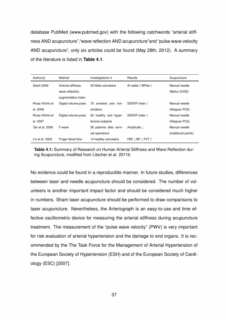
4.1 Summary of Research on Human Arterial Stiffness and Wave Reflec-
tion during Acupuncture, modified from Litscher et al. 2011b . . . . . . 37
X
List of Abbreviations
AI radial Augmentation index from radial artery
AIx brachial Augmentation index from the brachial artery
AP Pressure augmentation
BP Blood pressure
BPM Beats per minute
CA-MRSA Community-acquired methicillin-resistant staphylococcus aureus
cm Centimeter
◦ C Degree centigrade
EA Electro acupuncture
ECG Electrocardiogram
e.g. exempli gratia
ESC European Society of Cardiology
ESH European Society of Hypertension
FBF Finger blood flow
Fig. Figure
HA-MRSA Hospital-acquired methicillin-resistant staphylococcus aureus
Hb Hemoglobin
HRV Heart rate variability
Hz Hertz
LASER Light Amplification by Stimulated Emission of Radiation
LF/HF ratio Low-frequency/high-frequency ratio
XI
µm Mikrometer
mW Milliwatt
nm Nanometer
NIH United States National Institute of Health
NO Nitric oxide
PVT Paravertebral temperature
PWVao Aortic pulse wave velocity
SDDVP index Index of second derivate of digital volume pulse
TCM Traditional Chinese Medicine
TENS Transcutaneous electrical nerve stimulation
XII
1 Introduction
1.1 Acupuncture
The traditional practice of acupuncture is based on the hypothesis that all the body’s
physiological functions are modulated by 12 bilaterally distributed channels (six Yin
and six Yang channels), supplemented by two midline channels (one on the front
and the other one on the back of the body). Yin and Yang are in polarity to each
other which leads to a certain tension. Based on this tension, the hypothetical ”Qi”,
which is the universal vital energy and therefore regulates the body functions, be-
gins to flow. This is one of the fundamental laws of Chinese medicine and Chinese
philosophy. Yin and Yang must never be static but related to each other and are
therefore supplementary principles. The following aspects are essential for a contin-
uous flow of Qi: Yin and Yang have to be in opposition (which is relative and never
absolute), they are mutually dependent (no winter without summer, no day with-
out night, no birth without death), and a shift towards either Yin or Yang leads to a
stronger tension between these levels. Furthermore, they can consume each other
(water extinguishes fire, day ends night) and they can convert into each other. The
ideal state is reached when the mentioned aspects are in a harmonic and dynamic
equilibrium (see Figure 1.1) [Focks 2010].
1
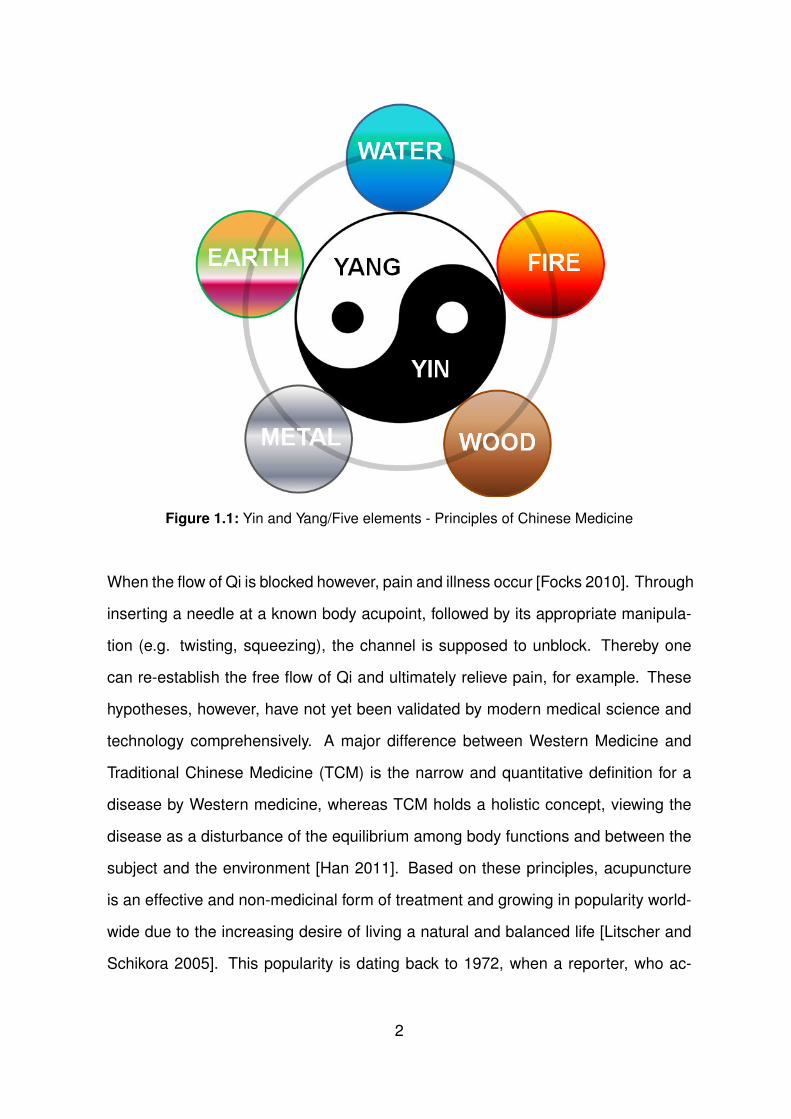
Figure 1.1: Yin and Yang/Five elements - Principles of Chinese Medicine
When the flow of Qi is blocked however, pain and illness occur [Focks 2010]. Through
inserting a needle at a known body acupoint, followed by its appropriate manipula-
tion (e.g. twisting, squeezing), the channel is supposed to unblock. Thereby one
can re-establish the free flow of Qi and ultimately relieve pain, for example. These
hypotheses, however, have not yet been validated by modern medical science and
technology comprehensively. A major difference between Western Medicine and
Traditional Chinese Medicine (TCM) is the narrow and quantitative definition for a
disease by Western medicine, whereas TCM holds a holistic concept, viewing the
disease as a disturbance of the equilibrium among body functions and between the
subject and the environment [Han 2011]. Based on these principles, acupuncture
is an effective and non-medicinal form of treatment and growing in popularity world-
wide due to the increasing desire of living a natural and balanced life [Litscher and
Schikora 2005]. This popularity is dating back to 1972, when a reporter, who ac-
2
companied an official visit of the former US President Nixon to the People’s Repub-
lic of China, had to undertake surgery and analgesia was solely achieved through
acupuncture [Hempen 2005].
With the growing demand for alternative forms of treatment, medical science had
to take notice of acupuncture. The World Health Organization (WHO) started deal-
ing with acupuncture in 1979 and created lists of medical indications. More than
40 disorders have been endorsed by the WHO so far, as conditions benefiting from
acupuncture treatment [Han 2011]. The United States National Institute of Health
(NIH) recommends acupuncture as an alternative or complementary therapy ap-
proach since its Consensus Conference in 1998. The effectiveness in symptoms
such as post operative nausea and vomiting, nausea and vomiting after chemother-
apy, but also for addictive illnesses, menstrual pain, headaches, tennis elbow, fi-
bromyalgia, back pain, carpal tunnel syndrome and bronchial asthma was addressed
[NIH 1998].
To be of the greatest utility, however, acupuncture should always be applied follow-
ing certain rules: A treatment begins with a complete diagnosis in accordance with
Traditional Chinese Medicine. Hence follows the therapeutic intention and strategy
which is based not only on ideal acupuncture, which is individually adjusted, but
on dietetics, Qi-Gong (Chinese kinetics) and Tuina (Chinese massage). The pa-
tient’s attention should be inward-facing, the treatment room has to be quiet and the
time of needling should last 20 to 30 minutes at least. Furthermore, the quantity
of treatment sessions and the individual responsiveness of the patient have to be
considered [Stux 2007].
Modern acupuncture researchers usually respect the traditional acupuncturist’s de-
cision on where to insert needles. These rationales of selection, based on theories
of TCM, usually cannot be completely understood and explained by modern medi-
cal science. Many studies concerning the applicability exist though, particular in the
3
field of analgesia and pain reduction through acupuncture treatment [Stux 2007].
For years now, neurophysiologists try to find biological pathways which could lead
to a scientific foundation of acupuncture. Some results could be observed so far:
Stimulation of acupoints leads to a temporary inhibition of algesia induced by the
mechanism of ”diffuse noxious inhibitory control” (DNIC) for mechanical, chemical
and thermal stimuli [Le Bars 2002]. Opiates, which are produced naturally by the
body, are released during acupuncture. This happens via activation of descending
inhibitory mechanisms in the cerebrospinal fluid and the substantia gelatinosa and
further on in the central plane in the substantia grisea centralis, the raphe nuclei,
the caudate nucleus, the thalamus and in the limbic system. The analgesic effect is
mainly mediated through endorphins and dynorphins [Irnich and Beyer 2002].
1.1.1 Ear Acupuncture
One of the oldest varieties of classical body acupuncture is ear acupuncture. It dates
back to ancient Egypt (Egyptian seafarers attempted to improve their vision through
piercing the earlobe), Greece (Hippocrates tried to cure acratia with a hemorrhage at
the outer ear) and Rome. The concept of somatotopy (derived from the Greek words
soma = body and topos = location) is the underlying basic concept. Somatotopy im-
plies the differentiated image of the human body on a certain area, in this case the
ear. This representation reminds someone of a bottom-up embryo. Compared to
body and traditional acupuncture, respectively, the most important principle of ear
acupuncture is that only existing disorders lead to irritated and therefore to identifi-
able ear acupoints. A so called ”active” ear acupoint is characterized by a pressure
pain or can show visible alterations, for example redness or ectasia [Thiedemann
2004]. Nonetheless, it lasted till the end of the 1950s that ear acupuncture was
4
established and further developed in Europe by French neurologist Paul Nogier.
Through a series of investigations he systematically demonstrated that specific in-
ternal organs and different regions of the ear have a functional relationship. These
results were published by Nogier in 1957 [Gori and Firenzuoli 2007]. Not until 1959
could ear acupuncture be found in Chinese literature, however. In further conse-
quence Chinese ear acupuncture developed, containing its own nomenclature and
cartography. Due to the fact that this study is based on Chinese acupuncture, its
system will be used and examined. It can be assumed that laser acupuncture stim-
ulation of ear acupoints can be performed to obtain an impact on specific organs
[Hecker and Steveling and Peuker 2002].
1.1.2 The de Qi Sensation
When using needle acupuncture, or in this study laser acupuncture, the de Qi sen-
sation has to be considered as a validity for acupunctural effects. The de Qi feeling
can be characterized as a sensation occurring during needling acupoints, although
it does not always occur and in every proband or patient. Probands with a de Qi
sensation often describe it as a dragging, dull or tingling feeling, with a spreading
or gathering of heat [Hempen 2005, Langevin 2001]. Today, only a few studies
concerning the investigation of the source of the de Qi exist. Furthermore, no spe-
cific, placebo-controlled studies could be found which examined the sensation dur-
ing laser acupuncture. The influence of de Qi on designated symptoms is not yet
clearly validated and some authors even raise questions if de Qi has any influence
on the outcome of acupuncture at all [White and Prescott and Lewith 2010].
5
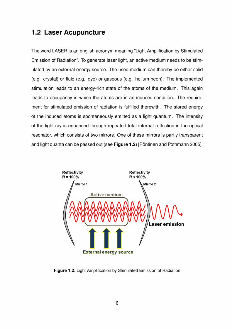
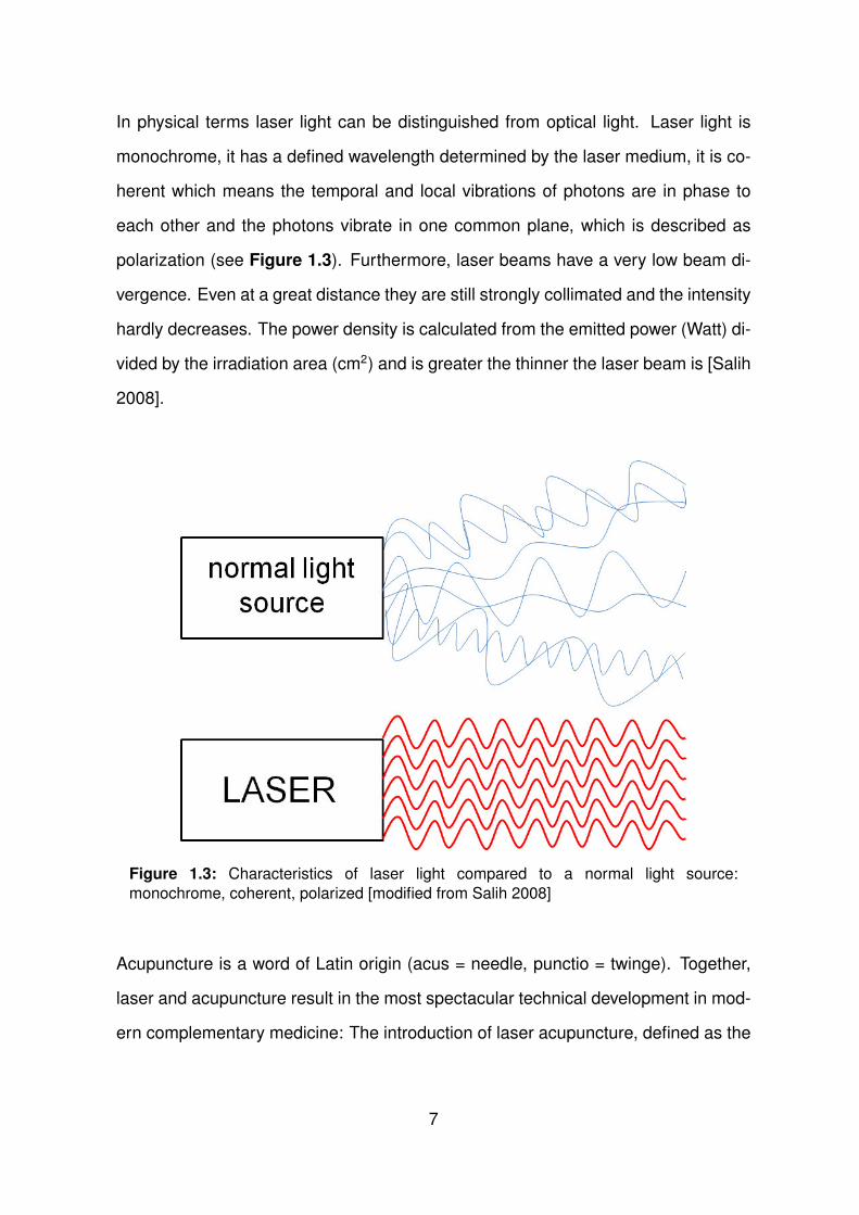
In physical terms laser light can be distinguished from optical light. Laser light is
monochrome, it has a defined wavelength determined by the laser medium, it is co-
herent which means the temporal and local vibrations of photons are in phase to
each other and the photons vibrate in one common plane, which is described as
polarization (see Figure 1.3). Furthermore, laser beams have a very low beam di-
vergence. Even at a great distance they are still strongly collimated and the intensity
hardly decreases. The power density is calculated from the emitted power (Watt) di-
vided by the irradiation area (cm2) and is greater the thinner the laser beam is [Salih
2008].
Figure 1.3: Characteristics of laser light compared to a normal light source:
monochrome, coherent, polarized [modified from Salih 2008]
Acupuncture is a word of Latin origin (acus = needle, punctio = twinge). Together,
laser and acupuncture result in the most spectacular technical development in mod-
ern complementary medicine: The introduction of laser acupuncture, defined as the
7
stimulation of traditional acupuncture points with low-intensity, nonthermal laser irra-
diation [Whittaker 2004]. Within this new technology development the goal has been
to maintain the aspects of therapeutic procedures and effects of traditional acupunc-
ture: the simultaneous procedure, the simple handling and needle equivalent stim-
ulation effects and characteristics. The result is a complex diagnostic system on
the one hand and the simple therapeutic procedure on the other [Schikora 2005].
Initially only one acupoint could be stimulated at a time which resulted in a practical
disadvantage of laser acupuncture due to the fact that classical Chinese acupunc-
ture uses treatment schemes [Whittaker 2004]. The development of a 12 channel
device by Weber and Schikora marked the turn [Weber 2005]. Laser acupuncture
has been clinically applied since the 1970s when the research group around Mester
from Hungary dealt with wound healing [Mester et al. 1971]. Although the pio-
neering nature of such work cannot be discounted, it was not until the late 1980s
and early 1990s that increased numbers of well-designed experiments appeared
[Snyder-Mackler et al. 1986, Snyder-Mackler et al. 1989, King et al. 1990, Beck-
erman et al. 1992, Gam and Thorsen and Lønnberg 1993, Whittaker 2004]. Today,
laser acupuncture is administered in a variety of pathologies and conditions of pain,
e.g. when having myofascial, postoperative and traumatic pain, headache, neural-
gia, back pain, rheumatism, tumor pain or muscular pain [Pontinen and Pothmann
2005]. The mechanisms of action, especially the physiological parameters, are not
utterly resolved however. Quantitative documented proof of the equivalence be-
tween laser acupuncture and classical needles is necessary. The effects of placebo
(so-called sham laser acupuncture) treatment should be considered. Not generating
any acupuncture effect, having an identical shape and size compared to verum nee-
dles, an identical application procedure, an application procedure not dependent on
the individual experience of the treating physician and the perception of acupoints
8
are the main requirements for future placebo laser acupuncture studies [Schikora
2005]. This study aims to contribute to still open questions.
1.2.1 Violet Laser Acupuncture
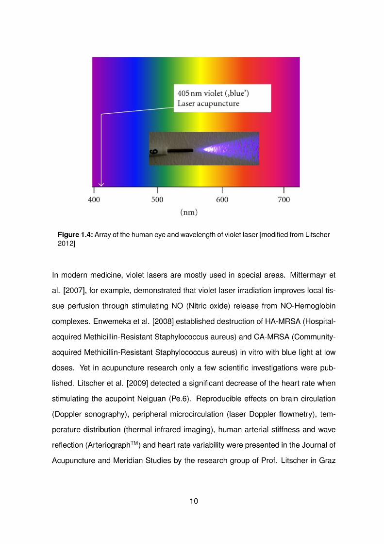
Within this study, modern violet laser acupuncture technology was used. Violet
laser acupuncture can stimulate different acupoints continuously and simultaneously
[Litscher 2012]. Nakamura et al. [2000] developed these small and convenient blue
and violet lasers which had not been available before. The most frequently used
lasers in experimental studies are violet lasers with a wavelength of 405 nm, an
output power of 100 mW and a diameter of 500 µm. Furthermore, each single
needle in modern devices can emit a different wavelength. As another important
fact the painless application of violet laser has to be mentioned [Litscher 2012]. One
major difference of violet laser compared to red and infrared laser is the evoked
de Qi sensation. Nearly 90 percent of Chinese volunteers distinguished stimulation
by violet laser and stated to have a de Qi sensation [Litscher et al. 2010]. This
sensation occurs although violet laser does not have the same penetration depth in
human skin (violet: 2 mm versus red/infrared: 2-3 cm). A de Qi sensation, which in
TCM is a prerequisite for effective acupuncture stimulation, could justify the scientific
investigation of violet laser stimulation in acupuncture [Anderson and Parrish 1981,
Litscher 2009, Litscher et al. 2009]. The patients normally do not notice if the laser
is started, when red (685 nm) or infrared (785 nm) lasers are used . It is important
to notice that both blue and violet laser are mentioned parallel in current literature.
The wavelength of 405 nm is not in fact blue but appears violet. The human eye can
process an electromagnetic spectrum from about 390 to 750 nm, therefore it has a
limited sensitivity to a color with a wavelength of 405 nm (see Figure 1.4).
9
[Litscher et al. 2010, Wang et al. 2011a, Litscher et al. 2011a, Litscher et al. 2011b,
Litscher et al. 2012a].
1.3 Modern Quantification Methods
1.3.1 The Arteriograph
Palpating the pulse, and therefore diagnosing the health state of a person, is one
of the earliest procedures a physician could perform in ancient China. In modern
medicine, several technical devices exist to evaluate the pulse wave. The pulse
wave is an index for arterial stiffness and can be used to predict cardiovascular mor-
tality [The Task Force for the Management of Arterial Hypertension of the European
Society of Hypertension (ESH) and of the European Society of Cardiology (ESC)
2007].
The ArteriographTM (TensioMed, Budapest, Hungary) is a relatively new non-invasive
device and compared to established tonometric and piezo-electronic methods easy
to use and time-effective [Baulmann et al. 2008]. The ArteriographTM indicates the
direct and indirect parameters for measuring the arterial stiffness: the aortal pulse
wave velocity (PWV), augmentation index (AIx), peripheral blood pressure and the
pulse pressure amplification [Baulmann et al. 2010].
The pulse wave velocity is widely used in clinical settings to indicate the risk of arte-
riosclerosis, coronary heart disease and stroke in healthy subjects. In aging people
the arterial stiffness becomes elevated. As a consequence systolic blood pressure
increases, causing a rise in left ventricular workload and subsequent hypertrophy,
and diastolic blood pressure decreases, leading to an impaired coronary perfusion.
Stiffer arteries lead to a higher PWV, a higher PWV indicates stiffer arteries, re-
spectively [Mattace-Raso et al. 2006]. Furthermore, with the PWV one can easily
11
evaluate the cardiovascular risk due to simple handling, accuracy and reproducibility
[Mattace-Raso et al. 2006, Inoue et al. 2009].
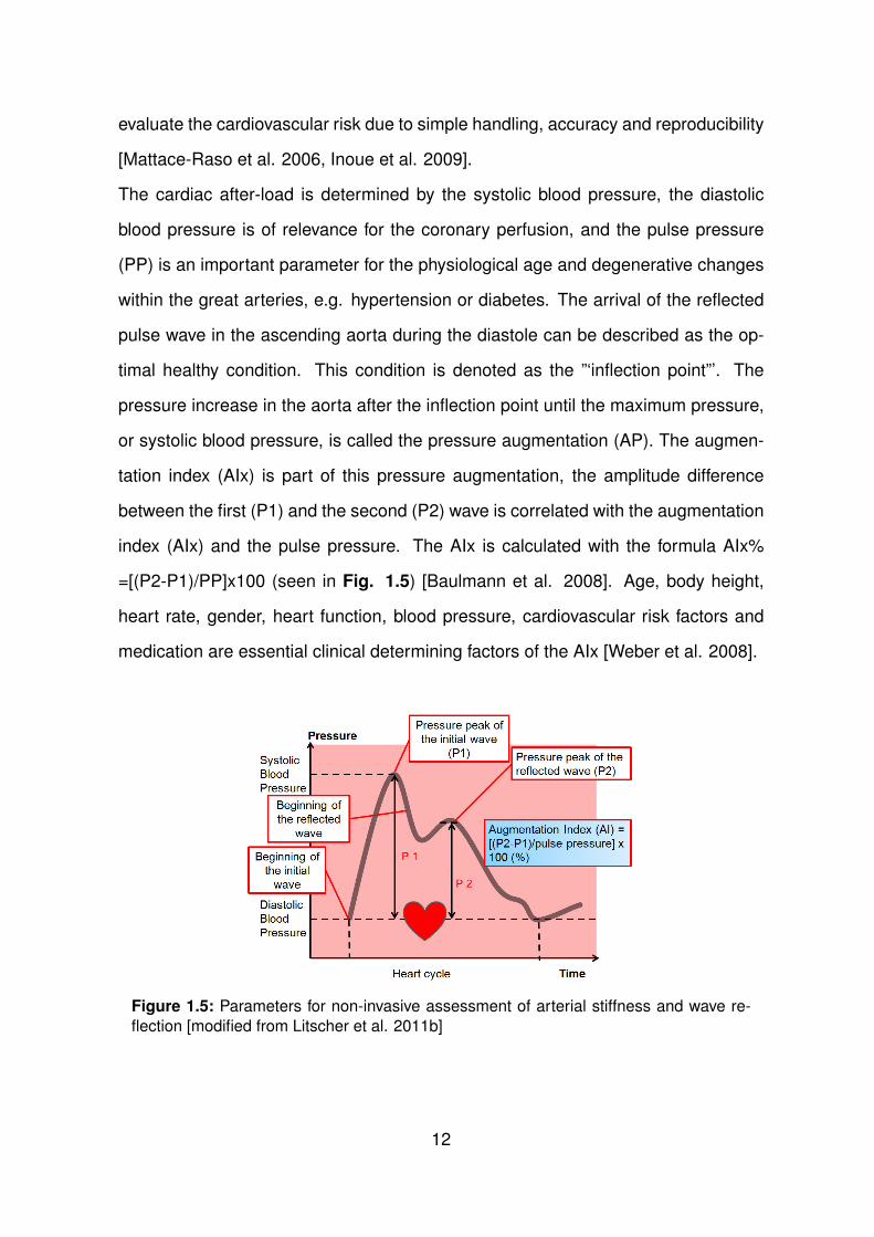
The cardiac after-load is determined by the systolic blood pressure, the diastolic
blood pressure is of relevance for the coronary perfusion, and the pulse pressure
(PP) is an important parameter for the physiological age and degenerative changes
within the great arteries, e.g. hypertension or diabetes. The arrival of the reflected
pulse wave in the ascending aorta during the diastole can be described as the op-
timal healthy condition. This condition is denoted as the ”‘inflection point”’. The
pressure increase in the aorta after the inflection point until the maximum pressure,
or systolic blood pressure, is called the pressure augmentation (AP). The augmen-
tation index (AIx) is part of this pressure augmentation, the amplitude difference
between the first (P1) and the second (P2) wave is correlated with the augmentation
index (AIx) and the pulse pressure. The AIx is calculated with the formula AIx%
=[(P2-P1)/PP]x100 (seen in Fig. 1.5) [Baulmann et al. 2008]. Age, body height,
heart rate, gender, heart function, blood pressure, cardiovascular risk factors and
medication are essential clinical determining factors of the AIx [Weber et al. 2008].
Figure 1.5: Parameters for non-invasive assessment of arterial stiffness and wave re-
flection [modified from Litscher et al. 2011b]
12
The arteriograph measurement is based on plethysmography, which is a measuring
procedure for volume fluctuation and therefore pressure changes in arteries. Be-
neath the inflated pressure cuff, which is applied to the upper arm, fluctuations in
pulsatile pressure in the artery lead to periodic pressure changes. These oscilla-
tions are measured by the arteriograph indirectly through the cuff. The pressure
recorded is dependent on three factors: the amplitude and duration of ventricular
ejection, the amplitude of the reflected wave, and the velocity of the reflected wave
from the periphery [Nelson et al. 2010].
Initially, the blood pressure is measured oscillometrically. A cuff pressure, that is
35mmHg above the systolic blood pressure measured, is inflated. Pressure fluctu-
ations can now be detected and analyzed on the computer as pulse waves (can be
seen in Figure 1.5) [Baulmann et al. 2008].
Previous studies could verify that the pulse wave velocity (PWV), measured by the
non-invasive arteriograph, is very close to the true and invasively determined one
[Horvath et al. 2010]. The pulse wave velocity in m/s is shown through the difference
in time between the beginning of the first wave and the beginning of the second or
reflected wave. This time difference is described as the distance from the jugulum
to the symphysis. The early and late systolic and diastolic waves are analyzed by
the arteriograph and the onset and the peaks of the waves are determined with first
and second derivatives. It is important to know that the arteriograph only records
and analyzes pulse waves when a supra-systolic pressure of 35mmHg is achieved
[Baulmann et al. 2008].
In summary it can be said that the quantification of arterial stiffness and pulse wave
reflexion, with the aid of PWV and AIx and blood pressure, makes it easy to evalu-
ate the damage to end organs when having arterial hypertension and furthermore
to make forward looking statements for healthy subjects directly and non-invasively
[Weber et al. 2008].
13
1.3.2 Heart Rate Variability
Today, the appearance of lethal arrhythmias in patients and the demand to constitute
either increased sympathetic or reduced vagal activity has led to enhanced efforts
in the development of quantitative markers of autonomic activity. Heart rate vari-
ability (HRV) represents such a promising and popular marker. In clinical medicine
HRV has become the accepted term to describe variations of the oscillation in the
interval between consecutive heartbeats and therefore the percentage change in
the RR interval (sequential chamber complexes) as well as the oscillation between
the consecutive instantaneous heart rates [Task Force of The European Society of
Cardiology and The North American Society of Pacing and Electrophysiology 1996].
The importance of HRV became obvious in the late 1980s, when HRV was associ-
ated with the mortality following an acute myocardial infarction [Bigger et al. 1992].
Furthermore, HRV indicates parameters of the neuro-vegetative activity, or the au-
tonomic function of the heart, respectively [Lollgen 1999].
The parasympathetic influence on the heart and the circulation is based on the re-
lease of acetylcholine by the vagus nerve. Stimulation of muscarinic receptors leads
to an increase of the potassium conductivity in the cellular membrane. As a re-
sult, stimulation of the slow diastolic depolarisation is evoked. The vagal and sym-
pathetic activity interact. The sympathetic stimulation is based on the release of
epinephrine and norepinephrine. These neurotransmitters activate β-adrenergic re-
ceptors which then phosphorylate membrane proteins via cyclic AMP. This results
in the acceleration of the slow diastolic depolarization. The vagal stimulation out-
weighs the sympathetic stimulation under resting condition and the RR interval vari-
ations depend on changes of the vagal modulation. Spectral analysis of HRV has
led to an understanding of autonomic effects of neural mechanisms on the sinus
node. The so-called high frequency (HF, between 0.15 and 0.40 Hz) is associated
with the parasympathetic nervous system, as can be seen in clinical observations
14
of autonomic maneuvers such as muscarinic receptor blockade and vagotomy [Ak-
selrod et al. 1981, HRV Manual]. The low frequency (LF, between 0.04 and 0.25
Hz) is usually taken as a marker for the sympathetic modulation, although this is
controversial. Consequently, the LF/HF ratio is considered by some investigators to
mirror sympathovagal balance or to reflect the sympathetic modulations [Task Force
of The European Society of Cardiology and The North American Society of Pacing
and Electrophysiology 1996, Lollgen 1999].
Important influence quantities on HRV are age, gender, body position, time of day
and medication [Task Force of The European Society of Cardiology and The North
American Society of Pacing and Electrophysiology 1996, Jung et al. 1996, Umetami
et al. 1998]. In a healthy person the heart rate depends on the breathing mechanism
and normally alternates more than 15 beats per minute. Values between 11 and 14
beats per minute are marginal, values under 10 beats per minute are pathological
[Lollgen 1999].
The ”Fire of Life” software, which was used in this study, analyzes the HRV. The
acquired data can be shown in a spectrogram and help to judge the function of the
autonomic nervous system. The reaction of the human body to either stress or re-
covery, and therefore to acupuncture also, is recognized and evaluated by the “Fire
of Life” software (for exemplification see Figure 1.6).
15

Figure 1.6: Fire of Life Spectrogram showing the difference between measurements of
a juvenile and a senile proband [modified from http://www.primedica.de/en/fire-of-life-2,
16.10.12]
Repeated measurements can indicate if therapeutic or interventional actions lead to
a reduction of stress and to a general improvement of the body functions [Schiller
Medilog 2012].
1.3.3 Thermography
The body temperature is one of the most important measurement parameters in
daily medical routine. An inflammation leading to an increase in temperature is
known to be a symptom and a follow-up parameter of many immunological diseases.
An induced hypothermia in anesthesia, on the other hand, is important for operations
at the heart or brain, because better tolerance towards hypoxia will be achieved. In
16
the normal processes of physiological thermoregulation the response of the human
skin blood flow is essential [Gekle and Singer and Jessen 2005]. An increase in
body temperature is always associated with a peripheral vasodilatation which is reg-
ulated by the autonomic nervous system. Deactivation of the sympathetic nervous
system leads to dilatation in the vessels, to an increase in peripheral skin blood
flow and therefore to increasing temperature while activation of the sympathetic ner-
vous system generates the opposite. This provides the necessary augmentation of
convective heat loss during heat exposure, while on the other hand cutaneous vaso-
constriction prevents heat loss during cold exposure [Gekle and Singer and Jessen
2005, Charkoudian 2010].
Today, a significant number of clinical studies investigating the benefit of thermog-
raphy as a diagnostical tool exist. Researchers could prove that infrared thermog-
raphy is an excellent non-invasive tool in the follow-up of hemangiomas, vascular
malformations and burns in pediatric patients and particularly indicate the value for
diagnosis of extremity thrombosis, inflammation, abscesses, gangrene and wound
infections, just to mention some [Saxena and Willital 2008]. Another field of infrared
thermography is cancer research. Thermographic imaging can detect temperature
changes as small as 0.1◦ C on the skin surface at an early stage of tumor de-
velopment. Therefore, it is suggested that thermographic imaging has a potential
in monitoring human tumor xenografts and their response to anticancer drugs and
should be used as standard in clinical routine [Song et al. 2007].
In conclusion, infrared thermography is non-invasive, without side effects, requires
no sedation and can be repeated as often as useful with objective results [Saxena
and Willital 2008]. Furthermore, heat radiation is emitted by every human body in
the form of energy. Energy is also called infrared radiation and cannot be seen by
the human eye [Lange 2008]. Consequently, infrared thermography is an impor-
tant method to evaluate the patients’ or healthy volunteers’ skin surface temperature
17
without influences caused by direct contact to the skin. Peripheral effects of laser-
acupuncture can be assessed [Litscher 2012].
18
2 Methods
2.1 Volunteers
This study was performed at the Stronach Research Unit for Complementary and
Integrative Laser Medicine at the Department of Anesthesiology and Intensive Care
Medicine Graz.
Within this study, the non-invasive parameters of thirteen healthy volunteers (M/F,
8/5; mean age ± standard deviation, 23.9 ± 1.7 [range 22 - 27] years; mean height
± standard deviation, 175.2 ± 7.2 cm; mean weight ± standard deviation, 69.4 ±
9.8 kg) were evaluated during violet laser acupuncture. In Figure 2.1 (Page 25) the
measurement procedure and the measurement times (a-f; thermographic imaging
and arteriograph measurement) are illustrated schematically before, during and after
violet laser stimulation.
None of the volunteers was taking medication. The study was approved by the local
ethics committee at the Medical University Graz. The volunteers were, as far as the
study design allowed, personally informed about the nature of the investigation and
all volunteers gave their written informed consent.
The volunteers were lying on a cot in the lab. The room temperature was kept
constant at 24°C. For recording the ECG, three electrodes (Skintact Premier F-55,
Leonhard Lang, Innsbruck, Austria) were attached on the thorax as standard.
19
2.2 Acupuncture Points
The laser needles were attached to the following acupuncture points:
• Nei Guan - “Inner Pass” (Pericardium 6)
Localization: Two cun 1 proximal to the transverse crease of the wrist, on the
palmar side of the hand, between the tendons of the Mm. palmaris longus and
flexor carpi radialis.
• Bai Hui - “Hundred Convergences” (Governing vessel 20)
Localization: Crossing point on an imaginary line between the apexes of both
ears and the midpoint of the calvaria, seven cun above the posterior and five
cun above the anterior hairline.
• Shen Men - “Spiritual Gate” (Ear Point 55)
Localization: In the bifurcating point between the superior and inferior crura
of the antihelix, close to the crus superior at the lateral third of the triangular
fossa [Focks 2010].
2.3 Violet Laser Acupuncture
Within this study a non-invasive Lasermicroneedle® system with integrated red and
blue/violet emission semiconductor diodes was used. For the first time, the laser
system includes violet (5) as well as red (5) laser needles, and each single needle
can emit light with a different wavelength. The wavelength accounted for 405 nm,
with an output power of 110 mW and a laser needle spot diameter of 500 µm. The
output power at the needle tip can be estimated with 100 mW based on coupling
losses [Litscher et al. 2010b]. The contact-laser needles were applied onto the skin
1Individual measurement unit defined as the width of the distal inter-phalangeal joint of the thumb.
20
through a patch, and two different (2 Hz and 100 Hz) frequency modes were used.
The irradiation lasted for 10 minutes each (600 seconds) which led to a very high
optical power density (range: kJ/cm2)[He et al. 2012].
2.4 Signal Analysis and Data Detection of Arterial
Stiffness and Pulse Wave
In this study an oscillometric non-invasive device, the ArteriographTM, was used.
The parameters in Table 2.1 were calculated. Signals were detected from an up-
per arm cuff. The cuff was attached following the rules of an ideal blood pressure
measurement:
• Volunteers were lying comfortable on a cot to keep down changes in blood
pressure. A consistent blood pressure results in consistent vessel stiffness
parameters and therefore to constant data.
• Secondly, the detection and accurate recording of the pressure alteration dur-
ing a pulse beat requires different time scales depending on the device. There-
fore a pleasurable environment has to be created for the test person to prevent
unwanted recording abortions [Baulmann et al. 2010].
Abbreviation Unit Parameter Description
HR BPM
(1/min)
Heart rate Heart rate.
MAP mmHg Mean arterial blood
pressure
Calculated from systolic and diastolic
blood pressure values.
21
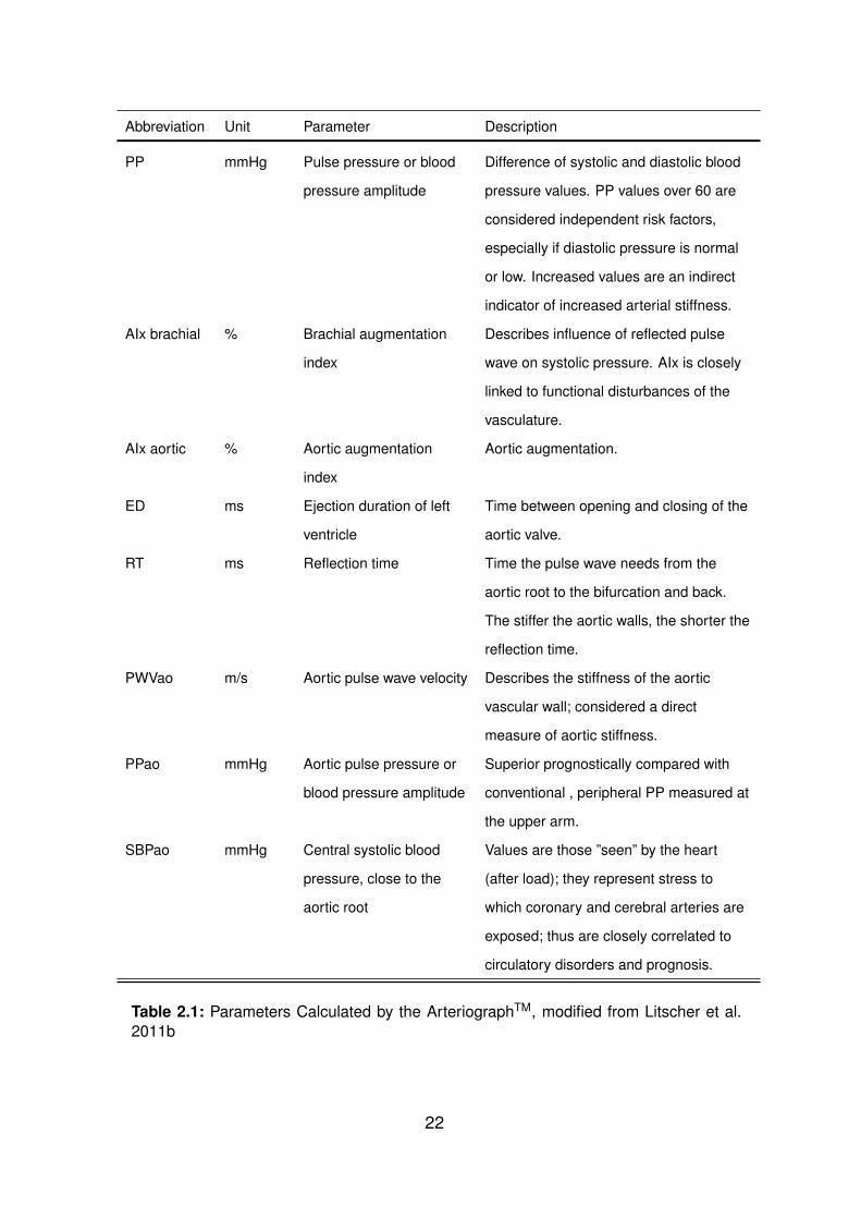
Abbreviation Unit Parameter Description
PP mmHg Pulse pressure or blood
pressure amplitude
Difference of systolic and diastolic blood
pressure values. PP values over 60 are
considered independent risk factors,
especially if diastolic pressure is normal
or low. Increased values are an indirect
indicator of increased arterial stiffness.
AIx brachial % Brachial augmentation
index
Describes influence of reflected pulse
wave on systolic pressure. AIx is closely
linked to functional disturbances of the
vasculature.
AIx aortic % Aortic augmentation
index
Aortic augmentation.
ED ms Ejection duration of left
ventricle
Time between opening and closing of the
aortic valve.
RT ms Reflection time Time the pulse wave needs from the
aortic root to the bifurcation and back.
The stiffer the aortic walls, the shorter the
reflection time.
PWVao m/s Aortic pulse wave velocity Describes the stiffness of the aortic
vascular wall; considered a direct
measure of aortic stiffness.
PPao mmHg Aortic pulse pressure or
blood pressure amplitude
Superior prognostically compared with
conventional , peripheral PP measured at
the upper arm.
SBPao mmHg Central systolic blood
pressure, close to the
aortic root
Values are those ”seen” by the heart
(after load); they represent stress to
which coronary and cerebral arteries are
exposed; thus are closely correlated to
circulatory disorders and prognosis.
Table 2.1: Parameters Calculated by the ArteriographTM, modified from Litscher et al.
2011b
22
2.5 Measuring HRV (heart rate variability)
For monitoring the cardiac setting an HRV medilog® AR12 (Huntleigh Healthcare,
Cardiff, UK, and Leupamed GmbH, Graz, Austria), with a sampling rate of 4096 Hz,
was used. An extremely accurate R-wave detection could be conducted with this
device. Raw data were saved on memory cards which can be read by a suitable
card reader connected to a computer.
To visualize how the body reacts to acupuncture, the new “Fire of Life” software
for analyzing the HRV was used. Furthermore, the low frequency (LF) and high
frequency (HF) ratio has been calculated.
2.6 Monitoring Temperature Distribution with Infrared
Thermography
In this study a Flir i5 infrared camera (Flir Systems Inc., Portland, USA), with a
wavelength ranging from 7.5 - 13 µm and a temperature distribution measurement
ranging between 0 - 250◦C, was used. The camera uses an array of infrared sensors
to measure the temperature on the surface area of the human body. The body sur-
face emits infrared beams which will then be registered by the camera. The camera
works contact-less, without feeding energy to the human body or manipulating it in
another way. It can detect differences in temperature as low as 0.1◦ C. Thermal im-
ages of the right-handed acupuncture points “Shenmen” and “Neiguan” were taken.
The collected data was transmitted to a notebook computer using the ThermaCAM
Researchers Pro 2.8 software (Flir Systems Inc., Portland, USA).
23
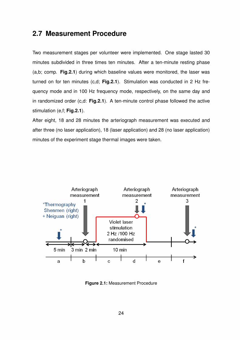
2.7 Measurement Procedure
Two measurement stages per volunteer were implemented. One stage lasted 30
minutes subdivided in three times ten minutes. After a ten-minute resting phase
(a,b; comp. Fig.2.1) during which baseline values were monitored, the laser was
turned on for ten minutes (c,d; Fig.2.1). Stimulation was conducted in 2 Hz fre-
quency mode and in 100 Hz frequency mode, respectively, on the same day and
in randomized order (c,d: Fig.2.1). A ten-minute control phase followed the active
stimulation (e,f; Fig.2.1).
After eight, 18 and 28 minutes the arteriograph measurement was executed and
after three (no laser application), 18 (laser application) and 28 (no laser application)
minutes of the experiment stage thermal images were taken.
Figure 2.1: Measurement Procedure
24
2.8 Statistical Analysis
The data were analyzed by one-way repeated measurements ANOVA on ranks
(SigmaPlot 11.0, Systat Software, Chicago, USA) and the Holm-Sidak method for
post hoc analysis. The level of significance was defined as p<0.05.
25
3 Results
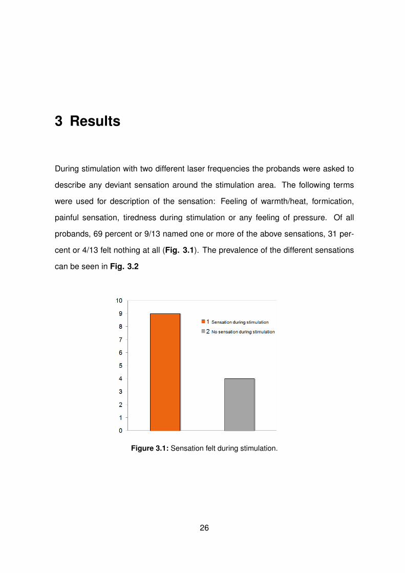
During stimulation with two different laser frequencies the probands were asked to
describe any deviant sensation around the stimulation area. The following terms
were used for description of the sensation: Feeling of warmth/heat, formication,
painful sensation, tiredness during stimulation or any feeling of pressure. Of all
probands, 69 percent or 9/13 named one or more of the above sensations, 31 per-
cent or 4/13 felt nothing at all (Fig. 3.1). The prevalence of the different sensations
can be seen in Fig. 3.2
Figure 3.1: Sensation felt during stimulation.
26
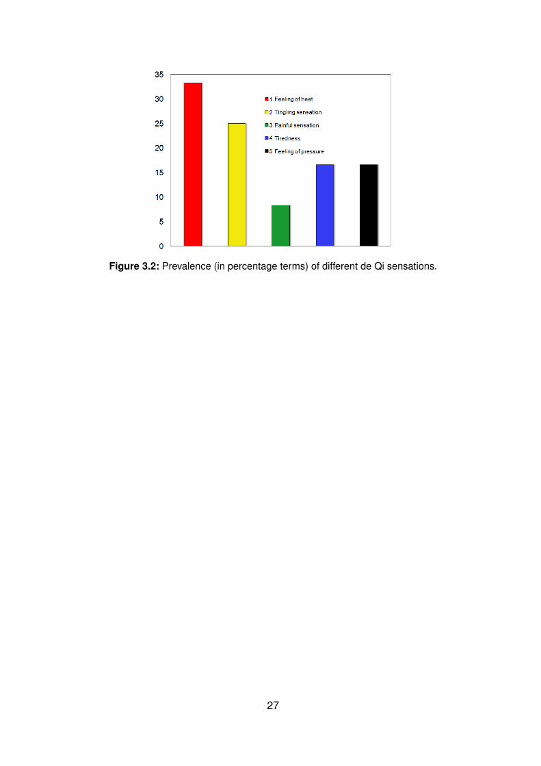
Figure 3.2: Prevalence (in percentage terms) of different de Qi sensations.
27
No significant changes were found during arteriograph measurement. The use of a
figure was refrained.
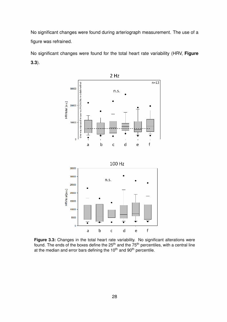
No significant changes were found for the total heart rate variability (HRV, Figure
3.3).
Figure 3.3: Changes in the total heart rate variability. No significant alterations were
found. The ends of the boxes define the 25th and the 75th percentiles, with a central line
at the median and error bars defining the 10th and 90th percentile.
28
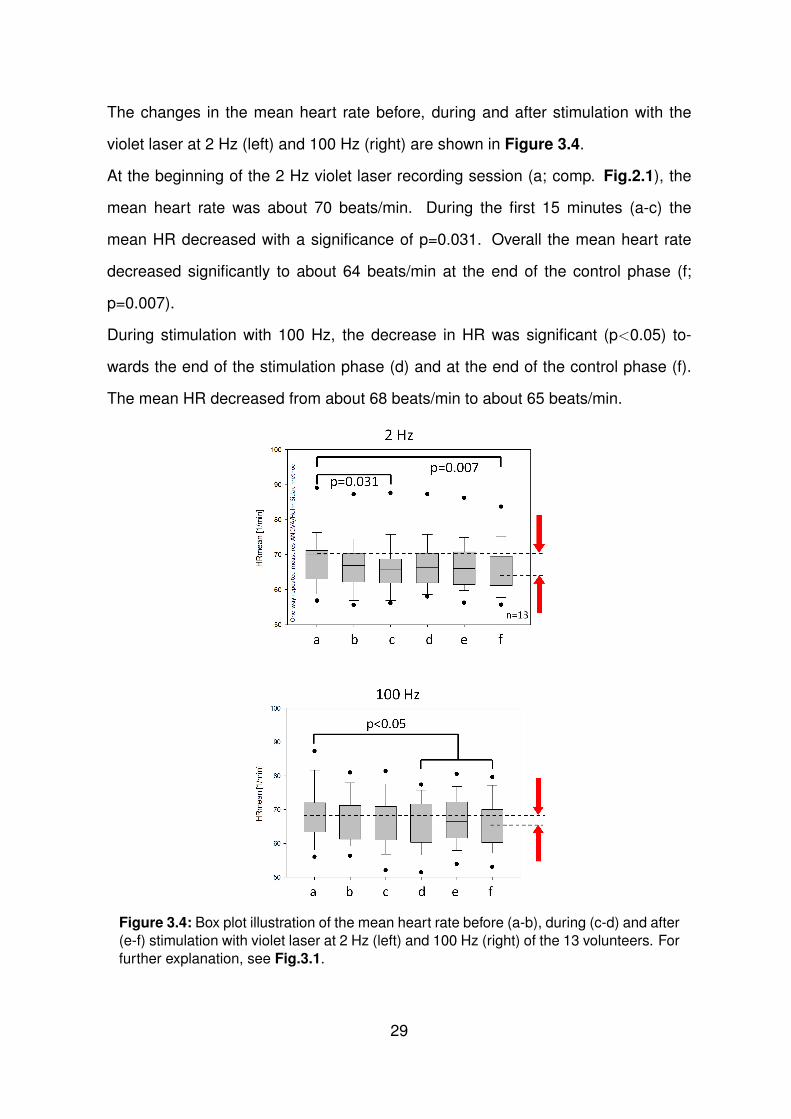
The changes in the mean heart rate before, during and after stimulation with the
violet laser at 2 Hz (left) and 100 Hz (right) are shown in Figure 3.4.
At the beginning of the 2 Hz violet laser recording session (a; comp. Fig.2.1), the
mean heart rate was about 70 beats/min. During the first 15 minutes (a-c) the
mean HR decreased with a significance of p=0.031. Overall the mean heart rate
decreased significantly to about 64 beats/min at the end of the control phase (f;
p=0.007).
During stimulation with 100 Hz, the decrease in HR was significant (p<0.05) to-
wards the end of the stimulation phase (d) and at the end of the control phase (f).
The mean HR decreased from about 68 beats/min to about 65 beats/min.
Figure 3.4: Box plot illustration of the mean heart rate before (a-b), during (c-d) and after
(e-f) stimulation with violet laser at 2 Hz (left) and 100 Hz (right) of the 13 volunteers. For
further explanation, see Fig.3.1.
29
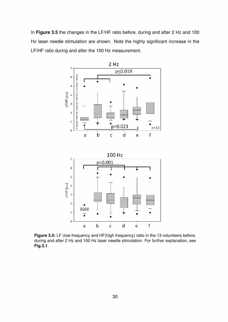
In Figure 3.5 the changes in the LF/HF ratio before, during and after 2 Hz and 100
Hz laser needle stimulation are shown. Note the highly significant increase in the
LF/HF ratio during and after the 100 Hz measurement.
Figure 3.5: LF (low-frequency and HF(high frequency) ratio in the 13 volunteers before,
during and after 2 Hz and 100 Hz laser needle stimulation. For further explanation, see
Fig.3.1.
30
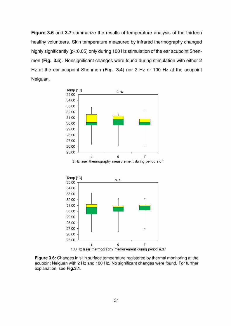
Figure 3.6 and 3.7 summarize the results of temperature analysis of the thirteen
healthy volunteers. Skin temperature measured by infrared thermography changed
highly significantly (p<0.05) only during 100 Hz stimulation of the ear acupoint Shen-
men (Fig. 3.5). Nonsignificant changes were found during stimulation with either 2
Hz at the ear acupoint Shenmen (Fig. 3.4) nor 2 Hz or 100 Hz at the acupoint
Neiguan.
Figure 3.6: Changes in skin surface temperature registered by thermal monitoring at the
acupoint Neiguan with 2 Hz and 100 Hz. No significant changes were found. For further
explanation, see Fig.3.1.
31
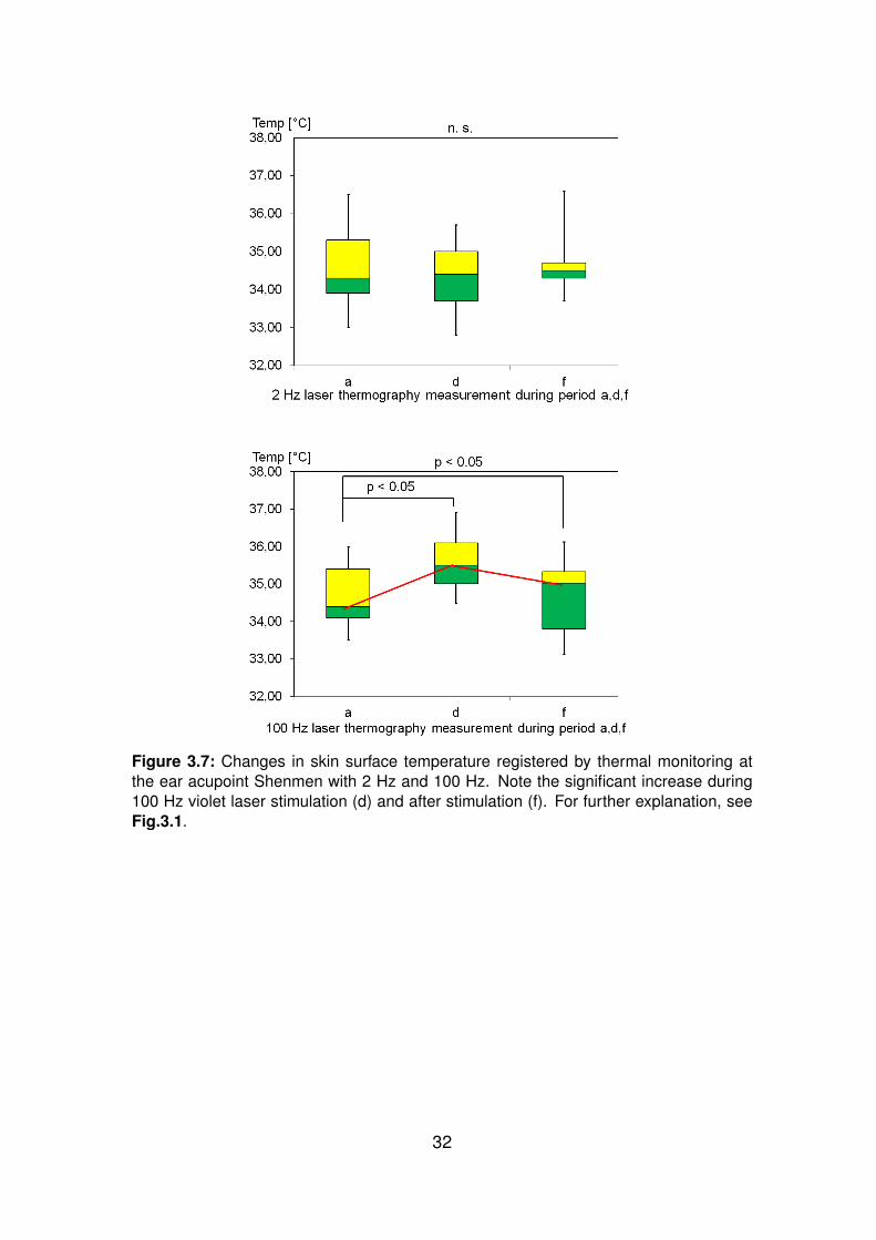
Figure 3.7: Changes in skin surface temperature registered by thermal monitoring at
the ear acupoint Shenmen with 2 Hz and 100 Hz. Note the significant increase during
100 Hz violet laser stimulation (d) and after stimulation (f). For further explanation, see
Fig.3.1.
32
4 Discussion
The aim of this study was to evaluate the influence of violet laser acupuncture on the
body’s autonomous systems, especially the cardiovascular system and the temper-
ature regulation. Two different frequencies (2 Hz and 100 Hz) were applied. Body
and ear acupuncture were used and three well-known acupoints, Neiguan, Baihui,
and the ear acupoint Shenmen.
When searching the scientific database PubMed (www.pubmed.gov) several acu-
puncture studies using the acupoint Neiguan (PC6) can be found. In Traditional Chi-
nese Medicine, the acupoint Neiguan is known to regulate and strengthen the heart
function, especially palpitations and chest distress and the Qi-circulation [Focks
2010]. Investigations led to new treatment indications for stimulating the acupoint
Neiguan. Acupressure with Sea Bands® 1 was used to control migraine-associated
nausea [Allais et al. 2012]. Self-administration, which was evaluated even better
than administration by the doctor, with simple battery-operated TENS caused a de-
crease in nausea in patients undergoing chemotherapy [Dundee et al. 1991]. 40 Hz
laser stimulation at Neiguan reduced the severity and incidence of hypotension after
epidural anesthesia. The authors stated that frequency is an important variable on
the outcome [Arai et al. 2012].
Stimulating the acupoint Baihui enhances the mood and is mainly used in headache
and vertigo therapy [Focks 2010]. Sun et al. examined the effects of high-frequency
1Elastic wristbands with a 1 cm protruding round plastic button; these devices apply continualpressure.
33
(100 Hz) laser acupuncture on a rat model. They could prove a significant decrease
of abnormally elevated glutamate (Glu) and acetylcholine (ACh) levels in the stria-
tum [Sun et al. 2012]. This could initiate further studies concerning Parkinson’s
disease, for example. Laser acupuncture could also improve motor recovery in rats
with cerebral ischemia which would suggest an improvement for patients in post-
stroke rehabilitation [Kim et al. 2012].
The ear acupoint Shenmen is used to reduce pain, stress, anxiety, and is known to
have an anti-inflammatory effect [Focks 2010]. Kao et al. [2012] significantly tapered
medication (benzodiazepines) in postmenopausal women with anxiety through au-
ricular acupressure at the ear acupoint Shenmen. In the treatment of primary insom-
nia the implementation of acupuncture at the ear acupoint Shenmen, along others,
could lead to improvement of sleep [Yeung et al. 2009]. The ear acupoint Shenmen
is also used to slow down the heart rate and activate the parasympathomimetic ner-
vous system [Hsu et al. 2007]. Clinical studies could demonstrate that acupuncture
stimulation on the mentioned points is equally effective or even superior to conven-
tional medical treatment [Pfab et al. 2011]. In most cases, however, it is suggested
to perform further and larger controlled studies to validate the results. As can be
seen above, several previously published research articles exist, examining the ef-
fectiveness of stimulating Neiguan, Baihui and Shenmen, together or independently.
Based on these, the present study tried to investigate the effectiveness of violet laser
acupuncture.
When searching for the key words ”violet laser acupuncture AND frequency” in the
scientific databases PubMed (www.pubmed.gov) and Ovid (www.ovidsp.ovid.com),
there is no publication listed until September 28th, 2012. Four preliminary scien-
tific studies have been performed by the TCM Research Center Graz, using violet
laser acupuncture [Litscher et al. 2010a, Wang et al. 2011a, Litscher et al. 2011a,
Litscher et al. 2011b]. Yet, in these studies continuos violet laser simulation was
34
applied. Evidence could be found that violet laser acupuncture stimulation at the
acupuncture point ”Dazhui” increased the brain circulation, peripheral circulation and
the local temperature [Litscher et al. 2010a, Litscher et al. 2011a]. In parameters
like heart rate and mean arterial blood pressure however, no significant changes
could be found when stimulating the acupuncture point ”Baihui” with continuous vio-
let laser stimulation [Litscher et al. 2011b].
For the first time, stimulation of the acupoints Neiguan, Baihui and the ear acupoint
Shenmen were evaluated simultaneously with two different violet laser frequencies
and stimulation was performed at body and ear acupuncture points. To measure
and identify effects on central and peripheral cardiovascular parameters, modern
quantification devices (see above) were used.
When using electro acupuncture (EA), different parameters including frequency, in-
tensity and duration have to be considered. Among these parameters, the frequency
seems to be the most critical element. Usually 2 Hz and 100 Hz are utilized as stan-
dard configurations for low- and high- frequency electro acupuncture [Han 2003,
Zhang et al. 2003, Xing et al. 2007, Li et al. 2008]. The efficacy of 2 Hz or 100 Hz
seems to be of importance when treating different diseases [Han 2011]. In rats with
neuropathic pain, for example, acupuncture analgesia with electro acupuncture of 2
Hz had a greater and more prolonged pain relief on mechanical allodynia and ther-
mal hyperalgesia than electro acupuncture with 100 Hz [Xing et al. 2007]. Neuro-
chemical studies revealed that stimulation with 2 Hz electro acupuncture accelerates
the release of enkephalin, beta-endorphin and endomorphin, while, in comparison
with 2 Hz, stimulation with 100 Hz electro acupuncture selectively increases the re-
lease of dynorphin in acupuncture analgesia [Li 2008, Han 2004, Han et al. 1991].
Silva, Silva and Prado [2012] and Zhang et al. [2003] suggested that different fre-
quencies of electro acupuncture cause different therapeutic effects and are therefore
mediated by different neural pathways. Arai et al. [2012] concluded in their investi-
35
gation that different frequencies have a different outcome on severity and incidence
of hypotension after epidural anesthesia. 2 Hz and 100 Hz frequency lasers seem
to be the most important ones in prospective studies. Wang et al. [2012] have found
that more genes related to neurogenesis were differentially regulated by 2 Hz EA
than 100 Hz EA. To do so, they used cDNA 2 micro arrays to investigate gene ex-
pressions in the region of the arcuate nucleus (Arc) in rats. The Arc is a region of
the hypothalamus and responsible for the effect of EA stimulation and the regulation
of pathophysiological processes, including autonomic activity.
The decreasing of the HR induced by violet laser acupuncture in the present study
is earlier, stronger and more prolonged when using 2 Hz compared to 100 Hz. How-
ever, there are still many different results when using either 2 Hz or 100 Hz violet
laser acupuncture. The aim of prospective examinations has to be the consideration
of application and indication of different laser frequencies. Furthermore, the correla-
tion between the HR changes stimulated by violet laser acupuncture and acupunc-
ture analgesia needs to be investigated.
No relevant results could be determined by analyzing the arteriograph data.
Likewise, Litscher et al. [2011] could not prove significance, that violet laser acupunc-
ture leads to an increase in the brachial augmentation index (AIx) and an decrease
in the aortic pulse wave velocity (PWV). However, minor fluctuations were observed.
Reasons could be the limited number of volunteers (n=10) and the missing compar-
ison to a control group with sham laser acupuncture. In this study, similar problems
can be stated. Only in one previous study, Satoh [2009] could detect reproducible
physiological alterations of human arterial stiffness and wave reflection using needle
acupuncture. PWV and the AIx are important parameters which provide enormous
information on the arterial vascular system and arterial stiffness. They are direct
indicators for cardiovascular risk [Baulmann 2008]. When searching the scientific
2complementary DNA, synthesized from a messenger RNA
36
database PubMed (www.pubmed.gov) with the following catchwords “arterial stiff-
ness AND acupuncture”,“wave reflection AND acupuncture”and “pulse wave velocity
AND acupuncture”, only six articles could be found (May 28th, 2012). A summary
of the literature is listed in Table 4.1.
Author(s) Method Investigations in Results Acupuncture
Satoh 2009 Arterial stiffness,
wave reflection,
augmentation index
25 Male volunteers AI radial ↑ BPdia ↑ Manual needle
(Baihui GV20)
Rivas-Vilchis et
al. 2008
Digital volume pulse 70 smokers and non-
smokers
SDDVP index ↑ Manual needle
(Neiguan PC6)
Rivas-Vilchis et
al. 2007
Digital volume pulse 65 healthy and hyper-
tensive subjects
SDDVP index ↑ Manual needle
(Neiguan PC6)
Tan et al. 2006 F-wave 56 patients after cervi-
cal operations
Amplitude ↓ Manual needle
(traditional points)
Lin et al. 2003 Finger blood flow 14 healthy volunteers FBF ↓ BP ↓ PVT ↑
Table 4.1: Summary of Research on Human Arterial Stiffness and Wave Reflection dur-
ing Acupuncture, modified from Litscher et al. 2011b
No evidence could be found in a reproducible manner. In future studies, differences
between laser and needle acupuncture should be considered. The number of vol-
unteers is another important impact factor and should be considered much higher
in numbers. Sham laser acupuncture should be performed to draw comparisons to
laser acupuncture. Nevertheless, the Arteriograph is an easy-to-use and time ef-
fective oscillometric device for measuring the arterial stiffness during acupuncture
treatment. The measurement of the “pulse wave velocity” (PWV) is very important
for risk evaluation of arterial hypertension and the damage to end organs. It is rec-
ommended by the The Task Force for the Management of Arterial Hypertension of
the European Society of Hypertension (ESH) and of the European Society of Cardi-
ology (ESC) [2007].
37
The effect on the parameters heart rate (HR), heart rate variability (HRV) and low
frequency/high frequency (LF/HF) was measured. No significance could be found
in the difference of the HRV before, during and after 2 Hz or 100 Hz violet laser
stimulation. The difference of the mean HR before, during and after 2 Hz or 100 Hz
violet laser stimulation is significant. In addition to the mentioned results, there is a
significant difference of the LF/HF ratio before, during and after 2 Hz or 100 Hz laser
stimulation. Maybe the high significance of the altered HR and LF/HF ratio results
out of the stimulation of three acupoints, Baihui, Shenmen (Extra, at both ears) and
Neiguan (at both arms) simultaneously.
In recent years, several studies were performed by Litscher et al. [Litscher 2009,
Litscher 2010, Gao et al. 2012a, Litscher et al. 2012a], which identified spe-
cific brain-modulated autonomic influences through computer analysis of heart rate
(HR) and heart rate variability (HRV). The significant decrease of the heart rates
in the present study is in accordance to previous trials with continuous violet laser
acupuncture [Litscher et al. 2012c, Litscher et al. 2009]. Gao et al. [2012] stimulated
anesthetized rats with violet laser acupuncture and found a significant change of the
HR during and after stimulation at the acupoint Baihui. It is questionable if these
results are comparable to results in humans and should be investigated further. In
two other trials the total HRV increased significantly after acupuncture stimulation
of the ear acupoint Shenmen and acupoint Tongli (HT 5, which was not used in the
present study), but did not last long in one case [Litscher et al. 2012a, Wang et
al. 2011b]. Although a significant difference of LF/HF ratio occurred in this study
and likewise in previous results [Litscher et al. 2009], Jones et al. [2011] could not
see significant changes during postural changes in healthy volunteers who had to
lie -10◦ head-down and then change their position. Nevertheless, they suppose that
electro acupuncture might reduce the blood pressure.
Nearly all of the above mentioned HRV studies were performed in China, while
38
the data was analyzed in Graz, Austria. Chinese volunteers/probands were en-
listed. Cultural differences, especially the probably deep belief in Traditional Chi-
nese Medicine, have to be considered. Further on, almost only metal needles and
repeated manipulation were used [Litscher et al. 2012a, Wang et al. 2011b, Litscher
et al. 2012b]. In two studies, only patients were needled [Wang et al. 2011b,
Litscher et al. 2012b]. It is inexplicable if acupuncture stimulation in patients differs
from young and healthy volunteers. It could be of importance in following studies to
evaluate the appropriate use of acupoints in different study designs.
Within this study, the aim was to visualize temperature changes around the laser
acupuncture stimulation point (Neiguan and Shenmen). When searching the scien-
tific database PubMed (www.pubmed.gov), no reviewed publication of temperature
effects when stimulating one of the acupuncture points, or both, could be found.
The skin temperature measured by infrared thermography changed highly signifi-
cantly (p<0.05) only during 100 Hz stimulation of the ear acupoint Shenmen. Non-
significant changes were found during stimulation with either 2 Hz at the ear acupoint
Shenmen nor 2 Hz or 100 Hz at the acupoint Neiguan.
As can be seen in the previous sections and results as well, study results of ther-
mography or thermal images and acupuncture vary considerably. Litscher et al.
[2011a] investigated the temperature distribution at the acupoint Dazhui (GV14) dur-
ing violet (405 nm) laser needle stimulation and could not only demonstrate a sig-
nificant increase in temperature (p<0.05) but also a temperature change at the so-
called “far field” area Zhiyang (GV9) which is located proximal at the same meridian.
Limitations of the study were the missing stimulation of a non-acupuncture (sham
acupuncture) point however. Some studies investigated the temperature distribution
of meridian like structures. Schlebusch, Maric-Oehler and Popp [2005] provided in-
formation that moxibustion (mugwort placed and burned on an acupoint) of the body
leads to the appearance of “light channels”, which can be compared to meridian like
39

structures as illustrated in textbooks of Traditional Chinese Medicine. On contrast,
Litscher [2005] tried to visualize structures which could be connected to meridians,
but failed to do so. No biological correlation could be found. Technical reflection
artifacts and equipment dependent measurement errors could be objectified and
quantified, however [Litscher and Ammer 2007]. In 2009 Agarwal-Kozlowski, Lange
and Beck [2009] could show that a significant increase in surface temperature oc-
curred within 2 min after needling the acupuncture point Hegu (Li 4), while needling
of a cutaneous or muscular point (sham acupuncture) or no needling manipulation
at all, resulted in a decrease of temperature.
Many authors come to similar conclusions concerning thermography and acupunc-
ture. Infrared thermography represents a commonly used and accurate procedure
in acupuncture studies to distinguish the effects, especially in studies with placebo
needling as a control. Main advantages are the easy to handle and contact-free
data acquisition and the realtime visualization, the superior sensitivity, contrast and
resolution [Bahr et al. 2007, Agarwal-Kozlowski and Lange and Beck 2009].
The limitations of infrared thermography and acupuncture, and one major critical
point, is the missing validity of the underlying mechanisms that lead to surface tem-
perature effects [Agarwal-Kozlowski and Lange and Beck 2009]. In future studies,
influence quantities such as skin type, the use of skin creams and most importantly
the measurement of placebo or sham acupuncture points as a control, have to be
considered.
In conclusion, it can be said that thermographic methods such as infrared cameras
at wavelength ranges of 2-5 µm and 7.5-13 µm and other High-Tech methods are
effective complementary methods in acupuncture research, which support demysti-
fication of this treatment method.
Today, there is still no detailed scientific foundation or evidence of the psychological
effects of acupuncture on volunteers or patients. Furthermore, to evaluate the phys-
40

iological basis, the sympathetic activity (e.g. hormone release, micro neurography)
and the endogenous opioid release should be investigated further. It is inevitable to
investigate the multimodal way Chinese medicine focuses on. In China, acupunc-
ture is usually combined with tuina, dietetics, herbal medicine and qi gong while in
western acupuncture trials only specific symptoms undergo examination [Agarwal-
Kozlowski and Lange and Beck 2009]. In 1984 Feinstein suggested that double-blind
placebo-controlled clinical trials are gold standard for demonstrating specific effects
over placebo. Yet in acupuncture studies double-blinding is hardly achievable. The
acupuncturist has to be aware of the applied method [Vincent and Lewith 1995]. The
main focus in future laser acupuncture studies should be on the study design.
41
5 Conclusion
For the first time, different quantification methods were used within this study to eval-
uate physiological alterations and effects on the human body when using two differ-
ent laser acupuncture frequencies. The ArteriographTM, the HRV medilog® AR12,
and the infrared thermography seem to be devices for a proper approach towards
effects like blood pressure, pulse wave velocity and temperature effects around stim-
ulation areas during acupuncture. They offer good reliability and reproducibility and
are non-invasive. In the future, further and larger studies have to be conducted to
consolidate results in laser acupuncture though. There are still too many variables
concerning the study design and open questions concerning the physiological as-
pects of acupuncture and its effect on pathological changes.
42
Bibliography
[1] Agarwal - Kozlowski K, Lange AC, Beck H. 2009.
Contact - free infrared thermography for assessing effects during acupuncture:
A randomized, single - blinded, placebo - controlled crossover clinical trial.
Anesthesiology, 111:632-639.
[2] Akselrod S, Gordon D, Ubel FA, Shannon DC, Barger AC, Cohen RC. 1981.
Power spectrum analysis of heart rate fluctuation: A quantitative probe of
beat-to-beat cardiovascular control. Science, 213:220-222.
[3] Allais G, Rolando S, Castagnoli Gabellari I, Burzio C, Airola G, Borgogno P,
Schiapparelli P, Allais R, Benedetto C. 2012.
Acupressure in the control of migraine-associated nausea. Neurol Sci,
33(Suppl 1):207-210.
[4] Anderson RR, Parrish JA. 1981.
The optics of human skin. The Journal of Investigative Dermatology, 77:13-19.
[5] Arai YCP, Ito A, Ohshima K, Hibino S, Niwa S, Kawanishi J, Numanami H,
Sakakima Y, Mizuno S, Tawada Y, Maruyama Y, Sato J, Nishihara M, Inoue S,
Ushida T. 2012.
Transcutaneous electrical nerve stimulation on the PC-5 and PC-6 points al-
leviated hypotension after epidural anaesthesia, depending on the stimulus
frequency. Evid Based Complement Med, Article ID 727121.
43
[6] Baulmann J, Schilling U, Rickerta S, Uena S, Dusing R, Illyesc M, Czirakic A,
Nickenig G, Mengden T. 2008.
A new oscillometric method for assessment of arterial stiffness: compari-
son with tonometric and piezo-electronic methods. Journal of Hypertension,
26(3):523-528.
[7] Baulmann J, Nurnberger J, Slany J, Schmieder R, Schmidt-Trucksass A,
Baumgart D, Cremerius P, Hess O, Mortensen K, Weber T. 2010.
Arterial stiffness and pulse wave analysis: consensus paper on basics, meth-
ods and clinical applications. Dtsch Med Wochenschr, 135:4-14.
[8] Beckerman H, De Bie RA, Bouter LM, De Cuyper HJ, Oostendorp RAB. 1992.
The efficacy of laser therapy for musculoskeletal and skin disorders: A criteria
- based meta - analysis of randomized clinical trials. Phys Ther, 72:483-491.
[9] Bigger Jr. JT, Fleiss JL, Steinman RC, Rolnitzky LM, Kleiger RE, Rottman JN.
1992.
Frequency domain measures of heart period, variability and mortality after
myocardial infarction. Circulation, 85(1):164-171.
[10] Charkoudian N. 2010.
Mechanisms and modifiers of reflex induced cutaneous vasodilation and vaso-
constriction in humans. J Appl Physiol, 109:1221-1228.
[11] Dundee JW, Yang J, McMillan C. 1991.
Non-invasive stimulation of the P6 (Neiguan) antiemetic acupuncture point in
cancer chemotherapy. Jounal of the Royal Society of Medicine, 84:210-212.
[12] Enwemeka CS, Williams D, Hollosi S, Yens D, Enwemeka SK. 2008.
Visible 405 nm SLD light photo - destroys methicillin - resistant staphylococcus
aureus (MRSA) in vitro. Lasers in Surgery and Medicine, 40:734-737.
44
[13] Feinstein AR. 1984.
Current problems and future challenges in randomized clinical trials. Circula-
tion, 70:767-774.
[14] Focks C, Hrsg. 2010.
Leitfaden Chinesische Medizin. 6. Aufl. Munchen: Elsevier Urban & Fischer.
[15] Gam AN, Thorsen H, Lønnberg F. 1993.
The effect of low - level laser therapy on musculoskeletal pain: a meta - anal-
ysis. Pain, 52(1):63-66.
[16] Gao XY, Liu K, Zhu B, Litscher G. 2012a.
Sino - European transcontinental basic and clinical high - tech Acupuncture
studies - part 1: Auricular acupuncture increases heart rate variability in anes-
thetized rats. Evidence - Based Complementary and Alternative Medicine, Ar-
ticle ID 817378.
[17] Gao XY, Wang L, Gaischek I, Michenthaler Y, Zhu B, Litscher G. 2012b.
Brain - modulated effects of auricular acupressure on the regulation of auto-
nomic function in healthy volunteers. Evidence - Based Complementary and
Alternative Medicine, Article ID 714391.
[18] Gekle M, Singer D, Jessen C. 2005.
Temperaturregulation und Warmehaushalt. In: Klinke R, Pape HC, Silbernagl
S, Hrsg. Physiologie. 5. Aufl. Stuttgart, New York: Georg Thieme Verlag, 493-
508.
[19] Gori L and Firenzuoli F. 2007.
Ear acupuncture in European traditional medicine. eCAM, 4(S1):13-16.
[20] Han JS, Chen XH, Sun SL, Xu XJ, Yuan Y, Yan SC, Hao JX, Terenius L. 1991.
Effect of low - and high - frequency TENS on Met - enkephalin - Arg - Phe and
45
dynorphin A [Abstract]. Pain, 47(3):295-298.
[21] Han JS. 2003.
Acupuncture: Neuropeptide release produced by electrical stimulation of dif-
ferent frequencies. TRENDS in Neurosciences, 26(1):17-22.
[22] Han JS. 2004.
Acupuncture and endorphins. Neurosci Lett, 361(1-3):258-261.
[23] Han JS. 2011.
Acupuncture analgesia: Areas of consensus and controversy. PAIN, 152:41-
48.
[24] He W, Wedig D, Wang L, Gaischek I, Litscher G. 2012.
Violet laser acupuncture - part 5: An investigation of different stimulation fre-
quencies on heart rate and variability. J Acupunct Stud, 5(6):290-294.
[25] Hecker HU, Steveling A, Peuker ET, Hrsg. 2002.
Lehrbuch und Repetitorium Ohr -, Schadel -, Mund -, Handakupunktur. 4. Aufl.
Stuttgart: Karl F. Haug Verlag.
[26] Hempen CH. 2005.
Taschenatlas Akupunktur. 6. Aufl. Stuttgart, New York: Georg Thieme Verlag.
[27] Horvath IG, Nemeth A, Lenkeya Z, Alessandri N, Tufano F, Kis P, Gaszner B,
Cziraki A. 2010.
Invasive validation of a new oscillometric device (Arteriograph) for measuring
augmentation index, central blood pressure and aortic pulse wave velocity.
Journal of Hypertension, 28:2068-2075.
[28] HRV Manual. URL, http://www.bfcenter.co.il/articles/HRV.pdf (as of
04.02.2013).
46
[29] Hsu CC, Weng CS, Sun MF, Shyu LY, Hu WC, Chang YH. 2007.
Evaluation of scalp and auricular acupuncture on EEG, HRV, and PRV. Am J
Chin Med,35(2):219-230.
[30] Inoue N, Maeda R, Kawakami H, Shokawa T, Yamamoto H, Ito C, Sasaki H.
2009.
Aortic pulse wave velocity predicts cardiovascular mortality in middle-aged
and elderly Japanese men. Circ J, 73:549-553.
[31] Irnich D, Beyer A. 2002.
Neurobiologische Grundlagen der Akupunkturanalgesie. Schmerz, 16:93-
102.
[32] Jones AYM, Kwan YL, Leung NTF, Yu RPW, Wu CMY, Warburton DER. 2011.
Electrical stimulation of acupuncture points and blood pressure responses to
postural changes: A pilot study. Am J Crit Care, 20(3):67-74.
[33] Jung J, Heisel A, Tscholl D, Fries R, Schieffer H, Uzbek C. 1996.
Assemssment of heart rate variability by using different commercially available
systems. American Journal of Cardiology, 78(1):118-120.
[34] Kao CL, Chen CH, Lin WY, Chiao YC, Hsieh CL. 2012.
Effect of auricular acupressure on peri- and early postmenopausal women
with anxiety: A double-blinded, randomized, and controlled pilot study. Evid
Based Coplement Med, Article ID 567639.
[35] King CE, Clelland JA, Knowles CJ, James R Jackson. 1990.
Effect of helium - neon laser auriculotherapy on experimental pain threshold.
Phys Ther, 70:24-30.
[36] Lange AC. 2008.
Einsatz der kontaktfreien Infrarot - Thermographie zum Nachweis von
47
Veranderungen der Hauttemperatur bei der Verum - und Sham - Akupunk-
tur. Eine prospektive, randomisierte, placebokontrollierte, einfach - blinde,
monozentrische Studie [Dissertation]. Hamburg: Universitatsklinikum Ham-
burg - Eppendorf.
[37] Langevin HM, Churchill DL, Fox JR, Badger GJ, Garra BS, Krag MH. 2001.
Biomechanical response to acupuncture needling in humans. J Appl Physiol,
91:2471-2478.
[38] Le Bars D. 2002.
The whole body receptive field of dorsal horn multireceptive neurones. Brain
Research Reviews, 40:29-44.
[39] Li Y, Zhang Y, Han JS, Wang Y. 2008.
Distinct responses of DREAM to electroacupuncture stimulation with differ-
ent frequencies during physiological and inflammatory conditions in rats. Neu-
rochem Res, 33:2070-2077.
[40] Lin CF, Liao JW, Tsai SJ, Chiang PJ, Ting H, Tang CJ, Lou KL, Hsieh LC,
Wang DW, Lin TB. 2003.
Depressor effect on blood pressure and flow elicited by electroacupuncture in
normal subjects. Autonomic Neuroscience: Basic and Clinical, 107:60-64.
[41] Litscher G. 2005.
Infrared thermography fails to visualize stimulation - induced meridian - like
structures. BioMedical Engineering OnLine, 4:38.
[42] Litscher G. 2007.
Thermography und Akupunktur. In: Bahr F, Bushe - Centmayer K, Dorfer L,
Jost F, Litscher G, Suwanda S, Zeitler H, Hrsg. Das große Buch der klassis-
chen Akupunktur. 1. Aufl. Munchen: Elsevier Urban & Fischer, 1-14.
48
[43] Litscher G, Ammer K. 2007.
Visualization of equipment dependent measurement erfors, but not of merid-
ian - like channels in complementary medicine - a thermographic human ca-
daver study. Thermology International, 17(1).
[44] Litscher G. 2009.
Modernization of traditional acupuncture using multimodal computer-based
high-tech methods - recent results of blue laser and teleacupuncture from the
Medical University of Graz. J Acupunct Meridian Stud, 2(3):202-209.
[45] Litscher G, Xie Z, Wang L, Gaischek I. 2009.
Blue 405 nm laser light mediates heart rate - investigations at the acupoint
Neiguan (Pe. 6) in Chinese adults. North Am J Med Sci, 1: 226-231.
[46] Litscher G. 2010.
Transcontinental and translational high-tech acupuncture research using
computer-based heart rate and “fire of life” heart rate analysis. Journal of
Acupuncture and Meridian Studies, 3(3):156-164.
[47] Litscher G, Huang T, Wang L, Zhang W. 2010a.
Violet laser acupuncture - part 1: Effects on brain circulation. J Acupunct
Meridian Stud, 3(4):255-259.
[48] Litscher G, Wang L, Xie Z, Gaischek I. 2010b.
Blauer Laser in der Akupunktur - ein erster Review - Report. Schweiz Z
Ganzheitsmed, 22:167-170.
[49] Litscher G, Wang L, Huang T, Zhang W. 2011a. Violet laser acupuncture -
part 3: Pilot study of potential effects on temperature distribution. J Acupunct
Meridian Stud, 4(3):164-167.
49
[50] Litscher G, Wang L, Gaischek I, Gao XY. 2011b.
Violet laser acupuncture - part 4: Acute effects on human arterial stiffness
and wave reflection. J Acupunct Stud, 4(3):168-174.
[51] Litscher G. 2012.
Integrative laser medicine and high-tech acupuncture at the Medical University
of Graz, Austria, Europe. Evid Based Complement Alternat Med, Article ID
103109.
[52] Litscher G, Cheng G, Cheng W, Wang L, Niu Q, Feng X, Gaischek I, Kuang
H. 2012a.
Sino - European transcontinental basic and clinical high-tech acupuncture
studies - part 2: Acute stimulation effects on heart rate and its variability
in patients with insomnia. Evid Based Complement Alternat Med, Article ID
916085.
[53] Litscher G, Cheng G, Wang L, Cheng W, Su H, Niu Q, Zou T, Wang Y, Feng
X, Gaischek I, Sheng Z, Kuang H. 2012b.
Biomedical teleacupuncture between China and Austria using heart rate vari-
ability - part 2: Patients with depression. Evid Based Complement Alternat
Med, Article ID 145904.
[54] Lollgen H. 1999.
Herzfrequenzvariabilitat. Deutsches Arzteblatt, 96:A-2029-2032 [Heft 31-32].
[55] Mattace-Raso FUS, van der Cammen TJM, Hofman A, van Popele NM, Bos
ML, Schalekamp MADH, Asmar R, Reneman RS, Hoeks APG, Breteler MMB,
Witteman JCM. 2006.
Arterial stiffness and risk of coronary heart disease and stroke : The Rotter-
dam study. Circulation, 113:657-663.
50
[56] Mester E, Spiry T, Szende B, Tota JG. 1971.
Effect of laser rays on wound healing. Am J Surg, 122(4):532-535.
[57] Mittermayr R, Osipov A, Piskernik C, Haindl S, Dungel P, Weber C, Vladimirov
YA, Redl H, Kozlov AY. 2007.
Blue laser light increases perfusion of a skin flap via release of nitric oxide
from hemoglobin. Mol Med, 13(1 - 2):22-29.
[58] Nakamura S, Pearton S, Fasol G. 2000.
The blue laser diode: The complete story, 2nd edition. Berlin, Germany:
Springer Verlag.
[59] Nelson MR, Stepanek J, Cevette M, Covalciuc M, Hurst RT, Tajik AJ. 2010.
Noninvasive measurement of central vascular pressures with arterial tonom-
etry: Clinical revival of the pulse pressure waveform? Mayo Clin Proc,
85(5):460-472.
[60] NIH Consensus Conference. 1998.
Acupuncture. JAMA, 280(17):1518-1524.
[61] Pfab F, Winhard M, Nowak - Machen M, Napadow V, Irnich D, Pawlik M, Bein
T, Hansen E. 2011.
Acupuncture in critically ill patients improves delayed gastric emptying: A ran-
domized controlled trial. Anesthesia and Analgesia, 112(1):150-155.
[62] Pontinen PJ, Pothmann R. 2005.
Laser in der Akupunktur - Grundlagen, Indikationen und Techniken fur
die Akupunktur-Schwerpunktpraxis. 3. Aufl. Stuttgart: MVS Medizinver-
lage/Hippokrates.
[63] Rivas-Vilchis JF, Hernandez-Sanchez F, Gonzalez-Camarena R, Suarez-
Rodrıguez LD, Escorcia-Gaona R, Cervantes-Reyes JA, Roman-Ramos R.
51
2007.
Assessment of the vascular effects of PC6 (Neiguan) using the second deriva-
tive of the finger photoplethysmogram in healthy and hypertensive subjects.
Am J Chin Med,35(3):427-436.
[64] Rivas-Vilchis JF, Escorcia-Gaona R, Cervantes-Reyes JA, Roman-Ramos R.
2008.
Vascular responses to manual PC6 acupuncture in nonsmokers and smokers
assessed by the second derivative of the finger photoplethysmogram wave-
form. Acupunct Meridian Stud,1(1):58-62.
[65] Salih N. 2008.
Randomisierte, placebo - kontrollierte Doppelblindstudie zur Unterscheid-
barkeit von Laserakupunktur und Placebo - Laserakupunktur anhand sen-
sorischer Wahrnehmungen [Dissertation]. Munchen: Ludwigs - Maximilian -
Universitat.
[66] Satoh H. 2009.
Acute effects of acupuncture treatment with Baihui (GV20) on human arterial
stiffness and wave reflection. J Acupunct Meridian Stud, 2(2):130-134.
[67] Saxena AK, Willital GH. 2008.
Infrared thermography: Experience from a decade of pediatric imaging. Eur J
Pediatr, 167:757-764.
[68] Schlebusch KP, Maric - Oehler W, Popp FA. 2005.
Biophotonics in the Infrared Spectral Range Reveal Acupuncture Merid-
ian Structure of the Body. The Journal of Alternative and Complementary
Medicine, 11(1):171-173.
52
[69] Schikora D. 2005.
Introduction. In: Litscher G, Schikora D. Eds. Laserneedle - Acupuncture, Sci-
ence and Practice. 1. Aufl. Lengerich, Pabst Science Publishers.
[70] Schiller medilog Darwin HRV. URL, http://www.schiller.ch/?lang=en#verz-
schiller§ 182§(as of: 18.07.2012).
[71] Siedentopf C, Haala I, Koppelstatter F, Verius M, Golaszewski S, Schikora D,
Felber S, Schlager A. 2005.
Placebo - Laser kontrollierte, Computer gesteuerte Doppelblind - Unter-
suchung - neue Ansatze fur die Akupunktur - Grundlagenforschung. Dt Ztschr
f Akup, 48(1):18-23.
[72] Silva ML, Silva JR, Prado WA. 2012.
Retrosplenial cortex is involved in analgesia induced by 2 - but not by 100
- Hz electroacupuncture in the rat tail - flick test. J Acupunct Meridian Stud,
5(1):42-45.
[73] Snyder-Mackler L, Bork C, Bourbon B, Trumbore D. 1986.
Effect of helium - neon laser on musculoskeletal trigger points. Phys Ther,
66:1087-1090.
[74] Snyder-Mackler L, Barry AJ, Perkins AI, Soucek MD. 1989.
Effects of helium - neon laser irradiation on skin resistance and pain in patients
with trigger points in the neck or back. Phys Ther, 69:336-341.
[75] Song C, Appleyard V, Murray K, Frank T, Sibbett W, Cuschieri A, Thompson
A. 2007.
Thermographic assessment of tumor growth in mouse xenografts. Int J Can-
cer, 121:1055-1058.
53
[76] Stux G. 2007.
Akupunktur Einfuhrung. 7. Aufl. Heidelberg: Springer Verlag.
[77] Sun Z, Jia J, Gong X, Jia Y, Deng J, Wang X, Wang X. 2012.
Inhibition of glutamate and acetylcholine release in behavioral improvement
induced by electroacupuncture in parkinsonian rats. Neuroscience Letters,
520:32-37.
[78] Tan T, Wang SC, Zhang GJ. 2006.
Effect of acupuncture and massage at tendon on F-wave of electromyogram
in the patient of flexor spasm of the upper limb after operation of cervical
spondylosis [Abstract]. Zhongguo Zhen Jiu,26(10):725-728.
[79] Task Force of The European Society of Cardiology and The North American
Society of Pacing and Electrophysiology. 1996.
Heart rate variability: Standards of measurement, physiological interpretation,
and clinical use. European Heart Journal, 17:354-381.
[80] Tiedemann N. 2004.
Randomisierte Doppelblindstudie zum Einfluss der Soft-Laser Ohrakupunktur
auf die Angst von Kindern bei der Zahnbehandlung [Dissertation]. Giessen,
Justus-Liebig-Universitat.
[81] The Task Force for the Management of Arterial Hypertension of the European
Society of Hypertension (ESH) and of the European Society of Cardiology
(ESC). 2007.
Guidelines for the management of arterial hypertension. European Heart
Journal, 28:1462-1536.
[82] Umetani K, Singer DH, McCraty R, Atkinson M. 1998.
Twenty - four hour time domain heart rate variability and heart rate: Relations
54
to age and gender over nine decades. J Am Coll Cardiol, 31:593-601.
[83] Vicent C, Lewith G. 1995.
Placebo controls for acupuncture studies. J R Soc Med, 88:199-202.
[84] Wang K, Zhang R, He F, Lin LB, Xiang XH, Ping XJ Han JS, Zhao GP, Zhang
QH, Cui CL. 2012.
Electroacupuncture frequency-related transcriptional response in rat arcuate
nucleus revealed region-distinctive changes in response to low- and high-
frequency electroacupuncture. Journal of Neuroscience Research, 90:1464-
1473.
[85] Wang L, Huang T, Zhang W, Litscher G. 2011a.
Violet laser acupuncture - part 2: Effects on peripheral microcirculation. J
Acupunct Meridian Stud, 4(1):24-28.
[86] Wang L, Valentini J, Sugimoto K, Cheng W, Cheng G, Geng H, Gaischek I,
Kuang H, Litscher G. 2011b.
Biomedical teleacupuncture between China and Austria using heart rate vari-
ability, part 1: Poststroke patients. Evidence - Based Complementary and
Alternative Medicine, Article ID 782489.
[87] Weber M. 2005.
Nadeln aus Licht - Vorstellung einer neuen Therapiemethode. Dt Ztschr f
Akup, 48(1):24-32.
[88] Weber T, Eber B, Zweiker R, Horn S, Sock S, Gruner P, Pichler M, Mayer G,
Eisserer G, Magometschnigg D, Illyes M. 2008.
Pulswellengeschwindigkeit, zentraler Blutdruck und Augmentationsindex -
”neue” Parameter zur Beschreibung eines Endorganschadens der arteriellen
Strombahn bei Hypertonie. Pathophysiologie, Methodik, prognostische Be-
55
deutung, Empfehlungen. J Hyperton, 12(1):7-13.
[89] White P, Prescott P, Lewith G. 2010.
Does needling sensation (de qi) affect treatment outcome in pain? Analysis of
data from a larger single - blind, randomised controlled trial. Acupunct Med,
28:120-125.
[90] Whittaker P. 2004.
Laser acupuncture: past, present, and future. Lasers in Medical Science,
19:69-80.
[91] Xing GG, Liu FY, Qu XX, Han JS, Wan Y. 2007.
Long - term synaptic plasticity in the spinal dorsal horn and its modulation
by electroacupuncture in rats with neuropathic pain. Experimental Neurology,
208:323-332.
[92] Yeung WF, Chung KF, Zhang SP, Yap TG, Law ACK. Electroacupuncture for
Primary Insomnia: A Randomized Controlled Trial. Sleep, 32(8):1039-1047.
[93] Zhang WT, Jin Z, Cui GH, Zhang KL, Zeng YW, Luo F, Chen AC, Han JS.
2003.
Relations between brain network activation and analgesic effect induced by
low vs. high frequency electrical acupoint stimulation in different subjects: A
functional magnetic resonance imaging study. Brain Res, 982(2):168-78.
56