noninvasive measurement of tissue magnesium and
TRANSCRIPT

HOME HELP FEEDBACK SUBSCRIPTIONS ARCHIVE SEARCH TABLE OF CONTENTS
(Circulation. 1995;92:2190-2197.) © 1995 American Heart Association, Inc.
Articles
Noninvasive Measurement of Tissue Magnesium and Correlation With Cardiac Levels Mark C.P. Haigney, MD; Burton Silver, PhD; Emmanuel Tanglao, MD; Howard S. Silverman, MD; J. Donald Hill, MD; Edward Shapiro, MD; Gary Gerstenblith, MD; Steven P. Schulman, MD
From the Department of Medicine, Division of Cardiology, Johns Hopkins Medical Institutions (M.C.P.H., H.S.S., E.S., G.G., S.P.S.), and the Department of Medicine, Good Samaritan Hospital (E.T.), Baltimore, Md; the Department of Medicine, Division of Cardiology, Uniformed Services University of the Health Sciences, Bethesda, Md (M.C.P.H.); Intracellular Diagnostics, Inc, Foster City, Calif (B.S.); and the Department of Cardiac Surgery, California Pacific Medical Center, San Francisco, Calif (J.D.H.).
Correspondence to Steven P. Schulman, MD, 536 Carnegie Bldg, Division of Cardiology, Johns Hopkins Medical Institutes, 600 N Wolfe St, Baltimore, MD 21205.
Abstract Background Intracellular magnesium ([Mg]i) plays an important role in the regulation of myocardial metabolism, contractility, and the maintenance of transsarcolemmal and
Abstract of this Article Similar articles found in:
Circulation Online PubMed
PubMed Citation This Article has been cited by:
other online articles Search Medline for articles by:
Haigney, M. C.P. || Schulman, S. P. Alert me when:
new articles cite this article Download to Citation Manager
Top Abstract Introduction Methods Results Discussion References

intracellular ionic gradients. An understanding of the role of magnesium in the clinical setting, however, is hampered by the lack of an assay of intracellular tissue magnesium levels.
Methods and Results We used energy-dispersive x-ray analysis to measure [Mg]i in sublingual epithelial cells and to correlate the level with those in atrial biopsy specimens from the same patients during cardiopulmonary bypass. Levels were also measured in acute myocardial infarction (AMI) patients before and after intravenous magnesium sulfate administration and compared with those from intensive care unit (ICU) patients and healthy individuals. A strong correlation between sublingual epithelial cell (mean, 32.1±0.3 mEq/L) and atrial tissue (mean, 32.1±0.3 mEq/L) [Mg]i was present in 18 cardiac surgery patients (r=.68, P<.002). Epithelial and atrial [Mg]i levels were lower than in healthy individuals (33.7±0.5 mEq/L, P<.01) studied at that time and correlated poorly with serum magnesium. Mean [Mg]i
in 22 AMI patients was 30.7±0.4 mEq/L, which was significantly lower than in 21 ICU patients and 15 healthy individuals (35.0±0.5 mEq/L and 34.5±0.7 mEq/L, respectively, P<.001). Intravenous magnesium sulfate was administered to most of the AMI patients (mean dose, 36±6 mmol). [Mg]i rose significantly in the AMI patients over the first 24 hours, and the magnitude of the
increase was greater in those who received higher doses of intravenous magnesium sulfate.
Conclusions Sublingual epithelial cell [Mg]i correlates well with atrial [Mg]i but not with serum magnesium. [Mg]i levels are low in patients undergoing cardiac surgery and those with AMI. Intravenous magnesium sulfate corrects low [Mg]i levels in AMI patients. Energy-dispersive x-ray analysis determination of sublingual cell [Mg]i may expedite the investigation of the role of magnesium deficiency in heart disease.
Key Words: magnesium • myocardium • myocardial infarction
Introduction
Top Abstract Introduction Methods Results Discussion References

Epidemiologists have suspected an etiologic role for dietary magnesium deficiency SCD that is based on an increased incidence of this disorder in areas with reduced concentrations of the ion in drinking water.1 2 3 4 Anderson and LeRiche5 found a 20% to 30% increase in SCD attributable to heart disease in regions of Ontario, Canada, having water hardness of <100 ppm compared with areas having >200 ppm water hardness, and
subsequent investigations reported a close inverse relation between sudden death rates and water magnesium content.6 7 The mechanism by which magnesium deficiency might increase SCD is unclear. The physiological functions of magnesium are protean, and a deficiency could lead to accelerated atherosclerosis,8 9 10 11 platelet aggregation,12 coronary thrombosis,13 coronary vasospasm,14 15 and arrhythmia as a vector of SCD. [Mg]i is a cofactor for all known ATPases and is essential for maintenance of normal
transmembrane ion gradients and metabolic homeostasis in the myocardium.16 Therefore, magnesium deficiency could have a deleterious impact on myocardial function in numerous ways.
Recently, attention has focused on the role of intravenous MgSO4 as a therapy for AMI.
A number of placebo-controlled trials have found a reduction in mortality in AMI patients receiving intravenous MgSO4,17 18 19 20 21 22 23 24 although the megatrial ISIS–4 showed no benefit.25 Serum magnesium levels were normal in LIMIT-2, but because <1% of total body magnesium is found in the serum, these measurements do not necessarily reflect the presence or absence of a deficiency. Attempts to ascertain the true state of magnesium repletion in infarct patients have yielded conflicting results. Rasmussen et al26 found that infarct patients retained an abnormal quantity of intravenous magnesium and concluded
that their stores are therefore low, but Urdal et al27 found that mononuclear cell magnesium levels were slightly higher than normal 4 to 11 days after MI. The reported coefficient of variation for this assay is 12% in healthy patients, however, and acute illness would tend to alter the lymphocyte subpopulations and nuclear-to-cytoplasmic ratio, making the test even less reliable.28 Lack of a reliable, noninvasive method for measuring physiologically relevant tissue levels of magnesium has hampered the understanding of magnesium deficiency in heart disease in general and acute coronary syndromes in particular.
In this article, we describe a novel method for measuring [Mg]i in sublingual epithelium
with EXA and apply it to patients undergoing cardiopulmonary bypass and AMI.
Methods

All patients entering the study gave informed consent. The Joint Committee on Clinical Investigation at the Johns Hopkins Hospital and the
Francis Scott Key Medical Center approved the infarct study protocol. The Institutional Review Board of the California Pacific Medical Center approved the cardiopulmonary bypass study.
Study Population To correlate the assay of sublingual [Mg]i with cardiac levels, 18 patients admitted to the California Pacific Medical Center for cardiac surgery were enrolled in the study. They included 12 patients undergoing coronary artery bypass graft surgery (2 with simultaneous aortic valve replacement), 2 patients undergoing mitral valve replacement, and 4 patients undergoing aortic valve replacement. To assess [Mg]i levels in patients with MI, 22 patients with an ST-elevation MI admitted to the CCU of the Johns Hopkins Hospital or Francis Scott Key Medical Center, 21 patients without AMI admitted to the ICU, and 15 healthy volunteers were evaluated.
Study Protocol To demonstrate that sublingual epithelial cell levels correlate with cardiac levels, tissue specimens were obtained before surgery in volunteers undergoing cardiopulmonary bypass. These values for sublingual magnesium content were then compared with atrial
biopsy specimens at bypass. To determine whether infarct patients are magnesium-deficient, sublingual samples were obtained from patients with ST-elevation MI, patients admitted to the ICU without AMI, and healthy volunteers. Furthermore, to assess the ability of acute intravenous MgSO4 to raise tissue magnesium levels, we measured [Mg]i on 2 consecutive days following admission after MgSO4 loading in a dose range of 0 to 72 mmol/24 h.
Sublingual epithelial cells were chosen for study because they are noncornified (unlike buccal mucosa), are aerobic, have a turnover time of <3 days, and are easily accessible. Previous work has shown an excellent correlation with intracellular elemental concentrations found in muscle biopsies (B.S., unpublished observation, 1993).
Sublingual specimens were obtained by gently scraping the tissue between the frenulum and Wharton's duct. The cells were applied to a low-background carbon slide and dehydrated with a standard cytology fixative (2.5% Carbowax, 95% ethanol, Medical Packaging Corp). These smears yielded many homogenous, well-defined cells for analysis with a >95% viability determined by Papanicolaou staining and the Kiss criteria for nonviability. Percoll density gradients demonstrated an average specific density of 1.037. Subjects undergoing surgery had sublingual specimens obtained 6 to 12 hours preoperatively (three specimens per patient). Serum magnesium was measured by atomic absorbance spectrophotometry. At operation, the surgeon secured three atrial biopsies, which were immediately freeze-dried for later EXA. Magnesium replacement was not routinely given.
Top Abstract Introduction Methods Results Discussion References
Infarct patients had serum magnesium, serum electrolytes, and sublingual magnesium samples obtained on presentation with ST-segment elevation 0.1 mV in two contiguous ECG leads and a history consistent with an AMI. Thrombolytics were given to all appropriate AMI subjects. Intravenous magnesium was given at the discretion of the admitting house officer. Repeated specimens of serum and epithelium were taken at 24 and 48 hours after admission. A history of previous medication use and prior cardiac conditions was obtained.
Control subjects included 21 patients with acute medical illnesses (but without history or laboratory evidence of recent myocardial ischemia) admitted to the ICU and 15 healthy house officers. A single sublingual specimen and a serum specimen were acquired within
24 hours of admission in the ICU patients; only a sublingual specimen was taken from the house officers.
Assay for Intracellular Magnesium The [Mg]i was measured with a novel application of EXA29 (B.S., US patent 4-717-826). X-ray microanalysis uses a specially configured scanning electron microscope to irradiate cells with a focused electron beam. Excitation of the cellular atoms results in displacement of inner orbital electrons, which are replaced by electrons from higher-energy shells with release of a characteristic quantum energy in the form of x-ray radiation. Measurement of x-ray fluorescence allows quantification of intracellular elements having an atomic weight equal to or greater than that of sodium by this relationship: EXA units equals x-ray intensity (peak divided by background) divided by unit cell volume.
EXA units are converted to milliequivalents per liter by a conversion constant for each element derived from a reference standard. This technique has been applied to measurement of intracellular elements in many tissues, including heart.30 31
Atrial specimens were obtained surgically from the right atrium at the time of cardiopulmonary bypass. The specimens were immediately frozen in a Freon cup suspended in liquid nitrogen and then cryosectioned in a refrigerated cryomicrotome (Ames Lab Tek). These samples were mounted frozen on a block, and the sections were transferred to a carbon planchet cooled with liquid nitrogen. Freeze-drying was performed at 10-6 mm Hg for 2 hours (Polaron Vacuum Coater).
Both sublingual and atrial specimens were examined on a scanning electron microscope (Philips Electron Optics, model XL 30) configured with an EXA analyzer using a silicon-lithium atmospheric thin window for light elements (Princeton Gamma Tech). Specimens were mounted at a 45° angle to the electron beam at a distance of 10 mm. Visualization at x5000 allows selection of an appropriate healthy-appearing, nucleated epithelial cell and measurement of cell volume. During each measurement of x-ray fluorescence, emission spectra were collected in the 500- to 5000-keV energy bands for 100 seconds; the detector was mounted at a 90° angle to the specimen at a distance of 20 mm. For the elements sampled, the only contamination between emission spectra is between calcium and potassium. A secondary peak for potassium appears in the spectral window for calcium,

but this accounts for <1% of the calcium peak and is subtracted by our analysis algorithm. We made 5 to 10 measurements in each specimen. The coefficient of variance between measurements was <2.0%.
Conversion of EXA units to milliequivalents per liter was based on a reference standard using a highly stable matrix synthetic glass containing known amounts of the elements to be analyzed and having an average atomic number similar to the biological sample examined. This standard was certified by Corning Diagnostics and the National Bureau of Standards.32 The coefficient of variance of 40 consecutive determinations of x-ray emission energies for magnesium with the reference standard was 0.82%.
Statistical Analysis All statistical analyses were performed by use of the STATVIEW II (Macintosh) statistical package, except that ANOVA with repeated measures was performed on SIGMASTAT (DOS) and linear regression analysis was executed on DELTAGRAPH 1.5 (Macintosh). Summary data are expressed as mean±SEM. A two-tailed unpaired Student's t test was used to compare serum magnesium levels at baseline between AMI and ICU patients. One-way ANOVA was used to contrast the [Mg]i levels among AMI, ICU, and healthy
patients. ANOVA with repeated measures was used to compare [Mg]i levels at baseline
and at 24 and 48 hours after bypass in the AMI patients. Student-Newman-Keuls post hoc analysis was performed on ANOVA. A linear regression analysis was performed to define the strength of the correlation between intracellular magnesium levels in the sublingual epithelium and in atrial biopsy specimens. A value of P<.05 was considered significant.
Results Cardiac Surgery Study Population Eighteen subjects admitted for cardiac surgery agreed to have sublingual scrapings taken before receiving atrial biopsies at the time of cardiopulmonary bypass. The average age of the 10 men and 8 women was 66±2 years. Their sublingual magnesium levels were compared with those of 16 athletes from the San Francisco, Calif, area.
Top Abstract Introduction Methods Results Discussion References

Serum and [Mg]i Levels in Surgical Patients The mean serum magnesium level for the cardiac surgery patients was within the normal range (1.87±0.06 mEq/L). Despite normal serum [Mg], the mean [Mg]i level in the sublingual epithelium was reduced compared to healthy volunteers (32.1±0.2 versus 33.7±0.5 mEq/L, P<.01). There was no significant difference in [Mg]i between men and women.
Correlation Between Sublingual and Atrial [Mg]i Levels The mean values for [Mg]i in the sublingual and atrial specimens for the 18 subjects were identical at 32.1±0.3 mEq/L. Fig 1 shows a linear regression comparing the individual values. A good correlation exists between the sublingual and cardiac cells (r=.68, P<.002).
View larger version
(13K): [in this window]
[in a new window]
Figure 1. Scatterplot showing linear regression comparing [Mg]i concentrations in atrial and sublingual cells in patients undergoing cardiopulmonary bypass (n=18, r=.68, P<.002).
Correlation Between [Mg]i and Serum Magnesium Linear regressions between the [Mg]i values for both atrial and sublingual specimens with serum values showed a poor correlation between serum and [Mg]i levels. Fig 2a shows the values for atrial [Mg]i plotted against serum magnesium (r=.22, P=NS); Fig 2b shows the corresponding values for sublingual and serum magnesium (r=.09, P=NS).

View larger version (16K):
[in this window] [in a new window]
Figure 2. a, Scatterplot comparing serum magnesium concentration and intracellular atrial concentrations in patients undergoing cardiopulmonary bypass (n=18). No correlation exists (r=.22). b, Scatterplot comparing serum magnesium concentration and intracellular sublingual epithelial concentrations in patients undergoing cardiopulmonary bypass (n=18). No correlation exists (r=.09).
Infarct Study Population The infarct study population consisted of 22 patients with AMI (15 men, 7 women) with a mean age of 59±3 years. Two patients described a previous history of angina; 3 had evidence of a previous MI on ECG; and 3 were taking diuretics at the time of the infarction. Nineteen patients received thrombolytics, whereas 3 did not because of a delay >12 hours before presentation (Table 1 ). Of the patients receiving thrombolytics, the mean time between the onset of symptoms and receipt of thrombolytics was 6.5±1.2 hours.
View this table: [in this window]
[in a new window]
Table 1. Infarct Patients
The control population admitted to the ICU comprised 21 patients (8 men, 13 women) with a mean age of 55±6 years (P=NS versus AMI). Table 2 lists the admitting diagnoses and medications. Two patients had a history of coronary artery disease. The control population included 15 healthy house officers (8 men, 7 women) with a mean age of 31±2 years; none were taking any medications.
View this table: [in this window]
[in a new window]
Table 2. ICU Patients

Serum Electrolytes Table 3 gives the mean serum electrolyte levels on admission to the CCU and ICU. The mean serum magnesium level in the AMI group (1.62±0.06 mEq/L) did not differ from that in the non-MI group (1.60±0.06 mEq/L). There was a small but statistically
significant difference in the mean serum sodium (139.3±0.6 and 135.3±1.3 mEq/L in the AMI and noninfarct groups, respectively; P<.05).
View this table: [in this window]
[in a new window]
Table 3. Serum Electrolyte Levels in AMI and ICU Patients
[Mg]i Levels in AMI Compared With Non-MI Patients and Control Subjects In contrast, the mean sublingual [Mg]i level was significantly lower in patients with AMI than in acutely ill, noninfarction patients or healthy control subjects (30.7±0.4 mEq/L in AMI versus 35.0±0.5 and 34.5±0.7 mEq/L in ICU and control subjects, respectively; P<.001; Fig 3 ). The difference between the ICU patients and control subjects was not significant.
View larger version
(11K): [in this window]
[in a new window]
Figure 3. Bar graph comparing mean [Mg]i concentration in the sublingual epithelium in AMI patients (n=22), patients admitted to ICU without myocardial ischemia (n=21), and healthy volunteers (NORM, n=15). *P<.001 vs AMI group.
Change in [Mg]i Levels With Intravenous Magnesium Therapy AMI patients received a mean dose of 36±6 mmol/24 h of intravenous MgSO4 (range, 0 to 72 mmol). No MgSO4 was given over the second 24 hours of the study. Mean serum
magnesium levels rose significantly from 1.62±0.06 mEq/L at admission to 2.34±0.14
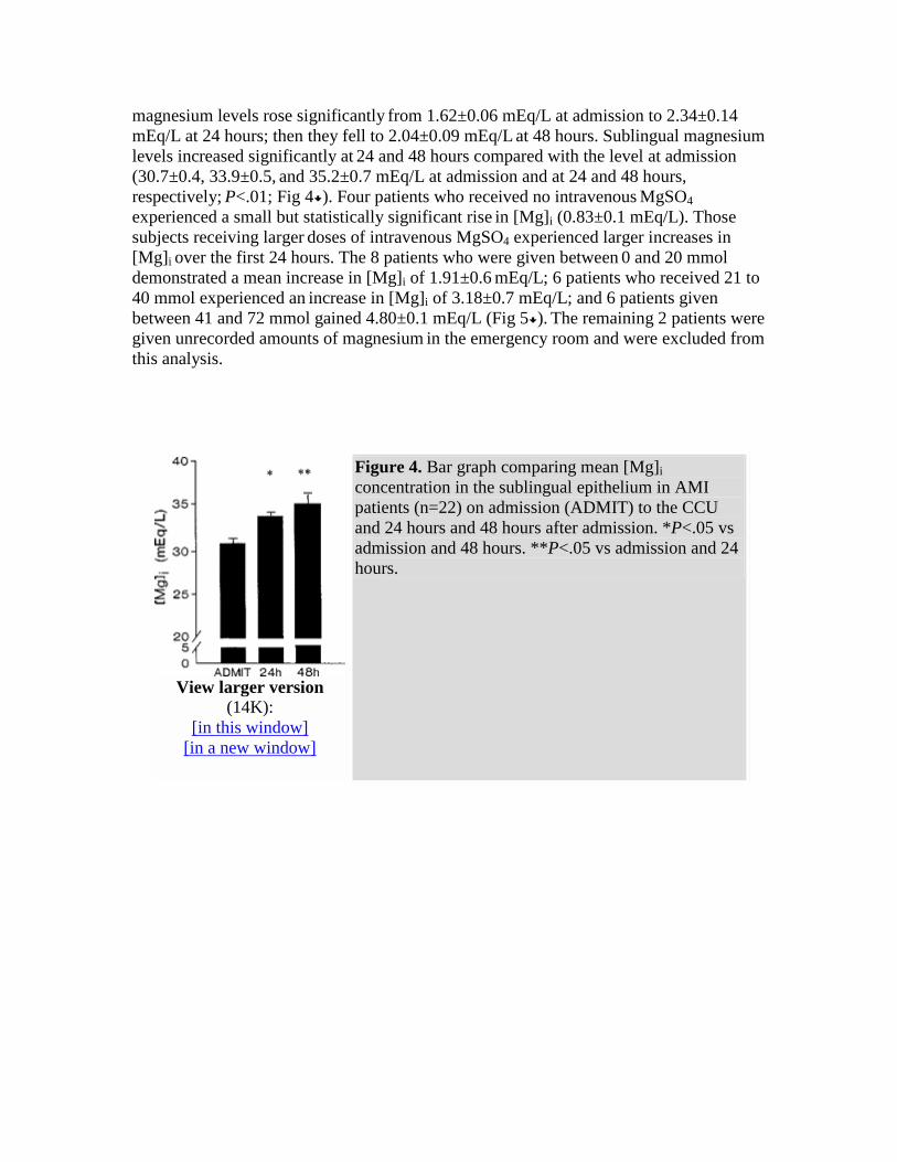
mEq/L at 24 hours; then they fell to 2.04±0.09 mEq/L at 48 hours. Sublingual magnesium levels increased significantly at 24 and 48 hours compared with the level at admission (30.7±0.4, 33.9±0.5, and 35.2±0.7 mEq/L at admission and at 24 and 48 hours, respectively; P<.01; Fig 4 ). Four patients who received no intravenous MgSO4 experienced a small but statistically significant rise in [Mg]i (0.83±0.1 mEq/L). Those subjects receiving larger doses of intravenous MgSO4 experienced larger increases in [Mg]i
over the first 24 hours. The 8 patients who were given between 0 and 20 mmol demonstrated a mean increase in [Mg]i of 1.91±0.6 mEq/L; 6 patients who received 21 to 40 mmol experienced an increase in [Mg]i of 3.18±0.7 mEq/L; and 6 patients given
between 41 and 72 mmol gained 4.80±0.1 mEq/L (Fig 5 ). The remaining 2 patients were given unrecorded amounts of magnesium in the emergency room and were excluded from this analysis.
View larger version
(14K): [in this window]
[in a new window]
Figure 4. Bar graph comparing mean [Mg]i concentration in the sublingual epithelium in AMI patients (n=22) on admission (ADMIT) to the CCU and 24 hours and 48 hours after admission. *P<.05 vs admission and 48 hours. **P<.05 vs admission and 24 hours.

View larger version
(13K): [in this window]
[in a new window]
Figure 5. Bar graph comparing the mean changes in [Mg]i concentration in the sublingual epithelium over 24 hours in AMI patients receiving 0, (n=4), 1 to 40 (n=10), and 41 to 72 mmol (n=6) IV MgSO4. *P<.05 vs patients receiving 0 mmol MgSO4.
Discussion This study applies EXA to whole cells collected noninvasively from the sublingual epithelium to assess [Mg]i levels in patients with heart disease and AMI. Measurements of [Mg]i were made in both sublingual and atrial cells from patients undergoing cardiac surgery, and these were compared with serum magnesium levels. Sublingual specimens were obtained from patients presenting with AMI and after magnesium therapy. The findings demonstrate that (1) measurements of [Mg]i in sublingual epithelial cells
correlate well with those of cardiac cells, (2) [Mg]i levels in patients undergoing cardiac surgery are lower than levels in control subjects despite normal serum magnesium, (3) subjects presenting with AMI have significantly lower [Mg]i levels compared with both healthy control subjects and acutely ill patients lacking myocardial ischemia, and (4) administration of intravenous MgSO4 raises [Mg]i levels within 24 hours.
Top Abstract Introduction Methods Results Discussion References

Rationale for Measuring [Mg]i Measurements of serum magnesium do not accurately assess tissue stores.33 Magnesium is predominantly an intracellular ion, and 1% of total body magnesium is found in the intravascular compartment.1 Skeletal muscle magnesium deficiency was demonstrated in patients with normal serum [Mg].34 In this study, we have demonstrated that no correlation exists between serum magnesium levels and both atrial and sublingual levels in patients undergoing cardiopulmonary bypass and confirmed that normal serum [Mg] often coexists with subnormal tissue levels. Therefore, direct assessment of [Mg]i
provides information not obtainable through routine serum electrolyte measurement.
It is difficult to overestimate the biological importance of magnesium. All known ATPases are Mg2+-dependent, including Na+,K+-ATPase, which maintains the transmembrane K+ gradient. G proteins and many important phosphotransferases also use Mg2+ as a cofactor. ATP must be complexed to Mg2+ to be metabolically available. In cardiac cells, O'Rourke et al35 demonstrated the importance of Mg2+ and Mg2+-nucleotide complexes in regulating the sarcolemmal calcium current, ICa. The inward rectifier current, IK1, is dependent on the presence of cytoplasmic Mg2+ ions,36 and the delayed
rectifier IK is inversely proportional to [Mg2+]i. It has been suggested that Mg2+ may serve as a "chronic regulator" of cell metabolism.37
Given the protean functions of [Mg]i, it is not surprising that magnesium deficiency is implicated in a number of cardiovascular disease states. Animal data suggest that dietary magnesium deficiency leads to accelerated atherosclerosis,8 9 and epidemiological data
also support a positive correlation.7 Magnesium-poor diets result in larger infarcts in dogs,38 impaired recovery from ischemia in rats,39 and increased pressor and arrhythmogenic responses to epinephrine in dogs.40 Sudden death is also associated with reduced magnesium intake.6 7 But although serum [Mg] deficiency is associated with arrhythmias,41 most patients with arrhythmias have normal serum concentrations, and both torsade de pointes and incessant ventricular tachycardia are reported to respond to magnesium therapy despite normal serum [Mg].42 43 To determine the true role of magnesium in heart disease, accurate measurements of [Mg]i are needed to confirm the presence of magnesium deficiency and to assess the effects of interventions on those levels.
EXA of Sublingual Cells The prior lack of a noninvasive, reproducible assay that correlates well with cardiac levels has hindered elucidation of the role of magnesium in heart disease. Mononuclear cells have been used for magnesium measurements in patients with heart failure,44 acute
medical illness, and MI26 ; patients at cardiac surgery45 ; and healthy elderly subjects.28 The poor correlation with cardiac [Mg]41 and the reported intrasubject coefficient of variability of 12%28 indicate that this test is inappropriate for precise measurements and may explain the conflicting results of Urdal et al,27 who found normal mononuclear levels in AMI patients despite increased magnesium retention.27 Erythrocyte [Mg] has been measured in hypertensive patients, but recently the correlation between erythrocyte and tissue levels was questioned.46 Muscle biopsies correlate well but are excessively invasive for routine purposes.45

EXA of sublingual cells offers an excellent alternative to existing methods of measuring [Mg]i. Sublingual cells are easily and noninvasively obtained. The cells are noncornified and metabolically active, and fixation renders their intracellular electrolyte levels stable for prolonged periods without special techniques. In this study, we have demonstrated a strong correlation between the total cellular magnesium content in sublingual and atrial
cells, establishing the relevance of the assay to cardiac tissues. EXA allows direct inspection of cells under a scanning electron microscope to exclude damaged cells from analysis. The assay is acceptably reproducible, with a coefficient of variance of 2%. The values for normal [Mg]i obtained in this study agree well with the published estimates of mammalian cardiac myocyte levels of 17 mmol/kg cell water.47
Method Limitations The primary limitation of EXA is that it measures total cellular magnesium content without distinguishing ionized species. In rat ventricular muscle, free Mg2+ makes up only 5% to 6% of total cell magnesium,44 with most of the remaining magnesium complexed to ATP. Total cellular magnesium is distributed among the cytosol (60%), mitochondria (38%), and detergent-insoluble components (2%).33 It is conceivable that the variations in [Mg]i noted in this study between healthy control subjects and patients with heart disease reflect differences in magnesium content in biologically irrelevant compartments. Against this, Corkey et al48 found that in rat hepatocytes relatively small reductions in total cellular [Mg] resulted in large decreases in [Mg2+]i because of the large number of binding sites for Mg. At any rate, measurements of tissue [Mg2+]i are
notoriously difficult and have been restricted in humans to serum and red blood cells; whether these compartments correlate with the myocardium is not known.49 Clinical investigations of the role of magnesium deficiency will probably be restricted to analysis of total [Mg]i for the foreseeable future.
Although we have demonstrated that [Mg] measurements in sublingual cells correlate well with those in cardiac cells, we have not investigated the relation between changes in sublingual levels and changes in cardiac levels after an intervention. It is possible that the
rise in sublingual [Mg] noted in the infarction patients after admission does not correspond to a change in cardiac levels. To confirm that interventions affect both tissue types similarly would require multiple samples of cardiac tissue over time, which is not feasible in human subjects.
[Mg]i in Patients Undergoing Cardiac Surgery The 18 patients undergoing cardiac surgery had a significantly lower mean sublingual magnesium level than 16 healthy volunteers from the same geographic area (32.1±0.2 versus 33.7±0.5 mEq/L, P<.01), despite normal serum magnesium levels. Hypomagnesemia after cardiac surgery was reported in 71% of patients in one report, despite normal preoperative serum magnesium levels.50 Hypomagnesemia was associated with a significantly higher incidence of arrhythmias and ventilator dependence. Cardiac surgery patients experience postoperative atrial and ventricular arrhythmias, and prophylactic treatment with intravenous magnesium reduces their incidence,51 perhaps because of the repletion of tissue magnesium deficiency.

[Mg]i in AMI Despite similar serum magnesium levels, [Mg]i levels in this study were significantly reduced in AMI patients compared with samples obtained from acutely ill patients without active myocardial ischemia and healthy individuals. The explanation for this magnesium deficiency is not clear. Only 3 of the 22 AMI patients were taking chronic diuretics; drug-induced magnesium diuresis is therefore unlikely to be responsible. The mechanisms controlling [Mg]i
homeostasis are imperfectly understood at this time. Several investigators have found that magnesium exchange occurs very slowly in vitro. Silverman et al52 detected no significant change in [Mg2+]i measured by the fluorescent compound Mg-Indo, despite incubation with 15 mmol/L MgCl, whereas Polimeni and
Page47 found that magnesium exchanges across the sarcolemma at the very slow rate of 0.21±0.02 pmol · s-1 · cm-2 membrane. It appears, however, that the magnesium flux across the sarcolemma may be hormonally regulated. Romani et al53 showed a 10% to 15% efflux of 28Mg from myocytes within 5 minutes of stimulation with 10 µmol/L norepinephrine, indicating a pool of magnesium in cardiac myocytes that can be mobilized rapidly in response to adrenergic stimulation. In our study, 1 patient in the AMI group was chronically taking a ß-blocker, and his [Mg]i on admission was above the
mean at 32.1 mEq/L. Because ß-blockade might protect the cell from catecholamine-induced magnesium loss, it is conceivable that this patient's partial preservation of magnesium stores was due to a drug effect of blocking a catecholamine-induced magnesium efflux. The infarct patients did not receive ß-blockers until 1 week after hospitalization, so the rise in [Mg]i seen after admission is not attributable to a pharmacological reduction in adrenergic tone. A large difference in plasma catecholamine
levels between the AMI and ICU patients might explain the disparity in [Mg]i levels between these groups at admission. Although plasma catecholamine levels were not measured, the ICU patients presented with life-threatening illnesses and should have had
high sympathetic tone, yet their magnesium levels were similar to those of healthy control subjects. Further studies correlating [Mg]i with plasma catecholamine levels are needed.
Alternatively, low [Mg]i levels in AMI may indicate an association between magnesium deficiency and AMI. Animal data have suggested a relation between hypomagnesemia and coronary vasospasm.54 Magnesium also has antiplatelet effects.55 Longitudinal follow-up of patients with coronary disease is necessary to investigate any etiologic role of magnesium deficiency in AMI.
[Mg]i levels rose in each AMI patient after hospitalization. In the 4 patients who did not receive intravenous MgSO4, a small but significant increase of 0.83±0.13 mEq/L after 24
hours was noted, with a much larger increase seen in subjects treated with intravenous MgSO4. Despite the fact that MgSO4
was not given after the first 24 hours, mean sublingual magnesium levels continued to rise, from 33.7±0.5 to 35.2±0.7 mEq/L at 48 hours. Mean serum [Mg] dropped from 2.34±0.14 to 2.04±0.09 mEq/L over the same period, suggesting that movement of magnesium from the vascular space to tissues continues over 48 hours. Despite the rise in [Mg]i during the first 24 hours, mean levels in AMI patients were still significantly lower than in the ICU group (P<.05). By 48 hours, however, the difference was no longer present (P=.7). In patients receiving >40 mmol IV MgSO4, mean [Mg]i was not significantly different from that of ICU patients at 24 hours

(34.6±0.7 versus 35.0±0.5 mEq/L, P=.7), suggesting that normalization of tissue levels can be achieved in 24 hours if 40 mmol IV MgSO4
is given.
It is difficult to assess the significance of magnesium deficiency in AMI patients. No patient died in the group studied, and the sample size is too small to address questions regarding outcome after infarction. Animal data suggest that magnesium deficiency
results in higher myocardial [Na+]i, worse recovery from ischemia,39 and larger infarcts.38 Although a number of small trials and one medium trial24 suggested that magnesium repletion reduces mortality in AMI, the megatrial ISIS-4 found no benefit.25 Possible interpretations for these contradictory findings include a significant increase in the time between administration of thrombolytics and intravenous MgSO4 in ISIS-4 compared with LIMIT-256 and regional differences in tissue magnesium deficiency resulting in variable responses to magnesium repletion. Alternatively, magnesium deficiency may predispose a patient to MI or to a worse outcome from AMI, but repletion over 24 hours may be too late or too slow to achieve a significant benefit. A prospective trial with extended follow-up in which [Mg]i levels are correlated with outcomes may better address the clinical significance of magnesium depletion or replenishment in acute coronary syndromes.
Conclusions EXA of sublingual epithelium offers a safe and reproducible method for measuring tissue magnesium in a cell that correlates well with cardiac levels. Elucidation of the role of magnesium in patients with heart disease can be expedited by applying EXA to examine the correlation of [Mg]i with the protean manifestations and consequences of ischemic disease and the response to different interventions.
Selected Abbreviations and Acronyms
AMI = acute myocardial infarction CCU = cardiac care unit EXA = energy-dispersive x-ray analysis ICU = intensive care unit ISIS = International Study of Infarct Survival LIMIT = Leicester Intravenous Magnesium Intervention Trial [Mg]i = intercellular magnesium SCD = sudden cardiac death

Acknowledgments We gratefully acknowledge the assistance of the medical house staff of the Francis Scott Key Medical Center and the Osler Department of Medicine at Johns Hopkins Hospital in enrolling patients in this study. We also thank Spring Metcalf and Rosalie Cosgrove for their expert assistance.
Footnotes The views expressed in this article are those of the authors and do not reflect the official policy or position of the US Air Force, Department of Defense, Uniformed Services University, or the US government.
Received June 14, 1994; revision received April 12, 1995; accepted May 16, 1995.
References
1. Eisenberg MJ. Magnesium deficiency and sudden death. Am Heart J. 1992;124:544-549. [Medline]
2. Schroeder HA. Relation between mortality from cardiovascular disease and treated water supplies. JAMA. 1960;172:1902-1908.
Top Abstract Introduction Methods Results Discussion References

3. Anderson TW, LeRiche WH, MacKay JS. Sudden death and ischemic heart disease: correlation with hardness of local water supply. N Engl J Med. 1969;280:805-807. [Medline]
4. Durlach J, Bara M, Guiet-Bara A. Magnesium level in drinking water and cardiovascular risk factor: a hypothesis. Magnesium. 1985;4:5-15. [Medline]
5. Anderson TW, LeRiche WH. Sudden death from ischemic heart disease in Ontario and its correlation with water hardness and other factors. Can Med Assoc J. 1971;105:155-160.
6. Allen HAJ. An Investigation of Water Hardness, Calcium, and Magnesium in Relation to Mortality in Ontario. Ontario, Canada: University of Waterloo; 1972. Thesis.
7. Karppanen H. Epidemiological studies on the relationship between magnesium intake and cardiovascular diseases. Artery. 1981;9:190-199. [Medline]
8. Bloom S. Coronary artery lesions in Mg-deficient hamsters. Magnesium. 1985;4:82-95. [Medline]
9. Seelig MS, Heggtveit HA. Magnesium interrelationships in ischemic heart disease. Am J Clin Nutr. 1974;27:59-79. [Medline]
10. Davis WH, Leary WP, Reyes AJ, Olhaberry JV. Monotherapy with magnesium increases abnormally low high density lipoprotein cholesterol: a clinical assay. Curr Ther Res. 1984;36:341-346.
11. Hellerstein E, Vitale JJ, White PL, Hegsted D, Zamchek N, Nakamura M. Influence of dietary magnesium on cardiac and renal lesions of young rats fed an atherogenic diet. J Exp Med. 1957;106:767-775.
12. Adams JH, Mitchell JRA. The effects of agents which modify platelet behaviour and of magnesium ions on thrombus formation in vivo. Thromb Haemost. 1979;42:603-610. [Medline]
13. Stevenson MM, Yoder I. Studies of platelet aggregation, plasma adenosine diphosphate breakdown, and blood coagulation in magnesium deficient calves and rats. Thromb Diath Haemorrh. 1972;23:299-305.
14. Gertz SD, Wajnberg RS, Kurgan A, Uretzky G. Effect of magnesium sulfate on thrombus formation following partial arterial constriction: implications for coronary vasospasm. Magnesium. 1987;6:225-235. [Medline]

15. Cohen L, Kitzes R. Prompt termination and/or prevention of cold-pressor-stimulation-induced vasoconstriction of different beds by magnesium sulfate in patients with Prinzmetal's angina. Magnesium. 1986;5:144-149. [Medline]
16. White RE, Hartzell HC. Magnesium ions in cardiac function. Biochem Pharmacol. 1989;38:859-867. [Medline]
17. Morton BC, Nair RC, Smith FM, McKibbon TG, Poznanski WJ. Magnesium therapy in acute myocardial infarction: a double blind study. Magnesium. 1984;3:346-352. [Medline]
18. Smith LF, Heagerty AM, Bing RF, Barnett DB. Intravenous infusion of magnesium sulphate after acute myocardial infarction: effects on arrhythmias and mortality. Int J Cardiol. 1986;12:175-180. [Medline]
19. Rasmussen HS, Gronback M, Cintin C, Balslev S, Norregard P, McNair P. One year death rate in 270 patients with suspected acute myocardial infarction, initially treated with intravenous magnesium or placebo. Clin Cardiol. 1988;11:377-381.
20. Shechter M, Hod H, Marks N, Kaplinsky E, Behar S, Rabinowitz B. Beneficial effect of magnesium sulfate in acute myocardial infarction. Am J Cardiol. 1990;66:271-274. [Medline]
21. Feldstedt M, Boesgaard S, Bouchelouche P, Svenningsen A, Brooks L, Lech Y, Aldershvile J, Skagen K, Godtfredsen J. Magnesium substitution in acute ischaemic heart syndromes. Eur Heart J. 1991;12:1215-1218. [Medline]
22. Abraham AS, Rosenmann D, Kramer M, Balkin J, Zion MM, Farbstein H, Eylayth U. Magnesium in the prevention of lethal arrhythmias in acute myocardial infarction. Arch Intern Med. 1987;147:753-755.
23. Ceremuzynski L, Jurgiel R, Kulakowski P, Gebalska J. Threatening arrhythmias in acute myocardial infarction are prevented by intravenous magnesium sulphate. Am Heart J. 1989;118:1333-1334. [Medline]
24. Woods KL, Fletcher S, Roffe C, Haider Y. Intravenous magnesium sulphate in suspected acute myocardial infarction: results of the second Leicester Intravenous Magnesium Intervention Trial (LIMIT-2). Lancet. 1992;339:1553-1558. [Medline]
25. ISIS Collaborative Group. ISIS-4: randomized study of intravenous magnesium in over 50 000 patients with suspected acute myocardial infarction. Circulation. 1993;88:I-292. Abstract.

26. Rasmussen HS, McNair P, Goransson L, Balslov S, Larsen OG, Aurup P. Magnesium deficiency in patients with ischemic heart disease with and without acute myocardial infarction uncovered by an intravenous loading test. Arch Intern Med. 1988;148:329-332.[Medline]
27. Urdal P, Landmark K, Basmo GM. Mononuclear cell magnesium and retention of magnesium after intravenous loading in patients with acute myocardial infarction. Scand J Clin Invest. 1992;52:763-766.
28. Martin BJ, Lyon TDB, Walker W, Fell GS. Mononuclear blood cell magnesium in older subjects: evaluation of its use in clinical practice. Ann Clin Biochem. 1993;30:23-27. [Medline]
29. Silver BB, Silver SL. Screening test for tissue calcium and magnesium in animals and human subjects. Fed Proc. 1983;42:40. Abstract.
30. Thandroyen FT, Bellotto D, Katayama A, Hagler HK, Willerson JT, Buja LM. Subcellular electrolyte alterations during progressive hypoxia and following reoxygenation in isolated neonatal rat ventricular myocytes. Circ Res. 1992;71:106-119. [Abstract]
31. Djaldetti M, Gilgal R, Bessler H, Magazanik A, Zahavi I. X-ray microanalysis of the elemental content in the hearts of mice treated with digoxin. Cardiology. 1988;75:344-347. [Medline]
32. Fiori CE, Blackburn DH. Low Z glass standards for biological X-ray microanalysis. J Microsc. 1982;127:223-226.
33. Dyckner T, Wester PO. The relation between extra- and intracellular electrolytes in patients with hypokalemia and/or diuretic treatment. Acta Med Scand. 1978;204:269-282. [Medline]
34. Lim P, Jacob E. Magnesium deficiency in patients on long term diuretic therapy for heart failure. Br Med J. 1972;3:620-622. [Medline]
35. O'Rourke B, Backx PH, Marban E. Phosphorylation-independent modulation of L-type calcium channels by magnesium-nucleotide complexes. Science. 1992;257:245-247. [Medline]
36. Vandenberg CA. Inward rectification of a potassium channel in cardiac ventricular cells depends on internal magnesium ions. Proc Natl Acad Sci U S A. 1987;84:2560-2564. [Medline]
37. Romani A, Scarpa A. Regulation of cell magnesium. Arch Biochem Biophys. 1992;298:1-12. [Medline]

38. Chang C, Varghese PJ, Downey J, Broom S. Magnesium deficiency and myocardial infarction size in the dog. J Am Coll Cardiol. 1985;5:280-289. [Medline]
39. Borchgrevink PC, Jynge P. Acquired magnesium deficiency and myocardial tolerance to ischemia. J Am Coll Nutr. 1987;6:355-363. [Medline]
40. Bean BL, Varghese PJ. Role of dietary magnesium deficiency in the pressor and arrhythmogenic response to epinephrine in the intact dog. Am Heart J. 1994;127:96-102. [Medline]
41. Gottlieb SS. Importance of magnesium in congestive heart failure. Am J Cardiol. 1989;63:39G-42G. [Medline]
42. Tzivoni D, Banai S, Schuger C, Benhorin J, Keren A, Gottlieb S, Stern S. Treatment of torsade de pointes with magnesium sulfate. Circulation. 1988;77:392-397. [Abstract]
43. Iseri LT, Chung P, Tobis J. Magnesium therapy for intractable ventricular tachyarrhythmias in normomagnesic patients. West J Med. 1983;138:823-829. [Medline]
44. Ralston MA, Murnane MR, Kelly RE, Altschuld RA, Unverferth DV, Leier CV. Magnesium content of serum, circulating mononuclear cells, skeletal muscle, and myocardium in congestive heart failure. Circulation. 1989;80:573-580. [Abstract]
45. Moller JB, Klaaborg KE, Alstrup P, Arendrup H, Klitgard NA, Pedersen KE. Magnesium content of the human heart. Scand J Thorac Cardiovasc Surg. 1991;25:155-158.[Medline]
46. Coghlan HC, Natello G. Erythrocyte magnesium in symptomatic patients with primary mitral valve prolapse: relationship to symptoms, mitral leaflet thickness, joint hypermobility, and autonomic regulation. Magn Trace Elem. 1992;10:205-214.
47. Polimeni PI, Page E. Magnesium in heart muscle. Circ Res. 1974;33:367-374.
48. Corkey BE, Duszynski J, Rich TL, Matschinsky B, Williamson KR. Regulation of free and bound magnesium in rat hepatocytes and isolated mitochondria. J Biol Chem. 1986;261:2567-2574. [Abstract]
49. Resnick LM, Altura BT, Gupta RK, Laragh JH, Alderman MH, Altura BM. Intracellular and extracellular magnesium depletion in type 2 (non-insulin-dependent) diabetes mellitus. Diabetologia. 1993;36:767-770.[Medline]

50. Aglio LS, Stanford GG, Maddi R, Boyd JL, Nussbaum S, Chernow B. Hypomagnesemia is common following cardiac surgery. J Cardiothorac Vasc Anesth. 1991;5:201-208. [Medline]
51. Fanning WJ, Thomas CS, Roach A, Tomichek R, Alford WC, Stoney WS. Prophylaxis of atrial fibrillation with magnesium sulfate after coronary artery bypass grafting. Ann Thorac Surg. 1991;52:529-533. [Abstract]
52. Silverman HS, Di Lisa F, Hui RC, Miyata H, Sollott SJ, Hansford RG, Lakatta EG, Stern MD. Regulation of intracellular free Mg2+ and contraction in single adult mammalian myocytes. Am J Physiol. 1994;266:C222-C233. [Medline]
53. Romani A, Marfella C, Scarpa A. Regulation of magnesium uptake and release in the heart and in isolated ventricular myocytes. Circ Res. 1993;72:1139-1148. [Abstract]
54. Turlapaty PDMV, Altura BM. Magnesium deficiency produces spasms of coronary arteries: relationship to etiology of sudden death and ischemic heart disease. Science. 1980;208:198-200. [Medline]
55. Adams JH, Mitchell JRA. The effect of agents which modify platelet behavior and of magnesium ions on thrombus formation in vivo. Thromb Haemost. 1979;42:603-610. [Medline]
56. Woods KL, Fletcher S. Long-term outcome after intravenous magnesium sulphate in suspected acute myocardial infarction: the second Leicester Intravenous Magnesium Intervention Trial (LIMIT-2). Lancet. 1994;343:816-819.[Medline]
This article has been cited by other articles:
• Shechter, M., Sharir, M., Labrador, M. J. P., Forrester, J., Silver, B., Bairey Merz, C. N. (2000). Oral Magnesium Therapy Improves Endothelial Function in Patients With Coronary Artery Disease. Circulation 102: 2353-2358 [Abstract] [Full Text]
HOME HELP FEEDBACK SUBSCRIPTIONS ARCHIVE SEARCH TABLE OF CONTENTS
Abstract of this Article Similar articles found in:
Circulation Online PubMed
PubMed Citation This Article has been cited by: Search Medline for articles by:
Haigney, M. C.P. || Schulman, S. P. Alert me when:
new articles cite this article Download to Citation Manager

CIRCULATION ART, THRO, VASC BIO ALL AHA JOURNALS CIRCULATION RESEARCH HYPERTENSION STROKE