normal caloric responses during acute phase of vestibular ... · 302 j clin neurol...
TRANSCRIPT

Copyright © 2016 Korean Neurological Association 301
Background and PurposezzWe report a novel finding of caloric conversion from normal re-sponses into unilateral paresis during the acute phase of vestibular neuritis (VN).MethodszzWe recruited 893 patients with a diagnosis of VN at Dizziness Clinic of Seoul Na-tional University Bundang Hospital from 2003 to 2014 after excluding 28 patients with isolated inferior divisional VN (n=14) and those without follow-up tests despite normal caloric responses initially (n=14). We retrospectively analyzed the neurotological findings in four (0.5%) of the patients who showed a conversion from initially normal caloric responses into unilateral paresis during the acute phase.ResultszzIn those four patients, the initial caloric tests were performed within 2 days of symp-tom onset, and conversion into unilateral caloric paresis was documented 1–4 days later. The clinical and laboratory findings during the initial evaluation were consistent with VN in all four patients except for normal findings in bedside head impulse tests in one of them.ConclusionszzNormal findings in caloric tests should be interpreted with caution during the acute phase of suspected VN. Follow-up evaluation should be considered when the find-ings of the initial caloric test are normal, but VN remains the most plausible diagnosis.Key Wordszz vestibular neuritis, caloric test, vestibulo-ocular reflex, head-impulse test.
Normal Caloric Responses during Acute Phase of Vestibular Neuritis
INTRODUCTION
Vestibular neuritis (VN) refers to a syndrome of acute unilateral vestibular deafferenta-tion due to inflammation of the peripheral vestibular organ.1 Patients with VN invariably suffer from spontaneous vertigo, nausea/vomiting, and unsteadiness.2 Along with sponta-neous horizontal-torsional nystagmus beating toward the side of the healthy ear, identify-ing unilateral peripheral vestibular hypofunction is the key step for the diagnosis of VN.3 In this regard, unilateral caloric paresis has been the diagnostic hallmark of VN even though unified diagnostic criteria have not been available for this disorder, which is the most-common cause of acute prolonged spontaneous vertigo.1
The increasing awareness of isolated spontaneous vertigo being caused by vascular le-sions necessitates a clearer differential diagnosis in this vertigo syndrome, which requires the objective documentation of the findings indicative of peripheral or central vestibulopa-thy. Thus, the findings of bithermal caloric tests are important in the differential diagnosis since the presence of normal caloric responses can reliably exclude the possibility of periph-eral vestibular dysfunction and indicate a central lesion as a cause of acute spontaneous vertigo.4
We report four patients whose caloric responses were initially normal, but became uni-laterally paretic several days later during the acute phase of VN. Recognition of these un-
Sun-Uk Leea* Seong-Ho Parkb* Hyo-Jung Kimc Ja-Won Kood Ji-Soo Kimb
a Department of Neurology, Ajou University School of Medicine, Ajou University Hospital, Suwon, Korea
bDepartments of Neurology and d Otolaryngology-Head and Neck Surgery, Seoul National University College of Medicine, Seoul National University Bundang Hospital, Seongnam, Korea
c Department of Biomedical Laboratory Science, Kyungdong University, Goseong, Korea
pISSN 1738-6586 / eISSN 2005-5013 / J Clin Neurol 2016;12(3):301-307 / http://dx.doi.org/10.3988/jcn.2016.12.3.301
Received September 23, 2015Revised November 22, 2015Accepted November 25, 2015
CorrespondenceJi-Soo Kim, MD, PhDDepartment of Neurology, Seoul National University College of Medicine, Seoul National University Bundang Hospital, 82 Gumi-ro 173beon-gil, Bundang-gu, Seongnam 13620, KoreaTel +82-31-787-7463Fax +82-31-719-6828E-mail [email protected]*These authors equally contributed to this study.
cc This is an Open Access article distributed under the terms of the Creative Commons Attribution Non-Com-mercial License (http://creativecommons.org/licenses/by-nc/3.0) which permits unrestricted non-commercial use, distribution, and reproduction in any medium, provided the original work is properly cited.
JCN Open Access ORIGINAL ARTICLE

302 J Clin Neurol 2016;12(3):301-307
Delayed Caloric Conversion in VNJCNusual manifestations of caloric responses in VN would aid the differential diagnosis of acute spontaneous vertigo.
METHODS
PatientsWe retrospectively reviewed our prospective database that includes all consecutive patients with a diagnosis of unilateral VN at the Dizziness Clinic of Seoul National University Bun-dang Hospital from May 2003 to November 2014. The diag-nosis of unilateral VN was based on the findings of a bedside examination and laboratory evaluation that include 1) acute spontaneous vertigo lasting more than 12 hours with unidi-rectional spontaneous nystagmus (SN, mostly horizontal-torsional), 2) no auditory symptoms including tinnitus, ear fullness, or hearing impairment, or hearing loss documented with pure-tone audiometry (PTA), 3) no additional neuro-logical signs or symptoms suggestive of a central lesion, and 4) a canal paresis of 25% or more on bithermal caloric tests or unilaterally positive head-impulse tests (HITs) for any of three semicircular canals (SCCs). The diagnosis of VN was made when the patients fulfilled all four of these criteria.
Bithermal caloric tests were performed in all 921 patients with a diagnosis of VN during the study inclusion period. Among them, 32 patients showed normal caloric responses initially, and finally 4 patients with a later conversion into uni-lateral caloric paresis were included in this study after exclud-ing the patients with isolated inferior divisional VN (n=14), and those who did not receive follow-up tests despite exhib-iting normal caloric responses initially (n=14). The diagnosis of isolated inferior divisional VN was based on 1) symptoms of acute unilateral vestibulopathy, 2) spontaneous torsional-downbeat nystagmus, 3) abnormal HIT findings for the pos-terior semicircular canals (PCs) but normal HIT findings for the anterior (ACs) and horizontal semicircular canals (HCs), 4) abnormal cervical vestibular-evoked myogenic poten-tials (cVEMPs), 5) normal findings of caloric tests, and 6) exclusion of central lesions using brain MRI and neurologi-cal examinations.5
Neurotological evaluationThe bedside neurotological examination included evalua-tions of SN with and without visual fixation, gaze-evoked nystagmus (GEN), head-shaking (HSN), vibration-induced (VIN), and positional nystagmus without fixation, ocular misalignment including skew deviation, head tilt, and eye movements including smooth pursuit, saccades and the ves-tibulo-ocular reflex (VOR) as measured with HITs. For HITs, the patients were instructed to fixate on the examiner’s nose. Meanwhile, the patient’s head was rotated rapidly by approxi-
mately 20° in the planes of the SCCs in a passive, unpredict-able manner so as to preclude any prediction. The results were considered to be positive only if corrective saccades were elic-ited repetitively.6
The eye movements were also assessed quantitatively using video-oculography (SMI, Teltow, Germany). SN was recorded with and without visual fixation while sitting. SN was consid-er to be abnormal only when it was present even during visual fixation or when the slow-phase velocity (SPV) of the SN ex-ceeded the values observed in normal controls (≥1.1°/s) with-out fixation. HSN and VIN were considered to be present only when the maximal SPV exceeded the values (mean+2 SDs) observed in normal controls after subtracting the SPV of SN, and when it lasted more than 5 s.7
We also measured the HITs using a magnetic search coil (MSC) technique in two patients (patients 1 and 2). A scleral annulus ring (CHRONOS VISION, Berlin, Germany) was placed on the subject’s left eye after anesthetizing the conjunc-tiva with 0.5% proparacaine hydrochloride (Alcon, Seoul, Korea). A second coil was fixed at the center of the forehead. The eye and head positions were calibrated with a gimbal that could be rotated independently around the three orthogonal axes. In addition to this in-vitro calibration using the gimbal system, we also used horizontal (-10° to +10°) and vertical (-10° to +10°) fixation spots for in-vivo eye calibration. Pa-tients were instructed to fixate on a red target placed 1.2 m in front of them. The head impulses comprised passive, unpre-dictable, low-amplitude, and high-acceleration head rotations in the planes of both HCs, the left AC and right PC, and the right AC and left PC while the patients sat upright. The eye and head position signals were digitized at 200 Hz by an ana-log-to-digital converter (EZAD, Seoul, Korea) and displayed on a computer screen to allow eye motion to be monitored during the testing. Digitized data were analyzed with MAT-LAB software (version R2011b, The MathWorks, Natick, MA, USA).8 At least seven impulses were delivered in each direc-tion. The gain of the VOR was calculated for each trial as the ratio of the mean velocity of the eye divided by the mean ve-locity of the head during a 40-ms window centered at the time of peak head acceleration.9-11
Reference data were obtained in 19 healthy subjects [14 women; age=44±13 years (mean±SD), age range=24–66 years] with no history of vestibular or neurological disorders. We de-fined the HIT findings as being abnormal when the mean VOR gain was less than the mean-2 SDs of the control data (<0.70 for the HC, <0.70 for the AC, and <0.74 for the PC).
Bithermal caloric testsAfter examining the tympanic membranes and determining the presence of SN, bithermal caloric tests were performed by

www.thejcn.com 303
Lee SU et al. JCNirrigating the ears for 25 s alternately with 150–250 mL of cold (30°C) and hot (44°C) water. The induced nystagmus was recorded with video-oculography either monocularly (ICS Medical, Schaumburg, IL, USA) or binocularly (Neu-rokinetics, Pittsburgh, PA, USA) until it had decayed to the null. The patients were supine while wearing video-oculog-raphy goggles in a dark room throughout the measurements. Asymmetry of vestibular function was calculated using Jong-kees’ formula. Canal paresis was defined as a response dif-ference of 25% or more between the ears.12
Other neurotological evaluationsPatients also underwent measurements of ocular torsion and subjective visual vertical (SVV), cVEMPs, and ocular vestib-ular-evoked myogenic potentials (oVEMPs). The methods used for each test have been described in detail previously.5,13
Representative case description: patient 1A 72-year-old woman with hypertension presented with vertigo and imbalance that had been present for 1 day. She had undergone a lumpectomy due to breast cancer 4 years
Fig. 1. Neurotological findings in patient 1. A: The initially normal caloric responses changed into complete left paralysis 4 days later. B: Video-oc-ulography shows spontaneous nystagmus (SN) beating rightward, upward, and clockwise (from the patient’s perspective). C: Ocular vestibular evoked myogenic potentials were decreased during left-ear stimulation. LH: horizontal position of the left eye, LV: vertical position of the left eye, LT: torsional position of the left eye, PSPV: peak slow-phase velocity. (Continued to the next page)
LH
5°
1 s
10 uV
10 uV
N:
N:
72
72
Lt ear
Rt ear
LV
LT
B C
A
Caloric response right ear
Caloric response right ear
Right ear cool PSPV: 15.29
Right ear cool PSPV: 7.67
Left ear warm PSPV: 21.47
Left ear warm PSPV: -2.85
Right ear warm PSPV: -22.72
Right ear warm PSPV: -16.65
Left ear cool PSPV: -17.87
Left ear cool PSPV: -2.48
0 20 40 60 80
0 20 40 60 80
0 20 40 60 80
0 25 50 75 100
Time (sec)
Time (sec)
Cool Warm Cool fit Warm fit Automatic PSPV Manual PSPV
Time (sec)
Time (sec)
20
10
0
-10
-20
15
10
5
0
-5
-10
-15
20
10
0
-10
-20
15
10
5
0
-5
-10
-15
Slow
pha
se v
eloc
ity (d
eg/s
ec)
Slow
pha
se v
eloc
ity (d
eg/s
ec)
Slow
pha
se v
eloc
ity (d
eg/s
ec)
Slow
pha
se v
eloc
ity (d
eg/s
ec)
Caloric response left ear
Caloric response left ear

304 J Clin Neurol 2016;12(3):301-307
Delayed Caloric Conversion in VNJCNpreviously. She denied any diplopia, tinnitus, aural fullness, or hearing impairments. Examination showed spontaneous right-beating horizontal-torsional nystagmus with an up-ward component under Frenzel goggles. Bedside HITs were normal in both horizontal directions. She did not show GEN, head tilt, or skew deviation. Remainder of the neurological examination was unrevealing.
Bithermal caloric tests produced normal findings, without asymmetry (Fig. 1A). Video-oculography performed 2 days after symptom onset also documented the SN that had been observed at the bedside (Fig. 1B). Vibratory stimuli to either mastoid augmented the SN, but the SN was not significant-ly altered by horizontal HSN.7 oVEMPs showed a decrease in the N1-P1 amplitude during left-ear stimulation with an asym-metry ratio of 0.36 (normal range: ≤0.22) (Fig. 1C). She also exhibited counterclockwise tilt of the SVV while left mon-ocular viewing (-8.8°, normal range: -3.1° to 3.0°; negative value indicates a counterclockwise tilt) and binocular viewing (-3.4°, normal range: -3.0° to 2.2°).12 However, cVEMPs and PTA were normal. In view of the normal bedside HITs and caloric tests in the presence of acute spontaneous vertigo and nystagmus, brain MRI including diffusion-weighted sequenc-
es was performed, but this did not reveal lesions responsi-ble for her symptoms and signs.
The vertigo and SN did not change significantly during the following 4 days, but a follow-up evaluation showed a posi-tive bedside HIT to the left, HSN to the right, abnormal ex-torsion of the left eye (Fig. 1D) (21.6°, normal range: 0–12.6°; positive value indicates extorsion), counterclockwise (from the patient’s perspective) SVV tilt, and complete left caloric paresis (Fig. 1A). Recording of HITs using the MSC technique documented decreased gains for the left HC (gain=0.13) and AC (gain=0.33), but normal gains for other SCCs including the left PC (gain=0.99) (Fig. 1E).8 Her vertigo and imbalance had resolved over the following 5 days, and no symptoms re-curred during a 2-month follow-up.
RESULTS
The bedside and laboratory neurotological findings of our patients are summarized in Fig. 2. All four patients present-ed with isolated acute vestibular syndrome characterized by spontaneous vertigo and unsteadiness for 1 day. Two patients had a previous medical history of hypertension. None of the
Fig. 1. (Continued from the previous page) D: Fundus photography shows extorsion of the left eye (normal range: 0–12.6°, positive value indi-cates extorsion). E: Magnetic search coil recordings of head impulse tests show decreased gains for the left horizontal (HCs) and anterior canals (ACs), but normal gains for other semicircular canals including the left posterior canal (PC). LAC: left AC, LHC: left HC, LPC: left PC, RAC: right AC, RHC: right HC, RPC: right PC.
D
E
Velo
city
(°/s
)
Velo
city
(°/s
)
Velo
city
(°/s
)
400300200100
0-100
400300200100
0-100
400300200100
0-100
1000
-100-200-300-400
1000
-100-200-300-400
1000
-100-200-300-400
RHC gain=1.00 RAC gain=0.85 RPC gain=0.87
LHC gain=0.13 LAC gain=0.33 LPC gain=0.99
Time (ms)HC normal range≥0.70
Time (ms) Time (ms)AC normal range≥0.70 PC normal range≥0.74
100 200 300 400 500 100 200 300 400 500 100 200 300 400 500
Head velocity Eye velocity Mean head velocity Mean eye velocity
www.thejcn.com 305
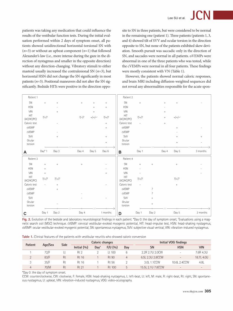
Lee SU et al. JCNpatients was taking any medication that could influence the results of the vestibular function tests. During the initial eval-uation performed within 2 days of symptom onset, all pa-tients showed unidirectional horizontal-torsional SN with (n=3) or without an upbeat component (n=1) that followed Alexander’s law (i.e., more intense during the gaze in the di-rection of nystagmus and smaller in the opposite direction) without any direction-changing. Vibratory stimuli to either mastoid usually increased the contralesional SN (n=3), but horizontal HSN did not change the SN significantly in most patients (n=3). Positional maneuvers did not alter the SN sig-nificantly. Bedside HITs were positive in the direction oppo-
site to SN in three patients, but were considered to be normal in the remaining one (patient 1). Three patients (patients 1, 3, and 4) showed tilt of SVV and ocular torsion in the direction opposite to SN, but none of the patients exhibited skew devi-ation. Smooth pursuit was saccadic only in the direction of SN, and saccades were normal in all patients. oVEMPs were abnormal in one of the three patients who was tested, while the cVEMPs were normal in all four patients. These findings were mostly consistent with VN (Table 1).
However, the patients showed normal caloric responses, and brain MRI including diffusion-weighted sequences did not reveal any abnormalities responsible for the acute spon-
Fig. 2. Evolution of the bedside and laboratory neurotological findings in each patient. *Day 0: the day of symptom onset, †Evaluations using a mag-netic search coil (MSC) technique. cVEMP: cervical vestibular-evoked myogenic potential, HIT: head-impulse test, HSN: head-shaking nystagmus, oVEMP: ocular vestibular-evoked myogenic potential, SN: spontaneous nystagmus, SVV: subjective visual vertical, VIN: vibration-induced nystagmus.
Patient 1
SN + + + + +
HSN - + +
VIN + +HIT
(AC/HC/PC)?/-/? ?/-/? +/+/-† ?/+/?
Caloric test - +
oVEMP +
cVEMP -
SVV + +Oculartorsion
+
Day* 1 Day 3 Day 4 Day 5 Day 6A
Patient 2
SN + + + -
HSN -
VIN +HIT
(AC/HC/PC)?/+/? + +/+/-†
Caloric test - +
oVEMP + -
cVEMP -
SVV + +Oculartorsion
+ -
Day 1 3 monthsDay 4 Day 5BPatient 3
SN + + + -
HSN + -
VIN + +HIT
(AC/HC/PC)?/+/? ?/+/?
Caloric test - + -
oVEMP +
cVEMP -
SVV - -Oculartorsion
-
Day 1 Day 2 Day 4 1 monthsC
Patient 4
SN + + + -
HSN - -
VIN - -HIT
(AC/HC/PC)?/+/? ?/+/?
Caloric test - +
oVEMP ?
cVEMP ?
SVV +Oculartorsion
+
Day 1 Day 2 Day 5D 2 months
Table 1. Clinical features of the patients with vestibular neuritis who showed caloric conversion
Patient Age/Sex SideCaloric changes Initial VOG findings
Initial (%) Day* F/U (%) Day SN HSN VIN1 72/F Lt Rt 2 2 Lt 100 6 2.2R 2.7U 2.0CW - 7.6R 4.3U
2 83/F Rt Rt 16 1 Rt 90 4 6.5L 2.3U 2.8CCW - 18.7L 4.0U
3 35/F Rt Rt 16 1 Rt 56 2 3.0L 1.1CCW 10.6L 2.4CCW 4.8L
4 70/M Rt Rt 21 1 Rt 100 5 15.5L 2.1U 7.8CCW - -
*Day 0: the day of symptom onset.CCW: counterclockwise, CW: clockwise, F: female, HSN: head-shaking nystagmus, L: left-beat, Lt: left, M: male, R: right-beat, Rt: right, SN: spontane-ous nystagmus, U: upbeat, VIN: vibration-induced nystagmus, VOG: video-oculography.

306 J Clin Neurol 2016;12(3):301-307
Delayed Caloric Conversion in VNJCNtaneous vestibular syndrome. Given the negative imaging findings, follow-up caloric tests were performed 1–4 days later, which eventually documented unilateral caloric paresis. In one patient (patient 1) with initially normal findings in bedside HITs and caloric tests, recording of HITs using the MSC technique 4 days later documented decreased gains for HC and AC on the lesion side along with a conversion into caloric paresis. In the other patient (patient 2), the record-ing of HITs using an MSC technique 5 days after symptom onset confirmed that the head impulse VOR gain was de-creased only in the HC in the presence of caloric paresis.
DISCUSSION
To the best of our knowledge, this is the first report of initial-ly normal caloric responses that subsequently converted into unilateral paresis during the acute phase of VN.
The caloric test evaluates the VOR by creating a tempera-ture gradient in the SCCs, which induces movement of the endolymph.14 Despite using nonphysiological low-frequency stimuli (<0.003 Hz), the caloric test enables the function of each labyrinth to be assessed separately, and also to differen-tiate peripheral from central vertigo. The findings of a caloric test are invariably abnormal during the acute stage of VN, and the abnormality is usually maintained for more than 1 year.12 Indeed, unilateral caloric paresis has been considered to be the diagnostic hallmark of VN.1,2
The caloric responses in our patients with a final diagnosis of VN were initially symmetrical even in the presence of SN. This indicates dissociation between the static and dynamic vestibular imbalances during the acute phase of VN. More-over, three of them (patients 2, 3, and 4) showed abnormal HITs but normal caloric responses. This further implies that the dynamic vestibular responses may vary according to the characteristics of the stimuli in peripheral12,15 as well as cen-tral vestibular disorders.
The primary vestibular afferents are categorized accord-ing to their spontaneous firing patterns.14 The regular affer-ents mainly encode the low-frequency movements of the en-dolymph (e.g., caloric responses), and innervate either the type II cells or a mixture of type I and type II cells. In contrast, the irregular afferents are known to encode higher frequency stimuli (e.g., head impulses), and innervate either the type I cells or a mixture of type I and type II cells. However, histo-pathological studies of VN have not shown any predilection for a specific type of hair cells.16,17 In addition, VN patients mostly show impaired responses on both caloric and HITs.18 Thus, the pattern of dissociation in vestibular dysfunction observed in our patients indicates a possibility of relative sparing of the low-frequency vestibular afferents (<0.003 Hz)
during the early evolution of the inflammation involving the vestibular nerve in some patients.
Indeed, the vestibular asymmetry may build up in a gradu-al manner in VN. The recovery of vestibular imbalances also occurs on different timescales according to the stimuli applied in VN; that is, the caloric asymmetry outlasts the abnormali-ties of the HITs, HSN, and VIN, which employ stimulus fre-quencies higher than that used in the caloric tests.12,18 Like-wise, the current study showed that vestibular abnormalities may emerge on different timescales during the acute phase of VN. Indeed, a previous study found that dizziness occurred within 1 week prior to a VN attack in 24% of patients,19 which is also indicative of a gradual or stepwise buildup of the ves-tibular asymmetry in VN. However, normal caloric responses should be rare in VN, if they occur at all, since only 18 (1.9%) of the present 907 patients showed normal results when 14 patients with an initial diagnosis of isolated inferior VN were excluded.
The subsequent conversion into unilateral caloric paresis has been described previously in patients with a progres-sion from isolated inferior VN into labyrinthitis involving the entire vestibular and auditory compartments.5 Accord-ingly, the initially normal caloric responses in those patients may be ascribed to sparing of the HC that belongs to the su-perior division. However, we excluded patients with an ini-tial diagnosis of isolated inferior VN in this study, and the included only patients who showed findings consistent with an involvement of the superior vestibular division from the beginning. Thus, the initially normal caloric responses can-not be explained by a selective sparing of the HC function in our patients.
Various factors could potentially influence the magnitude of caloric responses, such as visual fixation, the alertness or psychological status of the patient,20 blood flow to the skin,21 heat conductivity of the temporal bone,21 and even test repe-tition.22 Thus, caloric responses should be interpreted with caution due to both interindividual and intraindividual vari-ability. In our patients, the caloric tests were repeated using the same methods and equipment over a short period of time by the same technician. Furthermore, repeated caloric testing tends to decrease the caloric asymmetry,22 which was not the case in our patients. In addition, the caloric responses might be underestimated when the SN is too strong to be reversed by caloric stimulation. To avoid this, we thoroughly reviewed the raw SPV data in each patient. In addition, two patients showed a relatively small SN (<3.0°/s) that would not have in-fluenced the caloric responses. However, intraindividual vari-ability of the caloric responses remains a possibility in our pa-tients.
Differentiating between central and peripheral etiologies

www.thejcn.com 307
Lee SU et al. JCNin acute isolated spontaneous vertigo is important since 10–20% of patients experiencing posterior circulation strokes may present with isolated vertigo (pseudo-VN).23 Since the patterns of SN alone cannot differentiate peripheral from cen-tral causes of vertigo, other vestibular findings should be care-fully investigated to secure differential diagnosis.24 In this regard, the findings of HITs and caloric tests are important since pseudo-VNs due to lesions mostly involving the vestib-ulocerebellum almost always show normal HITs and caloric responses.23 The HITs and caloric tests are complementary in detecting peripheral vestibular hypofunction, since HIT findings may be normal in patients with incomplete periph-eral vestibular damage. Indeed, the sensitivity of bedside HITs is reportedly 63–72%.25 Given the relatively older ages and comorbid vascular risk factors in our patients, their acute spontaneous vertigo might be attributable to vascular inner ear damage. However, labyrinthine infarctions tend to involve both the vestibular and cochlear end organs, and isolated damage to the vestibular labyrinth would be extremely rare (if it occurs at all) in the presence of vascular inner ear damage.26
In conclusion, the results of caloric tests should be inter-preted with caution during the acute phase of spontaneous vertigo since VN patients can present with normal caloric responses during the hyperacute phase. Therefore, follow-up caloric tests should be considered when the initial evaluation is normal, but VN remains the most plausible diagnosis.
Conflicts of InterestDrs. Lee, Park, Koo, and H.J. Kim have no potential conflicts of interest to disclose. Dr. J.S. Kim serves as an associate editor of Frontiers in Neuro-otology and on the editorial boards of the Journal of Korean Society of Clinical Neurophysiology, Journal of Clinical Neurology, Frontiers in Neu-ro-ophthalmology, Journal of Neuro-ophthalmology, Journal of Vestibular Research, Journal of Neurology, and Medicine, and received research sup-port from SK Chemicals, Co. Ltd.
AcknowledgementsThis study was supported by a grant of the Korea Healthcare Technology R&D Project, Ministry of Health and Welfare, Republic of Korea (no. HI10C2020).
REFERENCES1. Jeong SH, Kim HJ, Kim JS. Vestibular neuritis. Semin Neurol 2013;33:
185-194.2. Strupp M, Brandt T. Vestibular neuritis. Semin Neurol 2009;29:509-
519.3. Baloh RW. Clinical practice. Vestibular neuritis. N Engl J Med 2003;
348:1027-1032.4. Cnyrim CD, Newman-Toker D, Karch C, Brandt T, Strupp M. Bed-
side differentiation of vestibular neuritis from central “vestibular
pseudoneuritis”. J Neurol Neurosurg Psychiatry 2008;79:458-460.5. Kim JS, Kim HJ. Inferior vestibular neuritis. J Neurol 2012;259:1553-
1560.6. Halmagyi GM, Curthoys IS. A clinical sign of canal paresis. Arch Neu-
rol 1988;45:737-739.7. Choi KD, Oh SY, Park SH, Kim JH, Koo JW, Kim JS. Head-shaking
nystagmus in lateral medullary infarction: patterns and possible mech-anisms. Neurology 2007;68:1337-1344.
8. Choi KD, Oh SY, Kim HJ, Kim JS. The vestibulo-ocular reflexes dur-ing head impulse in Wernicke’s encephalopathy. J Neurol Neurosurg Psychiatry 2007;78:1161-1162.
9. Chen L, Todd M, Halmagyi GM, Aw S. Head impulse gain and sac-cade analysis in pontine-cerebellar stroke and vestibular neuritis. Neu-rology 2014;83:1513-1522.
10. Lehnen N, Aw ST, Todd MJ, Halmagyi GM. Head impulse test reveals residual semicircular canal function after vestibular neurectomy. Neu-rology 2004;62:2294-2296.
11. Kim HJ, Park SH, Kim JS, Koo JW, Kim CY, Kim YH, et al. Bilaterally abnormal head impulse tests indicate a large cerebellopontine angle tumor. J Clin Neurol 2016;12:65-74.
12. Choi KD, Oh SY, Kim HJ, Koo JW, Cho BM, Kim JS. Recovery of ves-tibular imbalances after vestibular neuritis. Laryngoscope 2007;117: 1307-1312.
13. Kim S, Lee HS, Kim JS. Medial vestibulospinal tract lesions impair sacculo-collic reflexes. J Neurol 2010;257:825-832.
14. Baloh RW, Kerber KA, Honrubia V. Clinical Neurophysiology of the Ves-tibular System. 4th ed. New York, NY: Oxford University Press, 2011.
15. Maire R, van Melle G. Vestibulo-ocular reflex characteristics in pa-tients with unilateral Ménière’s disease. Otol Neurotol 2008;29:693-698.
16. Nadol JB Jr. Vestibular neuritis. Otolaryngol Head Neck Surg 1995;112: 162-172.
17. Schuknecht HF, Kitamura K. Second Louis H. Clerf Lecture. Vestib-ular neuritis. Ann Otol Rhinol Laryngol Suppl 1981;90(1 Pt 2):1-19.
18. Schmid-Priscoveanu A, Böhmer A, Obzina H, Straumann D. Caloric and search-coil head-impulse testing in patients after vestibular neuri-tis. J Assoc Res Otolaryngol 2001;2:72-78.
19. Lee H, Kim BK, Park HJ, Koo JW, Kim JS. Prodromal dizziness in ves-tibular neuritis: frequency and clinical implication. J Neurol Neurosurg Psychiatry 2009;80:355-356.
20. Proctor L, Glackin R. Factors contributing to variability of caloric test scores. Acta Otolaryngol 1985;100:161-171.
21. Baertschi AJ, Johnson RN, Hanna GR. A theoretical and experimental determination of vestibular dynamics in caloric stimulation. Biol Cy-bern 1975;20:175-186.
22. Furman JM, Jacob RG. Jongkees’ formula re-evaluated: order effects in the response to alternate binaural bithermal caloric stimulation using closed-loop irrigation. Acta Otolaryngol 1993;113:3-10.
23. Lee H, Sohn SI, Cho YW, Lee SR, Ahn BH, Park BR, et al. Cerebellar infarction presenting isolated vertigo: frequency and vascular topo-graphical patterns. Neurology 2006;67:1178-1183.
24. Kim JS, Lee H. Vertigo due to posterior circulation stroke. Semin Neu-rol 2013;33:179-184.
25. Jorns-Häderli M, Straumann D, Palla A. Accuracy of the bedside head impulse test in detecting vestibular hypofunction. J Neurol Neurosurg Psychiatry 2007;78:1113-1118.
26. Lee H, Kim JS, Chung EJ, Yi HA, Chung IS, Lee SR, et al. Infarction in the territory of anterior inferior cerebellar artery: spectrum of au-diovestibular loss. Stroke 2009;40:3745-3751.