normalizing 1
TRANSCRIPT

8/8/2019 Normalizing 1
http://slidepdf.com/reader/full/normalizing-1 1/15
tipE Regulates Na -dependent Repetitive Firingin Drosophila Neurons
Dianne D. Hodges,* ,1 Daewoo Lee,* Charles F. Preston,*Kevin Boswell,* Linda M. Hall, † and Diane K. O’Dowd* ,2
Department of Anatomy and Neurobiology and Department of Developmental and Cell Biology, University of California at Irvine, Irvine, California 92697-1280; and Functional Insect Genomics, 1105 Kennedy Place, Suite 4, Davis, California 95616
The tipE gene, originally identied by a tempera-
ure-sensitive paralytic mutation in Drosophila, en-odes a transmembrane protein that dramatically inu-nces sodium channel expression in Xenopus oocytes.
There is evidence that tipE also modulates sodiumhannel expression in the y; however, its role in regu-ating neuronal excitability remains unclear. He re weeport that the majority of neurons in both wild-type andipE mutant ( tipE ) embryo cultures re sodium-depen-ent action potentials in response to depolarizing cur-ent injection. However, the percentage of tipE neu-ons capable of ring repetitively during a sustainedepolarization is signicantly reduced. Expression of aipE transgene, in tipE neurons, restores repetitive
ring to wild-type levels. Analysis of underlying cur-ents reveals a slower rate of repolarization-dependentecovery of voltage-gated sodium currents during re-eated activation in tipE neurons. This phenotype islso rescued by expression of the tipE transgene.
These data demonstrate that tipE regulates sodium-ependent repetitive ring and recovery of sodium cur-ents during repeated activation. Furthermore, the du-ation of the interstimulus interval necessary to re aecond full-sized action potential is signicantly longern single- versus multiple-spiking transgenic neurons,uggesting that a slow rate of recovery of sodium cur-ents contributes to the decrease in repetitive ring inipE neurons.
INTRODUCTION
Cell-specic changes in electrical excitability duringearly development are critical in formation of matureneural circuits (Spitzer et al., 1994). Modulation of neu-ronal excitability has also been implicated in mediatingplasticity in the nervous system (Desai et al., 1999;Aizenman and Linden, 2000; Armano et al., 2000). Elec-trophysiological studies demonstrate that alterations inthe number, type, localization, and/ or posttranslationalmodications of voltage-gated ion channels can inu-ence neuronal excitability (Barish, 1986; Huguenard et
al., 1988; O’Dowd et al., 1988; Spitzer, 1991; Turrigian o et al., 1995; Massengill et al., 1997; Catterall, 2000). How-ever, the molecular mechanisms underlying regulationof excitability are less clear.
Using a genetic app roach in Drosophila, progress hasbeen made in identifying genes involved in mediatingneur onal excitability. Shaker an d ether-a-go-go (eag ), mu-tants with hyperexcitable phenotypes, exhibit anoma-lous repetitive ring in larval motor axons and identifypotassium channel genes important in determining theexcitabil ity propert ies of these neurons (Wu and
Ganetzky, 1992; Littleton and Ganetzky, 2000). Temper-ature-sensitive paralytic mu tants identify add itionalgenes, such as paralytic ( para ) and no action potential(nap ), that were recognized as playing a role in medi-ating n euronal excitability based on temperature-de-pendent blockade of action potential conduction in lar-val nerve bers (Wu and Ganetzky, 1992). The para geneencodes a voltage-gated sodium channel subunit(Loughney et al., 1989), whereas nap encodes an RNAhelicase involved in editing of para transcripts (Reenanet al., 2000).
1 Present address: Purd ue Pharma Ltd., 213 Technology Drive,rvine, CA 92618.
2 To whom correspondence and reprint requests should be ad-ressed at the Department of Anatomy and Neurobiology, Universityf California, Irvine, CA 92697-1280. Fax: (949) 824-1105. E-mail:kod owd @uci.edu .
doi:10.1006/mcne.2001.1088, available online at http://www.idealibrary.com on Molecular and Cellular Neuroscience 19, 402–416 (2002)MCN
1044-7431/02 $35.00© 2002 Elsevier Science (USA)
All rights reserved.02

8/8/2019 Normalizing 1
http://slidepdf.com/reader/full/normalizing-1 2/15
tipE mutant ies, similar to nap an d para mutants,xhibit a rapid and reversible temperature-sensitive pa-alysis (Kulkarni and Padh ye, 1982). A red uction in theumber of sodium channel binding sites in head mem-ranes from tipE ies (Jackson et al., 1986) and a de-rease in sodium current density in cultured tipE neu-
ons (O ’Dowd and Aldrich, 1988) suggest that tipE mayegulate sodium channel expression. Enhanced temper-ture sensitivity for nerve condu ction failure in para;ipE double mutants, compared with para alone, sup-orts the suggestion that tipE can modulate axonalondu ction prop erties (Ganetzky, 1986). The cloning of ipE revealed that the gene product is a novel integral
membrane protein with two membrane spanning re-ions (Feng et al., 1995a). The tipE protein does not form functional channel by itself when expressed in Xeno-
pus oocytes but coexpression of tipE with para cRNA
lters both the expression levels and the fast kineticroperties of the para -encoded voltage-gated sodiumhannels (Feng et al., 1995a; Warm ke et al., 1997). Takenogether these d ata suggest that tipE may de ne a newlass of proteins that regulates electrical excitabilityhrough an interaction with the para sodium channel.
Analysis of excitability in tipE mutants has beenimited to extracellular recordings in larval motor neu-ons that, interestingly, demonstrated apparently nor-
mal action potential propagation even at behaviorallyonpermissive temperatures (Ganetzky, 1986). Usingell cultures that contain subpopulations of primary
Drosophila neurons exhibiting distinct ring pheno-ypes, w e explored the role of tipE in regulation of eu ronal excitability. A line of tran sgenic ies carryinghe wild-type tipE gene under the control of a heat-hock promoter, in a tipE background (Feng et al.,995b), was crucial in determ ining th e electrical phen o-ypes associated w ith tipE. Our results demonstrate thatipE plays a role in regulating sodium -dependent repet-tive ring p roperties in cultured Drosophila neurons.
RESULTS
ipE Neurons Exhibit a Decrease in Repetitive Firing, Spontaneous Firing, and Action Potential Amplitude
To determine if the tipE gene plays a role in regulat-ng neu ronal excitability we comp ared the ring prop-rties of embryonic tipE and wild-type neurons grownn dissociated cell culture. Cultures from both geno-ypes contained heterogeneous populations of neurons:
some with simple neurites and others with elaboratelybranched processes. Neurons could be found in clus-ters, where there w as contact betw een neighboring cells(Figs. 1A and 1C), and in isolation (Figs. 1B and 1D).The w hole-cell recording technique was used to exam-ine the ring properties of neurons at 2 and 3 days invitro. All recordings were performed blind with respectto genotype. Initial studies were conducted to deter-mine if a correlation between morphological featuresand neu ronal excitability could be established . Becausethere did not appear to be systematic differences in theelectrical properties of isolated versus clustered neu-rons all d ata w ere group ed for statistical analysis.
The majority of neurons in both the tipE (76 5%,
n 10 platings) and the wild-type (82 5%, n 10platings) cultures were electrically excitable based onthe ability to elicit one or more action potentials inresponse to a 600-ms, suprathreshold depolarizing cur-rent step. The electrically excitable neurons weregroup ed into three broad classes: single spiking, grad edmu ltiple spiking, or m ultiple spiking (Figs. 2A a nd 2B).The single spiking neurons w ere characterized by asingle action potential elicited at the beginning of thestep depolarization (Fig. 2A). In this class of neurons,neither changes in holding p otential nor increases in the
FIG. 1. Wild-type and tipE Drosophila neurons grown in primarydissociated cell culture. Neu ronal clusters interconnected by overlap-ping branched neuritic processes in a wild-type (A) and a tipE (C)culture. Isolated neurons in a wild-type (B) and a tipE (D) culture.Cultures were grown for 2 days in vitro in DDM1, xed in 4%paraformaldehyde, and stained with uorescein-conjugated anti-HRPantibodies. Scale bar s, 20 m.
403ipE Regulates Neuronal Excitability

8/8/2019 Normalizing 1
http://slidepdf.com/reader/full/normalizing-1 3/15

8/8/2019 Normalizing 1
http://slidepdf.com/reader/full/normalizing-1 4/15
otential amplitude in the tipE neurons was reducedompared to wild type (Fig. 3D). In contrast, the actionotential duration was not different between the twoenotypes (Table 2). No signi cant changes in the inputesistance or the resting membrane potential were de-ected (Table 2). There was a large range in the size of he cultured neurons but the m ean capacitance of theopu lation examined in the tw o genotypes was similarTable 2).
Rescue of Reduced Repetitive Firing and Spontaneous Firing by Expression of the tipE Transgene in Differentiated tipE Neurons
The reduced repetitive ring, spontaneous ring, andaction potential am plitude in tipE neurons are consis-tent with the hypothesis that tipE regulates these prop -erties in Drosophila neurons. However, one caveat tothis interpretation is that the comparison was madebetween popu lations of neurons harvested from two ystrains in which the contribution of differences in ge-netic backgrounds is unknown. To determine if thealtered electrical properties in tipE neurons are theconsequence of a mu tation in the tipE gene we studieda transgenic line ( tipE :tipE ) containing the wild-typetipE transgene ( tipE ), under the control of a heat shock promoter, in the tipE background.
To monitor expression of the transgene and deter-
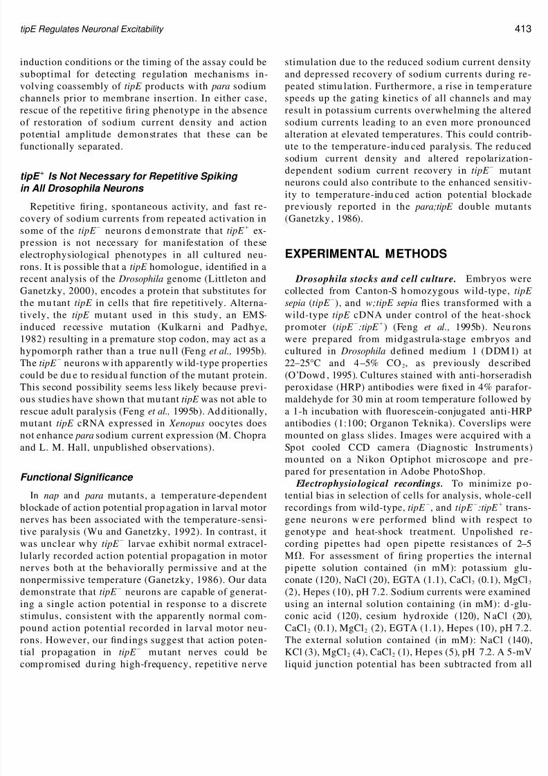
mine if it could be regulated in the neurons by heatshock, cultures w ere prepared from wild-type, tipE ,an d tipE :tipE embryos. Half of the cultures in eachgenotype were subjected to three 1-h heat shocks (seeExperimen tal Method s for d etails). N on-heat-shockedcultures were maintained continuously at room temper-ature. RNA was prepared from all the cultures at 42 hafter plating. Primers (M1 and M2) anking a singlepoint mutation in the tipE cDNA (removing an Rsa Irestriction enzym e site) generated PCR produ cts of dis-tinct sizes in wild-type and tipE neurons following
RsaI digestion (Fig. 4A). In this analysis we found thatwild-type an d tipE neurons, in both non-heat-shockedand heat-shocked cultures, expressed only wild-type ormutant tipE mRNA, respectively. In contrast, tipE :tipE neurons express both mutant and wild-type tipE mRNA, even in the absence of heat shock (Fig. 4B). Therelative abu ndan ce of the w ild-type tipE product in thetipE :tipE cultures was dram atically increased ( 10-fold) following heat shock. How ever, since the absolutelevels of tipE mRNA produced are not known it waspossible that, even in the absence of heat shock, wild-type tipE levels might be suf cient to rescue the mutantring p henotyp e. Therefore, in all the electrophysiology
TABLE 1
Subclass Distribution of Multiple Spiking (MS) Neurons
Tonic (%MS) Adaptive (%MS) Delayed (%MS)
Wild typ e 53.9 10.8 25.3 10.3 21.3 6.1tipE 57.4 13.1 20.4 11.7 18.8 13.2
Note. 9 platings; mean SEM.
IG. 3. The frequencies of repetitive ring, spontaneous ring, andction potential amplitude are decreased in tipE neurons. (A) Rep-esentative recordings illustrating the most abundant ring class in
wild-type and tipE neuron s. (B) There was a signi cant reduction inhe percentage of neurons in the multiple-spiking (MS) class in tipE ultures compared with wild-type, with a corresponding increase inhe % of neurons in the single-spiking (SS) class. The mean percentagef total excitable cells w ithin each ring class was determined byalculating the percentage observed in nine separate experiments in
which three or more neurons were examined. (C) The fraction of eurons ring spon taneous action potentials, as a function of the totalumber of neurons examined, was signi cantly lower in tipE com-ared with w ild-type. The m eans represent data obtained from threer m ore neuron s in four separate p latings for both genotypes. (D) Themplitude of the rst action potential (AP) in each train (peak torough) was signi cantly reduced in tipE neurons compared to wildype. Bars indicate SEMs. * P 0.05, ** P 0.01, *** P 0.001,tudent ’s t test.
405ipE Regulates Neuronal Excitability
8/8/2019 Normalizing 1
http://slidepdf.com/reader/full/normalizing-1 5/15
tudies comparisons included analysis of neurons inipE :tipE cultures examined after heat shock and inibling cultures that were maintained continuously atmbient temperature.
To address the role of tipE in neuronal excitability,ndependent of development, tipE :tipE cultures w ererown in the absence of heat shock for the rst 2 days
n vitro, by which time differences in the three major
ring classes w ere readily app arent between tipE an dwild-type neurons. In four independent experiments,half of the tipE :tipE cultures were exposed to two 1-hheat shocks at 42 and 49 h after plat ing while theremaining cultures were not heat shocked (Fig. 5, top).To control for th e affects of heat shock alone, w ild-typecultures prepared in parallel were exposed to the sameheat-shock regime. Cultures were coded and examinedblind with respect to genotype and heat-shock condi-tions. Examples of ring properties in three differentneurons recorded from a wild-type ( HS), a tipE :tipE ( HS), and a tipE :tipE ( HS) culture are illustratedin Fig. 5A. In the absence of heat shock, there w ere veryfew multiple-spiking neurons in the tipE :tipE cul-tures, with the majority of excitable cells split betweenthe graded mu ltiple-spiking and single-spiking ringclasses (Fig. 5B), similar to the distribution seen previ-ously in the tipE cultures. These data demonstrate thatthe level of wild-type tipE product in the absence of heat shock d oes not rescue the mu tant ring phenotypein transgenic cultures. However, the altered ring typedistribution w as fully rescued within 24 h after heatshock ( HS), with over 90% of the neurons in themultiple-spiking ring class and the remainder classi-ed as single spiking, similar to the distribution seen inwild-type cultures ( HS) examined in p arallel (Fig. 5B).The percentage of spontaneously ring neurons, low inth e tipE :tipE cultures in the absence of heat shock ( HS), was also rescued following heat shock ( HS)
(Fig. 5C). Unexpectedly, the reduced action potentialamplitude in the transgenic neurons was not rescuedfollowing heat shock (Fig. 5D).
Repolarization-Dependent Recovery of Sodium Currents and Excitability in Wild-Type, Mutant,and Transgenic Neurons
Previous studies had demonstrated that the wild-type tipE gene product u pregulates the amplitude andalters the kinetic properties of para sodium currents in a
ABLE 2
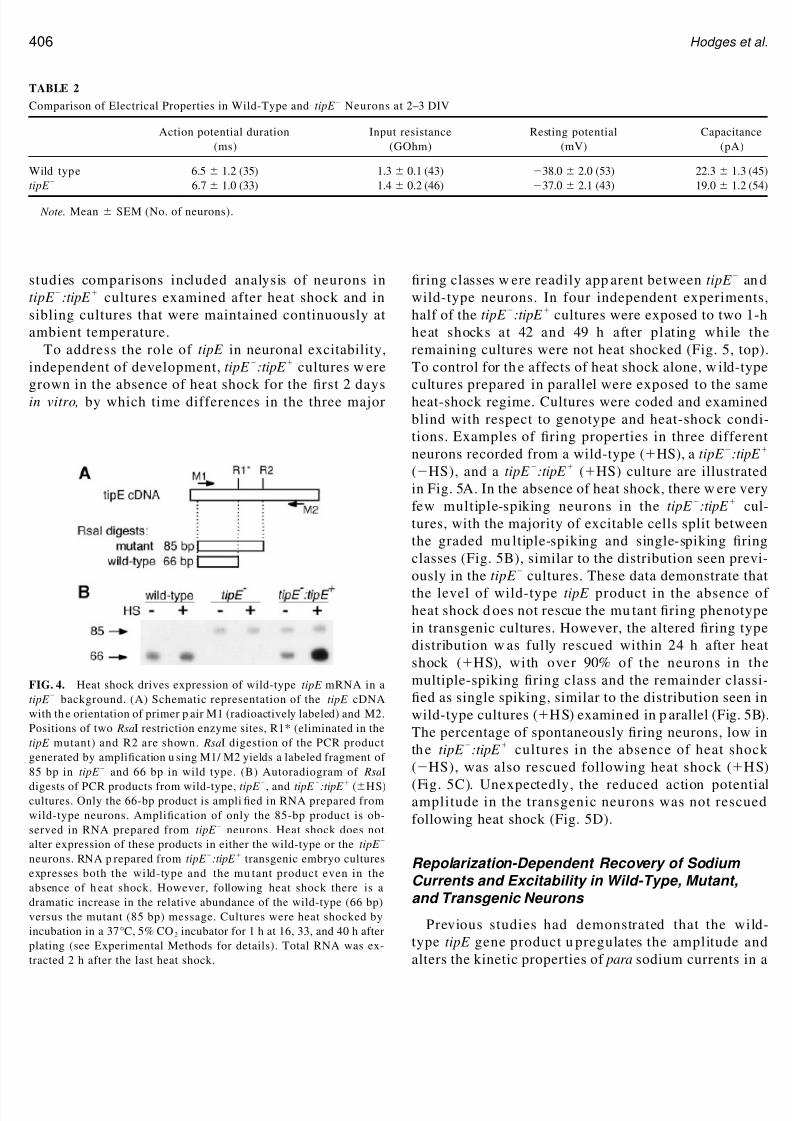
Comparison of Electrical Properties in Wild-Type and tipE Neurons at 2 –3 DIV
Action potential duration(ms)
Input resistance(GOhm)
Resting potential(mV)
Capacitance(pA)
Wild type 6.5 1.2 (35) 1.3 0.1 (43) 38.0 2.0 (53) 22.3 1.3 (45)pE 6.7 1.0 (33) 1.4 0.2 (46) 37.0 2.1 (43) 19.0 1.2 (54)
Note. Mean SEM (No. of neurons).
IG. 4. Heat shock drives expression of wild-type tipE mRNA in apE background. (A) Schematic representation of the tipE cDNA
with th e orientation of primer p air M1 (radioactively labeled) and M2.ositions of two Rsa I restriction enzyme sites, R1* (eliminated in thepE mutant) and R2 are shown. Rsa I digestion of the PCR productenerated by ampli cation u sing M1/ M2 yields a labeled fragment of 5 bp in tipE and 66 bp in wild type. (B) Autoradiogram of Rsa I
igests of PCR products from wild-type, tipE , and tipE :tipE ( HS)ultures. Only the 66-bp product is ampli ed in RNA prepared from
wild-type neurons. Ampli cation of only the 85-bp product is ob-erved in RNA prepared from tipE neurons. Heat shock does notlter expression of these products in either the wild-type or the tipE eurons. RNA p repared from tipE :tipE transgenic embryo culturesxpresses both the wild-type and the mu tant product even in thebsence of h eat shock. However, following heat shock there is aramatic increase in the relative abundance of the wild-type (66 bp)ersus the mutant (85 bp) message. Cultures were heat shocked by
ncubation in a 37 °C, 5% CO 2 incubator for 1 h at 16, 33, and 40 h afterlating (see Experimental Methods for details). Total RNA was ex-racted 2 h after the last heat shock.
406 Hodges et al.

8/8/2019 Normalizing 1
http://slidepdf.com/reader/full/normalizing-1 6/15
heterologous expression system (Feng et al., 1995a;Warmke et al., 1997). In addition, a decrease in sodiumcurrent d ensity in tipE versus wild-type neurons wasobserved at 1 day in culture (O ’Dowd and Aldrich,1988). Therefore, we a sked if there w ere changes in th esodium current properties that might contribute to the
mutant ring phenotypes seen in the present culturecondition. These studies were complicated by the factthat the sodium currents in the majority of the electri-cally excitable neurons could not be well voltage-clamp ed, p reclud ing a classical quantitative biophy sicalanalysis of the underlying sodium channel properties.However, comparison of features of the whole-cell cur-rents elicited by step depolarizations, using identicalrecording conditions for wild-type, mutant, and trans-genic neurons, allowed us to identify sodium currentproperties linked to tipE expression.
The rst comparison focused on the maximal sodiumcurrent activated in m utant and wild-type neurons.Individual neurons w ere stimulated with a series of increasing depolarizing voltage steps in the presence of cesium in the internal solution to block outward potas-sium currents. The sodium current density in each neu-ron was determined by normalizing the peak inwardsodium current to the wh ole-cell capacitance. Therewa s a 25% redu ction in the peak sodiu m curr ent den sityin the tipE versus wild-type neurons (Fig. 6A). Thesodium current density in the transgenic neurons in theabsence of heat shock was similarly low. However, thisreduced sodium current density was not rescued fol-lowing heat shock (Fig. 6A). The inability to rescuesodium current density is consistent with the inabilityto rescue the action potential amplitude, suggestingthat these tw o p roperties are linked. These data furtherdemonstrate that recovery of robust repetitive ringdoes not require rescue of the sodium current density.
In light of our nding that most tipE mutant neuronsare capable of ring a single action potential but arecompromised in their ability to re repetitively, weinvestigated the recovery of sodium currents during
repetitive activation. Neurons were subject to two iden-tical voltage steps from 75 to 5 mV, separated by a10-ms interstimulus interval at 75 mV (Fig. 6B). Theamplitude of the current elicited by the second step was
IG. 5.Expression of w ild-type tipE message in a tipE backgroundestores wild-type distribution of ring phenotypes and spontaneous
ring levels in differentiated neurons. The heat-shock regime used inhese experiments is illustrated on the top . Half of the cultures in eachenotype were subjected to two 1 h, 37 °C heat shocks at 42 and 49 hfter plating. Electrophysiological recordings were done at 66 –74 hfter plating (see Experimental Methods for details). (A) Representa-ve action potential trains recorded from neurons in the three differ-nt culture conditions tested, wild-type ( HS), tipE :tipE ( HS),n d tipE :tipE ( HS). (B) Heat shock rescued the altered ring classistribution seen in the mutant neurons. The distribution of neurons,ormalized to total number of excitable cells, among the three majorring classes was determined in the different cultures. The % multi-le-spiking (MS) and % single-spiking (SS) neurons are signi cantly
ifferent in wild type ( HS) and tipE :tipE ( HS) compared topE :tipE ( HS) (ANOVA, * P 0.05, ** P 0.01, Fisher ’s pro -
ected least signi cant difference). H eat shock d id not alter the d is-ribution of ring classes in wild-type cultures. Mean percentages
were determined by calculating the percentages in four independentxperiments in which 4 – 6 neurons/ culture condition were examinedn each experiment. The total number of neurons in each group was8 wild type ( HS), 22 tipE :tipE ( HS), and 21 tipE :tipE ( HS).C) The level of spontaneous ring is restored to wild-type levels inransgenic neurons following heat shock. The fraction of neuronsring spontan eous action potentials, as a function of the total num berf neurons examined, is signi cantly different between tipE :tipE eurons HS and HS (** P 0.01, Student ’s t test). (D) The action
potential (AP) amplitude is not rescued by indu ction of the tipE transgene: there is no signi cant difference between the mean ampli-tude in neurons in HS ( n 13) and HS ( n 12) tipE :tipE cultures ( P 0.05, Student ’s t test). Bars on all graph s ind icate SEM.
407ipE Regulates Neuronal Excitability

8/8/2019 Normalizing 1
http://slidepdf.com/reader/full/normalizing-1 7/15
ormalized to that elicited by the rst step. Under theseonditions, the amplitude of the second sodium current
was app roximately 90% of the rst sodium current, inwild-type neurons. In contrast, in tipE neurons theecond sodium current was only 80% of the controlmp litud e (Fig. 6B). The redu ced level of recovery seen
in the mutant was also seen in the transgenic neurons inthe absence of heat shock. Following heat shock, so-dium current recovery was restored to wild-type levels(Fig. 6B). There was no decrement in sodium currentamplitude following a 2-s interstimulus interval at 75mV, the standard time between repeated trials, in either
mutant or rescued transgenic neurons. These ndingsdemonstrate that the tipE mutation results in a slowerrate of repolarization-dependent recovery of sodiumcurrents during repetitive activation.
A straightforward interpretation of these d ata w ouldbe that the tipE mutation slows but does not block recovery of the underlying sodium channels from inac-tivation. In the absence of excess sodium channels, thiswould decrease the probability of repetitive spikingduring a sustained depolarization. The poor spaceclamp in the cultured neurons makes a more d etailed
investigation of the kinetics of recovery of sodiumcurrents from inactivation problematic. How ever, if slowed recovery from inactivation contributes to thetipE ring phenotype, then one would predict thatrepolarization should be necessary, and 2 s interstimu-lus interval suf cient, for ring a second spike in tipE (single-spiking) neu rons. Therefore, ve single-spikingneurons, in transgenic cultures in the absence of heatshock, were held between 50 and 60 mV and sub- jected to prolonged depolarizing steps (5 s) separatedby 2 s intervals. All neur ons red on e action p otential atthe onset of each d epolarizing step. These data demon-strate that an interstimulus interval is necessary and 2 sis suf cient for ring a second full-sized action poten-tial in tipE neur ons that ar e classi ed as single spiking.
The relationship between interstimulus duration andrecovery of excitability was examined in cultures of transgenic neurons, half that were heat shocked andhalf that served as controls. Neurons were h eld at volt-ages between 50 and 60 mV and given tw o identical,suprathreshold, depolarizing, current steps. The stepdepolarizations were separated by intervals of varyingduration (Fig. 7A). An example of the typical behavior
of a single-spiking transgenic neuron, in the absence of heat shock, is shown in the top traces in Fig. 7B. Thesecond action potential was reduced in amplitude whenthe two depolarizing pulses were separated by shortinterstimulus intervals of 2 and 10 ms ( rst and secondpair). The action potential amplitude was similar in thetwo steps when the interst imulus interval was in-creased to 100 ms (third pair, Fig. 7B). In contrast, in amultiple-spiking transgenic neuron, following heatshock rescue, there was no signi cant d ecrement in theamplitude of the action potential elicited by a second
IG. 6. Sodium currents in wild-type, tipE , and transgenic neuronstipE :tipE ). (A) Sodium current density is reduced in tipE neuronsut is not rescued by indu ction of the tipE transgene. Sodium currentensity was calculated by norm alizing the maximal am plitude so-ium current to the whole-cell capacitance in each neuron in which aodium current was observed. There is a signi cant decrease in meanodium current density in tipE compared to wild-type neurons. * P
.05, Student ’s t test, unpaired. The mean sodium current densities inransgenic neurons ( tipE :tipE ) under HS and HS conditions areot signi cantly different from each other ( P 0.05, Student ’s t test).B) Recovery of sod ium currents from repetitive activation is red ucedn the m utant and rescued by heat-shock induction of the tipE ransgene. This was examined u sing the two-step p rotocol illustrated ;olding potential and recovery voltages were 75 mV, test steps were
o 5 mV for 10 ms, interstimulus interval was 10 ms. The sodiumurrent amplitude elicited by the second test step was normalized tohat elicited by the rst step. The normalized sodium current ampli-ude in tipE neurons ( n 35) is signi cantly lower than in wild typen 29) (*** P 0.001, Student ’s t test, unpaired). The reductionbserved in the transgenic neurons in the absence of heat shock tipE :tipE HS; n 12) is rescued in the heat-shocked transgeniceurons ( tipE :tipE HS; n 13) (*** P 0.001, Student ’s t test,npaired) ( n 7). Bars in all graphs indicate SEM.
408 Hodges et al.

8/8/2019 Normalizing 1
http://slidepdf.com/reader/full/normalizing-1 8/15
step depolarization, even w hen the interstimu lus inter-val was as short as 2 ms (Fig. 7B, bottom). To quantifythese data, the action potential amp litud e elicited by th esecond step was n ormalized to that elicited by the rststep and plotted as a function of interstimulus interval,for a num ber of neu rons in each ring class (Fig. 7C). As
illustrated in the graph, the interstimulus duration re-quired for recovery of the ability to re a full-sizedaction p otential was longer in th e single-spiking than inthe multiple-spiking transgenic neurons. In gradedmultiple-spiking neurons the required interstimulusduration was intermediate to the other two classes. Thelonger interstimulus duration necessary to re a secondfull-sized action potential in single- versus graded ver-sus multiple-spiking classes is consistent with the sug-gestion that a slowed rate of repolarization-dependentrecovery of sodium currents contributes to the decrease
in repetitive ring intipE
neurons.In ad dition to sodium currents, voltage-gated potas-sium currents are critical in determining m any aspectsof neuronal excitability, including repetitive ringproperties (Wu and Ganetzky, 1992). Potassium cur-rents were induced by a series of 300-ms depolarizingvoltage steps between 55 and 55 mV. Individualneurons in both genotypes had varying levels of tran-sient and sustained currents. The peak and plateaucurrent amplitudes in wild-type and tipE neuronswere determined at 55 and normalized to the whole-cell capacitance. There was no signi cant difference inthe peak (wt 63.9 8 p A/ pF, n 25; tipE 62.6 4pA/ pF, n 48) nor in the plateau current density (wt33.5 3 pA/ pF, n 22; tipE 35.1 2.5 pA/ pF, n47) between the two genotypes. Since there were noprior studies indicating that tipE regulates potassiumchannels, we did not compare the properties of voltage-gated potassium currents in tipE and wild-type neu-rons further.
Coexpression of tipE and para mRNAin Wild-Type Neurons
Previous studies from our laboratory have demon-strated that the para gene encodes functional sodiumchannels in neurons cultured from wild-type Drosophilaembryos (O ’Dowd et al., 1989). If tipE is regulating paraexpression, thereby in uencing sodium currents andring properties in Drosophila neurons as suggestedfrom the oocyte stud ies, then tipE an d para must beexpressed at the same time and in the same cells. RT-PCR with two distinct p rimer sets (see ExperimentalMethods) was used to examine expression of para an d
IG. 7. Recovery of excitability in transgenic neurons. (A) Neu-ons in transgenic cultures were held at 55 mV and stimulated
with the two-step protocols illustrated using three interstimulus
ntervals (2, 10, 100 ms). (B) Whole-cell current clamp recordingsrom a single-spiking neuron in the absence of heat shock ( tipE :pE HS) (top trace) and a mu ltiple-spiking neu ron in the pres-nce of heat shock ( tipE :tipE HS) (bottom trace). In the single-piking neuron the action potential elicited by the second step inach p air was reduced in amplitude for the interstimulus intervalsf 2 and 10 ms. When the interstimulus interval was extended to00 ms the second action potential was similar in amplitude to therst. In a m ultiple-spiking n euron in a transgenic culture that hadeen heat shocked, there was no difference in act ion potent ialmplitude in the rst and second steps even when the interstimu-us interval wa s 2 ms. (C) The amplitud e of the rst action potentialmeasured from peak to trough) generated in the second step inach pa ir was normalized to the ampl itude o f the rst action
otential in the rst step and plotted as a function of interstimulusnterval. Multiple-spiking neurons showed no decrease in the nor-
malized act ion potent ial amplitude at any of the interst imulusntervals examined. In contrast, the normalized action p otentialmpli tude was s igni cantly redu ced in graded multiple spikingnd even further reduced in s ingle-spiking neurons at inter-als shorter than 20 ms. The values for s ingle- and mult iple-p ik ing classes , be tween 2 and 12 ms , w hen eva lua ted in aoint-by-point compar ison are signi cantly different (ANOVA,
0.001 (2 – 8 ms), P 0.01 (10 –12 ms), Fisher ’s protected leastigni cant difference). Each data point represents the mean valueor the indicated number of neurons in each ring class. Barsndicate SEM.
409ipE Regulates Neuronal Excitability

8/8/2019 Normalizing 1
http://slidepdf.com/reader/full/normalizing-1 9/15
ipE in RNA harvested from wild-type cultures between and 48 h in vitro. A third primer set was included tomplify ribosomal protein 49 (O ’Connell and Rosbash,984), serving as a control for normalization of input
RNA am oun ts. The au toradiogram in Fig. 8A illustrateshe PCR prod ucts generated from total RNA harvested
rom wild-type neurons at the indicated times in cul-ure. The percentage of the maximal levels of expres-ion, after norm alization to rp49 values, is show n in Fig.B. Expression of both genes w as initiated about 12 hfter plating, peaked at 33 h, and su bsequently d eclinedo app roximately 50 –70% of maximum by 48 h (Fig. 8B).
The similar tim e courses of tipE an d para expression areonsistent w ith a r ole for tipE in regulating para sodiumhannels during normal neuronal development.
To determine if para an d tipE are coexpressed inndividual neurons, we used a multiplex, single-cell
RT-PCR approach to examine their expression in wild-ype n eurons at 2 days in culture. These analyses wereerformed with two primer pairs in a single RT-PCR.
One primer pair (paraDP3/ DP4) anked the alterna-ively spliced exons i and a in the para gene, am plifyingistinct PCR products representing the para splice vari-nts containing one or both alternative exons i and a.
The second prim er set (tipEComF/ R) ampli ed a singlePCR product from the tipE transcripts. A representative
utoradiogram of PCR products am pli ed from RNAarvested from individual cultured neurons is shownn Fig. 8C. As p reviously rep orted there was cell-to-cell
variability in expression of para splice variants. Themajority of cells that express sodium currents and paramRNA also express tipE (Fig. 8C). In six experim ents, inwhich eight or more neurons w ere examined/ experi-men t, 92.2 3.1% of the para -expressing neurons alsoxpressed tipE. These data demonstrate that the paran d tipE gene products could interact directly or indi-ectly, in single neurons, to in uence repetitive ring.
DISCUSSION
Although little is known about the ring properties of Drosophila neurons in vivo (Ikeda and Kaplan, 1970;Tanouye et al., 1981) studies in cell culture reveal sub-
opulations of neurons with distinct ring p atterns elic-ted by depolarizing current pulses (O ’Dowd , 1995;
Zhao and Wu, 1997). Many of the multiple-spikingeurons in this study have spike discharge patterns thatlosely resemble those of the well-characterized regu-ar-spiking or fast-spiking ring classes of mammalian
FIG. 8. tipE an d para sodium channel mRNA expressions exhibit
s imilar temporal and spat ial dis tr ibut ions in cultured neurons.(A) Autoradiogram of the RT/ PCR products separated on an8% polyacrylamide gel reveals similar temporal patterns of expres-sion of tipE an d para mRNA in the cul tured neurons. rp49 isobserved at a constant level at all of the ages examined, indicatingthat expression of this gene is not developmentally regulated andthat starting RNA template concentrations were similar in eachsample. Lane W represents a negative control in which water issubst ituted for RNA. PCR products w ere generated from totalRNA prepared f rom cu l tu red neurons us ing th ree ind ividualprimer sets that amplify rp49, para, an d tipE transcripts. (B) Fortemporal comparison the product levels for tipE an d para werenormalized to the maximum levels observed for each. Data wereobtained from ve independent RNA preparat ions at each age.
Bars indicate SEMs. (C) Autoradiogram from a typical experimentshowing strong ampli cation of one or more of three alternativelyspliced para PCR produ cts from 7/ 10 cells, with 6/ 7 of theseneurons also expressing the tipE product. In addition, low levels of both para an d tipE are seen in one ad ditional neuron (Cell 9). Theexperiment was accepted for analysis on the basis of clean inter-mingled medium (M) and water (W) controls that w ere processedin parallel with the cells. PCR products from total RNA harvestedfrom whole pu pae (P) are shown for comparison. Amp li cation of the cel l contents was performed using 32 P-labeled primers thatamplify the region surrounding al ternative para exons i and a(paraDP3/ DP4) and tipE primers that detect all transcripts fromth e tipE region.
410 Hodges et al.

8/8/2019 Normalizing 1
http://slidepdf.com/reader/full/normalizing-1 10/15
ortical neurons described in the animal and in disso-iated cell culture (Connors and Gutnick, 1990; Massen-ill et al., 1997). The single-spiking neu rons in the Dro-ophila cultures are similar to the recently describedn-spiking neurons found in the rodent auditory cortex,
which re only one or two spikes that occur within 10
ms of the onset of a maintained intracellular depolar-zation (Metherate and Aramakis, 1999). The presencef similar ring classes in the cultured Drosophila neu-ons and rodent cortical neurons suggests strong con-ervation of the functional elements contributing to
CNS circuitry between these distantly related species.It should also be noted that the resting potentials of
h e Drosophila neurons reported in this study are moreepolarized than is standard for many m ature mamm a-ian neurons. However, hyperpolarizing shifts in mem-rane potential, from 40 to 65 mV, have been re-
orted during early development in some populationsf mamm alian cortical neurons (Agmon et al., 1996;
Zhou and Hablitz, 1996). This suggests that the depo-arized resting potentials could be related to the rela-ively young age at w hich most of the recordings werebtained, 2 –3 days of the neuronal birth date. Moreegative resting potentials of 55 mV have been re-orted for Drosophila “giant neurons ” examined atlightly later stages, between 2 and 5 days in cultureYao and Wu, 1999). In addition, w e have observed
more hyperpolarized resting potentials ( 55 mV) wh en
ecordings are done at 3 – 4 days (unpublished data).Despite the depolarized resting potentials, intracellu-
ar (wh ole cell) record ings revealed sp ontaneou s actionotentials in the absence of current injection in someeu rons. This does n ot seem likely to be injury-indu cedpiking as spontaneously active neurons were observedt a similar frequency in extracellular (cell attached)ecordings. Previous studies from our lab have alsoemonstrated the presence of action potential mediatedpontaneous excitatory postsynaptic currents in manyf these cultured neurons, in w hich activity in the p re-
ynaptic neuron is clearly independent of technicalrtifacts that could be potentially associated withwhole-cell recording electrodes (Lee and O ’Dowd,
999). Finally, recordings from neur ons in th e Drosoph-la embryonic nerve cord have revealed large spontane-usly active currents, thought to underlie action poten-ials, in neurons held at 40 mV, that were rarely seenn those held at more hyperpolarized potentials (Bainesnd Bate, 1998). Together these ndings suggest that
young embryonic Drosophil a neurons, both in vivo an dn vitro, are excitable at relatively d epolarized voltages.
tipE Regulates Sodium-Dependent Repetitive Firing
Assessment of the ring properties in primary neu-rons from genetic mutants is a useful strategy for ex-amining the role of speci c genes in regulating neuro-nal excitability. Alterations in spontaneous activity of neurons cultured from Hyperkinetic mutant embryos(Yao and Wu, 1999) supp orted a n early stud y ind icatingthat this gene, encoding a K channel subunit, is in-volved in regulation of neuronal ring properties(Ikeda and Kaplan, 1970). Our analysis of tipE neuronsrevealed reductions in repetitive ring, spontaneousring, action potential am plitud e, peak sodium currentdensity, and sodium current recovery during repeatedactivation, suggesting th at these are linked to each oth erand to tipE. Rescue experiments, involving expressionof the wild-type tipE transgene in tipE neurons, con-rmed that tipE is important in regulation of repetitivering, spontaneous ring, and the rate of recovery of sodium currents during repeated activation. Our dataalso demonstrate that induction of tipE expression intransgenic neurons beginning at 2 days, after neuronshave already established their ring properties, is suf-cient to rescue the mutant ring phenotypes. Thissuggests that regulation of tipE may play a role, notonly in establishment of neuronal ring phenotype, butalso in mod ulation of ring properties in differentiatedneurons.
The slower rate of recovery of sodium currents dur-ing rep etitive a ctivation in tipE neurons pred icts that adiminished sodium current w ill be available for gener-ation of the second spike in an action potential train inthe mutant neurons. This could thus contribute to thedecrease in p robability of m utant neurons ring repet-itively du ring sustained depolarization. Concomitantrescue of sodium current recovery and repetitive ring,following ind uction of the tipE transgene in tipE neu-rons, suggests linkage between these two phenotypes.The difference in the level of recovery of sodium cur-
rents during repolarization seen between wild-type andmutant neurons, though signi cant, was not large (ap-proximately 10%), and therefore it was not clear howthis property might in uence repetitive ring rates.However, analysis of the recovery of excitability as afunction of interstimulus interval in the different ringclasses is consistent with the suggestion that reducedrate of recovery of sodium currents contributes to thedecrease in repetitive ring in mutant neurons. In sin-gle-spiking neurons, an interstimulus interval was re-quired for recovery of the ability to re a second action
411ipE Regulates Neuronal Excitability

8/8/2019 Normalizing 1
http://slidepdf.com/reader/full/normalizing-1 11/15
otential. In ad dition, the du ration of the interstimu lusnterval necessary to re a second full-sized action po-ential w as signi cantly longer in single- versus multi-le-spiking transgenic neurons.
In Drosophila, as in mammals, the sodium channelshat underlie the whole-cell sodium currents are tran-
iently activated by a sustained depolarizing voltagetep and recovery from inactivation requires return toyperpolarized potentials (O ’Dowd and Aldrich, 1988).
A decrease in the rate of recovery from inactivation of he u nderlying sodium channels is one m echanism thatould contribute to the reduced recovery of sodiumurrents seen in the tipE neurons. Studies in otherystems h ave clearly d emonstrated a relationship be-ween rate of recovery of sodium channels from inacti-
vation and repetitive ring. In hippocampal pyramidaleurons spikes in the dend rites are attenuated by high-
requency stimulation and this is correlated with a rel-tively slow rate of recovery of sodium channels fromnactivation (Colbert et al., 1997; Jung et al., 1997). Aomputational model supports the hypothesis that de-ayed recovery of sodium channels from inactivationan result in attenuation of action potentials (Migliore,996). Ad ditionally, hyp erexcitability characterized bylevated ring frequencies in spinal sensory neuronsollowing injury has been associated with the emer-ence of sodium currents that recover rapidly fromnactivation (Cumm ins and Waxman, 1997; Cum mins et l., 2000). How ever, in the p resent stud y th e ma jority of he data on sodium currents were obtained from neu-ons that could not be well voltage-clamped. Therefore,
we cannot ru le out the possibility that a use-depend enthange in space constant, rather than a change in theodium channel inactivation properties, could contrib-te to th e observed d ecrease in recovery of the currents.
For example, a failure to reach the same m embraneotential during the two sequential depolarizing stepsould cause a reduction in amp litude of the sodiumurrent evoked by the second pu lse. We do not believehis was a factor since the latency and waveform of the
urrents, also in uenced by space constant, did notvary signi cantly between the two steps (Fig. 6B). In
ddition, for this mechanism to account for the differ-nces seen between tipE and wild-type neurons andhe rescue by tipE , it would necessitate invokingenotype-speci c differences in the properties of use-ep end ent alterations in space clamp . In either case, ourescue studies clearly demonstrate that tipE is impor-ant for regulating recovery of sodium currents fromepeated activation and sodium-dependent repetitive
ring. Therefore, isolation of vertebrate tipE ortho-
logues may identify novel pathways involved in regu-lation of sodium currents that can in uence action po-tential propagation in mammalian neurons.
Most of the spontaneously ring neurons in w ild-type cultures were in the multiple-spiking class. Alter-ations that decrease the probability of ring a second
spike in the mutant neurons in response to depolariza-t ion could also decrease the probability of ringspontaneously. How ever, additional changes in the u n-derlying currents may contribute to the reduced spon-taneous activity in the mutant neurons. For example, inoocytes, coexpression of the wild-type tipE product in-uenced both the density and the fast decay kinetics of th e para sodium currents (Warmke et al., 1997). The fastkinetic properties of sodium currents were not assessedin the present study due to inadequate voltage-clamp inexcitable cells. Therefore, tipE might also affect fast
gating properties of sodium channels that could con-tribute to the altered ring phenotypes observed.The oocyte studies further suggested that tipE might
be functioning like sodium channel subunits ( 1 and2) as these are known to in uence both expression
levels and fast kinetic properties of mammalian sodiumchannels (Isom et al., 1994). A newly identi ed sub-unit ( 3), cloned from human and rat, has been shownto in uence the rate of sodium current recovery frominactivation (Morgan et al., 2000), similar to the rolesuggested for tipE by the present study. Our single-cellRT-PCR analyses demonstrate that tipE is coexpressedwith para in most cells, and coimmunoprecipitation in Xenopus oocytes suggests that the two proteins canphysically associate (L. M. Hall and C. Ericsson, unpub-lished results). Taken together these data suggest that,although tipE has little amino acid sequence identitywith sodium channel subu nits, it could be fun ctioningas an auxiliary subunit imp ortant in regulating sodiumchannel function in wild-type Drosophila neurons. Aprediction of this hyp othesis is that wild-type neuronsthat re multiple spikes express more tipE than thosethat re only single action potentials. A quantitative
analysis of gene and / or protein levels, not u ndertakenin the present studies, would be necessary to addressthis question.
The inability of wild-type tipE transgene expressionto rescue the reduced sodium current d ensity and ac-tion potential amp litud e in transgenic neurons was sur-prising. It is possible that th ese featur es, while related toeach other, are not necessarily linked to tipE. However,we cannot rule out the p ossibility that tipE plays a rolein regulation of sodium current density and action po-tential amplitude in primary neurons. For example,
412 Hodges et al.
8/8/2019 Normalizing 1
http://slidepdf.com/reader/full/normalizing-1 12/15
nduction conditions or the timing of the assay could beuboptimal for detecting regulation mechanisms in-
volving coassembly of tipE products with para sodiumhannels prior to membrane insertion. In either case,escue of the repetitive ring phenotype in the absencef restoration of sodium current density and action
otential amplitude demonstrates that these can beunctionally separated.
ipE Is Not Necessary for Repetitive Spiking n All Drosophila Neurons
Repetitive ring, spontaneous activity, and fast re-overy of sodium currents from repeated activation inome of the tipE neurons d emonstrate that tipE ex-ression is not necessary for manifestation of theselectrophysiological phenotypes in all cultured neu-
ons. It is possible th at atipE
homologue, identi ed in aecent analysis of the Drosophila genome (Littleton andGanetzky, 2000), encodes a protein that substitutes forhe mu tant tipE in cells that re repetitively. Alterna-ively, the tipE mutant used in this study, an EMS-nduced recessive mutation (Kulkarni and Padhye,982) resulting in a premature stop codon, may act as aypomorph rather than a true nu ll (Feng et al., 1995b).
The tipE neurons w ith apparently w ild-type propertiesould be du e to residu al function of the mutant protein.
This second possibility seems less likely because previ-us studies have shown that mu tant tipE was not able toescue adult paralysis (Feng et al., 1995b). Ad d itionally,
mutant tipE cRNA expressed in Xenopus oocytes doesot enhance para sodium current expression (M. Choprand L. M. Hall, unpublished observations).
Functional Signicance
In nap an d para mutants, a temperature-dependentlockade of action potential prop agation in larval motorerves has been associated with the temperature-sensi-ive paralysis (Wu and Ganetzky, 1992). In contrast, it
was unclear why tipE larvae exhibit normal extracel-ularly recorded action potential propagation in motorerves both at the behaviorally permissive and at theonpermissive temperature (Ganetzky, 1986). Our dataemonstrate that tipE neurons are capable of generat-ng a single action potential in response to a discretetimulus, consistent with the apparently normal com-ound action potential recorded in larval motor neu-ons. However, our nd ings suggest that action poten-ial propagation in tipE mutant nerves could beomp romised du ring high-frequency, repetitive n erve
stimulation due to the reduced sodium current densityand depressed recovery of sodium currents during re-peated stimu lation. Furthermore, a rise in temp eraturespeeds up the gating kinetics of all channels and mayresult in potassium currents overwhelming the alteredsodium currents leading to an even more pronounced
alteration at elevated temperatures. This could contrib-ute to the temperature-indu ced paralysis. The redu cedsodium current density and altered repolarization-dependent sodium current recovery in tipE mutantneurons could also contribute to the enhanced sensitiv-ity to temperature-indu ced action potential blockadepreviously reported in the para;tipE double mutants(Ganetzky , 1986).
EXPERIMENTAL METHODS
Drosophila stocks and cell culture. Embryos werecollected from Canton-S h omozygous wild-type, tipE sepia (tipE ), and w;tipE sepia ies transformed with awild-type tipE cDNA under control of the heat-shock promoter ( tipE :tipE ) (Feng et al., 1995b). Neu ronswere prepared from midgastrula-stage embryos andcultured in Drosophila de ned medium 1 (DDM1) at22–25°C and 4 –5% CO 2, as previously described(O ’Dowd , 1995). Cultures stained with anti-horseradishperoxidase (HRP) antibodies were xed in 4% parafor-maldehyde for 30 min at room temperature followed bya 1-h incubation with uorescein-conjugated anti-HRPantibodies (1:100; Organon Teknika). Coverslips weremounted on glass slides. Images were acquired with aSpot cooled CCD camera (Diagnostic Instruments)mounted on a Nikon Optiphot microscope and pre-pared for presentation in Adobe PhotoShop.
Electrophysio logical recordings. To minimize p o-tential bias in selection of cells for analysis, whole-cellrecordings from wild-type, tipE , and tipE :tipE trans-gene neurons w ere performed blind with respect togenotype and heat-shock treatment. Unpolished re-
cording pipettes had open pipette resistances of 2 –5M . For assessment of ring properties the internalpipette solution contained (in mM): potassium glu-conate (120), NaCl (20), EGTA (1.1), CaCl 2 (0.1), MgCl 2
(2), Hepes (10), pH 7.2. Sodium currents were examinedusing an internal solution containing (in mM): d -glu-conic acid (120), cesium hyd roxide (120), N aCl (20),CaCl 2 (0.1), MgCl 2 (2), EGTA (1.1), Hepes (10), pH 7.2.The external solution contained (in mM): NaCl (140),KCl (3), MgCl 2 (4), CaCl 2 (1), Hep es (5), pH 7.2. A 5-mVliquid junction potential has been subtracted from all
413ipE Regulates Neuronal Excitability
8/8/2019 Normalizing 1
http://slidepdf.com/reader/full/normalizing-1 13/15
membrane potentials noted in this report. Whole-cellapacitance w as determined by measuring the area u n-
er the capacitative transient current record obtainedmmediately after break into the cell. Data were col-ected and analyzed using a List EPC-7 p atch-clampmpli er, a Dell computer, and pCLAMP softwareAxon Instruments). All recordings w ere p erformed atoom temperature. Heat-shock induction of tipE expression in trans-
genic neurons. Cultures were prepared from midgas-rula-stage embryos obtained from wild-type, tipE ,n d tipE :tipE ies. For PCR analysis of gene expres-ion, half of the cultures from each genotype were heathocked by transfer to a 37 °C, 5% CO 2 incubator for 1 ht 16, 33, and 40 h after plating. The remainder of theime they w ere maintained at am bient temp erature (22 –5°C). The sibling cultures were maintained continu-usly in a 5% CO 2 incubator at ambient temperature for2 h. Total RNA was extracted at 42 h (2 h after the lasteat shock) from both control and heat-shocked cul-ures. For the electrophysiological studies, half of theultures from each genotype w ere heat shocked byransfer to a 37 °C, 5% CO 2 incubator for 1 h at 42 and9 h after p lating. The remainder of the time they w ere
maintained in a 5% CO 2 incubator at ambient temper -
ture. The sibling cultures w ere m aintained continu-usly at ambient temperature. All electrophysiologicalecordings were done at 66 –74 h after plating. RT-PCR analy sis of gene expression in cultured neu-
ons. Total RNA from cultured neurons was preparedsing Tri-Reagent (Molecular Research Center, Inc.,
Cincinnati, OH) according to a single-step methodChomczynski and Sacchi, 1987). First-strand cDNA
was generated by rand om-primed reverse transcriptionf total RNA, and PCR ampli cation of the cDNA w aserformed as previously described (O ’Dowd et al.,
1995) using the p rimer p airs shown in Table 3. Am pli-ed products, visualized by inclusion of 2 –5 10 5 dpm
of 32
P-end-labeled forward primers in the PCR, wereseparated by electrophoresis on 8 or 10% nondenatur-ing polyacrylamide gels. The amount of product wasquanti ed by phosphorimager analysis (Molecular Dy-nam ics, Sunnyva le, CA).
Identi cation of w ild-type and mutant tipE PCRproducts was performed by Rsa I restriction enzymeanalysis of an aliquot of the PCR products using stan-dard procedures (Sambrook et al., 1989). In the devel-opmental study a single reverse transcription reactionwas performed on each RNA sample for each t imepoint. This was divided into three equal aliquots inwh ich PCR produ cts were am pli ed u sing p rimers spe-cic for ribosomal protein 49 ( rp49 ) (21 cycles) or para ortipE (25 cycles). Cycle numbers were chosen to yieldproducts within the linear range of ampli cation. Tominimize differences in reaction conditions, primers of similar size and speci c activities were used. Phospho-imager optical d ensity measurements for d evelopmen-tally regulated PCR products were normalized to opti-cal density values obtained from PCR ampli cation of rp49, a mRNA that is not developmentally regulated(O ’Connell and Rosbash, 1984). Single-cell amp li ca-
tion of total RNA aspirated from neurons after electro-physiological recordings was performed as previouslydescribed (O ’Dowd et al., 1995).
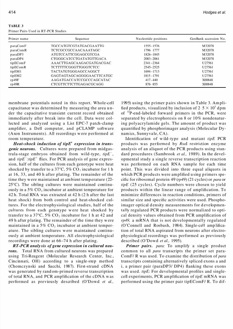
Primer pairs. para: To amplify a single productcommon to all para transcripts the primer set para-ComF/ R was used. To examine the distribution of paratranscripts containing alternatively spliced exons a andi, a primer pair (paraDP3/ DP4) anking these exonswas used. tipE: For developmental pro les and single-cell experiments, PCR am pli cation of tipE mRNA wasperformed using the primer pair tipEComF/ R. To dif-
ABLE 3
rimer Pairs Used in RT-PCR Studies
rimer name Sequence Nucleotide positions GenBank accession No.
paraComF TGCCATGTCGTATGACGAATTG 1555 –1576 M32078paraComR TCTCGCCGCCAACAAATAGC 1796 –1777 M32078paraDP3 ATGTCCATTCGGAGCGTCGA 1826 –1845 M32078paraDP4 CTGGGCATCCTGATATGTTGACA 2083 –2061 M32078tipEComF AAACTTGAGCAAGACGATGACGAC 2341 –2364 U27561tipEComR TCTTTTTCGGGTTGGGTCTCC 2545 –2525 U27561tipEM1 TACTATGTGGGAGCCAGGCT 1694 –1713 U27561tipEM2 GAGTAGTAGCAGGGGAACTTCATGC 1815 –1791 U27561rp49F AAGATGACCATCCGCCCAGCATAC 417 –440 X00848rp49R CTCGTTCTTCTTGAGACGCAGG 876 –855 X00848
414 Hodges et al.

8/8/2019 Normalizing 1
http://slidepdf.com/reader/full/normalizing-1 14/15
erentiate between wild-type and tipE mutant mRNA,rimers tipEM1/ M2 were used. rp49: rp49F/ R primerair was used for PCR ampli cation of ribosomal pro-ein transcripts. Sequences, nucleotide positions, and
GenBank accession numbers for all of the primer setssed are detailed in Table 3.
ACKNOWLEDGMENTS
This work was supported by NIH Grants NS27501 and NS01854 toD.K.O ’D., NIH Grant NS16204 and an American Cancer Society
cholar Award to L.M.H., and American Heart Association Postdoc-oral Grant 95-98 to D.D.H.
REFERENCES
Agmon, A., Hollrigel, G., and O ’Dowd, D. K. (1996). FunctionalGABAergic synaptic connections in neonatal mouse barrel cortex. J. Neurosci. 16: 4684 – 4695.
Aizenman, C. D., and Linden , D. J. (2000). Rapid, syn aptically d rivenincreases in the intrinsic excitability of cerebellar deep nuclearneurons. Nat. Neurosci. 3: 109 –111.
Arm ano, S., Rossi, P., Taglietti, V., and D ’Angelo, E. (2000). Long-termpotentiation of intrinsic excitability at the mossy ber-granule cellsynapse of rat cerebellum. J. Neurosci. 20: 5208 –5216.
Baines, R. A., and Bate, M. (1998). Electrophysiological developmentof central neurons in the Drosophila embryo. J. Neurosci. 18: 4673 –4683.
Barish, M. E. (1986). Differentiation of voltage-gated potassium cur-
rent and modulation of excitability in cultured amphibian spinalneurones. J. Physiol. 375: 225–229.Catterall, W. A. (2000). From ionic currents to molecular mechanisms:
The structure and function of voltage-gated sodium channels. Neu-ron 26: 13–25.
Chomczynski, P., and Sacchi, N. (1987). Single-step m ethod of RNAisolation by acid guanidinium thiocyanate –phenol – chloroform ex-traction. Anal. Biochem. 162: 156 –159.
Colbert, C. M., Magee, J. C., Hoffman, D. A., and Johnston, D. (1997).Slow recovery from inactivation of N a channels underlies theactivity-dependent attenuation of dend ritic action p otentials in hip-pocampal CA1 pyramidal neurons. J. N eurosci. 17: 6512 – 6521.
Connors, B. W., and Gutnick, M. J. (1990). Intrinsic ring patterns of diverse neocortical neurons. Trends Neurosci. 13: 99 –104.
Cummins, T., Dib-Hajj, S., Black, J., and Waxman, S. (2000). Sodiumchannels and the molecular pathophysiology of pain. In Progress in Brain Research, pp. 3 –19. Elsevier, Am sterdam .
Cumm ins, T., and Waxman, S. (1997). Downregulation of tetrodo-toxin-resistant sodium currents and upregulation of rapidlyrepriming tetrodotoxin-sensitive sodium current in small spinalsensory neurons after nerve injury. J. N eurosci. 17: 3503 –3514.
Desai, N. S., Rutherford, L. C., and Turrigiano, G. G. (1999). Plasticityin the intrinsic excitability of cortical pyr amidal neuron s. Nat. Neu-rosci. 2: 515 –520.
eng, G., Dea´ k, P., Chopr a, M., and Hall, L. M. (1995a). Cloning andfunctional analysis of tipE, a novel membr ane p rotein that enhan cesDrosophila para sodium channel function. Cell 82: 1001 –1011.
Feng, G., Deak, P., Kasbekar , D. P., Gil, D. W., and Ha ll, L. M. (1995b).Cytogenetic and molecular localization of tipE: A gene affectingsodium channels in Drosophila melanogaster. Genetics 139: 1679 –1688.
Ganetzky, B. (1986). Neurogenetic analysis of Drosophila mutationsaffecting sodium channels: Synergistic effects on viability andnerve conduction in double mutants involving tipE. J. N eurogenet. 3:
19 –31.Huguenard, J. R., Hamill, O. P., and Prince, D. A. (1988). Develop-mental changes in sodium conductances in rat neocortical neurons:App earance of a slowly inactivating component. J. Neurophysiol. 59:
778 –794.Ikeda, K., and Kaplan, W. (1970). Patterned neural activity of a mutant
Drosophila melanogaster. Proc. Natl. Acad. Sci. USA 66: 765 –772.Isom, L. L., DeJongh, K. S., and Catterall, W. A. (1994). Auxiliary
subunits of voltage-gated ion channels. Neuron 12: 1183 –1194.Jackson, F. R., Wilson, S. D., and Hall, L. M. (1986). The tip-E muta-
tions of Drosophila decrease saxitoxin binding and interact w ithother mutations affecting nerve membrane excitability. J. Neuro-genet. 3: 19 –31.
Jung, H. Y., Mickus, T., and Spruston, N. (1997). Prolonged sodium
channel inactivation contributes to dend ritic action potential atten-uation in hippocampal pyramidal neurons. J. Neurosci. 17: 6639 –6646.
Kulkarni, S. J., and Padhye, A. (1982). Temperature-sensitive paralyticmutations on the second and third chromosomes of Drosophilamelanogaster. Genet. Res. 40: 191–199.
Lee, D., and O ’Dowd, D. K. (1999). Fast excitatory synaptic transmis-sion m ediated by nicotinic acetylcholine receptors in Drosophilaneurons. J. N eurosci. 19: 5311 –5321.
Littleton, J. T., and Ganetzky, B. (2000). Ion channels an d synapticorganization: Analysis of the Drosophila genome. Neuron 26: 35– 43.
Loughney, K., Kreber, R., and Ganetzky, B. (1989). Molecular analysisof the para locus, a sodium channel gene in Drosophila. Cell 58:
1143 –1154.Massengill, J. L., Smith, M. A., Son, D. I., and O ’Dowd, D. K. (1997).
Differential expression of K4-AP currents and Kv3.1 potassiumchannel transcripts in cortical neurons that develop distinct ringphenotypes. J. N eurosci. 17: 3136 –3147.
Metherate, R., and Aramakis, V. B. (1999). Intrinsic electrophysiologyof neurons in thalamorecipient layers of developing rat auditorycortex. Dev. Brain. Res. 115: 131–144.
Migliore, M. (1996). Modeling the attenuation and failure of actionpotentials in the dendrites of hippocampal neurons. Biophys. J. 71:
2394 – 403.Morgan, K., Stevens, E., Shah, B., Cox, P., Kixon, A., Lee, K., Pinnock,
R., Hughes, J., Richardson, P., Mizuguchi, K., and Jackson, A.(2000). 3: An additional auxiliary subunit of the voltage-sensitive
sodium channel that modulates channel gating with distinct kinet-ics. Proc. Natl. Acad. Sci. USA 97: 2308 –2313.
O ’Connell, P. O., and Rosbash, M. (1984). Sequence, structure, an dcodon preference of the Drosophila ribosomal protein 49 gene. Nucleic Acids Res. 12: 5495 –5513.
O ’Dowd, D. K. (1995). Voltage-gated currents and ring properties of embryonic Drosophila neurons grown in a chemically de ned me-dium. J. Neurobiol. 27: 113 –126.
O ’Dowd, D. K., and Aldrich, R. W. (1988). Voltage-clamp analysis of sodium channels in wildtype and mutant Drosophila neurons. J. Neurosci. 8: 3633 –3643.
O ’Dowd, D. K., Gee, J. R., and Smith, M. A. (1995). Sodium currentdensity correlates with expression of speci c alternatively spliced
415ipE Regulates Neuronal Excitability

8/8/2019 Normalizing 1
http://slidepdf.com/reader/full/normalizing-1 15/15
sodium channel mRNAs in single neurons. J. Neurosci. 15: 4005 –4012.
O ’Dowd, D. K., Germeraad, S., and Aldrich, R. W. (1989). Alterationsin the expression and gating of Drosophila sodium channels bymutations in the para gene. Neuron 2: 1301 –1311.
O ’Dowd, D. K., Ribera, A. B., and Spitzer, N. C. (1988). Developmentof voltage-dependent calcium, sodium and potassium currents in Xenopus spinal neurons. J. N eurosci. 8: 792 – 805.
Reenan, R. A., Hanr ahan, C. J., and Ganetzky, B. (2000). The mle napts
RNA helicase mutation in Drosophila results in a splicing catastro-phe of the para Na channel transcript in a region of RNA editing. Neuron 25: 139 –149.
ambrook, J. M., Fritsch, E. F., and Maniatis, T. (1989). Molecular Cloning: A Laboratory M anual. Cold Spring Harbor Laboratory Press,Cold Spring Harbor, New York.
pitzer, N. C. (1991). A developmental handshake: Neuronal controlof ionic currents and their control of neuronal d ifferentiation. J. Neurobiol. 22: 659 – 673.
pitzer, N. C., Gu, X., and Olson, E. (1994). Action potentials, calciumtransients an d the control of d ifferentiation of excitable cells. Curr.Opin. Neurobiol. 4: 70 –77.
anouye, M. A., Ferrus, A., and Fujita, S. C. (1981). Abnormal action
potentials associated with the Shaker comp lex locus of Drosophila.Proc. Natl. Acad. Sci. USA 78: 6548 – 6552.
Turrigiano, G., LeMasson, G., and Marder, E. (1995). Selective regu-lation of current densities underlies spontaneous changes in theactivity of cultured neurons. J. N eurosci. 15: 3640 –3652.
Warmke, J., Reenan, R., Wang, P., Qian, S., Arena, J., Wang, J.,Wunderler, D., Liu, K., Kaczorowski, G., Wan der Ploeg, L.,
Ganetzky, B., an d Cohen, C. (1997). Fun ctional expr ession of Dro-sophila p ara sodium channels: Modulation by the membrane pro-tein tipE and toxin pharmacology. J. Gen. Physiol. 110: 119 –133.
Wu, C.-F., and Ganetzky, B. (1992). Neurogenetic studies of ion chan-nels in Drosophila. In Ion Channels, pp . 261 –314. Plenum, New York.
Yao, W. D., and Wu, C. F. (1999). Auxiliary hyperkinetic beta subunitof K channels: Regulation of ring properties and K currents inDrosophila neurons. J. Neurophysiol. 81: 2472 –2484.
Zhao, M.-L., and Wu, C.-F. (1997). Alterations in frequency codingand activity dependence of excitability in cultured neurons of Dro-sophila memory mutants. J. N eurosci. 17: 2187 –2199.
Zhou, F.-M., and Hablitz, J. J. (1996). Postnatal development of m em-brane properties of layer I neurons in rat neocortex. J. N eurosci. 16:
1131 –1139.
Received August 21, 2001Revised November 30, 2001Accepted December 7, 2001
416 Hodges et al.