notch ligand delta-like 1 promotes the metastasis of melanoma · pdf filethe metastasis of...
TRANSCRIPT
Notch ligand Delta-like 1 promotesthe metastasis of melanoma by
enhancing tumor adhesion
J.P. Zhang1*, N. Li2*, W.Z. Bai1, X.C. Qiu1, B.A. Ma1, Y. Zhou1, Q.Y. Fan1 and L.Q. Shan1
1Department of Orthopedic Surgery, Tangdu Hospital, The Fourth Military Medical University, Xi’an, China2Department of Oncology, Tangdu Hospital, The Fourth Military Medical University, Xi’an, China
Abstract
Notch signaling plays a vital role in tumorigenicity and tumor progression by regulating proliferation, invasion, and the tumor
microenvironment. Previous research by our group indicated that Notch ligand Delta-like 1 (Dll1) is involved in angiogenesis in
melanoma, and we noticed that it took a longer time to trypsinize Dll1-expressing B16 melanoma cells than the control cells. In
this article, we extended our study to investigate the effects of Dll1 on tumor cell adhesion and metastasis. Dll1 overexpression
activated Notch signaling in B16 tumor cells and significantly enhanced the adhering capacity of B16 tumor cells both in vitroand in vivo. B16-Dll1 cells also had a higher metastatic potential than their counterpart in the mouse model of lung metastasis.
Along with increased Dll1 expression, N-cadherin, but not E-cadherin, was upregulated in B16-Dll1 cells. These data sug-
gested that Notch ligand Dll1 may enhance the adhesion and metastasis of melanoma cells by upregulation of N-cadherin.
Key words: Delta-like 1; Notch signaling; Melanoma; Metastasis; Adhesion; N-cadherin
Introduction
Metastasis is the key factor in determining the stage,
relevant treatment, and prognosis of most solid cancers.
Although metastasis only occurs in approximately 10% of
melanoma patients, it remains the major cause of death
(1). The lungs are the most common site of metastasis in
melanoma (2). Tumor metastasis is a complex process
that includes local invasion, intravasation, and survival in
the circulation, extravasation, and colonization. The tumor
cells must overcome numerous hurdles to successfully
colonize in the target organ (3,4). In fact, the circulation
system is such a tough environment that only 0.01% of
the circulating tumor cells can survive to produce
metastasis (4). The adhesion of circulating tumor cells
to vascular endothelial cells is one of the key steps for
metastasis because it can protect tumor cells from
anoikis, fluid shear force, and attack from the immune
system (5). Many adhesion molecules, such as those
from the integrin or cadherin family, participate in the
attachment process of tumor cells and have been
reported to facilitate growth and transmigration of tumor
cells (5).
Notch signaling is an evolutionarily conserved path-
way that controls cell fate determination, cellular differ-
entiation, and pattern formation in many tissue types by
ligand-receptor interactions with neighboring cells. There
are four Notch receptors (Notch1-4) and five ligands
[Jagged1, Jagged2, and Delta-like ligands (Dll) 1, 3, and
4]. Upon activation of the Notch receptor, the intracellular
domain of the receptor is cleaved, and it can subsequently
translocate to the nucleus, where it interacts with the
transcription factor C promoter-binding factor 1/recombi-
nation signal binding protein J/k (RBP-J) to activate target
genes such as Hes family basic helix-loop-helix members
(6).
Notch signaling is implicated in various neoplastic
processes. Depending on organ and tissue type, Notch
signaling can function either as a promoter to support
tumor development or as a suppressor to inhibit tumor
growth (7-10). In melanoma, it has been seen that
activation of the Notch pathway is involved in the
tumorigenic process (11,12). Pinnix et al. (13) demon-
strated that activation of Notch1 conferred transforming
Correspondence: L.Q. Shan and/or Q.Y. Fan, Department of Orthopedic Surgery, Tangdu Hospital, The Fourth Military Medical
University, Xi’an 710038, China. E-mail: [email protected] and/or [email protected]
*These authors contributed equally to this study.
Received July 17, 2013. Accepted January 13, 2014. First published online March 31, 2014.
Brazilian Journal of Medical and Biological Research (2014) 47(4): 299-306, http://dx.doi.org/10.1590/1414-431X20143368
ISSN 1414-431X
www.bjournal.com.br Braz J Med Biol Res 47(4) 2014
properties, such as increased proliferative capacity and
unregulated cell adhesion and migration, to melanocytes.
It has also been found that Notch1 is a key effector of
the oncogene Akt and hypoxia in melanoma development,
and it can facilitate melanoma development by maintain-
ing cell proliferation and protecting cells from stress-
induced cell death (14). Overexpression of Notch1 can
also increase the aggressiveness of primary melanoma
cells through upregulation of b-catenin or N-cadherin
(15,16).
Previously, our group investigated the effects of the
Notch 1 ligand Dll1 on angiogenesis and tumor growth in
melanoma and found that overexpression of Dll1 pro-
moted tumor cell proliferation in vitro (17), but reduced
tumor growth in vivo. Interestingly, it took a longer time to
trypsinize Dll1-expressing B16 melanoma cells than the
control cells. Thus, we hypothesized that Dll1 may have
some effects in regulating the adhesion of melanoma
cells. In the present study, we extended our research
to evaluate the role of Dll1 in tumor adhesion and
metastasis.
Material and Methods
Cell cultureDll1-overexpressing (B16-Dll1) and control (B16-GFP,
green fluorescent protein) mouse melanoma cells that
express GFP were established as previously described
(17) and were maintained in RPMI 1640 medium
supplemented with 10% fetal calf serum, 100 U/mL
penicillin, and 100 mg/mL streptomycin (Life Tech-
nologies, USA). Human umbilical vein endothelial cells
(HUVECs) were isolated after treatment of umbilical veins
with collagenase and were maintained in complete
Medium 199 (M199; Gibco-BRL, USA) supplemented
with 20% fetal calf serum, 100 U/mL penicillin, 100 mg/mL
streptomycin, 0.25 mg/mL amphotericin B, 16 U/mL
heparin, 75 mg/mL endothelial cell growth supplement,
and 2 mM glutamine. All cells were grown at 376C in a
humidified atmosphere with 5% CO2 and subcultured by
trypsinization with 0.05% trypsin-0.02% EDTA when the
cells grew to confluence.
Quantitative real-time RT-PCRTotal RNA was isolated using Trizol reagent
(Invitrogen, USA) following the manufacturer’s instruc-
tions. Complementary DNA was prepared with a reverse-
transcription kit from Toyobo (Japan). RT-PCR was
performed using a kit (SYBR Premix EX Taq, Takara,
Japan) and the ABI PRISM 7300 real-time PCR system,
with b-actin as the internal control. The following primers
were used: b-actin, 59-CATCCGTAAAGACCTCTATGCC-
AAC-39 and 59-ATGGAGCCACCGATCCACA-39; Dll1, 59-
AGGGTGTGATGACCAACATGGA-39 and 59-TATCGGA-
TGCACTCATCGCAGTA-39; Hes1, 59-GCAGACATTCT-
GGAAATGACTGTGA-39 and 59-GAGTGCGCACCTC-
GGTGTTA-39; E-cadherin, 59-GAAAGCGGCTGATAC-
TGACC-39 and 59-CGTACATGTCAGCCGCTTC-39; and
N-cadherin, 59-TGTTTGACTATGAAGGCAGTGG-39 and
59-TCAGTCATCACCTCCACCAT-39.
Western blotCultured tumor cells were lysed in a radioimmunopre-
cipitation assay buffer (RIPA, 50 mM Tris-HCl, 150 mM
NaCl, 1 mM MgCl2, 0.5% octylphenoxypolyethoxyetha-
nol, 0.1 mM phenylmethylsulfonyl fluoride). Protein con-
centration in the extracts was determined using a
bicinchoninic acid (BCA) protein assay (Pierce, USA)
according to the manufacturer’s instructions. The samples
were separated by SDS-12% polyacrylamide gel electro-
phoresis and transferred to polyvinylidene difluoride
membranes (PVDF; Hybond-P, Amersham Biosciences,
USA). Blots were blocked with 5% non-fat milk in TBS-T
buffer for 1 h at room temperature and then probed using
the appropriate primary antibodies followed by horse-
radish peroxidase-conjugated rabbit anti-goat or goat anti-
rabbit IgG antibodies. The primary antibodies included
goat polyclonal IgG antibody to Dll1 and rabbit polyclonal
IgG antibodies to Hes1, N-cadherin, or E-cadherin (Santa
Cruz Biotechnology, USA). b-actin was detected simulta-
neously using 1:5000 dilution of monoclonal mouse anti-
b-actin antibody (Sigma, USA) as a loading control. The
membrane was developed using chemiluminescent
reagents (Super Signal West Femto Maximum
Sensitivity Substrate, Pierce).
Tumor cell adhesion assaysFor plate adhesion assays, tumor cells (46104/well)
were seeded on flat-bottom 96-well plates and incubated
at 376C for 30 min or 1 h. The plates were then gently
washed three times with PBS, and fresh medium
containing 20% 5 mg/mL 3-(4,5-dimethylthiazol-2-yl)-
2,5-diphenyltetrazolium bromide (MTT) was added.
After a further incubation at 376C for 4 h, the medium
was removed, and 150 mL dimethyl sulfoxide (Sigma)
was added to each well. The absorbance of each well
was measured at 490 nm after thorough shaking for
10 min.
For adhesion assays using endothelial cells, suspen-
sions of HUVECs in M199 were seeded onto 12-well
plates at a density of 26105/well. After the cells grew to
confluence, the plates were washed with PBS. Then,
56104 tumor cells in a volume of 2 mL RPMI 1640
medium were added into individual wells containing
monolayers of HUVECs. The tumor cells were incubated
with HUVECS for 1 h at 376C. The wells were then gently
washed 3 times with PBS to remove unbound tumor cells.
After trypsinization, single cell suspensions were resus-
pended in PBS containing 2% fetal calf serum and 0.05%
NaN3. The cells were subsequently incubated with 5 mLpropidium iodide for 30 min in the dark at room temper-
ature and analyzed using flow cytometry (FACSCalibur,
300 J.P. Zhang et al.
Braz J Med Biol Res 47(4) 2014 www.bjournal.com.br
BD Immunocytometry Systems, USA). Data were ana-
lyzed using the CellQuest software.
In vivo tumor cell adhesion assaysAll animal experiments were approved by the Animal
Experiment Administration Committee of the Fourth
Military Medical University. For in vivo tumor cell adhesion
assays, C57BL/6 mice were injected with 56105 mela-
noma cells through the tail vein and then killed after 2 h.
Ten minutes before the mice were killed, 100 mL Evans
blue was perfused through the tail vein to label pulmonary
vasculature. After the lungs were harvested, they were
sectioned at a thickness of 20 mm. The sections were
observed using a laser scanning system (Radiance 2000;
Bio-Rad Laboratories, USA) and a microscope (Eclipse
TE300; Nikon, Japan), and GFP-labeled melanoma cells
in the sections were counted. The number of GFP cells
was normalized to the total examined surface area of
the lung.
Mouse model of lung metastasisTo establish the lung metastasis model, tumor cells
were trypsinized, suspended in PBS, and injected into the
tail vein of C57BL/6 mice at a density of 56105/200 mLusing a 30G1/2 needle and a 1-mL syringe. Fourteen
days after injection, the animals were weighed and killed.
The lungs were removed, rinsed in PBS, and weighed.
The lung weight index was calculated as the ratio of lung
weight vs body weight. The harvested lungs were fixed in
10% neutral buffered formalin. Tumor foci on the surfaces
of the lungs were counted under a stereomicroscope.
Then, the whole lung was embedded in optimum cutting
temperature compound (Sakura Finetek, USA), sectioned
(12 mm thickness, 5 levels), and stained with hematoxy-
lin and eosin (H&E) according to routine protocols.
Histological observations were performed under a micro-
scope (BX51, Olympus, Japan). The percentage of total
area of the stained sections occupied by tumor was
measured using the Image-Pro Plus Phase 6 Imaging
System (MediaCybernetics, USA).
Statistical analysisData are reported as means±SD and were analyzed
using the unpaired Student t-test (GraphPad Prism 5.0 for
Windows, GraphPad Software, USA). P values less than
0.05 were considered to be statistically significant.
Results
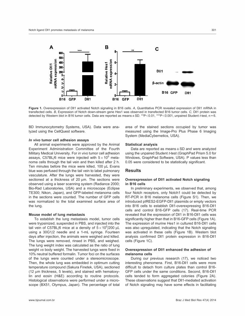
Overexpression of Dll1 activated Notch signalingin B16 cells
In preliminary experiments, we observed that, among
four Notch receptors, only Notch1 could be detected by
RT-PCR in B16 melanoma cells (Figure S1). Then, we
introduced pIRES2-EGFP-Dll1 plasmids or empty vectors
into B16 cells to establish Dll1-overexpressing B16-Dll1
cells and control B16-GFP cells (17). Real-time PCR
revealed that the expression of Dll1 in B16-Dll1 cells was
significantly higher than that in B16-GFP cells (Figure 1A).
The expression of murine Hes1 in cultured B16-Dll1 cells
was also upregulated, indicating that the Notch signaling
was activated in these cells (Figure 1B). Western blot
analysis confirmed Dll1 protein expression in B16-Dll1
cells (Figure 1C).
Overexpression of Dll1 enhanced the adhesion ofmelanoma cells
During our previous research (17), we noticed two
interesting phenomena. First, B16-Dll1 cells were more
difficult to detach from culture plates than control B16-
GFP cells under the same conditions. Second, B16-Dll1
cells tended to form aggregated colonies (Figure 2A).
These observations suggest that Dll1-mediated activation
of Notch signaling may have some effects in facilitating
Figure 1. Overexpression of Dll1 activated Notch signaling in B16 cells. A, Quantitative PCR revealed expression of Dll1 mRNA in
transfected cells. B, Expression of Notch down-stream gene Hes1 was observed in transfected B16 tumor cells. C, Dll1 protein was
detected by Western blot in B16 tumor cells. Data are reported as means±SD. **P,0.01, ***P,0.001, unpaired Student t-test, n=6.
Notch ligand Dll1 promotes metastasis of melanoma 301
www.bjournal.com.br Braz J Med Biol Res 47(4) 2014

tumor cell adhesion. To test this hypothesis, in vitro ad-
hesion assays were performed. As shown in Figure 2B, the
number of cells adhering to the bottom of the plates was
significantly higher for the B16-Dll1 group compared to the
B16-GFP group. An endothelial cell adhesion assay was
also carried out to determine whether overexpression of Dll1
may contribute to heterotypic adhesion of tumor cells. Similar
to the results of the plate adhesion assay, the number of
B16-Dll1 cells adhering to HUVECs was significantly higher
than that of B16-GFP cells (Figure 2C and D).
Overexpression of Dll1 led to tumor cell arrestin the lung
Attachment of circulating tumor cells to lung endothe-
lial cells is the initial step for lung metastasis. To in-
vestigate whether Dll1 expression could enhance tumor
cell adhesion in vivo, we studied the initial fate of GFP-
labeled control and B16-Dll1 cells. Two hours after the
Figure 2. Dll1 overexpression enhanced the
adhesion capacity of tumor cells. A, Growth
colonies of tumor cells were observed by see-
ding at low density. B, Plate adhesion assay. The
bound tumor cells were assessed by 3-(4,5-
dimethylthiazol-2-yl)-2,5-diphenyltetrazolium
bromide and compared. C, Endothelial cell (EC)adhesion assay. GFP-labeled tumor cells that
adhered to EC were analyzed by flow cytometry.
The total number of GFP-labeled cells was
calculated based on flow cytometry shown in C.
Data are reported as means±SD. **P,0.01,
***P,0.001, unpaired Student t-test, n=4.
Figure 3. Overexpression of Dll1 led to tumor
cell arrest in lung. A, Vessels of mouse lung were
labeled with Evans blue (red) and sectioned,
arrested GFP-labeled tumor cells (green) were
detected with fluorescent microscope, at 406magnification. Insets were amplified by laser
scanning microscope at 2006 magnification.
Arrows indicate the vessels of mouse lung
section. B, Average numbers of GFP-labeled
tumor cells were counted in lung sections with
the Image-Pro Plus Phase 6 Imaging System
and compared. Data are reported as
means±SD. **P,0.01, unpaired Student t-test,n=6.
302 J.P. Zhang et al.
Braz J Med Biol Res 47(4) 2014 www.bjournal.com.br

tumor cells were intravenously injected, approximately
twice as many B16-Dll1 tumor cells could be observed in
the lungs compared to B16-GFP cells (Figure 3A and B).
Furthermore, the B16-Dll1 cells in lung vessels tended to
appear as aggregated pellets or tumor emboli, as shown
in the magnified insets of Figure 3A.
OverexpressionofDll1promotes tumormetastasis invivo
To further investigate whether forced expression of
Dll1 would affect tumor metastasis in vivo, we studied the
metastatic potential of B16-Dll1 and B16-GFP cells in mouse
models of lung metastasis. Fifteen days after injection, lung
samples were collected and tumor colonies on the surfaces
of the harvested lungs were counted macroscopically
(Figure 4A). A dramatically increased number of tumor foci
was observed in the lungs of B16-Dll1-injected mice
compared to B16-GFP-injected mice (Figure 4B). Accord-
ingly, the lung weight index indicated that metastasis was
more remarkable for B16-Dll1 cells than B16-GFP cells
(Figure 4C). Figure 4D and E shows H&E staining of lung
tumor tissue and the percentage of tumor area in compar-
ison to total lung area. The results are in accordance with
those of macroscopic observations.
Dll1-mediated Notch activation induced N-cadherinexpression
Since Notch activation has been reported to influence
cadherin expression, we hypothesized that enhanced
Figure 4. Overexpression of Dll1 promotes
tumor metastasis in vivo. A, B16-Dll1 or B16-
GFP tumor cells were injected into the tail vein
of normal mice. Lungs were dissected and
photographed 15 days later. Tumor nodules are
observed on the surface of lungs (red arrows). B,Tumor nodules on the surface of the lung were
counted under a stereomicroscope and com-
pared. C, Lung weight index was calculated as
the ratio of lung weight versus body weight
of host mice, and was compared between the
two groups. D, Lungs in A were sectioned and
photographed with H&E staining. T indicates the
tumor foci in lungs. E, Tumor area in lung section
was assessed by the Image-Pro Plus Phase
6 Imaging System and nomalized to total lung
area. Data are reported as means±SD.
**P , 0.01, unpaired Student t-test, n=6.
Figure 5. Dll1-mediated Notch activation
induced N-cadherin (cad) expression. The ex-
pressions of N-cadherin (A) and E-cadherin
(B) were assessed by real-time RT-PCR, with
b-actin as a reference control. C, The protein
levels of Hes1, N-cadherin, E-cadherin in tumor
cells were examined with Western blot. Data are
reported as means±SD. **P,0.01, unpaired
Student t-test, n=6. NS: not significant.
Notch ligand Dll1 promotes metastasis of melanoma 303
www.bjournal.com.br Braz J Med Biol Res 47(4) 2014
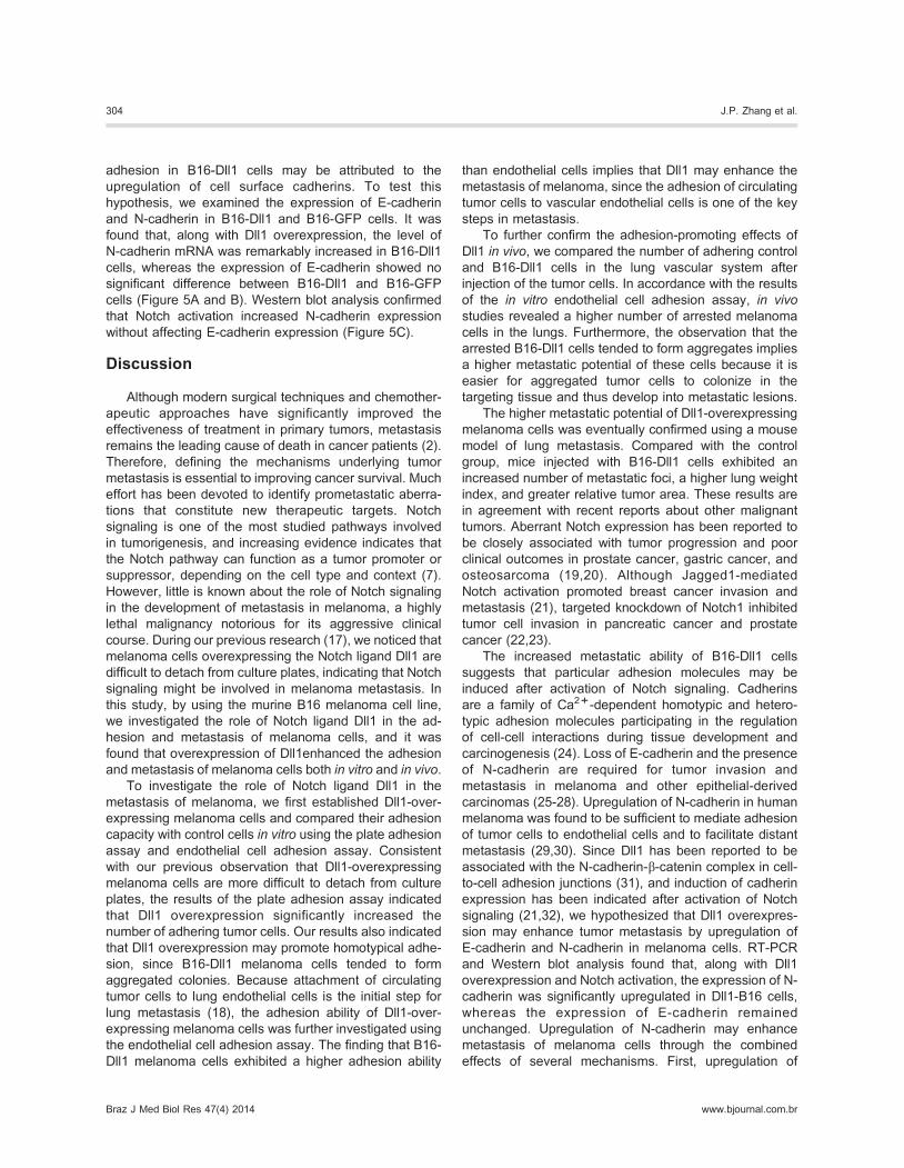
adhesion in B16-Dll1 cells may be attributed to the
upregulation of cell surface cadherins. To test this
hypothesis, we examined the expression of E-cadherin
and N-cadherin in B16-Dll1 and B16-GFP cells. It was
found that, along with Dll1 overexpression, the level of
N-cadherin mRNA was remarkably increased in B16-Dll1
cells, whereas the expression of E-cadherin showed no
significant difference between B16-Dll1 and B16-GFP
cells (Figure 5A and B). Western blot analysis confirmed
that Notch activation increased N-cadherin expression
without affecting E-cadherin expression (Figure 5C).
Discussion
Although modern surgical techniques and chemother-
apeutic approaches have significantly improved the
effectiveness of treatment in primary tumors, metastasis
remains the leading cause of death in cancer patients (2).
Therefore, defining the mechanisms underlying tumor
metastasis is essential to improving cancer survival. Much
effort has been devoted to identify prometastatic aberra-
tions that constitute new therapeutic targets. Notch
signaling is one of the most studied pathways involved
in tumorigenesis, and increasing evidence indicates that
the Notch pathway can function as a tumor promoter or
suppressor, depending on the cell type and context (7).
However, little is known about the role of Notch signaling
in the development of metastasis in melanoma, a highly
lethal malignancy notorious for its aggressive clinical
course. During our previous research (17), we noticed that
melanoma cells overexpressing the Notch ligand Dll1 are
difficult to detach from culture plates, indicating that Notch
signaling might be involved in melanoma metastasis. In
this study, by using the murine B16 melanoma cell line,
we investigated the role of Notch ligand Dll1 in the ad-
hesion and metastasis of melanoma cells, and it was
found that overexpression of Dll1enhanced the adhesion
and metastasis of melanoma cells both in vitro and in vivo.To investigate the role of Notch ligand Dll1 in the
metastasis of melanoma, we first established Dll1-over-
expressing melanoma cells and compared their adhesion
capacity with control cells in vitro using the plate adhesion
assay and endothelial cell adhesion assay. Consistent
with our previous observation that Dll1-overexpressing
melanoma cells are more difficult to detach from culture
plates, the results of the plate adhesion assay indicated
that Dll1 overexpression significantly increased the
number of adhering tumor cells. Our results also indicated
that Dll1 overexpression may promote homotypical adhe-
sion, since B16-Dll1 melanoma cells tended to form
aggregated colonies. Because attachment of circulating
tumor cells to lung endothelial cells is the initial step for
lung metastasis (18), the adhesion ability of Dll1-over-
expressing melanoma cells was further investigated using
the endothelial cell adhesion assay. The finding that B16-
Dll1 melanoma cells exhibited a higher adhesion ability
than endothelial cells implies that Dll1 may enhance the
metastasis of melanoma, since the adhesion of circulating
tumor cells to vascular endothelial cells is one of the key
steps in metastasis.
To further confirm the adhesion-promoting effects of
Dll1 in vivo, we compared the number of adhering control
and B16-Dll1 cells in the lung vascular system after
injection of the tumor cells. In accordance with the results
of the in vitro endothelial cell adhesion assay, in vivo
studies revealed a higher number of arrested melanoma
cells in the lungs. Furthermore, the observation that the
arrested B16-Dll1 cells tended to form aggregates implies
a higher metastatic potential of these cells because it is
easier for aggregated tumor cells to colonize in the
targeting tissue and thus develop into metastatic lesions.
The higher metastatic potential of Dll1-overexpressing
melanoma cells was eventually confirmed using a mouse
model of lung metastasis. Compared with the control
group, mice injected with B16-Dll1 cells exhibited an
increased number of metastatic foci, a higher lung weight
index, and greater relative tumor area. These results are
in agreement with recent reports about other malignant
tumors. Aberrant Notch expression has been reported to
be closely associated with tumor progression and poor
clinical outcomes in prostate cancer, gastric cancer, and
osteosarcoma (19,20). Although Jagged1-mediated
Notch activation promoted breast cancer invasion and
metastasis (21), targeted knockdown of Notch1 inhibited
tumor cell invasion in pancreatic cancer and prostate
cancer (22,23).
The increased metastatic ability of B16-Dll1 cells
suggests that particular adhesion molecules may be
induced after activation of Notch signaling. Cadherins
are a family of Ca2++-dependent homotypic and hetero-
typic adhesion molecules participating in the regulation
of cell-cell interactions during tissue development and
carcinogenesis (24). Loss of E-cadherin and the presence
of N-cadherin are required for tumor invasion and
metastasis in melanoma and other epithelial-derived
carcinomas (25-28). Upregulation of N-cadherin in human
melanoma was found to be sufficient to mediate adhesion
of tumor cells to endothelial cells and to facilitate distant
metastasis (29,30). Since Dll1 has been reported to be
associated with the N-cadherin-b-catenin complex in cell-
to-cell adhesion junctions (31), and induction of cadherin
expression has been indicated after activation of Notch
signaling (21,32), we hypothesized that Dll1 overexpres-
sion may enhance tumor metastasis by upregulation of
E-cadherin and N-cadherin in melanoma cells. RT-PCR
and Western blot analysis found that, along with Dll1
overexpression and Notch activation, the expression of N-
cadherin was significantly upregulated in Dll1-B16 cells,
whereas the expression of E-cadherin remained
unchanged. Upregulation of N-cadherin may enhance
metastasis of melanoma cells through the combined
effects of several mechanisms. First, upregulation of
304 J.P. Zhang et al.
Braz J Med Biol Res 47(4) 2014 www.bjournal.com.br

N-cadherin may result in effective attachment of tumor
cells to lung endothelial cells and thus protect them from
anoikis (33,34). Second, N-cadherin may promote the
migration of tumor cells through endothelial cells
(29,30,35). Third, N-cadherin may facilitate tumor cell
survival and proliferation in a new environment (36).
In conclusion, the data from this research indicated
that overexpression of Dll1 could potentiate the adhesion
and metastasis of melanoma through the upregulation of
N-cadherin expression. These results suggested that the
Notch ligand Dll1 may be a target for developing effective
strategies to reduce metastasis in melanoma. Besides
upregulation of N-cadherins, overexpression of Dll1 may
also enhance the metastasis of melanoma cells by
interaction with its receptor and may serve as an adhesion
molecule by itself (37). Therefore, the detailed mechanism
of Dll1-related enhancement of melanoma metastasis
needs to be further investigated.
Supplementary Material
Click here to view [pdf].
Acknowledgments
Research supported by grants from the Natural
Science Foundation of China (#81272072 and
#30901784).
References
1. Finn L, Markovic SN, Joseph RW. Therapy for metastatic
melanoma: the past, present, and future. BMC Med 2012;
10: 23, doi: 10.1186/1741-7015-10-23.
2. Eccles SA, Welch DR. Metastasis: recent discoveries and
novel treatment strategies. Lancet 2007; 369: 1742-1757,
doi: 10.1016/S0140-6736(07)60781-8.
3. Yilmaz M, Christofori G, Lehembre F. Distinct mechanisms
of tumor invasion and metastasis. Trends Mol Med 2007;
13: 535-541, doi: 10.1016/j.molmed.2007.10.004.
4. Fidler IJ. The pathogenesis of cancer metastasis: the ‘seed
and soil’ hypothesis revisited. Nat Rev Cancer 2003; 3: 453-
458, doi: 10.1038/nrc1098.
5. Lafrenie RM, Buchanan MR, Orr FW. Adhesion molecules
and their role in cancer metastasis. Cell Biophys 1993; 23:
3-89, doi: 10.1007/BF02796507.
6. Kato H, Taniguchi Y, Kurooka H, Minoguchi S, Sakai T,
Nomura-Okazaki S, et al. Involvement of RBP-J in biological
functions of mouse Notch1 and its derivatives. Development
1997; 124: 4133-4141.
7. Bolos V, Grego-Bessa J, De La Pompa JL. Notch signaling
in development and cancer. Endocr Rev 2007; 28: 339-363,
doi: 10.1210/er.2006-0046.
8. Baia GS, Stifani S, Kimura ET, McDermott MW, Pieper RO,
Lal A. Notch activation is associated with tetraploidy and
enhanced chromosomal instability in meningiomas.
Neoplasia 2008; 10: 604-612.
9. Zage PE, Nolo R, Fang W, Stewart J, Garcia-Manero G,
Zweidler-McKay PA. Notch pathway activation induces
neuroblastoma tumor cell growth arrest. Pediatr Blood
Cancer 2012; 58: 682-689, doi: 10.1002/pbc.23202.
10. Curry CL, Reed LL, Golde TE, Miele L, Nickoloff BJ,
Foreman KE. Gamma secretase inhibitor blocks Notch
activation and induces apoptosis in Kaposi’s sarcoma tumor
cells. Oncogene 2005; 24: 6333-6344.
11. Massi D, Tarantini F, Franchi A, Paglierani M, Di Serio C,
Pellerito S, et al. Evidence for differential expression of
Notch receptors and their ligands in melanocytic nevi and
cutaneous malignant melanoma. Mod Pathol 2006; 19: 246-
254, doi: 10.1038/modpathol.3800526.
12. Hoek K, RimmDL,WilliamsKR, ZhaoH, AriyanS, Lin A, et al.
Expression profiling reveals novel pathways in the transfor-
mation of melanocytes to melanomas. Cancer Res 2004; 64:
5270-5282, doi: 10.1158/0008-5472.CAN-04-0731.
13. Pinnix CC, Lee JT, Liu ZJ, McDaid R, Balint K, Beverly LJ,
et al. Active Notch1 confers a transformed phenotype to
primary human melanocytes. Cancer Res 2009; 69: 5312-
5320, doi: 10.1158/0008-5472.CAN-08-3767.
14. Bedogni B, Warneke JA, Nickoloff BJ, Giaccia AJ, Powell
MB. Notch1 is an effector of Akt and hypoxia in melanoma
development. J Clin Invest 2008; 118: 3660-3670, doi:
10.1172/JCI36157.
15. Balint K, Xiao M, Pinnix CC, Soma A, Veres I, Juhasz I, et al.
Activation of Notch1 signaling is required for beta-catenin-
mediated human primary melanoma progression. J Clin
Invest 2005; 115: 3166-3176, doi: 10.1172/JCI25001.
16. Liu ZJ, Xiao M, Balint K, Smalley KS, Brafford P, Qiu R, et al.
Notch1 signaling promotes primary melanoma progression
by activating mitogen-activated protein kinase/phosphatidy-
linositol 3-kinase-Akt pathways and up-regulating N-cad-
herin expression. Cancer Res 2006; 66: 4182-4190, doi:
10.1158/0008-5472.CAN-05-3589.
17. Zhang JP, Qin HY, Wang L, Liang L, Zhao XC, Cai WX, et al.
Overexpression of Notch ligand Dll1 in B16 melanoma cells
leads to reduced tumor growth due to attenuated vascular-
ization. Cancer Lett 2011; 309: 220-227, doi: 10.1016/
j.canlet.2011.06.008.
18. Behrens J. The role of cell adhesion molecules in cancer
invasion and metastasis. Breast Cancer Res Treat 1993;
24: 175-184, doi: 10.1007/BF01833258.
19. Yeh TS, Wu CW, Hsu KW, Liao WJ, Yang MC, Li AF, et al.
The activated Notch1 signal pathway is associated with
gastric cancer progression through cyclooxygenase-2.
Cancer Res 2009; 69: 5039-5048, doi: 10.1158/0008-
5472.CAN-08-4021.
20. Santagata S, Demichelis F, Riva A, Varambally S, Hofer
MD, Kutok JL, et al. JAGGED1 expression is associated
with prostate cancer metastasis and recurrence. Cancer
Res 2004; 64: 6854-6857, doi: 10.1158/0008-5472.CAN-04-
2500.
21. Leong KG, Niessen K, Kulic I, Raouf A, Eaves C, Pollet I,
et al. Jagged1-mediated Notch activation induces epithelial-
to-mesenchymal transition through Slug-induced repression
of E-cadherin. J Exp Med 2007; 204: 2935-2948, doi: 10.
1084/jem.20071082.
Notch ligand Dll1 promotes metastasis of melanoma 305
www.bjournal.com.br Braz J Med Biol Res 47(4) 2014

22. Wang Z, Banerjee S, Li Y, Rahman KM, Zhang Y, Sarkar
FH. Down-regulation of notch-1 inhibits invasion by inacti-
vation of nuclear factor-kappaB, vascular endothelial growth
factor, and matrix metalloproteinase-9 in pancreatic cancer
cells. Cancer Res 2006; 66: 2778-2784, doi: 10.1158/0008-
5472.CAN-05-4281.
23. Bin Hafeez B, Adhami VM, Asim M, Siddiqui IA, Bhat KM,
Zhong W, et al. Targeted knockdown of Notch1 inhibits
invasion of human prostate cancer cells concomitant with
inhibition of matrix metalloproteinase-9 and urokinase
plasminogen activator. Clin Cancer Res 2009; 15: 452-
459, doi: 10.1158/1078-0432.CCR-08-1631.
24. Vleminckx K, Kemler R. Cadherins and tissue formation:
integrating adhesion and signaling. Bioessays 1999; 21:
211-220, doi: 10.1002/(SICI)1521-1878(199903)21:3,211::
AID-BIES5.3.0.CO;2-P.
25. Hazan RB, Qiao R, Keren R, Badano I, Suyama K.
Cadherin switch in tumor progression. Ann N Y Acad Sci
2004; 1014: 155-163, doi: 10.1196/annals.1294.016.
26. Hirohashi S. Molecular aspects of adhesion-epigenetic
mechanisms for inactivation of the E-cadherin-mediated
cell adhesion system in cancers. Verh Dtsch Ges Pathol
2000; 84: 28-32.
27. Nieman MT, Prudoff RS, Johnson KR, Wheelock MJ.
N-cadherin promotes motility in human breast cancer cells
regardless of their E-cadherin expression. J Cell Biol 1999;
147: 631-644, doi: 10.1083/jcb.147.3.631.
28. Suyama K, Shapiro I, Guttman M, Hazan RB. A signaling
pathway leading to metastasis is controlled by N-cadherin
and the FGF receptor. Cancer Cell 2002; 2: 301-314, doi:
10.1016/S1535-6108(02)00150-2.
29. Sandig M, Voura EB, Kalnins VI, Siu CH. Role of cadherins in
the transendothelial migration of melanoma cells in culture.
Cell Motil Cytoskeleton 1997; 38: 351-364, doi: 10.1002/
(SICI)1097-0169(1997)38:4,351::AID-CM5.3.0.CO;2-6.
30. Qi J, Chen N, Wang J, Siu CH. Transendothelial migration
of melanoma cells involves N-cadherin-mediated adhesion
and activation of the beta-catenin signaling pathway. Mol
Biol Cell 2005; 16: 4386-4397, doi: 10.1091/mbc.E05-03-
0186.
31. Mizuhara E, Nakatani T, Minaki Y, Sakamoto Y, Ono Y,
Takai Y. MAGI1 recruits Dll1 to cadherin-based adherens
junctions and stabilizes it on the cell surface. J Biol Chem
2005; 280: 26499-26507, doi: 10.1074/jbc.M500375200.
32. Garcia A, Kandel JJ. Notch: a key regulator of tumor
angiogenesis and metastasis. Histol Histopathol 2012; 27:
151-156.
33. Rangarajan A, Syal R, Selvarajah S, Chakrabarti O, Sarin A,
Krishna S. Activated Notch1 signaling cooperates with
papillomavirus oncogenes in transformation and generates
resistance to apoptosis on matrix withdrawal through PKB/
Akt. Virology 2001; 286: 23-30, doi: 10.1006/viro.2001.
0867.
34. Hu YY, Zheng MH, Zhang R, Liang YM, Han H. Notch
signaling pathway and cancer metastasis. Adv Exp Med Biol
2012; 727: 186-198, doi: 10.1007/978-1-4614-0899-4_14.
35. Qi J, Wang J, Romanyuk O, Siu CH. Involvement of Src
family kinases in N-cadherin phosphorylation and beta-
catenin dissociation during transendothelial migration of
melanoma cells. Mol Biol Cell 2006; 17: 1261-1272, doi:
10.1091/mbc.E05-10-0927.
36. Li G, Satyamoorthy K, Herlyn M. N-cadherin-mediated
intercellular interactions promote survival and migration of
melanoma cells. Cancer Res 2001; 61: 3819-3825.
37. Murata A, Okuyama K, Sakano S, Kajiki M, Hirata T, Yagita
H, et al. A Notch ligand, Delta-like 1 functions as an
adhesion molecule for mast cells. J Immunol 2010; 185:
3905-3912, doi: 10.4049/jimmunol.1000195.
306 J.P. Zhang et al.
Braz J Med Biol Res 47(4) 2014 www.bjournal.com.br