notes on morphology of amnicola limosa (say, …
TRANSCRIPT
Malacological Review, 1988, 21: 81-92
NOTES ON MORPHOLOGY OF AMNICOLA LIMOSA (SAY, 1817) (GASTROPODA: HYDROBIIDAE) WITH COMMENTS ON
STATUS OF THE SUBFAMILY AMNICOLINAE
Robert Hershler and Fred G. Thompson
ABSTRACT
A morphological description is provided for Amnicola limosa (Say) (Gastropoda: Hydrobiidae), type-species for the genus. The group consisting of Amnicola and several European genera is retained in the Amnicolinae Tryon, 1866, based on shared distinctive aspects of reproductive biology. Subfamilial affinities probably lie with Hydrobiidae: Emmericiinae and/or Bithyniidae, the only other rissoacean groups having tubular glands in the penis.
INTRODUCTION
While the concept of a family-level group based on Amnicola Gould & Haldeman, 1840 and placed either within or near the Hydrobiidae dates back at least to Tryon (1866), the status of this group remains uncertain, in part because morphology of this common and widespread North American genus remains poorly known. Numerous workers (see Gould 1841; Stimpson 1865; Baker 1928; Berry 1943; Thompson 1968) have described shell and external morphology of these snails, yet certain aspects of these features require additional observations. In addition, details on internal anatomy are almost entirely lacking: only Radoman (1983: fig. 109a-c), for instance, dealt with female reproductive morphology, but his description lacked critical information (see Davis et al. 1985).
In this paper we provide additional details of morphology of the mud amnicola, Amnicola limosa (Say, 1817), which should now be considered the generic type-species in the wake of the recent ICZN opinion suppressing Paludina lustrica Say, 1821 (see Thompson 1974; Melville 1978). Based on these and other data in the literature, we conclude that Hydrobiidae: Amnicolinae consisting of the type genus and several European genera, merits recognition and may have affinities with Hydrobiidae: Emmericiinae and/or Bithyniidae.
MATERIALS AND METHODS
Anatomical descriptions and illustrations are based on study of relaxed alcohol lots, each consisting of 20-50 specimens, from the following three localities: Sail water River at Ridge Avenue, Dayton, Montgomery County, Ohio (University of Florida [UF] uncatalogued lot, coll. H.J. Walter, 25 VIII 1966); swamp of Black Warrior River, E-SE of Eutaw, Greene County, Alabama (UF uncatalogued lot, coll. L. Hubricht, 7 XI 1965); creek W of Crescent Lake, Oakland County, Michigan (UF uncatalogued lot, coll. F.G.T., 15 VI1961).
Shell, opercula and radulae were cleaned in commercial Qorox bleach and photographed using a Hitachi S-570 scanning electron microscope (SEM). Animals were dried using a Den ton DCP-1 Critical
(81)
82 Malacological Review
Point Drier and then photographed using SEM. Serial sections were made at 10 um and stained with Harris hematoxylin and eosin.
Amnicola limosa (Say)
Examination of material in the National Museum of Natural History (NMNH) and UF indicates that the mud amnicola is distributed along the east coast of the United States south to at least North Carolina, and occurs inland generally east of the Mississippi River. Purported (Berry 1943, and others) occurrences in the far west require confirmation. In Canada, its range was reported by Clarke (1981) as from Newfoundland west to Manitoba and Saskatchewan. These snails typically occur in lakes, ponds, and streams where they are found, often in great abundance, on aquatic vegetation.
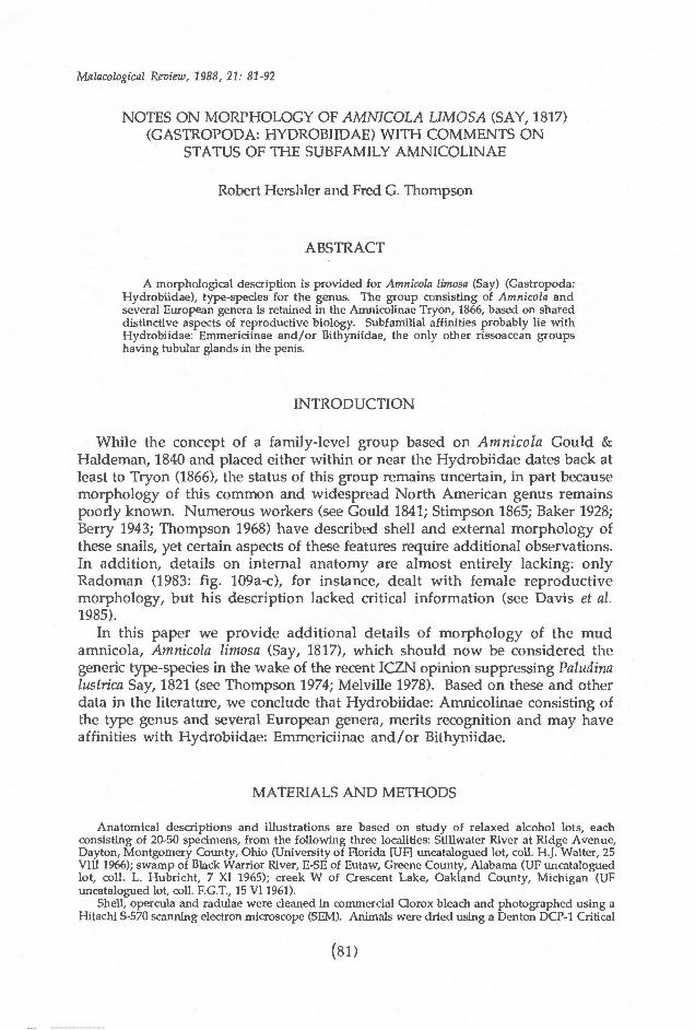
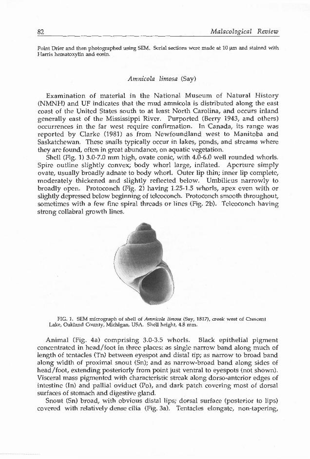
Shell (Fig. 1) 3.0-7.0 mm high, ovate conic, with 4.0-6.0 well rounded whorls. Spire outline slightly convex; body whorl large, inflated. Aperture simply ovate, usually broadly adnate to body whorl. Outer lip thin; inner lip complete, moderately thickened and slightly reflected below. Umbilicus narrowly to broadly open. Protoconch (Fig. 2) having 1.25-1.5 whorls, apex even with or slightly depressed below beginning of teleoconch. Protoconch smooth throughout, sometimes with a few fine spiral threads or lines (Fig. 2b). Teleoconch having strong collabral growth lines.
FIG. 1. SEM micrograph of shell of Amnicola limosa (Say, 1817), creek west of Crescent Lake, Oakland County, Michigan, USA. Shell height, 4.8 mm.
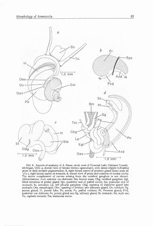
Animal (Fig. 4a) comprising 3.0-3.5 whorls. Black epithelial pigment concentrated in head/foot in three places: as single narrow band along much of length of tentacles (Tn) between eyespot and distal tip; as narrow to broad band along width of proximal snout (Sn); and as narrow-broad band along sides of head/foot, extending posteriorly from point just ventral to eyespots (not shown). Visceral mass pigmented with characteristic streak along dorso-anterior edges of intestine (In) and pallial oviduct (Po), and dark patch covering most of dorsal surfaces of stomach and digestive gland.
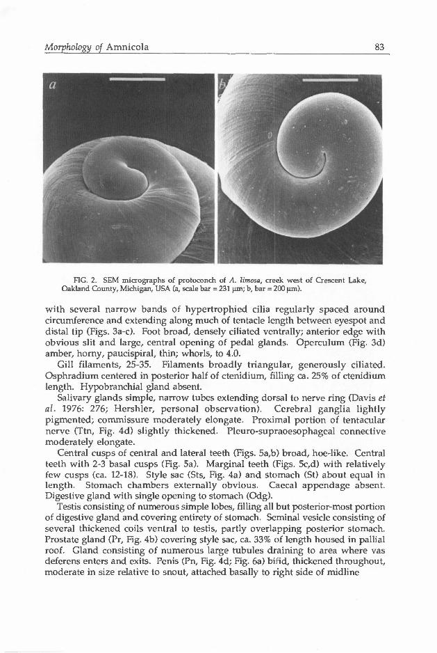
Snout (Sn) broad, with obvious distal lips; dorsal surface (posterior to lips) covered with relatively dense cilia (Fig. 3a). Tentacles elongate, non-tapering,
Morphology of Amnicola 83
FIG. 2. SEM micrographs of protocortch of A. limosa, creek west of Crescent Lake, Oakland County, Michigan, USA (a, scale bar = 231 urn; b, bar = 200 um).
with several narrow bands of hypertrophied cilia regularly spaced around circumference and extending along much of tentacle length between eyespot and distal tip (Figs. 3a-c). Foot broad, densely ciliated ventrally; anterior edge with obvious slit and large, central opening of pedal glands. Operculum (Fig. 3d) amber, horny, paucispiral, thin; whorls, to 4.0.
Gill filaments, 25-35. Filaments broadly triangular, generously ciliated. Osphradium centered in posterior half of ctenidium, filling ca. 25% of ctenidium length. Hypobranchial gland absent.
Salivary glands simple, narrow tubes extending dorsal to nerve ring (Davis et al. 1976: 276; Hershler, personal observation). Cerebral ganglia lightly pigmented; commissure moderately elongate. Proximal portion of tentacular nerve (Ttn, Fig. 4d) slightly thickened. Pleuro-supraoesophageal connective moderately elongate.
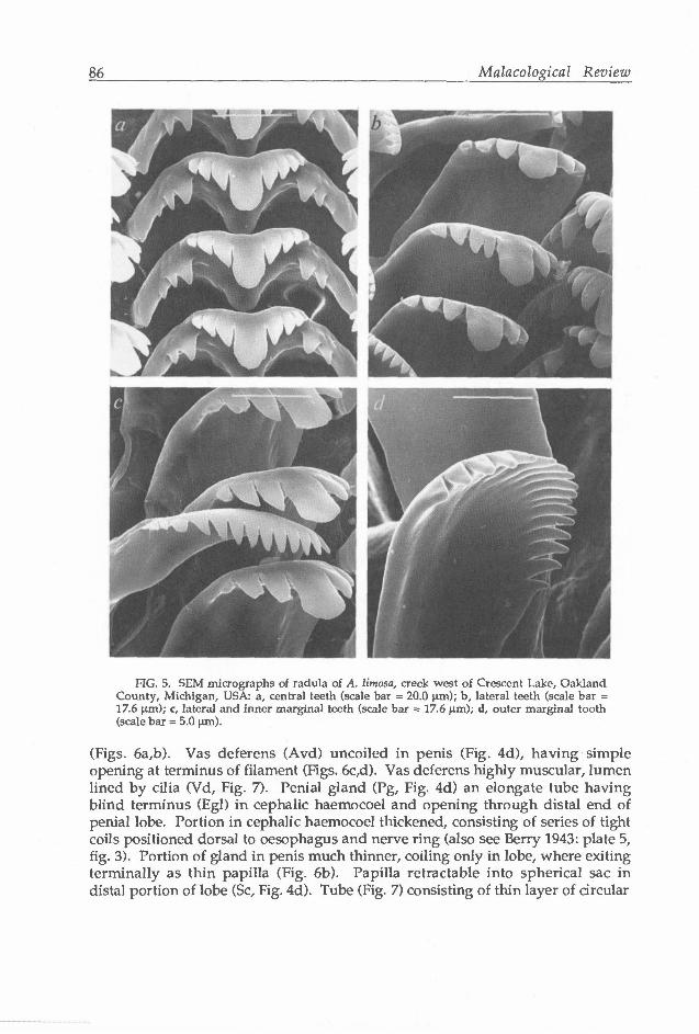
Central cusps of central and lateral teeth (Figs. 5a,b) broad, hoe-like. Central teeth with 2-3 basal cusps (Fig. 5a). Marginal teeth (Figs. 5c,d) with relatively few cusps (ca. 12-18). Style sac (Sts, Fig. 4a) and stomach (St) about equal in length. Stomach chambers externally obvious. Caecal appendage absent. Digestive gland with single opening to stomach (Odg).
Testis consisting of numerous simple lobes, filling all but posterior-most portion of digestive gland and covering entirety of stomach. Seminal vesicle consisting of several thickened coils ventral to testis, partly overlapping posterior stomach. Prostate gland (Pr, Fig. 4b) covering style sac, ca. 33% of length housed in pallial roof. Gland consisting of numerous large tubules draining to area where vas deferens enters and exits. Penis (Pn, Fig. 4d; Fig. 6a) bifid, thickened throughout, moderate in size relative to snout, attached basally to right side of midline
84 Malacological Review
FIG. 3. SEM micrographs of cephalic tentacles and operculum of A. limosa, creek west of Crescent Lake, Oakland County, Michigan, USA: a, dorsal view of snout and left cephalic tentacle (scale bar = 0.46 mm); b, right cephalic tentacle (scale bar = 0.50 mm); c, close-up of dilation on tentacle (scale bar = 75 urn); d, dorsal surface of operculum partly encrusted with diatoms (scale bar = 0.50 mm).
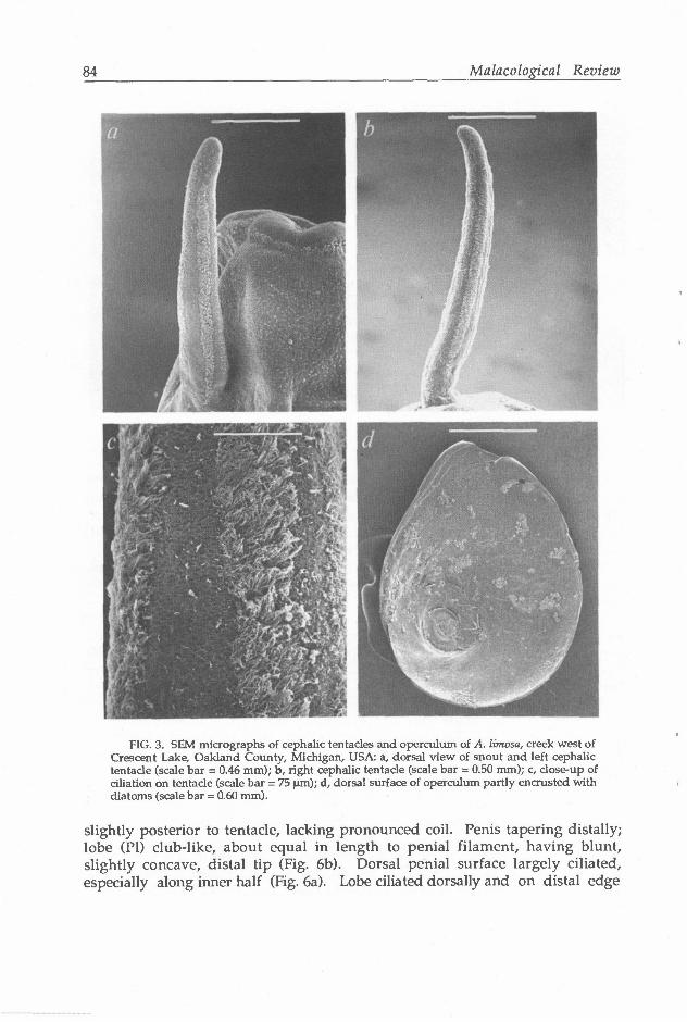
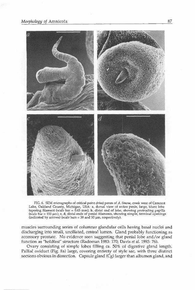
slightly posterior to tentacle, lacking pronounced coil. Penis tapering distally; lobe (PI) club-like, about equal in length to penial filament, having blunt, slightly concave, distal tip (Fig. 6b). Dorsal penial surface largely ciliated, especially along inner half (Fig. 6a). Lobe ciliated dorsally and on distal edge
Morphology of Amnicola 85
Oov
b pr
Epc
Pvd
Sts
1.0 mm
FIG. 4. Aspects of anatomy of A. limosa, creek west of Crescent Lake, Oakland County, Michigan, USA: a, dorsal view of female (minus operculum), with dense stipple indicating areas of dark melanic pigmentation; b, right lateral aspect of prostate gland (same scale as "a"); c, right lateral aspect of stomach; d, dorsal view of penis and contents of nuchal cavity. The entire complement of nerves arising from the cerebral ganglion is not shown. Abbreviations: Avd, anterior vas defer ens; Bm, buccal mass; Cbg, cerebral ganglion; Egl, blind terminus of penial gland; Epc, posterior end of pallial cavity; Est, posterior end of stomach; In, intestine; Lp, left pleural ganglion; Odg, opening of digestive gland into stomach; Oes, oesophagus; Oov, opening of oviduct into albumen gland; Ov, oviduct; Pg, penial gland; PI, penial lobe; Pn, penis; Po, pallial oviduct; Pr, Prostate gland; Pvd, posterior vas defer ens; Sc, penial gland sac; Sg, salivary gland; St, stomach; Sts, style sac; Tn, cephalic tentacle; Ttn, tentacular nerve.
86 Malacological Review
FIG. 5. SEM micrographs oi radula of A. limosa, creek west of Crescent Lake, Oakland County, Michigan, USA: a, central teeth (scale bar = 20.0 |xm); b, lateral teeth (scale bar = 17.6 urn); c, lateral and inner marginal teeth (scale bar = 17.6 um); d, outer marginal tooth (scale bar = 5.0 |jm).
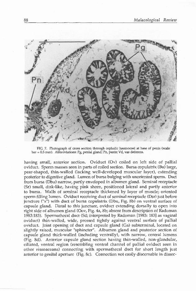
(Figs. 6a,b). Vas deferens (Avd) uncoiled in penis (Fig. 4d), having simple opening at terminus of filament (Figs. 6c,d). Vas deferens highly muscular, lumen lined by cilia (Vd, Fig. 7). Penial gland (Pg, Fig. 4d) an elongate tube having blind terminus (Egl) in cephalic haemocoel and opening through distal end of penial lobe. Portion in cephalic haemocoel thickened, consisting of series of tight coils positioned dorsal to oesophagus and nerve ring (also see Berry 1943: plate 5, fig. 3). Portion of gland in penis much thinner, coiling only in lobe, where exiting terminally as thin papilla (Fig. 6b). Papilla retractable into spherical sac in distal portion of lobe (Sc, Fig. 4d). Tube (Fig. 7) consisting of thin layer of circular
Morphology of Amnicola 87
FIG. 6. SEM micrographs of critical point dried penes of A. limosa, creek west of Crescent Lake, Oakland County, Michigan, USA: a, dorsal view of entire penis, large, blunt lobe tapering filament (scale bar = 0.43 mm); b, distal end of lobe, showing protruding papilla (scale bar = 100 urn); c, d, distal ends of penial filaments, showing simple, terminal openings (indicated by arrows) (scale bars = 38 and 50 um, respectively).
muscles surrounding series of columnar glandular cells having basal nuclei and discharging into small, unciliated, central lumen. Gland probably functioning as accessory prostate. No evidence seen suggesting that penial lobe and/or gland function as "holdfast" structure (Radoman 1983: 170; Davis et al. 1985: 76).
Ovary consisting of simple lobes filling ca. 50% of digestive gland length. Pallial oviduct (Fig. 8a) large, covering entirety of style sac, with three distinct sections obvious in dissection. Capsule gland (Cg) larger than albumen gland, and
88 Malacological Review
FIG. 7. Photograph of cross section through cephalic haemocoel at base of penis (scale bar = 0.5 mm). Abbreviations: Pg, penial gland; Pn, penis; Vd, vas deferens.
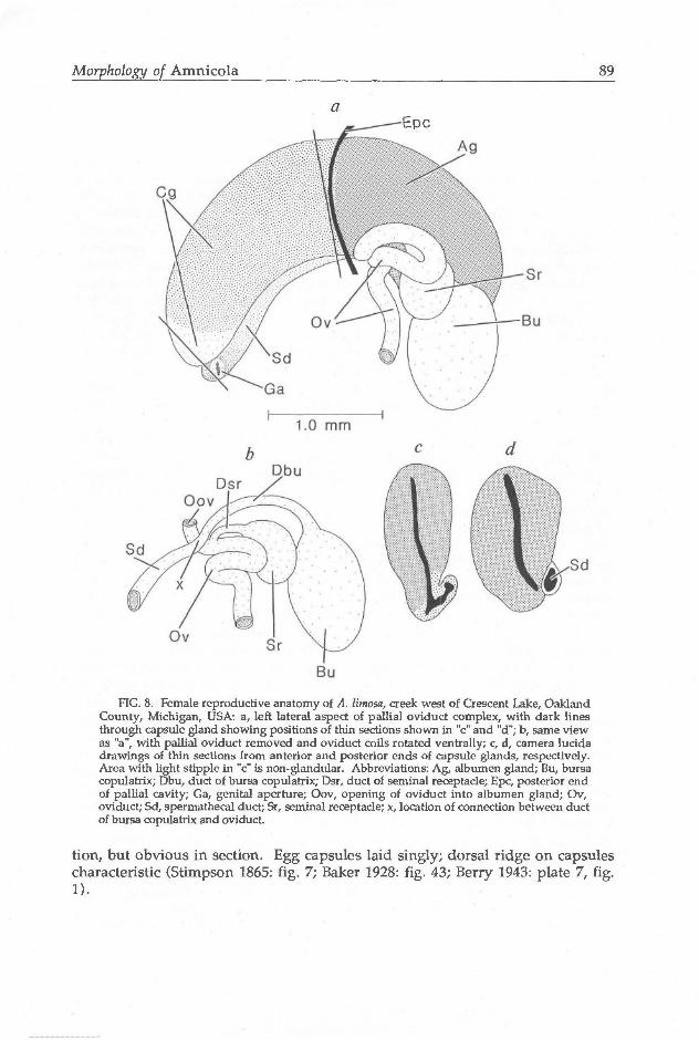
having small, anterior section. Oviduct (Ov) coiled on left side of pallial oviduct. Sperm masses seen in parts of coiled section. Bursa copulatrix (Bu) large, pear-shaped, thin-walled (lacking well-developed muscular layer), extending posterior to digestive gland. Lumen of bursa bulging with unoriented sperm. Duct from bursa (Dbu) narrow, partly enveloped in albumen gland. Seminal receptacle (Sr) small, disk-like, having pink sheen, positioned lateral and partly anterior to bursa. Walls of seminal receptacle thickened by layer of muscle; oriented sperm filling lumen. Oviduct receiving duct of seminal receptacle (Dsr) just before juncture ("x") with duct of bursa copulatrix (Dbu, Fig. 8b) on ventral surface of capsule gland. Distal to this juncture, oviduct extending dorsally to open into right side of albumen gland (Oov, Fig. 4a, 8b; absent from description of Radoman 1983:183). Spermathecal duct (Sd; interpreted by Radoman [1983: 183] as vaginal oviduct) thin-walled, wide, pressed tightly against ventral surface of pallial oviduct. Joint opening of duct and capsule gland (Ga) subterminal, located on slightly raised, muscular "sphincter". Albumen gland and posterior section of capsule gland thick-walled (including ventrally), with narrow, central lumens (Fig. 8d). Anterior capsule gland section having thin-walled, non-glandular, ciliated, ventral region (resembling ventral channel of pallial oviduct seen in other rissoaceans) connecting with spermathecal duct for short length just anterior to genital aperture (Fig. 8c). Connection not easily discernable in dissec-
Morphology of Amnicola 89
a Epc
FIG. 8. Female reproductive anatomy of A. limosa, creek west of Crescent Lake, Oakland County, Michigan, USA: a, left lateral aspect of pallia! oviduct complex, with dark lines through capsule gland showing positions of thin sections shown in "c" and "d"; b, same view as "a", with pallia! oviduct removed and oviduct coils rotated ventrally; c, d, camera lucida drawings of thin sections from anterior and posterior ends of capsule glands, respectively. Area with light supple in "c" is non-glandular. Abbreviations: Ag, albumen gland; Bu, bursa copulatrix; Dbu, duct of bursa copulatrix; Dsr, duct of seminal receptacle; Epc, posterior end of pallial cavity; Ga, genital aperture; Oov, opening of oviduct into albumen gland; Ov, oviduct; Sd, spermathecal duct; Sr, seminal receptacle; x, location of connection between duct of bursa copulatrix and oviduct.
tion, but obvious in section. Egg capsules laid singly; dorsal ridge on capsules characteristic (Stimpson 1865: fig. 7; Baker 1928: fig. 43; Berry 1943: plate 7, fig. 1).
90 Malacological Review
DISCUSSION
Numerous authors have considered Amnicola to be closely related to several European genera, including Bythinella Moquin-Tandon, Parabythinella Radoman, and Marstoniopsis Altena, as these taxa have very similar shells, penial shape, and penial gland type (data for European taxa in Fretter & Graham 1978; Radoman 1983). Several other distinctive features of Amnicola (dorsal ridge on egg capsule, spiral sculpture on protoconch) have been noted in at least some of the above European forms. Reliable illustrations of female reproductive systems for the European snails are lacking, but groundplans similar to that of Amnicola may be inferred for Bythinella (Radoman 1983: fig. 105) and Parabythinella (Radoman 1983: fig. 107) if one assumes that Radoman overlooked presence of spermathecal duct and posterior connection between oviduct and albumen gland in these taxa as he did for A. limosa (see above).
In most recent reviews of hydrobioid prosobranchs (Morrison 1949; Taylor 1966; Thompson 1968; Burch 1982), the "Amnicola-group" was assigned subfamilial rank within the Hydrobiidae, although Radoman (1983) and Davis et al. (1985) elevated the group to separate familial status. Radoman's (1983: 168, 170) diagnosis of the family listed character-states that are common among hydrobiids, with the exception being the bifid penis having accessory prostate: a distinctive feature, yet one no more divergent than numerous other penial types seen in hydrobiids. Possession of a spermathecal duct in the "Amnicola-group" suggests placement in the Littoridininae, which are currently defined as hydrobiids possessing this very character-state (Davis et al. 1982; Hershler 1985). Given the likelihood that this feature has evolved iteratively in various rissoaceans (Ponder 1988), and the fact that littoridinines are quite heterogeneous in other anatomical aspects such as penial morphology, division of its members among additional subfamilies has merit. On the basis of several features (morphology of egg capsules; penis shape and gland type; body pigmentation) of the Amnicola-group" that are not seen in littoridinines, we retain placement of the former in the subfamily Amnicolinae Tryon 1866 (=Bythinellinae of authors).
Relationships of amnicolines probably lie with either or both of the other rissoacean groups that possess a caecum-like accessory prostate in the penis, the Hydrobiidae: Emmericiinae and Bithyniidae. Although it has not yet been established that this structure is homologous among these groups, such a situation is likely, given the infrequent occurrence of a penial caecum in rissoaceans and striking similarity of external morphology and location of this structure in the above groups. A suggestion of close relationship between amnicolines and emmericiines, which have two tubular penial glands discharging through separate ducts in a trifid penis, is complicated by absence of a spermathecal duct in females of the latter, in which sperm travel inside the pallial oviduct (Radoman 1983: fig. 94c; Hershler, personal observation), but may still be tenable given plasticity of this aspect of rissoacean female reproductive morphology (see above). Bithyniids, which may represent a divergent lineage within the Rissoacea (Ponder 1988), possess a number of character-states (involving feeding mode, and morphology of pallial cavity, female reproductive system, and egg capsule; see Lilly 1953; Taylor 1966) not found in hydrobiids. Additional
Morphology of Amnicola 91
morphological data are needed to assess possible cladistic relationships among these three groups.
ACKNOWLEDGEMENTS
National Museum of Natural History (NMNH) staff of the Electron Microscopy Laboratory assisted with preparation of SEM micrographs, Mrs. Molly Ryan (NMNH, Invertebrate Zoology) assisted with preparation of illustrations and plates, and Mr. Paul Greenhall (NMNH, Invertebrate Zoology) provided diverse forms of assistance. We also thank Dr. S.-K. Wu and an anonymous reviewer for useful criticism of the manuscript.
LITERATURE CITED
BAKER, F.C. 1928. The fresh water Mollusca of Wisconsin; Part I. Gastropoda. Bulletin of the Wisconsin Geological and Natural History Survey, 70: 1-507 pp.
BERRY, E.G. 1943. The Amnicolidae of Michigan: distribution, ecology, and taxonomy. Miscellaneous Publications, Museum of Zoology, University of Michigan no. 57: 1-68 pp.
BURCH, J.B. Freshwater snails (Mollusca: Gastropoda) of North America. United States Environmental Protection Agency, Environmental Monitoring and Support Laboratory, Cincinnati, Ohio. 1-294 pp.
CLARKE, AH. 1981. The freshwater molluscs of Canada. National Museums of Sciences, National Museums of Canada, Ottawa. 1-446 pp.
DAVIS, G.M., KITIKOON, V. & TEMCHAROEN, P. 1976. Monograph on "Lithoglyphopsis" aperta, the snail host of Mekong River schistosomiasis. Malacologia, 15:241-287.
DAVIS, G.M., MAZURKIEWICZ, M. & MANDRACCHIA, M. 1982. Spurwinkia: morphology, systematics, and ecology of a new genus of North American marshland Hydrobiidae (Mollusca: Gastropoda). Proceedings of the Academy of Natural Sciences of Philadelphia, 134: 143-177.
DAVIS, G.M., KUO, Y.-H., HOAGLAND, K.E., CHEN, P.-L., TANG, H.-M. & CHEN, D.-J. 1985. Erhaia, a new genus and species of Pomatiopsidae from China (Gastropoda: Rissoacea). Proceedings of the Academy of Natural Sciences of Philadelphia, 137: 48- 78.
FRETTER, V. & GRAHAM, A. 1978. The prosobranch molluscs of Britain and Denmark. Part 3, Neritacea, Viviparacea, Valvatacea, terrestrial and freshwater Littorinacea and Rissoacea. Journal of Molluscan Studies, Supplement, 5: 101-152.
GOULD, A.A. 1841. A report on the Invertebrata of Massachusetts, comprising the Mollusca, Annelida, and Radiata. Folsom, Wells and Thurston, Cambridge, Massachusetts, i-xiii, 1-373 pp, 15 pis.
HERSHLER, R. 1985. Systematic revision of the Hydrobiidae (Gastropoda: Rissoacea) of the Cuatro Cienegas Basin. Malacologia, 26: 31-123.
LILLY, M.M. 1953. The mode of life and the structure and functioning of the reproductive ducts of Bithynia tentaculata (L.). Proceedings of the Malacological Society of London, 30:87-110.
MELVILLE, R.V. 1978. Opinion 1108. Conservation of Marstonia Baker, 1926 and of Amnicola lustrica Pilsbry, 1890 (Mollusca: Gastropoda). Bulletin of Zoological Nomenclature, 35: 94-96.
MORRISON, J.P.E. 1949. The cave snails of eastern North America (abstract). American Malacological Union, Annual Report for 1948, pp.13-14.
92 Malacological Review
PONDER, W.F. 1988. The truncatelloidean (= rissoacean) radiation - a preliminary phylogeny. Malacological Review, Supplement, 4: 129-164.
RADOMA.N, P. 1983. Hydrobioidea; a superfamily of Prosobranchia (Gastropoda); I. Systematics. Serbian Academy of Sciences and Arts, Monographs, DXLVII, Number 57:1-256 pp.
STIMPSON, W. 1865. Researches upon the Hydrobiinae and allied forms. Smithsonian Miscellaneous Collections, 201: 1-59 pp.
TAYLOR, D.W. 1966. A remarkable snail fauna from Coahuila, Mexico. Veliger, 9: 152- 228.
THOMPSON, F.G. 1968. The aquatic snails of the family Hydrobiidae of peninsular Florida. University of Florida Press, Gainesville. 1-268 pp.
THOMPSON, F.G. 1974. Comments on the proposed suppression of Paludina lustrica Say, 1821. Z.N.(S.) 730. Bulletin of Zoological Nomenclature, 31: 170-171.
TRYON, G.W. 1866. Researches upon the Hydrobiinae and allied forms (review). American Journal of Conchology, 2: 152-158.
ROBERT HERSHLER
Department of Invertebrate Zoology National Museum of Natural History Smithsonian Institution Washington, DC 20560, U.S.A.
FRED G. THOMPSON
Florida Museum of Natural History University of Florida Gainesville, Florida 32611, U.S.A.
MALACOLOGICAL REVIEW, P. O. Box 3037, Ann Arbor, Michigan 48106, U.S.A.