notes on trochila (ascomycota, leotiomycetes), with new
TRANSCRIPT

Notes on Trochila (Ascomycota, Leotiomycetes), with new species and combinations
Paula Andrea Gómez-Zapata1, Danny Haelewaters1,2,3,4, Luis Quijada2,3, Donald H. Pfister2,3, M. Catherine Aime1
1 Department of Botany and Plant Pathology, Purdue University, West Lafayette, Indiana, USA 2 Depart-ment of Organismic and Evolutionary Biology, Harvard University, Cambridge, Massachusetts, USA 3 Farlow Herbarium and Reference Library of Cryptogamic Botany, Harvard University Herbaria, Harvard University, 22 Divinity Avenue, Cambridge, MA 02138, USA 4 Faculty of Science, University of South Bohemia, České Budějovice, Czech Republic
Corresponding author: Paula Andrea Gómez-Zapata ([email protected]), Danny Haelewaters ([email protected]), M. Catherine Aime ([email protected])
Academic editor: N. Wijayawardene | Received 15 December 2020 | Accepted 7 January 2021 | Published 11 February 2021
Citation: Gómez-Zapata PA, Haelewaters D, Quijada L, Pfister DH, Aime MC (2021) Notes on Trochila (Ascomycota, Leotiomycetes), with new species and combinations. MycoKeys 78: 21–47. https://doi.org/10.3897/mycokeys.78.62046
AbstractStudies of Trochila (Leotiomycetes, Helotiales, Cenangiaceae) are scarce. Here, we describe two new species based on molecular phylogenetic data and morphology. Trochila bostonensis was collected at the Boston Harbor Islands National Recreation Area, Massachusetts. It was found on the stem of Asclepias syriaca, representing the first report of any Trochila species from a plant host in the family Apocynaceae. Trochila urediniophila is associated with the uredinia of the rust fungus Cerotelium fici. It was discovered during a survey for rust hyperparasites conducted at the Arthur Fungarium, in a single sample from 1912 collected in Trinidad. Macro- and micromorphological descriptions, illustrations, and molecular phylo-genetic analyses are presented. The two new species are placed in Trochila with high support in both our six-locus (SSU, ITS, LSU, rpb1, rpb2, tef1) and two-locus (ITS, LSU) phylogenetic reconstructions. In addition, two species are combined in Trochila: Trochila colensoi (formerly placed in Pseudopeziza) and T. xishuangbanna (originally described as the only species in Calycellinopsis). This study reveals new host plant families, a new ecological strategy, and a new country record for the genus Trochila. Finally, our work emphasizes the importance of specimens deposited in biological collections such as fungaria.
Keywords4 new taxa, biological collections, Boston Harbor Islands, fungarium specimens, fungicolous fungi, South America, taxonomy, Trinidad
MycoKeys 78: 21–47 (2021)
doi: 10.3897/mycokeys.78.62046
https://mycokeys.pensoft.net
Copyright Paula Andrea Gómez-Zapata et al. This is an open access article distributed under the terms of the Creative Commons Attribution License (CC BY 4.0), which permits unrestricted use, distribution, and reproduction in any medium, provided the original author and source are credited.
RESEARCH ARTICLE
A peer-reviewed open-access journal
Launched to accelerate biodiversity research
Paula Andrea Gómez-Zapata et al. / MycoKeys 78: 21–47 (2021)22
Introduction
The genus Trochila Fr. (Ascomycota, Leotiomycetes) was erected by Fries (1849) to accommodate four species previously placed in Phacidium Fr., Sphaeria Haller, and Xyloma Pers. Trochila craterium (DC) Fr. was the first species listed by Fries, based on Sphaeria craterium DC., which was later selected by Clements and Shear (1931) as the type species of Trochila. The other three species included by Fries (1849) were: T. ilicis (Fr.) Fr. [= Sphaeria ilicis Fr.], T. laurocesari (Desm.) Fr. [= Phacidium lau-rocerasi Desm.], and T. taxi (Fr.) Fr. [= Xyloma taxi Fr.]. Only the genus and one spe-cies (T. laurocerasi) were briefly described by Fries (1849). However, the type species, T. craterium, was well described macromorphologically by Lamarck and de Candolle (1805). The description can be translated loosely from French as “a fungus growing on the lower surface of ivy leaves, initially forming a flat white disc, then turning black-ish and concave opening by a split along radial lines, the disc usually surrounded by a whitish membrane” (Lamarck and de Candolle 1805). Later, the generic concept was expanded to include other types of apothecial opening. Rehm (1896) remarked that the covering layer of the apothecia could also open completely like a lid depending on host characters such as cuticle thickness. After the inclusion of this new character de-scribing the genus, Stegia ilicis (Chevall.) Gillet was transferred as Trochila ilicina (Nees ex Fr.) Courtec (Crouan and Crouan 1867; Rehm 1896).
In our current circumscription of the genus Trochila, apothecia are sunken in the host tissues and hymenia are exposed either by splitting along radial lines or by split-ting into a number of lobes that roll outward exposing the hymenium. The excipu-lum is composed of dark, globose-angular cells; asci contain eight ellipsoid, hyaline ascospores with oil guttules (except T. substictica Rehm and T. tetraspora E. Müll. & Gamundí, which both have asci containing four ascospores); and paraphyses possess yellowish guttules (Dennis 1978; Baral and Marson 2005). Thirty-three names have been applied in the genus (Index Fungorum 2021). Jaklitsch et al. (2016) suggest that only ca. 10 names should be accepted.
Fries (1849) included Trochila in “Patellariacei” (= Patellariaceae). Later, it was transferred to Dermateaceae, Helotiales (Fuckel 1869; Karsten 1869; Saccardo 1884; Lambotte 1888). Trochila remained in this family (Korf 1973; Dennis 1978) into the molecular era (Lumbsch and Huhndorf 2010). Jaklitsch et al. (2016) placed Trochila in the resurrected family Cenangiaceae based on morphological and molecular data. Lat-er, the relationships among genera in this family were supported in another, 5–15-lo-cus phylogeny of Leotiomycetes (Johnston et al. 2019).
Most species of Trochila have been described from their sexual morph. The asexual morph has the characteristics of the form-genus Cryptocline Petr. (Morgan-Jones 1973; Kiffer and Morelet 2000; Hyde et al. 2011). Two species of Trochila have been linked to their asexual morphs: T. craterium to C. paradoxa (De Not.) Arx and T. laurocerasi to C. phacidiella (Grove) Arx (von Arx 1957). The paucity of culture and molecular data of both Cryptocline and Trochila species has hindered the linkage of sexual and asexual morphs for most species. Trochila viburnicola Crous & Denman was the first species

Notes on Trochila 23
of the genus to be described based on the combination of morphology and molecular data, but only its asexual morph is known (Crous et al. 2018). The species was named referring to its host, Viburnum sp. (Dipsacales, Adoxaceae). In addition to T. vibur-nicola, two other species have been reported on this host genus, but only from their sexual morph, T. ramulorum Feltgen and T. tini (Duby) Quél. [currently Pyrenopeziza tini (Duby) Nannf.]. Due to the lack of sequences or cultures of these two species, a comparison with T. viburnicola is impossible (Feltgen 1903; Crous et al. 2018).
Most Trochila members have a restricted record of geographical distribution and ecological strategy. Trochila records typically originate from the Northern Hemisphere limited to temperate regions in Europe and North America (Ziolo et al. 2005; Stoykov and Assyov 2009; Crous et al. 2018; Stoykov 2019; Global Biodiversity Information Facility 2020). Nonetheless, a number of putative Trochila reports are known from the Southern hemisphere (Spegazzini 1888, 1910, 1921; Rehm 1909; Gamundí et al. 1978). In addition, species of Trochila are typically recorded as saprotrophs on dead leaves and branches of both herbaceous plants and trees. However, a few species have been found infecting living plant tissues. Trochila ilicina is reported as both a weak parasite and a saprotroph because of its presence on living, decaying, and fallen leaves of Ilex aquifolium (Aquifoliales, Aquifoliaceae) (Ziolo et al. 2005), T. laurocerasi as a parasite of living leaves of Prunus laurocerasus (Rosales, Rosaceae) (Gregor 1936), and T. symploci as a pathogen of living leaves of Symplocos japonica (Ericales, Symplocaceae) (Hennings 1900; Stevenson 1926).
Here, we describe two new species, T. bostonensis and T. urediniophila, collected at the Boston Harbor Islands National Recreation Area, Massachusetts and at Port of Spain, Trinidad, respectively. We also make two new combinations in Trochila based on morphological studies and phylogenetic analyses. We reveal two new host plant families (Apocynaceae and Asparagaceae) and a new ecological strategy (fungicolous symbiont) for the genus. Finally, we provide a comparative table of characters, based on literature review, for all currently accepted species of Trochila (sensu Index Fungorum 2021).
Material and methods
Collected samples
Samples were collected in the field and from fungaria. One collection of Trochila was discovered during the Boston Harbor Islands (BHI) National Recreation Area fungal ATBI (Haelewaters et al. 2018a). In this project, above-ground, ephemeral fruiting bod-ies of non-lichenized fungi were collected. In the field, specimens were placed in plastic containers or brown paper bags. BHI-F collection numbers were assigned. Date, specific locality when applicable, GPS coordinates, substrate, and habitat notes were recorded. Specimens were dried using a Presto Dehydro food dehydrator (National Presto Indus-tries, Eau Claire, Wisconsin) set at 35 °C for 7–9 hours. Collections were packaged, labeled, and deposited at FH. A second Trochila collection came to our attention during

Paula Andrea Gómez-Zapata et al. / MycoKeys 78: 21–47 (2021)24
a survey for hyperparasites of rust fungi at PUR. The specimen was found on the uredinia of the rust fungus Cerotelium fici on the underside of Ficus maxima leaves. Fungarium acronyms follow Thiers (continuously updated).
Morphological studies
Methods to study the morphological characteristics of the Trochila specimens followed the process given in Baral (1992). Macro- and micromorphological features were ex-amined on both fresh and dried apothecia for the specimen collected at the BHI and on dried apothecia for the specimen found at PUR. Apothecia from the BHI speci-men were observed under an EZ4 stereomicroscope (Leica, Wetzlar, Germany) and studied under a B1 compound microscope (Motic, Barcelona, Spain). Apothecia from the PUR specimen were examined on an SZ2-ILTS dissecting microscope (Olympus, Center Valley, Pennsylvania) and studied using a BH2-RFCA compound microscope (Olympus). Sections of apothecia were cut free-hand and mounted in water or pre-treated in 5% KOH. Sections were also mounted in Melzer’s reagent with and without KOH-pretreatment to determine dextrinoid or amyloid reactions. At least 10 measure-ments were made for each structure at 400–1000× magnification. Measurements for each character are given as (a–)b–c(–d), with b–c indicating the 95% confidence inter-val and a and d representing the smallest and large single measurement, respectively. Macro- and microphotographs were taken with a USB Moticam 2500 camera (Motic) (BHI specimen) or an Olympus SC30 camera (PUR specimen). Measurements were made using the following software suites: Motic Images Plus 2.0 and cellSens Standard 1.18 Imaging Software (Olympus). Color coding refers to Kelly (1965). Abbreviations were adopted from Baral (1992) and Baral and Marson (2005) as follows:
* living state;† dead state;IKI Lugol’s solution;KOH potassium hydroxide;
LBs lipid bodies;MLZ Melzer’s reagent;OCI oil content index;VBs refractive vacuolar bodies.
DNA isolation, PCR amplifications, sequencing
Genomic DNA was isolated from 1–3 apothecia per specimen using the E.Z.N.A. HP Fungal DNA Kit (Omega Bio-Tek, Norcross, Georgia), QIAamp DNA Micro Kit (Qiagen, Valencia, California), following the manufacturer’s instructions, and the Extract-N-Amp Plant PCR Kit (Sigma-Aldrich, St. Louis, Missouri), following Haelewaters et al. (2018a). We amplified the following loci: nuclear small and large ribosomal subunits (SSU and LSU), internal transcribed spacer region of the riboso-mal DNA (ITS), RNA polymerase II second largest subunit (rpb2), and translation elongation factor 1-α (tef1). Primer combinations were as follows: NS1/NS2 and NS1/NS4 for SSU (White et al. 1990); LR0R/LR5 for LSU (Vilgalys and Hester 1990; Hopple 1994); ITS1F/ITS4, ITS9mun/ITS4A, and ITS5/ITS2 for ITS (White et al.
Notes on Trochila 25
1990; Gardes and Bruns 1993; Egger 1995); RPB2-5F2/fRPB2-7cR for rpb2 (Liu et al. 1999; Sung et al. 2007); and EF1-983F/EF1-1567R and EF1-983F/EF1-2218R for tef1 (Rehner and Buckley 2005). All 25-µl PCR reactions were conducted on a Mastercycler ep gradient Thermal Cycler (Eppendorf model #5341, Hauppauge, New York) and consisted of 12.5 µl of 2× MyTaq Mix (Bioline, Swedesboro, New Jersey), 1 µl of each 10 µM primer, and 10.5 µl of 1/10 diluted DNA extract. Amplifications of rDNA and rpb2 loci were run under the following conditions: initial denaturation at 95 °C for 5 min (94 °C for LSU); followed by 40 cycles of denaturation at 95 °C for 30 sec (94 °C for LSU), annealing at 45 °C (ITS) / 50 °C (LSU) / 55 °C (SSU, rpb2) for 45 sec, and elongation at 72 °C for 45 sec (1 min for LSU); and final extension at 72 °C for 7 min (1 min for SSU). Amplification of tef1 was done with a touchdown PCR as follows: initial denaturation at 95 °C for 10 min; followed by 30 cycles of 95 °C for 1 min, 62 °C for 1 min (decreasing 1 °C every 3 cycles), 72 °C for 90 sec; then 30 cycles of 95 °C for 30 sec, 55 °C for 30 sec, and 72 °C for 1 min; and final extension at 72 °C for 7 min (Don et al. 1991; Haelewaters et al. 2018b). PCR prod-ucts were visualized by gel electrophoresis. Purification of successful PCR products and subsequent sequencing in both directions were outsourced to Genewiz (South Plain-field, New Jersey). Raw sequence reads were assembled and edited using Sequencher version 5.2.3 (Gene Codes Co., Ann Arbor, Michigan).
Sequence alignment and phylogenetic analysis
Edited sequences were blasted against the NCBI GenBank nucleotide database (http://ncbi.nlm.nih.gov/blast/Blast.cgi) to search for closest relatives. For phylogenetic place-ment of our isolates, we downloaded SSU, ITS, LSU, rpb1, rpb2, and tef1 sequences of Trochila from GenBank. We also downloaded sequence data of selected clades of Helotiales, mainly from Pärtel et al. (2017) but also other sources (details in Table 1), as a basis for our six-locus phylogenetic analysis. We selected representative taxa of Cenangiaceae, Cordieritidaceae, Rutstroemiaceae, and Sclerotiniaceae, with taxa in the family Chlorociboriaceae serving as outgroups (Johnston et al. 2019). Alignment of DNA sequences was done for each locus separately using MUSCLE version 3.7 (Edgar 2004), available on the Cipres Science Gateway 3.3 (Miller et al. 2010). The aligned sequences for each locus were concatenated in MEGA7 (Kumar et al. 2016). Maxi-mum likelihood (ML) inference was performed using IQ-TREE from the command line (Nguyen et al. 2015) under partitioned models (Chernomor et al. 2016). Nu-cleotide substitution models were selected under Akaike’s information criterion cor-rected for small sample size (AICc) with the help of the built-in program ModelFinder (Kalyaanamoorthy et al. 2017). Ultrafast bootstrap analysis was implemented with 1000 replicates (Hoang et al. 2017).
For the purpose of species delimitation, we constructed a second dataset of ITS–LSU consisting of isolates of Trochila and closely related taxa in the family Cenangiace-ae. We included Trochila spp., Calycellinopsis xishuangbanna, and Pseudopeziza colensoi, with Cenangiopsis spp. serving as outgroup. In this analysis, we included T. ilicina, for

Paula Andrea Gómez-Zapata et al. / MycoKeys 78: 21–47 (2021)26
Tabl
e 1.
Seq
uenc
es u
sed
in p
hylo
gene
tic a
naly
ses.
Acce
ssio
n nu
mbe
rs in
bol
dfac
e in
dica
te se
quen
ces t
hat w
ere
gene
rate
d du
ring
the
cour
se o
f thi
s stu
dy.
Isol
ate
Spec
ies
Fam
ilySS
UIT
SLS
Urp
b1rp
b2te
f1R
efer
ence
KL3
91Am
eghi
niell
a au
stral
isC
ordi
eriti
dace
aeK
X09
0893
KX
0908
41K
X09
0787
KX
0906
90Pä
rtel
et a
l. (2
017)
AD28
3531
TAn
nabe
lla a
ustra
liens
isC
ordi
eriti
dace
aeM
K32
8475
MK
3284
76Fr
yar e
t al.
(201
9)AF
TOL-
ID 5
9Bo
tryo
tinia
fuck
elian
aSc
lero
tinia
ceae
AY54
4695
AY54
4651
DQ
4711
16D
Q24
7786
DQ
4710
45Sp
ataf
ora
et a
l. (2
006)
HM
AS:1
8706
3C
alyc
ellin
opsis
xish
uang
bann
aC
enan
giac
eae
GU
9361
24K
R09
4163
MH
7293
38M
H72
9345
W.Y
. Zhu
ang
et a
l. (u
npub
l.)K
L375
Cen
angi
opsis
alp
estris
Cen
angi
acea
eK
X09
0837
KX
0907
84K
X09
0736
Pärt
el e
t al.
(201
7)K
L378
Cen
angi
opsis
alp
estris
Cen
angi
acea
eK
X09
0891
LT15
8470
KX
0908
39K
X09
0786
KX
0907
38Pä
rtel
et a
l. (2
017)
KL1
57C
enan
giop
sis a
lpest
risC
enan
giac
eae
KX
0908
58LT
1584
21K
X09
0806
KX
0907
09Pä
rtel
et a
l. (2
017)
KL1
74C
enan
giop
sis q
uerc
icola
Cen
angi
acea
eK
X09
0862
LT15
8425
KX
0908
11K
X09
0760
KX
0907
13K
X09
0663
Pärt
el e
t al.
(201
7)K
L377
Cen
angi
opsis
sp.
Cen
angi
acea
eK
X09
0890
KX
0909
00K
X09
0838
KX
0907
85K
X09
0737
Pärt
el e
t al.
(201
7)K
L276
“Cen
angi
um”
acuu
mPi
ceom
phal
e cla
deK
X09
0879
LT15
8445
KX
0908
28K
X09
0727
KX
0906
80Pä
rtel
et a
l. (2
017)
KL2
43“C
enan
gium
” ac
uum
Pice
omph
ale c
lade
KX
0908
73LT
1584
39K
X09
0822
KX
0907
67K
X09
0720
KX
0906
74Pä
rtel
et a
l. (2
017)
KL3
90C
enan
gium
ferr
ugin
osum
Cen
angi
acea
eK
X09
0892
LT15
8471
KX
0908
40K
X09
0739
Pärt
el e
t al.
(201
7)K
L167
Chl
oren
coeli
a to
rta
Cen
angi
acea
eLT
1584
24K
X09
0810
KX
0907
59Pä
rtel
et a
l. (2
017)
KP6
06C
hlor
enco
elia
versi
form
isC
enan
giac
eae
KX
0908
94K
X09
0788
KX
0907
40K
X09
0692
Pärt
el e
t al.
(201
7)K
L21
Chl
oren
coeli
a ve
rsifo
rmis
Cen
angi
acea
eK
X09
0846
LT15
8427
KX
0907
95Pä
rtel
et a
l. (2
017)
KL1
52C
hlor
ocib
oria
aer
ugin
asce
nsC
hlor
ocib
oria
ceae
LT15
8419
KX
0907
52K
X09
0706
KX
0906
57Pä
rtel
et a
l. (2
017)
KL2
47C
hlor
ocib
oria
aer
ugin
ella
Chl
oroc
ibor
iace
aeK
X09
0875
KX
0907
69K
X09
0722
KX
0906
76Pä
rtel
et a
l. (2
017)
KL2
38C
hlor
ocib
oria
gla
uca
Chl
oroc
ibor
iace
aeK
X09
0872
LT15
8438
KX
0908
21K
X09
0766
KX
0906
73Pä
rtel
et a
l. (2
017)
KL2
12C
ibor
ia v
iridi
fusca
Scle
rotin
iace
aeK
X09
0863
LT15
8429
KX
0908
12Pä
rtel
et a
l. (2
017)
KL2
54C
rum
enul
opsis
soro
riaC
enan
giac
eae
LT15
8442
KX
0908
26K
X09
0725
Pärt
el e
t al.
(201
7)K
L317
Dip
loca
rpa
blox
amii
Cor
dier
itida
ceae
KX
0908
85K
X09
0834
KX
0907
78K
X09
0745
KX
0906
88Pä
rtel
et a
l. (2
017)
SK80
Dip
lola
evio
psis
ranu
laC
ordi
eriti
dace
aeK
X09
0896
KP9
8478
2K
X09
0790
Etay
o et
al.
(201
5), P
ärte
l et a
l. (2
017)
TU
:109
263
Dum
ontin
ia tu
bero
saSc
lero
tinia
ceae
KX
0908
97LT
1584
12K
X09
0843
KX
0907
92K
X09
0697
Pärt
el e
t al.
(201
7)K
L111
Enco
elia
fimbr
iata
Cen
angi
acea
eK
X09
0852
KX
0908
00K
X09
0703
KX
0906
55Pä
rtel
et a
l. (2
017)
KL1
08En
coeli
a fu
rfura
cea
Cen
angi
acea
eK
X09
0851
KX
0907
99K
X09
0702
KX
0906
54Pä
rtel
et a
l. (2
017)
KL1
07En
coeli
a fu
rfura
cea
Cen
angi
acea
eK
X09
0850
LT15
8416
KX
0907
98K
X09
0749
KX
0907
01K
X09
0653
Pärt
el e
t al.
(201
7)K
L106
Enco
elia
furfu
race
aC
enan
giac
eae
KX
0908
49LT
1584
15K
X09
0748
KX
0906
52Pä
rtel
et a
l. (2
017)
KL9
2En
coeli
a fu
rfura
cea
Cen
angi
acea
eK
X09
0847
LT15
8482
KX
0907
96K
X09
0651
Pärt
el e
t al.
(201
7)K
L164
Enco
elia
hete
rom
era
Cen
angi
acea
eK
X09
0861
KX
0908
09K
X09
0758
KX
0907
12K
X09
0662
Pärt
el e
t al.
(201
7)K
L304
Enco
elia
hete
rom
era
Cen
angi
acea
eK
X13
8404
KX
1384
00Pä
rtel
et a
l. (2
017)
KL2
44H
elot
iale
s sp.
Cen
angi
acea
eK
X09
0874
LT15
8440
KX
0908
23K
X09
0768
KX
0907
21K
X09
0675
Pärt
el e
t al.
(201
7)K
L20
Hey
deria
abi
etis
Cen
angi
acea
eK
X09
0845
LT15
8426
KX
0907
47K
X09
0699
KX
0906
50Pä
rtel
et a
l. (2
017)
HM
AS:7
1954
Hey
deria
abi
etis
Cen
angi
acea
eAY
7892
95AY
7892
97AY
7892
96W
ang
et a
l. (2
005)
KL2
16H
eyde
ria p
usill
aC
enan
giac
eae
KX
0908
65LT
1584
30K
X09
0762
KX
0907
15K
X09
0665
Pärt
el e
t al.
(201
7)K
L299
Iono
mid
otis
frond
osa
Cor
dier
itida
ceae
KX
0908
82K
X09
0775
KX
0906
85Pä
rtel
et a
l. (2
017)
KL2
31Io
nom
idot
is fu
lvot
inge
nsC
ordi
eriti
dace
aeK
X09
0870
KX
0908
19K
X09
0765
KX
0907
19K
X09
0671
Pärt
el e
t al.
(201
7)

Notes on Trochila 27
Isol
ate
Spec
ies
Fam
ilySS
UIT
SLS
Urp
b1rp
b2te
f1R
efer
ence
KL2
39Io
nom
idot
is fu
lvot
inge
nsC
ordi
eriti
dace
aeK
X13
8403
KX
1384
07K
X13
8399
KX
1384
01Pä
rtel
et a
l. (2
017)
KL1
54Io
nom
idot
is irr
egul
aris
Cor
dier
itida
ceae
KX
0908
56K
X09
0804
KX
0907
54K
X09
0658
Pärt
el e
t al.
(201
7)K
L301
Iono
mid
otis
oliv
asce
nsC
ordi
eriti
dace
aeK
X09
0883
KX
0908
33K
X09
0776
KX
0907
32K
X09
0686
Pärt
el e
t al.
(201
7)C
BS:8
11.8
5La
mbe
rtell
a su
bren
ispor
aRu
tstro
emia
ceae
KF5
4541
6AB
9260
97M
H87
3604
Zha
o et
al.
(201
6), P
ärte
l et a
l. (2
017)
, Vu
et a
l. (2
019)
LL95
Llim
oniel
la te
rrico
laC
ordi
eriti
dace
aeK
X09
0895
KX
0908
42K
X09
0789
KX
0907
41K
X09
0693
Pärt
el e
t al.
(201
7)AF
TOL-
ID 1
69M
onili
nia
laxa
Scle
rotin
iace
aeAY
5447
14AY
5446
70FJ
2384
25D
Q47
0889
DQ
4710
57Sp
ataf
ora
et a
l. (2
006)
KL3
74Pi
ceom
phal
e bul
gario
ides
Pice
omph
ale
clad
eK
X09
0889
LT15
8469
KX
0908
36K
X09
0783
Pärt
el e
t al.
(201
7)K
L98
Pice
omph
ale b
ulga
rioid
esPi
ceom
phal
e cl
ade
KX
0908
48LT
1584
83K
X09
0797
KX
0907
00Pä
rtel
et a
l. (2
017)
PDD
:112
240
Pseu
dope
ziza
colen
soi
Cen
angi
acea
eM
H92
1874
MH
9852
97M
H98
6706
MH
9867
05P.R
. Joh
nsto
n an
d D
. Par
k (u
npub
l.)K
L267
Pycn
opez
iza
sejou
rnei
Scle
rotin
iace
aeK
X09
0878
LT15
8443
KX
0908
27K
X09
0772
KX
0907
26K
X09
0679
Pärt
el e
t al.
(201
7)AF
TOL-
ID 9
07Rh
abdo
cline
laric
isC
enan
giac
eae
DQ
4710
02D
Q47
0954
DQ
4711
46D
Q47
0904
DQ
4710
73Sp
ataf
ora
et a
l. (2
006)
KL2
92Ru
tstro
emia
firm
aRu
tstro
emia
ceae
KX
0908
81LT
1584
50K
X09
0832
KX
0907
74K
X09
0731
KX
0906
84Pä
rtel
et a
l. (2
017)
KL2
91Ru
tstro
emia
firm
aRu
tstro
emia
ceae
LT15
8449
KX
0908
31K
X09
0730
KX
0906
83Pä
rtel
et a
l. (2
017)
KL2
90Ru
tstro
emia
firm
aRu
tstro
emia
ceae
KX
0908
30K
X09
0729
KX
0906
82Pä
rtel
et a
l. (2
017)
KL2
22Ru
tstro
emia
firm
aRu
tstro
emia
ceae
KX
1384
02K
X13
8406
KX
1383
97Pä
rtel
et a
l. (2
017)
KL3
10Ru
tstro
emia
john
stoni
iRu
tstro
emia
ceae
KX
0908
84LT
1584
54K
X09
0777
KX
0907
33K
X09
0687
Pärt
el e
t al.
(201
7)K
L234
Rutst
roem
ia ju
nipe
riRu
tstro
emia
ceae
KX
0908
71K
X09
0820
KX
0906
72Pä
rtel
et a
l. (2
017)
KL2
17Ru
tstro
emia
lute
ovire
scens
Rutst
roem
iace
aeLT
1584
31K
X09
0814
KX
0907
63K
X09
0716
KX
0906
66Pä
rtel
et a
l. (2
017)
KL1
60Ru
tstro
emia
tilia
cea
Rutst
roem
iace
aeK
X09
0860
LT15
8423
KX
0908
08K
X09
0757
KX
0907
11K
X09
0661
Pärt
el e
t al.
(201
7)K
L393
Rutst
roem
iace
ae sp
.Ru
tstro
emia
ceae
KX
1384
05LT
1584
72K
X13
8408
KX
1383
98K
X09
0691
Pärt
el e
t al.
(201
7)K
L288
Rutst
roem
iace
ae sp
.Ru
tstro
emia
ceae
KX
0908
80LT
1584
46K
X09
0829
KX
0907
73K
X09
0728
KX
0906
81Pä
rtel
et a
l. (2
017)
CBS
:273
.74T
Sarc
otro
chila
long
ispor
aC
enan
giac
eae
KJ6
6383
6K
J663
877
KJ6
6391
8C
rous
et a
l. (2
014)
KL3
47Sc
leren
coeli
a fa
scicu
laris
Scle
rotin
iace
aeK
X09
0782
Pärt
el e
t al.
(201
7)K
L156
Scler
enco
elia
fraxi
nico
laSc
lero
tinia
ceae
KX
0908
57K
X09
0805
KX
0907
55K
X09
0708
KX
0906
59Pä
rtel
et a
l. (2
017)
KL3
44Sc
leren
coeli
a pr
uino
saSc
lero
tinia
ceae
KX
0908
88K
X09
0781
KX
0907
35Pä
rtel
et a
l. (2
017)
CBS
:499
.50
Scler
otin
ia sc
lerot
ioru
mSc
lero
tinia
ceae
DQ
4710
13D
Q47
0965
DQ
4709
16Sp
ataf
ora
et a
l. (2
006)
NY:
0123
1276
Skyt
tea
radi
atili
sC
ordi
eriti
dace
aeK
J559
538
KJ5
5956
0K
X09
0791
KX
0907
42K
X09
0694
Suija
et a
l. (2
015)
, Pär
tel e
t al.
(201
7)T
H90
Tham
noga
lla cr
ombi
eiC
ordi
eriti
dace
aeK
J559
583
KJ5
5953
5K
J559
557
KX
0907
43K
X09
0695
Pärt
el e
t al.
(201
7)BH
I-F9
74aT
Troc
hila
bos
tone
nsis
Cen
angi
acea
eM
T87
3949
MT
8739
47M
T87
3952
MT
8611
81M
T86
1183
This
study
BHI-
F974
bTTr
ochi
la b
osto
nens
isC
enan
giac
eae
MT
8739
50M
T87
3948
MT
8739
48M
T86
1182
MT
8611
84Th
is stu
dyK
L332
Troc
hila
crat
eriu
mC
enan
giac
eae
KX
0908
86K
X09
0779
Pärt
el e
t al.
(201
7)K
L336
Troc
hila
laur
ocer
asi
Cen
angi
acea
eK
X09
0887
LT15
8460
KX
0908
35K
X09
0780
KX
0907
34K
X09
0689
Pärt
el e
t al.
(201
7)F1
8316
TTr
ochi
la u
redi
niop
hila
Cen
angi
acea
eM
T87
3946
MT
8739
51Th
is stu
dyC
BS:1
4420
6TTr
ochi
la v
ibur
nico
laC
enan
giac
eae
MH
1079
21M
H10
7967
MH
1080
11M
H10
8031
Cro
us e
t al.
(201
8)K
L253
Velu
tarin
a ru
fo-o
livac
eaC
enan
giac
eae
KX
0908
77K
X09
0825
KX
0907
71K
X09
0724
KX
0906
78Pä
rtel
et a
l. (2
017)

Paula Andrea Gómez-Zapata et al. / MycoKeys 78: 21–47 (2021)28
which only a single ITS sequence is available. The same methods as above were applied: alignment using MUSCLE (Edgar 2004), selection of nucleotide substitution models with the help of ModelFinder (Kalyaanamoorthy et al. 2017), ML using IQ-TREE (Nguyen et al. 2015; Chernomor et al. 2016; Hoang et al. 2017). Phylogenetic recon-structions with bootstrap values (BS) were visualized in FigTree version 1.4.3 (http://tree.bio.ed.ac.uk/software/figtree/).
Results
Nucleotide alignment dataset and phylogenetic inferences
The concatenated six-locus dataset consisted of 11343 characters, of which 2655 were parsimony-informative. The percentage of parsimony-informative characters per locus was 9.3% for SSU, 48.1% for ITS, 21.4% for LSU, 48.9% for rpb1, 30.0% for rpb2, and 19.2% for tef1. A total of 71 isolates were included, of which Chlorociboria aerugina-scens (Nyl.) Kanouse ex C.S. Ramamurthi, Korf & L.R. Batra, C. aeruginella (P. Karst.) Dennis, and C. glauca (Dennis) Baral & Pärtel (Helotiales, Chlorociboriaceae) served as outgroup taxa. The following models were selected by ModelFinder (AICc): TNe+R3 (SSU, –lnL = 23478.796); GTR+F+I+G4 (ITS, –lnL = 18385.043); TN+F+R4 (LSU, –lnL = 28398.591); SYM+I+G4 (rpb1, –lnL = 41387.214); GTR+F+R10 (rpb2, –lnL = 57025.083); and GTR+F+R8 (tef1, –lnL = 35467.940). Our ML analysis reveals five high to maximum-supported clades (Fig. 1): Cenangiaceae, Cordieritidaceae, Rutstro-emiaceae, Sclerotiniaceae, and a clade with Piceomphale bulgarioides (P. Karst.) Svrček and “Cenangium” acuum Cooke & Peck (Piceomphale clade sensu Pärtel et al. 2017). As previously reported (e.g., Pärtel et al. 2017; Johnston et al. 2019), several genera in their current circumscription are polyphyletic: Encoelia (Fr.) P. Karst. in Cenangiaceae and Rutstroemiaceae, Ionomidotis E.J. Durand ex Thaxt. in Cordieritidaceae, Rutstro-emia P. Karst. in Rutstroemiaceae, and Trochila in Cenangiaceae. Trochila laurocerasi is placed as a sister taxon to Calycellinopsis xishuangbanna W.Y. Zhuang and Pseudopeziza colensoi (Berk.) Massee. The other species of Trochila, including the type species T. cra-terium and the here described species, form a monophyletic clade (BS = 81).
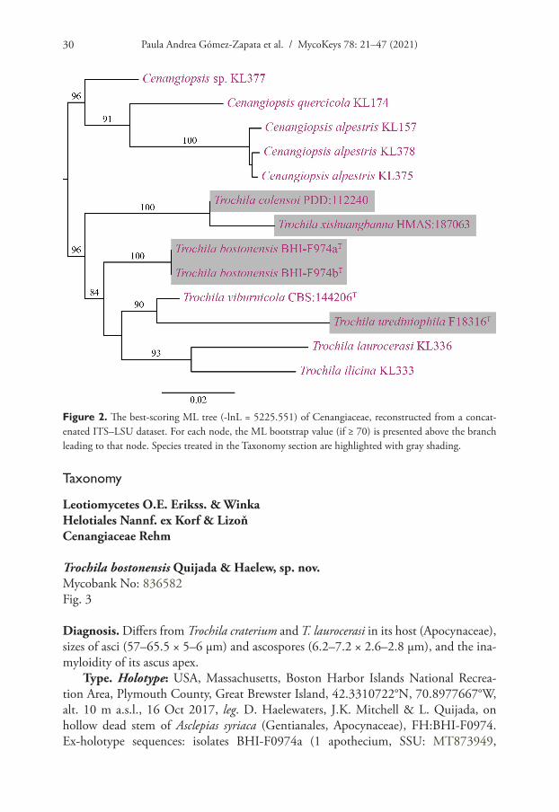
The second two-locus dataset consisted of 2284 characters (ITS: 924, LSU: 1360), of which 2040 were parsimony-informative (ITS: 782, LSU: 1258). A total of 13 iso-lates were included, of which Cenangiopsis alpestris (Baral & B. Perić) Baral, B. Perić & Pärtel, C. quercicola (Romell) Rehm, and Cenangiopsis sp. served as outgroup taxa. The following models were selected by ModelFinder (AICc): GTR+F+I+G4 (ITS, –lnL = 5810.483) and TIM+F+R2 (LSU, –lnL = 5595.374). Calycellinopsis xishuangbanna, Pseudopeziza colensoi, and all Trochila species form a monophyletic clade with high support (BS = 96) (Fig. 2). Both new species of Trochila are distinct from previously described species. The undescribed Trochila species found on uredinia of Cerotelium fici is retrieved as sister to T. viburnicola (BS = 90).

Notes on Trochila 29
Figure 1. The best-scoring ML tree (-lnL = 87544.854) of Cenangiaceae, Cordieritidaceae, Rutstroemi-aceae, Sclerotiniaceae, and the Piceomphale clade, reconstructed from a concatenated six-locus dataset (SSU, ITS, LSU, rpb1, rpb2, and tef1). For each node, the ML bootstrap value (if ≥ 70) is presented above or in front of the branch leading to that node. The arrow denotes the genus Trochila. Species with an asterisk (*) are treated in the Taxonomy section.
Paula Andrea Gómez-Zapata et al. / MycoKeys 78: 21–47 (2021)30
Taxonomy
Leotiomycetes O.E. Erikss. & WinkaHelotiales Nannf. ex Korf & LizoňCenangiaceae Rehm
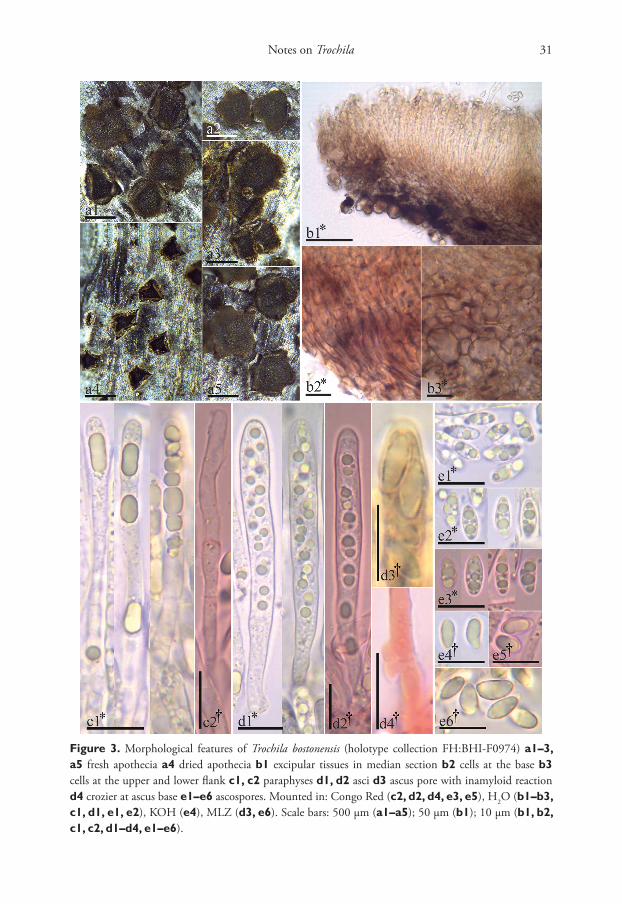
Trochila bostonensis Quijada & Haelew, sp. nov.Mycobank No: 836582Fig. 3
Diagnosis. Differs from Trochila craterium and T. laurocerasi in its host (Apocynaceae), sizes of asci (57–65.5 × 5–6 µm) and ascospores (6.2–7.2 × 2.6–2.8 µm), and the ina-myloidity of its ascus apex.
Type. Holotype: USA, Massachusetts, Boston Harbor Islands National Recrea-tion Area, Plymouth County, Great Brewster Island, 42.3310722°N, 70.8977667°W, alt. 10 m a.s.l., 16 Oct 2017, leg. D. Haelewaters, J.K. Mitchell & L. Quijada, on hollow dead stem of Asclepias syriaca (Gentianales, Apocynaceae), FH:BHI-F0974. Ex-holotype sequences: isolates BHI-F0974a (1 apothecium, SSU: MT873949,
Figure 2. The best-scoring ML tree (-lnL = 5225.551) of Cenangiaceae, reconstructed from a concat-enated ITS–LSU dataset. For each node, the ML bootstrap value (if ≥ 70) is presented above the branch leading to that node. Species treated in the Taxonomy section are highlighted with gray shading.
Notes on Trochila 31
Figure 3. Morphological features of Trochila bostonensis (holotype collection FH:BHI-F0974) a1–3, a5 fresh apothecia a4 dried apothecia b1 excipular tissues in median section b2 cells at the base b3 cells at the upper and lower flank c1, c2 paraphyses d1, d2 asci d3 ascus pore with inamyloid reaction d4 crozier at ascus base e1–e6 ascospores. Mounted in: Congo Red (c2, d2, d4, e3, e5), H2O (b1–b3, c1, d1, e1, e2), KOH (e4), MLZ (d3, e6). Scale bars: 500 µm (a1–a5); 50 µm (b1); 10 µm (b1, b2, c1, c2, d1–d4, e1–e6).

Paula Andrea Gómez-Zapata et al. / MycoKeys 78: 21–47 (2021)32
ITS: MT873947, LSU: MT873952, rpb2: MT861181, tef1: MT861183) and BHI-F0974b (1 apothecium, SSU: MT873950, ITS: MT873948, LSU: MT873953, rpb2: MT861182, tef1: MT861184).
Etymology. bostonensis – referring to Boston, Massachusetts, the locality of the type collection.
Description. Apothecia erumpent singly or in groups of 2–3, protruding from the bark by lifting and rolling outward the host periderm, sessile on a broad base, closed and barely visible when dry, rehydrated 0.4–1.1 mm diam., 0.1–0.2 mm thick; mature flat to slightly cupulate, dark grayish red brown (47.D.gy.r.Br) to black (267.Black). Margin toothed and lighter than the disc, apothecia star-shaped, with 3–6 teeth of 0.1–0.3 mm in length, each tooth deep yellowish brown (75.deepyBr). Asci *(46.5–)55.5–66.5(–73) × (5.5–)6.0–6.5(–7.0) µm, †(50.5–)57–65.5(–66) × (4.5–)5.0–6.0 µm, 8-spored, cylindrical, pars sporifera *30–52 µm; apex rounded to subconical, inamyloid (IKI, KOH-pretreated or not), slightly thick-walled at apex, lateral walls thin; base slightly tapered and arising from croziers. Ascospores *(6.3–)6.7–7.7(–8.6) × 2.7–3.4 µm, †(5.8–)6.2–7.2 × 2.6–2.8 µm, el-lipsoid-cuneate, inequilateral, ends rounded or subacute, aseptate, hyaline, smooth, thick-walled, oligoguttulate, containing 2–5 grayish yellow (90.gy.Y) oil drops (LBs), 1–2.4 µm diam., OCI = (45–)60–75(–90)%. Paraphyses slightly to medium clavate, terminal cell *(17.5–)18–23(–29.5) × 3–4 µm, secondary cells *(8–)9–10(–11) × 2.5–3 µm, lower cells *(7.5–)8.5–10.5(–11.5) × 2.5–3 µm, unbranched, thin-walled, smooth, with one or sev-eral cylindric to globose refractive drops (VBs, not present after KOH-pretreated), *3.5–14 × 2–3.5 µm. Medullary excipulum 17.5–54 µm thick, grey yellowish brown (80.gy.yBr), upper part of textura porrecta, lower part dense textura intricata, cells with tiny globose deep yellow (85.deepY) refractive drops (VBs). Ectal excipulum of thin-walled textura globu-losa–angularis at base and lower flanks, dark yellowish brown (78.d.yBr) to dark brown (59.d.Br), (40–)55–78 µm thick, cells *(7.0–)9.5–13(–15.5) × (3.0–)5.0–8.5(–10) µm; at upper flanks and margin of textura prismatica, 30–40 µm thick, cells *(5.5–)6.5–7.5(–8.5) × 2.5–3.5 µm, entirely without drops and slightly gelatinized, cells slightly thick-walled with irregular patches of dark brown exudates in areas of mutual contact, cortical cells in flanks covered by amorphous refractive deep yellow (88.d.Y) granular exudates, at margin some cells protruding like short hairs (*6.5–14 × 2.5–3.5 µm). Asexual state unknown.
Notes. Trochila bostonensis is the only species of the genus found on a member of Apocynaceae (Table 2). It was growing in the outer layer of a dead stem of Asclepias syriaca, which had fallen on the ground. The host was close to the shore in a shrub-by thicket of Rhus. There are two similar species. Trochila laurocerasi has wider asci (6.0–8.0 µm vs. 4.5–6.0 µm) and larger ascospores (6.3–10 × 2.5–4.6 µm vs. 5.8–7.2 × 2.6–2.8 µm) compared to T. bostonensis. Ascus and ascospore length are similar in T. bostonensis and T. craterium, although ascospores are slightly larger in T. craterium. The two species mostly differ in the width of their asci (7–12 µm in T. craterium vs. 4.5–6.0 µm in T. bostonensis). We used the measurements in dead state to compare T. bostonensis with other species in the genus (see Table 2).

Notes on Trochila 33
Table 2. Comparative table of currently accepted species of Trochila (except T. viburnicola). For each spe-cies, the following characters are presented: host plant, host family, measurements of asci and ascospores (dead state). The asterisk (*) indicates a fungal host.
Species Host Plant Host Family Asci (µm) Ascospores (µm) ReferenceLength Width Length Width
T. andromedae Andromeda polifolia Ericaceae 80 12 15–18 4–5 Karsten (1871)T. astragali Astragalus glycyphyllos Fabaceae 50–60 6–7 8 4 Rehm (1896)T. atrosanguinea Carex rigida Cyperaceae 45–68 7–8 7–8 2–3 Rostrup (1885)
Carex vulgaris CyperaceaeT. bostonensis Asclepias syriaca Apocynaceae (50.5)57–
65.5(66)(4.5)5–6 (5.8)6.2–
7.22.6–2.8 This study
T. chilensis Lardizabala biternata Lardizabaleae 70–80 8–9 14–15 4 Spegazzini (1910)T. cinerea Pyrola sp. Ericaceae no data no data 6–7 1.5 Patouillard (1886)T. colensoi Cordyline sp. Asparagaceae 60–70 8–10 9–12.5 3.5–5 Dennis (1961)T. conioselini Conioselinum sp. Apiaceae 38–40 6–7 10–13 3 Rostrup (1886)
Gmelina sp. ApiaceaeT. craterium Cassiope tetragona Araliaceae 50–60 8–12 6–8 4–5 Rehm (1896)
Hedera algeriensis Araliaceae no data 7 6–8.2 3–4.5 Greenhalgh and Morgan-Jones (1964)
Hedera helix AraliaceaeT. epilobii Epilobium
angustifoliumOnagraceae 75–95 17–20 15–17 8 Karsten (1871)
T. exigua Nardus stricta Poaceae 32 6 8–10 0.8 Rostrup (1888)T. fallens Salix sp. Salicaceae 50–60 7–9 9–14 3.5–4.5 Karsten (1871)T. ilicina Ilex aquifolia Aquifoliaceae 75–80 9–10 9–11 3.5–4.5 Rehm (1896)
Ilex aquifolium Aquifoliaceae 60–76 8.5–10 10–12.5 3.5–4.5 Greenhalgh andMorgan-Jones (1964)Ilex colchica Aquifoliaceae
Ilex platyphylla Aquifoliaceae 57.6–93.4 6.6–9.6 9.8–15.9 2.7–5.1 Ziolo et al. (2005)T. jaffuelii Lapageria rosea Philesiaceae 50–70 25 13–14 6–7 Spegazzini (1921)T. juncicola Juncus compressus Juncaceae 40–45 5–6 8–9 1–1.5 Rostrup (1886)T. laurocerasi Laurocerasus officinalis Rosaceae 45–60 8–9 7–10 3.5–4 Rehm (1896)
Photinia serrulata RosaceaePrunus laurocerasus Rosaceae 50–65 6–9 7.5–10 3–3.75 Greenhalgh and Morgan-
Jones (1964)Prunus lusitanica RosaceaeT. leopoldina Nectandra rigida Lauracaee 45–50 7 8–9 3 Rehm (1909)T. majalis Fagus sylvatica Fagaceae 38–45 7–8 7–9 3–3.5 Kirschstein (1944)T. molluginea Galium molluginis Rubiaceae 55–60 7 10–12 2.5 Mouton (1900)T. oleae Olea europaea Oleacae no data no data no data no data Fries (1849)T. oxycoccos Vaccinium oxycoccos Ericaceae 60–70 11–14 14–18 5 Karsten (1871)T. perexigua Hippophae rhamnoides Elaeagnaceae 80 15 14 7 Spegazzini (1881)T. perseae Persea lingue Lauraceae 50–60 10 9–10 3 Spegazzini (1910)T. plantaginea Plantago major Plantaginaceae 42–50 12–16 18–25 4–4.5 Karsten (1871)T. prominula Juniperus sabina Cupressaceae 65–70 10–12 18–20 6 Saccardo (1878)T. puccinioidea Carex sp. Cyperaceae no data no data no data no data De Notaris (1863)T. ramulorum Viburnum opulus Viburnaceae 40–55 5.5–7 5–7 1.5–2 Feltgen (1903)T. rhodiolae Rhodiola sp. Crassulaceae 40 5–6 10 1–1.5 Rostrup (1891)T. staritziana Ailanthus glandulosa Simaroubaceae no data no data no data no data Kirschstein (1941)
Rhus glabra AnacardiaceaeT. substictica Solidago virgaurea Asteraceae 60 9 12–14 6 Rehm (1884)T. symploci Symplocos japonica Symplocaeae 65–85 5–7 8–11 4–5 Hennings (1900)T. tami Tamus communis Dioscoreaceae 40–55 6–7 5–8 2.5–4 Grelet and de Crozals (1928)T. tetraspora Nothofagus dombeyi Nothofagaceae 58–72 7.7–9.6 12–15 3.4–4.8 Gamundí et al. (1978)T. urediniophila Cerotelium fici* Phakopsoraceae* (86.4)102.4–
111.2(121.8)(9.1)10.5–11.6(13.1)
(7.6)9.0–9.7(10.9)
(5.1)6.3–7.1(8.1)
This study
T. xishuangbanna no data no data 55–60 3.5–4 8–11 1.2–1.7 Zhuang et al. (1990)T. winteri Drymis Winteri Winteraceae 40–50 10–12 12–13 5 Spegazzini (1888)
Paula Andrea Gómez-Zapata et al. / MycoKeys 78: 21–47 (2021)34
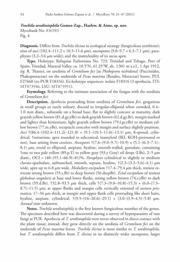
Trochila urediniophila Gomez-Zap., Haelew. & Aime, sp. nov.Mycobank No: 836583Fig. 4
Diagnosis. Differs from Trochila ilicina in ecological strategy (fungicolous symbiont); sizes of asci (102.4–111.2 × 10.5–11.6 µm), ascospores (9.0–9.7 × 6.3–7.1 µm), para-physes (3.2–3.6 µm wide); and the inamyloidity of its ascus apex.
Type. Holotype: Reliquiae Farlowiana No. 723; Trinidad and Tobago, Port of Spain, Trinidad, Maraval Valley, ca. 10.5°N, 61.25°W, alt. ±301 m a.s.l., 1 Apr 1912, leg. R. Thaxter, on uredinia of Cerotelium fici [as Phakopsora nishidana] (Pucciniales, Phakopsoraceae) on the underside of Ficus maxima (Rosales, Moraceae) leaves, PUL F27668 (ex-PUR F18316). Ex-holotype sequences: isolate F18316 (3 apothecia, ITS: MT873946, LSU: MT873951).
Etymology. Referring to the intimate association of the fungus with the uredinia of Cerotelium fici.
Description. Apothecia protruding from uredinia of Cerotelium fici, gregarious in small groups or rarely solitary, discoid to irregular-ellipsoid when crowded, 0.4–1.0 mm diam., subsessile on a broad base, flat to slightly concave at maturity, dark grayish yellow brown (81.d.gy.yBr) to dark grayish brown (62.d.gy.Br), margin marked and lighter than hymenium, light grayish yellow brown (79.l.gr.yBr) to medium yel-low brown (77.m.yBr), receptacle concolor with margin and surface slightly pruinose. Asci †(86.4–)102.4–111.2(–121.8) × (9.1–)10.5–11.6(–13.1) µm, 8-spored, cylin-drical, †uniseriate; apex rounded to subconical, inamyloid (IKI, KOH-pretreated or not), base arising from croziers. Ascospores †(7.6–)9.0–9.7(–10.9) × (5.1–)6.3–7.1(–8.1) µm, ovoid to ellipsoid, aseptate, hyaline, smooth-walled, guttulate, containing †one to two pale yellow (89.p.Y) to yellow gray (93.y Gray) oil drops (LBs), 2–5 µm diam., OCI = (40–)55.1–66.9(–81)%. Paraphyses cylindrical to slightly or medium clavate-spathulate, unbranched, smooth, septate, hyaline, †(2.3–)3.2–3.6(–4.1) µm wide, apex up to 6.8 µm wide. Medullary excipulum †17.4–79.4 µm thick, textura in-tricata strong brown (55.s.Br) to deep brown (56.deepBr). Ectal excipulum of textura globulosa–angularis at base and lower flanks, strong yellow brown (74.s.yBr) to dark brown (59.d.Br), †32.8–93.5 µm thick, cells †(7.3–)9.0–10.8(–15.3) × (6.0–)7.5–8.7(–11.5) µm; at upper flanks and margin cells vertically oriented of textura pris-matica, 17–34 µm thick, at margin and upper flank cells protruding like short hairs, hyaline, aseptate, cylindrical, †(9.5–)16–20.6(–29.1) × (3.0–)3.9–4.5(–5.8) µm. Asexual state unknown.
Notes. Trochila urediniophila is the first known fungicolous member of the genus. The specimen described here was discovered during a survey of hyperparasites of rust fungi at PUR. Apothecia of T. urediniophila were never observed in direct contact with the plant tissue; instead, they grew directly on the uredinia of Cerotelium fici on the underside of Ficus maxima leaves. Trochila ilicina is most similar to T. urediniophila, but T. urediniophila differs from T. ilicina in its distinctly wider ascospores, larger
Notes on Trochila 35
Figure 4. Morphological features of Trochila urediniophila, holotype collection (PUL F27668) a1–a4 dried apothecia growing on uredinia of Cerotelium fici a2, a3 substrate (uredinia) on which the ap-othecia grow (arrows) b1 transverse section of apothecia; arrow pointing out the substrate b2, b3 details of excipulum at margin and upper flanks b4 cells at base c1–c3 asci d1 paraphyses e1–e3 ascospores e2, e3 oil drops (LBs) inside ascospores. Mounted in: Congo Red (c1, e2), H2O (b2, c3, d1, e1, e3), KOH (b1, b3, b4, c2). Scale bars: 1 mm (a1–a3); 500 µm (a4); 200 µm (b1); 50 µm (b2); 20 µm (b3, b4, c2, c3, d1); 2 µm (c1, e1–e3).

Paula Andrea Gómez-Zapata et al. / MycoKeys 78: 21–47 (2021)36
asci, inamyloid ascus apex, and wider apex of the paraphyses. The uredinia of the host fungus, C. fici, become a solidified mass that changes in color from dark orange yel-low (72.d.OY) without apothecia of Trochila to brownish black (65.brBlack) where apothecia are present.
A second duplicate of the Reliquiae Farlowiana No. 723 is also deposited at PUR (accession PUR F1098). However, no apothecia were present on this specimen, nor could additional specimens of T. urediniophila be found on any of the other specimens of C. fici housed at PUR. At least eight other duplicates are housed at BPI, CINC, CUP, F, ISC, MICH, and UC (MyCoPortal 2020). It is unknown whether any of them may host T. urediniophila.
New combinations
Trochila colensoi (Berk.) Quijada, comb. nov.Mycobank No: 836591
≡ Cenangium colensoi Berk., Hooker, Bot. Antarct. Voy. Erebus Terror 1839–1843, II, Fl. Nov.-Zeal.: 201 (1855). [Basionym]
= Pseudopeziza colensoi (Berk.) Massee, J. Linn. Soc., Bot. 31: 468 (1896)
Notes. Cenangium colensoi is described from dead leaves of Cordyline sp. (Aspara-gales, Asparagaceae) in New Zealand (Hooker 1855). The host had been mistakenly reported as Phormium (Asparagales, Asphodelaceae) by Berkeley in Hooker (1855) and only recently corrected after re-study of the type collection (Landcare Research 2020). Cenangium colensoi was later combined in Pseudopeziza and described in more detail by Massee (1896). Both authors commented on the watery-grey disc and brownish receptacle of the apothecia. The apothecia develop among the rigid vascular bundles of the epidermis, first covered by the cuticle, then erumpent and opening by a narrow slit, becoming discoid when mature (Hooker 1855; Massee 1896). The habit of this fungus fits well with typical macromorphological features of the genus Trochila – a dark brown to black receptacle, which develops beneath the host tissues and eventually becomes erumpent to expose the hymenium by split-ting along radial lines or by its splitting into lobes (von Höhnel 1917; Greenhalgh and Morgan-Jones 1964; Dennis 1978; Baral and Marson 2005). Microscopically, P. colensoi was described with a parenchymatous excipulum (angular-globose or iso-diametric cells), hyaline under the hymenium and dark brown at the cortex (Berke-ley in Hooker 1855; Massee 1896), which is also in agreement with the excipular features of Trochila species. Finally, the hymenium of P. colensoi was described as composed of inamyloid, 8-spored asci with elliptical hyaline ascospores and slender paraphyses (op. cit.).
In 2018, P.R. Johnston collected two specimens (PDD:112240, PDD:112242, Landcare Research 2020) on leaves of Cordyline australis (Asparagaceae). The
Notes on Trochila 37
morphology, ecology (host), and locality of these new collections agree with P. colen-soi. The photographs of both specimens reveal features such as guttules in ascospores and paraphyses, protruding hyaline cells in the cortical layer of the upper flank and margin, and hyaline gelatinized hyphae covering the dark globose-angular cells of the ectal excipulum at the base and lower flanks. The latter excipular feature of the recepta-cle is reminiscent of Zhuang’s (1990) description of Calycellinopsis xishuangbanna. An ITS sequence of this species was generated from the recent material (PDD:112240) and included in the Leotiomycetes-wide ITS phylogeny of Johnston et al. (2019). Their results and those in this study (Figs 1, 2) show that P. colensoi is placed among species of Trochila.
Trochila xishuangbanna (W.Y. Zhuang) Quijada, comb. nov.Mycobank No: 836592
≡ Calycellinopsis xishuangbanna W.Y. Zhuang, Mycotaxon 38: 121 (1990). [Basionym]
Notes. The genus Calycellinopsis was proposed with a single species, C. xishuangbanna, which is a petiole-inhabiting fungus (Zhuang 1990). The genus was placed in Dermateaceae because of its isodiametric dark brownish excipular cells (Zhuang 1990). In 2002, a second collection of the same species was sampled (HMAS:187063), which was sequenced (Zhuang et al. 2010). Additional morphological details were provided, and the genus was placed in Helotiaceae (Zhuang et al. 2010). Trochila was treated in Dermateaceae until recently because of its excipular features (Fuckel 1869; Karsten 1869; Saccardo 1884; Lambotte 1888; Lumbsch and Huhndorf 2010). Collections of Calycellinopsis have a well-developed excipulum, with an outer layer of angular to isodiametric cells with brownish walls and cortical cells at flanks and margin with pro-truding hyaline cells. The medullary excipulum is subhyaline and composed of textura angularis to textura intricata (Zhuang 1990; Zhuang et al. 2010).
Species in Trochila usually have a poorly developed excipulum. For example, T. bostonensis and T. craterium produce only a thin layer of globose to angular dark excipular cells (von Höhnel 1917; Greenhalgh and Morgan-Jones 1964; Baral and Marson 2005). However, other species, such as T. laurocerasi and T. urediniophila, have a well-developed excipulum (op. cit.). The excipulum of Calycellinopsis is very similar to those species of Trochila with a well-developed excipulum, composed of an outer layer of dark textura globulosa–angularis and an inner layer of hyaline medulla made of textura angularis–porrecta–intricata. At the flanks and margin of the excipu-lum, Calycellinopsis has protruding hyaline cells similar to Trochila species with a well-developed excipulum (Fig. 4). Although limited details about the living features can be obtained from the original description of Calycellinopsis, its hymenial features are consistent with Trochila. The ascospores of Calycellinopsis are described with several guttules, a feature that is also observed in species of Trochila.
Paula Andrea Gómez-Zapata et al. / MycoKeys 78: 21–47 (2021)38
Discussion
Taxonomy of Trochila
This study represents the first attempt to investigate the systematics of Trochila us-ing both morphological features and DNA sequences. We have added four species to Trochila, bringing the total number of species described in the genus to 37. Most Tro-chila species have been delimited based on the size of asci and ascospores, but we find that amyloidity of ascus apex, excipular features, details of the paraphyses, and pres-ence vs. absence of guttules are also diagnostic (Table 2). For this study, we also ap-plied a two-dataset approach for phylogenetic analyses (e.g., Aime and Phillips-Mora 2005; Haelewaters et al. 2019). Our phylogenetic reconstruction of a six-locus dataset resolved Trochila as polyphyletic with respect to C. xishuangbanna and P. colensoi (Fig. 1). Because morphological data of these two taxa agree with Trochila, we recombined them in this genus. The second, two-locus dataset was used for species delimitation, which showed T. bostonensis and T. urediniophila as distinct from the other Trochila species. Our molecular phylogenetic results (Figs 1, 2) and morphological compari-sons of Trochila species (Table 2) will facilitate future taxonomic studies in the genus.
Host associations
Thus far, members of Trochila have been reported from 31 families of both monocots and dicots (Table 2). In this study, we add two plant family hosts, Apocynaceae (for T. bostonensis) and Asparagaceae (for T. colensoi). In addition, we reveal a new ecological niche (for T. urediniophila) – a species that associates with uredinia of the rust species Cerotelium fici. This sample was collected in 1912 as a rust specimen and deposited in the Arthur Fungarium (PUR) at Purdue University. More than a century later, the ex-siccatae sample was scanned for the presence of hyperparasites of rust fungi from South America. Apothecia of T. urediniophila were found exclusively on uredinia without any direct contact with the host plant. Due to the age and limited available material, ultrastructural examinations of the interaction between these two fungi could not be made. However, T. urediniophila is the first species in the genus that fruits exclusively from another fungus, hinting at more complex associations among Trochila species and other fungi on which they might act as mycoparasites.
Trochila in the Neotropics
South America is known to be one of the most biodiverse continents in the world (Dourojeanni 1990; Hawksworth 2001). However, its fungal communities are thought to be severely understudied (Mueller and Schmit 2007). Members of Trochila are no exception to this. Six species of Trochila have been described from South America. These are T. chilensis Speg., T. jaffuelii Speg., and T. perseae Speg. from Chile; T. leopoldina Rehm from Brazil; and T. tetraspora, and T. winteri Speg. from Argentina (Spegazzini 1888, 1910, 1921; Rehm 1909; Gamundí et al. 1978). Their type collections need to be

Notes on Trochila 39
re-examined to determine if these species are in fact members of Trochila. One of our new species, T. urediniophila, was collected in Port of Spain, Trinidad. Little data are available regarding the Funga (sensu Kuhar et al. 2018) of Trinidad and Tobago (Baker and Dale 1951; Dennis 1954a, b). The most recent work on the fungal diversity from this country was published online (Jodhan and Minter 2006) derived from reference collections and data from scientific literature. Based on the available literature, no records of Trochila are known in Trinidad. As a result, T. urediniophila represents the first published report of the genus from Trinidad, and by extension from the Caribbean (Minter et al. 2001).
Trochila species are likely more broadly distributed than generally thought, and certainly not limited to the Northern Hemisphere. This is often the case for many fungi that are based on limited regional collecting and thus may not represent the full extent of their distributional ranges due to, for example, the lack of studies in sub-tropical and tropical ecosystems (Groombridge 1992; Hawksworth and Mueller 2005; Mueller and Schmit 2007; Aime and Brearley 2012; Cheek et al. 2020).
The importance of biological collections
Our work emphasizes the importance of specimens preserved in biological collections – such as fungaria and herbaria – for studies of biodiversity and applied biological sci-ences, and for climate change research (Hawksworth and Lücking 2017; Andrew et al. 2019; Lang et al. 2019; Ristaino 2020; Wijayawardene et al. 2020). Because of the well-preserved specimens deposited at PUR, the genus Trochila is now known to be present in Trinidad and to form fungicolous associations. Another interesting example of the use of collections is Trochila colensoi. Known only from the type specimen for more than 100 years, additional specimens were only reported following the correc-tion of the host substrate (as Cordyline rather than Phormium), which was based on re-examination of the type specimen preserved at K. Biological collections are not only important for morphological studies, but also as sources of genetic and genomic infor-mation (Bruns et al. 1990; Brock et al. 2009; Redchenko et al. 2012; Dentinger et al. 2016; this study). The single-oldest fungal specimen used for DNA extraction and se-quencing was the type of Hygrophorus cossus (Sowerby) Fr. (Agaricales, Hygrophorace-ae), collected in 1794 and deposited at K (Larsson and Jacobsson 2004). Our material of T. urediniophila gathered by Roland Thaxter in 1912 proves again that old samples can be used successfully for modern molecular phylogenetic analyses.
Acknowledgements
The National Park Service at the Boston Harbor Islands (BHI) National Recreation Area and the University of Massachusetts – Boston School for the Environment are acknowledged for facilitating the fungal ATBI. The National Park Service issued the scientific research and collecting permits (#BOHA-2012-SCI-0009, PI B.D. Farrell; #BOHA-2018-SCI-0002, PI D. Haelewaters). Thanks are due to: Marc Albert (Bos-ton Harbor Islands Stewardship Program) for immense support with everything that is
Paula Andrea Gómez-Zapata et al. / MycoKeys 78: 21–47 (2021)40
Boston Harbor Islands-related; Russ Bowles and his staff (Division of Marine Opera-tions, University of Massachusetts Boston) for expert navigation and transportation to Great Brewster Island; Peter R. Johnston (Manaaki Whenua Landcare Research) for providing important information about Pseudopeziza colensoi and for improvements to the manuscript. D. Haelewaters acknowledges support for fieldwork at the BHI and molecular work from Boston Harbor Now (2017–2018) and the New England Botanical Club (2017 Les Mehrhoff Botanical Research Award). L. Quijada thanks the support of the Farlow Fellowship, the Department of Organismic and Evolutionary Biology at Harvard University, and the Harvard University Herbaria. This work was supported in part by the U.S. National Science Foundation (DEB-2018098 to D. Haelewaters; DEB-1458290 to M.C. Aime) and the U.S. Department of Agriculture (National Institute of Food and Agriculture Hatch project 1010662 to M.C. Aime).
References
Aime MC, Brearley FQ (2012) Tropical fungal diversity: closing the gap between species es-timates and species discovery. Biodiversity and Conservation 21: 2177–2180. https://doi.org/10.1007/s10531-012-0338-7
Aime MC, Phillips-Mora W (2005) The causal agents of witches’ broom and frosty pod rot of cacao (chocolate, Theobroma cacao) form a new lineage of Marasmiaceae. Mycologia 97: 1012–1022. https://doi.org/10.3852/mycologia.97.5.1012
Andrew C, Diez J, James TY, Kauserud H (2019) Fungarium specimens: a largely untapped source in global change biology and beyond. Philosophical Transactions of the Royal Soci-ety B 374(1763): 20170392. https://doi.org/10.1098/rstb.2017.0392
Baker RED, Dale WT (1951) Fungi of Trinidad and Tobago. Mycological Papers 33: 1–123.Baral H-O (1992) Vital versus herbarium taxonomy: morphological differences between living
and dead cells of ascomycetes, and their taxonomic implications. Mycotaxon 44: 333–390.Baral H-O, Marson G (2005) In vivo veritas. Over 10000 images of fungi and plants (micro-
scopical drawings, water colour plates, photo macro- & microphotographs), with materials on vital taxonomy and xerotolerance. Ed. 3. Privately distributed DVD-ROM.
Brock PM, Döring H, Bidartondo MI (2009) How to know unknown fungi: The role of a herbar-ium. New Phytologist 181: 719–724. https://doi.org/10.1111/j.1469-8137.2008.02703.x
Bruns TD, Fogel R, Taylor JW (1990) Amplification and sequencing of DNA from fungal herbarium specimens. Mycologia 82: 175–184. https://doi.org/10.2307/3759846
Cheek M, Lughadha EN, Kirk P, Lindon H, Carretero J, Looney B, Douglas B, Haelewaters D, Gaya E, Llewellyn T, Ainsworth M, Gafforov Y, Hyde K, Crous P, Hughes M, Walker BE, Forzza RC, Meng WK, Niskanen T (2020) New scientific discoveries: Plants and fungi. Plants, People, Planet 2(5): 371–388. https://doi.org/10.1002/ppp3.10148
Chernomor O, Von Haeseler A, Minh BQ (2016) Terrace aware data structure for phylogenomic inference from supermatrices. Systematic Biology 65: 997–1008. https://doi.org/10.1093/sysbio/syw037
Clements FE, Shear CL (1931) The genera of fungi. H.W. Wilson Company, Bronx, New York, 496 pp.
Notes on Trochila 41
Crouan PL, Crouan MH (1867) Florule du Finistère. Contenant les descriptions de 360 espèces nouvelles de sporogames, de nombreuses observations et une synonymie des plantes cellu-laires et vasculaires qui croissant spontanément dans ce département. Friedrich Klincksieck and J.B. et A. Lefournier, Paris & Brest, 262 pp. https://doi.org/10.5962/bhl.title.11601
Crous PW, Quaedvlieg W, Hansen K, Hawksworth DL, Groenewald JZ (2014) Phacidium and Ceuthospora (Phacidiaceae) are congeneric: taxonomic and nomenclatural implications. IMA Fungus 5: 173–193. https://doi.org/10.5598/imafungus.2014.05.02.02
Crous PW, Schumacher RK, Wingfield MJ, Akulov A, Denman S, Roux J, Braun U, Burgess TI, Carnegie AJ, Váczy KZ, Guatimosim E, Schwartsburd PB, Barreto RW, Hernández-Restrepo M, Lombard L, Groenewald JZ (2018) New and Interesting Fungi. 1. Fungal Systematics and Evolution 1: 169–215. https://doi.org/10.3114/fuse.2018.01.08
De Notaris G (1863) [printed 1864] Proposte di alcune rettificazioni al profilo dei Discomiceti. Commentario della Società Crittogamologica Italiana 1(5): 357–388.
Dennis RWG (1954a) Operculate Discomycetes from Trinidad and Jamaica. Kew Bulletin 9(3): 417–421. https://doi.org/10.2307/4108810
Dennis RWG (1954b) Some Inoperculate Discomycetes of Tropical America. Kew Bulletin 9(2): 289–348. https://doi.org/10.2307/4114399
Dennis RWG (1961) Some Inoperculate Discomycetes from New Zealand. Kew Bulletin 15(2): 293–320. https://doi.org/10.2307/4109373
Dennis RWG (1978) British Ascomycetes. J. Cramer, Vaduz, Liechtenstein, 585 pp.Dentinger BTM, Gaya E, O’Brien H, Suz LM, Lachlan R, Diaz-Valderrama JR, Koch RA,
Aime MC (2016) Tales from the crypt: genome mining from fungarium specimens im-proves resolution of the mushroom tree of life. Biological Journal of the Linnean Society 117: 11–32. https://doi.org/10.1111/bij.12553
Don RH, Cox PT, Wainwright BJ, Baker K, Mattick JS (1991) ‘Touchdown’ PCR to circumvent spurious priming during gene amplification. Nucleic Acids Research 19(14): 4008–4008. https://doi.org/10.1093/nar/19.14.4008
Dourojeanni MJ (1990) Entomology and biodiversity conservation in Latin America. Ameri-can Entomologist 36: 88–93. https://doi.org/10.1093/ae/36.2.88
Edgar RC (2004) MUSCLE: multiple sequence alignment with high accuracy and high throughput. Nucleic Acids Research 32: 1792–1797. https://doi.org/10.1093/nar/gkh340
Egger KN (1995) Molecular analysis of ectomycorrhizal fungal communities. Canadian Jour-nal of Botany 73: S1415–S1422. https://doi.org/10.1139/b95-405
Etayo J, Flakus A, Suija A, Kukwa M (2015) Macroskyttea parmotrematis gen. et sp. nov. (Helo-tiales, Leotiomycetes, Ascomycota), a new lichenicolous fungus from Bolivia. Phytotaxa 224: 247–257. https://doi.org/10.11646/phytotaxa.224.3.3
Feltgen J (1903) Vorstudien zu einer Pilz-flora des Grossherzogthums Luxemburg. I. Theil – Ascomycetes. Nachträge III. Recueil des mémoires et des travaux publiés par la Société de botanique du grand-duché de Luxembourg 16: 3–328.
Fries E (1849) Summa Vegetabilium Scandinaviae. Sectio Posterior. Cl. XX. Fungi. A. Bonnier, Stockholm & Leipzig, 261–572.
Fryar SC, Haelewaters D, Catcheside DE (2019) Annabella australiensis gen. & sp. nov. (Heloti-ales, Cordieritidaceae) from South Australian mangroves. Mycological Progress 18: 973–981. https://doi.org/10.1007/s11557-019-01499-x

Paula Andrea Gómez-Zapata et al. / MycoKeys 78: 21–47 (2021)42
Fuckel L (1869) Symbolae mycologicae. Beiträge zur Kenntniss der rheinischen Pilze. Mit VI lithographirten und colorirten Tafeln. Jahrbücher des Nassauischen Vereins für Naturkunde 23–24: 1–459. https://doi.org/10.5962/bhl.title.47117
Gamundí, IJ, Arambarri, AM, Giaiotti A (1978) Micoflora de la hojarasca de Nothofagus dombeyi. Darwiniana 21: 81–114.
Gardes M, Bruns TD (1993) ITS primers with enhanced specificity for basidiomycetes – ap-plication to the identification of mycorrhizae and rusts. Molecular Ecology 2: 113–118. https://doi.org/10.1111/j.1365-294X.1993.tb00005.x
Global Biodiversity Information Facility (2020) Trochila Fr. GBIF Backbone Taxonomy. https://www.gbif.org/species/2575690 [accessed 17 August 2020]
Greenhalgh G, Morgan-Jones G (1964). Some species of Trochila and an undescribed discomycete on leaves of Prunus laurocerasus. Transactions of the British Mycological Society 47: 311–320. https://doi.org/10.1016/S0007-1536(64)80002-4
Gregor MJF (1936) A disease of cherry laurel caused by Trochila laurocerasi (Desm.) Fr. Annals of Applied Biology 23: 700–704. https://doi.org/10.1111/j.1744-7348.1936.tb06121.x
Grelet L-J, de Crozals A (1928) Discomycètes nouveaux (3ième série). Bulletin trimestriel de la Société mycologique de France 44: 336–340.
Haelewaters D, Dirks AC, Kappler LA, Mitchell JK, Quijada L, Vandegrift R, Buyck B, Pfister DH (2018a) A preliminary checklist of fungi at the Boston Harbor islands. Northeastern Naturalist 25(sp9): 45–77. https://doi.org/10.1656/045.025.s904
Haelewaters D, De Kesel A, Pfister DH (2018b) Integrative taxonomy reveals hidden species within a common fungal parasite of ladybirds. Scientific Reports 8: e15966. https://doi.org/10.1038/s41598-018-34319-5
Haelewaters D, Pfliegler WP, Gorczak M, Pfister DH (2019) Birth of an order: comprehensive molecular phylogenetic study reveals that Herpomyces (Fungi, Laboulbeniomycetes) is not part of Laboulbeniales. Molecular Phylogenetics and Evolution 133: 286–301. https://doi.org/10.1016/j.ympev.2019.01.007
Hammond PM (1992) Species inventory. In: Groombrigde B (Ed.) Global biodiversity, status of the earth’s living resources. Chapman & Hall, London, 17–39. https://doi.org/10.1007/978-94-011-2282-5_4
Hawksworth DL (2001) The magnitude of fungal diversity: the 1.5 million species estimate revisit-ed. Mycological Research 105: 1422–1432. https://doi.org/10.1017/S0953756201004725
Hawksworth DL, Lücking R (2017) Fungal diversity revisited: 2.2 to 3.8 million species. In: Heit-man J, Howlett B, Crous P, Stukenbrock E, James T, Gow N (Eds) The Fungal Kingdom. ASM Press, Washington, 79–95. https://doi.org/10.1128/microbiolspec.FUNK-0052-2016
Hawksworth DL, Mueller GM (2005) Fungal communities: their diversity and distribution. In: Digthon J, White JF, Oudemans P (Eds) The fungal community: its organisation and role in the ecosystem. CRC Press, Boca Raton, 27–37. https://doi.org/10.1201/9781420027891.ch2
Hennings P (1900) Fungi japonici. Botanische Jahrbücher fur Systematik, Pflanzengeschichte und Pflanzengeographie 28: 273–280.
Hoang DT, Chernomor O, Von Haeseler A, Minh BQ, Vinh LS (2017) UFBoot2: improving the ultrafast bootstrap approximation. Molecular Biology and Evolution 35: 518–522. https://doi.org/10.1093/molbev/msx281
Höhnel F von (1917) Über die Gattung Trochila Fries. Annales Mycologici 15: 330–334.

Notes on Trochila 43
Hooker JD (1855) The botany of the Antarctic Voyage of H.M. discovery ships Erebus and Ter-ror, in the years 1839–1843. II. Flora Novae-Zealandiae. Part II. Flowerless plants. Lovell Reeve, London, 378 pp.
Hopple JS (1994) Phylogenetic investigations in the genus Coprinus based on morphological and molecular characters. PhD Dissertation, Duke University, Durham.
Hyde KD, McKenzie EHC, KoKo TW (2011) Towards incorporating anamorphic fungi in a natural classification – checklist and notes for 2010. Mycosphere 2: 1–88.
Index Fungorum (2021) Index Fungorum. http://www.indexfungorum.org/names/Names.asp [accessed 22 June 2020]
Jaklitsch W, Baral H-O, Lücking R, Lumbsch HT (2016) Syllabus of plant families. In: Frey W (Ed.) Adolf Engler’s Syllabus der Pflanzenfamilien. Part 1/2 Ascomycota (13th edn). Borntraeger Science Publishers, Stuttgart, 322 pp.
Jodhan D, Minter DW (2006) Fungi of Trinidad & Tobago. http://www.cybertruffle.org.uk/trinfung [accessed 14 November 2019]
Johnston PR, Quijada L, Smith CA, Baral H-O, Hosoya T, Baschien C, Pärtel K, Zhuang K-Y, Haelewaters D, Park D, Carl S, López-Giráldez F, Wang Z, Townsend JP (2019) A mul-tigene phylogeny toward a new phylogenetic classification of Leotiomycetes. IMA Fungus 10: 1–22. https://doi.org/10.1186/s43008-019-0002-x
Kalyaanamoorthy K, Minh BQ, Wong TKF, von Haeseler A, Jermiin LS (2017) ModelFinder: Fast model selection for accurate phylogenetic estimates. Nature Methods 14: 587–589. https://doi.org/10.1038/nmeth.4285
Karsten PA (1869) Monographia Pezizarum fennicarum. Notiser ur Sällskapets pro Fauna et Flora fennica förhandlingar X: 99–206.
Karsten PA (1871) Mycologia Fennica I. Discomycetes. Bidrag Kännedom Finland Natur Folk 19: 1–263.
Kelly KL (1965) ISCC-NBS Colour-name charts illustrated with centroid colors. Inter-Society Colors Council. National Bureau of Standards, Circular 553 (Supplement). US Govern-ment Printing Office, Washington, 44 pp.
Kiffer E, Morelet M (2000) The Deuteromycetes, mitosporic fungi: classification and generic key. Science Publishers, Enfield, 273 pp.
Kirschstein W (1941) De plerisque novis ascomycetibus et paucis novis fungis imperfectis. Hedwigia 80: 119–137.
Kirschstein W (1944) Über neue, seltene und kritische Kleinpilze. Hedwigia 81: 193–224.Korf RP (1973) Discomycetes and Tuberales. In: Ainsworth GC, Sparrow FK, Sussman AS
(Eds) The Fungi: An Advanced Treatise. Vol. 4a. Academic Press, London, 249–319.Kuhar F, Furci G, Drechsler-Santos ER, Pfister DH (2018) Delimitation of Funga as a valid
term for the diversity of fungal communities: the Fauna, Flora & Funga proposal (FF&F). IMA Fungus 9: 71–74. https://doi.org/10.1007/BF03449441
Kumar S, Stecher G, Tamura K (2016) MEGA7: Molecular Evolutionary Genetics Analysis version 7.0 for bigger datasets. Molecular Biology and Evolution 33: 1870–1874. https://doi.org/10.1093/molbev/msw054
Lamarck JBPA, de Candolle AP (1805) Flore française, ii. Desray, Paris, 600 pp.Lambotte E (1888) La flore mycologique de la Belgique. Mémoires de la Société royale des sci-
ences de Liège, sér. 2, 14: 1–350.
Paula Andrea Gómez-Zapata et al. / MycoKeys 78: 21–47 (2021)44
Landcare Research (2020) Collection details. Pseudopeziza colensoi (Berk.) Massee (1896) [1895–97]. https://nzfungi2.landcareresearch.co.nz/default.aspx?selected=NameDetails&TabNum=0&NameId=1CB1B798-36B9-11D5-9548-00D0592D548C [accessed 16 August 2020]
Lang PL, Willems FM, Scheepens JF, Burbano HA, Bossdorf O (2019) Using herbaria to study glob-al environmental change. New Phytologist 221: 110–122. https://doi.org/10.1111/nph.15401
Larsson E, Jacobsson S (2004) Controversy over Hygrophorus cossus settled using ITS sequence data from 200 year-old type material. Mycological Research 108: 781–786. https://doi.org/10.1017/S0953756204000310
Liu YJ, Whelen S, Hall BD (1999) Phylogenetic relationships among ascomycetes: Evidence from an RNA polymerase II subunit. Molecular Biology and Evolution 16: 1799–1808. https://doi.org/10.1093/oxfordjournals.molbev.a026092
Lumbsch HT, Huhndorf SM (2010) Myconet Volume 14. Part One. Outline of Ascomycota – 2009. Part Two. Notes on Ascomycete Systematics. Nos. 4751–5113. Fieldiana Life and Earth Sciences 2010: 1–64. https://doi.org/10.3158/1557.1
Massee GE (1896) Redescriptions of Berkeley’s types of fungi. Journal of the Linnean Society 31: 463–524. https://doi.org/10.1111/j.1095-8339.1896.tb00812.x
Miller MA, Pfeiffer W, Schwartz T (2010) Creating the CIPRES Science Gateway for inference of large phylogenetic trees. In: Proceedings of the Gateway Computing Environments Work-shop, 14 Nov. 2010, New Orleans, LA, 1–8. https://doi.org/10.1109/GCE.2010.5676129
Minter DW, Rodríguez-Hernández M, Mena-Portales J (2001) Fungi of the Caribbean. An annotated checklist. PDMS Publishing, Isleworth, 950 pp.
Morgan-Jones G (1973) Genera coelomycetarum. VII. Cryptocline Petrak. Canadian Journal of Botany 51: 309–325. https://doi.org/10.1139/b73-039
Mouton V (1900) Quatrième notice sur des ascomycètes nouveaux ou peu connus. Bulletin de la Société Royale de Botanique de Belgique 39: 37–53.
Mueller GM, Schmit JP (2007) Fungal biodiversity: what do we know? What can we predict? Biodiversity and Conservation 16: 1–5. https://doi.org/10.1007/s10531-006-9117-7
MyCoPortal (2020) Mycology Collections data Portal. http://mycoportal.org/portal/index.php [accessed 7 December 2020]
Nguyen L-T, Schmidt HA, Von Haeseler A, Minh BQ (2015) IQ-TREE: A fast and effective stochastic algorithm for estimating maximum likelihood phylogenies. Molecular Biology and Evolution 32: 268–274. https://doi.org/10.1093/molbev/msu300
Pärtel K, Baral H-O, Tamm H, Põldmaa K (2017) Evidence for the polyphyly of Encoelia and Encoelioideae with reconsideration of respective families in Leotiomycetes. Fungal Diver-sity 82: 183–219. https://doi.org/10.1007/s13225-016-0370-0
Patouillard MN (1886) Quelques champignons de la Chine, récoltés par M. l’abbé Delavay dans la province du Yunnan. Revue Mycologique 8: 179–194.
Redchenko O, Vondrák J, Košnar J (2012) The oldest sequenced fungal herbarium sample. The Lichenologist 44: 715–718. https://doi.org/10.1017/S002428291200031X
Rehm H (1896) Abt. 3. Ascomyceten: hysteriaceen und discomyceten. In: Rabenhorst L (Ed.) Kryptogamen-Flora von Deutschland, Oesterreich und der Schweiz. Verlag von Eduard Kummer, Leipzig.
Rehm H-J (1909) Ascomycetes exs. Fasc. 45. Annales Mycologici 7: 524–530.

Notes on Trochila 45
Rehner SA, Buckley E (2005) A Beauveria phylogeny inferred from nuclear ITS and EF1-α se-quences: evidence for cryptic diversification and links to Cordyceps teleomorphs. Mycologia 97: 84–98. https://doi.org/10.3852/mycologia.97.1.84
Ristaino JB (2020) The importance of mycological and plant herbaria in tracking plant killers. Frontiers in Ecology and Evolution 7: e521. https://doi.org/10.3389/fevo.2019.00521
Rostrup E (1885) Islands svampe. Botanisk Tidsskrifte 14: 218–229.Rostrup E (1886) Svampen fra Finmarken. Botanisk Tidsskrifte 15: 229–236.Rostrup E (1888) Oversigt over Grönlands svampe. Meddelelser om Grønland 3: 516–590.Rostrup E (1891) Tillaeg til “Grönlands svampe (1888)”. Meddelelser om Grønland 3: 591–643.Saccardo PA (1878) Fungi Veneti novi vel critici. Series IX. Michelia 4: 361–434.Saccardo PA (1884) Conspectus generum discomycetum hucusque cognitorum. Botanisches
Centralblatt 18: 247–256.Spatafora JW, Sung GH, Johnson D, Hesse C, O’Rourke B, Serdani M, Spotts R, Lutzoni
F, Hofstetter V, Miadlikowska J, Reeb V, Gueidan C, Fraker E, Lumbsch T, Lücking R, Schmitt I, Hosaka K, Aptroot A, Roux C, Miller AN, Geiser DM, Hafellner J, Hestmark G, Arnold AE, Büdel B, Rauhut A, Hewitt D, Untereiner WA, Cole MS, Scheidegger C, Schultz M, Sipman H, Schoch CL (2006) A five-gene phylogeny of Pezizomycotina. My-cologia 98: 1018–1028. https://doi.org/10.1080/15572536.2006.11832630
Spegazzini C (1888) Fungi Fuegiani. Boletin de la Academia Nacional de Ciencias en Córdoba 11: 135–311. https://doi.org/10.5962/bhl.title.4055
Spegazzini C (1910) Hongos chilenos. Revista de la Facultad de Agronomia y Veterinaria. 6: 130–132.
Spegazzini C (1921) Mycetes chilenses. Boletin de la Academia Nacional de Ciencias 25: 1–124.Stevenson JA (1926) Foreign plant diseases. A manual of economic plant diseases which are new
or not widely distributed in the United States. United States Department of Agriculture Federal Horticulture Board, Washington DC. https://doi.org/10.5962/bhl.title.114939
Stoykov DY, Assyov B (2009) The genus Trochila in Bulgaria. Mycotaxon 109: 351–359. https://doi.org/10.5248/109.351
Stoykov DY (2019) New records of Trochila (Cenangiaceae, Helotiales) from the Balkans. Phytologia Balcanica 25: 245–248.
Suija A, Ertz D, Lawrey JD, Diederich P (2015) Multiple origin of the lichenicolous life habit in Helotiales, based on nuclear ribosomal sequences. Fungal Diversity 70: 55–72. https://doi.org/10.1007/s13225-014-0287-4
Sung GH, Sung JM, Hywel-Jones NL, Spatafora JW (2007) A multi-gene phylogeny of Cla-vicipitaceae (Ascomycota, Fungi): Identification of localized incongruence using a com-binational bootstrap approach. Molecular Phylogenetics and Evolution 44: 1204–1223. https://doi.org/10.1016/j.ympev.2007.03.011
Thiers B (2020) [continuously updated] Index Herbariorum: A global directory of public herbaria and associated staff. New York Botanic Garden’s Virtual Herbarium. http://sweetgum.nybg.org/ih/ [accessed 14 July 2020]
Vilgalys R, Hester M (1990) Rapid genetic identification and mapping of enzymatically ampli-fied ribosomal DNA from several Cryptococcus species. Journal of Bacteriology 172: 4238–4246. https://doi.org/10.1128/JB.172.8.4238-4246.1990

Paula Andrea Gómez-Zapata et al. / MycoKeys 78: 21–47 (2021)46
von Arx JA (1957) Revision der zu Gloeosporium gestellten Pilze. Verhandelingen der Koninklijke Nederlandse Akademie van Wetenschappen, Afd. Natuurkunde, Tweede Reeks 51: 1–153.
Vu D, Groenewald M, de Vries M, Gehrmann T, Stielow B, Eberhardt U, Al-Hatmi A, Groe-newald JZ, Cardinali G, Houbraken J, Boekhout T, Crous PW, Robert V, Verkley GJM (2019) Large-scale generation and analysis of filamentous fungal DNA barcodes boosts cov-erage for kingdom fungi and reveals thresholds for fungal species and higher taxon delimi-tation. Studies in Mycology 92: 135–154. https://doi.org/10.1016/j.simyco.2018.05.001
Wang Z, Binder M, Hibbett DS (2005) Life history and systematics of the aquatic discomycete Mitrula (Helotiales, Ascomycota) based on cultural, morphological, and molecular studies. American Journal of Botany 92: 1565–1574. https://doi.org/10.3732/ajb.92.9.1565
White TJ, Bruns T, Lee S, Taylor J (1990) Amplification and direct sequencing of fungal ribo-somal RNA genes for phylogenetics. In: Innis MA, Gelfand H, Sninsky JS, White TJ (Eds) PCR protocols: A guide to methods and applications. Academic Press, San Diego, 315–322. https://doi.org/10.3181/00379727-150-39089
Wijayawardene NN, Hyde KD, Al-Ani LKT, Tedersoo L, Haelewaters D, Rajeshkumar KC, Zhao RL, Aptroot A, Leontyev DV, Saxena RK, Tokarev YS, Dai DQ, Letcher PM, Ste-phenson SL, Ertz D, Lumbsch HT, Kukwa M, Issi IV, Madrid H, Phillips AJL, Selbmann L, Pfliegler WP, Horváth E, Bensch K, Kirk PM, Kolaříková K, Raja HA, Radek R, Papp V, Dima B, Ma J, Malosso E, Takamatsu S, Rambold G, Gannibal PB, Triebel D, Gau-tam AK, Avasthi S, Suetrong S, Timdal E, Fryar SC, Delgado G, Réblová M, Doilom M, Dolatabadi S, Pawłowska J, Humber RA, Kodsueb R, Sánchez-Castro I, Goto BT, Silva DKA, de Souza FA, Oehl F, da Silva GA, Silva IR, Błaszkowski J, Jobim K, Maia LC, Barbosa FR, Fiuza PO, Divakar PK, Shenoy BD, Castañeda-Ruiz RF, Somrithipol S, La-teef AA, Karunarathna SC, Tibpromma S, Mortimer PE, Wanasinghe DN, Phookamsak R, Xu J, Wang Y, Tian F, Alvarado P, Li DW, Kušan I, Matočec N, Maharachchikumbura SSN, Papizadeh M, Heredia G, Wartchow F, Bakhshi M, Boehm E, Youssef N, Hustad VP, Lawrey JD, Santiago ALCMA, Bezerra JDP, Souza-Motta CM, Firmino AL, Tian Q, Houbraken J, Hongsanan S, Tanaka K, Dissanayake AJ, Monteiro JS, Grossart HP, Suija A, Weerakoon G, Etayo J, Tsurykau A, Vázquez V, Mungai P, Damm U, Li QR, Zhang H, Boonmee S, Lu YZ, Becerra AG, Kendrick B, Brearley FQ, Motiejūnaitė J, Sharma B, Khare R, Gaikwad S, Wijesundara DSA, Tang LZ, He MQ, Flakus A, Rodriguez-Flakus P, Zhurbenko MP, McKenzie EHC, Stadler M, Bhat DJ, Liu JK, Raza M, Jeewon R, Nassonova ES, Prieto M, Jayalal RGU, Erdoğdu M, Yurkov A, Schnittler M, Shchepin ON, Novozhilov YK, Silva-Filho AGS, Liu P, Cavender JC, Kang Y, Mohammad S, Zhang LF, Xu RF, Li YM, Dayarathne MC, Ekanayaka AH, Wen TC, Deng CY, Pereira OL, Navathe S, Hawksworth DL, Fan XL, Dissanayake LS, Kuhnert E, Grossart HP, Thines M (2020) Outline of Fungi and fungus-like taxa. Mycosphere 11: 1060–1456. https://doi.org/10.5943/mycosphere/11/1/8
Zhao YJ, Hosaka K, Hosoya T (2016) Taxonomic re-evaluation of the genus Lambertella (Rutstro-emiaceae, Helotiales) and allied stroma-forming fungi. Mycological Progress 15: 1215–1228. https://doi.org/10.1007/s11557-016-1225-5
Zhuang WY (1990) Calycellinopsis Xishuangbanna gen. et sp. nov. (Dermateaceae), a petiole-inhabiting fungus from China. Mycotaxon 38: 121–124.

Notes on Trochila 47
Zhuang WY, Luo J, Zhao P (2010) The fungal genus Calycellinopsis belongs in Helotiaceae not Dermateaceae. Phytotaxa 3: 54–58. https://doi.org/10.11646/phytotaxa.3.1.8
Ziolo E, Madej T, Blaszkowski J (2005) Trochila ilicina (Helotiales, Ascomycota), a fungus new-ly found in Poland. Acta Mycologica 40: 181–184. https://doi.org/10.5586/am.2005.016