novel nanoparticles for tuberculosis...
TRANSCRIPT

1
Novel nanoparticles for Tuberculosis chemotherapy
B SEMETE*, L KALOMBO, P CHELULE, Y BENADIE, L BOOYSEN, L KATATA, S NAIDOO, H SWAI *CSIR, Material Science and Manufacturing, Polymers and Bioceramics, PO Box 395, Pretoria, 0001
Email:
Abstract Current therapeutic management of tuberculosis is inadequate due to non-compliance, lengthy course of treatment and drug- related side effects. In order to address these setbacks, we are developing a nanotechnology drug delivery system whereby anti-TB drugs can be administered in a single dose that maintains an active level of the drug for at least a week. Poly(lactic-co-glycolic acid) (PLGA) was used to prepare a multiple nano-emulsion which was subsequently lyophilized via freeze drying or spray drying. The resulting nanoparticles were characterised based on size, zeta potential, morphology and drug encapsulation efficiency. Particle uptake was analyzed in CaCo-2 cells via confocal microscopy, as well as in vivo, by analyzing macrophages of peritoneum cells of mice. Nano-sized polymeric PLGA particles of size ranging from 200-300 nm (zeta potential ~16 mV) were synthesized, with a smooth spherical surface and a drug encapsulation efficiency of 50-65% for INH and RIF. It was observed from the in vitro release assays, that the drugs were released in a slow manner compared to free drugs. The particles were taken up in vitro by CaCo-2 cells as well as in vivo by macrophages of the peritoneum. The uptake of the particles by cells will enable improved bioavailability of the encapsulated drugs, in that intracellular drug release can be obtained. Due to the slow degradation, and thus slow release mechanism, of the carrier systems it is envisaged that using nano-based delivery systems can prolong drug release, thus minimising dose and dosage frequency of anti-TB drugs. 1. Introduction Tuberculosis (TB) is currently one of the leading causes of death in South African adults. Annually, approximately 250 000 new cases of TB occur and it is ranked 5th amongst the top 22 high-burden countries of the world, which collectively account for over 80% of all TB cases (WHO, 2007). A major reason for the escalation of TB is the evolution of the AIDS epidemic. It is estimated that approximately 70% of adult TB cases aged 15-49
years are HIV-infected and approximately 1.7% of new cases are multi-drug resistant (WHO 2007). Although an effective therapeutic regimen is available, patient non-compliance resulting in treatment failure, as well as the emergence of multi-drug resistant (MDR) and extremely drug resistant (XDR) strains of TB still pose a challenge (Centres for Disease Control, 2006). The first-line drugs that are currently administered to treat TB include rifampicin (RIF), isoniazid (INH), pyrazinamide (PYR) and ethambutol (ETB). The current doses administered are high compared to the required minimum inhibitory concentration (MIC) of the drugs (Smith 1999). This is because the drugs have poor bioavailability and low permeability, which is a function of degradation of drugs before reaching their target site (Smith 1999). Thus the drawbacks of conventional chemotherapy necessitate the development of a carrier system which can release drugs slowly over prolonged time periods and reduce the current costs of treatment. The paper focuses on the development of a nanotechnology-based drug delivery system for improved bioavailability and sustained release of anti-TB drugs. The application of nanotechnology in the health-care sector, also referred to as nanomedicine, has gained ground over the past 5 years. The field of drug development could potentially benefit greatly from nanomedicine in terms of addressing current shortfalls such as lack of specificity, toxicity of the therapeutic compounds, low solubility leading to lowered bioavailability and thus reduced efficacy (Langer, 2000). The nanometer size-ranges of delivery systems offer certain distinct advantages for drug delivery. Due to their sub-cellular and sub-micron size, nanoparticles can penetrate deep into tissues through fine capillaries, cross the fenestration present in the epithelial lining (e.g., liver), and are generally taken up efficiently by the cells. This allows efficient delivery of therapeutic agents to target sites in the body (Singh and Kim, 2000). The small size also facilitates intracellular and para-cellular absorption or uptake of the particles.
2
To date various hydrophilic surfactants such as polyethylene glycol (PEG) have been used to enhance the bioavailability, by increasing the mean residence time of the encapsulated drug through minimising opsonisation of the particles (Storm et al, 1995). Poloxamers, poloxamines and other polymers such as chitosan can also be used for this purpose. Furthermore, the reported ability to functionalise these polymeric nanoparticles with ligands such as antibodies, peptides, carbohydrates and DNA has enabled researchers to target specific diseased cells within the body, thus further increasing the specificity and efficacy of the therapeutic drugs (Farokhzad and Langer, 2006; Torchilin, 2006). Much of the advances in drug delivery have been primarily in cancer treatment, however various groups are working on anti-TB drug encapsulation. Dutt and Khuller (2001) formulated entrapped INH and RIF in PLGA microparticles for sustained drug delivery following subcutaneous administration. In vivo studies revealed sustained release of 6-7 weeks in mice, whereas a release of only 24hrs was observed for free drugs. Pandey et al (2003) also encapsulated RIF, INH and PZA in PLGA nanoparticles for oral delivery by the multiple emulsion technique. The nanoparticles were administered at every 10th day and after 5 oral doses of treatment, no tubercle bacilli could be detected in the tissues. This formulation achieved an encapsulation efficiency of 56.9% for RIF, 66.3% for INH and 68% for PZA The group has conducted various studies in anti-TB drug delivery with very positive data. In addition, Kisisch et al., 2007 illustrated that encapsulated moxifloxacin accumulated in macrophages approximately three-fold times that of the free drug, and was detected in the cells for at least six times longer than free moxifloxacin at the same extracellular concentration. Inhibition of intracellular M. tuberculosis growth with encapsulated moxifloxacin was achieved at the concentration of 0.1ug/ml, whereas the same effect with free moxifloxacin required a concentration of 1 ug/ml. In this paper we report the encapsulation of anti-TB drugs using spray-drying and freeze-drying technologies. The focus is specifically on RIF and INH encapsulation. We will illustrate how we have managed to modify physiochemical properties of the particles and attain sustained drug release over a period of days, both in vitro and in vivo. We further indicate that our particles are taken up by
cells and also that the activity of the drugs against Mycobacterium tuberculosis (M.tb) is still maintained in the process of encapsulation. 2. Methods 2.1 Preparation of PLGA nanoparticles
Poly, DL, Lactic-co-Glycolic Acid (PLGA) 50:50 (Mw: 45000-75000) nanoparticles loaded with anti-tuberculosis drug were prepared using a modified double emulsion solvent evaporation technique (Lampretch et al., 1999) followed by freeze drying (FD). A solution of the hydrophilic drug (INH) was dissolved in 2 ml of an aqueous phosphate buffer solution (PBS) pH 7.4 was emulsified for a short period with a solution of 100mg PLGA dissolved in 8 ml of ethyl acetate (EA), by means of a high speed homogeniser (Silverson L4R) with a speed varying between 3000 and 5000rpm. The mass ratio of the drug to the polymer was 1:1. This water-in-oil (w/o) emulsion obtained was transferred into a specific volume of an aqueous solution of 1% w/v of the polyvinyl alcohol (PVA) (Mw: 13000-23000 and partially hydrolysed (87-89%)) as a stabiliser. The mixture was further emulsified for 5min by homogenisation at 5000 or 8000rpm. When hydrophobic drug (e.g. rifampicin) was used, it was dissolved together with PLGA in the organic solvent and the mass ratio polymer/drug was kept constant to 1:2.
The water-in-oil-in water (w/o/w) double emulsion obtained was gently stirred overnight at room temperature to remove the organic solvent. Thereafter, drug-loaded PLGA nanoparticles were harvested by ultracentrifugation (37000g for 15min) followed by a series of wash steps with de-ionised water. The recovered pellet was re-dispersed in a small volume of de-ionised water to be lyophilized for 3 days.
An extensive investigation has been conducted on another method of preparing PLGA nanoparticles via spray drying (SD), where the double emulsion (w/o/w) obtained was directly fed into a bench top Buchi mini-spray dryer (Model B-290) and spray dried at a temperature ranging between 95 and 110°C, with an atomizing pressure varying between 6 and 7bars. PEG was used in the formulation as excipient to increase the in vivo residence time of nanoparticles in the blood stream (Torchilin, 1995).

3
In order to increase the residence time in the gastro-intestinal tract, a mucoadhesive and positively charged ligand such as chitosan was added in the formulation as recommended in previous reports (Takeuchi, 2005 and Cui, 2006). The summary of formulations investigated is presented in Table 1.
Formul-ation
1% PVA Vol (ml)
Speed of homogeni-
sation (rpm) a
Type of drying Drug
A 20 3000-5000 FD INH
A1 20 5000-8000 FD INH
B 40 3000-5000 FD INH
B1 40 5000-8000 FD INH
C 40 3000-5000 FD RIF
C1 40 5000-8000 FD RIF
D 40 5000-8000 SD INH
F 40 5000-8000 SD INH
Table 1 . Different formulations investigated
a: The first speed is for first emulsion and the second relates to the second emulsion. 2.2 Particle characterization 2.2.1 Particle Size and Zeta potential Particle size and distribution were measured by Dynamic Laser Scattering or Photon Correlation Spectroscopy using Malvern Zetasizer Nano ZS (Malvern Instruments Ltd, UK). For each sample 1-3mg of nanoparticles were suspended in filtered water (0.2µm filter), then vortexed and/or sonicated for a few minutes. Each sample was measured in triplicate. The zeta potential was also determined using the same instrument. 2.2.2 Determination of encapsulation and loading efficiency To determine drug concentration in the particles, i.e the encapsulation efficiency (EE), an indirect method was used. For that, 10 mg of nanoparticles were suspended in a small volume of de-ionised water (10ml) and washed by centrifugation to remove the un-encapsulated drug. The supernatant was analysed using a UV-spectrophotometer (UV-Vis, Thermo Spectronic Helios) set at a corresponding wavelength of the specific drug (e.g. 262 and 330nm for INH and RIF: respectively).
Drug concentrations were determined using a standard calibration curve. The amount of drug incorporated into polymeric nanoparticles is then deduced from the difference between the initial amount put in the formulation and the amount found in the supernatant after washing of nanoparticles. Drug encapsulation and loading efficiency were calculated as follows:
The drug entrapment efficiency is obtained as follows: EE = [(Mt-Mu) / Mt] X 100 With: EE: Encapsulation efficiency in (%)
Mt: Total mass of drug used in the formulation Mu: Mass of un-encapsulated drug obtained from the supernatant.
Whereas the Drug loading (in %) is calculated as the ratio: (MD/MT) X100 With: MD: Actual mass of drug encapsulated into
nanoparticles and MT: Mass of nanoparticles
2.2.3 Analysis of surface morphology Surface morphology of ATD-loaded PLGA nanoparticles was studied by scanning electron microscopy (LEO 1525 Field Emission SEM). 2.3 In vitro release studies In vitro drug release of INH and RIF was performed in phosphate buffered saline (0.1 M PBS, pH 7.4). In this method, three replicates of each sample (50 mg) nanoparticles were dispersed in 100 ml of PBS in a shaker incubated at 37°C. All the assays were performed in triplicate. At specified intervals, 500 µl of sample was taken and replaced with the same volume of fresh PBS. The collected samples were analyzed by HPLC using the method adapted from Mohan et al. 2003 with minor modifications. The second method involved placing the suspended particles in a dialysis membrane with a molecular weight cut-off of 10-12 KDa, and immersing the membrane into a PBS solution. For positive controls, 10mg of each of the drugs was dialysed into 100 ml of PBS. Sample collection and analysis was performed in a similar manner as that of PBS suspensions.

4
2.4 Particle uptake studies 2.4.1 In vitro particle uptake Particle uptake studies were performed in CaCo-2 cells, a colon carcinoma cell line purchased from Highveld Biologicals. The cells were cultured in 25 cm3 flasks at 37°C in humidified atmosphere (90% humidity), 5% CO2 , DMEM, 1% non-essential amino acid, 1% glutamine, 10% Fetal bovine serum (FBS). (Penicillin, 100U/ml and streptomycin, 100 µg/ml). Stocks of the cells were prepared in culture medium, containing 20% FBS, and 10% sterile glycerol. The cells were maintained according to routine cell culture procedures. For the analysis of particle uptake, cells were seeded onto a polycarbonate filter (0.4 µm pores, 4.71 cm2-Costar/Millicell (Millipore, Co.) at a density of 1X 10⁵ cells/cm2 in the same conditions as above. The transepithelial electrical resistance (TEER) was measured with the Millicell-ERS (Millipore, Co.) The values measures were an indication of the confluency of the cells and how tight the junctions are between the cells. A TEER measurement >250 Ω/cm2 indicated the formation of an intact monolayer with tight enough junctions to perform the drug transport studies (Artursson et al., 2001). The cells were grown for approximately 25 days in order to get to a value close to 200 Ω/cm2. Once this value was obtained the cells were analysed by confocal microscopy. In order to visualise the lysosomes, the cells were stained with 1X lysotracker green, following the incubation with the nanoparticles. Particles that were labeled with Rhodamine 6G were used in the experiment. The incubations were performed over a 3-hr period. 2.4.2 In vivo particle uptake Rhodamine 6G labeled PLGA nanoparticles were administered via the oral route once daily over five days and another group via the intra-peritoneal (ip) route once to unchallenged Balb/C mice. Thioglycolate was administered via the oral and intraperitoneal routes respectively as a positive control. The cells were harvested after peritoneal lavage. The particle uptake by macrophages was determined via fluorescence activation cell sorting (FACS). Antibodies specific to macrophage and specifically phagocytotic macrophages, anti-MOMA-2 and anti-CD11c were utilised for the distinction of macrophage cells from the rest of the peritoneal exudate cell (PECS) population.
2.5 Determination in vitro efficacy Direct BACTEC 460 BACTEC 12B vials (Becton Dickinson) were primed with 5% CO2 and 100 µl the antimicrobial, which contains Polymyxin B, Amphotericin B, Naladixic acid, Trimethoprim, Azlocillin (PANTA™ Becton Dickinson) in reconstitution fluid. 100ul of McFarland No.1 stock of the H37RV (1X10⁸/ml) was added to each vial and incubated at 37°C. A subset of vials was treated with reported MIC concentration of RIF, PLGA-RIF, INH and PLGA-INH. Three vials were used for each group. Two controls were included, one a 10 X dilution and the second at 100 X dilution of the stock. Readings were taken daily until the bacterial growth index reached a maximum in the control samples. 2.6 Determination of in vivo release profile PLGA-drug loaded nanoparticles were administered via the oral gavage to unchallenged laca mice on day 1 at a therapeutic dose of RIF 12 mg/kg, INH 10 mg/kg and PZA 25 mg/kg. Mice were divided into two groups. One group received free RIF, INH and PZA and that the other received PLGA-RIF, INH and PZA. Mice were bled via the retro-orbital plexus once each day for a period of 5 days. Drug concentrations were determined in plasma by LC-MS. Results and discussion 2.7 Particle Characterization 2.7.1 Particle size, size distribution (SD) and zeta potentials (ZP), The size of nanoparticles was optimized against a benchmark of 500nm as it is reported that smaller particles (average size < 500nm) undergo passive diffusion through cells and different tissues in the body (Jani, 1990). The increase of shear rate by increasing the speed of the homogeniser caused a decrease in particle size. Table 2. Characteristics of nanoparticles (n=3)
Formu-lation
Size (nm) SD ZP (mV) EE
A 486± 7 0.53 ± 0.11 --18 ± 3 35 ± 4
A1 373± 23 0.27 ± 0.07 -11 ± 3 51 ± 3
B 300 ± 9 0.24 ± 0.06 -14 ± 3 53 ± 5
B1 210± 13 0.12 ± 0.03 -14 ± 2 60 ± 3
C 328± 18 0.3 ± 0.09 -15 ± 2 48 ± 3
5
C1 280± 23 0.21 ± 0.08 -10 ± 4 58 ± 5
D 297± 22 0.15 ± 0.06 +16 ± 2 65 ± 2
F 321± 33 0.21 ± 0.06 +19 ± 1 70 ± 3
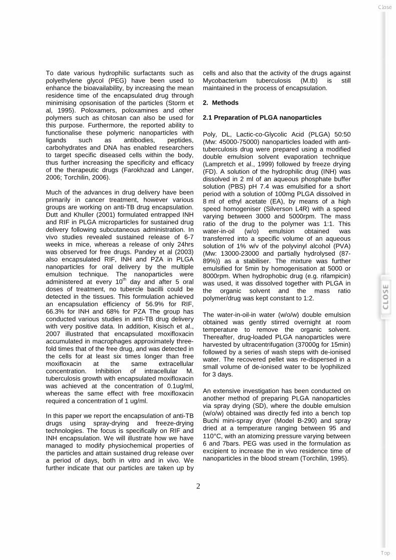
All samples made via freeze-drying showed a negative zeta potential. The addition of chitosan to provide positive surface charge resulted in microparticles (data not shown). This problem has been alleviated by spray-drying the double emulsion containing chitosan in the formulation (i.e. D and F in Table 2). Although RIF was loaded in the polymeric matrix of the carrier, there were no significant differences with INH-loaded nanocarriers in terms of all characteristics investigated, i.e size, size distribution and Zeta potential. 2.7.2 Encapsulation efficiency (EE) Table 2 also shows that a considerable fraction of the active compound (i.e. INH) was incorporated into the nanoparticles. The encapsulation was improved by increasing the amount of PVA molecules for the same amount of particles. This might be attributed to a better coverage of the stabilizer onto the surface of particles. Although RIF is highly hydrophobic, which should imply high encapsulation efficiency attributed to its low diffusion rate through the aqueous phase, its encapsulation is still relatively low when compared to INH samples. This might be attributed to the fact that it is located in the matrix (as opposed to the inner aqueous core), which might make it susceptible to loss during washing. The current EE for freeze dried samples is at 60%, where for spray dried samples it is 70%. Further work in improving the EE is in progress. 2.7.3 Analysis of surface morphology As shown in Table 2, high volume of stabilizer i.e PVA, led to well distributed and uniform INH-loaded PLGA nanoparticles with an average size around 200nm, characterized by a very smooth surface as depicted by the SEM image in Figure 1.
Figure 1 . SEM picture of freeze dried formulation A1 (Prepared with 20ml PVA 1%w/w), The bar represents 200nm).
Figure 2. SEM picture of freeze dried formulation B1, (Prepared with 1% PVA). The bar represents 200nm. It has been reported that small size (less than 500nm) and a spherical shape give rise to an enhanced efficiency of cell internalisation. A spherical particle possesses the right curvature allowing its attachment onto the cell (Trewyn, 2008) It is equally admitted that spherical particles offer maximum volume for drug incorporation. We also observed that the use of lactose in the spray-dried formulation helped to decrease the dimpling effect on both INH and RIF-loaded nanoparticles (SEM results not shown). 2.7.4 In vitro drug release profile Minimal drug release was observed from nanoparticles suspended in PBS or in particles suspended in the dialysis membrane as depicted in Figure 3. Evidence of initial burst release can be observed in the first 1-2 hours. The release lasted for 24-48 hours for both drugs as depicted in Figure 3A, in contrast to free drugs whose release was complete in 6 hours (Figure 3B). Additionally, there was no major difference in the amounts of drug released in both methods. In the two methods, approximately 15-40% of surface-entrapped drugs were removed during the washing stage. Thus, the

6
amount of drugs released from nanoparticles only accounted for less than 2% of encapsulated drugs. We postulated that the rest of the drugs (62-78%) are possibly trapped in the nanoparticles.
-0.05
0
0.05
0.1
0.15
0.2
0.25
0.3
0.35
0 10 20 30 40 50 60
Time (Hrs)
Dru
g C
onc
(ug/
ml)
PLGA-RIF
PLGA-INH
-10
0
10
20
30
40
50
60
70
80
90
0 10 20 30 40 50 60
Time (Hrs)
Dru
g C
onc
(ug/
ml)
Free INH
Free RIF
Figure 3:A: In vitro release of INH and RIF from PLGA nanoparticles suspended in 0.1M PBS pH 7.4. B: Positive control of free drugs following dialysis (10mg/100ml PBS) with stirring at 37 deg C. Dutt and Khuller (2001) also made the same observation in one of their studies where they observed that when pre-washed drug-loaded nanoparticles were suspended in PBS, there was negligible (initial 3–7% followed by <1%) in vitro drug release for 6 weeks. Polymer biodegradation and adequate agitation usually play a crucial role in drug release from nanoparticle systems (Ahlin et al., 2002). It is also hypothesized that drug release from NPs is through bioerosion of nanoparticles (Siepmann and Gopferich, 2001). Since hydrolytic enzymes were not included in PBS, the slow rate of nanoparticle degradation could be contributed to this factor. We believe that faster release could be observed in the environment with hydrolytic enzymes, especially in the body tissues. 2.7.5 In vitro particle uptake To determine particle uptake by CaCo-2 cells, cell were treated with 10 ug/ml PLGA at a density of 1X 10⁵ cells/cm2 .It was evident as indicated in Figure
4 that the particles of the specified size range were taken up within 30 min. However, after 60 min, a greater proportion of the particles was taken up. Based on this data, it can be suggested that the Rhodamine-loaded nanoparticles co-localised with the lysosomes, as indicated by the orange colour in Figure 4.
A B Figure 4: A indicates a Z-stack of 30 min incubation and B depicted a 60 min incubation period. Our results are in agreement with similar reports where it has been illustrated that nanoparticles are taken up by cells (Kisich et al., 2007). This results support the data by Kisich et al., 2007, where it was illustrated that free drugs did not accumulate in the cells at the same concentration as encapsulated drugs. Thus, nanoparticulate encapsulation of drugs may enhance drug uptake into the cells. The use of CaCo-2 cells in this specific study enabled the in vitro analysis of uptake in a cell model of the intestine. Thus, it can be suggested that the prepared particles should be taken up by cells of the intestine in patients. 2.7.6 In vivo particle uptake An in vivo method was followed as described in section 2.4.2 to confirm particle uptake. Initially macrophages were characterised from the subpopulation of PECS as indicated in Figure 5B, using thioglycolate-induced macrophage proliferation, a positive control and anti-CD11c and MOMA-2 to characterize cells. In general macrophages are large in size and also granular, thus they are detected in the higher ranges of the forward and side scatters.
B
A

7
Figure 5A: FACS data of PECS subsequent to saline administration to mice. Figure 5B: FACS data of PECS subsequent to thioglycolate administration to mice. The analysis of PECS subsequent to intraperitoneal and oral administration of rhodamine-labeled PLGA nanoparticles indicated that peritoneal macrophages did take up the particle as indicated in Figure 6. The method was slightly modified in order to detect for rhodamine fluorescence.
Figure 6: A indicates macrophage cells subsequent to ip administration and B indicates oral administration of Rhodamine labeled particles From this data, it can be suggested that PLGA particles are taken up by macrophages. This is indicated by a population of macrophages that is positive for the Rhodamine particles. Although uptake of particles by macrophages has previously been reported (Ahsan et al., 2002), much of the research was conducted in vitro. With this approach, we were able to illustrate that subsequent to intraperitoneal and oral administration, fluorescently labeled PLGA nanoparticles of the reported size range were taken up by the macrophages of the peritoneum. Further
assays still need to be conducted to illustrate whether the uptake of the particles elicits immune response or not. 2.7.7 In vitro efficacy MIC concentrations of INH and RIF were added into vials inoculated with H37RV. From the reading taken on the days indicated in Figure 7, it was evident that the encapsulated drugs still exhibited in vitro efficacy against H37RV, comparable to the free drugs. The BACTEC 460 assay is generally conducted to analyse the susceptibility of M.tb to the test drugs. Similar to data presented by Kisich et al., 2007, we were able to illustrate in vitro that the processes utilised to encapsulate the drugs do not have any effect on the potency of the drugs.
BACTEC 460 Spray dried samples
0
200
400
600
800
1000
1200
Day 1 Day 2 Day 3 Day 4 Day 6 Day 7 Day 8 Day 9
Days
GI
Free INH
SD INH
Free RIF
SD RIF
No drug (control 1)
No drug (control 2)
Figure 7: BACTEC 460 data of encapsulated RIF and INH Since we postulate that our approach should minimise the dose administered to patients, we are also in the process of conducting BACTEC 460 assays with various lower concentrations of the encapsulated drugs. The data will de utilised to determine the new MIC concentration for encapsulated RIF and INH. 2.7.8 In vivo release profile As depicted in Figure 8, a sustained release profile of RIF, INH and PZA was observed over a period of at least 5 days when compared to free drugs which reached levels below the MIC within 18 hrs.

8
Figure 8: in vivo release of free drugs vs spray dried nanoparticles encapsulating RIF, INH and PZA. With encapsulated drugs, drug concentration in plasma above the MIC level of RIF and INH were sustained for 4.5 days. Although PZA levels in plasma were below the MIC, a sustained release profile was observed over the same period of 4.5 days. The data presented is in agreement with results from Pandey et al., 2003, where a sustained drug release profile was observed over a period of 10 days. However, the difference between our results and those of Pandey et al., (2003) is the use of a spray-drying technology, where the surface of the particles were modified to increase the residence time in the circulation. The scalability of our technology also makes it feasible for easier entry into the market. A more detailed study with a larger number of mice will be performed to illustrate with statistical significance that a slow release profile is feasible. Further work is also planned to illustrate the efficacy of the formulation in TB-challenged mice.
2.8 Conclusions This study presents a novel technology that can be applied to address patient non-compliance in TB treatment programmes. The ability to release the drugs at the MIC concentration over a period of days presents the promise of reducing the doses currently administered, and also minimising the dose frequency, from a daily intake of antibiotics to a postulated once a week intake. 2.9 References Ahlin, P., Kristl, J., Kristl, A. et al, 2002. Investigation of polymeric nanoparticles as carriers of enalaprilat for oral administration. International Journal of Pharmaceutics, 239:113–2. Ahsan, F., Rivas, I.P., Khan, M.A. et al. 2002. Targeting to macrophages: role of physicochemical properties of particulate carriers—liposomes and microspheres—on the phagocytosis by macrophages. Journal of Controlled Release, 79:29–40. Artursson P., Palm K., Luthman K. 2001. Caco-2 monolayers in experimental and theoretical predictions of drug transport. Advanced Drug Delivery Reviews, 46:27-43.
Bawarski, W.E., Childelowsky, E., Bharali D.J. et al. 2008. Emerging nanopharmaceuticals, Nanomedicine: NBM 2008;xx:1-10, doi:10.1016/j.nano.2008.06.002. Centres for Disease control and Prevention 2006. Emergence of Mycobacterium tuberculosis with Extensive Resistance to Second-Line Drugs -Worldwide, 2000—2004. MMWR Morbidty Mortality Weekly Report 55:301-5.
Cui, F., Qian, F. and Yin, C. 2006. Preparation and characterization of mucoadhesive polymer-coated nanoparticles. International Journal of Pharmaceutics 316(1-2):154-161.
Dutt, M. Khuller, G.K. 2001. Therapeutic efficacy of poly (DL-lactideco-glycotide)-encapsulated antitubercular drugs against Mycobacterium tuberculosis infection induced in mice. Antimicrobia Agents Chemotherapy, 45:363–6.
Dutt, M., Khuller, G.K. 2001. Chemotherapy of Mycobacterium tuberculosis infections in mice with a combination of isoniazid and rifampicin entrapped in Poly (DL-lactide-co-glycolide) microparticles. Journal of Antimicrobial Chemotherapy, 47: p. 829-835. Farokhzad O.C. and Langer R. 2006. Nanomedicine: Developing smarter therapeutic and diagnostic modalities. Advanced Drug Delivery Reviews, 58: 1456-1459.
Release profile Free Drugs vs Spray dried Nanocarriers
0.01
0.1
1
10
100
0 20 40 60 80 100 120
Time [Hrs]
Pla
sma
Con
c. [u
g/m
l]
INH-PLGA RIF-PLGA PZA-PLGAFree INH Free RIF Free PZAMIC INH MIC RIF MIC PZA

9
Jani, P.,. Halbert G. W, Langridge J. et al. 1990. Nanoparticles uptake by the rat gastrointestinal mucosa: quantitation and particle size dependency. Journal of Pharmaceutics and Phamacology. 42: 821-826, Kisich, K.O., Gelperina, S., Higgins, M.P. et al. 2007. Encapsulation of moxifloxacin within poly(butyl cyanoacrylate) nanoparticles enhances efficacy against intracellular Mycobacterium tuberculosis. International Journal of Pharmaceutics 345:154–162.
Lamprecht A., Ubrich N., Pérez M.H. et al. 1999. Biodegradable monodispersed nanoparticles prepared by pressure homogenisation-emulsification. International Journal of Pharmacy, 184:97-105.
Langer, R. 2000. Biomaterial in drug delivery and tissue engineering: One laboratory's experience. Accounts of Chemical Research, 33:94-101.
Mohan, B., Sharda, N., Singh, S. 2003. Evaluation of recently reported USP gradient HPLC method for analysis of anti-tuberculosis drugs for its ability to resolve degradation products of rifampicin. Journal of Pharmacy and Biomedical Analysis 31:607-612.
Pandey, R., Zahoor, A., Sharma, S. et al. 2003. Nanoparticle encapsulated antitubercular drugs as potential oral drug delivery system against murine tuberculosis. Tuberculosis, 83:373-378.
Siepmann, J., Gopferich, A. 2001. Mathematical modeling of bioerodible, polymeric drug delivery systems. Advanced Drug Delivery Reviews, 48:229–247.
Singh, B.N. and Kim, K.H. 2000. Floating drug delivery systems: an approach to oral controlled drug delivery via gastric retention. Journal of Controlled Release, 63:235-259 Smith P.J. van Dyk J. and Fredericks A.1999. Determination of rifampicin, isoniazid and pyrazinamide by high performance liquid chromatography after their simultaneous extraction from plasma. International Journal of Lung Disease 3(11):S325-S328. Takeuchi H, J. Thongborisute, Y. Matsui, H. et al, 2005 Novel mucoadhesion tests for polymers and polymer-coated particles to design optimal mucoadhesive drug delivery systems. Advanced Drug Delivery Reviews,Volume 57, Issue 11,3, Pages 1583-1594. Trewyn, B.G., Nieweg, J.A., Zhao, Y. et al. 2008. Biocompatible mesoporous silica nanoparticles with different morphologies for animal cell membrane penetration. Chemical Engineering Journal, 137, 23-29.
Torchilin, V. 2006. Multifunctional nanocarriers, Advanced Drug Delivery Reviews, 58:523-1555.
Torchilin, V.P., Trubetskoy, V.S., 1995. Which polymers can make nanoparticulate drug carriers long-circulating? Advanced. Drug Delivery Reviews, 16:141-155. WHO. Ten statistical highlights in global public health-Part 1. World health Statistics 2007 [cited 2008 9 January] Available from : http://www.who.int WHO: Global tuberculosis control 2007: key findings. [cited 2008 8 January]. Available from: http://www.who.int/tb/publications/global_report/2007/key