nrf-1, an activator involved in nuclear- mitochondrial interactions
TRANSCRIPT

NRF-1, an activator involved in nuclear- mitochondrial interactions, utilizes a new DNA-binding domain conserved in a family of developmental regulators
Ching-man A. Virbasius, Joseph V. Virbasius, and Richard C. Scarpulla 1
Department of Cell, Molecular, and Structural Biology, Northwestern University Medical School, Chicago, Illinois 60611 USA
Nuclear respiratory factor 1 (NRF-1) was first discovered as an activator of the cytochrome c gene and was subsequently found to play a broader role in nuclear-mitochondrial interactions. We have now cloned a HeLa cDNA encoding NRF-1 using degenerate oligomers derived from tryptic peptide sequences for PCR amplification. The cDNA-encoded protein was indistinguishable from the authentic HeLa cell factor on denaturing gels, displayed the expected NRF-1 DNA-binding specificity, and made the same guanine nucleotide contacts as HeLa NRF-1 on binding known NRF-1 recognition sites. Antiserum raised against the highly purified recombinant protein recognized the identical DNA-protein complex formed using either a crude nuclear fraction or nearly homogeneous HeLa NRF-1. Recombinant NRF-1 also activated transcription through specific sites from several NRF-l-responsive promoters, confirming both the transcriptional activity and specificity of the cDNA product. Portions of NRF-1 are closely related to sea urchin P3A2 and the erect wing (EWG) protein of Drosophila. Both are recently identified developmental regulatory factors. The region of highest sequence identity with P3A2 and EWG was in the amino-terminal half of the molecule, which was found by deletion mapping to contain the DNA-binding domain, whereas the carboxy-terminal half of NRF-1 was highly divergent from both proteins. The DNA-binding domain in these molecules is unrelated to motifs found commonly in DNA-binding proteins; thus, NRF-1, P3A2, and EWG represent the founding members of a new class of highly conserved sequence-specific regulatory factors.
[Key Words: Oxidative phosphorylation; nuclear respiratory factors; mitochondria; transcription]
Received July 27, 1993; revised version accepted October 1, 1993.
The vertebrate mitochondrion contains its own genome, along with the machinery required for its autonomous transcription, translation, and replication (Attardi and Schatz 1988; Clayton 1991; Wallace 1992). Mitochon- drial DNA, however, has a coding capacity limited to only 13 respiratory chain polypeptides, and 2 ribosomal and 22 transfer RNAs. Most respiratory proteins and all of those necessary for maintenance and expression of the mitochondrial genome are encoded in the nuclear DNA. Thus, understanding the genetic control of mitochon- drial function largely becomes a problem of identifying the nuclear genes involved and investigating potential mechanisms of regulated expression.
Investigations of nuclear-mitochondrial interactions in mammalian cells have led to the cloning of nuclear gene products required for mitochondrial DNA tran- scription and replication (Clayton 1991). The human and mouse genes for the RNA subunit of MRP endonuclease,
1Corresponding author.
a ribonucleoprotein enzyme that is thought to cleave light-strand transcripts to form primers for heavy-strand DNA replication, have been cloned (Chang and Clayton 1989; Topper and Clayton 1990). A second nuclear gene product linking transcription and replication is mito- chondrial transcription factor 1 (mtTF1), now called mt- TFA (Xu and Clayton 1992). This factor recognizes the divergent heavy- and light-strand promoters to stimulate transcription initiation by mitochondrial RNA polymer- ase in vitro (Fisher et al. 1987). Because light-strand tran- scripts cleaved by mitochondrial RNA processing (MRP) endonuclease prime heavy-strand replication, mtTFA has the potential to modulate both transcription and rep- lication of mitochondrial DNA (Clayton 1991). The im- portance of mtTFA to mitochondrial function in vivo is supported by the observation that a null mutation in the yeast counterpart to mtTFA (ABF2) results in the loss of mitochondrial DNA (Diffley and Stillman 1991). Inter- estingly, this phenotype can be rescued by expression of human mtTFA in yeast (Parisi et al. 1993).
Regulatory elements common to nuclear genes with
GENES & DEVELOPMENT 7:2431-2445 © 1993 by Cold Spring Harbor Laboratory Press ISSN 0890-9369/93 $5.00 2431
Cold Spring Harbor Laboratory Press on April 9, 2018 - Published by genesdev.cshlp.orgDownloaded from

Virbasius et al.
products that function in the mitochondria have also been described (Nagley 1991; Wallace 1993). Analysis of cytochrome c and cytochrome oxidase promoters has led to the identification of transcriptional activators desig- nated as nuclear respiratory factors (NRF)-I (Evans and Scarpulla 1989, 1990; Chau et a1.1992) and -2 (Virbasius and Scarpulla 1991; Virbasius et al. 1993). Functional NRF-1 sites have been found in genes encoding cy- tochrome c and at least one subunit each of respiratory complexes III, W, and V (Evans and Scarpulla 1989, 1990; Chau et al. 1992}, suggesting a role for the factor in the coordinate expression of respiratory chain subunits. NRF-1 may also participate in mitochondrial gene ex- pression through its sequence-specific activation of genes encoding both the MRP RNA (Evans and Scarpulla 1990) and mtTFA (this paper; Virbasius and Scarpulla 1994). Activity of the proximal mtTFA promoter is highly dependent on NRF-1 in both transfected cells and in in vitro transcription assays (Virbasius and Scarpulla 1994). Similarly, expression of the gene encoding 5-ami- nolevulinate (5-ALA} synthase, the rate-limiting enzyme in the biosynthesis of heme for respiratory cytochromes, requires two NRF-1 recognition sites within its pro- moter region (Braidotti et al. 1993). These findings are consistent with an integrative role for NRF-1 in control- ling nuclear-mitochondrial interactions in higher organ- isms. In addition, functional NRF-l-binding sites are present in the genes for tyrosine aminotransferase and the translation initiation factor eIF-2c~ (Chau et al. 1992). Like 5-ALA synthase, these two proteins participate in the rate-limiting steps of their respective pathways of tyrosine catabolism and protein synthesis. Thus, NRF-1 may integrate a number of metabolic processes by regu- lating the genes encoding key enzymes.
As a prelude to molecular cloning, NRF-l-binding ac- tivity was purified over 30,000-fold to near homogeneity and was found to reside in a single polypeptide of 68 kD (Chau et al. 1992). Here, we use the sequences of tryptic peptides to obtain a cDNA clone that encodes a protein with the DNA-binding and transcriptional specificities expected for NRF-1. Deletion mapping of NRF-1 estab- lishes that its DNA-binding domain coincides with a region of high sequence similarity with P3A2 (Calzone et al. 1991; Hoog et al. 1991) and erect wing (EWG)(Desi- mone and White 1993), two recently identified develop- mental regulatory factors. Thus, these proteins define a new family of sequence-specific regulators that share a conserved DNA-binding domain. On the basis of func- tional analysis of over a dozen NRF-1 sites, we predict that the NRF-1 DNA-binding domain participates in the expression of >50 mammalian genes of known se- quence.
Results
Molecular cloning and overexpression of recombinant NRF-1
Tryptic peptides were derived from -50 pmoles of puri- fied HeLa NRF-1 (Chau et al. 19921. Two peptide peaks
from HPLC chromatography were chosen for microse- quencing. NRF-1(72) yielded 26 residues (SMILEDLE- SALAEHAPAPQEVNSELP) from a single homogenous peptide, whereas NRF-l(38) was a mixture of two pep- tides with a novel 8-residue sequence (VSWTQALR) de- rived from the secondary product. The major NRF-l(38) peptide was from Ku antigen, a known contaminant of DNA-binding proteins (Kadonaga 1991). A series of de- generate oligonucleotides was designed for PCR ampli- fication of total eDNA prepared from HeLa poly(A) + RNA. Two of these probes yielded a 269-nucleotide PCR product containing an open reading frame that was sub- sequently used as a probe to obtain a 3-kb HeLa cDNA clone. The putative NRF-1 cDNA had a 503-amino-acid open reading flame containing the complete sequence of the PCR product flanked by the two NRF-1 peptides (Fig. 1}.
The cDNA product was overexpressed in Escherichia coli using an inducible T7 expression system (Studier et al. 1990). A transformant containing the NRF-l-coding region transcribed from an inducible T7 promoter gave a major 68-kD protein on induction (Fig. 2, lanes 2,3). The 68-kD mass of the induced protein is identical to that determined previously for highly purified HeLa NRF-1 (Chau et al. 1992) but is greater than the 54-kD mass predicted by the NRF-1 open reading frame. As only the open reading frame fragment was cloned into the expres- sion vector, the mass of NRF-1 appears to be overesti- mated on denaturing gels. The induced 68-kD protein was purified to >95% purity from sonified extracts by ammonium sulfate precipitation (lane 4) and heparin- agarose fractionation (lane 5).
To determine whether the eDNA-encoded recombi- nant protein was the same as that present in DNA-pro- tein complexes formed using HeLa cell NRF-1, goat an- tiserum was raised against the purified recombinant pro- tein and tested for its ability to "supershift" NRF-1- DNA complexes in a gel-retardation assay. DNA- protein complexes of identical migration were formed with a labeled rat cytochrome c NRF-1 oligomer (RC4
- 172 / - 147) using crude nuclear extract (Fig. 3, lane 1), affinity-purified NRF-1 (lane 4), or recombinant NRF-1 (lane 7). In each case, the complex was supershifted with antiserum raised against recombinant NRF-1 (lanes 3,6,9) under conditions where preimmune serum had no effect (lanes 2,5,8). Variations in the antibody complexes formed with affinity-purified and recombinant proteins result from differences in the antigen-antibody ratios present in each reaction. The multiple complexes ob- served previously with binding of NRF-2 to its recogni- tion site in the cytochrome oxidase subunit Vb (MCO5b + 13/+33)gene (Virbasius et al. 1993)were unaffected by the addition of anti-NRF-1 or preimmune serum to binding reactions (lanes 10-12). These results provide a direct link between the NRF-1 cDNA product and the NRF-l-binding activity present in HeLa cells.
DNA- bin cling specificity of recom bin an t NRF-1
The recombinant protein should display the same DNA-
2432 GENES & DEVELOPMENT
Cold Spring Harbor Laboratory Press on April 9, 2018 - Published by genesdev.cshlp.orgDownloaded from

Cloning of transcription factor NRF-1
Met Glu Glu His 119 ATG GAG GAA CAt
30 Gin Val His Val AIs Thr
194 CAG GTC CAT GTG GCT ACT
Thr Set Tyr Asp Asp Ser 269 ACC TCT TAC GAT GAC TCA
Pro Val Gly Met Ala AIs 344 CCT GTG GGA ATG GCC GCT
Pro Set Ile Arg Lys Arg 419 CCA TCT ATC CGG AAG AGG
130 Val Gly Gln Gln Ala Ile
494 GTG GGA CAG CAA GCT ATT
Pro Leu Glu Ash Val Val 569 CCT TTG GAG AAT GTG GTG
Pro Ala Pro Gln GI~ Val 644 CCT GCG CCA CAG GAG GTT
Thr Gln Ala Gln Leu Arg 719 ACC CAG GCC CAG CTT CGG
230 Lys Glu Set Cys Lys Pro
794 AAA GAA AGC TGC AAG CCC
Glu Gln Lys Gln Arg Val 869 GAG CAA RAG CAG AGG GTT
Glu Asp Leu Leu Tyr AIa 944 GRA GAC CTT TTG TAT GCC
Val Pro Set Gin Thr Val 1019 GTA CCA TCA CAG ACT GTA
330
Ala Thr Val Ala Thr Leu 1094 GCA ACA GTA GCC ACA TTG
Val Ala Asp Gly GIu Val 1169 GTG GCT GAT GGA GAG GTG
Set Glu Ala Thr Gln Ala 1244 TCA GAG GCC RCC CAG GCG
Thr Val Thr Met Ala Leu 1319 ACAGTC ACT ATGGCG CTT
430 Gly Gly Gln Ile Val Leu
1394 GGG GGA CAG ATC GTC TTG
Leu Val Gin Ile Pro Val 1469 CTG GTC CAG ATC CCT GTG
Ala Met Ala Pro Val ThE 1544 GCC ATG GCC CCT GTG ACC
Leu Glu Gln End 1619 TTG GAA CAG TGA
AGCGCTGCCGCTTCTGAGT
20 TCCTGGT•CAGAACTTTACACAGAAAAGGTGCTCAAAGGATATTTGTTTAATGAATGTGGTATGCTGACATTTAAAcAGGACAAAA•TTGTAGAACTTC
i0 Gly Val Thr Gln Thr Glu His Met Ala Thr Ile GIu Ala His AIa Val Ala Gin Gln Val Gln GGA GTG ACC CAA ACC GAA CAT ATG GCT ACC ATA GAA GCA CAT GCA GTG GCC CAG CAA GTG CAG
Tyr Thr Glu His Set Met Leu Set TAC ACC GAG CAT AGT ATG CTG AGT
Asp Ile Leu ASh Set Thr Ala Ala GAT ATA CTC AAC TCC ACA GCA GCT
Ala Ala Ala Val Ala Thr Gly Lys GCT GCT GCT GTG GCA ACA GGA AAG
110 Gln Gln Thr Arg Leu Leu Arg Lys CAA CAA ACA CGT TTG CTT CGG AAA
Val Leu Cys Ile Set Pro Ser Ly8 GTC CTC TGT ATC TCA CCC TCCAAA
Arg Lys Tyr Lys Ser Met Ile Leu CGT AAG TAC AAG AGC ATG ATC CTG
ASh Set Glu Leu Pro Pro Heu Thr A~C T~A GAA CTG CCG 9CT CTC ACC
210 Ala Phe Ile Pro Glu Met Leu Lys GCA TTT ATC CCA GAG ATG CTC AAG
Ile Trp Trp Pro Glu Asp Ile Pro ATC TGG TGG CCT GAA GAT ATC CCC
Ser TrD Thr Gln Ala Le u Ara Thr ~C~ ~GG ACC CAG GCA CTA CGG ACC
Phe Glu Asp Gln Gin Thr Gln Thr TTT GAA GAT CAG CAA ACG CAA ACA
310 Val Gln Thr Phe Ser ASh Pro Asp GTC CAG ACT TTT AGT AAC CCT GAT
Ala Asp Ala Set Glu Leu Pro Thr GCT GAT GCT TCA GAA TTG CCA ACC
Glu Gln Ash Trp Ala Thr Leu Gin GAA CAA AAT TGG GCC ACG TTA CAG
Val Ala Set Leu Ala Glu Ala Ala GTG GCA TCG TTG GCA GAG GCC GCA
410 ASh $er Glu Ala Ala Ala His Ala AAC AGC GAA GCT GCC GCC CAT GCT
Set Gly Glu Thr Ala Ala Ala Val TCT GGG GAA ACC GCA GCA GCC GTC
Ser Met Tyr Gln Thr Val Val Thr AGC ATG TAC CAG ACT GTG GTG ACC
Thr Arg Ile Ser Asp Set Ala Val ACC AGG ATA TCA GAC AGC GCA GTC
19
118
193 50
Ala Asp Glu Asp Set Pro Ser Set Pro Glu Asp GCT GAT GAA GAC TCG CCT TCT TCG CCC GAG GAC 268
70 Asp Glu Val Thr Ala His Leu Ala Ala Ala Gly GAT GAG GTG ACA GCT CAT CTG GCA GCT GCA GGT 343 90
Lys Arg LyB Arg Pro His Val Phe Glu Set ADn AAA CGG AAA CGG CCT CAT GTA TTT GAG TCT PAT 418
Leu Arg Ala Thr Leu Asp Glu Tyr Thr Thr Arg CTT CGA GCC ACG TTA GAT GAA TAT ACT ACT CGT 493
150 Pro Ash Pro Val Phe Lys Val Phe Gly Ala Ala CCT AAC CCT GTC TTT AAA GTG TTT GGT GCA GCA 568
170 G~u ~ s ~ LeU Glu Set Kla Leu Ala Glu His A1s GAA GAC CTG GAG TCT GCT CTG ~ 643 190 Ile Asp Gly Ile Pro Val Ser Val Asp Lys Met ATC GAC GGA ATT CCA GTC TCT GTG GAC AAA ATe 718
Tyr Ser Thr Gly Arg Gly Lys Pro Gly Trp Gly TAC TCT ACA GGT CGG GGA AAA CCA GGC TGG GGr 793
250 Trp Ala Asn Val Arg Ser Asp Val Arg Thr Glu TGG GCA AAT GTC CGG AGT GAT GTC qqq hq A GAA 868
270 Ile Val LyB ASh Cys Tyr LyB Gln Hil Gly Arg ATA GTT AAA AAC TGT TAT AAA CAG CAT GGG CGG 943 290 Gln Ala Thr Ala Thr His Set Ile Ala His Leu CAG GCC ACA GCC ACA CAT AGT ATA GCT CAT CTT 1018
Gly Thr Val Set Leu Ile Gln Val Gly Thr Gly GGC ACT GTC TCA CTT ATC CAG GTT GGT ACG GGG 1093
350 Thr Val Thr Val Ala Gln Val Ash Tyr Set Ala ACG GTC ACC GTT GCC CAA GTG AAT TAT TCT GCC 1168
370 Gly Gly Glu Met Thr Ile Gln Thr Thr Gin Ala GGA GGT GAG ATG ACC ATC CAG ACG ACG CAA GCA 1243 390 Val Ala Ala Ser Gin Glu Met Gln Gln Gly Ala GTG GCA GCT TCT CAG GAG ATG CAG CAG GGA GCT 1318
Val Ala Thr Leu Ala Glu Ala Thr Leu Gln Gly GTC GCC ACC CTG GCT GAG GCC ACC TTA CAA GGT 1393
450 Gly Ala Leu Thr Gly Val Gln Asp Ala Ash Gly GGA GCA CTT ACT GGA GTC CAA GAT GCT AAT GGC 1468
470 Set Leu Ala Gln Gly Ash Gly Pro Val Gln Val AGC CTC GCC CAG GGC AAC GGA CCA GTG CAG GTG 1543 490 Thr Met Asp Gly Gin Ala Val GIu Val Val Thr ACC ATG GAC GGC CAA GCT GTG GAG GTG GTG ACA 1618
Figure 1. Nucleotide and predicted amino acid sequence of the HeLa NRF-1 cDNA 5'-untranslated and coding region. Amino acid sequences matching those of tryptic peptides from the purified NRF-1 protein are underlined. The sequence co- inciding with the PCR product amplified from HeLa cDNA using primers derived from the NRF-1 peptide sequences is indi- cated with a bold underline. The complete 2970-nucleotide NRF-1 cDNA sequence has been submitted to GenBank under ac- cession number L22454.
binding properties ascribed previously to NRF-1 (Evans and Scarpulla 1989, 1990; Chau et al. 1992). Recombi- nant NRF-1 was thus used for competition DNase I foot- printing of the rat cytochrome c promoter region (Fig. 4). In the absence of competitor, the recombinant protein yielded several intense enhanced cleavages at the 5' and 3' ends of the footprint, with the absence of cleavages throughout the intervening protected region (Fig. 4, lane 3). This pattern is identical to that observed previously using preparations of HeLa NRF-1 (Evans and Scarpulla 1990). The footprint was eliminated by the inclusion of an excess of unlabeled oligonucleotides of previously characterized NRF-1 sites from nuclear genes with prod- ucts that function in the mitochondria. These include cytochrome c (RC4, lane 4), cytochrome oxidase subunit VIc (COXVIc, lane 5), and mouse MRP RNA (mMRP, lane 6). Moreover, a sequence from the cytochrome cl gene with two mismatches from the NRF-1 consensus did not compete (hCC1, lane 9), but a mutated derivative, active in both NRF- 1 binding and transcriptional activity {Evans and Scarpulla 1990), did (hCC1UP, lane 10). The unrelated cytochrome c ATF/CREB site (RC4 - 2 8 1 / -256 , lane 11) served as a negative control.
In addition to the known NRF-1 sites, we observed strong similarities to the NRF-1 consensus in recently isolated genes encoding cytochrome oxidase subunit Vb (COXVb) (Basu and Avadhani 1991) and mtTFA (Tomi- naga et al. 1992). Oligomers of each of these sites were
tested for their ability to stimulate the activity of a trun- cated cytochrome c promoter in transfected cells. The COXVb - 1 0 9 / - 8 7 and mtTFA - 7 3 / - 4 6 oligomers stimulated promoter activity 12.3-+3.4-fold and 6.0---1.9-
kD
200
97
E ,-,=
"E ® .E == O ' ~ ' ,P G
S T D - ~" E ~ 0~o~
68
43
29
1 2 3 4 5
Figure 2. Expression and purification of recombinant NRF-1. Coomassie-blue stained SDS-PAGE of molecular mass stan- dards (lane I} and 50 txl of log-phase culture of E. coli strain BL21(DE3) transformed with the NRF-l-coding region in the pET3d expression vector uninduced (lane 2) or induced by the addition of 0.4 mM IPTG (lane 3). A lysate of an induced culture was purified by ammonium sulfate precipitation (lane 4) and fractionation of the pellet on a heparin-agarose column (lane 5}. Lanes 4 and 5 contain equal amounts (4 ~g) of total protein.
GENES & DEVELOPMENT 2433
Cold Spring Harbor Laboratory Press on April 9, 2018 - Published by genesdev.cshlp.orgDownloaded from

V i r b a s i u s et al.
!
preimmune - + immune - -
MCO5b RC4 -1721-147 +13/+33
I I I
nuclear affinity recombinant nuclear extract purified protein extract
I ! ! I I a I . . + - + - . + -
+ - . + - + - . +
: : ?;s
1 2 3 4 5 6 7 8 9 10 11 12
Figure 3. Recognition of HeLa NRF-1 by antiserum directed against the recombinant protein. Binding reactions contained 14 ~g of HeLa nuclear extract (lanes 1-3, 10-12), 12 ng of affin- ity-purified HeLa NRF-1 {lanes 4-6), or 20 ng of bacterial NRF-1 heparin-agarose fraction (lanes 7-9). Labeled oligonucleotides contained either an NRF- 1-binding site from the rat cytochrome c gene (RC4 - 172/- 147, lanes 1-9) or an NRF-2-binding site from the mouse COXVb gene (Virbasius et al. 1993) {lanes 10- 12). Following the binding reaction, 1 ~1 of preimmune serum or goat antiserum was added to bacterially produced NRF-1, and the complexes were subsequently resolved on a native poly- acrylamide gel.
ers are identical to those observed wi th the HeLa protein, providing further support for the conclusion that the iso- lated cDNA encodes NRF-1.
Transcriptional activity and specificity of recombinant NRF-1
The binding of NRF-1 to its recognition site has been correlated with site-specific effects on promoter activity in transfected cells (Evans and Scarpulla 1989, 1990; Chau et al. 1992). The cloning of NRF-1 now affords an opportunity for a direct demonstrat ion of its transcrip- tional activity and specificity. Promoter activation by NRF-1 was thus tested in an in vitro transcription assay using both a wild-type promoter from the rat cy- tochrome c gene (RC4CAT/-326} and the same tem- plate containing an insertional disruption of the NRF-1 site ( R C 4 C A T / - 3 2 6 ; L I - 1 6 2 / - 159) that d iminishes both NRF-1 binding and the activity of the transfected
competitor r
.J.. , -.L'
o -. i ' . o o oo ~ , = , = =
fold, respectively, when cloned in cis, results s imilar to the value of 10.1 +3.1-fold obtained wi th the cytochrome c NRF-1 site (RC4 - 171 / - 147). Both also formed spe- cific complexes wi th affinity-purified HeLa NRF-1 (not shown). The COXVb and mtTFA oligomers were also found to be specific competitors in the DNase I foot- printing assay using recombinant NRF-1 (Fig. 4, lanes 7,8). These results further substantiate the binding spec- ificity of the recombinant protein and indicate that COXVb and mtTFA genes are l ikely to have NRF-I-re- sponsive promoters.
If the recombinant protein is NRF-1, it should contact DNA through specific guanine nucleotides spanning one t u m of the DNA helix, as demonstrated using prepara- tions of the HeLa protein (Evans and Scarpulla 1990; Chau et al. 1992). Recombinant NRF-1 was thus used for methyla t ion interference footprinting of known sites from RC4, COXVIc, and MRP RNA genes. The pat tem of guanine nucleotide contacts in each case was indistin- guishable from that obtained using HeLa NRF- 1 and con- forms to the consensus derived previously (Fig. 5). The RC4 site was known to deviate from the others by mak- ing additional downstream contacts (Evans and Scarpulla 1990). The same pa t tem is observed here wi th recombi- nant NRF- 1. Therefore, the binding interactions between recombinant NRF-1 and cognate sites in several promot-
w w ............. ~W : ~
1 2 3 4 5 6 7 8 9 1 0 1 1
Figure 4. B i n d i n g of r e c o m b i n a n t N R F - 1 t o t h e r a t c y t o c h r o m e
c p r o m o t e r reg ion . A n e n d - l a b e l e d R C 4 p r o m o t e r f r a g m e n t con -
t a i n i n g t h e N R F - l - b i n d i n g s i t e w a s s u b j e c t e d to D N a s e I d iges-
t i o n following incubation in a mixture without added protein (lane 2) or with the addition of 20 ng of NRF-1 heparin-agarose fraction (lanes 2-11}. Competitor oligonucleotides indicated above lanes 4-11 were added at a 200-fold molar excess before the addition of the labeled fragment. The extent of the NRF-1 footprint is indicated by the vertical bar at right. {G) G reaction of the labeled fragment.
2434 GENES & DEVELOPMENT
Cold Spring Harbor Laboratory Press on April 9, 2018 - Published by genesdev.cshlp.orgDownloaded from

Cloning of transcription factor NRF-1
RC4
-1721-147
COXVlc
+20/+46
mMRP
-3111-292
~ o. ~ ca. o D. C3. 0 -~
F B F B F B F B F B F B
i
~ o
RC4
-172 -147 I • • o o t TGCTAGCC CGCATGCGCGCGCACC TT ACGATCGG GCGTACGCGCGCGTGGAA
0 • • • • •
COXVIc
+ 4 6 +20 I • • I CTAGCAGCACGCATGCGCAGGAGC CGA GATC GTCGTGCGTACG CGTCCTCG GCT
• • 0 •
mMRP RNA
-311 -292 I • o o • I TAGTGCGCACGCGCAGGAG ATCACGCGTGCG CGTC CTC
O • •
Consensus: T T A cGCGCAcGCGCG
Figure 5. Recognition of NRF-l-binding sites by recombinant NRF-1 through characteristic guanine contacts. Fragments con- taining representative NRF-l-binding sites from the indicated promoters were labeled on upper or lower strands, partially methylated, and subjected to preparative scale mobility retar- dation using recombinant NRF-1. Free DNA IF) and DNA iso- lated from bound complexes (B) were cleaved with piperidine, and the products were analyzed on denaturing gels. (O) Guano- sine bases that {when methylated) strongly inhibit NRF-1 bind- ing; (0) partial interference. Summarized below are the DNA sequences of each site and the positions of guanine nucleotide contacts compared with the consensus sequence and contacts derived from analysis of binding of HeLa NRF-1 to 10 known binding sites (Evans and Scarpulla 1990; Chau et al. 1992).
promoter (Evans and Scarpulla 1989). The results dem- onstrate that a functional NRF-1 site is required for ac-
t ivation of transcription by the recombinant protein (Fig. 6A, lanes 2-4). Significant s t imulat ion was observed us- ing 0.1 ~g of NRF-1, whereas 0.4 ~g was inhibitory. This inhibition likely results from the competi t ive displace- ment of transcription complexes from the promoter tem- plate at high NRF-1 concentrations. In contrast, the linker insertion muta t ion resulted in a reduced level of transcription and completely el iminated activated ex- pression (lanes 5-7). The transcripts were init iated at the same position observed for the in vivo cytochrome c transcripts in liver RNA (lane 1), indicating that they accurately reflect promoter activation through the nor- mal initiation complex.
The cytochrome c promoter has mult iple cis-acting elements and therefore does not show complete depen- dence on NRF-1 for its activity (Evans and Scarpulla 1989). To enhance the NRF-l-dependent signal, four tan- dem sites from the cytochrome c (4XRC4) or the MRP
< RC4CATI -326 Z t r RC4CAT/ -326 LI -162/ -159 =.
• 0 0.1 0.4 0 0.1 0.4 pg NRF-1
1 2 3 4 5 6 7
RC4CAT/-66 ,¢ z No 4XRC4 4XmMRP
¢: insert - 1 7 2 / - 1 4 7 - 3 1 1 1 - 2 9 2
0 0.1 0.4 0 0.1 0.4 0 0.1 0.4 I.IgNRF-1
1 2 3 4 5 6 7 8 9 10
Figure 6. Transcriptional activation by recombinant NRF-1 through specific NRF-1 recognition sites. (A) In vitro transcrip- tion reactions with HeLa nuclear extract were gamed out with 500 ng of a plasmid containing the RC4 promoter (lanes 2-4) or a promoter with a linker insertion disrupting the NRF- 1-binding site (lanes 5-7). Heparin-agarose-purified bacterial NRF-1 was added as indicated (lanes 3,4,6,7). Transcription products were analyzed by primer extension and compared with the primer extension product of 20 ~g of rat liver RNA {lane 1). (B) Products of in vitro transcription reactions using a truncated RC4 pro- moter construction RC4CAT/-66 (lanes 2-4) or the same pro- moter with four tandem copies of the RC4 (lanes 5-7) or mMRP (lanes 8-I0) NRF-1-binding sites cloned upstream.
GENES & DEVELOPMENT 2435
Cold Spring Harbor Laboratory Press on April 9, 2018 - Published by genesdev.cshlp.orgDownloaded from

Virbasius et al.
RNA (4XmMRP) promoters were cloned into an RC4 vector deleted of sequences upstream from - 66, and the resulting constructs were used for in vitro transcription. Compared with the vector with no insert (Fig. 6B, lanes 2--4), those with the RC4 (lanes 5-7) or mMRP (lanes 8-10) NRF-1 sites displayed a strong, dose-dependent in- crease in transcription in response to added recombinant NRF-1. In this case, no inhibition was observed at 0.4 lag of NRF-1 because of the increased binding capacity of the promoter template for NRF-1. As with the intact cy- tochrome c promoter, initiation occurred at the same site used by the liver initiation complex in the synthesis of cytochrome c mRNA. These results establish that NRF-1 is a transcriptional activator that can function both in the proper promoter context and in a minimal promoter to direct the synthesis of high levels of accu- rately initiated transcripts.
NRF-1 has a new DNA-binding domain conserved in developmental regulatory factors
It was of interest to determine whether NRF-1 shares structural features with other proteins. A computer search revealed a region of extensive sequence similarity with two recently described developmental regulatory factors (Fig. 7). The first, P3A2, has been implicated in the correct expression of a cytoskeletal actin gene during sea urchin development (Calzone et al. 1991; Hoog et al. 1991). The second, the EWG gene product of Drosophila
melanogaster participates in both nervous system and flight muscle development (Desimone and White 1993). Binding to DNA has been demonstrated for P3A2 but not EWG, and neither has yet been shown to function di- rectly in transcriptional activation.
Alignment of NRF-1 with P3A2 and EWG reveals a stretch of striking sequence conservation among all three proteins between NRF-1 residues 65 and 284 (Fig. 7). This region coincides with the novel DNA-binding domain identified previously for P3A2 (P3A2 residues 25-258) (Hoog et al. 1991) and corresponding here to NRF-1 residues 61-290. In contrast, the three proteins share little similarity in their carboxy-terminal halves or in an amino-terminal extension present in NRF-1 and EWG.
To determine whether the highly conserved region co- incided with the NRF-1 DNA-binding domain, a dele- tion series of truncated NRF-1 molecules (summarized diagrammatically in Fig. 8A) was expressed by in vitro transcription and translation, and the products were as- sayed for binding to radiolabeled RC4 - 1 7 2 / - 147. As shown in Figure 8B, lane A, the intact eDNA yielded a translation product migrating at 68 kD. This protein was unaltered by deletion of the 3'-untranslated region to a position just downstream from the predicted NRF-1 translational terminator (lane B), confirming that the translation product is derived from the NRF-1 open read- ing frame. To demonstrate that the 68-kD translation product had the correct binding specificity, it was tested
NRF-1 ........................................................... MEEHGVTQTEHMATI 15 P3A2 .......................................................................... EWG ATTSYRLWAPAGSQRSSTGNVVVTTTSSGSHSSNGANGGTGGTSAGSSTLGSGLNVTTITATSGGQLQSAGNT 75
NRF-1 P3A2 EWG
NRF-1 P3A2 EWG
EAHAVAQQVQQVHVATYTEHSMLSADEDSPSSPEDTSYDDSDILNSTAADEVTAHLAAAGPVGMAAAAAVATGKK 90 .................... MMISEDISEPSSP.DTPFDDSDLLNSSMTDDVSAQLAASGPIGVRAAAAIATGKK 54 SQSNGTTYKIEMLEEDIQSLGSDDDDEDLISSDGSLYEG..DLGSMPVNDDVAHQLAAAGPVGVAAAAAIASSKK 148
I II I0 IOIO IIIOIIOIO IIIIOIO II
NRF-I P3A2 EWG
RKRPHVFESNPSIRKRQQTRLLRKLRATLDEYTTRVGQQAIVLCISPSKPNPVFKVFGAAPLENVVRKYKSMILE 165 RKRPHSFETNPSIRRRQQTRLIRKLKATIDEYATRVGQQAVVLTCTPGKHDGNFKVFGAAPLENIMRNLKGIVLQ 129 RKRPHCFETNPSVRKRQQNRLLRNVRAIIYEFTGRVGKQAVVLVATPGKPNTSYKVFGAKPLEDVLRNLKNIVMD 223 IIIII IIOIIIOIDIII IIOI 001 O IO0 III IIOOI OI I OIIIII III 0 I I ODD
NRF-I P3A2 EWG
DLESALAEHAPAPQEVNS...ELPPLTIDGIPVSVDKMTQAQLRAFIPEMLKYSTGRGKPGWGKESCKPIWWPED 237 DLDNALAQRAPQPSNENSDLYELPPLVIDGIPTSVNKMTQAQLRAFIPLMLKYSTGRGKPGWGKESCRPVWWPSD 204 ELDNALAQQAPPPPQDDPSLFELPGLVIDGIPTPVEKMTQAQLRAFIPLMLKYSTGRGKPGWGRESTRPPWWPKE 298 010 III II I I III IOIIIII I IIIIIIIIIIII IIIIIIIIIIIIIIOII OI IIl 0
NRF-1 P3A2 EWG
IPWANVRSDVRTEEQKQRVSWTQALRTIVKNCYKQHGREDLLYAF.EDQQTQTQATATHS ............. IA 298 LPWANVRSDVRSEDEKRKVSWTHALVTIVINCYKHHGRDDLLPEFIEDKCKEIEASQNQ ......... VASLPTA 270 LPWANVRMDARSEDDKQKISWTHALRKIVINCYKYHGREDLLPTFADDED.KVNALISQSGDEDEDMELSNPPTI 372 OIIIIII I IOIO I OOIII II II IIII IIIOIII I OI I
Figure 7. Alignment of NRF- 1 protein se- quence with those of developmental regu- latory factors P3A2 and EWG. Sequences of human NRF-1, sea urchin P3A2, and Drosophila EWG were aligned using the Nat-1
P3A2 GAP program of the Genetics Computer EWG
Group (program manual, v.7, 1991J. (I} Residues identical in all three proteins~ ([31 Nay-1 positions where all three proteins contain van2
r.WG
similar (conservative} amino acid substitu- tions grouped as follows: (A S T); (D E); (N Nat-1 Q); (R K); (I L M V); (F Y W). Dots denote P3a2
EWG gaps introduced for optimum alignment.
HLVPSQTWQTFSNPDGTVSLIQVGTGATVATLADASELPTTVTVAQ ............................ 345 TLLPSHAVVHTINNPDGTVSLIQVDTGATVATLAD ........................................ 305 HTVTTMTPPTGNSNQPQQVNVVKINSAGTVITTHTAQSNTPAPTIIQSTNNQHVTTTATLPASTKIEICQAPAQN 447
O 0 0 I I O0 0 O II I
NRF-1 .............. VNYSAVADGEVEQNWATLQGGEMT...IQTTQAS...EATQAVA ......... SLAEAAVA 391 P3A2 ................... VTQVQQLTNLQTLQQVRLQPLQIQHALGNQQAEATQAVQ ......... TLAEVAAA 352 EWG QQHHQHHQTHLPNAVHIQPVAGGQPQTIQLTTASGTATATAVQTTAAA..VSAAQAHAHSQSQAHSQSSANQTVT 520
I0 I Ol 0 I011 0 1 O 0
ASQ ...................... EMQQGATVTMALNSEAAAHAVATLAEATLQ..GGGQIVLSGETAAAVGAL 442 QGG .................... DGELTEGQTVT ............. TLPEGT ....... QLVLASD ..... GSL 382 AQQIANAQVCIEPITLSDVDYTTQTVLSQNADGTVSLIQVDPNNPIITLPDGTTAQVQGVATLHQGEGGATIQTV 595
0 I II O I 0 [] DO
TGVQDANG ................................ LVQIPVSMYQTVVTSLA..QGNGPVQVAMAPVTTR 483 QAINDGTAQG ............................... IVIPASVYQTVVAG ..... DGQPIQIANVNIAQQ 421 QSLTDVNGHENMTVDLTETQDGQIYITTEDGQGYPVSVSNVISVPVSMYQSVMANVQQIQTNSDGTVCLAPMQVE 670
[] I [] DI IDIIDIDD [] []
ISD ................. SAVTMDGQAV..EWTLEQ ........................... 503 SGG .......... GTTMAAIKNAVMQSQPIPSQVATLVVNAASHDQHT .................. 459 NGDQLETITMSPGMHQMMIQGGPGQEPQLV..QVVSLKDATLLSKAMEAINSGNVKSEDTIIMEQ 733
I D I~DI
2436 GENES & DEVELOPMENT
Cold Spring Harbor Laboratory Press on April 9, 2018 - Published by genesdev.cshlp.orgDownloaded from
Cloning of transcription |actor NRF-1
N R F . 1 5 0 3 A A
[ w/////////////////////~ = " I
5O3 5o3
& 4 7 6 - 5 0 3
& 4 1 9 - 5 0 3
A 3 3 1 - 5 0 3
& 3 0 5 - 5 0 3
A 2 6 4 - 5 0 3
& 2 3 8 - 5 0 3
I
4 1 - 7 7 J
a 1 - 1 0 9 K
& 1 - 1 4 4 L
DNA Binding
+
+
+
÷
+
+
.
+
+
+
B kD A B C D E F G H kD
2 1 8 - - 218 100--
72-- 100 43-- 72
43 2 9 - -
C A B C D E F G H
B I J K L
B I J K L
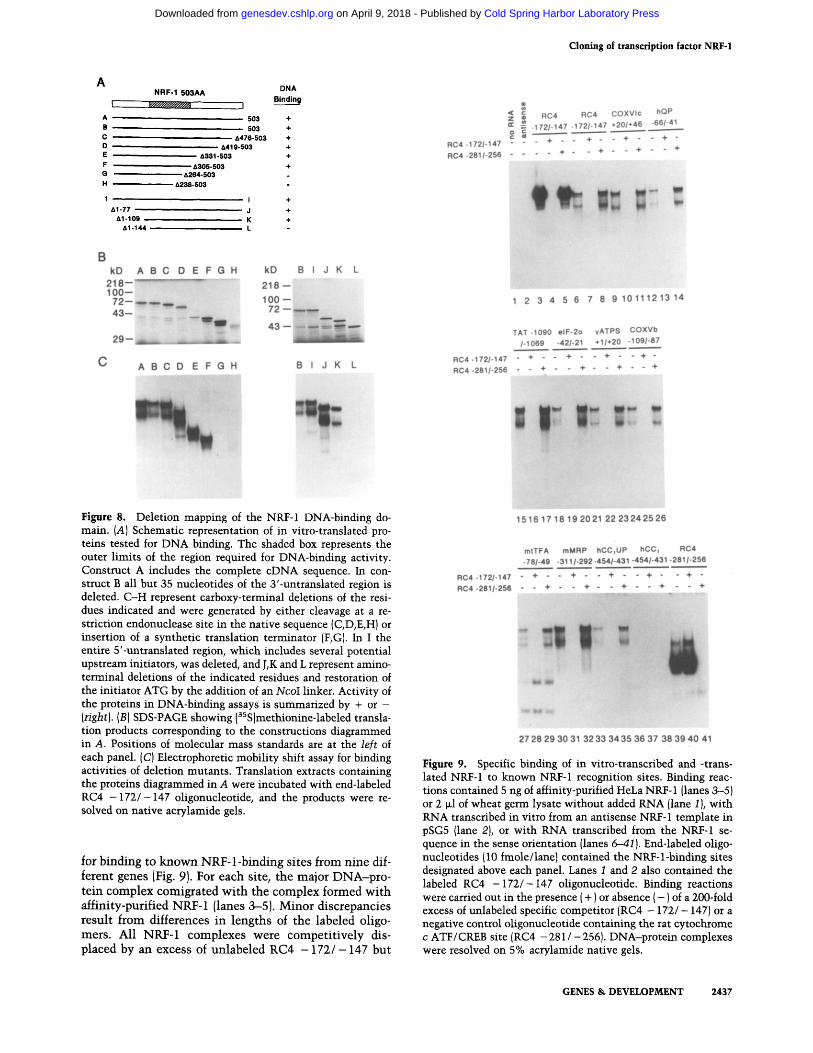
Figure 8. Deletion mapping of the NRF-1 DNA-binding do- main. (A) Schematic representation of in vitro-translated pro- teins tested for DNA binding. The shaded box represents the outer limits of the region required for DNA-binding activity. Construct A includes the complete cDNA sequence. In con- struct B all but 35 nucleotides of the 3'-untranslated region is deleted. C-H represent carboxy-terminal deletions of the resi- dues indicated and were generated by either cleavage at a re- striction endonuclease site in the native sequence (C,D,E,H) or insertion of a synthetic translation terminator (F,G). In I the entire 5'-untranslated region, which includes several potential upstream initiators, was deleted, and J,K and L represent amino- terminal deletions of the indicated residues and restoration of the initiator ATG by the addition of an NcoI linker. Activity of the proteins in DNA-binding assays is summarized by + or - (right). (B) SDS-PAGE showing [3SS]methionine-labeled transla- tion products corresponding to the constructions diagrammed in A. Positions of molecular mass standards are at the left of each panel. (C) Electrophoretic mobility shift assay for binding activities of deletion mutants. Translation extracts containing the proteins diagrammed in A were incubated with end-labeled RC4 - 1 7 2 / - 147 oligonucleotide, and the products were re- solved on native acrylamide gels.
for binding to known NRF-1-binding sites from nine dif- ferent genes (Fig. 9). For each site, the major DNA-pro- tein complex comigrated with the complex formed with affinity-purified NRF-1 (lanes 3-5). Minor discrepancies result from differences in lengths of the labeled oligo- mers. All NRF-1 complexes were competitively dis- placed by an excess of unlabeled RC4 - 1 7 2 / - 147 but
~ i R C 4 R C 4 C O X V I c h Q P
= . - 1 7 2 / - 1 4 7 - 1 7 2 / - 1 4 7 + 2 0 / + 4 6 - 6 6 / - 4 1 g
R C 4 - 1 7 2 / - 1 4 7 " " " + " " + " " + " " + "
R C 4 - 2 8 1 / - 2 5 6 . . . . + " " + " " + " " +
1 2 3 4 5 6 7 8 9 1 0 1 1 1 2 1 3 1 4
T A T - 1 0 9 0 o I F - 2 o y A T P S C O X V b
/ - 1 0 6 9 - 4 2 1 - 2 1 + 1 / + 2 0 - 1 0 9 1 - 8 7
R C 4 - 1 7 2 / - 1 4 7 - + " " + " " + " " + "
R C 4 - 2 8 1 / - 2 5 6 - - + - - + " " + " " +
1516 17 18 19 2021 22 2 3 2 4 2 5 2 6
m t T F A m M R P h C C 1 U P h C C 1 R C 4
- 7 8 1 - 4 9 - 3 1 1 / - 2 9 2 - 4 5 4 1 - 4 3 1 - 4 5 4 1 - 4 3 1 - 2 8 1 / - 2 5 6
R C 4 - 1 7 2 1 - 1 4 7 - + " " + " " + " " + " " + "
R C 4 - 2 8 1 1 - 2 5 6 - - + - " + - - + - - + - - +
27 28 29 30 31 32 33 34 35 36 37 38 39 40 41
Figure 9. Specific binding of in vitro-transcribed and -trans- lated NRF-1 to known NRF-1 recognition sites. Binding reac- tions contained 5 ng of affinity-purified HeLa NRF-1 (lanes 3-5) or 2 ~1 of wheat germ lysate without added RNA (lane 1 ), with RNA transcribed in vitro from an antisense NRF-1 template in pSG5 (lane 2), or with RNA transcribed from the NRF-1 se- quence in the sense orientation (lanes 6-41 ). End-labeled oligo- nucleotides (10 fmole/lane) contained the NRF-l-binding sites designated above each panel. Lanes 1 and 2 also contained the labeled RC4 - 1 7 2 / - 1 4 7 oligonucleotide. Binding reactions were carried out in the presence (+) or absence ( - ) of a Z00-fold excess of unlabeled specific competitor (RC4 - 172/ - 147) or a negative control oligonucleotide containing the rat cytochrome c ATF / CREB site (RC4 - 281 / - 256). DNA-protein complexes were resolved on 5% acrylamide native gels.
GENES & DEVELOPMENT 2437
Cold Spring Harbor Laboratory Press on April 9, 2018 - Published by genesdev.cshlp.orgDownloaded from

Virbasius et al.
not by an excess of RC4 - 2 8 1 / - 256 ATF/CREB oligo- mer. The faster migrating complex appears to result from a truncated product formed during in vitro synthesis, as indicated by the elimination of the lower complex by carboxy-terminal deletion to residue 305 (Fig. 8C, lane F).
Having confirmed the correct binding specificity, the carboxy-terminal deletions (Fig. 8A, constructs A-H) were expressed (Fig. 8B, lanes A-H) and assayed for DNA-binding activity (Fig. 8C, lanes A-H). DNA bind- ing was unaffected until residues between 305 and 264 were removed (lanes F,G). Likewise, when the amino- terminal deletions (Fig. 8A, constructs I-L) were ex- pressed {Fig. 8B, lanes I-L) and assayed (Fig. 8C, lanes I-L), binding was lost on removal of residues 109-144 (lanes K,L). The precise deletion of the 5'-untranslated region in construct I removes several potential initiation codons without affecting the translated product, further confirming the identity of the NRF-1 reading frame. The carboxy-terminal boundary of the DNA-binding domain, determined here between NRF-1 residues 264 and 305, compares favorably with that determined previously for P3A2 between NRF-1 residues 255 and 290. The amino- terminal boundary determined here between residues 109 and 144 is somewhat downstream from the P3A2 boundary between NRF-1 residues 61 and 126 but is overlapping in the region between residues 109 and 126. These results establish that the major region of sequence similarity among these proteins resides in their DNA- binding domains. Thus, NRF-1, P3A2, and EWG define a new family of regulatory factors that share a highly con- served DNA-binding motif.
Discussion
Identification of the cDNA-encoded product as NRF-1
Purification and molecular cloning of NRF-1 were un- dertaken as a requisite for further investigating its struc- tural characteristics and biological functions. Previ- ously, we had purified NRF-1 >30,O00-fold from HeLa nuclear extracts and demonstrated that a single 68-kD polypeptide accounted for specific binding to the known NRF-1 sites (Chau et al. 1992). The tryptic peptide se- quences described here were derived from -50 pmoles of the protein purified from >200 liters of HeLa cells.
The evidence presented here is consistent with the isolated cDNA encoding NRF-1. Both peptides derived from the purified protein were encoded in the cDNA, the expressed product of which migrated identically to HeLa NRF-1 on denaturing gels. Interestingly, the masses of NRF-1, P3A2, and EWG were all overestimated on dena- turing gels by 30-50%, possibly reflecting shared struc- tural features. Recombinant NRF-1 also binds specifi- cally to the known NRF-1 sites through characteristic guanine contacts encompassing a single helical turn. Al- though the binding site is palindromic, the protein ap- pears to bind as a monomer. Heterodimeric DNA-pro- tein complexes were not detected when intact NRF-1 was mixed with derivatives truncated at either carboxyl
or amino termini, and the mass of HeLa NRF-1 was es- timated at - 52 kD by glycerol gradient centrifugation in both the presence and absence of its binding site (C. Vir- basius, unpubl.). Finally, in vitro transcription experi- ments unequivocally establish that the recombinant pro- tein has the transcriptional activity and specificity ex- pected for NRF-1. Taken together, the results presented here allow us to conclude with reasonable certainty that the cloned cDNA encodes NRF-1. However, given the existence of families of related transcription factors, it still remains a formal possibility that the true biological activity results from a protein that has escaped our de- tection.
Conservation of the NRF-1 DNA-binding domain in P3A2 and E WG
The striking conservation of the NRF-1 DNA-binding domain in P3A2 and EWG suggests that these proteins constitute a new family of regulatory factors with di- verse functions in eukaryotic development. The P3A2 DNA-binding domain coincides with that defined here for NRF-1 and with the region of highest sequence con- servation among all three factors. Although NRF-1 and P3A2 are acidic proteins with predicted isoelectric points of 4.71 and 5.49, respectively, nearly all of the lysine and arginine residues [33/34 for NRF-1 and 34/37 for P3A2) are clustered into two sequence blocks within the most highly conserved regions of the DNA-binding domain. The sequence between NRF-1 residues 89 and 160 {Fig. 7) is 25% lysine plus arginine and has 85% sequence conservation {identical plus similar residues} with P3A2. Likewise, the NRF-1 sequence between res- idues 199 and 274 is 20% lysine plus arginine and has 91% sequence conservation with P3A2.
In keeping with this structural conservation, the P3A2 recognition sites strongly resemble those for NRF-1. Both proteins make major groove contacts through alter- nating GC base pairs, and high-affinity binding occurs through a tandem repeat of the T/CGCGCA motif {Evans and Scarpulla 1990; Calzone et al. 1991). An ap- parent difference is that P3A2 can bind a monomer of this sequence at reduced affinity, whereas stable binding of NRF-1 requires a tandem direct repeat of this half-site {Table 1). No NRF-1 binding was detected to sequences from the cytochrome Cl (hCC1 - 4 5 4 / - 4 3 1 ) and COX- VIc (COXVIc - 4 6 / - 20) genes containing perfect NRF-1 half-sites {Evans and Scarpulla 1990]. Two nucleotide changes in the hCC~ site that restore the direct repeat {hCC~UP - 4 5 4 / - 431) also restore binding by NRF-1. This is confirmed here for these hCCI sites using the recombinant protein (Figs. 4 and 9), making it unlikely that NRF-1 would bind with high affinity to several of the P3A2 target sites. Also, in the highest affinity P3A2- binding sites, the half-site motifs are separated by inter- vening nucleotides and, in one case, are rotated by one- half helical turn {Calzone et al. 1991). These features have not been observed in the known NRF-1 recognition sites. It should be noted that P3A2 has been proposed to be a negative regulator of transcription through its dis-
2438 GENES & DEVELOPMENT
Cold Spring Harbor Laboratory Press on April 9, 2018 - Published by genesdev.cshlp.orgDownloaded from

C l o n i n g o f t r a n s c r i p t i o n f a c t o r N R F - 1
T a b l e 1. NRF-l-binding sites
Gene n a m e a Sequence Locat ion b
I. Funct ional binding si tes rat c y t o c h r o m e c ~ h u m a n c y t o c h r o m e c 2 rat c y t o c h r o m e c oxidase subun i t VIc 2 m o u s e c y t o c h r o m e c oxidase subun i t Vb 3 bovine c y t o c h r o m e c oxidase subun i t VIIa 4 h u m a n ub iqu inone binding prote in 2 bovine ATP syn thase 7 - subun i t s m o u s e M R P R N A 2 h u m a n M R P R N A 2 h u m a n mi tochondr i a l t ranscr ip t ion factor A rat 5 -aminolevu l ina te syn thase 6
rat tyros ine aminot ransfe rase s h u m a n eukaryo t i c in i t ia t ion factor 2 a - subun i t s
Consensus II. Potent ia l b inding si tes ident if ied in GenBank/EMBL data base c
A. Metabolic enzymes h u m a n arylsulfatase A 7 h u m a n branched chain c~-keto acid dehydrogenase 8 h u m a n carbonyl reduc tase 9 h u m a n prote in disulf ide i somerase /pro ly l 4-hydroxylase Bto h u m a n a-enolase 1 rat g lu t ama te dehydrogenase ~2 h u m a n steroid 5-a- reductase ~3 m o u s e orn i th ine decarboxylase 14 h u m a n orni th ine decarboxylase ~s rat o m i t h i n e decarboxylase 16 rat fa t ty acid syn thase *z rat N a + / K + ATPase e~-I subun i t 18 h u m a n ca lc ium-ac t iva ted neut ra l protease ~9 h u m a n ca theps in D 2°
B. Signal transduction m o u s e GM-CSF 2t m o u s e hepa tocy t e g rowth factor- l ike prote in 22 rat dopamine D 1 receptor 23 h u m a n insu l in receptor 24 h u m a n insul in- l ike g rowth factor receptor 2s h u m a n in terferon receptor 26
h u m a n cyclophi l in 27 h u m a n l ipoprote in receptor- l ike pro te in 28 rat ca lmodul in II129
h u m a n calret inin 3° h u m a n Go-or 31
h u m a n ADP r ibosyla t ion factor 13~ h u m a n prote in phospha tase 2A 0~ 33
m o u s e cyclic nuc leo t ide phosphodies te rase 34 Chromosome maintenance and nucleic acid metabolism h u m a n D N A po lymerase ~3s h u m a n topo i somerase 136 h u m a n H1 R N A 37 h u m a n h n R N P core pro te in A138
m o u s e S 16 r ibosomal pro te in 39 m o u s e h i s tone H2a.24° m o u s e h i s tone H3 4~
C C C G C A T G C G C G C C A G C A T G C G C G C A C G C A T G C G C A C G C A C A T G C G C A T G C G C A A G C G C C T G C G C A G G C G C A C G C G C A C G C G C G T G C G C A C G C G C A C G C G C A C G C G C A G G C G C A G G C G C G C G C G C A C G C G C A A G C G C A T G C G C A T G C A C A T G C G C A T C C G C A T G C G C G
P y G C G C A P y G C G C P u
C G A G C A C G C G C A T C C G C A T G C G C A
C G C G C A G G C G C A C G C G C A C G C G C C C G G G C A G G C G C A C G A G C A T G C G C A T G C G C A C G C G C G T G C G C A C G C G C A C G C G C A C G C G C A C G C G C A C G C G C A C G C G C A A G C G C G C G C G C A C G C G C G T G C G C A T G C G C A G G C G C A C G C G C A
C A C G C A C G C G C G C G C G C A C G C G C A C A C G C A C G C G C A G G C G C A C G C G C G C G C G C A C G C G C C C G C G C A C G C G C C T G C G C A C G C G C T C G C G C A C G C G C G G G C G C A T G C G C A C G C G C A C G C G C G C G C G C A C G C G C A G G C G C A G G C G C A A G C G C A C G C G C G G G C G C A G G C G C A C G C G C A G G C G C A T G C G C A C G C G C C
T G C G C A A G C G C A C G C G C A G G C G C A G G C G C A C G C G C G T G C G C A G G C G C A T A C G C A T G C G C A T G C G C A G G C G C A T G C G C A G G C G C A C G A G C A C G C G C G C G C A C A C G C G C A
- 1 6 6 / - 1 5 5
- 1 6 9 / - 158 + 3 9 / + 28 - 9 2 / - 103 - 1 1 / - 2 2
- 5 3 / - 64 +4/+ 15
- 3 0 8 / - 297 - 2 9 3 / - 282
- 5 9 / - 7 O
- 7 7 / - 8 8
- 5 9 / - 48 - 1 0 8 5 / - 1074
- 3 7 / - 26
- 7 1 / - 6 1 - 1 4 9 / - 138
- 4 6 / - 5 7
- 7 9 / - 90 - 7 3 4 / - 723 - 2 2 7 / - 216 - 2 6 9 / - 280 - 2 6 6 / - 277" - 3 3 3 / - 344" - 2 6 8 / - 279" - 6 1 0 / - 6 2 1 - 5 6 2 / - 549"
- 1 8 / - 7 - 1 7 5 / - 1 8 6
- 9 8 1 / - 992 - 1 0 1 4 / - 1003" - 1 0 6 6 / - 1077 - 1 0 3 6 / - 1025
- 2 9 / - 40 - 8 5 / - 96 +i/-ii
- 1 7 3 / - 162" - 1 5 1 / - 140 - 2 0 9 / - 220" - 1 9 7 / - 208"
- 5 3 / - 42 - 8 3 5 / - 846 - 2 1 5 / - 2 0 4 - 5 3 2 / - 543
- 7 9 / - 90
- 2 3 5 / - 246 - 1 9 / - 8
- 1 4 5 / - 156 - 5 5 8 / - 569" - 5 2 1 / - 5 1 0 - 3 9 8 / - 4 0 1 "
- 3 5 / - 4 6 "
- 2 1 0 / 1 9 9 - 4 0 / - 51
(Table 1 continued on following page.}
G E N E S & D E V E L O P M E N T 2 4 3 9
Cold Spring Harbor Laboratory Press on April 9, 2018 - Published by genesdev.cshlp.orgDownloaded from

Virbasius et al.
T a b l e 1. NRF-I-binding sites (Continuted)
Gene name a Sequence Location b
D.
human RCC142 h u m a n cdc2 43 Other human bcl-244 human GADD153 growth/DNA damage inducible gene 4s hamster GADD 153 46
human 86-kD heat shock protein 47 human synapsin p8 rat synapsin 148 mouse myb proto-oncogene 49
TGCGCACGCGCA CCAGCATGCGCA
CACGCACGCGCA CGCGCACGCGCA
CGCGCACGCGCA CGCGCAGGCGCA TGCGCACGCGCC TGCGCACGCGCC GGCGCACGCGCC
- 7 8 / - 67"
- 717/- 706
- 889/- 900 - 175/- 186"
-365/-354* - 286/- 257 -241 / -252 - 240/- 251 - 295/- 306
aThe search was confined to primate and rodent genes. For part I, references are given for the demonstration of NRF-1 binding. For part II, publications of the gene sequence are cited. bCoordinates are given relative to the first transcription start site, if known. Otherwise the authors' numbering system is followed. In some cases, the reverse complement of the published sequence is given to conform to the consensus. Sites identical to those of known function are indicated by an asterisk (*). CRodent and primate sequences in the GenBank (release 76) and EMBL data bases (release 34) were searched with the Findpatterns program (Genetics Computer Group Manual, 1991), allowing one mismatch to the consensus given. Only mismatches found in the known sites in part I were allowed in a further screen of the identified sequences. Furthermore, only sites in upstream regions of published genomic sequences were included in the table. References: l{Evans and Scarpulla 1989), 2(Evans and Scarpulla 1990), 3(this work), 4(unpubl.), 5(Chau et al. 1992), 6(Braidotti et al. 1993), Z(Kreysing et al. 1990), 8(Mitsubuchi et al. 1991), 9(Forrest et al. 1991), t°(Tasanen et al. 1992), ~(Giallongo et al. 1990), ~2(Das et al. 1993), la(Jenkins et al. 1991), 14(Katz and Kahana 1988), lS(Moshier et al. 1992), 16(Wen et al. 1989), lZ(Amy et al. 1990), 18(Yagawa et al. 1990), W(Miyake et al. 1986), 2°(Cavailles et al. 1993), 21(Miyatake et al. 1985), 22(Degen et al. 1991), 2a{Zhou et al. 1992), ~4(Tewari et al. 1989), 2S(Mamula and Goldfine 1992), 26(Lutfalla et al. 1992), 27(Haendler and Hofer 1990), 28{Kutt et al. 1989), 29(Nojima 1989), S°(Parmentier and Lefort 1991), al(Tsukamoto et al. 1991), a2(Lee et al. 1992), a3(Khew-Goodall et al. 1991), 34(Kurihara et al. 1990), aS(Pearson et al. 1991), 36{Kunze et al. 1991), 37(Baer et al. 1990), aa{Biamonti et al. 1989), 39(Wagner and Perry 1985), 4°{Hurt et al. 1989), 4~(Sittman et al. 1983), 42(Furuno et al. 1991), 43(Ku et al. 1993), 44{Adachi and Tsujimoto 1990), 45(Park et al. 1992), 46(Luethy et al. 1990), 47(Walter et al. 1989), 48(Sauerwald et al. 1990), 49(Bender and Kuehl 1986).
p lacement of a zinc finger protein that binds the same sequence (Hoog et al. 1991). In contrast, NRF-1 clearly functions as a positive activator of transcription. Thus, it remains to be determined whether the structural conser- vation between P3A2 and NRF-1 in their DNA-binding domains wil l be precisely reflected in their binding and transcriptional specificities.
The EWG protein is required for viabil i ty of Droso- phila embryos and for the proper development of the embryonic nervous system (Desimone and White 1993). Its molecular mass of 116 kD on denaturing gels is greater than that observed for P3A2 (62 kD) and NRF-1 (68 kD) and exceeds the mass predicted by its amino acid sequence (77 kD]. Like P3A2, the sequence conservation wi th EWG is largely confined to the NRF-1 DNA-bind- ing domain. Although its nuclear localization and struc- tural conservation wi th P3A2 and NRF-1 are consistent wi th a function in gene regulation, there is as yet no evidence for DNA-binding or transcriptional effects, nor have potential target genes been identified.
NRF-1 and the nuclear control of mitochondrial function
The mitochondrion serves to compartmental ize diverse cellular metabolic systems largely regulated by enzymes encoded in the nuclear DNA. The apparatus for electron
transport and oxidative phosphorylation is unique in that both nuclear and mitochondrial genomes contribute protein subunits (Attardi and Schatz 1988; Clayton 1991; Wallace 1992). The sole purpose of the mitochon- drial genetic system is to complement the contribution of nuclear genes in main ta in ing respiratory function. Such interplay between the two genomes might involve novel pathways of intracellular communicat ion.
One possibility is that NRF-1 may help to coordinate the expression of respiratory chain subunits wi th com- ponents of the mitochondrial transcription and replica- tion machinery. Such a model is consistent wi th the finding of functional NRF-1 recognition sites in genes encoding respiratory subunits, the MRP RNA [Evans and Scarpulla 1990; Chau et al. 19921 and mtTFA. The latter two have the capability of communica t ing changes in nuclear gene expression to the mitochondria through their effects on mitochondrial DNA replication and tran- scription. In keeping with this hypothesis, we have re- cently established that the proximal promoter for the mtTFA gene is almost completely dependent on a NRF-1 recognition site for its activity both in transfected cells and in an in vitro transcription assay using recombinant NRF-1 (Virbasius and Scarpulla 1994). NRF-1 control over mitochondrial function is also supported by the re- cent observation that the activity of 5-ALA synthase gene promoter is highly dependent on tandem NRF-1 recognition sites [Braidotti et al. 1993). Thus, NRF-1 con-
2440 GENES & D E V E L O P M E N T
Cold Spring Harbor Laboratory Press on April 9, 2018 - Published by genesdev.cshlp.orgDownloaded from

Cloning of transcription factor NRF-1
trol of this nuclear gene may serve to regulate the levels of the heine cofactor required by the respiratory cy- tochromes encoded by both genomes. These observa- tions constitute a compelling case for an important in- tegrative function for NRF-1 in communication between nuclear and mitochondrial genetic compartments.
Other regulatory functions for NRF-1 in coordinate gene expression
Our interest in nucleus-encoded mitochondrial func- tions, along with the remarkable consistency in the ap- pearance of NRF-1 sites in the majority of genes in this category, has led to a direct functional characterization of these sites (Table 1). However, the identification of NRF-1 sites in the tyrosine aminotransferase and eIF-2oL genes suggested a broader integrative function for NRF-1 (Chau et al. 1992) and prompted a systematic search for potential binding sites in published gene sequences (Ta- ble 1). The sites listed have only a single mismatch with the consensus derived from the 14 tested binding sites; and in each case, the mismatch is known to be allowed at that position in the functional sites. These imposed constraints in selecting potential binding sites make it likely that the genes containing these sites are targets for NRF-1. In fact, 14 of the 48 putative target genes in Table 1 have NRF-1 sites identical to those of known function. It should be emphasized, however, that the effects of NRF-1 on basal promoter activity are influenced by pro- moter context. For example, mutation of the NRF-1 sites in the cytochrome c and COXVIc genes (Evans and Scar- pulla 1989, 1990) results in a more modest effect on pro- moter activity than mutation of the NRF-1 sites in the mtTFA (Virbasius and Scarpulla 1994) or 5-ALA syn- thase genes (Braidotti et al. 1993). Also, the conservation of the NRF-1 DNA-binding domain in P3A2 and EWG suggests that this domain may be conserved in a mam- malian family of related factors that mediate different biological functions through similar recognition sites. Thus, a rigorous analysis ultimately requires an evalua- tion of the NRF-1 sites within the proper promoter con- text and the identification of the cognate activator pro- tein.
With these caveats in mind, some interesting observa- tions emerge from Table 1. In addition to the cy- tochrome c and MRP RNA genes, there are several genes (omithine decarboxylase, GADD153, and synapsin I) where the NRF-1 site is conserved in a similar location in different species. A majority of the genes are ubiqui- tously expressed, consistent with the wide distribution of the NRF-1-binding activity. In cases where ubiquitous and tissue-specific members of a gene family exist (cytochrome c, 5-ALA synthase, enolase, and the Na/ K ATPase), NRF-1 sites are detected only in the widely expressed gene, suggesting involvement of NRF-1 in general, rather than tissue-specific cellular functions. Among a variety of metabolic enzymes encoded by these genes, several (omithine decarboxylase and the branched-chain a-keto acid dehydrogenase) catalyze the rate-limiting step of their pathways (polyamine synthe-
sis and branched-chain amino acid catabolism, respec- tivelyl. This is also true of tyrosine aminotransferase, 5-ALA synthase, and eIF-2a, supporting a role for NRF-1 in integrating a variety of metabolic pathways by mod- ulating the expression of a key activity. Putative NRF-1 sites in genes encoding a number of receptors and com- ponents of signal transduction networks may indicate a role in the establishment or maintenance of regulatory systems that influence these or other cellular functions. Also prominent in the list are genes involved in chromo- some maintenance and nucleic acid metabolism. This may reflect a requirement for coordinating the expres- sion of the replication, transcription, and translation ma- chinery with organelle biogenesis under certain condi- tions. Similarly, NRF-1 sites are found in genes that may be directly involved in cell cycle regulation (cdc2, RCC1) or are regulated by cell growth (omithine decarboxylase, DNA polymerase-a, and GADD153). Maintenance of mitochondria might be expected to require sensitivity to proliferative signals, and it is tempting to speculate that NRF-1 may function in transducing such signals. Thus, although the best defined biological role for NRF-1 is in the nuclear control of mitochondrial function, the NRF-1 protein or related proteins having the NRF-1 DNA-binding domain may have the potential for inte- grating diverse functions required for cell maintenance, growth, and proliferation.
Materials and methods
Purification and amino acid sequencing of NRF-1
DEAE-agarose and heparin-agarose fractionation of HeLa nu- clear extracts have been described (Virbasius et al. 1993), except NRF-1 fractions were eluted with HEPES-D, 0.45 M KC1, diluted to 0.1 M KC1 with HEPES-D, 0.1% NP-40, and loaded onto a NRF-l-specific affinity column as described (Chau et al. 1992). Affinity-purified NRF-1 was isolated by SDS-PAGE, transferred to nitrocellulose, and the NRF-1 band was identified by Pon- ceau-S staining (Aebersold et al. 1987). In situ tryptic digestion and peptide sequencing was performed by William S. Lane (Har- vard Microchemistry Facility, Cambridge MA).
Amplification of NRF-1 sequence and cDNA library screening
One sense primer and two sets of antisense primers were used to amplify the NRF-l-coding sequence. One set of primers, 5'- GCIGAICATGCICCIGCICCICAIGAIGTIAACTC-3' derived from the NRF-l(72) peptide and 5'-GCYTGNGTCCANGA- NAC-3' derived from the NRF-l(38) peptide, yielded a PCR product. Briefly, cDNA was synthesized using AMV reverse transcriptase {Promega) with 2 p.g of oligo(dT)-primed HeLa poly(A) RNA in a total volume of 20 Izl. The product (2 ~1) was mixed with two different pairwise combinations of sense and antisense primers and amplified with AmpliTaq DNA polymer- ase (Perkin-Elmer Cetus) for 50 cycles (94°C for 1 rain, 50°C for 2 min, 72°C for 2 rain). The products were ligated to M13mpl8 for sequencing. A 269-bp PCR product, encoding portions of the two NRF-1 peptides, was subcloned into pGEM4 Blue. The in- sert was labeled by nick translation for screening a HeLa cDNA library in KZAPII (a gift of Dr. R. Morimoto, Northwestern Uni- versity, Evanston, IL). The 3-kb insert of one of two positive
GENES & DEVELOPMENT 2441
Cold Spring Harbor Laboratory Press on April 9, 2018 - Published by genesdev.cshlp.orgDownloaded from

Virbasius et al.
phages was subcloned into pGEM7zf(+) (Promega} using the flanking XhoI and XbaI sites in the phage to generate pGEM7zf- NRF1. Subclones in M13 were sequenced on both strands using Sequenase (U.S. Biochemical).
Plasmid constructions
The full-length NRF-1 cDNA was put into the pSG5 expression vector (Green et al. 1988} in the sense orientation by ligating the upstream EcoRI-PstI fragment and downstream PstI-BamHI fragment of pGEM7zf-NRF1 into the EcoRI and BamHI sites of pSG5 to produce pSG5NRF1/1-2970. The antisense construc- tion was generated by ligating the XhoI-BamHI fragment into the same sites of a pSG5 vector modified by insertion of a XhoI linker at the EcoRI site. pSGSNRF1/1-1662 (construct B in Fig. 8A) was generated by addition of a BamHI linker at the AccI site 35 bp downstream of the termination codon, removing the 3'- untranslated region, pSGSNRF 1 / 1-1030 and pSGSNRF 1 / 1-908 (constructs F and G in Fig. 8A) were generated by exonuclease III digestion from the AccI site, followed by addition of an Asp718I linker and cloning into a pGEM7zf(+) containing a synthetic universal translation terminator (Pharmacia) in its Sinai site. Deleted fragments were recloned to pSG5 using flanking PstI and BamHI sites. Amino-terminal truncations were generated either by PCR cloning or restriction enzyme cleavages. Briefly, pSG5NRF1/348-1662 (construct J in Fig. 8A) was generated by PCR using a sense primer, CCCATGGGAATGGCCGC, and an antisense primer, CCACGGCAGAATAATTC, matching se- quence downstream of the natural EcoRV site. The PCR product was cut with NcoI and EcoRV, and the 480-bp fragment was cloned into pGEM5Zf( + ) (Promega). An EcoRI linker was added at an adjacent EagI site. EcoRI and HincII digestion of this vec- tor released a fragment that was then ligated with the HinclI/ BamHI fragment of pSGSNRF1/1-1662 into the EcoRI and BamHI sites of pSG5. pSG5NRF1/444-1662 and pSG5NRF1/ 551-1662 (constructs K and L in Fig. 8A) were generated by digestion of pSGSNRF1/1-1662 with AflIII and DraI, respec- tively, followed by the addition of NcoI linkers. Recloning into pSG5 was the same as described for pSG5NRF1/349-1662. pSG5NRF1/119-1662 (construct I in Fig. 8A) was generated by ligating the 221-bp NcoI-PstI fragment from pET3dNRF1 (see below) into pGEM5zf and cloning back into pSG5.
In vitro transcription, translation, and mobility shift assay
The pSG5 vectors described above include a T7 promoter up- stream of the cloning site. To generate runoff transcription tem- plates, pSGSNRFI/1-2970 was linearized with BamHI to gen- erate the full-length (construct A in Fig. 8A), NcoI (C, A476- 503), EcoNI (D, A419-503), BglI {E, d1331-503), or EcoRV (H, A238-503). The other carboxy-terminal and all amino-terminal deletions were linearized with BamHI. In vitro transcription was performed by using T7 polymerase (Promega) and RNA translated in wheat-germ extract (Promega). Reactions con- tained unlabeled methionine for use in mobility-shift assays or [aSS]methionine for analysis of the protein products on SDS-- polyacrylamide gels. Mobility shift assays were done as de- scribed (Evans and Scarpulla 1990). Binding reactions contained 1 ~g of sonicated calf thymus DNA and 2 ~g of BSA in HEPES- D, 100 mM KC1. When indicated, a 200-fold excess of NRF-1- specific or -nonspecific competitors was added. In addition to oligonucleotides described previously (Evans and Scarpulla 1990; Chau et al. 1992), the following oligonucleotides were employed in binding assays:
C O X V b - 1 0 9 / - 8 7 GATCCAGAACTGCGCATGTGCGGCGTCA
G T C T T G A C G C G T A C A C G C C G C T G A T C G A
m t T F A - 7 8 / - 4 9 C G C T C T C C C G C G C C T G C G C C A A T T
GGGCGCGGACGCGGTTAAGGCGGG
For antiserum supershift experiments, 1 ~1 of anti-NRF-1 or preimmune serum was added to binding reactions that had been incubated for 15 rain as described above. After an additional 15 rain of incubation, the reaction was fractionated by electropho- resis on 4% (58 : 1) acrylamide/bisacrylamide gels.
Expression and purification of the recombinant NRF-1 and antiserum preparation
NRF-1 was expressed using the T7 expression system {Studier et al. 1990). An NcoI site was introduced at the NRF-1 initiation codon by PCR using a sense primer, GAACTCCATGGAG- GAACAC, and the same antisense primer as above. The PCR product was digested with NcoI and PstI to give a 221-bp frag- ment, ligated with a PstI-BamHI fragment containing the rest of NRF-1-coding region to the NcoI and BamHI sites of pET3d and used to transform E. coli BL21(DE3). Partial purification of the overexpressed protein was as described (Pognonec et al. 1991). The ammonium sulfate fraction was diluted 10-fold with HEPES-D and applied to a 1-ml heparin-agarose column in HEPES-D, 0.1 M KC1. NRF-1 was eluted in a 0.1-1 M KC1 gra- dient. Goat anti-NRF-1 serum was raised against the heparin- agarose peak fraction (East Acres Biologicals, Southbridge, MA).
Methylation interference and footprinting
Methylation interference and DNase I footprinting were de- scribed previously (Evans and Scarpulla 1990). A 130-ng recom- binant NRF-1 ammonium sulfate pellet was used in the prepar- ative shift of methylated fragments, and a 20-ng NRF-1 heparin- agarose fraction dialyzed to 0.1 M KC1 was used in footprinting. When indicated, a 200-fold excess of NRF-1-specific or -nonspe- cific competitors was added before the addition of labeled frag- ment in footprinting.
In vitro transcription
In vitro transcription and analysis of transcripts were done as described (Virbasius et al. 1993), except 54 ~g of HeLa nuclear extract and 0.5 ~g DNA template were used. The recombinant NRF-1 used was the dialyzed heparin-agarose peak fraction. Constructions used as templates have been described (Evans and Scarpulla 1989, 1990).
Transient transfection
For transfection, 3x 10⁶-4X 10⁶ COS cells were resuspended in 0.8 ml of ZAP buffer (20 mM HEPES, 137 mM NaC1, 0.5 mM KC1, 0.7 mM Na2HPO4, 6 mM dextrose adjusted to pH 7.05) and mixed with 5 ~g of reporter plasmid and 15 ~g of pGEM4blue carrier. The cells were then subjected to a single pulse {270 V, 960 ~F} using a Bio-Rad Gene Pulser. The cells were harvested after 48 hr, and extracts were analyzed for chloramphenicol acetyltransferase (CAT) activity and CAT-coding DNA in Hirt supematants as described previously (Evans and Scarpulla 1988). The reporter plasmids used were either RC4CATBA/ - 66BA or RC4CATBA/- 66BA with NRF-! oligonucleotides from MCOSb, mtTFA, or RC4 cloned upstream as described
2442 GENES & DEVELOPMENT
Cold Spring Harbor Laboratory Press on April 9, 2018 - Published by genesdev.cshlp.orgDownloaded from

Cloning ot transcription [actor NRF-1
previously (Evans and Scarpulla 1990). Values were the average of six separate determinations -+ S.D.
A c k n o w l e d g m e n t s
We thank William S. Lane of the Harvard Microchemistry Fa- cility for his invaluable guidance and expertise in performing the peptide mapping and amino acid sequencing of NRF-1. This work was supported by U.S. Public Health Service grant GM32525-10 from the National Institutes of Health. R.C.S. is the recipient of Faculty Research Award FRA-361 from the American Cancer Society•
The publication costs of this article were defrayed in part by payment of page charges• This article must therefore be hereby marked "advertisement" in accordance with 18 USC section 1734 solely to indicate this fact.
References
Adachi, M. and Y. Tsujimoto. 1990. Potential Z-DNA elements surround the breakpoints of chromosome translocation within the 5' flanking region of bcl-2 gene. Oncogene 5: 1653-1657.
Aebersold, R.H., J. Leavitt, R.A. Saavedra, L.E. Hood, and S.B.H. Kent. 1987. Internal amino acid sequence analysis of pro- teins separated by one- or two-dimensional gel electropho- resis after in situ protease digestion on nitrocellulose. Proc. Natl. Acad. Sci. 84: 6970-6974.
Amy, C.M., B. Williams-Ahlf, J. Naggert, and S. Smith. 1990. Molecular cloning of the mammalian fatty acid synthase gene and identification of the promoter region. Biochemistry 271: 675-679•
Attardi, G. and G. Schatz. 1988. Biogenesis of mitochondria. Annu. Rev. Cell Biol. 4: 289-333.
Baer, M., T.W. Nilsen, C. Costigan, and S. Altman. 1990. Struc- ture and transcription of a human gene for H1 RNA, the RNA component of human RNase P. Nucleic Acids Res. 18: 97-103.
Basu, A. and N.G. Avadhani• 1991. Structural organization of nuclear gene for subunit Vb of mouse mitochondrial cy- tochrome c oxidase. J• Biol. Chem. 266: 15450-15456•
Bender, T.P. and W.M. Kuehl. 1986. Murine myb protooncogene mRNA: eDNA sequence and evidence for 5' heterogeneity. Proc. Natl. Acad. Sci. 83: 3204-3208.
Biamonti, G., M. Buvoli, M.T. Bassi, C. Morandi, F. Cobianchi, and S. Riva. 1989. Isolation of an active gene encoding hu- man hnRNP protein A1. Evidence for alternative splicing. J. Mol. Biol. 207: 491-503.
Braidotti, G., I.A. Borthwick, and B.K. May. 1993. Identification of regulatory sequences in the gene for 5-aminolevulinate synthase from rat. J. Biol. Chem. 268:1109-1117.
Calzone, F.J., C. Hoog, D.B. Teplow, A.E. Cutting, R.W. Zeller, R.J. Britten, and E.H. Davidson. 1991. Gene regulatory fac- tors of the sea urchin embryo. I. Purification by affinity chro- matography and cloning of P3A2, a novel DNA binding pro- tein. Development 112: 335-350•
Cavailles, V., P. Augereau, and H. Rochefort. 1993. Cathepsin D gene is controlled by a mixed promoter, and estrogens stim- ulate only TATA-dependent transcription in breast cancer cells. Proc. Natl. Acad. Sci. 90: 203-207.
Chang, D.D. and D.A. Clayton. 1989. Mouse RNase MRP RNA is encoded by a nuclear gene and contains a decamer se- quence complementary to a conserved region of mitochon- drial RNA substrate. Cell 56: 131-139.
Chau, C.A., M.J. Evans, and R.C. Scarpulla. 1992. Nuclear res-
piratory factor 1 activation sites in genes encoding the gamma-subunit of ATP synthase, eukaryotic initiation fac- tor 2a, and tyrosine aminotransferase. Specific interaction of purified NRF-1 with multiple target genes• J. Biol. Chem. 267: 6999-7006.
Clayton, D.A. 1991. Replication and transcription of vertebrate mitochondrial DNA. Annu. Rev. Cell Biol. 7: 453-478.
Das, A.T., A.C. Amberg, H. Malingre, P. Moerer, R. Charles, A.F. Moorman, and W.H. Lamers. 1993. Isolation and char- acterization of the rat gene encoding glutamate dehydroge- nase. Eur. J. Biochem. 211: 795-803.
Degen, S.J., L.A. Stuart, S. Hart, and C.S. Jamison. 1991. Char- acterization of the mouse eDNA and gene coding for a he- patocyte growth factor-like protein: Expression during de- velopment. Biochemistry 30:9781-9791.
Desimone, S.M. and K. White. 1993. The Drosophila erect wing gene, which is important for both neuronal and muscle de- velopment, encodes a protein which is similar to the sea urchin P3A2 DNA binding protein• Mol. Cell. Biol. 13: (6) 3641-3949.
Diffley, J.F. and B. Stillman. 1991. A close relative of the nu- clear, chromosomal high-mobility group protein HMG1 in yeast mitochondria. Proc. Natl. Acad. Sci. 88: 7864-7868.
Evans, M.J. and R.C. Scarpulla. 1988. Both upstream and intron sequence elements are required for elevated expression of the rat somatic cytochrome c gene in COS-1 cells. Mol. Cell. Biol. 8: 35--41.
• 1989. Interaction of nuclear factors with multiple sites in the somatic cytochrome c promoter. Characterization of upstream NRF-1, ATF and intron Spl recognition sites. ]. Biol. Chem. 264: 14361-14368.
• 1990. NRF-I: A trans-activator of nuclear-encoded res- piratory genes in animal cells. Genes & Dev. 4: 1023-1034.
Fisher, R.P., J.N. Topper, and D.A. Clayton. 1987. Promoter selection in human mitochondria involves binding of a tran- scription factor to orientation-independent upstream regula- tory elements. Cell 50: 247-258.
Forrest, G.L., S. Akman, J. Doroshow, H. Rivera, and W.D. Ka- plan. 1991. Genomic sequence and expression of a cloned human carbonyl reductase gene with daunorubicin reduc- tase activity• Mol. Pharmacol. 40: 502-507.
Furuno, N., K. Nakagawa, U. Eguchi, M. Ohtsubo, T. Nishim- oto, E. Soeda, and M. Ohtubo. 1991. Complete nucleotide sequence of the human RCC1 gene involved in coupling between DNA replication and mitosis. Genomics 11: 459- 461.
Giallongo, A., D. Oliva, L. Cali, G. Barba, G. Barbieri, and S. Feo. 1990. Structure of the human gene for alpha-enolase. Eur. ]. Biochem. 190: 567-573.
Green, S., I. Isseman, and E. Sheer. 1988. A versatile in vivo and in vitro eukaryotic expression vector for protein engineering. Nucleic Acids Res. 16: 369.
Haendler, B. and E. Hofer. 1990. Characterization of the human cyclophilin gene and of related processed pseudogenes. Eur. J. Biochem. 190: 477-482.
Hoog, C., F.J. Calzone, A.E. Cutting, R.J. Britten, and E.H. Dav- idson. 1991. Gene regulatory factors of the sea urchin em- bryo. II. Two dissimilar proteins, P3A1 and P3A2, bind to the same target sites that are required for early territorial gene expression• Development 112:351-364.
Hurt, M.M., N. Chodchoy, and W.F. Marzluff. 1989. The mouse histone H2a.2 gene from chromosome 3. Nucleic Acids Res. 17: 8876.
Jenkins, E.P., C.L. Hsieh, A. Milatovich, K. Normington, D.M. Berman, U. Francke, and D.W. Russell. 1991. Characteriza- tion and chromosomal mapping of a human steroid 5 alpha-
GENES & DEVELOPMENT 2443
Cold Spring Harbor Laboratory Press on April 9, 2018 - Published by genesdev.cshlp.orgDownloaded from

Virbasius et al.
reductase gene and pseudogene and mapping of the mouse homologue. Genornics 11:1102-1112.
Kadonaga, J.T. 1991. Purification of sequence-specific binding proteins by DNA affinity chromatography. Methods Enzy- rnol. 208: 10-12.
Katz, A. and C. Kahana. 1988. Isolation and characterization of the mouse ornithine decarboxylase gene. J. Biol. Chem. 263: 7604-7609.
Khew-Goodall, Y., R.E. Mayer, F. Maurer, S.R. Stone, and B.A. Hemmings. 1991. Structure and transcriptional regulation of protein phosphatase 2A catalytic subunit genes. Biochemis- try 30: 89-97.
Kreysing, J., K. von Figura, and V. Gieselmann,. 1990. Structure of the arylsulfatase A gene. Eur. L Biochern. 191: 627-631.
Ku, D.H., S.C. Wen, A. Engelhard, N.C. Nicolaides, K.E. Lipson, T.A. Marino, and B. Calabretta. 1993. c-myb transactivates cdc2 expression via Myb binding sites in the 5'-flanking re- gion of the human cdc2 gene. J. Biol. Chem. 268: 2255-2259.
Kunze, N., G.C. Yang, M. Dolberg, R. Sundarp, R. Knippers, and A. Richter. 1991. Structure of the human type I DNA topoi- somerase gene. J. Biol. Chem. 266: 9610-9616.
Kurihara, T., K. Monoh, K. Sakimura, and Y. Takahashi. 1990. Alternative splicing of mouse brain 2',3'-cyclic-nucleotide 3'-phosphodiesterase mRNA. Biochern. Biophys. Res. Corn- rnun. 170: 1074-1081.
Kutt, H., J. Herz, and K.K. Stanley. 1989. Structure of the low- density lipoprotein receptor-related protein (LRP) promoter. Biochirn. Biophys. Acta 1009: 229-236.
Lee, C.-M., R.S. Haun, S.-C. Tsai, J. Moss, and M. Vaughan. 1992. Characterization of the human gene encoding ADP- ribosylation factor 1, a guanine nucleotide-binding activator of cholera toxin. J. Biol. Chem. 267: 9028-9034.
Luethy, J.D., J. Fargnoli, J.S. Park, A.J. Fornace, Jr., and N.J. Holbrook. 1990. Isolation and characterization of the ham- ster gadd 153 gene. Activation of promoter activity by agents that damage DNA. J. Biol. Chem. 265: 16521-16526.
Lutfalla, G., K. Gardiner, D. Proudhon, E. Vielh, and G. Uze. 1992. The structure of the human interferon alpha/beta re- ceptor gene. J. Biol. Chem. 267: 2802-2809.
Mamula, P.W. and I.D. Goldfine. 1992. Cloning and character- ization of the human insulin-like growth factor-I receptor gene 5'-flanking region. DNA Cell Biol. 11: 43-50.
Mitsubuchi, H., Y. Nobukuni, F. Endo, and I. Matsuda. 1991. Structural organization and chromosomal localization of the gene for the E 1 beta subunit of human branched chain alpha- keto acid dehydrogenase. J. Biol. Chem. 266: 14686-14691.
Miyake, S., Y. Emori, and K. Suzuki. 1986. Gene organization of the small subunit of the human calcium-activated neutral protease. Nucleic Acids Res. 14: 8805-8807.
Miyatake, S., T. Otsuka, T. Yokota, F. Lee, and K. Arai. 1985. Structure of the chromosomal gene for granulocyte-macro- phage colony stimulating factor: Comparison of the mouse and human genes. EMBO J. 4: 2561-2568.
Moshier, J.A., D.L. Osborne, M. Skunca, J. Dosescu, J.D. Gilbert, M.C. Fitzgerald, G. Polidori, R.L. Wagner, R. Friezner, S.J. Degen, and G.D. Luk. 1992. Multiple promoter elements govern expression of the human omithine decarboxylase gene in colon carcinoma cells. Nucleic Acids Res. 20: 2581- 2590.
Nagley, P. 1991. Coordination of gene expression in the forma- tion of mammalian mitochondria. Trends Genet. 7: 1-4.
Nojima, H. 1989. Structural organization of multiple rat cal- modulin genes. J. Mol. Biol. 208: 269-282.
Parisi, M.A., B. Xu, and D.A. Clayton. 1993. A human mito- chondrial transcriptional activator can functionally replace a yeast mitochondrial HMG-box protein both in vivo and in
vitro. Mol. Cell. Biol. 13: 1951-1961. Park, J.S., J.D. Luethy, M.G. Wang, J. Fargnoli, A.J. Fornace, Jr.,
O.W. McBride, and N.J. Holbrook. 1992. Isolation, charac- terization and chromosomal localization of the human GADD153 gene. Gene 116: 259-267.
Parmentier, M. and A. Lefort. 1991. Structure of the human brain calcium-binding protein calretinin and its expression in bacteria. Eur. J. Biochern. 196: 79-85.
Pearson, B.E., H.P. Nasheuer, and T.S. Wang. 1991. Human DNA polymerase alpha gene: Sequences controlling expres- sion in cycling and serum-stimulated cells. Mol. Cell. Biol. 11: 2081-2095.
Pognonec, P., H. Kato, H. Sumimoto, M. Kretzschmar, and R.G. Roeder. 1991. A quick procedure for purification of func- tional recombinant proteins over-expressed in E. coli. Nu- cleic Acids Res. 19: 6650.
Sauerwald, A., C. Hoesche, R. Oschwald, and M.W. Kilimann. 1990. The 5'-flanking region of the synapsin I gene. A G + C-rich, TATA- and CAAT-less, phylogenetically con- served sequence with cell type-specific promoter function. J. Biol. Chem. 265: 14932-14937.
Sittman, D.B., R.A. Graves, and W.F. Marzluff. 1983. Structure of a cluster of mouse histone genes. Nucleic Acids Res. 11: 6679--6697.
Studier, F.W., A.H. Rosenberg, J.J. Dunn, and J.W. Dubendorff. 1990. Use of T7 RNA polymerase to direct expression of cloned genes. Methods Enzyrnol. 185: 60-89.
Tasanen, K., J. Oikarinen, K.I. Kivirikko, and T. Pihlajaniemi. 1992. Promoter of the gene for the multifunctional protein disulfide isomerase polypeptide. Functional significance of the six CCAAT boxes and other promoter elements. J. Biol. Chem. 267: 11513-11519.
Tewari, D.S., D.M. Cook, and R. Taub. 1989. Characterization of the promoter region and 3' end of the human insulin re- ceptor gene. J. Biol. Chem. 264: 16238-16245.
Tominaga, K., S. Akiyama, Y. Kagawa, and S. Ohta. 1992. Up- stream region of a genomic gene for human mitochondrial transcription factor 1. Biochim. Biophys. Acta 1131: 217- 219.
Topper, J.N. and D.A. Clayton. 1990. Characterization of hu- man MRP/Th RNA and its nuclear gene: Full length MRP/ Th RNA is an active endoribonuclease when assembled as an RNP. Nucleic Acids Res. 18: 793-799.
Tsukamoto, T., R. Toyama, H. Itoh, T. Kozasa, M. Matsuoka, and Y. Kaziro. 1991. Structure of the human gene and two rat cDNAs encoding the alpha chain of GTP-binding regulatory protein Go: Two different mRNAs are generated by alterna- tive splicing. Proc. Natl. Acad. Sci. 88: 2974-2978.
Virbasius, J.V. and R.C. Scarpulla. 1991. Transcriptional activa- tion through ETS domain binding sites in the cytochrome c oxidase subunit IV gene. Mol. Cell. Biol. 11: 5631-5638.
~ . 1994. Activation of the human mitochondrial transcrip- tion factor A gene by nuclear respiratory factors: A potential regulatory link between nuclear and mitochondrial gene ex- pression in organelle biogenesis. Proc. Natl. Acad. Sci. {in pressl.
Virbasius, J.V., C.A. Virbasius, and R.C. Scarpulla. 1993. Iden- tity of GABP with NRF-2, a multisubunit activator of cy- tochrome oxidase expression, reveals a cellular role for an ETS domain activator of viral promoters. Genes & Dev. 7: 380-392.
Wagner, M. and R.P. Perry. 1985. Characterization of the multi- gene family encoding the mouse $16 ribosomal protein: Strategy for distinguishing an expressed gene from its pro- cessed pseudogene counterparts by an analysis of total geno- mic DNA. Mol. Cell. Biol. 5: 3560-3576.
2444 GENES & DEVELOPMENT
Cold Spring Harbor Laboratory Press on April 9, 2018 - Published by genesdev.cshlp.orgDownloaded from

Cloning of transcription factor NRF-1
Wallace, D.C. 1992. Diseases of the mitochondrial DNA. Annu. Rev. Biochem. 61: 1175-1212.
~ . 1993. Mitochondrial diseases: Genotype versus pheno- type. Trends Genet. 9: 128-133.
Walter, T., B. Drabent, H. Krebs, M. Tomalak, S. Heiss, and B.J. Benecke. 1989. Cloning and analysis of a human 86-kDa heat-shock-protein-encoding gene. Gene 83:105-115.
Wen, L., J.K. Huang, and P.I. Blackshear. 1989. Rat ornithine decarboxylase gene. Nucleotide sequence, potential regula- tory elements, and comparison to the mouse gene. J. Biol. Chem. 264: 9016-9021.
Xu, B. and D.A. Clayton. 1992. Assignment of a yeast protein necessary for mitochondrial transcription initiation. Nucleic Acids Res. 20: 1053-1059.
Yagawa, Y., K. Kawakami, and K. Nagano. 1990. Cloning and analysis of the 5'-flanking region of rat Na +/K( + ]-ATPase alpha 1 subunit gene. Biochim. Biophys. Acta 1049: 286- 292.
Zhou, Q.Y., C. Li, and O. Civelli. 1992. Characterization of gene organization and promoter region of the rat dopamine D1 receptor gene. J. Neurochem. 59: 1875-1883.
GENES & DEVELOPMENT 2445
Cold Spring Harbor Laboratory Press on April 9, 2018 - Published by genesdev.cshlp.orgDownloaded from

10.1101/gad.7.12a.2431Access the most recent version at doi: 7:1993, Genes Dev.
C A Virbasius, J V Virbasius and R C Scarpulla developmental regulators.utilizes a new DNA-binding domain conserved in a family of NRF-1, an activator involved in nuclear-mitochondrial interactions,
References
http://genesdev.cshlp.org/content/7/12a/2431.full.html#ref-list-1
This article cites 71 articles, 31 of which can be accessed free at:
License
ServiceEmail Alerting
click here.right corner of the article or
Receive free email alerts when new articles cite this article - sign up in the box at the top
Copyright © Cold Spring Harbor Laboratory Press
Cold Spring Harbor Laboratory Press on April 9, 2018 - Published by genesdev.cshlp.orgDownloaded from