nrg oncology nrg-br001 a phase 1 study of stereotactic ... · 3 nrg-br001 version date...
TRANSCRIPT
1 NRG-BR001 version date 11/13/159/29/14
NRG ONCOLOGY
NRG-BR001 ClinicalTrials.gov NCT02206334
A Phase 1 Study of Stereotactic Body Radiotherapy (SBRT) for the Treatment of Multiple Metastases
This trial is sponsored by the National Cancer Institute (NCI) and will be led by NRG Oncology. Study Team Steven Chmura, MD, PhD/Radiation Oncology University of Chicago 5840 S. Maryland Ave, MC9006 Chicago, Il 60637 773-702-0817/Fax:773-834-7233 [email protected] Radiation Oncology Co-Chair Joseph K. Salama, MD Duke University Medical Center Box 3085 Durham, NC 27710 919-668-7338/Fax 919-668-7345 [email protected] Lung Committee Co-Chair/Radiation Oncology Clifford Robinson, MD Washington University in St. Louis 4921 Parkview Place, Box 8224 St. Louis, MO 63110 314-362-8567/Fax:314-362-7769 [email protected] Genitourinary Committee Co-Chair/Radiation Oncology Thomas M. Pisansky, MD Mayo Clinic 200 First St. SW Rochester, MN 55905 507-266-5232/Fax: 507-284-0079 [email protected]
Medical Oncology Co-Chair Virginia Borges, MD University of Colorado Anschutz Medical Center 19801 E 17
th Avenue, RC1S 8112
Aurora, CO 80045 303-949-9964/Fax:720-848-1774 [email protected] Medical Physics Co-Chair Hania Al-Hallaq, PhD University of Chicago 5758 S. Maryland Ave., MC9006 Chicago, IL 60637 773-702-3309/Fax:773-834-7233 [email protected] Medical Physics Co-Chair Martha Matuszak, PhD University of Michigan 1500 E. Medical Center Dr. Ann Arbor, MI 734-232-6195/FAX 734-936-7859 [email protected] Senior Statistician Kathryn Winter, MS NRG Oncology 1818 Market Street, Suite 1600 Philadelphia, PA 19103 215-574-3198/Fax:215-928-0153 [email protected]
Protocol Agent
Agent Supply NSC # IND #
N/A N/A N/A Exempt

2 NRG-BR001 version date 11/13/159/29/14
Participating Sites
U.S. Only Canada Only U.S. and Canada Approved International Member Sites
Document History
Version/Update Date Broadcast Date
Amendment 3 November 13, 2015 December 28, 2015
Amendment 2 September 29, 2014 October 6, 2014
Amendment 1 September 19, 2014 October 6, 2014
Update August 4, 2014 August 4, 2014
Activation August 4, 2014 August 4, 2014
Update June 30, 2014 June 30, 2014
Pre-Activation May 5, 2014 June 30, 2014
NRG Oncology
1-800-227-5463, ext. 4189 This protocol was designed and developed by the NRG Oncology. It is intended
to be used only in conjunction with institution-specific IRB approval for study entry. No other use or reproduction is authorized by NRG Oncology nor does
NRG Oncology assume any responsibility for unauthorized use of this protocol.

3 NRG-BR001 version date 11/13/159/29/14
NRG ONCOLOGY
NRG-BR001
A Phase 1 Study of Stereotactic Body Radiotherapy (SBRT) for the Treatment of Multiple Metastases
TABLE OF CONTENTS (6/30/14)
NRG ONCOLOGY ........................................................................................................................................ 1 SCHEMA ....................................................................................................................................................... 5 1.0 INTRODUCTION ............................................................................................................................... 6
1.1 Oligometastases ........................................................................................................................... 6 1.2 Stereotactic Body Radiotherapy (SBRT) in Oligometastases ....................................................... 7 1.3 NRG-BR001 Rationale and Study Design ................................................................................... 7 1.4 Hypothesis ..................................................................................................................................... 8
2.0 OBJECTIVES .................................................................................................................................... 8 2.1 Primary Objective .......................................................................................................................... 8 2.2 Secondary Objective ..................................................................................................................... 8
3.0 PATIENT SELECTION..................................................................................................................... 8 3.1 Conditions for Patient Eligibility .................................................................................................... 8 3.2 Conditions for Patient Ineligibility .................................................................................................. 9
4.0 PRETREATMENT EVALUATIONS/MANAGEMENT .................................................................... 10 4.1 Required Evaluations/Management ............................................................................................ 10 4.2 Highly Recommended Evaluations/Management ....................................................................... 10
5.0 REGISTRATION PROCEDURES ................................................................................................... 10 5.1 RT-Specific Pre-Registration Requirements .............................................................................. 10 5.2 Digital RT Data Submission to NRG Oncology Using TRIAD .................................................... 12 5.3 Regulatory Pre-Registration Requirements ............................................................................... 13 5.4 Registration ................................................................................................................................. 14
6.0 RADIATION THERAPY................................................................................................................... 15 6.1 Dose Specifications ..................................................................................................................... 15 6.2 Technical Factors ........................................................................................................................ 16 6.3 Localization, Simulation, and Immobilization .............................................................................. 17 6.4 Treatment Planning/Target Volumes .......................................................................................... 20 6.5 Critical Structures ........................................................................................................................ 27 6.6 Documentation Requirements ..................................................................................................... 37 6.7 Compliance Criteria ..................................................................................................................... 38 6.8 R.T. Quality Assurance Reviews................................................................................................. 39 6.9 Radiation Therapy Adverse Events ............................................................................................ 39 6.10 Radiation Therapy Adverse Event Reporting .............................................................................. 42
7.0 DRUG THERAPY ........................................................................................................................... 45 8.0 SURGERY ..................................................................................................................................... 45 9.0 CONCOMITANT SYSTEMIC THERAPY ....................................................................................... 45
9.1 Permitted Systemic Therapy ....................................................................................................... 45 9.2 Permitted Supportive Therapy .................................................................................................... 45 9.3 Non-permitted Supportive Therapy ............................................................................................. 46
10.0 TISSUE/SPECIMEN SUBMISSION ................................................................................................ 46 11.0 PATIENT ASSESSMENTS ............................................................................................................. 46
11.1 Study Parameters ....................................................................................................................... 46

4 NRG-BR001 version date 11/13/159/29/14
12.0 DATA COLLECTION ....................................................................................................................... 46 12.1 Summary of Data Submission..................................................................................................... 47 12.2 Summary of Dosimetry Digital Data Submission ........................................................................ 47
13.0 STATISTICAL CONSIDERATIONS ................................................................................................ 48 13.1 Primary Endpoint ......................................................................................................................... 48 13.2 Secondary Endpoints .................................................................................................................. 48 13.3 Sample Size ................................................................................................................................ 48 13.4 Analysis Plan ............................................................................................................................... 51 13.5 Gender and Minorities ................................................................................................................. 51
REFERENCES ............................................................................................................................................ 53 APPENDIX I ................................................................................................................................................ 55 APPENDIX II ............................................................................................................................................... 57 APPENDIX III .............................................................................................................................................. 58

5 NRG-BR001 version date 11/13/159/29/14
NRG ONCOLOGY
NRG-BR001
A Phase 1 Study of Stereotactic Body Radiotherapy (SBRT) for the Treatment of Multiple Metastases
SCHEMA
Patients with metastatic breast, adenocarcinoma of the prostate or non-small cell lung cancer with ≤ 4
metastases; all metastases not resected must be amenable to SBRT See Section 3.0 for details
↓
REGISTER
↓ SBRT (in 3 or 5 fractions) to all existing metastases in 1-3 weeks
See Table 6-1 in Section 6.1 for dose levels and Table 13-1 in Section 13.3 for Dose Limiting Toxicities (DLTs)
See Section 5.0 for pre-registration requirements and Section 6.0 for details of radiation therapy planning and delivery Patient Population: (See Section 3.0 for Eligibility) Oligometastases arising from the breast, lung or prostate; oligometastases defined as ≤ 4 distinct metastases visualized on standard imaging studies; all metastases not resected must be amenable to SBRT; local and regional disease treated per standard of care with no evidence of progression Required Sample Size: maximum of 84 evaluable patients Note: Questions that need to be answered at the time of study entry on the OPEN system are available at: http://www.rtog.org/ClinicalTrials/ProtocolTable/StudyDetails.aspx?study=1311

6 NRG-BR001 version date 11/13/159/29/14
1.0 INTRODUCTION Metastatic spread of disease is the leading cause of cancer related death. The standard of care for metastatic breast, prostate, and lung cancer (MBC/MPC/MLC) is to deliver palliative chemotherapy, biologic and/or hormonal therapy when appropriate with radiation or surgery reserved for the management of symptomatic or non-responsive metastases. More recently oligometastases, or the clinical scenario where a patient presents with distant relapse with a limited number of metastases has been described (Hellman1995). Numerous publications regarding ablative therapies of few metastases have emerged (Salama 2012). These preliminary studies raise the question of whether aggressive treatment of metastases is warranted in addition to effective systemic management to prevent further progression of metastatic disease or possibly improve survival duration. However, reports detailing the safety and tolerability of treating multiple and/or overlapping metastases are scarce (Salama 2012, Milano 2012). While the safety and toxicity of SBRT for single metastases within individual organs is known, the treatment of multiple metastases with SBRT is much more complicated with greater exposure of surrounding normal organs to radiation. Therefore, in this study we seek to determine the safety and tolerability of SBRT to treat patients with multiple metastases in many different anatomical sites from breast, prostate, and non-small cell lung cancer.
1.1 Oligometastases
The spread of the primary tumor to distant organs is common in patients with cancer. Several hypotheses based on clinical evidence have attempted to explain metastatic tumor cell spread. Halsted (1907) emphasized the anatomic proximity of the primary tumor to the lymphatic and vascular supply and suggested that tumor cells spread in an orderly manner from the primary site, to regional lymph nodes, and then to distant organs. In contrast, the “systemic hypothesis”, which was first suggested by Keynes (1954) and advanced by Fisher (1980), proposes two principal types of cancer: those that cannot metastasize and those that have metastasized widely before clinical detection.
Support of a third model, the spectrum hypothesis, reflects the wide array of survival seen in clinical practice and encompasses patients with limited metastatic or oligometastatic disease. Oligometastases is a proposed clinical state where disease has spread to only limited metastases and long term control may be achievable. This has specifically been seen in the resection of lung metastases from sarcoma, liver metastases from colon primaries, and isolated metastasectomies of breast cancer (McDonald 1994; Nordlinger 1994; Staren 1992). Additionally, long-term survivors are reported following surgical resection of adrenal metastases, and aggressive treatment of brain metastases with surgery and/or radiotherapy/radiosurgery (Alexander 1995; Fong 1999; Hasegawa 2003; Petrovich 2002; Sampson 2005). While these reported series have been primarily retrospective, taken together they identify long -term survival from tumor ablation in patients with limited metastatic disease.
Population based analysis has demonstrated modest improvements in overall survival in MBC consistent with the utilization of more effective systemic therapies. Greenberg et. al. (1996) reported the outcomes of over 1,500 women with metastatic breast cancer treated at M.D. Anderson Cancer Center with doxorubicin and alkylating agent-based chemotherapy. A small number of patients demonstrated long-term progression free survival, with 3.1% and 1.6% remaining free of relapse at 5 and 15 years respectively (Greenberg 1996). The improvement in survival of a small number after chemotherapy suggest that interventions aimed at the eradication of oligometastases may further enhance the proportion of long term survivors supporting ablative approaches in this setting. Patients with metastatic prostate cancer have improved response to hormonal therapy and longer survival duration when there is a lesser burden of disease (Walsh 1997). Significant survival gains are also now possible with secondary anti-androgen therapy (deBono 2011; Sher 2012;

7 NRG-BR001 version date 11/13/159/29/14
Ryan 2013) chemotherapy (Tannock 2004) and systemic radioisotope therapy (Parker 2013). Nonetheless, disease progression tends to be at sites of tumor bulk, which gives rise to the hypothesis that tumor debulking may lead to further gains. Finally in metastatic lung cancer, retrospective studies have demonstrated improved outcomes for metastatic NSCLC patients treated to all known metastases (Hasselle 2012, Cheruvu 2011). Based on this clinical experience, a potential alternative treatment paradigm for this subset of MBC, MLC and MPC patients is eradication of the oligometastatic foci prior to or concurrent with standard palliative systemic therapy. However, limited data exists demonstrating the toxicity and normal tissue tolerance of such an approach.
1.2 Stereotactic Body Radiotherapy (SBRT) in Oligometastases
Stereotactic Body Radiotherapy (SBRT) reduces the overall time of radiation treatment and offers a greater potential for cell kill compared to standard fractionation schemas of 1.8-3.0 Gy/Day. This technique allows for rapid delivery of tumorcidal doses of radiotherapy and provides ablative treatment to metastases not amenable to surgical intervention.
Single studies have demonstrated that SBRT for ablative treatment of non-symptomatic, solitary metastases is technically feasible, with acceptable toxicity and high rates of in-field cancer control (Salama 2011). SBRT or ablative RT approaches have been studied in 4 Radiation Therapy Oncology Group (RTOG) clinical trials: RTOG 0236, 0813/0915, 0438 and 0613 for solitary lesions in lung, liver and spinal metastases respectively. These trials have provided a framework for successful delivery and quality assurance of complex radiotherapy in the cooperative group setting. These trials have demonstrated that ablative doses with the Biologically Effective Dose (BED) >100 are needed to achieve high control rates. However, nearly all accrual on these trials represented solitary metastases with one course of SBRT. Little is known about toxicity and timelines for delivery of multiple potentially overlapping courses of SBRT +/- ablative surgery necessary for eradication of all known metastases.
SBRT is rapidly deployable and can be delivered to newly diagnosed oligometastatic disease with little or no interruption of systemic therapy. An oligometastatic patient population of MBC/MPC/MLC is fundamentally different than using radiotherapy for palliation of disease, as it aims to ablate the remaining tumor deposits. To date, only a few single institutions studies have attempted to assess the toxicity of treating multiple and potentially overlapping sites of disease simultaneously. If acceptable toxicity and tolerance is demonstrated, a planned Phase IIR/III randomized trial can be expanded to include patients with more extensive metastases. This planned PhIIR/III trial will specifically focus on MBC and compare standard palliative systemic therapy with radiation and/or surgery reserved for treatment of symptomatic metastases versus up-front ablative therapy of oligometastases with systemic therapy for progression.
1.3 NRG-BR001 Rationale and Study Design (6/30/14)
Experimental and clinical observations discussed above suggest a select population of newly MBC/MPC/MLC patients may benefit from ablative radiation and/or surgery to all known oligometastases. Building on prior clinical trials from RTOG, the goal of this Phase I study is to determine the feasibility and tolerability of using SBRT in the setting of multiple oligometastases. Rationale for Eligibility Currently, there is limited information about the integrated combination of SBRT and surgery to treat multiple metastases in the same patient. This scenario will be addressed in this protocol. When metastases are in close proximity (i.e., less than 5 cm), there is limited information regarding the toxicity and safety of SBRT. Therefore if the patient has only two metastases, they must be within 5 cm of each other to be eligible to participate. There are single institution studies of treatment of > 2 metastatic sites (Milano 2012; Salama 2011;Hoyer 2006; Tong 2012) with SBRT but this has not been studied in the multi institutional setting. Therefore, patients with three

8 NRG-BR001 version date 11/13/159/29/14
to four lesions are eligible to participate in this protocol. Proximity for these lesions is not a factor in determining eligibility.
Rationale for Ineligibility The safe prescription dose for a single metastasis in the lung, liver and spine is known from previous multi-institutional and RTOG protocols (Rusthoven 2009a; Rusthoven 2009b; Sahgal 2013; Timmerman 2010; Lee 2009). Therefore, patients with a single metastasis are not eligible to participate. Retrospective studies and multi-institution studies have shown that two metastases separated by more than 5 cm of normal tissue can be safely treated with the same dose or combination of doses used for a single metastasis. Thus, this scenario will not be treated in this protocol.
Based on all available data, a consensus of RTOG experts selected the SBRT doses to be used in this study. Doses were selected based on high rates of treated metastasis control, and low reported rates of normal tissue toxicity from prior phase I and phase II studies. The expert consensus panel also tabulated all known normal tissue tolerances to ensure as low risk to surrounding normal tissues as possible (Rusthoven 2009a; Rusthoven 2009b; Hoyer 2006; Salama 2011; Milano 2012; Tong 2012; Sahgal 2013; Timmerman 2010; Lee 2009; Salama 2012).
The recommended SBRT dose will be determined for the following and will apply for each of the seven anatomical sites specified in the protocol:
Three or four metastases that are simultaneously treated
Two metastases that are 5 cm or less from each other and simultaneously treated with SBRT;
Three or four metastases, two or three to be treated with SBRT, and the other(s) having been surgically removed
1.4 Hypothesis
We hypothesize that there will be acceptable toxicity at 6 months after treatment of 3-4 or 2 anatomically close metastases (≤ 5cm of normal tissue between metastases) with or without surgical resection in 3 or 5 SBRT fractions using CTCAE v. 4.0 criteria (≥ grade 3)
2.0 OBJECTIVES 2.1 Primary Objective 2.1.1 To determine the recommended SBRT dose for each of the metastatic locations being treated
given the individual and overlapping fields when multiple metastases are treated with SBRT in a national clinical trials network setting
2.2 Secondary Objective 2.2.1 To estimate rates of ≥ grade 3 CTCAE, v. 4.0 adverse events other than a dose-limiting toxicity
(DLT) which is possibly, probably, or definitely related to treatment and which occurs within 6 months from the start of SBRT to multiple metastases
2.2.2 To estimate the rates of long-term adverse events occurring up to 2 years from the end of SBRT 2.2.3 To explore the most appropriate and clinically relevant technological parameters to ensure quality
and effectiveness throughout radiation therapy processes, including imaging, simulation, patient immobilization, target and critical structure definition, treatment planning, image guidance and delivery
3.0 PATIENT SELECTION NOTE: PER NCI GUIDELINES, EXCEPTIONS TO ELIGIBILITY ARE NOT PERMITTED 3.1 Conditions for Patient Eligibility (6/30/14)
For questions concerning eligibility, please contact the study data manager. Questions that need to be answered at the time of study entry on the OPEN system are available at: http://www.rtog.org/ClinicalTrials/ProtocolTable/StudyDetails.aspx?study=1311

9 NRG-BR001 version date 11/13/159/29/14
3.1.1 Metastatic breast cancer (MBC) OR metastatic non-small cell lung cancer (NSCLC) OR metastatic adenocarcinoma of the prostate. The sites of allowed metastases are: peripheral lung, central lung, medistinal/cervical lymph node, liver, spinal/paraspinal, osseous, and abdominal-pelvic
NOTE: After the required number of evaluable patients have been accrued for a given dose level, the accrual for that metastatic location will be temporarily suspended while the safety of that dose level is assessed. A patient can only be entered onto the trial if all of their metastatic locations are open to accrual (e.g. If central lung is temporarily suspended for safety assessment and the patient has a central lung metastases, regardless of other metastases, they cannot enroll until the safety of dose to central lung is determined.)
3.1.2 Primary tumor site without progression at registration 3.1.3 All metastases not resected must be amenable to SBRT 3.1.4 The patient must meet ONE of the three following criteria:
3-4 radiographically distinct metastases of any distribution in the allowed anatomical sites OR
2 radiographically distinct metastases that must be anatomically close (i.e., with less than or equal to 5cm of normal tissue between them) OR
3 or 4 distinct metastases, 2 or 3 to be treated with SBRT and the other (s) having been surgically removed
3.1.6 Evaluation by a radiation oncologist within 45 days prior to study registration 3.1.7 Evaluation by a medical oncologist within 45 days prior to study registration 3.1.8 The following imaging workup to document metastases within 45 days prior to study registration:
CT scans of the chest, abdomen and pelvis with radionuclide bone scan OR whole body PET/CT
3.1.9 History/physical examination within 45 days prior to study registration 3.1.10 Zubrod Performance Status ≤ 2 within 45 days prior to study registration 3.1.11 Age ≥ 18; 3.1.12 CBC/differential obtained within 30 days prior to registration on study, with adequate bone
marrow and liverfunction defined as follows:
Absolute neutrophil count (ANC) ≥ 500 cells/mm3; and
Platelets ≥ 50,000 /mm3; and
Hemoglobin ≥ 8.0 g/dl (Note: The use of transfusion or other intervention to achieve Hgb ≥ 8.0 g/dl is acceptable.);
If liver metastases present, AST and ALT must be < 3X ULN 3.1.13 Patient must provide study specific informed consent prior to study entry 3.1.14 For females of child-bearing potential, negative serum/urine pregnancy test within 14 days prior to
study registration 3.2 Conditions for Patient Ineligibility (9/19/14)(11/13/15) 3.2.1 Progression of primary tumor site (breast, prostate, or lung) at time of registration 3.2.2 Metastases with indistinct borders making targeting not feasible 3.2.3 Known brain metastases 3.2.4 Prior palliative radiotherapy to metastases 3.2.5 Metastases located within 3 cm of the previously irradiated structures:
Spinal cord previously irradiated to > 40 Gy (delivered in ≤ 3 Gy/fraction)
Brachial plexus previously irradiated to > 50 Gy (delivered in ≤ 3 Gy/fraction)
Small intestine, large intestine, or stomach previously irradiated to > 45 Gy (delivered in ≤ 3 Gy/fraction)
Brainstem previously irradiated to > 50 Gy (delivered in ≤ 3 Gy/fraction)
Whole Lung lung previously irradiated with prior V20Gy > 30% (delivered in ≤ 3 Gy/fraction)
Primary tumor irradiated with SBRT
Metastasis irradiated with SBRT
3.2.6 Severe, active co-morbidity, defined as follows:

10 NRG-BR001 version date 11/13/159/29/14
Unstable angina and/or congestive heart failure requiring hospitalization within the last 6 months prior to registration;
Transmural myocardial infarction within the last 6 months prior to registration;
Acute bacterial or fungal infection requiring intravenous antibiotics at the time of registration;
Chronic Obstructive Pulmonary Disease exacerbation or other respiratory illness requiring hospitalization or precluding study therapy within 30 days prior to registration
Severe hepatic disease, defined as a diagnosis of Child-Pugh Class B or C hepatic disease.
HIV positive with CD4 count < 200 cells/microliter. Note that patients who are HIV positive are eligible, provided they are under treatment with highly active antiretroviral therapy (HAART) and have a CD4 count ≥ 200 cells/microliter within 30 days prior to registration. Note also that HIV testing is not required for eligibility for this protocol.
End-stage renal disease (i.e., on dialysis or dialysis has been recommended). 3.2.7 Pregnancy or women of childbearing potential not willing/able to use medically acceptable forms
of contraception during protocol treatment or for at least 6 months following treatment; this exclusion is necessary because the treatment involved in this study may be significantly teratogenic.
4.0 PRETREATMENT EVALUATIONS/MANAGEMENT
NOTE: This section lists baseline evaluations needed before the initiation of protocol treatment that do not affect eligibility.
4.1 Required Evaluations/Management Note that failure to perform one or more of these tests may result in assessment of a protocol violation.
4.1.1 For patients with prostate cancer, serum prostate-specific antigen (PSA) performed within 60 days prior to registration
4.2 Highly Recommended Evaluations/Management
Note that these evaluations/interventions are highly recommended as part of good clinical care of patients on this trial but are not required.
4.2.1 Histological confirmation from at least one metastasis is highly recommended for breast cancer patients
4.2.2 Prior to initiating SBRT to the liver, an MRI of the liver is highly recommended for radiation treatment planning
4.2.3 MRI of the vertebral column is highly recommended for all patients with suspected epidural tumor extension
5.0 REGISTRATION PROCEDURES
Access requirements for OPEN, Rave, and TRIAD: Site staff will need to be registered with CTEP and have a valid and active CTEP-IAM account. This is the same account (user id and password) used for the CTSU members' web site. To obtain an active CTEP IAM account, go https://eapps-ctep.nci.nih.gov/iam.
5.1 RT-Specific Pre-Registration Requirements (8/4/14)
For detailed information on the specific technology requirement required for this study, please refer to the table below and utilize the web link provided for detailed instructions. The check marks under the treatment modality columns indicate whether that specific credentialing requirement is required for this study. Specific credentialing components may require you to work with various QA centers; however, the IROC Houston QA Center will notify your institution and NRG Oncology when all credentialing requirements have been met and the institution is RT credentialed to enter patients onto this study.

11 NRG-BR001 version date 11/13/159/29/14
RT Credentialing
Requirements
Web Link for Procedures and Instructions:
http://irochouston.mdanderson.org
Treatment Modality
Key Information SBRT IMRT/VMAT
Facility
Questionnaire
The IROC Houston electronic facility questionnaire
(FQ) should be completed or updated with the most
recent information about your institution. To access
this FQ go to
http://irochouston.mdanderson.org/questionnaires/
Credentialing Status
Inquiry Form
To determine whether your institution needs to
complete any further credentialing requirements,
please complete the “Credentialing Status Inquiry
Form” found under credentialing on the IROC
Houston QA Center website
(http://irochouston.mdanderson.org)
Benchmark Cases
A Benchmark CT Scan is to be downloaded from the
IROC Houston website
(http://irochouston.mdanderson.org), planned per
NRG-BR001 specifications and submitted to TRIAD
for evaluation.
Phantom Irradiation
An IROC Houston anthropomorphic phantom must
be successfully completed (if the institution has not
previously met this credentialing requirement) if the
institution plans to deliver SBRT with IMRT.
Credentialing for IMRT allows the institution to also
use 3D-CRT SBRT, but credentialing for 3D-CRT
SBRT does not allow the institution to use IMRT.
Flattening-filter-free (FFF) photon beam delivery,
Tomotherapy and Cyberknife treatment delivery
modalities must be credentialed individually.
Instructions for requesting and irradiating the phantom
are available on the IROC Houston website under
credentialing (http//irochouston.mdanderson.org),
IGRT Verification
Study
Submit a series of daily treatment images along with a
spreadsheet of IGRT data from 2 anonymized cancer
patients treated with SBRT to the appropriate site (1.
lung/liver and 2. spine). The anonymized data must
come from a patient treated with an identical motion
management strategy (i.e., gating, breath hold,
abdominal compression, motion tracking), as used
with the phantom irradiation.
Pre-Treatment
Review
See section 6.0 for details
Credentialing Notification Issued to:
Institution IROC Houston QA Center will notify the institution and
NRG Oncology that all desired credentialing
requirements have been met.
Radiation Oncologist
Radiation Oncology Physics Team

12 NRG-BR001 version date 11/13/159/29/14
NRG Oncology does not have a single specific credentialing method for stereotactic body radiotherapy (SBRT). SBRT credentialing consists of a combination of other existing credentialing steps. SBRT requires the use of image-guided radiotherapy (IGRT) with its associated credentialing requirement. Therefore, IGRT credentialing is required for this protocol. Since SBRT can be used for various sites in the body, treatment technique credentialing must include some method of addressing motion when lesions in or near the thorax are treated. Credentialing of individual treatment modalities is required for this protocol. However, institutions need to credential for only the most complex modality they intend to use. The increasing complexity level for treatment modalities is three-dimensional conformal radiation therapy (3DCRT), intensity-modulated radiation therapy (IMRT) and volumetric arc therapy (VMAT). Treating with Tomotherapy or the CyberKnife requires separate credentialing. The selected treatment technique(s) have to be coupled with the motion management technique that will be used by the institution. Taken together, the various required credentialing steps can be large. However, the number of steps can be reduced through a process of “grandfathering” institutions with appropriate previous credentialing. Please contact IROC Houston for more information.
5.1.1 3D-CRT SBRT credentialing with the lung phantom is required. Credentialing for IMRT/ VMAT allows the institution to also use 3D-CRT. As part of SBRT credentialing, institutions intending to use a motion management technique as outlined in Section 6.3 must be credentialed using the IROC Houston phantom placed on a moving table supplied by IROC Houston.
5.1.2 NOTE: If the institution wishes to utilize a single isocenter setup to treat multiple lesions, which is discouraged (see Section 6.2.5) for lesions more than 10cm away, the SBRT credentialing must be performed with two lesions (lung and spine) in the lung SBRT phantom provided by IROC Houston. Furthermore the phantom must be irradiated in the same manner (i.e., utilizing a single isocenter to treat both the lung and spine metastases concurrently) and with appropriate motion management.
5.1.3 NOTE: If the institution has previously irradiated the liver 3D-CRT SBRT phantom but not the lung 3D-CRT SBRT phantom, please contact IROC prior to discuss grandfathering.
5.2 Digital RT Data Submission to NRG Oncology Using TRIAD (6/30/14)
TRIAD is the image exchange application used by NRG Oncology. TRIAD provides sites participating in NRG Oncology clinical trials a secure method to transmit DICOM RT and other objects. TRIAD anonymizes and validates the images as they are transferred.
TRIAD Access Requirements
Site physics staff who will submit images through TRIAD will need to be registered with The Cancer Therapy Evaluation Program (CTEP) and have a valid and active CTEP Identity and Access Management (IAM) account. Please refer to Section 5.0 of the protocol for instructions on how to request a CTEP-IAM account.
To submit images, the site physics user must have been assigned the 'TRIAD site user' role on the relevant Group or CTSU roster. NRG Oncology users should contact your site Lead RA to be added to your site roster. Users from other cooperative groups should follow their procedures for assignment of roster roles.
RAs are able to submit standard of care imaging through the same method.
TRIAD Installations When a user applies for a CTEP-IAM account with proper user role, he/she will need to have the TRIAD application installed on his/her workstation to be able to submit images. TRIAD installation documentation can be found on the website http://www.rtog.org/CoreLab/TRIAD.aspx.
This process can be done in parallel to obtaining your CTEP IAM account username and password.

13 NRG-BR001 version date 11/13/159/29/14
If you have any questions regarding this information, please send an email to the TRIAD Support mailbox at [email protected]
5.3 Regulatory Pre-Registration Requirements (6/30/14) 5.3.1 This study is not on the CTSU Menu but is supported by the NCI Cancer Trials Support Unit
(CTSU) and OPEN. Prior to the recruitment of a patient for this study, investigators must be registered members of a NRG Oncology. Each investigator must have an NCI investigator number and must maintain an “active” investigator registration status through the annual submission of a complete investigator registration packet (FDA Form 1572 with original signature, current CV, Supplemental Investigator Data Form with signature, and Financial Disclosure Form with original signature) to the Pharmaceutical Management Branch (PMB), CTEP, DCTD, NCI. These forms are available on the CTSU web site: http://ctep.cancer.gov/investigatorResources/investigator_registration.htm. For questions, please contact the CTEP Investigator Registration Help Desk by e-mail at [email protected] Each investigator or group of investigators at a clinical site must obtain IRB approval for this protocol and submit IRB approval and supporting documentation to the CTSU Regulatory Office before they can enroll patients. Study centers can check the status of their registration packets by querying the Regulatory Support System (RSS) site registration status page of the CTSU member web site by entering credentials at https://www.ctsu.org. For sites under the CIRB initiative, IRB data will automatically load to RSS.
Requirements for NRG-BR001 site registration:
CTSU IRB Certification (for sites not participating via the NCI CIRB)
CTSU IRB/Regulatory Approval Transmittal Sheet (for sites not participating via the NCI CIRB)
IRB/REB approval letter (for sites not participating via the NCI CIRB)
IRB/REB approved consent (English and native language versions*) *Note: Institutions must provide certification/verification of IRB/REB consent translation to NRG Oncology (described below).
IRB/REB assurance number renewal information as appropriate.
CTSU RT Facilities Inventory Form (if applicable) NOTE: Per NCI policy all institutions that participate on protocols with a radiation therapy component must participate in the IROC Houston monitoring program. For non-lead group institutions an RT Facilities Inventory Form must be on file with CTSU. If this form has been previously submitted to CTSU it does not need to be resubmitted unless updates have occurred at the RT facility.
Non-English Speaking Canadian and Non-North American Institutions:
*Translation of documents is critical. The institution is responsible for all translation costs. All regulatory documents, including the IRB/REB approved consent, must be provided in English and in the native language. Certification of the translation is optimal but due to the prohibitive costs involved NRG Oncology will accept, at a minimum, a verified translation. A verified translation consists of the actual REB approved consent document in English and in the native language, along with a cover letter on organizational/letterhead stationery that includes the professional title, credentials, and signature of the translator as well as signed documentation of the review and verification of the translation by a neutral third party. The professional title and credentials of the neutral third party translator must be specified as well.

14 NRG-BR001 version date 11/13/159/29/14
Submitting Regulatory Documents: Submit completed forms along with a copy of your IRB Approval and Informed Consent to the CTSU Regulatory Office, where they will be entered and tracked in the CTSU RSS. CTSU Regulatory Office 1818 Market Street, Suite 1100 Philadelphia, PA 19103 Phone: 1-866-651-2878 Fax: 215-569-0206 E-mail: [email protected] (for regulatory document submission only)
5.3.2 Pre-Registration Requirements FOR INTERNATIONAL INSTITUTIONS For institutions that do not have an approved LOI for this protocol:
International sites must submit an LOI to NRG Oncology to receive approval to participate in this trial. For more details see link below: http://www.rtog.org/Researchers/InternationalMembers/LetterofIntent.aspx
For institutions that have an approved LOI for this protocol:
All requirements indicated in your LOI Approval Notification must be fulfilled prior to enrolling patients to this study.
5.4 Registration (6/30/14) 5.4.1 OPEN Registration Instructions
Patient registration can occur only after evaluation for eligibility is complete, eligibility criteria have been met, and the study site is listed as ‘approved’ in the CTSU RSS. Patients must have signed and dated all applicable consents and authorization forms.
Patient enrollment will be facilitated using the Oncology Patient Enrollment Network (OPEN). OPEN is a web-based registration system available on a 24/7 basis. All site staff will use OPEN to enroll patients to this study. It is integrated with the CTSU Enterprise System for regulatory and roster data and, upon enrollment, initializes the patient position in the Rave database. OPEN can be accessed at https://open.ctsu.org or from the OPEN tab on the CTSU members’ web site https://www.ctsu.org. Prior to accessing OPEN site staff should verify the following:
All eligibility criteria have been met within the protocol stated timeframes.
All patients have signed an appropriate consent form and HIPPA authorization form (if applicable).
Access requirements for OPEN:
See Section 5.0 for obtaining a CTEP-IAM account.
To perform registrations, the site user must have been assigned the 'Registrar' role on the relevant NRG Oncology roster.
To perform registrations on protocols for which you are a member of NRG Oncology, you must have an equivalent 'Registrar' role on the NRG Oncology roster. Role assignments are handled through the Groups in which you are a member.
The OPEN system will provide the site with a printable confirmation of registration and treatment information. Please print this confirmation for your records.
Further instructional information is provided on the CTSU members' web site OPEN tab or within the OPEN URL. For any additional questions contact the CTSU Help Desk at 1-888-823-5923 or [email protected].
In the event that the OPEN system is not accessible, participating sites can contact web support
for assistance with web registration: [email protected] or call the Registration Desk at (215) 574-3191, Monday through Friday, 8:30 a.m. to 5:00 p.m. ET. The registrar will ask the site to fax

15 NRG-BR001 version date 11/13/159/29/14
in the eligibility checklist and will need the registering individual’s e-mail address and/or return fax number. This information is required to assure that mechanisms usually triggered by the OPEN web registration system (e.g. drug shipment and confirmation of registration) will occur.
6.0 RADIATION THERAPY (6/30/14)
NOTE: See Section 5.5 for information on installing TRIAD for submission of digital RT data prior to enrolling patients. NOTE: PRE-TREATMENT REVIEWS for the first patient registered at each institution are required for each disease site. A sites second case may be required to have a Pre-treatment review by the PI. Three (3) business days are required to complete a pre- treatment review. See Section 6.8 for more details.
The goal of SBRT is to deliver appropriate metastasis directed radiotherapy while minimizing exposure of surrounding normal tissues. The dose used to treat a given metastasis will be based on the location of the metastasis, as normal tissue toxicity is likely to arise from the organs at risk surrounding the metastasis. The starting dose level for each metastatic location has been selected based on available evidence and expert consensus. Patients who are being considered for this protocol will be evaluated by a multi-disciplinary committee (see sections 3.1.6 and 3.1.7). This will ensure any baseline evaluations are performed that are deemed needed for the particular patient. Metastatic lesions with the following distribution are eligible to be treated with SBRT on this protocol:
3-4 radiographically distinct metastases of any distribution in the allowed anatomical sites OR
2 radiographically distinct metastases that must be anatomically close (i.e., with less than or equal to 5cm of normal tissue between them) OR
3 or 4 distinct metastases, 2 or 3 to be treated with SBRT and the other (s) having been surgically removed
All metastases must be located in the metastatic locations listed in Section 6.4.1. See Section 3.0 for more details on eligibility.
SBRT must begin within 4 weeks of study registration. SBRT for all metastases should be completed within 3 weeks of the first dose of SBRT. It is recommended that metastases are treated on an every other day schedule. Not all metastases need to receive radiation therapy on the same day.
Most commercially available photon producing treatment units are allowed. As such, conventional linear accelerators, specialized linear accelerators with image guidance (e.g., Novalis, Trilogy, Synergy, Artiste, TrueBeam) are allowed. These units can be used with conformal dose delivery or IMRT. Specialized dose painting accelerators (e.g., Cyberknife, or Tomotherapy) are allowed provided they meet the technical specifications of the protocol and are used in a fashion that passes the credentialing required by the protocol. Conventional linear accelerators without add-on IGRT must have some other IGRT capability like CT-on-rails in the treatment room.
IGRT is required for this study. Either 3DCRT or IMRT (including VMAT) are all acceptable planning techniques. However, SBRT is, in general, a 3-D conformal treatment. Indeed, IMRT (including VMAT) can result in dosimetric inaccuracies especially in circumstances where tumor motion is either unknown or not properly accounted. Planning techniques may differ for each lesion to be treated provided that the tumor motion is properly accounted for with each technique when the target or targets are in or near the thorax region.
6.1 Dose Specifications (6/30/14) 6.1.1 Dose Fractionation

16 NRG-BR001 version date 11/13/159/29/14
Patients will receive 3 or 5 fractions of radiation as determined by the location of the metastases to be irradiated. There should be a minimum of 40 hours between treatments for an individual metastasis. However, a patient may receive radiation for different metastases on consecutive days. The initial starting dose for metastases in each of the defined metastatic locations (see Section 6.4.1) is based on available evidence and/or expert consensus for treatment of a single metastasis within this location. However, it is not known if these doses are tolerable for patients with multiple metastases treated together in a single SBRT course. If sufficient treatment related toxicity is observed in any of the metastatic locations at the “initial starting dose”, then that location will subsequently be treated with the “decreased DLT dose” described below. If no treatment related toxicity is seen in any metastatic site, after treating sufficient patients, then the Initial starting dose will be the recommended phase II dose.
The dose per fraction shown in Table 6-1 is to be prescribed to the prescription line covering 95% of the PTV (see Section 6.4.3). NOTE: Metastases in different locations may be treated to different doses in the same patient.
Table 6-1
Prescription Doses
Metastatic Locations Initial Starting Dose Decreased DLT Dose
Lung--Peripheral 45 Gy (3 fractions)
42 Gy (3 fractions)
Lung—Central 50 Gy (5 fractions)
47.5 Gy (5 fractions)
Mediastinal/Cervical Lymph Node
50 Gy (5 fractions)
47.5 Gy (5 fractions)
Liver 45 Gy (3 fractions)
42 Gy (3 fractions)
Spinal/Paraspinal 30 Gy (3 fractions)
27 Gy (3 fractions)
Osseous 30 Gy (3 fractions)
27 Gy (3 fractions)
Abdominal-pelvic metastases (lymph node/adrenal gland)
45 Gy (3 fractions)
42 Gy (3 fractions)
6.2 Technical Factors (8/4/2014) 6.2.1 Physical Factors
Only photon (x-ray) beams with photon energies > 6MV will be allowed. For metastases located within 3 cm of the lungs, photon energies of 6-10MV are required. Cobalt-60 and charged particle beams (including electrons, proton, and heavier ions) are not allowed. For lung central and lung peripheral metastases, photon beam energies > 10 MV are allowed only for a limited number (≤50% of all beams or all beam angles) of beams that must travel more than a cumulative distance of 10 cm through soft tissue (not lung) to reach the isocenter OR a shorter distance if the tumor abuts the chest or abdominal wall (i.e., to spare skin dose). FFF photon beams are allowed if the institution has performed SBRT credentialing with FFF beams.
6.2.2 Minimum Field Aperture (Field Size) Dimension Because of uncertainties in beam commissioning resulting from electronic disequilibrium within small beam apertures, an equivalent square field dimension of 3 cm is required for any field used for treatment delivery for sites using standard 3-D conformal techniques where nearly all of the PTV is encompassed for each beam. It is understood that this may exceed the technical

17 NRG-BR001 version date 11/13/159/29/14
requirements for small lesions. In such cases, the prescription dose is still prescribed to the edge of the defined planning treatment volume (PTV). For sites using dose painting including IMRT techniques, where by design the entire PTV is not encompassed for each beam, smaller beam apertures are allowed. In addition, if the site has specifically commissioned the beams for smaller field sizes, and if these same beams have been employed in IROC Houston credentialing, they may reduce the minimum field aperture requirement to the size commissioned after pre-approval by the Medical Physics Co-chairs.
6.2.3 All institutions must use heterogeneity correction algorithms approved by IROC Houston independent of the treatment planning technique. All doses should be reported in terms of dose-to-water and not in terms of dose-to-medium.
6.2.4 Stereotactic Targeting For the purposes of this protocol, the term ‘stereotactic’ implies the targeting, planning, and directing of radiation beams along any trajectory in 3-D space toward a target of known 3-D coordinates. The coordinate system is defined by reliable ‘fiducials.’ A fiducial may be external or internal to the patient’s body. External fiducials may relate to a frame or treatment device. Internal fiducials may be implanted markers OR reliably identifiable anatomy that is clearly visible on orthogonal kV imaging including the tumor itself. In all cases, the relationship between the fiducial and the actual tumor position in real time should be reliably understood for both planning and treatment.
6.2.5 Isocenter Placement When using a gantry mounted linear accelerator for this protocol, the isocenter is defined as the
common point of gantry, collimator, and couch rotation for the treatment unit. For other types of treatment units (e.g., Tomotherapy or CyberKnife), a reference point in space that is typically positioned at the center of the target is used instead of a mechanical isocenter.
When treating multiple lesions, it is best to use multiple isocenters, each centered on a separate lesion,and to treat different targets on different days in order to decrease treatment time for a single day. For widely spaced lesions (over 10 cm apart), localization is improved when the isocenter is placed in the center of each target and image guidance is performed individually for each target. This is due to the limitation of most IGRT systems which ignore necessary rotational corrections when table shift coordinates are derived. Some platforms, including Cyberknife and Tomotherapy, are inherently non-isocentric. These platforms take special account in the setup and treatment process to rigorously detect and account for rotations to avoid errors making them exempt from the separate isocenter for each lesion requirement. For other platforms, the use of a single isocenter to treat multiple lesions in proximity to each other may be allowed if the institution has credentialed successfully for SBRT treatment of 2 lesions with a single isocenter setup. Please contact the PIs directly before utilizing a single isocenter to treat multiple metastases.
6.2.6 Composite Dose Calculations Composite plans should be generated to incorporate the dose to surrounding normal tissues from each metastasis treated. The composite doses to critical normal tissues will be used to evaluate plan compliance. Composite planning refers to dose summation from multiple treatment sites on a single CT scan that encompasses the relevant anatomy. Composite treatment planning is best accomplished by obtaining a planning CT dataset that incorporate all targets and relevant critical structures in the imaging study. If this is not possible due to restrictions on the size of the imaging study that can be managed by the treatment planning system, CT datasets should be divided into two parts and treatment fields should be adjusted so that dose spillage from the treatment of targets in one dataset to the next is minimal such that the dose contributions do not require summation. The two datasets should be obtained so that they have some amount of overlap that can be used to fuse the information using a rigid registration technique. The use of deformable registration to sum dose is not allowed. In general, it is best to perform CT scanning with the patient in the same position. This implies that, for example, all lesions planned on a gated CT scan must be treated with gating. If technical limitations are encountered in summing dose, contact the Study Chairs with questions regarding composite planning.
6.3 Localization, Simulation, and Immobilization 6.3.1 Patient Positioning (Immobilization)

18 NRG-BR001 version date 11/13/159/29/14
Patients will be positioned in a stable position capable of allowing accurate reproducibility of the target position throughout treatment. Positions uncomfortable for the patient should be avoided so as to prevent uncontrolled movement during treatments. Patient immobilization must be reliable enough to ensure that the gross tumor volume (GTV) does not deviate beyond the confines of the planning treatment volume (PTV). Positioning patients on flat couches and relying solely on image-guidance for reproducible set-up is strongly discouraged.
6.3.2 Simulation
All patients will undergo CT-based treatment planning in custom made immobilization devices. CT scan range must allow simultaneous view of the patient anatomy and fiducial system for stereotactic targeting (if used) and be adequate to ensure contouring of all targeted metastases, as well as necessary organs at risk (OAR), defined below. High-resolution CT scans should be obtained with uniform slice thickness of ≤ 3mm throughout. If a single CT scan cannot be obtained due to a large spatial separation between metastases (i.e., cervical and femoral metastases), or planning system slice number limitation, multiple CT scans are allowed provided that OAR are entirely encompassed in a single CT scan. CT imaging should be performed so that a composite dose distribution including all treated metastases can be created. Ideally all metastases will be treated in one treatment position. When treating multiple metastases such as a lung and extremity, varying the treatment position may be necessary (i.e. simulation with arms up and arm to the side). Thus, more treatment positions can be used at the discretion of the treating oncologist, but every effort should be made to obtain a composite distributions.
The use of IV contrast is required for liver metastases. For other metastases (central & peripheral lung, cervical/mediastinal, abdominal-pelvic, and spinal/paraspinal), the use of IV contrast is encouraged but will be left to the discretion of the treating physician. The use of other contrast agents is left to the discretion of the treating oncologist. Vascular contrast from the planning dataset is recommended to be converted to water equivalent density if used for planning. Planning datasets without intravenous contrast may be used for dose calculation.
6.3.3 Respiratory Motion Assessment and Management All metastases with potential for respiratory motion should be evaluated by appropriate means including 4D CT scan, implanted fiducial marker, or flouroscopy at the time of simulation. Respiratory motion management (RMM) including abdominal compression, active breathing control, breath hold, end expiratory gating, or fiducial marker tracking, is recommended for any metastasis to be treated with motion > 5mm. A recommended approach would be to use an ITV technique for motion < 1cm, but for motion > 1cm (typically too large for a free breathing ITV) motion management including but not limited to abdominal compression, active-breathing control (ABC), gating, breath hold, etc. should be used.
If a treatment for multiple metastases (i.e., lung and spine) is designed on a CT scan employing motion management (i.e., abdominal compression), all metastases should be treated with the chosen motion management technique in order to generate an accurate composite dose calculation (see Section 6.2.6).
6.3.4 Localization Using Daily IGRT (6/30/14) As an SBRT protocol, this study requires the use of IGRT. NRG Oncology defines IGRT as a computer assisted process that uses imaging devices that generate a series of coordinates for shifting the patient support system in three orthogonal directions (sometimes including rotational changes) to position the treatment beams relative to target regions. The allowed technologies are as follows: cone-beam CT (CBCT) using either a specially mounted kV imaging head or the MV treatment beam with an opposed electronic imaging panel, dual fixed-position in-room kV imaging systems that are orthogonal or near orthogonal, an in-room standard diagnostic CT scanner that is geometrically linked to the treatment unit, and the Tomotherapy approach. Although all of these units are allowed, some might not be appropriate for some disease sites. For example, orthogonal imaging techniques result in overlapping structures that are not as easily visualized compared to 3D cone-beam approaches. Simple portal imaging approaches that do not use computer assistance are not considered to be suitable for this study.

19 NRG-BR001 version date 11/13/159/29/14
When the treatment equipment is not equipped with any device that allows direct visualization of anatomical structures using the treatment beam, the recommendations of AAPM Task Group Report 142 for testing the coincidence of the imaging and treatment reference points must be implemented. For example, verification of treatment and imaging isocenter coincidence must be performed routinely for the CyberKnife, Tomotherapy units as well as any BrainLab equipment that does not include an electronic portal imaging device (EPID) that intercepts the treatment beam.
The minimum IGRT requirement for each metastatic location is listed in Table 6-2. Volumetric imaging refers to 3D modalities (e.g., kV cone-beam, MV cone-beam, CT on rails) while orthogonal imaging refers to 2D modalities (e.g., kV OBI, ExacTrac). For volumetric imaging, appropriate CT window/level thresholds must be employed for registration at each metastatic location as outlined in Table 6-3. Additional IGRT may be employed at the discretion of the treating physician (i.e., orthogonal kV imaging prior to required volumetric imaging or volumetric imaging even if only orthogonal kV imaging is required). Note that when orthogonal kV imaging is employed for sites where respiratory motion is expected and not controlled via motion management techniques, care must be taken to ensure accurate targeting of the ITV within the treatment. For example, static kV imaging at an undetermined breath hold position would not be adequate IGRT for treating a free-breathing lung tumor.
Table 6-2
*NOTE: When osseous/rib metastases are classified into another metastatic location, follow the IGRT guidelines for that site. **NOTE: When a metastasis contains an implanted fiducial that is clearly visible on kV orthogonal or volumetric imaging, either method can be used +NOTE: Registration using a soft tissue surrogate for the tumor is recommended for lung, liver,
and abdominal-pelvic metastases for both 3D and 2D IGRT datasets.
Use of a shortened CT planning scan for registration may be important for IGRT systems that cannot handle a large number of CT slices. A subset of the planning CT scan can be uploaded to the IGRT system for localization of each metastasis. The CT data should include the metastasis of interest plus at least 5cm superiorly and inferiorly. Please note that composite dose must be calculated on a single CT scan encompassing all pertinent OAR (see Section 6.2.6).
Metastatic Location Minimum IGRT Requirement
No Fiducials With Fiducials**
Lung--Peripheral+ Volumetric (3D) Orthogonal kV (2D)
Lung—Central+ Volumetric (3D) Orthogonal kV (2D)
Mediastinal/Cervical LN Volumetric (3D) N/A
Liver+ Volumetric (3D) Orthogonal kV (2D)
Spinal Orthogonal kV (2D) Orthogonal kV (2D)
Osseous* Orthogonal kV (2D) N/A
Abdominal-pelvic+ Volumetric (3D) Orthogonal kV (2D)

20 NRG-BR001 version date 11/13/159/29/14
6.4 Treatment Planning/Target Volumes 6.4.1 Metastasis Location Definition (8/4/14)(11/13/15)
To determine a safe SBRT dose when treating multiple metastases, each metastasis targeted with SBRT will be assigned to one of the seven “Metastasis Locations” as described in Table 6-3. Dose fractionation will differ for each metastatic location as shown in Table 6-1.
Metastatic Locations:
Lung Central: GTV within 2 cm of proximal bronchial tree as described in RTOG 0813/0915: Tumor within or touching the zone of the proximal bronchial tree, defined as a volume 2 cm in all directions around the proximal bronchial tree (carina, right and left main bronchi, right and left upper lobe bronchi, intermedius bronchus, right middle lobe bronchus, lingular bronchus right and left lower lobe bronchi). [See Figure 6-1] Tumors that are immediately adjacent to mediastinal or pericardial pleura (PTV touching the pleura) also are considered central tumors and are eligible for this protocol. A visual representation is shown below in Figure 6-1.
Figure 6-1
Lung Peripheral: Metastases within the lung parenchyma with GTV outside of the proximal bronchial tree as described above.
Mediastinal/Cervical LN: Mediastinal: GTV arising within the anatomic space between the lungs, above the diaphragm, and below the thoracic inlet at the level of the top of the sternal notch. Cervival Lymph nodes: GTV occurring within cervical lymph node Levels I-VI and/or retropharyngeal spaces

21 NRG-BR001 version date 11/13/159/29/14
Sternal metastases will be assigned to the mediastinal/cervical lymph node location based on potential for normal tissue toxicity.
Liver: GTV arising within the liver.
Rib metastases immediately adjacent to the liver will be assigned to the liver metastasis location
Spinal: Metastases will be assigned to the spinal/paraspinal site if the GTV arises within the vertebral bodies expanded by 1 cm. Spinal metastases, shown in Figure 6-2 in black, can involve:
(a) The vertebral body only OR
(b) The vertebral body and pedicle OR
(c) Posterior elements only
Figure 6-2
For each of these metastases, the GTV PTV delineation will include:
(a) the involved vertebral body and both pedicles (solid red line in Figure 6-2a) OR
(b) a more generous delineation of the involved vertebral body and both pedicles (dashed red line in Figure 6-2b) OR
(c) the involved vertebral body, both pedicles, and the anterior and posterior elements of the spine (solid red line in Figure 6-2b) OR
(d) the spinous process and laminae (solid red line in Figure 6-2c)
The target volume may be chosen at the discretion of the treating Radiation Oncologist based on the extent of tumor involvement.
Spinal metastases with epidural extension will only be included if there is > 3 mm gap between the edge of the epidural metastasis and edge of the spinal cord.
Metastases arising in the ribs within 1 cm of the edge of the vertebral body should be included in the spinal metastasis location but osseous metastases planning guidelines are to be used.
Osseous: GTV arising within an ossesous structure, part of the axial skeleton, not included in the spinal definition.

22 NRG-BR001 version date 11/13/159/29/14
Rib metastases that are within 1 cm of the vertebral bodies will be classified into the spinal metastasis location given the similar normal tissues at risk.
Rib/scapular metastases within the thorax adjacent to lung parenchyma will be classified into the lung metastasis location given the similar normal tissues at risk.
Rib/osseous metastases adjacent to mediastinal or cervical structures will be classified into the mediastinal/cervical lymph node location given the similar normal tissues at risk.
Rib metastases adjacent to the liver will be classified into the liver location given the similar normal tissues at risk
Rib metastases adjacent to the stomach/abdominal wall will be classified into the intra-abdominal location given the similar normal tissues at risk
Sternal metastases will be considered part of the mediastinal/cervical lymph nodes location given the similar normal tissues at risk.
Abdominal-pelvic: GTV arising within the anatomic space defined by the diaphragm superiorly, the genitourinary diaphragm inferiorly including the peritoneal and retroperitoneal spaces, not including liver, osseous, or spinal metastases.
6.4.2 Dosimetry Target Volume Definition Based on Metastatic Location: Specific SBRT planning parameters depend on the location of the treated metastasis as well as mechanism used for motion management/evaluation. The table below defines appropriate planning CT window/leveling, recommended additional modality scans to be fused, as well as how to define the GTC, ITV, CTV, and PTV for each metastatic location. Only rigid registration will be permitted for multi-modality fusion. In general the GTV is defined as the entirety of the metastasis as seen on planning CT scan aided by additional diagnostic imaging studies (i.e., PET/CT or MRI). Use of additional diagnostic studies is left to the discretion of the treating physician. The CTV=GTV; there is no margin added for microscopic extension. In general, either a helical CT or 4DCT will be used for defining the GTV/ITV depending upon the tumor motion encountered, although both scans may be acquired at the time of simulation. Typically, the ITV is generated using either expiratory/inspiratory phase scans or from reconstructed maximum intensity projection (MIP) scans. Maximum/minimum intensity projections (MIP/MinIP) should be used with caution because the MIP reconstruction for lung or MinIP reconstruction for liver may erroneously define an ITV in cases of significant irregular breathing or when tumors abut soft tissue structures (e.g., the diaphragm for MIP) or fat (for the MinIP).
Table 6-3
Metastatic Location Planning Parameter
Lung Central
Lung Peripheral
Liver Abdominal-pelvic
Mediastinal/ Cervical Lymph Nodes
Osseous Spinal
CT window/level
Pulmonary/ mediastinal
Pulmonary/ mediastinal
Hepatic Soft tissue Pulmonary/ mediastinal
Bone/soft tissue
Bone/soft tissue
Additional Studies
PET/CT PET/CT PET/CT MRI
PET/CT MRI
PET/CT PET/CT MRI
PET/CT MRI
Multiphase CT
N/A N/A N/A Yes N/A N/A N/A
Anatomy of focus for multi modality fusion
Bony Anatomy
Bony Anatomy
Liver Bony anatomy
Bony anatomy
Bony anatomy
Bony anatomy

23 NRG-BR001 version date 11/13/159/29/14
GTV definition
metastasis metastasis metastasis metastasis metastasis metastasis
metastasis
CTV definition
= GTV/ITV* = GTV/ITV* = GTV/ITV*
=GTV/ITV* = GTV/ITV*
+
= GTV = GTV
PTV axial expansion
= CTV + 5mm**
= CTV + 5mm**
= CTV + 5mm**
= CTV + 5mm**
= CTV + 5mm**
= CTV + 5mm**
= PTV in RTOG 0631** (see Figure 6-2)
PTV craniocaudal expansion
= CTV + 7mm**
= CTV + 7mm**
= CTV + 7mm**
= CTV + 7mm**
= CTV + 7mm**
= CTV + 7mm**
= PTV in RTOG 0631** (see Figure 6-2)
*NOTE: A GTV to ITV expansion of greater than 1cm in any one direction is strongly discouraged and alternative respiratory management technique is suggested.
**NOTE: When osseous/rib metastases are classified into other specific metastatic locations, the planning guidelines for that metastatic location should be used. If rib metastases are grouped into the spinal metastasis location, then the metastasis should be contoured as defined for osseous metastases, but the prescription doses for the spinal region should be used. +NOTE: Mediastinal lymph nodes should undergo motion assessment and an ITV should be
generated to account for motion.
6.4.3 Planning Techniques (8/4/14)
General Considerations: A variety of planning techniques can be used to deliver SBRT for each metastasis. General guidelines include the following:
Multiple coplanar or non-coplanar beam arrangements are acceptable.
Typically 7-13 static radiation beams with equal weighting are used. It is recommended that at least 10 beams be used when possible.
A minimum field dimension of 3 cm should be observed treating small metastases.
Dynamic conformal arcs are acceptable. It is recommended that arcs span at least 340 degrees.
For non-IMRT or dose painting techniques, the conformal field aperture size and shape should correspond nearly identically to the projection of the PTV along a beam’s eye view (i.e., no additional “margin” for dose buildup at the edges of the blocks or MLC jaws beyond the PTV). The only exception will be when observing the minimum field dimension of 3 cm when treating small lesions.
The prescription isodose line covering 95% the PTV will generally be 80-90% but may range from 60-90% where the maximum dose is 100%. As a result, a “hotspot” will exist within the PTV that is equal to the prescription dose divided by the prescription isodose line (i.e., 45Gy/0.6 = 75Gy when 45Gy is prescribed to the 60% isodose).
Doses higher than the prescription isodose (i.e., hotspots) should be manipulated to occur within the target.
Dose calculations: All dose distributions shall include corrections for tissue heterogeneities. IROC Houston approved algorithms must be used. All doses should be reported in terms of dose-to-water and not in terms of dose-to-medium.
Successful treatment planning will require accomplishment of all of the following criteria: These criteria will be assessed on dose calculated independently for each metastasis (i.e., not from composite dose calculations)

24 NRG-BR001 version date 11/13/159/29/14
1. Normalization: The treatment plan should be initially normalized such that 100% corresponds to the maximum dose within the PTV (MAXPTV). While this point will typically correspond to the PTV center of mass, it can be located elsewhere within the PTV.
2. Prescription Isodose Surface Coverage: The prescription isodose surface will be chosen such that 95% of the target volume (PTV) is conformally covered by the prescription isodose surface. Doses less than 95% of the prescription dose are restricted to the outside edges of the PTV as shown in Figure 6-3. The prescription isodose surface selected MUST be ≥ 60% and ≤90% of the dose maximum within the PTV (MAXPTV). The MAXPTV corresponds to the normalization point (100%) of
the plan as noted in number 1 above. 3. Target Dose Heterogeneity: Rather than prioritizing target dose homogeneity,
SBRT treatment planning prioritizes adequate minimum target coverage and rapid dose fall-off gradients outside of the target. Hot spots within targets are generally accepted without consequence since targets are mostly tumor. The only exception is when the hotspot within the PTV also intersects an OAR (see Figure 6-3).
4. Critical Organ Doses: Respect all critical organ dose-volume limits listed in Section 6.5) below
5. High-Dose Spillage: a. Location: Any dose > 105% of the prescription dose should occur within the
PTV and not within the normal tissues outside the PTV. See Figure 6-3
b. Volume: Acceptable isodose distributions should be as conformal as possible. To this end the ratio of prescription isodose volume to PTV should be as small as possible.
i. The ratio of the prescription isodose volume to the PTV volume should be < 1.2. Acceptable variations include a ratio of 1.2-1.5. Ratios above 1.5 will be considered unacceptable variations. The prescription line for each lesion will be contoured for calculation of this ratio. The prescription line will be labelled as V_5000 with the 5000 changing to reflect the prescription dose in cGy. Contours with identical doses should be distinguished according to the convention described in section 6.5.
ii. Guidelines for the ratio of the 50% prescription isodose volume to the PTV volume (R50%) and for the maximum dose at 2cm (D2cm) from the PTV are given in Table 6-4. Because it may become more difficult to restrict the 50% isodose volume when dose is summed from treatment of multiple metastases, this ratio should be evaluated for dose calculated for a single metastasis (i.e., not for composite dose). Additionally, the 50% isodose volume may be elongated deliberately in order to avoid OAR thereby making it difficult to meet the guidelines in Table 6-4. This is acceptable as long as normal tissue constraints are met.
iii. Given that conformal tumor coverage is often more difficult to achieve in lung than in more homogeneous organs, these ratios should serve as a guide for liver, abdominal-pelvic, mediastinal/cervical metastases as well.
iv. Elliptically shaped metastases as well as extremity metastases may not meet these guidelines. This is acceptable as long as normal tissue constraints are respected.
v. These criteria will not be required in treating very small tumors (< 2.5 cm axial GTV dimension or < 1.5 cm craniocaudal GTV dimension) in which the required minimum field size of 3 cm (see Section 6.2) results in the inability to meet a conformity ratio of 1.5.

25 NRG-BR001 version date 11/13/159/29/14
Table 6-4
NOTE: For values of PTV dimension or volume not specified, linear interpolation between table entries is required.
NOTE: For tumors within 2 cm of the skin, it may be difficult to meet the values for D2cm and R50%. In these cases, these criteria will not be used.
PTV Volume (cc)
Ratio of 50% Prescription Isodose Volume to PTV
Volume, R50%
Maximum Dose at 2cm (D2cm) from PTV in any direction as %
of Prescribed Dose
1.8 < 7.5 <57.0
3.8 < 6.5 <57.0
7.4 < 6.0 <58.0
13.2 < 5.8 <58.0
22.0 < 5.5 <63.0
34.0 < 5.3 <68.0
50.0 < 5.0 <77.0
70.0 < 4.8 <86.0
95.0 < 4.4 <89.0
126.0 < 4.0 <91.0
163.0 < 3.7 <94.0

26 NRG-BR001 version date 11/13/159/29/14
Figure 6-3
6.4.4 Planning Priorities Every attempt should be made to successfully satisfy all of the planning goals and OAR criteria without deviation. In some circumstances, it may not be possible to meet all the ideal criteria leading to plans with an acceptable deviation. Thus, suggested priority of planning goals in order of importance is:
1. Respect spinal cord, cauda equina, sacral plexus and brachial plexus dose constraints.
2. Meet dose “compactness” constraints including the prescription isodose surface coverage, high dose spillage (location and volume), and intermediate dose spillage (D2cm, and R50%) as these define the “essence” of SBRT. Dose compactness should be assessed for plans based on treatment dose for a single lesion at a time.
3. Meet critical structure constraints other than those listed in 1. The OAR constraints are last in priority (except for nervous system tolerance), because they are the least validated. The “essence” of a stereotactic plan is captured mostly in the dose compactness criteria, thereby justifying their higher priority. As an example in a case where not all goals can be met, it would be suggested to meet dose compactness goals without deviation even at the expense of a non-spinal cord normal tissue having acceptable deviation. Unacceptable deviations should be avoided in all cases.

27 NRG-BR001 version date 11/13/159/29/14
4. In cases where PTV coverage cannot be achieved while avoiding unacceptable deviations to OAR, coverage of a section of PTV including or immediately adjacent to the OAR may be as low as 70% of the prescription dose ONLY in this situation (see Section 6.5.4).
6.5 Critical Structures (9/19/14)(11/13/15)
Note: All required structures must be labeled as listed in the table below for digital RT data submission. Resubmission of data will be required if labeling of structures does not conform to the standard DICOM name listed. The following table outlines the naming of the various normal and critical structures for submission to TRIAD. If multiple lesions named PTV_5000 exist, each should be labelled according to numerical order of the anatomical sites in Table 6-1 (e.g., PTV_5000_1 is a peripheral lung lesion while PTV_5000_4 is a liver lesion). If multiple lesions exit within a single anatomical site, each lesion can be distinguished by adding a letter to the end of the PTV name (PTV_5000_1a and PTV_5000_1b). Table 6-5
Standard Name Description
GTV
ITV
PTV_5000 For central lung, mediastinal, and cervical Lymph node lesions
PTV_4500 For Peripheral Lung, abdominal and liver lesions
PTV_3000 For Spinal/Paraspinal and Osseous lesions
PTV_4750 For central lung, mediastinal, and cervical Lymph node lesions
PTV_4200 For Peripheral Lung, abdominal and liver lesions
PTV_2700 For Spinal/Paraspinal and Osseous lesions
Lung Central/Lung Peripheral/Mediastinal/Cervical Lymph Node metastases
BrachialPlexus
BrachialPlex_L
BrachialPlex_R
BronchialTree
GreatVessels
BronchTree_20 Proximal Bronchial Tree expanded by 2cm
SpinalCord
ChestWall
Heart
SkinOAR Lungs
Lung_R
Lung_L
Trachea
Stomach
Kidney_R
Rib
Kidney_L
Larynx

28 NRG-BR001 version date 11/13/159/29/14
Esophagus
External
NonPTV External minus PTV
NonPTV_20 External minus PTV with a 2cm expansion
PTV_20 PTV with a 2cm expansion
SBRT Central Lung, Mediastinal, Cervical Lymph node (additional structures)
Esoph_NonAdj
Trachea_NonAdj
GrVess_NonAdj
Abdominal-pelvic metastases (liver, adrenal, lymph nodes):
NonPTV External minus PTV
Liver
Stomach
ChestWall
Duodenum
SmallBowel
LargeBowel
SpinalCord
SacralPlexus
CaudaEquina
Kidney_R
Kidney_L
Kidneys
External
SkinOAR
Heart
IVC
Spinal Metastases/Osseous
SpinalCord_PRT As described in RTOG 0631
SpinalCord
NonPTV
NonPTV_10
NonPTV_15
Lung_L Thoracic and Cervical Spine
Lung_R Thoracic and Cervical Spine
Lungs Thoracic and Cervical Spine
Esophagus Thoracic and Cervical Spine
BrachialPlexus Thoracic and Cervical Spine
External
Kidney_L Thoracic and Cervical Spine
Rib Thoracic and Cervical Spine
Kidney_R Thoracic and Cervical Spine
ChestWall Thoracic and Cervical Spine
Trachea Thoracic and Cervical Spine
Larynx Thoracic and Cervical Spine
Heart Thoracic and Cervical Spine
SkinOAR
GreatVessels
Stomach Lumbar, Thoracic and Cervical Spine

29 NRG-BR001 version date 11/13/159/29/14
Duodenum Lumbar Spine
Rectum Lumbar Spine
SacralPlexus Lumbar Spine
CaudaEquina Lumbar Spine
6.5.1 Planning SBRT Near Prior Radiotherapy Volumes:
The toxicity of delivering SBRT to multiple metastases in close proximity to prior conventionally fractionated EBRT volumes is not known. Therefore, overlap of protocol treatment SBRT isodoses with prior fractionated external beam volumes must be avoided (see Section 3.2.5).
6.5.2 Organs at Risk For all metastases specific organs at risk (OAR) must be contoured. The specific OAR to be contoured will depend on the location of metastases to be treated. In general, OAR within 3cm any single metastasis should be contoured.
Lung Central/Lung Peripheral/Mediastinal/Cervical Lymph Node metastases:
Proximal tracheobronchial tree (as defined by Timmerman et al 2006)
Lungs, left/right/combined
Heart
Great vessels
Esophagus (from cricoid to gastro-esophageal junction)
Spinal cord
Chest wall
Brachial plexus
Skin
Liver
Kidney, left/right
Larynx
Stomach
Abdominal-pelvic metastases (liver, adrenal, lymph nodes):
Stomach
Duodenum
Spinal cord
Kidney, left/right
Bowel,large/small
Rectum
Bladder
Skin
Lungs, left/right/combined
Liver
Chestwall
Sacral plexus
Cauda equina
Spinal Metastases:
For all spinal metastases, the partial spinal cord volume as per RTOG 0631 should be defined.
30 NRG-BR001 version date 11/13/159/29/14
For thoracic and cervical spinal metastases follow guidelines for pulmonary/ mediastinal/cervical metastases depending upon nearby organs at risk
For lumbar metastases follow guidelines for Abdominal-pelvic metastases.
Osseous Metastases:
OAR for osseous metastases will depend on the location of the osseous metastasis NOTE: OAR listed above should be contoured if located within 3cm of the osseous metastases.
6.5.3 Contouring of Normal Tissue Structures In order to verify each of these limits, the organs must be contoured such that appropriate
volume histograms can be generated. Instructions for the contouring of these organs are as follows:
Spinal Cord The spinal cord will be contoured based on the bony limits of the spinal canal ending at L2. The spinal cord should be contoured starting at least 10 cm above the superior extent of the PTV and continuing on every CT slice to at least 10 below the inferior extent of the PTV.
Cauda Equina Starting at the conus (end of spinal cord, typically around L1 or L2) include the entire spinal canal into the sacrum to the filum. Sacral Plexus Include the nerve roots from L5 to S3 on each side from the neuroforamina to the coalescing of the nerves at the obturator internus muscle.
Esophagus The esophagus will be contoured using mediastinal windowing on CT to correspond to the mucosal, submucosa, and all muscular layers out to the fatty adventitia. The esophagus should be contoured starting at least 10 cm above the superior extent of the PTV and continuing on every CT slice to at least 10 cm below the inferior extent of the PTV. Brachial Plexus The defined ipsilateral brachial plexus originates from the spinal nerves exiting the neuroforamine on the involved side from around C5 to T2. However, for the purposes of this protocol, only the major trunks of the brachial plexus will be contoured using the subclavian and axillary vessels as a surrogate for identifying the location of the brachial plexus. This neurovascular complex will be contoured starting proximally at the bifurcation of the brachiocephalic trunk into the jugular/subclavian veins (or carotid/subclavian arteries) and following along the route of the subclavian vein to the axillary vein ending after the neurovascular structures cross the second rib. If PTV of all metastases are more than 10 cm away from the brachial plexus, this structure need not be contoured.
Heart The heart will be contoured along with the pericardial sac. The superior aspect (or base) for purposes of contouring will begin at the level of the inferior aspect of the aortic arch (aortopulmonary window) and extend inferiorly to the apex of the heart. Trachea and Proximal Bronchial Tree The trachea and proximal bronchial tree will be contoured as two separate structures using mediastinal windows on CT to correspond to the mucosal, submucosa and cartilage rings and airway channels associated with these structures. For this purpose, the trachea will be divided into two sections: the proximal trachea and the distal 2 cm of trachea. The proximal
31 NRG-BR001 version date 11/13/159/29/14
trachea will be contoured as one structure, and the distal 2 cm of trachea will be included in the structure identified as proximal bronchial tree.
Proximal Trachea Contouring of the proximal trachea should begin at least 10 cm superior to the extent of the PTV for lung metastases or 5 cm superior to the carina (whichever is more superior) and continue inferiorly to the superior aspect of the proximal bronchial tree.
Proximal Bronchial Tree The proximal bronchial tree will include the most inferior 2 cm of distal trachea and the proximal airways on both sides as indicated in Figure 6-1. The following airways will be included according to standard anatomic relationships: the distal 2 cm of trachea, the carina, the right and left mainstem bronchi, the right and left upper lobe bronchi, the intermedius bronchus, the right middle lobe bronchus, the lingular bronchus, and the right and left lower lobe bronchi. Contouring of the lobar bronchi will end immediately at the site of a segmental bifurcation. If there are parts of the proximal bronchial tree that are within GTV, they should be contoured separately, as “proximal bronchial tree GTV”, not as part of the “proximal bronchial tree”.
Whole Lung Both the right and left lungs should be contoured as one structure. Contouring should be carried out using pulmonary windows. All inflated and collapsed lung should be contoured; however, gross tumor (GTV) and trachea/ipsilateral bronchus as defined above should not be included in this structure.
Proximal Bronchial Tree Plus 2 cm
As part of determining if lung metastases are central or peripheral, adhering to the eligibility the zone of the proximal bronchial tree, the SBRT protocols defined an artificial structure 2 cm larger in all directions from the proximal bronchial tree. If the GTV falls within this structure, the patient is eligible for this protocol. Most treatment planning systems have automatic contouring features that will generate this structure without prohibitive effort at the time of treatment planning. This structure is not required by the protocol, but its construction is suggested to facilitate appropriateness of patient selection. Alternately, participating sites may use ruler tools in the treatment planning software to ensure protocol compliance.
Skin The skin will be defined as the outer 0.5 cm of the body surface. As such it is a rind of uniform thickness (0.5 cm) which envelopes the entire body in the axial planes. The cranial and caudal surface of the superior and inferior limits of the planning CT should not be contoured as skin unless skin is actually present in these locations (e.g., the scalp on the top of the head). Great Vessels The great vessels (aorta and vena cava, not the pulmonary artery or vein) will be contoured using mediastinal windowing on CT to correspond to the vascular wall and all muscular layers out to the fatty adventitia. The great vessel should be contoured starting at least 10 cm above the superior extent of the PTV and continuing on every CT slice to at least 10 cm below the inferior extent of the PTV. For right sided tumors, the vena cava will be contoured, and for left sided tumors, the aorta will be contoured. Non-adjacent Wall of a Structure For the esophagus, trachea and proximal bronchial tree, and great vessels, the nonadjacent wall corresponds to the half circumference of the tubular structure not immediately touching the GTV or PTV, These contours would start and stop superiorly and inferiorly just as with the named structure. The half lumen of the structure should be included in this contour.

32 NRG-BR001 version date 11/13/159/29/14
Stomach The entire stomach and its contents should be contoured as a single structure as a continuation of the esophagus and ending at the first part of the duodenum.
Duodenum The wall and contents of the 1st, 2nd, and 3rd parts of the duodenum will be contoured as one structure beginning where the stomach ends and finishing as the superior mesenteric artery crosses over the third part of the duodenum.
Bowel (Large/Small) From the ileocecal area to include the ascending, transverse, descending and sigmoid colon as one structure. Rectum The entire rectum with contents from the peritoneal reflection of the sigmoid to the anus.
Bladder This organ will be contoured as bladder wall exclusive of urinary contents
Kidney (renal cortex) Both the right and left kidney, excluding renal pelvis/collecting system, should be contoured in their entirety (the renal cortex)
Liver The entire liver minus the GTV targets. Bile ducts May use the portal vein from its juncture with the splenic vein to its right and left birfurcation in the liver as a surrogate to identify the bile ducts. Femoral Heads The ball of the head and socket joint. Rib Ribs within 5 cm of the PTV should be contoured by outlining the bone and marrow. Typically, several portions of adjacent ribs will be contoured as one structure. Adjacent ribs, however, should not be contoured in a contiguous fashion (i.e., do not include the inter-costal space as part of the ribs).
PTV + 2 cm As part of the QA requirements for “low dose spillage” listed above, a maximum dose to any point 2 cm away in any direction is to be determined (D2cm). To facilitate this QA requirement, an artificial structure 2 cm larger in all directions from the PTV is required. Most treatment planning systems have automatic contouring features that will generate this structure without prohibitive effort at the time of treatment planning. If possible this structure should be constructed as a single contour that is 2 cm larger than the PTV.
Other Structures The constraints tables below contain other structures. These are required if the structure is within 10 cm of the PTV.
6.5.4 Critical Organ Dose-Volume Limits This primary purpose of this study is to determine the safety of delivering SBRT to multiple metastases within one treatment course. To that end composite dose distributions of organs at risk are critical to understand toxicity. Composite dose plans including all treated metastases and organs at risk must be submitted as well as individual SBRT plans to evaluate protocol

33 NRG-BR001 version date 11/13/159/29/14
compliance. To facilitate composite planning, dose to all metastases should be calculated on a single CT scan simultaneously with in-plane resolution of at least 2 x 2 x 3mm. If this is not possible, composite plans should be generated incorporating the dose from each metastasis treated. The composite doses to critical normal tissues will be used to evaluate plan compliance.
Tables 6-6 and 6-7 lists maximum dose limits to a point or volume within several critical organs based on the dose fractionation schema (three or five fractions) assigned based on metastatic tumor location. If both three and five fraction dose schemes are being used in a given patient, use Table 6-6 for locations being treated with a three fraction regimen. For locations being treated with a five fraction regimen, use Table 6-7. If a given organ has > 1 Gy dose contribution from both the three and five fraction plans, then the three fraction dose constraints in Table 6-6 will be used. The spinal cord doses are absolute limits, and treatment delivery that exceeds these limits will constitute a major protocol violation. However, some OAR (ie, the esophagus, trachea, bronchi and heart within the lung) may be situated adjacent to the treated GTV/PTV. As such, there is no specified limit as tumors that are immediately adjacent to that organ will not be able to be treated to any of the prescription doses without irradiating a small volume of that organ to the prescribed dose. In such a case, the planning needs to be done so that there is no hot spot within that organ, even if that organ is part of the GTVPTV, i.e., that no part of any OAR receives more than 105% of the prescribed dose In addition, the volume of the OAR in question needs to be minimized, both in length and in the width (i.e., circumference), with efforts made to reduce the dose to the contralateral wall of the organ. For non-spinal cord organs at risk with known sensitivity to high doses of radiation (including the bowel, esophagus, and stomach) included within a PTV or immediately adjacent to PTVs, a prescription dose at the lower end of acceptable variation should be used. Additionally, every effort should be made to cover the GTV with the prescription dose while ensuring rapid falloff to the organ at risk. Coverage of a section of PTV including or immediately adjacent to the OAR may be as low as 70% of the prescription dose ONLY in this situation. Every effort should be made to cover 100% of the GTV by the prescription dose at the lower end of acceptable variation. Since the tumor and normal tissue may not allow strict avoidance, the larger volume limits will not be scored as protocol Deviations Unacceptable if exceeded. The total allowable doses over either a three or five fraction treatment regimen based on the schema assigned and are listed in Tables 6-6 and 6-7.
For tumors that are not immediately adjacent to any OAR, centers are encouraged to observe
prudent treatment planning principles in avoiding unnecessary radiation exposure to critical normal structures; we expect that the OAR doses will be as low as achievable (ideally, < 6 Gy/fraction).
NOTE: No studies of OAR limits for multiple metastases have been reported in the literature. Thus, organ limits from previously developed protocols, as shown in tables 6-6 and 6-7 below, will be utilized.
Table 6-6 OAR Dose Limits for 3 fraction SBRT
Serial Organ Volume Volume Dose (Gy)
Avoidance Endpoint (Reference)
Spinal Cord <0.03 cc 22.5 Myelitis (Timmerman)
<1.2 cc 13 Myelitis (Timmerman)

34 NRG-BR001 version date 11/13/159/29/14
Ipsilateral Brachial Plexus < 0.03 cc 26 Brachial Plexopathy (Timmerman)
<3 cc 22 Brachial Plexopathy (Timmerman)
Cauda Equina <0.03 cc 25.5 Neuritis (Timmerman)
<5 cc 21.9 Neuritis (AAPM TG-101)
Sacral Plexus <0.03 cc 24 Neuropathy (AAPM TG-101)
<5 cc 22.5 Neuropathy (AAPM TG-101)
Trachea and Ipsilateral Bronchus*
<0.03 cc 30 Stenosis/Fistula (Z4099)
<5cc 25.8 Stenosis/Fistula (Timmerman)
Esophagus*
<0.03 cc 27 Stenosis/Fistula (Timmerman 2006
/RTOG 0618)
<5cc 17.7 Stenosis/Fistula (Z4099)
Heart/Pericardium <0.03cc 30 Pericarditis (Z4099)
<15 cc 24 Pericarditis (Z4099)
Great vessels*
<0.03cc 45 Aneurysm (Z4099)
<10 cc 39 Aneurysm (Z4099)
Skin <0.03cc 33 Ulceration (Z4099)
<10cc 31 Ulceration (Timmerman)
Stomach <0.03cc 30 Ulceration/Fistula (Timmerman)
<10cc 22.5 Ulceration/Fistula (Timmerman)
Duodenum* <0.03cc 24 Ulceration (Timmerman 2006)
<10cc 15 Ulceration (Timmerman 2006)
Bowel* <0.03 cc 34.5 Ulceration
(Timmerman)
<20cc 24 Colitis/Fistula (Z4099)
Rectum* <0.03 cc 49.5 Ulceration (Timmerman)

35 NRG-BR001 version date 11/13/159/29/14
<3.5 cc 45 Proctitis/Fistula (Timmerman)
< 20 cc 27.5 Proctitis/Fistula (Timmerman)
Bladder 0.03cc 33 Cystitis/Fistula (Timmerman)
<15 cc 16.8 Cystitis/Fistula (AAPM TG-101)
Ureter <0.03 cc 40 Stenosis (Timmerman)
Penile bulb < 3cc 25 Impotence (Timmerman)
Femoral heads <10 cc 24 Necrosis (Timmerman)
Bile duct < 0.03 cc 36 Stenosis (Timmerman)
Renal hilum/vascular trunk <15 cc 19.5 Malignant Hypertension (Timmerman)
Rib < 0.03 cc 50 Pain or Fracture (Timmerman
<5 cc 40 Pain or Fracture (Timmerman
Parallel Organ Volume Volume Dose (Gy)
Avoidance Endpoint (Reference)
Lung (total) <15% lung volume
20 Pneumonitis/Lung Function (RTOG 0618)
< 37% lung volume
11 Pneumonitis (Timmerman)
1500 cc 10.5 Basic Lung Function (Z4099)
1000 cc 11.4 Pneumonitis (Z4099)
Ipsilateral kidney <130 cc 12.3 Nephritis (Timmerman 2006)
Total Kidney <200cc 15 Basic Renal Function
(Timmerman)
Liver <700 cc 17.1 Liver function
(Timmerman 2006/Z4099)
*NOTE: Avoid circumferential irradiation. Table 6-7 OAR Dose Limits for 5 fraction SBRT
Serial Organ Volume Volume Dose (Gy) Avoidance Endpoint
Spinal Cord <0.03 cc 28 Myelitis (Timmerman)
<0.35 cc 22 Myelitis (Timmerman)
<1.2 cc 15.6 Myelitis (Timmerman)

36 NRG-BR001 version date 11/13/159/29/14
Ipsilateral Brachial Plexus < 0.03 cc 32 Brachial Plexopathy
(RTOG 0813)
<3 cc 30 Brachial Plexopathy
(RTOG 0813)
Cauda Equina <0.03 cc 32 Neuritis (AAPM TG-101)
<5 cc 30 Neuritis (AAPM TG-101)
Sacral Plexus <0.03 cc 32 Neuropathy (AAPM TG-101)
<5 cc 30 Neuropathy (AAPM TG-101)
Trachea and Ipsilateral Bronchus* <0.03cc 40 Stenosis/Fistula (Timmerman)
<5cc 32 Stenosis/Fistula (RTOG 0813)
Esophagus*
<0.03cc 35 Stenosis/Fistula (Timmerman)
<5 cc 27.5 Stenosis/Fistula (RTOG 0813)
Heart/Pericardium <0.03 cc 38 Pericarditis (Timmerman)
<15 cc 32 Pericarditis (RTOG 0813)
Great vessels* <0.03 cc 53 Aneurysm (Timmerman)
<10 cc 47 Aneurysm (RTOG 0813)
Skin < 0.03cc 38.5 Ulceration (Timmerman)
< 10cc 36.5 Ulceration (Timmerman)
Stomach < 0.5cc 35 Ulceration/Fistula
(Timmerman)
< 5cc 26.5 Ulceration/Fistula
(Timmerman)
Duodenum* < 0.5 cc 30 Ulceration (RTOG 1112)
< 5 cc 18.3 Ulceration (Timmerman
2006)
Bowel* < 0.03 cc 40 Ulceration (Timmerman)
<20 cc 28.5 Colitis/Fistula (Timmerman)
Rectum* <0.03 cc 55 Ulceration (Timmerman)
<3.5 cc 50 Proctitis/Fistula (Timmerman)
<20 cc 32.5 Proctitis/Fistula (Timmerman

37 NRG-BR001 version date 11/13/159/29/14
Bladder < 0.03 38 Cystitis/Fistula (Timmerman)
<15 cc 20 Cystitis/Fistula (Timmerman)
Ureter < 0.03 cc 45 Stenosis (Timmerman)
Penile Bulb <3 cc 30 Impotence (Timmerman)
Femoral head <10 cc 30 Necrosis (Timmerman)
Bile Duct <0.03 cc 41 Stenosis (Timmerman)
Renal hilum/Vascular Trunk <15 cc 23 Malignant Hypertension (Timmerman)
Rib <0.03 cc 57 Pain or Fracture (Timmerman)
<5 cc 45 Pain or Fracture (Timmerman)
Parallel Organ Volume Volume Dose (Gy) Avoidance Endpoint
Lung (total) < 37% lung volume
13.5 Pneumonitis (Timmerman)
< 1500 cc 12.5 Basic Lung Function
(RTOG 0813)
< 1000 cc 13.5 Pneumonitis (RTOG 0813)
Ipsilateral Kidney < 130 cc 14.5 Basic Renal Function
(Timmerman)
Total Kidney < 200cc 18 Basic Renal Function
(Timmerman)
Liver <700 cc 21 Liver Function (Timmerman)
*NOTE: Avoid circumferential irradiation. 6.5.5 Rib/Chest wall Dose Constraints
Recent reports have highlighted that the rib and chest wall in proximity to the treated lesion may represent an organ at risk for complication. Tumor location, particularly when located peripherally, will enhance the potential risk for chest wall toxicity. While target coverage should not be compromised to limit dose to the rib/chest wall, every effort should be made to minimize dose to this OAR.
6.6 Documentation Requirements (6/30/14) 6.6.1 Treatment Interruptions In general, treatment interruptions should be avoided by preventative medical measures and
nutritional, psychological, and emotional counseling. Treatment breaks, including indications, must be clearly documented on the treatment record.
6.6.2 The “NRG-BR001 Datasheet” is available in the Forms section of the website: http://www.rtog.org/ClinicalTrials/ProtocolTable/StudyDetails.aspx?study=1311 Sites will record dose-volume values for all required structures on this datasheet. The datasheet must be completed and submitted with the digital RT data via TRIAD for review.

38 NRG-BR001 version date 11/13/159/29/14
6.6.3 In addition to IGRT credentialing (see Section 5.1), it is recommended that IGRT images in the treatment position for every fraction (and a table of subsequent ‘shifts’) for each patient be submitted for subsequent future evaluation.
6.7 Compliance Criteria (9/19/14)(11/13/15) 6.7.1 Treatment Duration Per Protocol: Treatment should be completed within 3 weeks Acceptable Variation: Treatment completing >3 but < 4 weeks Unacceptable Deviation: Treatment completed > 4 weeks 6.7.2 PTV Dosimetry Compliance
Tables 6-8 and 6.9 describe variations and deviations in the protocol prescription dose (dose covering 95% of the PTV). These criteria should be evaluated for each metastasis independently (i.e., while suppressing dose from all other metastases), particularly for metastases treated on separate days. This may not be possible for metastases treated on the same day with a single plan (e.g., VMAT).
Table 6-8 Initial Starting Dose
Metastatic Locations Per Protocol Variation Acceptable
Lung--Peripheral: 45 Gy (3 fractions)
< 45.5 Gy but > 42.5 Gy
Lung—Central 50 Gy (5 fractions)
<50.5 Gy but ≥48.5 Gy
Mediastinal/Cervical Lymph Node 50 Gy (5 fractions)
<50.5 Gy but ≥48.5 Gy
Liver 45 Gy (3 fractions)
< 45.5 Gy but > 42.5 Gy
Spinal/Paraspinal 30 Gy (3 fractions)
<30.3 Gy but ≥27.0 Gy
Osseous 30 Gy (3 fractions)
<30.3 Gy but ≥27.0 Gy
Abdominal-pelvic metastases (lymph node/adrenal gland)
45 Gy (3 fractions)
< 45.5 Gy but > 42.5 Gy
NOTE: Protocol deviations outside of the” Variation Acceptable” range will be classified as “Deviation Unacceptable” for protocol compliance scoring.
Table 6-9 Decreased DLT Dose
Metastatic Locations Per Protocol Variation Acceptable
Lung--Peripheral 42 Gy (3 fractions)
< 42.5 Gy but > 40.0 Gy
Lung—Central 47.5 Gy (5 fractions)
<48.0 Gy but ≥46.0 Gy
Mediastinal/Cervical Lymph Node 47.5 Gy (5 fractions)
<48.0 Gy but ≥46.0 Gy
Liver: 42 Gy (3 fractions)
< 42.5 Gy but > 40.0 Gy
Spinal/Paraspinal 27 Gy (3 fractions)
<27.3 Gy but ≥24.3 Gy
Osseous 27 Gy (3 fractions)
<27.3 Gy but ≥24.3 Gy
Abdominal-pelvic metastases 42 Gy < 42.5 Gy but > 40.0 Gy

39 NRG-BR001 version date 11/13/159/29/14
NOTE: Protocol deviations outside of the” Variation Acceptable” range will be classified as “Deviation Unacceptable” for protocol compliance scoring.
6.7.3 Organ at Risk Dosimetry Compliance
Respect spinal cord, cauda equina, sacral plexus and brachial plexus dose constraints. Any dose to spinal cord, cauda equina, sacral plexus above that listed in Tables 6-6 and 6-7 will be considered an unacceptable deviation. For all other OAR, when OAR dose criteria provided in Section 6.5 cannot be accomplished by following planning priorities outlined in Section 6.4.45, doses to serial OAR of more than 105% of the dose prescribed to the PTV will be scored as unacceptable deviations. Doses to parallel OAR exceeding 110% of the doses in the tablesprescribed to the PTV will be scored as unacceptable deviations
6.8 R.T. Quality Assurance Reviews (6/30/14)((11/13/15) A full 3D dosimetry plan pre-treatment review for the first patient from each institution registered for this study will be performed. This must be accomplished PRIOR TO DELIVERY of radiation treatment. Additional pre-treatment review cases may be requested for individual institutions. If these plans are within protocol compliance, then subsequent review of cases will be done as a timely review, defined below.
A PRE-TREATMENT REVIEW for the first patient registered at the institution on this protocol must be submitted to TRIAD for review PRIOR TO DELIVERY of radiation treatment. If the initial case at the institution treats a single metastasis with SBRT, the institution will also be required to submit their first multiple metastases case to be treated with SBRT for pre-treatment review PRIOR TO DELIVERING ANY PROTOCOL TREATMENT. The plan(s) will be reviewed centrally by the PIs, and feedback regarding protocol compliance will be forwarded to the participating institution. Based on the results of any of the reviews described above, a request for additional pre-treatment reviews might be necessary. In general, the treatment plan for subsequent patients enrolled at a site will not be required to be centrally reviewed prior to treatment, but will be reviewed for protocol compliance at a later date. Allow 3 business days for the results of the pre-treatment review process. The pre-treatment review process will not start until all required data is submitted to TRIAD.
The Radiation Oncology Co-Chairs will perform an RT Quality Assurance Review after complete data for the first 20 cases enrolled has been received at NRG Oncology. The Radiation Oncology Co-Chairs will perform the next review after complete data for the next 20 cases enrolled has been received at NRG Oncology. The final cases will be reviewed within 3 months after this study has reached the target accrual or as soon as complete data for all cases enrolled has been received at NRG Oncology, whichever occurs first. These reviews will be on going and performed at the NRG Oncology semi-annual meetings as well as at NRG Oncology
6.9 Radiation Therapy Adverse Events All Radiation Therapy AEs will be scored according to the NCI Common Terminology Criteria for Adverse Events (CTCAE) version 4
Adverse events related to SBRT for the treatment of metastases are dependent on the location of the metastases treated as well as from exposure of surrounding normal tissues. For all treated metastases, fatigue is likely to occur and should be transient lasting < 8 weeks. Other adverse events are likely to be related to the specific metastatic location receiving SBRT:
Lung (Central and Peripheral), Mediastinal/Cervical Lymph Node Metastases: Cardiac and Pericardial Injury
(lymph node/adrenal gland) (3 fractions)

40 NRG-BR001 version date 11/13/159/29/14
Although cardiac and pericardial injury is uncommon in the conventionally fractionated course of RT, with large doses per fraction of SBRT a number of possible side-effects can be seen.
Gastrointestinal/Esophageal Injury The radiation effects on the esophagus can be acute: esophagitis (i.e., dysphagia, causing pain on swallowing, typically relatively soon after RT course is completed, and typically resolves on its own within days to a week or longer), or chronic, typically manifesting with dysphagia due to stenosis, or esophageal ulceration, with perforation in the extreme cases.
Central Airway/Bronchial Injury This bronchial injury with subsequent focal collapse of lung may impair overall pulmonary
status. It also makes further assessment of tumor response more difficult as the collapsed lung approximates the treated tumor. Because atelectatic lung and tumor have similar imaging characteristics, radiology reports will often describe the overall process as progressive disease while the actual tumor may be stable or shrinking.
The consequences of bronchial toxicity, e.g., cough, dyspnea, hypoxia, impairment of
pulmonary function test parameters, pleural effusion or pleuritic pain (associated with collapse), should all be graded according to the Common Terminology Criteria for Adverse Events (CTCAE), v. 4; MedDRA, v. 12.0. Lung Injury Radiation pneumonitis is a subacute (weeks to months from treatment) inflammation of the end bronchioles and alveoli. Radiation fibrosis is a late manifestation of radiation injury to the irradiated lung. Given the small amount of lung that is typically included in the SBRT portals, lung toxicity has not been as dose-limiting as in conventionally fractionated large field RT, but it is nevertheless seen, can be symptomatic, and may be confused with other causes of respiratory deterioration, including infections, and tumor recurrence. Given that larger volumes of lung may be irradiated in this protocol compared to SBRT for primary tumors, it is very important that a Radiation Oncologist participate in the care of the patient, as the clinical picture may be very similar to acute bacterial pneumonia, with fatigue, fever, shortness of breath, nonproductive cough, and a pulmonary infiltrate on chest x-ray. The infiltrate on chest x-ray should include the area treated to high dose, but may extend outside of these regions. The infiltrates may be characteristically “geometric” corresponding to the radiation portal, but may also be ill defined. Patients reporting symptoms as above will be promptly evaluated and treated. Mild radiation pneumonitis may be treated with nonsteroidal anti-inflammatory agents or steroid inhalers. More significant pneumonitis will be treated with systemic steroids, bronchodilators, and pulmonary toilet. Supra- and concurrent infections should be treated with antibiotics. Consideration of prophylaxis of opportunistic infections should be considered in immunocompromised patients.
Liver/abdominal-pelvic metastases Very likely (80-90%): Fatigue (which generally goes away after the radiation therapy is completed); skin irritation, redness, itchiness, discomfort; temporary changes in blood work (decrease in blood counts, increase in liver enzymes), without symptoms Less likely (30%): Nausea, vomiting (during therapy) – more common if stomach or gastrointestinal track irradiated; gastric, esophagus, small bowel or large bowel irritation/ulceration, bleeding, fistula, obstruction or changes in motility following therapy (may require medications or surgery) (<10% permanent changes); chest wall pain, rib fracture (< 10%) Less likely, but serious (<20%): Radiation-induced liver disease (RILD) (<5%). Classic RILD is a clinical diagnosis of anicteric ascites, hepatomegaly and elevation of alkaline phosphatase relative to other transaminases that may occur 2 weeks to 3 months following radiation to the

41 NRG-BR001 version date 11/13/159/29/14
Liver; non-classic RILD includes elevation of liver enzymes and/or any decline in liver function within 12 weeks from start of therapy (~20%). RILD can lead to liver failure that could lead to death. There is an increased risk of liver toxicity in patients with large tumors and in patients with pre-existing liver disease; permanent thrombocytopenia (<1%); this may lead to bleeding; kidney injury (<1%); this may lead to changes on imaging and more rarely the need for medication.
Spinal metastases Radiation Myelitis Given the proximity and position of spinal cord in relation to the radiosurgery target, every effort should be made to minimize the radiation dose to the spinal cord. Radiation myelitis is a subacute or chronic clinical syndrome after radiation. The symptoms may include paresthesia,sensory changes, and motor weakness including paralysis. There is no active treatment for radiation myelitis; therefore, it is important to prevent any injury to the spinal cord. Corticosteroids are used when clinical symptoms develop. Radiation Esophagitis Patients with thoracic spine treatment will likely develop esophageal mucositis within the first 2 weeks. These symptoms subside with time; however, adequate symptomatic treatment including hydration is advised. There are no long-term adverse events reported with spine radiosurgery. However, it is prudent to minimize the radiation spillage in the normal esophagus. The consequences of esophageal toxicity, e.g., swallowing difficulty, dysphagia, cough, dehydration, and fistula, should be documented. Radiation Laryngitis or Pharyngitis Patients with cervical spine treatment will likely develop laryngopharyngeal mucositis within the first 2 weeks. These symptoms subside with time; however, adequate symptomatic treatment including hydration is advised. No long-term laryngopharyngeal toxicity has been reported with spine radiosurgery. However, it is prudent to minimize the radiation spillage in the normal larynx and pharynx. The consequences of toxicity, e.g., swallowing difficulty, dysphagia, cough, dysphonia dehydration, and fistula, should be documented. Tracheal Injury Although no cases of tracheal injury have been reported with spine radiosurgery, it is prudent to minimize the radiation spillage in the normal trachea. The consequences of tracheobronchial toxicity, e.g., cough, dyspnea, hypoxia, impairment of pulmonary function test parameters, pleural effusion or pleuritic pain (associated with collapse), should be documented. Radiation Pneumonitis There have been no reported cases of symptomatic radiation pneumonitis with spine radiosurgery. However, it is prudent to minimize the radiation spillage in the lung tissue. It is strongly recommended to use radiation beams directed from posterior to avoid passage of radiation through the lungs. Patients with symptoms of pneumonitis will be promptly evaluated and treated. Mild radiation pneumonitis may be treated with non-steroidal anti-inflammatory agents or steroid inhalers. More significant pneumonitis will be treated with systemic steroids, bronchodilators, and pulmonary toilet. Supra- and concurrent infections should be treated with antibiotics. Consideration of prophylaxis of opportunistic infections should be considered in immunocompromised patients. Compression Fracture of Treated Vertebra Radiation doses in excess of 19 Gy for a single fraction are associated with higher rates of vertebral body compression (Saghal 2013).
In this protocol, doses per fraction this high are not used, so that the estimated rate of vertebral body compression fracture following spinal metastases treatment should be approximately 10%. Other Adverse Events
42 NRG-BR001 version date 11/13/159/29/14
Short-term or long-term injury to the kidney or upper airway has not been reported. If other severe adverse events occur, details should be documented.
Osseous:
Erythema, desquamation and alopecia are common side effects from radiation therapy for osseous metastases; other effects are determinate on location of metastasis, and may include pain, edema and neuralgia.
6.10 Radiation Therapy Adverse Event Reporting (6/30/14) This study will utilize the NCI Common Terminology Criteria for Adverse Events (CTCAE) version
4.0 for adverse event (AE) reporting. The CTCAE version 4.0 is located on the CTEP website at http://ctep.cancer.gov/protocolDevelopment/electronic_applications/ctc.htm
All appropriate treatment areas should have access to a copy of the CTCAE version 4.0.
Adverse events (AEs) that meet expedited reporting criteria defined in the table(s) below will be reported via the CTEP-AERS (CTEP Adverse Event Reporting System) application accessed via either the CTEP web site (https://webapps.ctep.nci.nih.gov/openapps/plsql/gCTEP-AERS_main$.startup).
6.10.1 Adverse Events (AEs) and Serious Adverse Events (SAEs) Reporting Requirements Definition of an AE: Any untoward medical occurrence associated with the use of a drug in humans, whether or not considered drug related. Therefore, an AE can be any unfavorable and unintended sign (including an abnormal laboratory finding), symptom, or disease temporally associated with the use of a medicinal (investigational) product, whether or not considered related to the medicinal (investigational) product (attribution of unrelated, unlikely, possible, probable, or definite). (International Conference on Harmonisation [ICH], E2A, E6). [CTEP, NCI Guidelines: Adverse Event Reporting Requirements. February 29, 2012; http://ctep.cancer.gov/reporting/CTEP-AERS.html]
Routine adverse event reporting guidelines are on the website (http://www.rtog.org/ResearchAssociates/AdverseEventReporting.aspx).
Definition of an SAE: Any adverse experience occurring during any part of protocol treatment
and 30 days after that results in any of the following outcomes:
Death;
A life-threatening adverse experience;
Inpatient hospitalization or prolongation of existing hospitalization;
A persistent or significant disability/incapacity;
A congenital anomaly/birth defect;
Important medical events that do not result in death, are not life threatening, or do not require hospitalization may be considered an SAE, when, based upon medical judgment, they may jeopardize the patient and may require medical or surgical intervention to prevent one of the outcomes listed in the definition.
Due to the risk of intrauterine exposure of a fetus to potentially teratogenic agents, the pregnancy
of a study participant must be reported via CTEP-AERS in an expedited manner. Serious adverse events (SAEs) that meet expedited reporting criteria defined in the table below
will be reported via CTEP-AERS. SAEs that require 24 hour CTEP-AERS notification are defined in the expedited reporting table below. Contact the CTEP-AERS Help Desk if assistance is required.
CTEP-AERS REPORTING REQUIREMENTS

43 NRG-BR001 version date 11/13/159/29/14
All serious adverse events that meet expedited reporting criteria defined in the reporting table below will be reported via CTEP-AERS, the CTEP Adverse Event Reporting System, accessed via the CTEP web site,
https://webapps.ctep.nci.nih.gov/openapps/plsql/gCTEP-AERS_main$.startup.
Submitting a report via CTEP-AERS serves as notification to NRG Oncology and satisfies NRG Oncology requirements for expedited adverse event reporting.
CTEP-AERS provides a radiation therapy (RT)-only pathway for events experienced that involve radiation therapy only. These events must be reported via the CTEP-AERS radiation therapy-only pathway.
In the rare event when Internet connectivity is disrupted, a 24-hour notification must be made to the NRG Oncology Office by phone, (1-800-227-5463, and ext.4189). An electronic report must be submitted immediately upon re-establishment of the Internet connection.
CTEP-AERS-24 Hour Notification requires that a CTEP-AERS 24-hour notification is electronically submitted within 24 hours of learning of the adverse event. Each CTEP-AERS 24-hour notification must be followed by a CTEP-AERS 5 Calendar Day Report. Serious adverse events that require 24 hour CTEP-AERS notification are defined in the expedited reporting table below.
Supporting source document is not mandatory. However, if the CTEP-AERS report indicates in the Additional Information section that source documentation will be provided, then it is expected. If supporting source documentation accompanies an CTEP-AERS report, include the protocol number, patient ID number, and CTEP-AERS ticket number on each page, and fax supporting documentation the NRG Oncology dedicated SAE FAX, 215-717-0990.
A serious adverse event that meets expedited reporting criteria outlined in the following table but is assessed by the CTEP-AERS System as “expedited reporting NOT required” must still be reported to fulfill NRG Oncology safety reporting obligations. Sites must bypass the “NOT Required” assessment; the CTEP-AERS System allows submission of all reports regardless of the results of the assessment.
Phase 1 and Early Phase 2 Studies: Expedited Reporting Requirements for Adverse Events that Occur on Studies within 30 Days of the Last Administration of the Investigational Intervention
1, 2
FDA REPORTING REQUIREMENTS FOR SERIOUS ADVERSE EVENTS (21 CFR Part 312)
NOTE: Investigators MUST immediately report to the sponsor (NCI) ANY Serious Adverse Events, whether or not they are considered related to the investigational agent(s)/intervention (21 CFR 312.64)
An adverse event is considered serious if it results in ANY of the following outcomes:
1) Death 2) A life-threatening adverse event 3) An adverse event that results in inpatient hospitalization or prolongation of existing hospitalization
for ≥ 24 hours 4) A persistent or significant incapacity or substantial disruption of the ability to conduct normal life
functions 5) A congenital anomaly/birth defect. 6) Important Medical Events (IME) that may not result in death, be life threatening, or require
hospitalization may be considered serious when, based upon medical judgment, they may jeopardize the patient or subject and may require medical or surgical intervention to prevent one of the outcomes listed in this definition. (FDA, 21 CFR 312.32; ICH E2A and ICH E6).

44 NRG-BR001 version date 11/13/159/29/14
ALL SERIOUS adverse events that meet the above criteria MUST be immediately reported to the NCI via CTEP-AERS within the timeframes detailed in the table below.
Hospitalization
Grade 1 and Grade 2 Timeframes Grade 3-5 Timeframes
Resulting in
Hospitalization
≥ 24 hrs
10 Calendar Days
24-Hour 5 Calendar Days Not resulting
in Hospitaliz
ation ≥ 24 hrs
Not required
NOTE: Protocol specific exceptions to expedited reporting of serious adverse events are found in the Specific Protocol Exceptions to Expedited Reporting (SPEER) portion of the CAEPR.
Expedited AE reporting timelines are defined as:
o “24-Hour; 5 Calendar Days” - The AE must initially be reported via CTEP-AERS within 24 hours of learning of the AE, followed by a complete expedited report within 5 calendar days of the initial 24-hour report.
o “10 Calendar Days” - A complete expedited report on the AE must be submitted within 10 calendar days of learning of the AE.
1Serious adverse events that occur more than 30 days after the last administration of
investigational intervention and have an attribution of possible, probable, or definite require reporting as follows:
Expedited 24-hour notification followed by complete report within 5 calendar days for:
All Grade 3, 4, and Grade 5 AEs Expedited 10 calendar day reports for:
Grade 2 AEs resulting in hospitalization or prolongation of hospitalization
2 For studies using PET or SPECT IND agents, the AE reporting period is limited to 10 radioactive half-lives, rounded UP to the nearest whole day, after the agent/intervention was last administered. Footnote “1” above applies after this reporting period.
Effective Date: May 5, 2011
6.10.2 Acute Myeloid Leukemia (AML) or Myelodysplastic Syndrome (MDS)
AML or MDS that is diagnosed as a secondary malignancy during or subsequent to treatment in patients on NCI/CTEP-sponsored clinical trials must be reported via the CTEP-AERS system within 30 days of AML/MDS diagnosis.
Secondary Malignancy A secondary malignancy is a cancer caused by treatment for a previous malignancy (e.g., treatment with investigational agent/intervention, radiation or chemotherapy). A secondary malignancy is not considered a metastasis of the initial neoplasm. CTEP requires all secondary malignancies that occur following treatment with an agent under an NCI IND/IDE be reported via CTEP-AERS. Three options are available to describe the event:

45 NRG-BR001 version date 11/13/159/29/14
Leukemia secondary to oncology chemotherapy (e.g., acute myelocytic leukemia [AML])
Myelodysplastic syndrome (MDS)
Treatment-related secondary malignancy Any malignancy possibly related to cancer treatment (including AML/MDS) should also be reported via the routine reporting mechanisms outlined in each protocol. Second Malignancy A second malignancy is one unrelated to the treatment of a prior malignancy (and is NOT a metastasis from the initial malignancy). Second malignancies require ONLY routine reporting via CDUS unless otherwise specified.
7.0 DRUG THERAPY Not applicable to this study 8.0 SURGERY
Not applicable to this study 9.0 CONCOMITANT SYSTEMIC THERAPY 9.1 Permitted Systemic Therapy
Experimental therapeutics must have a wash out period of 30 days prior to the administration of protocol specified SBRT therapy
9.1.1 Metastatic Breast Cancer (MBC) Participants may receive continuing standard of care systemic therapy management for their
disease as follows: All hormonal therapy and bone supportive therapy may be continued during protocol specified SBRT. The ongoing use of biologic therapy with trastuzumab, pertuzumab, and lapatinib is permitted concurrently with protocol specified SBRT. Administration of everolimus and ado-trastuzumab emantasine should follow the chemotherapy guideline listed below in Section 9.3.
9.1.2 Metastatic Prostate Cancer (MPC) Androgen suppression therapy, ketoconazole, steroids, estrogens, abiraterone acetate, and
enzalutamide are allowed concurrently with protocol specified SBRT. Bone metastasis supportive therapy is permitted to continue during the administration of protocol specified SBRT. Sipuleucel-T therapy should follow the chemotherapy guidelines listed below in Section 9.3.
9.1.3 Metastatic Lung Cancer (MLC) Biologic therapy with standard of care drugs, including erlotinib and afatinib, may continue during
protocol specified SBRT. Bevacizumab and cetuximab administration should follow the chemotherapy guidelines listed below in Section 9.3
9.2 Permitted Supportive Therapy All supportive therapy for optimal medical care will be given during the study period at the
discretion of the attending physician(s) within the parameters of the protocol and documented on each site’s source documents as concomitant medication.
9.2.1 All supportive therapy for optimal medical care will be given during the study period at the discretion of the attending physician(s) within the parameters of the protocol and documented on each site’s source documents as concomitant medication.
9.2.2 Anticonvulsants if indicated for reasons other than brain metastasis 9.2.3 For patients with liver and/or abdominal-pelvic metastases (as well as any other patient at the
discretion of the treating oncologist) anti-emetics may be given prior to each fraction of SBRT to prevent nausea.
9.2.4 For patients with lung (central or peripheral) and/or mediastinal/cervical lymph node metastases to be treated with radiation, corticosteroid premedication can be used at the discretion of the treating oncologist (in which case, its use needs to be reported).
9.2.5 Anticoagulants as indicated for thrombotic disease 9.2.6 Antidiarrheal as indicated by symptomatic diarrhea

46 NRG-BR001 version date 11/13/159/29/14
9.2.7 Analgesic premedication to avoid general discomfort during simulation and treatment is recommended when appropriate
9.2.8 Hematopoietic Growth Factors should not be used during SBRT protocol therapy. All subjects should have stable hematologic parameters to meet entrance criteria without requirement for transfusions or chronic cytokine support. Neupogen or neulasta may have been given as part of prior chemotherapy as indicated by standard parameters, but ANC must be recovered to entrance values for the protocol without ongoing support.
9.2.9 Herbal products are at the treating physicians’ discretion and should be captured on the concomitant medication forms.
9.2.10 Nutritional supplementation may be administered per standard indications and should be captured on the concomitant medication forms.
9.2.11 Highly active antiretroviral therapy (HAART) is permitted for HIV affected individuals 9.3 Non-permitted Supportive Therapy 9.3.1 Cytotoxic chemotherapy is not permitted during the administration of protocol specific SBRT.
Cytotoxic chemotherapy should be held long enough for bone marrow function to be recovered from any anticipated nadir (i.e. 14-21 days for 14-28 day cycles of scheduled chemo, 7 days for weekly regimens) prior to the initiation of protocol specified SBRT. Cytotoxic chemotherapy must not overlap the SBRT and can be resumed 14 days after completion of protocol specified SBRT.
10.0 TISSUE/SPECIMEN SUBMISSION Not applicable to this study 11.0 PATIENT ASSESSMENTS 11.1 Study Parameters
See Appendix I 12.0 DATA COLLECTION (6/30/14)
This study will utilize Medidata Rave® for remote data capture (RDC) of all data. Access to the trial in Rave is granted through the iMedidata application to all persons with the appropriate roles in RSS. To access iMedidata/Rave, the site user must have an active CTEP-IAM account and the appropriate Rave role (Rave CRA, Read-Only, Site Investigator) on either the LPO or participating organization rosters at the enrolling site; see Section 5.0 of the protocol. Each person responsible for data entry must be on the NRG Oncology roster in order to receive access to Medidata Rave®.
Upon initial site registration approval for the study in RSS (Regulatory Support System), all persons with Rave roles assigned on the appropriate roster will be sent a study invitation e-mail from iMedidata ([email protected]) to activate their account. To accept the invitation, site users must log into the Select Login (https://login.imedidata.com/selectlogin) using their CTEP-IAM user name and password, and click on the “accept” link in the upper right-corner of the iMedidata page. Please note, site users will not be able to access the study in Rave until all required Medidata and study specific trainings are completed. Trainings will be in the form of electronic learnings (eLearnings), and will be listed in the upper right pane of the iMedidata screen.
Users that have not previously activated their iMedidata/Rave accounts will also receive a separate invitation from iMedidata to activate their account. Account activation instructions are located on the CTSU website, Rave tab under the Rave resource materials (Medidata Account Activation and Study Invitation Acceptance). Additional information on iMedidata/Rave is available on the CTSU website under the Rave tab at www.ctsu.org/RAVE/ or by contacting the CTSU Help Desk at 1-888-823-5923 or by e-mail at [email protected].

47 NRG-BR001 version date 11/13/159/29/14
12.1 Summary of Data Submission (6/30/14) Adverse event data collection and reporting, which are required as part of every clinical trial, are done to ensure the safety of patients enrolled in the studies as well as those who will enroll in future studies using similar agents. Adverse events are reported in a routine manner at scheduled times during the trial using Medidata Rave. Additionally, certain adverse events must be reported in an expedited manner for timelier monitoring of patient safety and care. The following sections provide information about expedited reporting. For this trial the Protocol Specific Adverse Events and Other Adverse Events is used for routine AE reporting in Rave.
For reporting of secondary cancers or other report forms available in Rave:
Folder Form/Item
Registration via the OPEN System Subject Enrollment Form
Enrollment When pushed into RAVE there will be 5 forms representing registration
Demography Form
Step Information Form
Treatment Assignment Form
Eligibility Checklist Form
Eligibility Checklist II Form
Baseline Workup
Patient History
Digital Data (RT Plan)
Month 1 OLIGO METS RT Administration Form
OLIGO METS RT Treatment if was RT administered=”yes”
Protocol Specific RT if was RT administered=”yes”
Disease specific AE form
Lab form (liver mets only)
Follow-up #1 (35-45 days post RT completion)
Disease specific AE form
Other AE
Post treatment Labs
Patient Contact
Follow up (if patient contact=yes)
Disease assessment (if disease assessed by imaging = yes
Non protocol Tx (if non protocol TX=yes)
Primary COD (if status= dead)
COD details (if status=dead
Follow-up #2-9 (q3 months post RT for 2 years)
Disease specific AE form
Other AE
Post treatment Labs
Patient Contact
Follow up (if patient contact=yes)
Disease assessment (if disease assessed by imaging = yes
Non protocol Tx (if non protocol TX=yes)
Primary COD (if status= dead)
COD details (if status=dead
12.2 Summary of Dosimetry Digital Data Submission NOTE: Submit to TRIAD; see Section 5.0 for account access and installation instructions

48 NRG-BR001 version date 11/13/159/29/14
Item Due
Preliminary Dosimetry Information (DD)
Digital Data Submission – Treatment Plan Submit toTRIAD Within 1 week of start of RT
Digital data submission includes the following:
CT data, critical normal structures, all GTV, CTV, and PTV contours
Digital beam geometry for initial and boost and composite beam sets
Doses for initial boost and composite sets of concurrently treated beams
Digital DVH data for all required critical normal structures, GTV, CTV, and PTVs for total dose plan
All required structures MUST be labeled per Table 6-5 in Section 6.5.
The “NRG-BR001 Datasheet” is available in the Forms section of the web site, http://www.rtog.org/ClinicalTrials/ProtocolTable/StudyDetails.aspx?study=1311
Submit via TRIAD with the digital data listed above.
Upon submission of the digital data via TRIAD, complete an online digital data transmission form (DT) located in the Forms section on the web site at http://www.rtog.org/ClinicalTrials/ProtocolTable/StudyDetails.aspx?study=1311
Note: All simulation and portal films and/or digital film images will be kept by the institution and only submitted if requested.
Final Dosimetry Information Within 1 week of RT end
Radiotherapy Form (T1) [copy to HQ] Daily Treatment Record (T5) [copy to HQ] Modified digital patient data as required through consultation with Image-Guided Therapy QA Center
NOTE: ALL SIMULATION AND PORTAL FILMS AND/OR DIGITAL FILM IMAGES WILL BE KEPT BY THE INSTITUTION AND ONLY SUBMITTED IF REQUESTED.
13.0 STATISTICAL CONSIDERATIONS 13.1 Primary Endpoint 13.1.1 Dose limiting toxicity for each of 7 metastatic locations when multiple metastases are
treated with SBRT in national clinical trials network setting. 13.2 Secondary Endpoints 13.2.1 Rates of ≥ grade 3 CTCAE, v. 4.0 adverse events other than a dose-limiting toxicity (DLT), which are possibly, probably, or definitely related to treatment and which occur within 6 months from the start of SBRT to multiple metastases 13.2.2 Rates of CTCAE v. 4.0 adverse events occurring up to 2 years from end of SBRT 13.2.3 Exploring clinically relevant technological parameters per Section 2.2.3 13.3 Sample Size 13.3.1 Evaluation of Adverse Events for MTD
Adverse events will be scored according to the NCI CTCAE version 4 criteria. The recommended SBRT dose will be determined separately for each of the following 7 metastatic locations based on potential for normal tissue toxicity: Lung (peripheral), Lung (central), Liver, Abdominal-pelvic, Mediastinal/Cervical Lymph Nodes, Osseous, and Paraspinal. Dose limiting toxicity (DLT) for each metastatic location is defined as any of the AEs outlined by metastatic location in Table 13-1 that occurs within 6 months from the start of treatment and is reported as being probably or definitely related to protocol treatment.

49 NRG-BR001 version date 11/13/159/29/14
Table 13-1 Dose Limiting Toxicity (DLT) Definitions by Disease Site
Adverse Events Occurring within 6 months of RT Start that are Definitely or Probably Related to Treatment
Lung (periphery)
Lung (central)
Liver Abdominal-pelvic
Mediastinal nodes/Cervical
nodes
Osseous Paraspinal
Pain AEs (Grade 3) [Note: there are no grade 4 or 5 pain AEs in CTCAE v. 4.0]
Chest pain – cardiac
X X X X X X X
Abdominal pain X X X X X X X
Pain of skin X X X X X X
Pelvic pain X X X X X
Breast pain X X X X X
Pain in extremity
X
Flank pain X X
Chest wall pain X X X
Bone pain X X X X X X X
Arthralgia X
Esophageal pain
X X X X X
Urinary tract pain X
Necrosis AEs (Grade 3-5)
Anal necrosis X X
Esophageal necrosis
X X X X X X
Gastric necrosis
X X X X X X
Pancreatic necrosis
X X
Peritoneal necrosis
X X X
Rectal necrosis X X
Gall bladder necrosis
X X X X
Hepatic necrosis
X X X X
Lung (periphery)
Lung (central)
Liver
Abdominal-pelvic
Mediastinal nodes/Cervical
nodes
Osseous Paraspinal
Necrosis AEs (Grade 3-5) continued

50 NRG-BR001 version date 11/13/159/29/14
13.3.2 Dose De-escalation
Evaluable patients will be defined as any eligible patient who begins protocol SBRT treatment for a given metastatic location. For each of the 7 metastatic locations, after 6 evaluable patients have been followed for a minimum of 6 months from the start of treatment, if there are 0 or 1 patients with a DLT (as defined in Table 13-1), the initial starting dose level will be judged to be acceptable and determined to be the recommended SBRT dose for that metastatic location. For each metastatic location, if there are 2 or more patients with DLTs (as defined in Table 13-1) at the initial starting dose level, then the dose will be de-escalated, per the protocol specification (as described in Table 6-1 of section 6.1). If this occurs, then after 6 evaluable patients have been followed for a minimum of 6 months from the start of treatment at the de-escalated dose, if there are 0 or 1 patients with a DLT (as defined in Table 13-1), the de-escalated dose will be declared to be the recommended SBRT dose for that metastatic location. If there are 2 or more patients with a DLT (as defined in Table 13-1) at the de-escalated dose level, then there will be no recommended dose for that metastatic location. If at any time a grade 5 treatment-related adverse event is observed, the study chairs will review the event. All patients will be followed for long-term toxicity for a maximum of 2 years.
After the required number of evaluable patients have been accrued for a given dose level, the accrual for that metastatic location will be temporarily suspended while the safety of that dose level is assessed. A patient can only be entered onto the trial if all of their metastatic locations are open to accrual (e.g. If central lung is temporarily suspended for safety assessment and the
Gastrointestinal stroma necrosis
X X X X
Abdominal soft tissue necrosis
X X X X
Avascular necrosis
X
Pelvic soft tissue necrosis
X X X
Soft tissue necrosis lower limb
X
Soft tissue necrosis upper limb
X
Fistula AEs (Grade 4-5)
Colonic fistula X X
Esophageal fistula
X X X
Gastrointestinal fistula
X X X X
Bronchial fistula X X X X
Bronchopleural fistula
X X X X
Pulmonary fistula
X X X X
Tracheal fistula X X X
Urinary fistula X
Anemia AEs (Grade 3-5)
X X X X

51 NRG-BR001 version date 11/13/159/29/14
patient has a central lung metastases, regardless of other metastases, they cannot enroll until the safety of dose to central lung is determined).
The number of evaluable patients that will be needed for the phase I portion of this study depends on the number metastatic locations for each patient entered and whether or not the dose is de-escalated for a given metastatic location. If each evaluable patient has multiple lesions within the same metastatic location and therefore contributes only to the determination of the recommended SBRT dose for that metastatic location, then 42 evaluable patients will be needed to assess the initial starting dose. If each metastatic location requires an evaluation at the de-escalated dose, again, a maximum of 42 evaluable patients will be required across the 7 metastatic locations, giving a total maximum number of 84 evaluable patients. It is projected that many evaluable patients will contribute to more than one metastatic location and that the number of evaluable patients required across the 7 metastatic locations will be more in the range of 30-35. It is projected that no more than 2 patients per dose level per metastatic location will be found to be ineligible or not start protocol treatment.
13.3.3 Patient Accrual (6/30/14) Allowing time for institutions to get this protocol through their IRB and be approved through the credentialing process, accrual will begin approximately 3-6 months from the date that the study is initially broadcast to NRG Oncology members. Patient accrual is projected to be 5 patients per month, each contributing to the dose level assessment of 1 - 4 metastatic locations. The time to accrue the 6 evaluable patients per metastatic location at a given dose level will depend on the number of and specific metastatic locations for each accrued patient.
13.3.4 Study Monitoring The study data, especially adverse events, will be closely monitored by the study team including but not limited to the study PI, co-PIs, and statistician, as listed on the protocol cover page. Given the projected monthly accrual and required data submission, a minimum of monthly conference calls will be held with the full study team (as described above) to review the study data, especially AEs, and associated radiation dose levels. More frequent calls will be held as needed. Information will be disseminated to institutional PIs per standard practice of NRG Oncology.
13.4 Analysis Plan 13.4.1 Interim Reporting
Interim reports with statistical analyses are prepared every six months until the primary endpoint results have been presented. In general, the interim reports will contain information about:
the patient accrual rate with projected completion date
institutional accrual
pretreatment characteristics
compliance rates of treatment delivery with respect to the protocol prescription
the frequency and severity of adverse events due to protocol therapy 13.4.2 Reporting of the Recommended SBRT Dose
This analysis will be undertaken when the Recommended SBRT Dose has been established for all 7 of the metastatic locations. The usual components of the analysis are:
tabulation of all cases entered and reasons for any patients excluded from the analysis
institutional accrual
patient accrual rate
distribution of important pretreatment characteristics
compliance rates of treatment delivery with respect to the protocol prescription
observed results with respect to the appropriate endpoint described in Section 13.1.1 within each metastatic location
AEs will also be reported by the eligibility groupings listed in Section 3.1.4 13.5 Gender and Minorities
Projected Distribution of Gender and Minorities

52 NRG-BR001 version date 11/13/159/29/14
Gender
Ethnic Category Females Males Total
Hispanic or Latino 3 2 5
Not Hispanic or Latino 39 40 79
Ethnic Category: Total of all subjects 42 42 84
Gender
Racial Category Females Males Total
American Indian or Alaskan Native 2 2 4
Asian 2 2 4
Black or African American 2 6 8
Native Hawaiian or other Pacific Islander 0 0 0
White 36 32 68
Racial Category: Total of all subjects 42 42 84

53 NRG-BR001 version date 11/13/159/29/14
REFERENCES
Alexander E.3rd, Moriarty TM., Davis RB., Wen PY., et al. Stereotactic radiosurgery for the definitive, noninvasive treatment of brain metastases. J Natl Cancer Inst. 1995; 87, 34-40. Cheruvu, P, Metcalfe SK,Metcalfe J, Chen Y., et al. Comparison of outcomes in patients with stage III versus limited stage IV non-small cell lung cancer. Radiat Oncol. 2011; 6:80
Cristofanilli M, BG., Ellis MJ, et al .Circulating Tumor Cells, Disease Progression and Survival in Metastatic Breast Cancer. N Engl J Med, 2004; 351, 781-791. de Bono JS, Logothetis CJ, Molina A, Fizazi K, et al. Abiraterone and increased survival in metastatic prostate cancer. N Engl J Med., 2011; May 26;364(21):1995-2005
Fisher B. Laboratory and clinical research in breast cancer--a personal adventure: the David A. Karnofsky memorial lecture. Cancer Res. 1980; 40, 3863-3874.
Fong Y, FJ., Sun RL, et al Clinical Score for predicting recurrence after hepatic resection for metastatic colorectal cancer: Analysis of 1001 consecutive cases. Ann Surg. 1999; 230, 309-318.
Greenberg, PA., Hortobagyi GN., Smith TL., Ziegler LD., et al. . Long-term follow-up of patients with complete remission following combination chemotherapy for metastatic breast cancer. J Clin Oncol. 1996;14, 2197-2205.
Halsted W. The results of radical operations for the cure of cancer of the breast. Ann Surg. 1907; 46,
Hasegawa T., Kondziolka, D., Flickinger, JC., Germanwala, A., and Lunsford, LD. Brain metastases treated with radiosurgery alone: an alternative to whole brain radiotherapy? Neurosurgery. 2003;52, 1318-1326; discussion 1326. Hasselle, MD, Haraf DJ, Rusthoven KE, Golden DW., et al. Hypofractionated image-guided radiation therapy for patients with limited volume metastatic non-small cell lung cancer. J Thorac Oncol. 2012; 7(2):376-81 Hellman S, Weichselbaum R.R. Oligometastases. J. Clin. Oncol. 1995; 13,8–10 Hoyer M, Reed H, Traberg HA, Ohlhuis L, et al. Phase II study on stereotactic body radiotherapy of colorectal metastases. Acta Oncol 2006;45(7):823-30
Keynes G. Carcinoma of the brest, the unorthodox view. Proc Cardiff M So. 1954; 40.
Lee MT, Kim JJ, Dinniwell R, Brierley J et al. Phase I study of individualized stereotactic body radiotherapy of liver metastses. J. Clin. Oncol. 2009; Apr 1;27(10); 1585-91 Lussier YA., Xing, HR., Salama, JK., Khodarev, NN., et al. MicroRNA expression characterizes oligometastasis(es). PLoS One. 2011; 6, e28650.
McDonald M.L., Deschamps C., Ilstrup, D.M., Allen MS., . Pulmonary resection for metastatic breast cancer. Ann Thorac Surg. 1994; 58, 1599-1602. Milano MT, Katz AW, Zhang H, Okunieff P. Oligometastases treated with stereotactic body radiotherapy: long-term follow-up of prospective study. Int J of Rad Onc, Bio, Phys 2012;83(3):878-86.

54 NRG-BR001 version date 11/13/159/29/14
Nordlinger B., Vaillant, J.C., Guiguet, M., Balladur, P., et al. 1994 Survival benefit of repeat liver resections for recurrent colorectal metastases: 143 cases. Association Francaise de Chirurgie. J Clin Oncol. 1994; 12,1491-1496. Parker C, Nilsson S, Heinrich D, Helle SI, et al. Alpha emitter radium-223 and survival in metastatic prostate cancer. N Engl J Med. 2013 Jul 18;369(3):213-23.
Petrovich Z., Yu C., Giannotta SL., O'Day S., and Apuzzo, ML. Survival and pattern of failure in brain metastasis treated with stereotactic gamma knife radiosurgery. J Neurosurg. 2002; 97, 499-506.
Rusthoven KE, Kavanagh BD, Burri SH, Chen C et al. Multi-institutional phase I/II trial of stereotactic body radiation therapy for lung metastases. J Clin Oncol. 2009 Apr 1; 27(10):1579-84 Rusthoven KE, Kavanagh BD,Cardenes H, Stieber VW, et al. J Clin Oncol. 2009 Apr 1; 27(10):1572-8 Ryan CJ, Smith MR, deBono JS, Molina A, et al. Abiraterone in metastatic prostate cancer without previous chemotherapy. N Engl J Med. 2013 Jan 10;368(2):138-48. Epub 2012 Dec 10.
Sahgal A, Atenafu EG, Chao S, Al-Omair A et al. Vertebral Compression Fracture After Spine Stereotactic Body Radiotherapy: A Multi-Institutional Analysis With a Focus on Radiation Dose and the Spinal Instability Neoplastic Score. J Clin Oncol. 2013; 3426-3431
Salama JK., Hasselle M.D., Chmura SJ., Malik, R., et al. Stereotactic body radiotherapy for multisite extracranial oligometastases: Final report of a dose escalation trial in patients with 1 to 5 sites of metastatic disease. Cancer. 2011,Jun; 1;118(11):2962-70.
Salama JK, Kirkpatrick J, Yin FF. Stereotactic body radiotherapy treatment of extracranial metastases. Nat Rev Clin Oncol. 2012; Nov; 9(11):654-65 Sampson M.C., K.A., Constine LS. Stereotactic Body Radiation Therapy for Extracranial Oligometastases: Does the Sword Have a Double Edge? Seminars in Radiation Oncology. 2005;67-76.
Scher HI, Fizazi K, Saad F, Taplin ME, et al. Increased survival with enzalutamide in prostate cancer after chemotherapy. N Engl J Med. 2012 ,Sep 27;367(13):1187-97. Epub 2012 Aug 15.
Staren E.D., Salerno C., Rongione, A., Witt, TR., and Faber, LP. Pulmonary resection for metastatic breast cancer. Arch Surg. 1992;127, 1282-1284. Tannock IF, de Wit R, Berry WR, Horti J et al. Docetaxel plus prednisone or mitoxantrone plus prednisone for advanced prostate cancer. N Engl J Med. 2004 Oct 7;351(15):1502-12.
Timmerman R, McGarry R., Yiannoutsos C, et al. Excessive toxicity when treating central tumors in a phase II study of stereotactic body radiation therapy for medically inoperable early-stage lung cancer. J Clin Oncol. 2006; 24, 4833-4839 Timmerman R, Paulus R, Galvin J, Michalski J et al. Sterotactic Body Radiation Therapy for Inoperable Early Stage Lung Cancer. JAMA. 2010 March 17; 303(11): 1070-76 Tong CC, Ko EC, Sung MW, Cesaretti JA, et al. Phase II trial of concurrent sunitini and image-guided radiotherapy for oligometastases. PLos One. 2012;7(6):e36979 Walsh PC, Immediate versus deferred treatment for advanced prostatic cancer: Initial results of the Medical Research Council trial. The Medical Research Council Prostate Cancer Working Party Investigators Group. J Urol. 1997,Oct;158(4):1623-4.

55 NRG-BR001 version date 11/13/159/29/14
APPENDIX I (9/19/14)
STUDY PARAMETER TABLE: PRE-TREATMENT ASSESSMENTS (See Sections 3.0 and 4.0 for additional details)
Assessment Time Points
≤ 45 days prior to registration
Evaluation by a
radiation
oncologist
X
Evaluation by a
medical oncologist
X
History/physical
examination
X
Performance Status X
CBC w/ diff & ANC,
platelets, ALT, AST
≤ 30 days prior to registration See Section 3.1.9
Serum/urine
pregnancy test (if
applicable)
≤ 14 days prior to registration
CT Scans of the
chest/abdomen/
pelvis with
radionuclide bone
scan OR whole body
PET/CT
X
Prostate cancer
patients:
PSA
≤ 60 days prior to registration
See Section 4.1.1
Breast cancer
patients:
Histological
confirmation from at
least one metastasis
Highly Recommended
see Section 4.2.1
MRI of the Liver Highly Recommended
see Section 4.2.2
MRI of the vertebral
column
Highly Recommended
see Section 4.2.3

56 NRG-BR001 version date 11/13/159/29/14
APPENDIX I (continued)
ASSESSMENTS DURING TREATMENT
ASSESSMENTS IN FOLLOW-UP
Assessment Time Points
Week 1
during
SBRT
Week 2
during SBRT
(if applicable)
Week 3 during
SBRT
(if applicable)
Last day of SBRT
Physical examination X X X X
Performance status X X X X
ALT, AST If SBRT delivered to
liver
Adverse event evaluation X X X X
Assessment Time Points
35-45 days after completion of
SBRT
Every 3 months after
completion of SBRT for a
total of 2 years
Physical examination X X
Evaluation by a radiation
oncologist
X X
Performance status X X
ALT, AST
X
(If SBRT delivered to the liver)
X
(If SBRT delivered to the liver)
Hgb Recommended if clinical symptoms of
anemia on physical exam post-treatment
Diagnostic Imaging
X
(based on treating Radiation
Oncologist’s discretion)
Adverse event evaluation X X

57 NRG-BR001 version date 11/13/159/29/14
APPENDIX II
ZUBROD PERFORMANCE SCALE
0 Fully active, able to carry on all predisease activities without restriction
1 Restricted in physically strenuous activity but ambulatory and able to carry work of a light or sedentary nature. For example, light housework, office work
2 Ambulatory and capable of all self-care but unable to carry out any work activities. Up and about more than 50% of waking hours
3 Capable of only limited self-care, confined to bed or chair 50% or more of waking hours
4 Completely disabled. Cannot carry on self-care. Totally confined to bed
5 Death

58 NRG-BR001 version date 11/13/159/29/14
APPENDIX III
AJCC STAGING SYSTEM
BREAST, 7th
EDITION
Edge, SB, ed. AJCC Cancer Staging Manual. 7
th ed. New York, NY: Springer; 2010.
Primary Tumor (T)
The T classification of the primary tumor is the same regardless of whether it is based on clinical or
pathologic criteria, or both. Size should be measured to the nearest millimeter. If the tumor size is
slightly less than or greater than a cutoff for a given T classification, it is recommended that the size be
rounded to the millimeter reading that is closest to the cutoff. For example, a reported size of 1.1 mm is
reported as 1 mm, or a size of 2.01 cm is reported as 2.0 cm. Designation should be made with the
subscript “c” or “p” modifier to indicate whether the T classification was determined by clinical (physical
examination or radiologic) or pathologic measurements, respectively. In general, pathologic
determination should take precedence over clinical determination of T size.
TX Primary tumor cannot be assessed
T0 No evidence of primary tumor.
Tis Carcinoma in situ
Tis (DCIS) Ductal carcinoma in situ
Tis (LCIS) Lobular carcinoma in situ
Tis (Paget’s) Paget’s disease of the nipple NOT associated with invasive carcinoma and/or carcinoma
in situ (DCIS and/or LCIS) in the underlying breast parenchyma. Carcinomas in the
breast parenchyma associated with Paget’s disease are categorized based on the size
and characteristics of the parenchymal disease, although the presence of Paget’s
disease should still be noted
T1 Tumor ≤20 mm in greatest dimension
T1mi Tumor ≤1 mm in greatest dimension
T1a Tumor >1 mm but ≤5 mm in greatest dimension
T1b Tumor >5 mm but ≤10 mm in greatest dimension
T1c Tumor >10 mm but ≤20 mm in greatest dimension
T2 Tumor >20 mm but ≤50 mm in greatest dimension
T3 Tumor >50 mm in greatest dimension
T4 Tumor of any size with direct extension to the chest wall and/or to the skin (ulceration or
skin nodules).
Note: Invasion of the dermis alone does not qualify as T4
T4a Extension to the chest wall, not including only pectoralis muscle adherence/invasion
T4b Ulceration and/or ipsilateral satellite nodules and/or edema (including peau d’orange) of
the skin, which do not meet the criteria for inflammatory carcinoma
T4c Both T4a and T4b
T4d Inflammatory carcinoma (see “Rules for Classification”) Note: Inflammatory carcinoma is restricted to cases with typical skin changes involving a third or more of the skin of the breast. While the histologic presence of invasive carcinoma invading dermal lymphatics is supportive of the diagnosis, it is not required, nor is dermal lymphatic invasion without typical clinical findings sufficient for a
diagnosis of inflammatory breast cancer.
Regional Lymph Nodes (N) Clinical
NX Regional lymph nodes cannot be assessed (e.g., previously removed)
N0 No regional lymph node metastases
N1 Metastases to movable ipsilateral level I, II axillary lymph node(s)
N2 Metastases in ipsilateral level I, II axillary lymph nodes that are clinically fixed or matted; or in
clinically detected* ipsilateral internal mammary nodes in the absence of clinically evident
axillary lymph node metastases

59 NRG-BR001 version date 11/13/159/29/14
N2a Metastases in ipsilateral level I, II axillary lymph nodes fixed to one another (matted) or to
other structures
N2b Metastases only in clinically detected* ipsilateral internal mammary nodes and in the
absence of clinically evident level I, II axillary lymph node metastases
N3 Metastases in ipsilateral infraclavicular (level III axillary) lymph node(s) with or without level I,
II axillary lymph node involvement; or in clinically detected* ipsilateral internal mammary
lymph node(s) with clinically evident level I, II axillary lymph node metastases; or metastases
in ipsilateral supraclavicular lymph node(s) with or without axillary or internal mammary
lymph node involvement
N3a Metastases in ipsilateral infraclavicular lymph node(s)
N3b Metastases in ipsilateral internal mammary lymph node(s) and axillary lymph node(s)
N3c Metastases in ipsilateral supraclavicular lymph node(s)
*Note: Clinically detected is defined as detected by imaging studies (excluding lymphoscintigraphy) or by clinical examination and having characteristics highly suspicious for malignancy or a presumed pathologic macrometastasis based on fine needle aspiration biopsy with cytologic examination. Confirmation of clinically detected metastatic disease by fine needle aspiration without excision biopsy is designated with an (f) suffix, for example, cN3a(f). Excisional biopsy of a lymph node or biopsy of a sentinel node, in the absence of assignment of a pT, is classified as a clinical N, for example, cN1. Information regarding the confirmation of the nodal status will be designated in site-specific factors as clinical, fine needle aspiration, core biopsy, or sentinel lymph node biopsy. Pathologic classification (pN) is used for excision or sentinel lymph node biopsy only in conjunction with a pathologic T assignment.
Regional Lymph Nodes Pathologic (pN)*
pNX Regional lymph nodes cannot be assessed (e.g., previously removed, or not removed for
pathologic study)
pN0 No regional lymph node metastasis identified histologically Note: Isolated tumor cell clusters (ITC) are defined as small clusters of cells not greater than 0.2 mm, or single tumor cells, or a cluster of fewer than 200 cells in a single histologic cross-section. ITCs may be detected by routine histology or by immunohistochemical (IHC) methods. Nodes containing only ITCs are excluded from the total positive node count for purposes of N classification but should be included in the total number of nodes evaluated.
pN0(i-) No regional lymph node metastases histologically, negative IHC
pN0(i+) Malignant cells in regional lymph node(s) no greater than 0.2 mm (detected by H&E or IHC
including ITC)
pN0
(mol-)
No regional lymph node metastases histologically, negative molecular findings (RT-PCR)
pN0
(mol+)
Positive molecular findings (RT-PCR),** but no regional lymph node metastases detected by
histology or IHC
pN1 Micrometastases; or metastases in 1-3 axillary lymph nodes; and/or in internal mammary
nodes with metastases detected by sentinel lymph node biopsy but not clinically detected***
pN1mi Micrometastases (greater than 0.2 mm and/or more than 200 cells, but none greater than
2.0 mm)
pN1a Metastases in 1-3 axillary lymph nodes, at least one metastasis greater than 2.0 mm
pN1b Metastases in internal mammary nodes with micrometastases or macrometastases detected
by sentinel lymph node biopsy but not clinically detected***
pN1c Metastases in 1-3 axillary lymph nodes and in internal mammary lymph nodes with
micrometastases or macrometastases detected by sentinel lymph node biopsy but not
clinically detected
pN2 Metastases in 4-9 axillary lymph nodes; or in clinically detected**** internal mammary lymph
nodes in the absence of axillary lymph node metastases
pN2a Metastases in 4-9 axillary lymph nodes (at least one tumor deposit greater than 2.0 mm)

60 NRG-BR001 version date 11/13/159/29/14
pN2b Metastases in clinically detected**** internal mammary lymph nodes in the absence of axillary lymph node metastases
pN3 Metastases in ten or more axillary lymph nodes; or in infraclavicular (level III axillary) lymph nodes; or in clinically detected**** ipsilateral internal mammary lymph nodes in the presence of one or more positive level I, II axillary lymph nodes; or in more than three axillary lymph nodes and in internal mammary lymph nodes with micrometastases or macrometastases detected by sentinel lymph node biopsy but not clinically detected***; or in ipsilateral supraclavicular lymph nodes
pN3a Metastases in ten or more axillary lymph nodes (at least one tumor deposit greater than 2.0 mm); or metastases to the infraclavicular (level III axillary lymph) nodes
pN3b Metastases in clinically detected**** ipsilateral internal mammary lymph nodes in the presence of one or more positive axillary lymph nodes; or in more than three axillary lymph nodes and in internal mammary lymph nodes with micrometastases or macrometastases detected by sentinel lymph node biopsy but not clinically detected***
pN3c Metastases in ipsilateral supraclavicular lymph nodes
Notes: *Classification is based on axillary lymph node dissection with or without sentinel lymph node biopsy. Classification based solely on sentinel lymph node biopsy without subsequent axillary lymph node dissection is designated (sn) for “sentinel node,” for example, pN0(sn). **RT-PCR: reverse transcriptase/polymerase chain reaction ***”Not clinically detected” is defined as not detected by imaging studies (excluding lymphoscintigraphy) or not detected by clinical examination. ****”Clinically detected” is defined as detected by imaging studies (excluding lymphoscintigraphy) or by clinical examination and having characteristics highly suspicious for malignancy or a presumed pathologic macrometastasis based on fine needle aspiration biopsy with cytologic examination.
Distant Metastasis (M)
M0 No clinical or radiographic evidence of distant metastases
cM0(i+) No clinical or radiographic evidence of distant metastases, but deposits of molecularly or
microscopically detected tumor cells in circulating blood, bone marrow, or other nonregional
nodal tissue that are no larger than 0.2 mm in a patient without symptoms or signs of
metastases
M1 Distant detectable metastases as determined by classic clinical and radiographic means
and/or histologically proven larger than 0.2 mm
Anatomic Stage/Prognostic Groups
Stage 0 Tis N0 M0
Stage IA T1* N0 M0
Stage IB T0
T1*
N1mi
N1mi
M0
M0
Stage IIA T0
T1*
T2
N1**
N1**
N0
M0
M0
M0
Stage IIB T2
T3
N1
N0
M0
M0
Stage IIIA T0
T1*
T2
T3
T3
N2
N2
N2
N1
N2
M0
M0
M0
M0
M0
Stage IIIB T4
T4
T4
N0
N1
N2
M0
M0
M0
Stage IIIC Any T N3 M0

61 NRG-BR001 version date 11/13/159/29/14
Stage IV Any T Any N M1
Notes:
*T1 includes T1mi.
**T0 and T1 tumors with nodal micrometastases only are excluded from Stage IIA and are classified
Stage IB.
M0 includes M0(i+).
The designation pM0 is not valid; any M0 should be clinical.
If a patient presents with M1 prior to neoadjuvant systemic therapy, the stage is considered Stage
IV and remains Stage IV regardless of response to neoadjuvant therapy.
Stage designation may be changed if postsurgical imaging studies reveal the presence of distant
metastases, provided that the studies are carried out within 4 months of diagnosis in the absence
of disease progression and provided that the patient has not received neoadjuvant therapy.
Post neoadjuvant therapy is designated with “yc” or “yp” prefix. Of note, no stage group is
assigned if there is a complete pathologic response (CR) to neoadjuvant therapy, for example,
ypT0ypN0cM0.

62 NRG-BR001 version date 11/13/159/29/14
APPENDIX III
AJCC STAGING SYSTEM (continued)
PROSTATE, 7th Edition DEFINITIONS OF TNM
Source: Edge, SB, ed. AJCC Cancer Staging Manual. 7
th ed. New York, NY: Springer; 2010.
Primary Tumor, Clinical (T) TX Primary tumor cannot be assessed T0 No evidence of primary tumor T1 Clinically inapparent tumor neither palpable nor visible by imaging
T1a Tumor incidental histologic finding in 5% or less of tissue resected T1b Tumor incidental histologic finding in more than 5% of tissue resected T1c Tumor identified by needle biopsy (e.g., because of elevated PSA)
T2 Tumor confined with prostate* T2a Tumor involves one-half of one lobe or less T2b Tumor involves more than one-half of one lobe but not both lobes T2c Tumor involves both lobes
T3 Tumor extends through the prostate capsule** T3a Extracapsular extension (unilateral or bilateral) T3b Tumor involves the seminal vesicle(s)
T4 Tumor is fixed or invades adjacent structures other than seminal vesicles such as external sphincter, rectum, bladder, levator muscles and/or pelvic wall
*Note: Tumor found in one or both lobes by needle biopsy, but not palpable or reliably visible by imaging, is classified as T1c **Note: Invasion into the prostatic apex or into (but not beyond) the prostatic capsule is classified not as T3 but as T2. Primary Tumor, Pathologic (pT) * pT2 Organ confined
pT2a Unilateral, one-half of one side or less pT2b Unilateral, involving more than one-half of side but not both sides pT2c Bilateral disease
pT3 Extraprostatic extension pT3a Extraprostatic extension or microscopic invasion of bladder neck** pT3b Seminal vesicle invasion
pT4 Invasion of rectum, levator muscles, and/or pelvic wall *Note: There is no pathologic T1 classification **Note: Positive surgical margin should be indicated by an R1 descriptor (residual microscopic disease).

63 NRG-BR001 version date 11/13/159/29/14
APPENDIX III
AJCC STAGING SYSTEM (continued)
PROSTATE, 7th
Edition DEFINITIONS OF TNM
Source: Edge, SB, ed. AJCC Cancer Staging Manual. 7
th ed. New York, NY: Springer; 2010.
Regional Lymph Nodes (N) Clinical NX Regional lymph nodes were not assessed N0 No regional lymph node metastasis N1 Metastasis in regional lymph node(s) Pathologic pNX Regional nodes not sampled pN0 No positive regional nodes pN1 Metastases in regional node(s)
Distant Metastasis (M)* M0 No distant metastasis M1 Distant metastasis
M1a Nonregional lymph node(s) M1b Bone(s) M1c Other site(s) with or without bone disease
*Note: When more than one site of metastasis is present, the most advanced category is used; pM1c is most advanced. Histologic Grade (G) Gleason X Gleason score cannot be processed Gleason ≤6 Well-differentiated (slight anaplasia]) Gleason 7 Moderately differentiated (moderate anaplasia) Gleason 8-10 Poorly differentiated/undifferentiated (marked anaplasia) Anatomic Stage/Prognostic Groups* Stage I T1a-c N0 M0 PSA <10 Gleason ≤6 T2a N0 M0 PSA <10 Gleason ≤6 T1-2a N0 M0 PSA X Gleason X Stage IIA T1a-c N0 M0 PSA <20 Gleason 7
T1a-c N0 M0 PSA ≥10<20 Gleason ≤6 T2a N0 M0 PSA <20 Gleason ≤7
T2b N0 M0 PSA <20 Gleason ≤7
T2b N0 M0 PSA X Gleason X
Stage IIB T2c N0 M0 Any PSA Any Gleason T1-2 N0 M0 PSA ≥20 Any Gleason T1-2 N0 M0 Any PSA Gleason ≥8 Stage III T3a-b N0 M0 Any PSA Any Gleason Stage IV T4 N0 M0 Any PSA Any Gleason

64 NRG-BR001 version date 11/13/159/29/14
Any T N1 M0 Any PSA Any Gleason Any T Any N M1 Any PSA Any Gleason
*Note: When either PSA or Gleason is not available, grouping should be determined by T stage and/or either PSA or Gleason as available.

65 NRG-BR001 version date 11/13/159/29/14
APPENDIX III
AJCC STAGING SYSTEM (continued)
LUNG, 7th
EDITION
Edge, SB, ed. AJCC Cancer Staging Manual. 7
th ed. New York, NY: Springer; 2010.
Primary Tumor (T)
TX Primary tumor cannot be assessed, or tumor proven by the presence of malignant cells in
sputum or bronchial washings but not visualized by imaging or bronchoscopy
T0 No evidence of primary tumor.
Tis Carcinoma in situ
T1 Tumor 3 cm or less in greatest dimension, surrounded by lung or visceral pleura, without
bronchoscopic evidence of invasion more proximal than the lobar bronchus (i.e., not in the
main bronchus)*
T1a Tumor 2 cm or less in greatest dimension
T1b Tumor more than 2 cm but 3 cm or less in greatest dimension
T2 Tumor more than 3 cm but 7 cm or less with any of the following features (T2 tumors with
these features are classified T2a if 5 cm or less): Involves main bronchus, 2 cm or more
distal to the carina; Invades the visceral pleura PL1 or PL2); Associated with atelectasis
or obstructive pneumonitis that extends to the hilar region but does not involve the entire
lung
T2a Tumor more than 3 cm but 5 cm or less in greatest dimension
T2b Tumor more than 5 but 7 cm or less in greatest dimension
T3 Tumor more than 7 cm or one that directly invades any of the following: parietal (PL3),
chest wall (including superior sulcus tumors), diaphragm, phrenic nerve, mediastinal
pleura, parietal pericardium; or tumor in the main bronchus (less than 2 cm distal to the
carina* but without involvement of the carina; or associated atelectasis or obstructive
pneumonitis of the entire lung or separate tumor nodule(s) in the same lobe
T4 Tumor of any size that invades any of the following: mediastinum, heart, great vessels,
trachea, recurrent laryngeal nerve, esophagus, vertebral body, carina, separate tumor
nodules in a different ipsilateral lobe
*The uncommon superficial spreading tumor of any size with its invasive component limited to the
bronchial wall, which may extend proximally to the main bronchus, is also classified as T1a.
Regional Lymph Nodes (N)
NX Regional lymph nodes cannot be assessed
N0 No regional lymph nodes metastasis
N1 Metastasis to ipsilateral peribronchial and/or ipsilateral hilar lymph nodes, and
intrapulmonary nodes including involvement by direct extension
N2 Metastasis to ipsilateral mediastinal and/or subcarinal lymph node(s)
N3 Metastasis in contralateral mediastinal, contralateral hilar, ipsilateral or contralateral
scalene, or supraclavicular lymph node(s)
Distant Metastasis (M)
M0 No distant metastasis
M1 Distant metastasis
M1a Separate tumor nodule(s) in a contralateral lobe tumor with pleural nodules or malignant
pleural (or pericardial) effusion*
M1b Distant metastasis
* Most pleural (and pericardial effusions with lung cancer are due to tumor. In a few patients, however,

66 NRG-BR001 version date 11/13/159/29/14
multiple cytopathologic examinations of pleural (pericardial) fluid are negative for tumor, and the fluid is
nonbloody and is not an exudate. Where these elements and clinical judgment dictate that the effusion is
not related to the tumor, the effusion should be excluded as a staging element, and the patient should be
classified as M0.
STAGE GROUPING
Occult Carcinoma TX, N0, M0
Stage 0 Tis, N0, M0
Stage IA T1a-b, N0, M0
Stage IB T2a, N0, M0
Stage IIA T2b, N0, M0
T1a-b, N1, M0
T2a, N1, M0
Stage IIB T2b, N1, M0
T3, N0, M0
Stage IIIA T1a-b, N2, M0
T2a-b, N2, M0
T3, N1-2, M0
T4, N0-1, M0
Stage IIIB T1a-b, N3, M0
T2a-b, N3, M0
T3, N3, M0
T4, N2-3, M0
Stage IV Any T, Any N, M1a-b

Sample consent form version: November 13, 2015September 29, 2014 To be attached to protocol version: November 13, 2015September 29, 2014
1
NRG-BR001
NRG-BR001 Consent Form
Study Title for Study Participants: Testing the Safety and Effects of Radiation at
Different Doses to Multiple Breast, Lung and Prostate Cancer Metastases
Official Study Title for Internet Search on http://www.ClinicalTrials.gov:
NRG-BR001, A Phase 1 Study of Stereotactic Body Radiotherapy (SBRT) for the
Treatment of Multiple Metastases
What is the usual approach to my breast, lung or prostate cancer? (11/13/15)
You are being asked to take part in this study because you have received treatment to your breast, lung or
prostate (the first location of your cancer). Now the cancer has spread to other parts of your body that have not
yet been treated. You have received treatment to either your breast, lung, or prostate (the first location of your
cancer). The cancer has now spread to other parts of your body that have not been specifically treated. People
who are not in a study are usually treated with medications (chemotherapy, hormonal therapy and others) or
surgery to help treat their cancer and relieve symptoms. Even if you participate in this study, medications can be
given and possibly surgery can be performed or more radtiation can be delivered after the study radiation is
completed .
What are my other choices if I do not take part in this study?
If you decide not to take part in this study, you have other choices. For example:you may choose to have
the usual approach described above (receive medications or undergo surgery)
you may choose to take part in a different study, if one is available
or you may choose not to be treated for cancer, but you may want to receive comfort care to relieve
symptoms.
Why is this study being done?
The purpose of this study is to test the safety of giving a few, focused, high doses of radiation (commonly
referred to as stereotactic body radiotherapy [SBRT]) to all known sites of cancer within your body. This study
will determine the recommended SBRT dose for each of the locations being treated. In this study the safety of
SBRT to 3-4 cancerous deposits in any organ of the body will be tested. Additionally, this study will test the
safety of giving SBRT to 2 cancerous deposits that are close together, or 2-3 cancerous deposits that are
remaining following surgery to 1-2 other cancerous deposits.
There will be about 84 people taking part in this study.
What are the study groups?
The dose of radiation to be given to study participants will be based on the location(s) that the cancer has spread
to within the body. The first six study participants with cancer in the same location will receive the starting
dose of radiation. If the starting dose does not cause serious side effects, this will be determined to be the safe
dose for this location of the body. If there are serious side effects with the starting dose, then a predetermined

Sample consent form version: November 13, 2015September 29, 2014 To be attached to protocol version: November 13, 2015September 29, 2014
2
NRG-BR001
lower dose will be used to treat additional patients. For different parts of the body where your cancer has
spread, you may receive different doses of radiation.
How long will I be in this study?
You will receive radiation for about 1 to 3 weeks. After the radiation treatment, you will be followed on a
regular basis for 2 years by the study team. Your doctor will continue to watch you for side effects and follow
your condition for as long as you are under his or her care.
What extra tests and procedures will I have if I take part in this study? (11/13/15)
Most of the exams, tests, and procedures you will have are part of the usual approach for your cancer. However,
there are some extra tests that you will need to have if you take part in this study.
Before you begin the study:
If you have tumors in your liver or spine, an MRI may be needed to assist with the planning of the radiation
During the study
If you have cancer in your liver that will be treated as part of the study, blood tests to monitor your liver
function will be drawn.
You will be followed for safety by the study team, which requires regularly scheduled office visits with your
doctors (including your radiation oncologist), at least once per week while receiving SBRT.
You will also be scheduled for office visits 35-45 days after completion of SBRT and every 3 months after
completion of SBRT for a total of 2 years. Office visits will include documention of any symptoms you may
be experiencing and a complete physical examination by your radiation oncologist.
What possible risks can I expect from taking part in this study?
If you choose to take part in this study, there is a risk that:
You may lose time at work or home and spend more time in the hospital or doctor’s office than usual
You may be asked sensitive or private questions which you normally do not discuss
The radiation used in this study may affect how different parts of your body work such as your liver, kidneys,
heart,bones, skin and blood. The study doctor will be testing your blood and will let you know if changes occur
that may affect your health.
Here are important points about side effects:
The study doctors do not know who will or will not have side effects.
Some side effects may go away soon, some may last a long time, or some may never go away.
Some side effects may interfere with your ability to have children.
Some side effects may be serious and may even result in death.
Here are important points about how you and the study doctor can make side effects less of a problem:
Tell the study doctor if you notice or feel anything different so they can see if you are having a side
effect.

Sample consent form version: November 13, 2015September 29, 2014 To be attached to protocol version: November 13, 2015September 29, 2014
3
NRG-BR001
The study doctor may be able to treat some side effects.
The study doctor may discontinue or adjust the radiation treatment to try to reduce side effects.
The tables below show the most common and the most serious side effects of radiation that researchers know
about. There might be other side effects that researchers do not yet know about. If important new side effects
are found, the study doctor will discuss these with you.
Possible Side Effects of Research Radiation Therapy
COMMON, SOME MAY BE SERIOUS
In 100 people receiving radiation therapy, more than 20 and up to 100 may have one or more of the
following:
Reddening, tanning, or peeling of the skin
Mild pain
Hair loss
Tiredness
Diarrhea, nausea, decreased appetite
Anemia, which may require transfusion
Infection, especially when white blood cell count is low
Frequent urination
Fatigue
OCCASIONAL, SOME MAY BE SERIOUS
In 100 people receiving radiation therapy, from 4 to 20 may have one or more of the following:
Thickening and numbness of the skin
Sores or ulcers on the skin or near the cancer location
Permanent hair loss
Bleeding from the skin
Sores in mouth which may cause difficulty swallowing
Cough
Shortness of breath
Pain in your ribs
Belly pain
Sexual dysfunction which may include the inability to develop or maintain a penile erection during
sexual intercourse and/or pain during intercourse

Sample consent form version: November 13, 2015September 29, 2014 To be attached to protocol version: November 13, 2015September 29, 2014
4
NRG-BR001
You may also experience the additional risks specific to the area of the body where you receive the radiation.
Possible Side Effects of Radiation Therapy to the Lung, Neck or Chest
COMMON, SOME MAY BE SERIOUS
In 100 people receiving radiation therapy to the lung, neck or chest, more than 20 and up to 100 may have
one or more of the following:
A common effect of this treatment in previous studies was scaring of the lung tissue that can lead to
cough, thick mucous (phlegm), difficulty breathing, and other symptoms of pneumonia. There can also be
permanent scaring of a portion of the lung or ribs. Efforts will be made to reduce this risk and limit its
effect. However, it is possible you will have shortness of breath at rest or during exercise, may need to
receive oxygen, and/or may have chest wall pain. A few patients may need oxygen therapy permanently.
In rare cases this can be life threatening
Tiredness, which is temporary
The skin in the treatment area may become reddened and/or dry, and chest hair in the treatment area may
fall out and may not grow back
OCCASIONAL, SOME MAY BE SERIOUS
In 100 people receiving radiation therapy to the lung, neck or chest, from 4 to 20 may have one or more of
the following:
Cough
Difficulty breathing
Chest radiotherapy can cause changes in normal lungs. These changes can be as unimportant as small
amounts of "scarring" seen on x-rays that does not cause symptoms. Sometimes chest radiotherapy can
cause lung damage that leads to symptoms such as chest pain, shortness of breath, cough, or fever.
Rarely, these symptoms can be severe or life threatening. Treatment for this lung damage involves pain
medicines, anti-inflammatory medicines (corticosteroids), and rarely, oxygen therapy, which may be
permanent. You should tell your doctors immediately if you have any of these symptoms
Irritation of the esophagus, which may result in heartburn or pain on swallowing
Fever
Chest wall discomfort or pain
Rib fracture, which may cause pain

Sample consent form version: November 13, 2015September 29, 2014 To be attached to protocol version: November 13, 2015September 29, 2014
5
NRG-BR001
RARE, AND SERIOUS
In 100 people receiving radiation therapy to the lung, neck or chest, 3 or fewer may have one or more of the
following:
Irritation of the lining around the heart, which can cause chest pain, shortness of breath, and irregular or
rapid heartbeat; rarely, this can require surgery to correct.
Irritation and/or damage to the muscle of the heart; rarely, this can cause a heart attack, heart failure,
and/or death
Irritation and/or damage to the spinal cord (the major nerve within the spine), which can lead to weakness,
tingling or numbness of the lower body and legs; very rarely, this can lead to inability to move or control
the lower half of the body.
Damage or scarring of nerves in the chest, which may result in a hoarse voice or a tingling “pins and
needles” sensation, or pain in the chest and rib area, depending on the nerve affected
Damage or scarring of nerves at the top of the lungs, which may result in a tingling “pins and needles”
sensation or pain or weakness of the muscles of the arm and hand, since these nerves provide sensation
and muscle control for the arm and hand
Narrowing of the esophagus (tube to the stomach), which can result in swallowing difficulty
Thinning of the wall of the esophagus; rarely, this can cause a hole in the esophagus and/or a hole in your
lung which could result in difficulty with eating and breathing
Irritation of the large blood vessels surrounding the heart; rarely, this can cause bleeding (coughing up
blood) and/or death
Irritation of the voice box which can cause hoarseness and/or pain
Damage to the blood vessels in the neck
Possible Side Effects of Radiation Therapy to the Liver or Abdomen (Belly)
COMMON, SOME MAY BE SERIOUS
In 100 people receiving radiation therapy to the liver or abdomen (belly), more than 20 and up to 100 may
have one or more of the following:
Fatigue (which generally goes away after the radiation therapy is completed)
Skin irritation, redness, sunburn or ulcer in the skin of upper abdomen and chest wall, itchiness,
discomfort
Temporary changes in blood work (decrease in blood counts, increase in liver enzymes), without
symptoms
OCCASIONAL, SOME MAY BE SERIOUS
In 100 people receiving radiation therapy to the liver or abdomen (belly), from 4 to 20 may have one or
more of the following:
Nausea, vomiting (during therapy) (more common if stomach or gastrointestinal track receives radiation)
Gastric, esophagus, small bowel or large bowel irritation/ulceration, bleeding, obstruction, connection
with other tissues, or changes in bowel habits (may require medications or surgery)
Chest wall pain requiring medications, rib fracture
Temporary bleeding due to low platelet count

Sample consent form version: November 13, 2015September 29, 2014 To be attached to protocol version: November 13, 2015September 29, 2014
6
NRG-BR001
RARE, AND SERIOUS
In 100 people receiving radiation therapy to the liver or abdomen (belly), 3 or fewer may have one or more
of the following:
Liver toxicity,classic radiation toxicity that can cause swelling of your abdomen (belly) and pain in the
liver and spleen (right and left upper abdomen) within 3 months of completing therapy.
Non-typical liver toxicity includes elevation of liver enzymes and/or any decline in liver function within
12 weeks from start of therapy. This can cause similar symptoms to those above, plus fatigue, confusion,
itchiness and/or change in skin color. This can lead to liver toxicity that can lead to death. There is an
increased risk of liver toxicity in patients with large tumors and in patients with pre-existing liver disease
Permanent low platelets which may lead to bleeding
Kidney injury which may lead to a need for medication
Possible Side Effects of Radiation Therapy to the Spine
COMMON, SOME MAY BE SERIOUS
In 100 people receiving radiation therapy to the spine, more than 20 and up to 100 may have one or more of
the following:
Inflammation of the lining of the mouth and esophagus (passageway from mouth to stomach), one or more
of the following which can result in difficulty swallowing, and if you cannot swallow water, dehydration
can occur (your body does not have as much water and fluids as it should)
Inflammation of the back of the throat, which can result in difficulty swallowing, and if you cannot
swallow water, dehydration can occur
Inflammation of the part of the airway that includes the vocal cords, which can result in hoarseness or loss
of voice
OCCASIONAL, SOME MAY BE SERIOUS
In 100 people receiving radiation therapy to the spine, from 4 to 20 may have one or more of the following:
Inflammation of the lungs due to radiation treatment, which can result in cough, phlegm (thick mucous),
difficulty breathing, and/or pneumonia
Fracture or compression of the treated bones of the spine, which can result in pain and which may need
nonsurgical or surgical treatment
Discomfort or anxiety due to 60-90 minutes lying in a specific position, possibly within a frame device,
for the planning session and 60 minutes for treatment; your doctor may give you medicine to decrease the
discomfort and/or anxiety
RARE, AND SERIOUS
In In 100 people receiving radiation therapy to the spine, 3 or fewer may have one or more of the following:
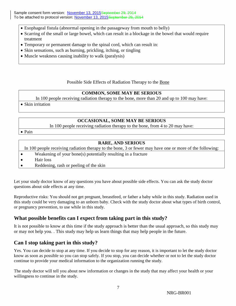
Sample consent form version: November 13, 2015September 29, 2014 To be attached to protocol version: November 13, 2015September 29, 2014
7
NRG-BR001
Esophageal fistula (abnormal opening in the passageway from mouth to belly)
Scarring of the small or large bowel, which can result in a blockage in the bowel that would require
treatment
Temporary or permanent damage to the spinal cord, which can result in:
Skin sensations, such as burning, prickling, itching, or tingling
Muscle weakness causing inability to walk (paralysis)
Possible Side Effects of Radiation Therapy to the Bone
COMMON, SOME MAY BE SERIOUS
In 100 people receiving radiation therapy to the bone, more than 20 and up to 100 may have:
Skin irritation
OCCASIONAL, SOME MAY BE SERIOUS
In 100 people receiving radiation therapy to the bone, from 4 to 20 may have:
Pain
RARE, AND SERIOUS
In 100 people receiving radiation therapy to the bone, 3 or fewer may have one or more of the following:
Weakening of your bone(s) potentially resulting in a fracture
Hair loss
Reddening, rash or peeling of the skin
Let your study doctor know of any questions you have about possible side effects. You can ask the study doctor
questions about side effects at any time.
Reproductive risks: You should not get pregnant, breastfeed, or father a baby while in this study. Radiation used in
this study could be very damaging to an unborn baby. Check with the study doctor about what types of birth control,
or pregnancy prevention, to use while in this study.
What possible benefits can I expect from taking part in this study?
It is not possible to know at this time if the study approach is better than the usual approach, so this study may
or may not help you. . This study may help us learn things that may help people in the future.
Can I stop taking part in this study?
Yes. You can decide to stop at any time. If you decide to stop for any reason, it is important to let the study doctor
know as soon as possible so you can stop safely. If you stop, you can decide whether or not to let the study doctor
continue to provide your medical information to the organization running the study.
The study doctor will tell you about new information or changes in the study that may affect your health or your
willingness to continue in the study.
Sample consent form version: November 13, 2015September 29, 2014 To be attached to protocol version: November 13, 2015September 29, 2014
8
NRG-BR001
The study doctor may take you out of the study: If your health changes and the study is no longer in your best interest
If new information becomes available
If you do not follow the study rules
If the study is stopped by the sponsor, Institutional Review Board (IRB) or the Food and Drug
Administration (FDA)
What are my rights in this study?
Taking part in this study is your choice. No matter what decision you make, and even if your decision changes,
there will be no penalty to you. You will not lose medical care or any legal rights.
For questions about your rights while in this study, call the ________________________ (insert name of
center) Institutional Review Board at __________________ (insert telephone number). (Note to Local
Investigator: Contact information for patient representatives or other individuals at a local institution who are
not on the IRB or research team but take calls regarding clinical trial questions can also be listed here.)
What are the costs of taking part in this study?
You and/or your health plan/insurance company will need to pay for the radiation and all of the other costs of
caring for your cancer while in this study, including the cost of tests, procedures, or medicines to manage any
side effects, unless you are told that certain tests are supplied at no charge. Before you decide to be in the study,
you should check with your health plan or insurance company to find out exactly what they will pay for.
You will not be paid for taking part in this study.
What happens if I am injured or hurt because I took part in this study?
If you are injured or hurt as a result of taking part in this study and need medical treatment, please tell your
study doctor. The study sponsors will not offer to pay for medical treatment for injury. Your insurance
company may not be willing to pay for study-related injury. If you have no insurance, you would be
responsible for any costs.
If you feel this injury was a result of medical error, you keep all your legal rights to receive payment for this
even though you are in a study.
Who will see my medical information?
Your privacy is very important to us and the researchers will make every effort to protect it. Your information
may be given out if required by law. For example, certain states require doctors to report to health boards if they
find a disease like tuberculosis. However, the researchers will do their best to make sure that any information
that is released will not identify you. Some of your health information, and/or information about your specimen,
from this study will be kept in a central database for research. Your name or contact information will not be put
in the database.
There are organizations that may inspect your records. These organizations are required to make sure your
information is kept private, unless required by law to provide information. Some of these organizations are:
The study sponsor: NRG Oncology

Sample consent form version: November 13, 2015September 29, 2014 To be attached to protocol version: November 13, 2015September 29, 2014
9
NRG-BR001
The Institutional Review Board, IRB, is a group of people who review the research with the goal of
protecting the people who take part in the study.
The Food and Drug Administration (FDA) and the National Cancer Institute (NCI) in the U.S., and
similar ones if other countries are involved in the study.
Where can I get more information?
You may visit the NCI Web site at http://cancer.gov/ for more information about studies or general information
about cancer. You may also call the NCI Cancer Information Service to get the same information at: 1-800-4-
CANCER (1-800-422-6237).
A description of this clinical trial will be available on http://www.ClinicalTrials.gov, as required by U.S. Law.
This Web site will not include information that can identify you. At most, the Web site will include a summary
of the results. You can search this Web site at any time.
Who can answer my questions about this study?
You can talk to the study doctor about any questions or concerns you have about this study or to report side
effects or injuries. Contact the study doctor __________________ (insert name of study doctor[s]) at
__________________ (insert telephone number).
My Signature Agreeing to Take Part in the Main Study (9/29/14)
I have read this consent form or had it read to me. I have discussed it with the study doctor and my questions
have been answered. I will be given a signed copy of this form. I agree to take part in the main study.
Participant’s signature________________________________
Date of signature_____________________________________
Signature of person(s) conducting the informed consent discussion___________________________________
Date of signature_____________________________________