nuclear medicine 4203 scanning & imaging respiratory system
TRANSCRIPT
Nuclear Medicine 4203Scanning & Imaging
Respiratory System

Anatomy and Physiology
Trachea divides into the right and left mainstem bronchi
These divide to form lobar bronchiRight side has upper, middle and lower lobe bronchi
Left side has upper and lower bronchi
Lobes further divide into segments

Lung Segments

Lung Anatomy
Main pulmonary arteries divide into each lung and follow the divisions of the bronchi and bronchioles to the level of the alveoli.
Each alveolus is supplied by a terminal pulmonary arteriole, which turns to capillaries.
Adults have 250-300 million alveoli

GI Motility online (May 2006) | doi:10.1038/gimo73
Figure 3 Schematic diagram of lung anatomy with cross-sections of bronchi, bronchioles alveolar ducts, and alveoli.

Lung Physiology
Gravity and patient position have a significant impact on both ventilation and perfusion.
In upright position, intrapleural pressure is significantly more negative at the apices than at the base of the lung.
Also in upright position, the apex receives only 1/3 of the blood flow compared to the base.

Radiopharmaceuticals
Perfusion: 99mTc macroaggregated albumin (MAA)Localizes by capillary blockageFewer than 1 in 1000 capillaries are blockedInjection should include 200,000-600,000 particlesNormal adult dose is 3-5 mCi of activity and 1-2 ml of volumeSyringe should be agitated before injectionShould be injected while patient is supine during respiration (some radiologists will prefer upright injection)Care should be taken not to draw back blood into the syringe~this will cause small labeled blood clots~causing focal hot spots on the image.

Radiopharmaceuticals
Perfusion:Contraindication to injecting 99mTc MAA
Severe pulmonary hypertension
Known Right-to Left shunt
In both cases, number of particles should be reduced to 100,000-200,000 particles.

Radiopharmaceuticals
Radioactive Inert Gas:133 Xe
Half life 5.3 daysGamma ray energy of 81 keVUsual dose of 10-20 mCiUsually done prior to 99mTc MAA perfusion
Imaged in posterior view1-initial breath2-equilibrium3-washout
Requires patient cooperationAdministered using delivery and rebreathing unit.


Radiopharmaceuticals
Radiolabeled AerosolsMap the distribution of aerated lung volume99mTc diethylene triamine pentaacetic acid (DTPA) 30-50 mCi of activity in 2-3 ml volume.Oxygen is supplied to the delivery system.Patient breaths in and out through a mouthpiece and the nose should be pinched off.Advantage is views can be taken in all 8 camera positions, to match perfusion.


Radiopharmaceuticals
Technegas and PertechnegasDelivered in a micro-aerosol generator
Still in FDA trials in U.S., but used commonly in other countries.
Advantage: pertechnegas can be delivered in only 1-2 breaths and multiple images can be obtained.

Indications
Suspected pulmonary embolusChest painShortness of BreathHypoxiaCoughing
Chest radiograph should be done 12-24 hrs. prior to VQ scan for comparison.CTA is generally preferred, but a VQ scan will still be warranted if:
Pt. has contrast allergyRenal failurePregnant (this is debatable)

Normal Perfusion Lung Scan
Uniform activity seen except a decreased area of cardiac silhouette and aortic knob.

Normal Perfusion

Normal Ventilation
133 XeNormal half-time washout for Xenon is 30-45 seconds.
May be deposited in the liver and result in increase activity in right upper quadrant.
Normal 99mTc Aerosol images resemble perfusion images.
Normal to see trachea and bronchi
Swallowed activity can be seen in the esophagus and stomach.

Perfusion Defects
Area of absent or diminished perfusion.
Classified as segmental or nonsegmental.Segmental may involve all or part of a bronchopulmonary anatomic segment. These are classically wedge shaped.
Nonsegmental do not correspond to anatomic segments and are generally not wedge shaped. These are NOT associated with pulmonary emboli. Can be caused by hilar or mediastinal structures, neoplasms, bullae, pneumonia, edema or other infiltrates.

High Probability StudyPulmonary Embolus

Do they Match?
A mismatch refers to a defect seen on perfusion, but is normal on the ventilation.
Segmental mismatch is a classic pulmonary emboli.
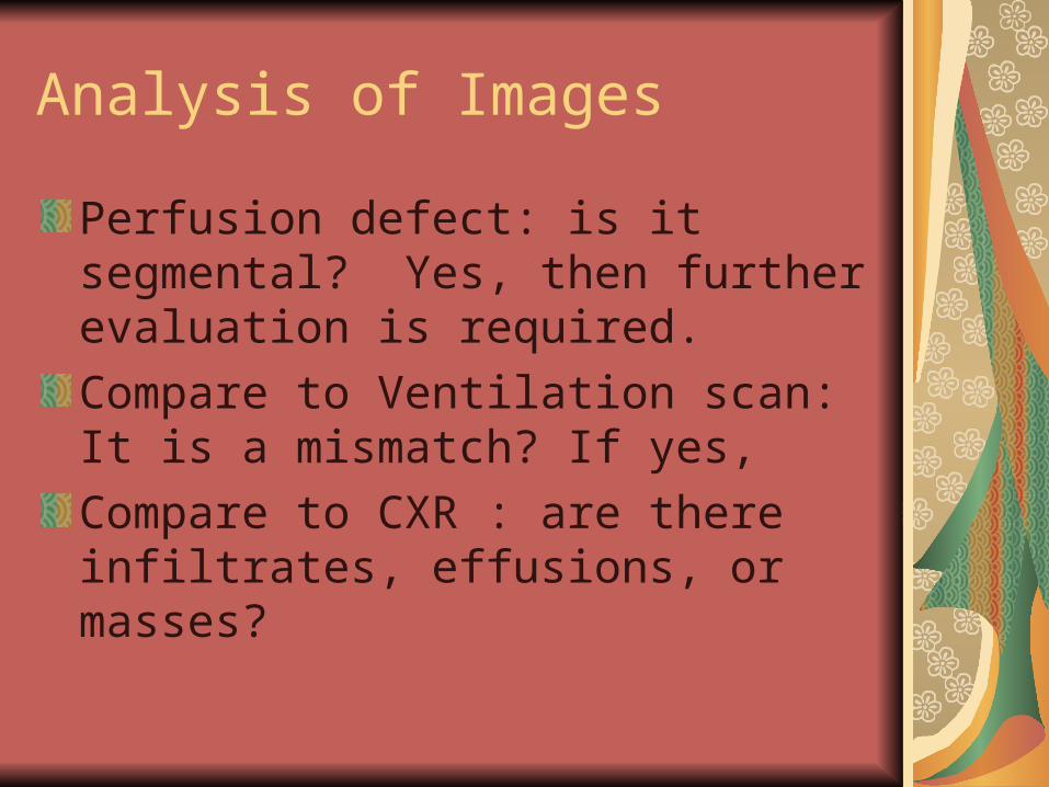
Analysis of Images
Perfusion defect: is it segmental? Yes, then further evaluation is required.
Compare to Ventilation scan: It is a mismatch? If yes,
Compare to CXR : are there infiltrates, effusions, or masses?

PIOPED II CriteriaProspective Investigation Of Pulmonary Embolism Diagnosis
High probabilityGreater than 80% likelihood of pulmonary emboli
Intermediate probability20-80% likelihood of pulmonary emboli
Low probabilityLess than 20% likelihood of pulmonary emboli
Very low probabilityLess than 10% likelihood of pulmonary emboli
IndeterminateShould be used only when technical factors limit the study
NormalNo perfusion defects


Stripe sign ~ Very unlikely to be pulmonary emboli
Fissure sign~ caused by pleural fluid in the fissures, pleural scarring or thickening, or COPD.
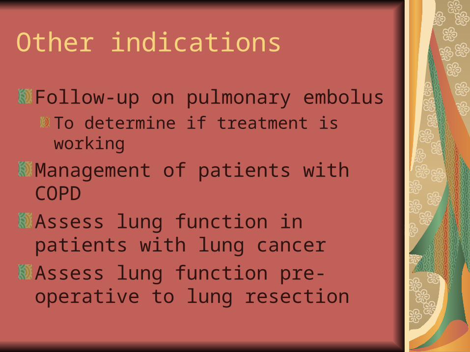
Other indications
Follow-up on pulmonary embolusTo determine if treatment is working
Management of patients with COPD
Assess lung function in patients with lung cancer
Assess lung function pre-operative to lung resection