nucleotide-dependent processes in the thylakoid lumen of plant
TRANSCRIPT

NUCLEOTIDE-DEPENDENT PROCESSES IN THE THYLAKOID LUMEN OF PLANT CHLOROPLASTS
Björn Lundin
Division of Molecular Genetics
Department of Physics, Chemistry and Biology Institute of Technology Linköping University
SE-581 83 Linköping, SWEDEN
Linköping 2008

© Björn Lundin, 2008 © Cover design: Björn Lundin All rights reserved ISBN: 978-91-7393-965-2 ISSN: 0345-7524 Linköping studies in science and technology. Dissertation No. 1165 Published articles and Figure 2 have been reprinted with the permission of the respective copyright holder: Paper I © 2004, The National Academy of Sciences of the USA Paper II © 2007, Elsevier B.V. Paper III © 2007, Blackwell Publishing Ltd Paper IV © 2008, Springer This work has been supported by the Swedish Research Council and the Graduate Research School in Genomics and Bioinformatics (FGB). We thank also the Salk Institute Genomic Analysis Laboratory for providing the sequence-indexed Arabidopsis T-DNA insertion mutants. Printed by: LIU-TRYCK, Linköping, Sweden, 2008
II

Ph. D. Thesis Thesis No. 1165
Nucleotide-Dependent Processes in the Thylakoid Lumen
of Plant Chloroplasts
Björn Lundin
Supervisor: Docent Cornelia Spetea Wiklund Division of Molecular Genetics Department of Physics, Chemistry and Biology Linköping University 581 83 Linköping, Sweden Opponent: Professor Zach Adam Institute of Plant Sciences
The Hebrew University 761 00 Rehovot, Israel
Linköping, 4 April, 2008
III

IV

“If you have no results, you haven’t worked” Anonymous
V

VI

ABSTRACT
Plants, algae and photosynthetic bacteria are able to harvest the sunlight and use its energy to
transform water and carbon dioxide to carbohydrate molecules and oxygen, both important to
sustain life on Earth. This process is called photosynthesis and is the route by which almost all
energy enters the biosphere. As most simple things in life, the process of photosynthesis is easily
explained but unfortunately not that easy to reproduce. If we could, we would be living in a much
different world with almost unlimited energy. Light energy is harvested by chlorophyll molecules,
bound to proteins in the chloroplast thylakoid membrane and drives the oxygen-evolving
complex, to extract electrons from water. Electrons are then transferred to NADPH through
photosystem II (PSII) to cytochrome b6f and photosystem I, the major photosynthetic protein
complexes. The cytochrome b6f complex also transfers protons into the lumenal space of the
thylakoid. These protons together with those from water oxidation create an electrochemical
gradient across the thylakoid membrane, which fuels the ATP synthase to produce ATP. ATP,
NADPH and carbon dioxide are used during the dark reactions to produce sugars in the
chloroplast stroma. The thylakoid lumenal space where the water oxidation occurs has until
recently been viewed as a proton sink with very few proteins. With the publication of the genome
of Arabidopsis thaliana it seems to be a much more complex compartment housing a wide variety
of biochemical processes.
ATP is a nucleotide and the major energy currency, but there are also other nucleotides
such as AMP, ADP, GMP, GDP and GTP. Chloroplast metabolism has mostly been associated
with ATP, but GTP has been shown to have a role in integration of light harversting complexes
into the thylakoid. In this work, we have demonstrated the occurrence of nucleotide-dependent
processes in the lumenal space of spinach by bringing evidence first for nucleotide (ATP)
transport across the thylakoid membrane, second for nucleotide inter-conversion (ATP to GTP)
by a nucleoside diphosphate kinase, and third the discovery that the PsbO extrinsic subunit of
PSII complex can bind and hydrolyse GTP to GDP. The active PSII complex functions as a
dimer but following light-induced damage, it is monomerised allowing for repair of its reaction
center D1 protein. PsbO is ubiquitous in all oxygenic photosynthetic organisms and together
with other extrinsic proteins stabilises the oxygen-evolving complex. We have modelled the
GTP-binding site in the PsbO structure and showed that the GTPase activity of spinach PsbO
induces changes in the protein structure, dissociation from the complex and stimulates the
degradation of the D1 protein, possibly by inducing momerisation of damaged PSII complexes.
As compared to spinach, Arabidopsis has two isoforms of PsbO, PsbO1 and PsbO2, expressed in
VII

a 4:1 ratio. A T-DNA insertion knockout mutant of PsbO1 showed a retarded growth rate, pale
green leaves and a decrease in the oxygen evolution while a PsbO2 knockout mutant did not
show any visual phenotype as compared to wild type. Unexpectedly, during growth under high
light conditions the turnover rate of the D1 protein was impaired in the PsbO2 knockout,
whereas it occurred faster in the PsbO1 knockout as compared to wild type. We concluded that
the PsbO1 protein mainly functions in stabilizing the oxygen evolving complex, whereas the
PsbO2 protein regulates the turnover of the D1 protein. The two PsbO proteins also differ in
their GTPase-activity (PsbO2 >> PsbO1). Although their amino acid sequences are 90%
identical, they differ in the GTP-binding region which could explain the difference in their
GTPase activity. Based on these data, we propose that the GTPase activity of PsbO(2) leads to
structural changes in interacting loops and plays a role in the initial steps of D1 turnover such as
the PSII monomerisation step.
The nucleotide-dependent processes we discovered in the thylakoid lumen raise
questions of transporters to facilitate these processes. As stated earlier, we provided biochemical
evidence of an ATP thylakoid transporter, and most recently have identified a transporter that
may be important for the export of lumenal phosphate back to the stroma. More transporters for
GDP, metal ions and others solutes have still to be identified.
VIII

POPULÄRVETENSKAPLIG SAMMANFATTNING
Växter, alger och fotosyntetiska bakterier lärde sig för 2.5-3 biljarder år sen att absorbera
solljusets energi och använda det för att omvandla vatten och koldioxid till kolväten. Denna
process kallas för fotosyntes och är den enskilt största energibindaren på jorden. Ljusenergin
absorberas av klorofyll-molekyler bundna till proteiner lokaliserade i kloroplastens tylakoid-
membran som driver själva vattenspjälkningen. Elektroner från vattnet vandrar till NADPH
genom de stora fotosystem-komplexen; Fotosystem II, Cytochrome b6f komplexet och
Fotosystem I. Cytochrome b6f komplexet har också som funktion att låta protoner vandra till
lumen, som är lokaliserad inne i tylakoiderna. Dessa protoner tillsammans med protonerna från
vattnet skapar en protongradient över membranet, vilket fungerar som drivmedel för ATP-
syntasens ATP produktion. ATP, NADPH och koldioxid används i mörker-reaktionen för att
producera sockerarter i stroma. Lumen där själva vattenspjälkningen sker, har tills nyligen antagits
vara en proton-uppsamlare med väldigt få proteiner. Med publikationen av backtravs genom har
en mer komplex syn på detta utrymme vuxit fram.
Nukleotiden ATP, är den huvudsakliga energitransportören nukleotid, men det finns
också andra nukleotider, till exempel AMP, ADP, GMP, GDP och GTP. Kloroplastens
metabolism har för det mesta blivit kopplat till ATP, men GTP har visat sig ha en roll vid
integrationen av ljusuppsamlande komplex in i tylakoid-membranet. I detta arbete har vi
demonstrerat förekomsten av nukleotid-beroende processer i lumen; för det första genom
nukleotid (ATP) transport över membranet; för det andra via nukelotid-konvertering, ATP till
GTP, med hjälp av en Nukleosidediphosphatekinase i lumen; för det tredje, upptäckten att
proteinet PsbO, som binder in till PSII och optimerar och skyddar vattenklyvningen, kan binda
och hydrolysera GTP till GDP.
De fotosyntetiska organismerna, särskilt växter, har svårt att röra på sig och kan inte
fysiskt skydda sig från överexponering av ljus och väder. Ljus, som är fotosyntesen drivkraft, är
också en direkt fara. För starkt ljus ger skada på fotosystem-komplexen och för att skydda sig för
det har flera skyddsåtgärder ”uppfunnits”. Reaktionscenter D1 proteinet är det protein som utstår
mest skada och har en av cellens snabbaste omsättning. Det aktiva PSII komplexet, som fungerar
som en dimer, måste monomeriseras innan det skadade D1 proteinet kan bytas ut. PsbO finns
närvarande i alla fotosyntetiska organismer och stabiliserar vattenklyvnings komplexet. GTP
aktiviteten av spenats PsbO ger strukturförändringar, dissociering från PSII och stimulerar
degradering av D1 proteinet. Arabidopsis thaliana har till skillnad från spenat, två isoformer av
PsbO; PsbO1 och PsbO2.
IX

X
En knockout-mutant av PsbO1 visar ett långsammare växtsätt, ljusgröna blad och en
minskning av syrgas-produktionen. PsbO2 knockout mutanter uppvisar däremot inte några
skillnader jämfört med vildtyps plantor. Oväntat så hade PsbO2 knockout mutanten, växande
under starkt ljus nedsatt D1 reparation medan det skedde fortare i PsbO1 knockout mutanter när
man jämförde det mot vildtyp. Vi kan dra slutsatsen att PsbO1 proteinets huvudsakliga funktion
är att stabilisera syrgaskomplexet medan PsbO2 proteinet reglerar D1 proteinets reparations-
cykel. Dessutom skiljer sig Arabidopsis isoformer av PsbO i deras GTPase aktivitet (PsbO2 >>
PsbO1). Fast deras aminosyra-sekvens är 90% identisk, skiljer de sig i GTP bindande områden,
vilket skulle kunna förklara aktiviteten. Baserat på detta föreslår vi att PsbO(2)s GTPase aktivitet
leder till struktuella förändringar och spelar en viktig roll i monomeriseringsteget av PSIIs D1
protein reparations-cykel.
De nukleotidberoende processer vi visat ger upphov till fler frågor om transportörer
över tylakoid-membranet, som kan transportera in och ut nödvändiga ämnen för processerna. Vi
har visat förekomsten av en ATP thylakoid transportör och vi har nyligen identifierat en
fosfattransportör som kan ha en viktig roll för regulationen av Pi innehållet i lumen. Andra
transportörer för till exempel GDP, metall joner och andra lösliga ämnen finns fortfarande kvar
att upptäcka.

ORIGINAL PUBLICATIONS
This thesis is based on the following publications, which will be referred to in the text by their
Roman numerals (I-V).
I. Spetea, C., Hundal, T., Lundin, B., Heddad, M., and Andersson, B. Multiple
evidence for nucleotide metabolism in the chloroplast thylakoid lumen. Proc. Natl. Acad.
Sci. USA 101, 1409-1414 (2004).
II. Lundin, B*., Thuswaldner, S*., Shutova, T., Eshaghi, S., Samuelsson, G,. Barber,
J., Andersson, B., and Spetea, C. Subsequent events to GTP binding by the plant
PsbO protein: structural changes, GTP hydrolysis and dissociation from the photosystem
II complex. Biochim. Biophys. Acta (Bioenerg.) 1767, 500-508 (2007).
*These authors equally contributed to this work.
III. Lundin B., Hansson, M., Schoefs, B., Vener, A. V., and Spetea, C. Arabidopsis
PsbO2 protein regulates dephosphorylation and turnover of the photosystem II reaction
centre D1 protein. Plant J. 49, 528–539 (2007).
IV. Lundin, B., Thuswaldner, S., and Spetea, C. Arabidopsis PsbOs differ in their GTPase
activity. In: Photosynthesis: Energy from the Sun (Allen, J.F. Ed) Springer publisher, chapter 5,
773-775 (2008).
V. Ruiz Pavón, L., Lundh, F., Lundin, B., Mishra, A., Persson, B. L., and Spetea, C.
Arabidopsis ANTR1 is a thylakoid Na+-dependent phosphate transporter -functional
characterization in Escherichia coli. J.Biol. Chem. revised manuscript submitted 25.02.2008.
XI

OTHER PUBLICATIONS
Spetea, C., Ruiz Pavón, L., Thuswaldner, S., Lundin, B., Lundh, F., Persson, B. L.,
Schoefs, B., and Adamska, I., Screening for solute transporters in plant photosynthetic
membranes. In: Photosynthesis: Energy from the Sun (Allen, J.F. Ed) Springer publisher, chapter
4, 1075-1078 (2008).
XII

ABBREVIATIONS
ATP adenosine 5’-triphosphate (energy carrier produced during photosynthesis) ADP adenosine 5’-diphosphate CP chlorophyll-binding protein CD circular diochrism Cyt b6f cytochrome b6f Chl chlorophyll D1 32 kDa D1 subunit of the photosystem II reaction center DNA deoxyribonucleic acid (where genes are coded) GTP guanosine 5’-triphosphate kDa kilo Dalton (weight unit for proteins) LC liquid chromatography LHC light-harvesting complex MALDI matrix-assisted laser desorption ionization MS mass spectrometry m/z mass over charge ratio (x-axis in MS spectrum) NADPH nicotinamide adenine dinucleotide phosphate, reduced form (reducing
potential produced in photosynthesis) NDPK nucleoside diphosphate kinase OEC oxygen-evolving complex Pi Inorganic phosphate Psb(O,P,Q,R) 33, 23, 16, 10 kDa extrinsic subunits of the photosystem II complex psbo(1,2) knockout mutants of PsbO(1,2) PSI photosystem I PSII photosystem II PQ plastoquinone RC reaction center SDS-PAGE sodium dodecyl sulfate polyacrylamide gel electrophoresis TAAC thylakoid ATP/ADP carrier TOF time of flight (detector in MS) WT wild type
XIII

XIV

TABLE OF CONTENTS
Abstract ............................................................................................................................................. VII
Populärvetenskaplig sammanfattning ............................................................................................ IX
Original Publications .......................................................................................................................XI
Other Publications .......................................................................................................................... XII
Abbreviations .................................................................................................................................. XIII
1. Introduction .......................................................................................................... 1
1.1. Photosynthesis: an overview ..................................................................................................... 1
1.2. Photosynthetic energy conversion ........................................................................................... 3
1.3. Photosynthetic complexes ......................................................................................................... 5 2. Functional genomics in plants .............................................................................. 9
2.1. Prediction studies ........................................................................................................................ 9
2.2. Photosynthetic preparations .................................................................................................... 10
2.3. Localisation studies .................................................................................................................... 11
2.4. Structural analyses ...................................................................................................................... 13
2.5. Phenotypic analyses ................................................................................................................... 14
3. Present investigation ............................................................................................ 16
3.1. Aim of this study ........................................................................................................................ 16
3.2. Working model ........................................................................................................................... 17
3.3. Nucleotide-dependent processes in the thylakoid lumen .................................................... 18
3.4. Plant PsbO as a GTPase ........................................................................................................... 20
3.5. ATP and phosphate thylakoid transporters ........................................................................... 27
4. Conclusions .......................................................................................................... 29
Acknowledgements ......................................................................................................................... 31
References ........................................................................................................................................ 33
XV

XVI

CHAPTER 1
1. Introduction
1.1. Photosynthesis: an overview
Plants, algae and photosynthetic bacteria are able to harvest sunlight and use its energy to
transform water and carbon dioxide to carbohydrate molecules and oxygen, both important to
sustain life on Earth. This process is called photosynthesis, and has evolved 2.5 to 3 billion years
ago from being dependent on substrates such as H2S, NH3 and Fe2+, to oxidising H2O
(McFadden, 1999; Olson, 2006) . It is the route by which almost all energy enters the biosphere.
Sunlight energy is harvested by chlorophyll molecules bound to hydrophobic proteins, located in
the chloroplast thylakoid membrane and drives the oxygen-evolving complex, where the water-
splitting reaction occurs (Lichtententhaler, 1987; Olson, 2006; Barber, 2007). Electrons are then
transferred to NADPH through photosystem II (PSII) via the cytochrome b6f complex (Cyt b6f)
and finally to photosystem I (PSI), the major photosynthetic protein complexes together with the
ATP synthase. The Cyt b6f complex also transfers protons into the lumenal space of the
thylakoid, in conjugation with the electron transport. These protons together with those from
water oxidation generate a proton gradient across the thylakoid membrane, which fuels the ATP
synthase to produce ATP. ATP, NADPH and carbon dioxide are then used in the dark reactions
to produce sugars in the chloroplast stroma. The thylakoid lumenal space where the water
oxidation occurs has until recently been viewed as a proton sink with very few proteins. With the
publication of the genome sequence of Arabidopsis thaliana (AGI, 2000) and proteomics works
(Peltier et al., 2002; Kieselbach and Schröder, 2003) it seems to be a much more complex
compartment housing a wide variety of biochemical processes.
1.1.1. Chloroplast
The chloroplast is the photosynthetic organelle in plants and green algae. It is surrounded by a
double membrane (outer and inner envelope), and contains the soluble stroma and thylakoids
(figure 1). The chloroplast, similarly to mitochondria, has its own genome, and thus the ability to
synthesise essential proteins for the photosynthetic machinery. However, a significant number of
chloroplast proteins are nuclear-encoded. For example, 10-15% of the nuclear gene products are
predicted to be targeted to the chloroplast (Peltier et al., 2002). Proteins that are targeted to the
1

chloroplast have an N-terminal chloroplast transit peptide (cTP), which is processed when the
precursor protein is translocated across the chloroplast inner envelope. Proteins then have two
options; either remain in the stroma or move further into the thylakoid membrane or lumen.
Proteins in the thylakoid lumen are targeted and transported via an additional transit peptide, the
lumenal transit peptide (lTP), which is located directly after the cTP. Soluble lumenal proteins
with lTPs are translocated by at least two different pathways: Sec pathway (ATP-dependent) and
twin-arginine translocation (tat) pathway (pH-dependent) (Robinson et al., 2001; Peltier et al.,
2002).
Figure 1. Schematic illustration of the chloroplast membrane and soluble compartments.
1.1.2. Thylakoid
The thylakoid membrane in plants can be divided into two different structures, namely grana and
stroma lammelae (Menke, 1962; Menke, 1990). The grana regions usually cluster together into
grana stacks, made up from repeating units that contain paired layers. These grana stacks can be
further divided into surface-exposed, core and margin regions (Nelson and Yocum, 2006). The
grana stacks are interconnected by the stroma lammelae (figure 1). Recently, the three
dimensional structure of the plant thylakoid membrane has been revealed using electron
tomography (Shimoni et al., 2005). In this structure the top layer bends slightly upwards and
fuses with the layer above it and the other layer bends downwards and fuses with the layer below
it. This proceeds with a counter clockwise rotation of 25° around the axis (figure 1). The
thylakoid harbours the four photosynthetic complexes, PSII, Cyt b6f, PSI and ATP synthase
driving the light reactions. These complexes are located differentially in the thylakoid membrane.
The dimeric PSII complex is located in the grana, PSI and the ATP synthase are located in the
stroma lammelae and the surface-exposed grana, whereas Cyt b6f is evenly distributed across the
two thylakoid structures (Anderson, 1980; Allen and Forsberg, 2001; Anderson, 2002). However
2

other reports suggest that Cyt b6f, which transports electrons between PSII and PSI, might
mostly be localised in the grana margins (Allen and Forsberg, 2001).
1.1.3. Lumen
The thylakoid lumenal space is located inside the thylakoid and is continuous between the grana
and stroma lammelae, as revealed in the recent 3-D structure (Shimoni et al., 2005). It has been
predicted to contain around 200 proteins, although no more than 80 proteins have been
identified using proteomics (Peltier et al., 2002; Schubert et al., 2002). There is also tremendous
evidence for various biochemical activities in the lumen, such as chaperones (Schlicher and Soll,
1996), immunophilins (Fulgosi et al., 1998), carbonic anhydrases (Park et al., 1999), violaxanthin
deepoxidases (Hieber et al., 2000), peroxidases (Kieselbach et al., 2000) and proteases (Adam,
2001). This growing complex view of the thylakoid lumen also raises the question of nucleotide-
dependent processes in this chloroplast compartment.
1.2. Photosynthetic energy conversion
Photosynthesis is a unique and complex process converting sunlight into chemical energy. This
process can be divided into two sets of reactions, the light and the dark reactions. In the light-
driven reactions, light energy is used by chlorophyll bound to proteins located in the chloroplast
thylakoid membrane, to drive the oxygen-evolving complex to form ATP from ADP, reduce
electron carrier molecules, such as NADP+ to NADPH, and produce oxygen as a by-product.
The dark reactions, called so because they proceed without the need for light, represent the
second part of photosynthesis, where the energy of ATP and reducing power of NADPH are
used for carbon dioxide fixation into sugars (Barber, 2007).
An overall equation of the oxygenic photosynthetic reactions is the following:
CO2 + H2O + light -> (CH2O)n + O2
The photosynthetic pigments such as chlorophylls, carotenoids and phycobilins are
embedded in the thylakoid membrane in units called photosystems (PS) (Lichtententhaler, 1987).
One photosystem can contain 250-750 pigment molecules (Dunahay et al., 1984; Neale and
Priscu, 1995), and consist of two subcomplexes, an antenna protein complex (LHC) and a
reaction centre (RC) complex, that capture the light at different wavelengths. There are several
types of chlorophyll, among which chlorophyll a occurs in all photosynthetic eukaryotes and
cyanobacteria. Chlorophyll b is found in vascular plants, bryophytes, green algae and euglenoids,
3

and functions as an accessory pigment to broaden the spectrum of the light that can be absorbed.
When a chlorophyll b, molecule absorbs the light, the energy is transferred to chlorophyll a which
leads the energy to the oxygen-evolving complex (OEC) (Mathews and Holde, 1990; Raven et al.,
1992).
When the light energy is absorbed by the pigments, the energy is transferred until it
reaches the reaction center, which has one special pair of chlorophyll molecules. When one of
these two chlorophylls absorbs the light energy, it looses one electron which is then transferred
to a primary acceptor molecule, pheophytin (Pheo) (Raven et al., 1992).
There are two different photosystems in algae and plants, called photosystem II and I,
having their own reaction center molecules, P680 and P700, with maximum absorption at 680
and 700 nm, respectively. In both photosystems, the primary step driven by the light energy is a
charge separation from where one electron is provided to the electron transfer chain (figure 2)
(Baker et al., 2007).
The water-splitting reaction takes place on the lumenal side of PSII complex, and is
performed by the OEC complex, composed of four Mn2+ and one Ca2+ ion (figure 2). Water is
split into protons, oxygen and electrons. A tyrosine residue (Yz), positioned on the D1 protein,
functions as an electron transfer intermediate between P680 and the OEC complex. It reduces by
electron transfer P680 to its ground state, while itself is reduced by an electron from the splitting
Figure 2. A schematic representation of the electron and proton transfer chain and of the photosynthetic
subunits involved.
4

of water (Baker et al., 2007).
Electrons are transferred via redox components through PSII to the Cyt b6f and PS I,
where the second type of charge separation occurs, involving P700 (figure 2). Electrons are
transferred to the primary acceptor Chl A0, then to PhQ A1, FeSx, FeSA and FeSB, from where
they leave PSI and reduce ferredoxin (Fd). The reduced ferredoxin reduces NADP+, via
ferredoxin-NADP+ oxidoreductase to NADPH (Merchant and Sawaya, 2005). The Cytochrome
b6f complex also transfers protons into the lumenal space of the thylakoid. The proton gradient
across the thylakoid membrane drives the ATP synthase. ATP together with NADPH are used
during carbon dioxide fixation (Mathews and Holde, 1990; Raven et al., 1992) (Baker et al., 2007).
For details of the light reactions, see the recent review by (Merchant and Sawaya, 2005).
Photosynthetic organisms, especially plants, have a hard time to cope with the
environment. They are literally rooted to the ground and cannot with ease move from their spot.
Sunlight, which drives photosynthesis, can be damaging to the photosynthetic machinery and
especially when the plants are exposed to high radiation. To overcome this problem, they have
developed several protection systems; from turning of leaves to extensive repair systems and
movement of the LHCII between PSII and PSI.
1.3. Photosynthetic complexes The four photosynthetic complexes are the PSII complex, Cyt b6f complex, the PSI complex and
the ATP synthase. Among them, the PSII complex has been the main focus of research in the
present investigation. There is overwhelming structural and functional information about each of
these complexes, for recent reviews see (Dekker and Boekema, 2005; Nelson and Yocum, 2006).
1.3.1. Photosystem II
Photosystem II (PSII), a multi-subunit membrane–protein complex, uses light energy to perform
the most thermodynamically demanding and unique reaction of photosynthesis, namely the
oxidation of water to molecular oxygen and reducing equivalents. X-ray crystal structures
obtained for the PSII complex isolated from Synechococcus elongatus, Thermosynechococcus vulcanus and
Thermosynechococcus elongatus, under various resolutions (3.8 to 3.0) (Zouni et al., 2001; Kamiya and
Shen, 2003; Ferreira et al., 2004; Loll et al., 2005) (figure 3). The PSII dimer has a 105 Å of depth,
205 Å of length and 110 Å of width at 3.5 Å resolution, and a molecular mass of 650 kDa
(Ferreira et al., 2004). Each monomer contains more than 20 different proteins where 16 are
intrinsic and four are extrinsic (Hankamer et al., 1997; De Las Rivas et al., 2004). Based on these,
5
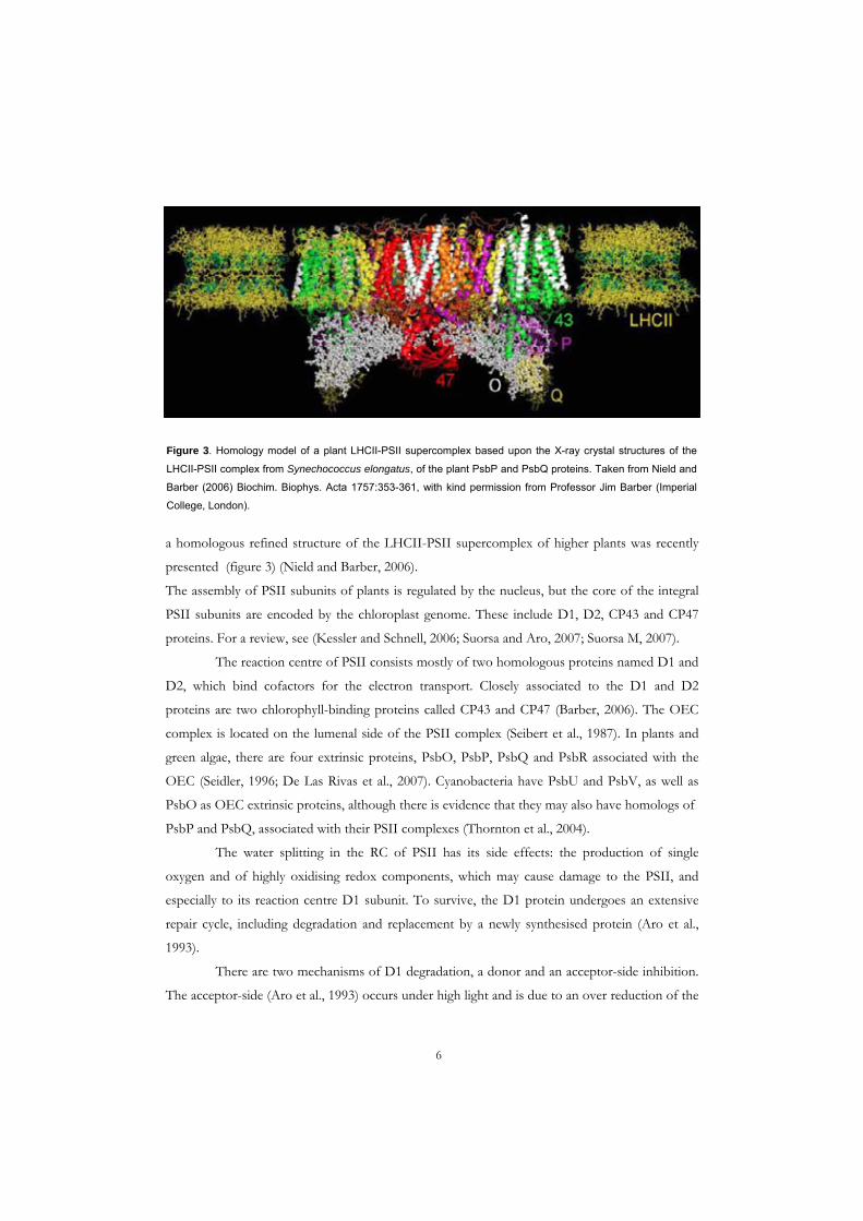
a homologous refined structure of the LHCII-PSII supercomplex of higher plants was recently
presented (figure 3) (Nield and Barber, 2006).
Figure 3. Homology model of a plant LHCII-PSII supercomplex based upon the X-ray crystal structures of the
LHCII-PSII complex from Synechococcus elongatus, of the plant PsbP and PsbQ proteins. Taken from Nield and
Barber (2006) Biochim. Biophys. Acta 1757:353-361, with kind permission from Professor Jim Barber (Imperial
College, London).
The assembly of PSII subunits of plants is regulated by the nucleus, but the core of the integral
PSII subunits are encoded by the chloroplast genome. These include D1, D2, CP43 and CP47
proteins. For a review, see (Kessler and Schnell, 2006; Suorsa and Aro, 2007; Suorsa M, 2007).
The reaction centre of PSII consists mostly of two homologous proteins named D1 and
D2, which bind cofactors for the electron transport. Closely associated to the D1 and D2
proteins are two chlorophyll-binding proteins called CP43 and CP47 (Barber, 2006). The OEC
complex is located on the lumenal side of the PSII complex (Seibert et al., 1987). In plants and
green algae, there are four extrinsic proteins, PsbO, PsbP, PsbQ and PsbR associated with the
OEC (Seidler, 1996; De Las Rivas et al., 2007). Cyanobacteria have PsbU and PsbV, as well as
PsbO as OEC extrinsic proteins, although there is evidence that they may also have homologs of
PsbP and PsbQ, associated with their PSII complexes (Thornton et al., 2004).
The water splitting in the RC of PSII has its side effects: the production of single
oxygen and of highly oxidising redox components, which may cause damage to the PSII, and
especially to its reaction centre D1 subunit. To survive, the D1 protein undergoes an extensive
repair cycle, including degradation and replacement by a newly synthesised protein (Aro et al.,
1993).
There are two mechanisms of D1 degradation, a donor and an acceptor-side inhibition.
The acceptor-side (Aro et al., 1993) occurs under high light and is due to an over reduction of the
6

PQ pool. This in turn leads to the formation of P680 triplet state that can cause the formation of
singlet oxygen. The single oxygen, produced in the vicinity of the D1 protein, leads to an
excessive damage and the need for repair (Aro et al., 1993). The donor-side inhibition occurs
when the donor-side of PSII can not keep up with the withdrawal of electrons from P680.
Leading to long lived P680+ and the oxidised tyrosine electron donor Yz+, which causes an
inactivation of the PSII electron transport (Aro et al., 1993).
The D1 repair cycle starts with phosphorylation at its N-terminus by the STN8 kinase,
activated by the reduction of the PQ pool; second, the oxidative damage to the D1 protein due to
highly reactive singlet oxygen; third, the monomerisation of PSII and detachment of CP43, PsbP
and PsbQ; fourth, migration of the momomerised PSII complex to the stroma lamellae; fifth,
dephosporylation of the D1 protein; sixth, multi-step degradation: first, by a primary cleavage by
DegP2 resulting in a 23 kDa N-terminus and 10 kDa C-terminal fragments followed by a
secondary cleavage by FtsH; seventh, a cotranslation insertion of a new D1 protein; eight, PSII
monomer migration to grana and dimerisation of the PSII complex; ninth, activation of the PSII
dimer to a functional state. (Adir et al., 1990; Aro et al., 1993; Spetea et al., 1999; Lindahl et al.,
2000; Haußühl et al., 2001; Zaltsman et al., 2005; Kapri-Pardes et al., 2007).
1.3.1.1. Extrinsic proteins
The 33, 23, 16 and 10 kDa extrinsic proteins of PSII (PsbO, PsbP, PsbQ and PsbR) are
associated on the lumenal side of thylakoid membrane (figure 3). They were thought to be
directly involved in oxygen evolution since the removal of the 23 and 16 kDa subunits led to a
partial inactivation of the oxygen evolution (Seidler, 1996). The PsbO subunit, also called the
manganese stabilising protein, is conserved in all known oxygenic photosynthetic organisms,
showing a 40–50% sequence homology between cyanobacteria and higher plants. PsbO
optimizes the ionic environment, provides a polar channel for protons/water, and stabilises the
metal cluster (Seidler, 1996; De Las Rivas and Barber, 2004; Murray and Barber, 2007).
PsbP together with PsbQ have been thought to be responsible of Ca+ and Cl- binding
(Seidler, 1996), but a recent paper states that PsbP is required to maintain the active Mn2+, Ca-, Cl-
cluster in vivo, and that PsbQ is not necessary for either PSII function or growth (Ifuku et al.,
2005).
In the present investigation, the focus has been on the PsbO protein. A review by (De
Las Rivas et al., 2007) goes through the other extrinsic proteins in more detail.
7

1.3.2. Cytochrome b6f (Cyt b6f)
The oxidation of plastoquinol occurs by an integral membrane protein complex, Cyt b6f. It
functions as a plastoquinone-plastocyanin oxidoreductase, transferring electrons from
plastoquinol to plastocyanin, a copper-containing protein. This electron transfer is accompanied
by the translocation of protons across the membrane, this creates a proton gradient across the
thylakoid membrane, which drives the ATP synthesis in the stroma (Cramer et al., 2006).
1.3.3. Photosystem I (PSI)
The plant photosystem I (PSI) complex, is a monomer as compared to cyanobacterial PSI which
is a trimer as to PSII which is a dimer, reviewed in (Melkozernov et al., 2006). PSI accepts
electrons from the cyt b6f complex via plastocyanin. PSI reaction centre Chl absorbs light with a
maximum wavelength of 700 nm (P700). The electrons are transported through a chain of redox
cofactors to the ferredoxin-NADP+ complex, completing the electron flow from H2O to
NADP+. PSI has a peripheral LHC (LHCI), but can share the LHCII with PSII. Under changing
light conditions (state transitions) LHCII detaches itself from PSII and connects to PSI (Allen
and Forsberg, 2001)
1.3.4. ATP synthase
There are three types of ATPases, P, F and V–type, where the F-type is located in the inner
membrane of mitochondria and in the thylakoid membrane of chloroplasts. The sequence of
thylakoid ATP synthase is termed CF1CF0 complex. By analogy with mitochondrial ATPase
which transfers protons into the matrix, the thylakoid ATP synthase is driven by the H+
emigration from the lumenal space to the stroma (Mathews and Holde, 1990). The mechanism of
the proton-driven ATP synthesis can be described in three steps; first, translocation of protons
carried out by F0; second, catalysis of formation of the phosphoanhydride bond of ATP carried
out by F1; third, coupling of the dissipation of the proton gradient with ATP synthesis (Mathews
and Holde, 1990).
8

CHAPTER 2
2. Functional genomics in plants
Identification and functional characterisation of proteins from organisms with sequenced
genomes (eg. Arabidopsis, (AGI, 2000)) involves several steps in a functional genomic approach.
The prediction provides information about the sequence of the protein of interest, in which
species it can be found, the putative function and where it can be localised. Also, by using
bioinformatics, protein candidates for a certain biochemical activity can be found. Then,
localisation- and characterisation studies in vivo and in vitro provide the experimental validation of
the predictions. An important part of the characterisation is represented by phenotypic studies of
knockout mutants for the gene candidate.
2.1. Prediction studies There are numerous databases, servers and tools that can be accessed over the Internet or used as
a stand alone program; a few of them and routine analyses are listed below.
2.1.1. Databases and tools
Below, I briefly describe the databases and tools that I have used in the present investigation.
NCBI (http://www.ncbi.nlm.nih.gov/) was established in 1988 as a National Resource for
Molecular Biology Information, creates public databases, conducts research in computational
biology, develops software tools for analysing genome data, and disseminates biomedical
information (NCBI).
ExPASy (http://www.expasy.ch/) is a server dedicated for the analysis of protein sequences and
structures. Swiss-Prot is the most popular and used database available at this server (Gasteiger et
al., 2003).
BLAST is a widely used tool at several servers (NCBI, ExPASy), and searches for homologies to
sequences in the same organism and other species.
ClustalW is a tool found at EBI, used to align sequences and to identify conserved regions.
9

TargetP (http://www.cbs.dtu.dk/services/TargetP/) predicts the sub-cellular location of
eukaryotic proteins. The location assignment is based on the predicted presence of any of the N-
terminal presequences: chloroplast transit peptide (cTP), mitochondrial targeting peptide (mTP)
or secretory pathway signal peptide (SP) (Nielsen et al., 1997; Olof Emanuelsson, 2000).
PyMOL (http://pymol.sourceforge.net/) is a PDB viewer program, based upon an opensource
project, and visualises protein crystal structures saved in a pdf format (PyMOL, 2006).
CAVER (http://loschmidt.chemi.muni.cz/caver/index.php) calculates tunnels leading from
cavities inside static or dynamic protein or protein complexes. It does this by calculating the least
costing path, by giving numerical numbers corresponding to the resistance of closely located
molecules. It can be used both as a plugin for the PyMOL program or as an online application
(Petřek et al., 2006; Damborský et al., 2007).
Signal (http://signal.salk.edu/) is used to search for availability of public collection of Arabidopsis
knockout mutants and the insert location (Alonso et al., 2003).
Geneinvestigator (https://www.genevestigator.ethz.ch/) is a reference expression database and
meta-analysis system. It allows to study the expression and regulation of genes in a broad variety
of contexts by summarizing information from hundreds of microarray experiments into easily
interpretable results (Zimmermann et al., 2004; Zimmermann et al., 2005).
2.2. Photosynthetic preparations
For the main parts of this thesis, Arabidopsis thaliana has been used as plant m
aterial (papers I, III and IV). Spinach was used in the earlier stages of this work (papers I and II).
Arabidopsis is a small flowering plant that was discovered by Johannes Thal already in the
sixteenth century. It is favoured by scientists for its small genome which was fully sequenced in
2000. The genome has a total of 125 Mbp with 25,498 genes, encoding proteins belonging to
11,000 families (AGI, 2000).
Isolation of chloroplasts, thylakoids and various PSII components are described in the attached
papers. Here I provide a summarised flow scheme (figure 4); first, the leaves are homogenised in
a mixer; second, chloroplasts are isolated through centrifugation and washing steps; third,
thylakoids are extracted by the disruption of the chloroplast envelope using osmotic shock
followed by differential centrifugation; forth, using thylakoids, various preparations
10

Figure 4. Flow scheme for preparation of various chloroplast sub-fractions.
can then be obtained: LHCII-PSII supercomplexes, PSII membranes (BBY), PSII cores and the
soluble lumen content, by using detergents or mechanical pressure combined with various
centrifugation steps. There are major differences between the different types of preparations. The
PSII membranes are fragments from the grana regions enriched in LHCII-PSII dimeric
complexes, and containing all of the OECs extrinsic proteins. PSII cores are PSII monomers
largely devoid of LHCII as well of all extrinsic proteins except for PsbO.
2.3. Localisation studies To localise your protein of interest, there are many standard techniques; those used in the present
investigation are listed below.
2.3.1. Sodium dodecyl sulfate polyacrylamide gel electrophoresis, SDS-PAGE
Gel electrophoresis is a technique where proteins are separated in a solid matrix using an electric
field. The rate of the migration is based on the protein net charge, size, electric field, and also on
11

the composition of the mediums (Westermeier, 2001). The most common used variant is SDS-
PAGE, a discontinuous system originally described by Laemmli (Laemmli, 1970). SDS is an
anionic detergent, which binds strongly to and denatures proteins. SDS removes the intrinsic
charge of the protein, and a constant negative net charge per unit is obtained. When run under
the same conditions on a polyacrylamide gel, different sized proteins will travel at various speed,
and therefore be separated on the gel.
2.3.2 Immuno detection
The protein of interest can be probed with antibodies raised against a homologous protein from
another organism or against a specific peptide from this protein. The protein, that can be in an
isolated form or associated to a complex, can be detected by Western blotting or fished out using
immonoprecipitation.
2.3.3. Radioactive labelling
Radioactive assays are used to give experimental proof of protein function. The methods use
radioactively labelled compounds such as 14C, 32P, relevant for the function of the protein. The
radio-labelled protein is detected on a phosphorimage of SDS-gels. The radio-labelled nucleotides
are detected by phosphorimage of a thin layer chromatographic plate. The amount of
radioactivity is determined by liquid scintillation spectrometry.
2.3.4. Organelle import
Organelle import is a classic method for localisation studies. The protein of interest is synthesized
in vitro in a radiolabelled precursor form, containing a targeting peptide at its N-terminus that
directs the protein for example into the chloroplast. The protein can also be synthesised as a
fusion construct with a recognition label at the C-terminus, such as a green fluorescent protein
(GFP) tag, which can be visualised in a fluorescence microscope.
2.3.5. Mass spectrometry
The protein of interest can be enzymatically cleaved and extracted from a protein mixture, SDS-
gel or membrane and the obtained peptides can be analysed without breaking the covalent
structure of these molecules by mass spectrometry (MS) techniques. In the matrix-assisted laser
desorption ionization (MALDI) technique (Tanaka et al., 1988), and further developed in the
time-of-flight (TOF), ionized peptides are accelerated in an electric field and enter a flight tube.
During the flight in this tube, different molecules are separated according to their mass to charge
12

ratio (m/z) and reach the detector at different times. The masses of the peptides are then
analysed and searched in various databases. MS techniques can also be used for quantitative
applications, for example to compare the levels of a certain protein in knockouts mutants and
wild type plants (Edvardsson et al., 2007).
2.4. Structural analyses
There are four different aspects of the structure of a protein. Its primary structure is the amino
acid sequence of the polypeptide chain. The secondary structure (sub structure) shows the α-
helix, β-sheet and random coils distribution in the protein. The tertiary structure shows how the
protein is folded in space. The quaternary structure shows how proteins are folded together in
complexes. Different techniques listed below have been used to investigate protein structure in
this thesis (Mathews and Holde, 1990).
2.4.1 Circular dichroism
Circular dichroism (CD) is a technique to characterise the secondary structure of proteins. The
CD spectra are generated based on the optical spectra of the polypeptide chain. When a protein
is exposed to circulary polarised light the different structures of the protein give characteristic
bands, reflecting the electronic excitation energies. Secondary structure elements such as the α-
helix, β-sheet and random coil all give specific bands. This analysis can therefore lead to the
determination of the secondary structure of a protein. CD can also be used to detect
conformational changes induced with substrates (Bulheller et al., 2007).
2.4.2. Intrinsic fluorescence
Intrinsic fluorescence of the aromatic amino acids tryptophan, tyrosine and phenylalanine is used
to monitor structural changes in proteins. Among them, tryptophan has the highest fluorescence
and quantum yield. The tryptophan fluorescence is used as a tool to determine if the residue is
buried in the protein core or exposed on the surface, and thus to investigate changes in the
tertiary structure of a protein during folding and substrate binding. For reviews, see (Cioni and
Strambini, 2002; Bulheller et al., 2007).
13

14
2.5. Phenotypic analyses
2.5.1. Plant fitness
The overall fitness of knockout mutants has been compared to that of wild type (WT) plants
during growth under normal (120 µmol photons m-2 s-1) and under high light (1000 µmol photons
m-2 s-1).
2.5.2. Leaf weight
The weight of leaves is in direct correlation with how large they are. Comparing the leaf weight in
plants under various growth conditions, as in paper III, may provide indications for an impaired
function. The reduced growth of a knockout mutant points to impaired functions due to the
absence of an important protein.
2.5.3. Chlorophyll a/b ratio
To determine total chlorophyll Chl (a+b) concentration and Chl a/b ratio of a photosynthetic
preparation, samples are measured in a spectrophotometer at specific wavelengths, 663 and 645
nm, corresponding to maximum absorption by chlorophyll a and chlorophyll b, respectively. The
equation of Arnon (Arnon, 1949) was previously routinely used, but recently the equation of
Porra (based upon a modification of Arnon) has gained popularity. A ratio of around 3 is typical
for thylakoids, around 2 for isolated PSII membranes and greater-than or equal to 4 for PSII
cores (Porra, 2002).
2.5.4. Chlorophyll content
The chlorophyll content of a photosynthetic tissue gives indications about the rate of senescence
as well as the efficiency of photosynthesis. For comparing young to mature and senescent tissues,
a ratio of the chlorophyll content per mass of tissue is calculated.
2.5.5. Oxygen evolution
Molecular oxygen produced during water oxidation by OEC of PSII, can be used as an indicator
how well the photosynthetic apparatus performs. It can be measured by using an oxygraph with a
Clark-type electrode. By using saturated light and electron acceptors, which withdraw electrons
from different parts of the photosynthetic machinery, we can locate the irregularity in the
electron transfer chain.

2.5.6. Chlorophyll fluorescence
The light energy that is absorbed by the leaf may take three directions; it can be dissipated as
heat, drive photosynthesis or can be emitted as fluorescent light. The emission of fluorescent
light from the leaf is called chlorophyll fluorescence. Although the emitted light is low (only 1-2%
of the total light absorbed), it can be measured using a fluorometer. The three directions are
always in conjugation with each other, if one goes up the others go down so that the energy
always is the same as the light energy coming in. By measuring the yield of chlorophyll
fluorescence, information about the other directions can be gained, as reviewed in (Maxwell and
Johnson, 2000).
Once PSII has absorbed light and QA has accepted one electron, it is not able to accept
another one until it has passed the first electron to the next electron carrier QB. During this time,
the reaction centre is called “closed”. In darkness the reaction centers are open and by exposing
them to light they will gradually be closed. An increase in the ratio between closed and open
reaction centers leads to an increase in the yield of chlorophyll fluorescence.
15

CHAPTER 3
3. Present Investigation
3.1. Aim of this study
The lumenal space, of the thylakoid was known in the past to harbour just a few proteins, but
recent biochemical and proteomic reports point to a much more active role for this compartment
in photosynthetic regulation. This new and complex view of the lumenal space raises several
questions of the chloroplast function and regulation. One of these questions concerns the
presence of nucleotides, nucleotide-binding proteins and nucleotide regulation in the thylakoid
lumen of plant chloroplasts, which was a general aim of this work.
This chapter summarises the findings of the papers I to V. A short description of the aim of each
paper is listed below.
Paper I: To identify nucleotide-dependent reactions in the thylakoid lumen of spinach
chloroplasts and an ATP transporter in thylakoid membrane.
Paper II: To characterise the GTP-binding properties and GTPase activity of spinach
PsbO.
Paper III: To study the roles of Arabidopsis PsbO isoforms.
Paper IV: To investigate if Arabidopsis PsbO isoforms differ in their GTPase activity.
Paper V: To identify and functionally characterise a phosphate transporter in the
Arabidopsis thylakoid membrane.
16

3.2. Working model
Figure 5. Working model during the present investigation
1. ATP import and ADP export across the thylakoid membrane, via the thylakoid
ATP/ADP carrier, TAAC (Paper I)
2. Intra-conversion of ATP to GTP, by transfer of the γ-phosphate from ATP to GDP via
Nucleoside diphosphate kinase, NDPK3 (Paper I)
3. GTP binding and hydrolysis by the PSII extrinsic PsbO protein (Papers I, II and IV)
4. Following light-induced damage to PSII, the PsbO1 protein of Arabidopsis thaliana is
exchanged for the PsbO2 protein. The GTPase activity of the PsbO2 protein (PsbO in
spinach), initiates the monomerisation of the PSII dimer as an initial step of D1 turnover
(Papers III and IV)
5. Lumenal Pi is exported to the stroma via the thylakoid anion transporter 1, ANTR1
(Paper V)
17

3.3. Nucleotide-dependent processes in the chloroplast
The outer and inner envelope of the chloroplast host several nucleotide-dependent processes and
have the function to transport and exchange proteins, solutes and metabolites between the
cytosol and the stroma. Most chlorophyll proteins are nuclear-encoded and need to be imported
through these membranes from the cytosol. Two protein complexes are responsible for the
protein import, namely the translocon at the outer envelope membrane of chloroplasts, Toc, and
the translocon at the inner envelope membrane of chloroplasts, the Tic apparatus. Toc binds the
preprotein, coming from the cytosol, in the presence of ATP and GTP and the preproteins
transit peptide associates with the Tic complex and forms a Toc and Tic supercomplex, reviewed
in (Chen and Schnell, 1999; Spetea and Thuswaldner, 2008). The insertion of the protein from
the Tic complex is made by the help of hydrolysis of stromal ATP. In addition to protein import,
the inner envelope holds other nucleotide-dependent processes such as lipid translocation and
transporters (Lu et al., 2007; Spetea and Thuswaldner, 2008).
Inside the chloroplast are nucleotides, such as ATP, produced by the ATP synthase on
the stromal side of the thylakoid membrane. These are involved not just in the photosynthetic
dark reactions but also play an important roll in energy-dependent processes such as
phosphorylation, transport, degradation and folding of proteins (Spetea and Thuswaldner, 2008).
Protein phosphorylation on the stromal side of the thylakoid membrane has an important role in
how photosynthetic organisms cope with the environment. Excess light to PSII relative to PSI
leads to the reduction of the PQ pool (Vener et al., 1997) and the phosphorylation of the light
harvesting complex signals for a reversible association of LHCII between PSII and PSI, called
state transition (Zito et al., 1999; Bellafiore et al., 2005). During high light stress, PSII core
proteins, especially the PSII D1 protein that harbours most of the redox components get
phosphorylated, dephosphorylated and degraded (Aro et al., 1993). The turnover rate of the D1
protein is the fastest known in chloroplast, with a half life of 30 minutes. The degradation step
following light-induced damage has been shown to be nucleotide dependent (Spetea et al., 1999;
Spetea et al., 2000).
While the stromal side of the thylakoid membrane has several nucleotide-dependent
processes involved in the regulation of photosynthetic light reactions, there have been no reports
on the presence of nucleotides in the thylakoid lumen before the initiation of the present
investigation.
It has earlier been reported that the presence of GTP bound to the thylakoid membrane
enhances the D1 degradation rate (Spetea et al., 1999; Spetea et al., 2000) In paper I, we have
18

attempted to find GTP-binding proteins in the thylakoid membrane with a role in D1 protein
degradation (figure 5).
For this purpose, we incubated dark-controlled and pre-illuminated thylakoid
membranes with [α-32P]8-N3GTP, which upon UV exposure binds covalently to polypeptides
containing GTP-binding sites. Two radioactive bands of 33 and 36.5 kDa could be detected. The
band of 33 kDa was detected mainly in the preilluminated thylakoids and to a lower extent in the
dark sample. When isolated PSII cores were incubated with [α-32P]8-N3GTP, the 33 kDa protein
was detected, but not the 36.5 kDa protein. The labelling of the 33 kDa band was inhibited by
DCMU, which points to an involvement in the electron transport. The photolabelled 33 kDa was
washed away by alkaline TRIS and crossreacted with an antibody against the OEC33 (PsbO)
protein. Upon incubation of thylakoid membranes with [γ-32P]8-N3ATP, only the 36.5 kDa was
detected and stronger than compared with [α-32P]8-N3GTP, pointing to a higher affinity for ATP
than GTP. Thylakoid subfractionation showed that the 36.5 kDa was mainly located to the
stroma-exposed membranes.
Nucleoside diphosphate kinases (NDPK) catalyses the transfer of the γ-phosphate of a
triphosphate nucleoside to a diphosphate nucleoside by a ping-pong mechanism. In plants several
isoforms have been purified from cytosol (NDPK1) and chloroplast (NDPK2, 3) and
mitochondria (mtNDPK) (Spetea and Thuswaldner, 2008). To test for NDPK activity, thylakoid
lumen from spinach was incubated with [γ-32P]ATP in the presence and absence of externally
added GDP. Radioactive GTP was detected only in the presence of GDP, and when incubated
with [γ-32P]GTP radioactive ATP was detected. The Km for ADP was four-fold higher than for
GDP, indicating a preference of lumenal NDPK for GDP. Bioinformatic searches indicated
NDPK3 as a candidate for lumenal NDPK. Western blotting and immunoprecipitation with a
peptide-specific antibody as well as chloroplast import studies validated the lumenal location of
NDPK3.
Nucleotide metabolism and GTP binding to PsbO pointed to existence of nucleotide
transport across the thylakoid membrane (figure 5, steps 1-3). To test if externally added ATP
could be transported across the thylakoid membrane from the stroma to the lumen, intact
thylakoid membranes were incubated with [γ-32P]ATP in darkness and under illumination. The
thylakoids were washed and reisolated and tested for the presence of [γ-32P]GTP. As shown in
paper I, radioactive GTP could be formed by the help of the lumenal NDPK3. When spinach
thylakoids were immunoblotted with an antibody against the ADP/ATP carrier (AAC) protein
from bovine mitochondria a major band of 36.5 kDa was detected. As detected earlier a
corresponding band of 36.5 kDa was detected to bind ATP using [α-32P]8-N3GTP and [γ-32P]8-
19

N3ATP. To verify that the 36.5 kDa nucleotide-binding protein was responsible for the transport,
thylakoids were immunoprecipitated with the AAC antibody or preimmune serum. The AAC
antibody reduced the NDPK mediated formation of GTP to 20% as compared with the
preimmune serums 72%. This gives support to the fact that the 36.5 kDa band correspond to the
nucleotide transporter homologous to the bovine AAC.
3.4. Plant PsbO as a GTPase
3.4.1. GTP-binding proteins
Small GTP-binding proteins (GTPases) have a
molecular mass of 20-40 kDa and constitute a
superfamily with more than 100 members. Small
GTPases can be classified in five families: Ras, Rho,
Rab, Sar1/Arf and Ran (Takai et al., 2001). The Ras
GTPases are key regulators for several events such as
differentiation, vesicle transport, nuclear assembly and
signalling pathways, as reviewed in (Lunquist, 2006;
Spetea and Thuswaldner, 2008). GTPase change from
an active to an inactive state by binding a GTP molecule followed by hydrolysis to GDP (figure
6). The rate-limiting step of the GDP/GTP exchange reaction is the dissociation of GDP from
the GDP bound form (Takai et al., 2001). Proteins regulating this event are known as GTPase
activating proteins (GAP) which promote the inactive state and GTP exchange factors (GEFs)
which facilitate the exchange of GDP for GTP, and thus favours the active state (Yang, 2002;
Siderovski and Willard, 2005; Spetea and Thuswaldner, 2008). The GDP dissociation inhibitors
(GDIs) reviewed in (DerMardirossian and Bokoch, 2005; Siderovski and Willard, 2005) are also
regulators of GTPases. They have three functional activities; first, they inhibit the dissociation of
GDP, thus holding the GTPase in its inactive form; second, GDIs can inhibit GTP hydrolysis;
third, they can maintain GTPases in a soluble phase and thus preventing it to bind to the
membrane.
Figure 6. GTP binding protein in its
active form turns inactive by hydrolysing
GTP to GDP.
Sequence analyses of small GTPases have revealed that GDP/GTP-binding proteins
have well-conserved binding motifs. The phosphate binding loop (P-loop or G1) consists of a
glycine rich sequence followed by a lysine and serine or threonine. (GX4GKS/T, PROSITE
PS00017). This P-loop may interact directly with one of the α/β-phosphates of the bound
GTP/GDP (Saraste et al., 1990; Rensland et al., 1995). The binding regions called G2 and G3
20

interact with Mg2+ and γ-phosphate, respectively. G2 consists of an aspartic acid followed by two
amino acids and one glycine (DX2G), whereas G3 consists of a conserved Threonine (Bourne et
al., 1991; Leipe et al., 2002). A fourth conserved region (G4), involved in the binding of the
guanine ring, consists of hydrophobic and polar amino acids followed by (N/T)KX(D/E)
(Bourne et al., 1991; Leipe et al., 2002).
In paper I, we discovered that the 33 kDa subunit (PsbO) of the PSII complex in
spinach could bind GTP. We further characterised spinach and Arabidopsis PsbOs as GTPases in
paper II and IV.
3.4.2. GTP-binding regions of PsbO
PsbO is exposed to the lumenal side of the PSII where it is responsible for the stability of the Mn
cluster of the OEC complex. Plant PsbOs structural information, have been gathered through
various biophysical approaches (Xu et al., 1994; Svensson et al., 1996; Shutova et al., 1997).
Cyanobacterial PsbO has two regions, a head domain that interacts with PSII and a β-barrel core
(Ferreira et al., 2004). In figure 7, the PsbO protein structure of the homology spinach PsbO and
Thermosynechococcus elongatus is shown, where the colours (red, blue, magenta) correspond
to the GTP-binding motifs of GTP in spinach; G2-G3, G1 and G4 respectively. In the structure
homology model of spinach PsbO based upon the X-ray crystal structure of cyanobacterial PSII
(Ferreira et al., 2004), we have predicted two loops, as the Switch I and II, corresponding to the
regions flanking G2 and G3 motifs, namely the long β1- β2 and the short β2- β3 (paper II).
Switch I, interacts with CP47 on the lumenal surface of the other PSII monomer constituting the
dimeric complex (De Las Rivas J, 2004). A function for Switch II has so far not been reported,
however it can be speculated to interact with another lumenal protein. In figure 4, of paper II, we
show a proposed location/orientation of the GTP molecule in the PsbO structure, namely inside
the PsbO β-barrel domain. The surface of PsbO is rich in basic and acid residues, thus making it
a hydrophilic protein, while the central part of the β-barrel is not hollow but full of bulky
hydrophobic residues (De Las Rivas J, 2004). To search for a potential pocket inside the PsbO
protein, we have used the CAVER program for finding a tunnel originating from the G2 domain.
This program has been recently used to identify water and proton channels leading from the Mn
cluster (Murray and Barber, 2007).
In this work we have found four potential tunnels in the homolgous PsbO structure
from spinach and five potential tunnels in the structure from Thermosynechococcus elongatus. Only
one of them exited on the lumenal side of both of the PsbO proteins (figure 7), between the
loops of β3-β4, β4-β5 and β6-β7 and inside the loop of β4-β5, respectively. The average radius of
21

Figure 7. (A) Prediction of GTP-binding
site in the homology structural model of
the spinach PsbO. The G1, G2, G3 and
G4 as detected in blue, red and magenta,
respectively. SwI and II correspond to the
predicted Switch I and Switch II. The
tunnel is indicated as a grey density
region and exits through the lumenal exit
of the β-barrel, between the loops of β3-
β4, β4-β5 and β6-β7. (B) Overlay of the
predicted GTP-binding site of spinach
PsbO in Thermosynechococcus
elongatus 3-D structure. Tunnel is
indicated as a grey density region and
exits inside the loop of β4-β5.
these tunnels was 2 Å and the lumenal exiting tunnel had the least cost of the five. The lumenal
tunnel structure/cavity matches with prediction of the GTP-binding site in the PsbO structure.
The tunnel inside the spinach PsbO has a different exit and is much less restricted than to the
corresponding tunnel of Thermosynechococcus. The N-terminus tail of PsbO could function as a lid
and control the binding or dissociation of GTP/GDP. Beside the location of the GTP-binding
site inside the β-barrel, close to the lumenal exit of the protein, it is important to emphasise that
conformational changes in the Switch I (and II) loops upon GDP/GTP exchange may weaken
the interaction between PSII monomers (figure 5).
3.4.3. Mg GTP induces structural changes in PsbO
To study the interaction of GTP and PsbO, the secondary structure was investigated by far-UV
CD spectroscopy upon the addition of GTP to the spinach protein in solution. Figure 2 in paper
II shows that GTP induces changes in the PsbO structure in the presence of Mg2+ while without
the metal ion no structural changes are observed. Eukaryotic PsbOs contain a single tryptophan
22

23
residue Trp-241 in the mature form of spinach PsbO (figure 8), which intrinsic fluorescence is
used to monitor structural changes in its surroundings. The differential spectra with or without
MgGTP showed that the tryptophan fluorescence was affected and shifted to a shorter
wavelength, indicating that this amino acid becomes more buried inside the core of the protein.
3.4.4. GTPase activity of PsbO in its isolated and associated form
GTP hydrolysis experiments of the PsbO protein were conducted in its isolated form or
associated to PSII in different PSII preparations such as PSII membranes, NaCl-washed PSII
membranes and PSII cores. The GTPase activity was measured in darkness at pH 6.0 and 7.4,
corresponding respectively to the physiological pH under in vivo light and dark conditions in the
lumen. Only PSII membranes and isolated PsbO proteins showed significant difference between
the different pH conditions. By calculating the amount of PsbO per mol PsbO in the various
PSII preparations we could give a better comparison of the GTPase activity, see figure 3C in
paper II.
The results show that the PsbO protein has a low intrinsic GTPase activity, which is
enhanced when associated to a PSII dimer. This point to the fact that the PsbO as a GTPase
requires a certain conformational change, which is best achieved when bound to the dimeric
form of PSII. The implication of this finding is a potential role of this activity in the PSII
monomerisation step of D1 turnover (figure 5).
3.4.5. GTP stimulates the light-induced release of the PsbO protein and D1 degradation
Previously, it was reported that GTP enhanced the degradation of the D1 protein in photo-
inhibited thylakoid membranes as well as PSII complexes (Spetea et al., 1999; Spetea et al., 2000).
In paper II we show that GTP stimulates the light-induced release of PsbO protein under similar
experimental conditions as those inducing D1 degradation.
Following GTP hydrolysis, the inactive GDP form of the protein may be released from
the membrane. It has previously been known that the PsbO protein dissociates from the PSII
complex upon light exposure (Hundal et al., 1990). In paper II, we have investigated potential
roles of GTP in the mechanism of PsbOs dissociation from the PSII complex (figure 5, step 3-5).
In figure 4A of paper II we show the effect of GTP on PsbO in dark and light-exposed NaCl-
washed PSII membranes at pH 6.0 and 7.4. At pH 6.0 the PsbO release was stimulated by GTP
in both dark and light exposed membranes, while at pH 7.4 the release was only stimulated in
light-exposed membranes.

Figure 8. Prediction and location of GTP-binding domains in the structure of the PsbO protein, as shown in
paper II. PsbO sequences from the following organisms were aligned using ClustalW software: Spinacia
oleracea (spiol), Arabidopsis thaliana (Arath), Chlamydomonas reinhardtii (Chlam) and from
Thermosynechococcus elongatus (Synel). Black boxes mark the changes in the amino sequence between
PsbO1 and PsbO2 in Arabidopsis thaliana. The blue background marks the G1 domain, red the G2-G3 domains,
magenta the G4 domain. The yellow background flanking G2-G3 domain indicates Switch I (Sw I) and Switch II
(Sw II) regions. The eukaryotic tryptophan residue, located in the C-terminus of PsbO is indicated in green.
Figure 4B from paper II shows a decrease in oxygen evolution, upon light exposure and
addition of GTP to LHCII-PSII supercomplexes, which have PSII in its dimeric form that
retains all OEC proteins and have a high level of GTPase activity. The effect occurs upon
exposure to various light conditions, from darkness, low, growth to highlight. Western blot of the
supernatant and pellet of the membranes from the LHCII-PSII supercomplexes, showed an
enhanced release of PsbO and corresponding degradation of the D1 protein at enhancing light
intensities. The most remarkable effect of GTP was on degradation of D1 protein at 200 µmol
photons m-2 s-1, corresponding to the largest effect of GTP on the release of PsbO.
24

3.4.6. Different roles of PsbOs in Arabidopsis thaliana
Paper I and II provided strong evidence for new roles for PsbO in addition to stabilising the
OEC. As stated in chapter 1.3.1.1., Arabidopsis thaliana has two PsbO isoforms. In this work
(Papers III and IV), using T-DNA insertion knockout mutants, we attempted to study their roles
in PSII and determine their GTPase activity.
Previously, a PsbO1 lacking mutant, that has a single point mutation, was shown to
have retarded growth, light green leaves and a lower PSII content (Murakami et al., 2002; 2005).
Two knockout mutants of each of the psbO genes were obtained from the SALK
Institute; mutant lacking PsbO1 (SALK 093396), called psbo1 with an insertion in the 3´-UTR
region, and a mutant lacking PsbO2 (SALK 024770) called psbo2, with an insertion in one of the
exons. The psbo1 showed similar phenotype as Murakami´s knockout mutant, while the psbo2 had
dark green leaves slightly elongated (Paper III).
Immuno detection of PSII proteins (CP43, D1, D2, Lhcb2 and PsbP) showed an overall
reduction (75%) in psbo1 and increased (125%) in psbo2 as compared with wild type (WT), while
PSI content did not significantly differ among mutants and wild type. This would explain their
visual difference seen between the mutants and wild type plants. To analyse the photosynthetic
performance, we measured the oxygen evolution in thylakoids from psbo1 and psbo2. The
obtained values, 70% and 145% respectively to WT, matched their PSII content, while activity
from LHCII-PSII supercomplexes did not significantly vary. This indicates that both of the PsbO
proteins can stabilise the OEC and support the oxygen evolution but in various degrees under
stress light conditions. Phenotypic-response due to high light stress of plants, showed that the
average leaf weight was reduced with the most severe effect on psbo2 under extended high light,
pointing to a critical role for PsbO2 protein in PSII D1 repair cycle. To confirm this, we
measured PSII activity and D1 degradation in thylakoids subjected to light stress, see figure 5 in
paper III. The important finding that the D1 degradation was impaired in psbo2, implied that
PsbO2 may be essential for the initial steps of D1 degradation during high light stress. As
described by (Aro et al., 2005), reversible phosphorylation of PSII proteins regulates the
functional stability of PSII complexes, and that dephosphorylation of D1 protein is a prerequisite
for its degradation (Rintamaki et al., 1996). Interestingly, the in vivo phosphorylation levels of the
PSII D1 and D2 protein in the psbo1 were significantly lower (60%) compared to that of WT and
psbo2 knockout. This was confirmed by mass spectrometry. The endogenous level of protein
phosphorylation is the result of phosphorylation/dephosphorylation reactions. To investigate the
mechanism behind the lower phosophorylation levels in psbo1, we studied in vitro phosphorylation
25

and dephosphorylation, see figure 7 and 8 from paper III. We could conclude that psbo1 had a
faster D1 dephosphorylation and degradation in thylakoids as compared to wild type and psbo2.
3.4.7. Arabidopsis PsbOs differ in their GTPase activity
In paper IV, we have investigated the GTPase activity of Arabidopsis PsbOs in the corresponding
knockout mutants, psbo1 and psbo2. As in paper II, isolated PSII membranes and NaCl washed
PSII membranes were incubated with [α-32P]GTP in darkness at pH 6.0. We calculated the
amount of hydrolysed GTP to GDP expressed first per mg chl and then per mol protein, see
figure 1 in paper IV. The PsbO2 protein in psbo1 showed a three fold higher activity as compared
to the PsbO1 protein in psbo2.
Following GTP hydrolysis, the inactive GDP-form may be released from the membrane
(figure 5, step 4-5). Previously, (Hundal et al., 1990), showed that PsbO dissociates from its
docking site upon photoinactivation of the PSII electron transport. Figure 2 paper IV, show the
release of Arabidopsis PsbOs from illuminated PSII membranes under various light conditions
and in the presence with or without GTP. PsbO2 protein is more efficiently released in the psbo1
compared to WT and the PsbO1 protein in psbo2.
Arabidopsis PsbO1 protein has a structural role in the stabilisation of the OEC complex
while the PsbO2 protein regulates the PSII turnover. The reasons for their differential roles are
not known. PsbO1 and PsbO2 differ in 11 amino acids, of which two amino acids are located in
the targeting sequence. Previously, (Murakami et al., 2005) showed that mutation of three amino
acids between PsbO1 and PsbO2 in the C-terminal region, could affect oxygen evolution. Six out
of the remaining nine are in or close by the GTP-binding domain, and only one is not conserved.
The change from an Ala131 (PsbO1) -> Thr (PsbO2 as well as spinach PsbO), i.e., non polar to
polar amino acid change in the G1 domain, could explain the change of the GTPase activity
between psbo1 and psbo2.
Most striking is that the putative GTP-binding motifs are completely absent or only
partially conserved in Chlamydamonas (eukaryotic unicellular, green alga) and Synechococcus
(prokaryotic, cyanobacterium) PsbO, allowing us to suggest that GTP-binding to PsbO is a plant-
specific feature, as is the GTP-dependent D1 protein degradation (Spetea et al., 1999; paper II)
26

27
3.5. ATP and phosphate thylakoid transporters
3.5.1. Thylakoid ATP transporter
In paper I, we provide evidence of nucleotide transport in the chloroplast thylakoid membrane,
by using the lumenal NDPK activity, which can form GTP using externally added ATP. Immuno
blotting and transport inhibition, using an antibody against a bovine ATP/ADP carrier indicated
that the responsible 36.5 kDa protein in spinach is a homologous of the bovine ADP/ATP
carrier.
This protein responsible for the activity described in paper I has recently been identified
as the product of the At5g01500 gene and functionally characterised as a thylakoid ATP/ADP
carrier (TAAC) in Arabidopsis (figure 5, step 1) (Thuswaldner et al., 2007).
3.5.2. Thylakoid phosphate transporter
An active nucleotide metabolism in the thylakoid lumen, reported in paper I, implies the
existence of additional transporters (figure 9) than TAAC, such as those recycling inorganic
phosphate (Pi) to the soluble stroma (figure 5, step 5). Among the transporters in the chloroplast
envelope there are several translocators for Pi, which all functions as antiport systems using Pi or
Figure 9. An updated overview of transporters related to nucleotide metabolism in chloroplasts. Modified from
Spetea and Thuswaldner (2008), with kind permission from Cornelia Spetea (Linköping University).

phosphorylated C3- and C6-compounds as counter substrates (Flugge, 1999; Rausch and Bucher,
2002).
In Arabidopsis thaliana there are six genes encoding for anion transporters (ANTR1-6)
homologous to the membrane Na+-dependent phosphate transporter NaPi-1 (Werner et al.,
1998; Reimer and Edwards, 2004). ANTR1-3 have been predicted as chloroplast proteins (Roth
et al., 2004). ANTR2 has been characterised as an envelope protein by proteomics and immuno-
detection (Rolland et al., 2003; Roth et al., 2004). ANTR1 has been localised, by transient
expression of green fluorescent protein fusion construct, to the chloroplast (Roth et al., 2004).
In paper V we show, using two different peptide specific antibodies that the Arabidopsis
ANTR1 is a thylakoid membrane protein. The recombinant expressed ANTR1 facilitates Na+-
dependent Pi transport into Escherichia coli (E-coli).
Structural analyses of the 512 amino acids of the ANTR1 protein, show a high score for
a putative transit peptide; the theoretical mass for its processed form is 51 kDa and shows an
80% similarity with the ANTR2 protein. NaPi-1 transporter have been predicted to have 12
trans-membrane domains (Werner et al., 1998; Reimer and Edwards, 2004). Prediction of
ANTR1 topology at AREMEMNON showed 8-12 putative trans-membrane domains. See figure
1A-B in paper V. The ANTR1 protein has a 50% similarity to the rabbit NaPi-1 transporter and
60% to VGLUT2 transporters.
Recently VGLUTs have been shown to transport glutamate as well as Pi by two
independent mechanisms (Juge et al., 2006). To verify if ANTR1 also transports glutamate, an
uptake study of L-[3,4-3H]glutamate in E-coli, figure 4, 6A and table 1 in paper V, showed that
glutamate can bind to but is not transported by ANTR1. To further establish a physiological role
of ANTR1 in chloroplast Pi metabolism, phenotypic analyses by using knockout mutants are
required.
28

CHAPTER 4
4. Conclusion
Paper I
• The thylakoid lumen of spinach and Arabidopsis contains NDPK3, which converts ATP
and GDP to ADP and GTP.
• Spinach PsbO can bind GTP
• ATP transport occurs across the spinach thylakoid membrane into the lumen via a
protein homologous to the bovine ADP/ATP carrier.
Paper II
• Four GTP-binding motifs have been identified in spinach PsbO
• MgGTP induces specific changes in the structure of the spinach PsbO protein
• Spinach PsbO has a low intrinsic GTPase activity, which is enhanced when it is
associated with the PSII complex in its dimeric form
Paper III
• Both Arabidopsis PsbO proteins are able to support oxygen evolution
• PsbO1 can not substitute for PsbO2 in D1 protein turnover under high light stress
Paper IV
• Between the two PsbO proteins (PsbO1 and PsbO2) in Arabidosis, PsbO2 is the main
GTPase
Paper V
• Arabidopsis ANTR1 is a membrane transporter, located in the thylakoid membrane
transporting phosphate out of the lumen into the stroma.
29

The findings of this work, as summarised above, bring multiple evidence for nucleotide
dependent processes in the thylakoid lumen and relevant transporters in the thylakoid membrane.
Against old paradigms, this work provides a new and complex view of this subcellular
compartment, displaying an active nucleotide metabolism with roles in the PSII D1 turnover as
well as other crucial thylakoid-associated processes, yet to be identified and characterised.
30

31
ACKNOWLEDGMENTS
I have mainly two persons to thank for this thesis; first my main supervisor Docent Cornelia Spetea Wiklund, that has been very patient with me and taught me almost everything I know about science. It has been very nice that you have leant your ear for the troubles I have had over the years; second I want to thank my wife Mia, without you my life would not be as rich and this thesis would never have been written! I would like to thank my co-authors: Torill Hundal, Mounia Heddad, Iwona Adamska, Bertil Andersson, Maria Hansson, Benoít Schoefs, Alexander V. Vener, Sophie Thuswaldner, Tatiana Shutova, Said Eshaghi, Göran Samuelsson, James Barber, Lorena Ruiz Pavón, Fredrik Lundh, Arti Mishra, Bengt L. Persson and Cornelia Spetea I also want to express my gratitude to the others in the plant group! Professor Alexander V Vener, who is my second supervisor and have given me many valuable advices over the years. Masha who not only helped me in the lab but also with the repair of my flute. Lorena “Rose of the jungle”. What can I say: I am going to miss our chats and your laughter! Rikard “Rikardo” (Prego), I am really going to miss you! Thank you for the time and all the help! Björn the second, you got to keep the name Björn at a high standard. Hanna, wow a girl. I am sorry that we have not been able to spend so much time together. Patrik “the new guy”, good luck with your PhD and Lan “China doll”, I hope your dreams will come true! A special thank to Inger Carlberg for the kilos of spinach that you nicely gave to us. I especially want to thank the ex-members of our group Anna, Maria, Sophie, Julia, Markus, Alexey, Arti and Georgios. Big hugs to Anna and Maria that helped me in the lab when I was so lost! I want to thank the floor 13 at IBK - Cell Biology and give big bear hugs to: Lotta, Johan, Eva, Emelie, Thomas, Anders, Ingmar, Anna L, Anna G, Cecilia “Cilla”, Cecelia M, Kerstin, Monica, Elisavet, Maria D, Anette, Ingrid, Åke, Cecilia, Maggan, Anita S, Anna-Lotta, Annelie and Anita L and all the others! I loved the coffee breaks we have had, and the coffee cakes on Thursdays ☺. Thank you Eva and Emelie for the conversations on not just the weather or the lab but also on computer games and names. I also warmly want to thank all the people at the Cell Biology/IKE that have been very nice to me and helpful of lending materials and explaining procedures. I want to thank all the secretaries, lab personal, our everyday helper Håkan and the boys at MTA that keep the equipment working. Of course, I want to thank my “old” family; my parents Bo and Inger, that have really given me support and given me many, many things and helped me through many of the worries of life; my “little” sister Maria and her husband Erik and their two young ones Alicia and Jonathan. I also want to thank my “new” family from my wife´s side that have warmly welcome me without

32
thought; Henrik, Birgitta, Susanna and Janke thank you for everything! Then I want to thank Erik, Sarah, Christina, Kalle, Bo and Birgitta! I want to thank all of my friends! Lina, Tobias, Jonathan, Per, Martin, Mohit, FeiFei, Feng, Sun Bo, Joakim, Leif, Lisa and Roland. I give thanks to the scholarships that I have got, without them many of the trips to conferences would not have taken place. Thank you Dennis and the others at LIU-TRYCK, for the help with printing this thesis. Thank you all that have helped me, talked to me and pushed me, you know who you are! Thank you from all my heart! Björn
This work has been supported by the Swedish Research Council and the Graduate Research School in Genomics and Bioinformatics (FGB). We thank also the Salk Institute Genomic Analysis Laboratory for providing the sequence-indexed Arabidopsis T-DNA insertion mutants.

REFERENCES
Adam, Z. (2001) Chloroplast proteases and their role in photosynthesis regulation. In Aro, E. and Andersson, B. (eds.), Regulation of Photosynthesis, Vol. 11, pp. 265-276.
Adir, N., Shochat, S. and Ohad, I. (1990) Light-dependent D1 protein synthesis and translocation is regulated by reaction center II. Reaction center II serves as an acceptor for the D1 precursor. J Biol Chem., 265, 12563-12568.
AGI. (2000) Analysis of the genome sequence of the flowering plant Arabidopsis thaliana. Nature, 408, 796-815.
Allen, J.F. and Forsberg, J. (2001) Molecular recognition in thylakoid structure and function. Trends in Plant Science, 6, 317-326.
Alonso, J.M., Stepanova, A.N., Leisse, T.J., Kim, C.J., Chen, H., Shinn, P., Stevenson, D.K., Zimmerman, J., Barajas, P., Cheuk, R., Gadrinab, C., Heller, C., Jeske, A., Koesema, E., Meyers, C.C., Parker, H., Prednis, L., Ansari, Y., Choy, N., Deen, H., Geralt, M., Hazari, N., Hom, E., Karnes, M., Mulholland, C., Ndubaku, R., Schmidt, I., Guzman, P., Aguilar-Henonin, L., Schmid, M., Weigel, D., Carter, D.E., Marchand, T., Risseeuw, E., Brogden, D., Zeko, A., Crosby, W.L., Berry, C.C. and Ecker, J.R. (2003) Genome-Wide Insertional Mutagenesis of Arabidopsis thaliana. Science, 301, 653-657.
Anderson, B.A.J. (1980) Lateral heterogeneity in the distribution of chlorophyll-protein complexes of the thylakoid membranes of spinach chloroplasts. Biochimica Biophysica Acta, 593, 427-440.
Anderson, J. (2002) Changing concepts about the distribution of Photosystems I and II between grana-appressed and stroma-exposed thylakoid membranes. Photosynth Res., 73, 157-164.
Arnon, D.I. (1949) Copper enzymes in isolated chloroplasts. Plant physiol., 24, 1-15.
Aro, E.-M., Virgin, I. and Andersson, B. (1993) Photoinhibition of Photosystem II. Inactivation, protein damage and turnover. Biochimica Biophysica Acta, 1143, 113-134.
33

Aro, E.M., Suorsa, M., Rokka, A., Allahverdiyeva, Y., Paakkarinen, V., Saleem, A., Battchikova, N. and Rintamaki, E. (2005) Dynamics of photosystem II: a proteomic approach to thylakoid protein complexes. J Exp Bot., 56, 347-356.
Baker, N.R., Harbinson, J. and Kramer, D.M. (2007) Determining the limitations and regulation of photosynthetic energy transduction in leaves. Plant Cell Environ., 30, 1107-1125.
Barber, J. (2006) Photosystem II: an enzyme of global significance. Biochem Soc Trans., 34, 619-631.
Barber, J. (2007) Biological solar energy. Philosophical Transactions of the Royal Society A: Mathematical, Physical and Engineering Sciences, 365, 1007-1023.
Bellafiore, S., Barneche, F., Peltier, G. and Rochaix, J.-D. (2005) State transitions and light adaptation require chloroplast thylakoid protein kinase STN7. Nature, 433, 892-895.
Bourne, H.R., Sanders, D.A. and McCormick, F. (1991) The GTPase superfamily: conserved structure and molecular mechanism. Nature, 349, 117-127.
Bulheller, B.M., Rodger, A. and Hirst, J.D. (2007) Circular and linear dichroism of proteins. Physical Chemistry Chemical Physics, 9, 2020-2035.
Chen, X. and Schnell, D.J. (1999) Protein import into chloroplasts. Trends in Cell Biology, 9, 222-227.
Cioni, P. and Strambini, G.B. (2002) Tryptophan phosphorescence and pressure effects on protein structure. Biochimica Biophysica Acta - Protein Structure and Molecular Enzymology, 1595, 116-130.
Cramer, W.A., Zhang, H., Yan, J., Kurisu, G. and Smith, J.L. (2006) Transmembrane Traffic in the Cytochrome b6f Complex. Annu Rev Biochem., 75, 769-790.
Damborský, J., Petřek, M., Banáš, P. and Otyepka, M. (2007) Identification of Tunnels in Proteins, Nucleic Acids, Inorganic Materials and Molecular Ensembles. Biotechnology Journal, 2, 62-67.
34

De Las Rivas, J., Balsera, M. and Barber, J. (2004) Evolution of oxygenic photosynthesis: genome-wide analysis of the OEC extrinsic proteins. Trends in Plant Science, 9, 18-25.
De Las Rivas, J. and Barber, J. (2004) Analysis of the Structure of the PsbO Protein and its Implications. Photosynt Res., 81, 329-343.
De Las Rivas, J., Heredia, P. and Roman, A. (2007) Oxygen-evolving extrinsic proteins (PsbO,P,Q,R): Bioinformatic and functional analysis. Biochimica Biophysica Acta, 1767, 575-582.
Dekker, J.P. and Boekema, E.J. (2005) Supramolecular organization of thylakoid membrane proteins in green plants. Biochimica Biophysica Acta, 1706, 12-39.
DerMardirossian, C. and Bokoch, G.M. (2005) GDIs: central regulatory molecules in Rho GTPase activation. Trends in Cell Biology, 15, 356-363.
Dunahay, T.G., Staehelin, L.A., Seibert, M., Ogilvie, P.D. and Berg, S.P. (1984) Biochimica Biophysica Acta, 764, 179-193.
Edvardsson, A., Shapiguzov, A., Petersson, U.A., Schroder, W.P. and Vener, A.V. (2007) Immunophilin AtFKBP13 Sustains All Peptidyl-Prolyl Isomerase Activity in the Thylakoid Lumen from Arabidopsis thaliana Deficient in AtCYP20-2. Biochemistry, 46, 9432-9442.
Ferreira, K.N., Iverson, T.M., Maghlaoui, K., Barber, J. and Iwata, S. (2004) Architecture of the Photosynthetic Oxygen-Evolving Center. Science, 303, 1831-1838.
Flugge, U.-I. (1999) Phosphate translocators in plastids. Annu Rev Plant Physiol Plant Mol Biol., 50, 27-45.
Fulgosi, H., Alexander, V.V., Altschmied, L., G, R., Herrmann and Andersson, B. (1998) A novel multi-functional chloroplast protein: identification of a 40 kDa immunophilin-like protein located in the thylakoid lumen. EMBO, 17, 1577-1587.
35

Gasteiger, E., Gattiker, A., Hoogland, C., Ivanyi, I., Appel, R.D. and Bairoch, A. (2003) ExPASy: the proteomics server for in-depth protein knowledge and analysis: Nucleic Acids.
Hankamer, B., Barber, J. and Boekema, E.J. (1997) Structure and Membrane Organization of Photosystem II in Green Plants. Annu Rev Plant Physiol Plant Mol Biol., 48, 641-671.
Haußühl, K., Andersson, B. and Adamska, I. (2001) A chloroplast DegP2 protease performs the primary cleavage of the photodamaged D1 protein in plant photosystem II. EMBO, 20, 713-722.
Hieber, A.D., Bugos, R.C. and Yamamoto, H.Y. (2000) Plant lipocalins: violaxanthin de-epoxidase and zeaxanthin epoxidase. Biochimica Biophysica Acta, 1482, 84-91.
Hundal, T., Virgin, I., Styring, S. and Andersson, B. (1990) Changes in the organization of photosystem II following light-induced D1 protein degradation. Biochimica Biophysica Acta, 1017, 235-241.
Ifuku, K., Yamamoto, Y., Ono, T.-a., Ishihara, S. and Sato, F. (2005) PsbP Protein, But Not PsbQ Protein, Is Essential for the Regulation and Stabilization of Photosystem II in Higher Plants. Plant physiol., 139, 1175-1184.
Juge, N., Yoshida, Y., Yatsushiro, S., Omote, H. and Moriyama, Y. (2006) Vesicular Glutamate Transporter Contains Two Independent Transport Machineries. J Biol Chem., 281, 39499-39506.
Kamiya, N. and Shen, J.-R. (2003) Crystal structure of oxygen-evolving photosystem II from Thermosynechococcus vulcanus at 3.7-A resolution. Proc Natl Acad Sci U S A., 100, 98-103.
Kapri-Pardes, E., Naveh, L. and Adam, Z. (2007) The Thylakoid Lumen Protease Deg1 Is Involved in the Repair of Photosystem II from Photoinhibition in Arabidopsis. Plant Cell, 19, 1039-1047.
Kessler, F. and Schnell, D.J. (2006) The Function and Diversity of Plastid Protein Import Pathways: A Multilane GTPase Highway into Plastids. Traffic, 7, 248-257.
36

Kieselbach, T., Bystedt, M., Hynds, P., Robinson, C. and Schröder, W.P. (2000) A peroxidase homologue and novel plastocyanin located by proteomics to the Arabidopsis chloroplast thylakoid lumen. FEBS Letters, 480, 271-276.
Kieselbach, T. and Schröder, W. (2003) The proteome of the chloroplast lumen of higher plants. Photosynth Res., 78, 249-264.
Laemmli, U.K. (1970) Cleavage of Structural Proteins during the Assembly of the Head of Bacteriophage T4. Nature, 227, 680-685.
Leipe, D.D., Wolf, Y.I., Koonin, E.V. and Aravind, L. (2002) Classification and evolution of P-loop GTPases and related ATPases. Journal of Molecular Biology, 317, 41-72.
Lichtententhaler, H. (1987) Chlorophylls and Carotenoids: Pigments of Photosynthetic Biomembranes. Methods in enzymology, 148, 350-382.
Lindahl, M., Spetea, C., Hundal, T., Oppenheim, A.B., Adam, Z. and Andersson, B. (2000) The Thylakoid FtsH Protease Plays a Role in the Light-Induced Turnover of the Photosystem II D1 Protein. Plant Cell, 12, 419-432.
Loll, B., Kern, J., Saenger, W., Zouni, A. and Biesiadka, J. (2005) Towards complete cofactor arrangement in the 3.0[thinsp]A resolution structure of photosystem II. Nature, 438, 1040-1044.
Lu, B., Xu, C., Awai, K., Jones, A.D. and Benning, C. (2007) A Small ATPase Protein of Arabidopsis, TGD3, Involved in Chloroplast Lipid Import. J Biol Chem., 282, 35945-35953.
Lunquist, E.A. (2006) Small GTPases. WormBook, 1-18.
Mathews, C.K. and Holde, K.E.v. (1990) Biochemistry.
Maxwell, K. and Johnson, G.N. (2000) Chlorophyll fluorescence--a practical guide. J Exp Bot., 51, 659-668.
37

McFadden, G.I. (1999) Endosymbiosis and evolution of the plant cell. Current Opinion in Plant Biology, 2, 513-519.
Melkozernov, A.N., Barber, J. and Blankenship, R.E. (2006) Light Harvesting in Photosystem I Supercomplexes. Biochemistry, 45, 331-345.
Menke, W. (1962) Structure and chemistry of plastids. Annu. Rev. Plant Physiol., 13, 27-44.
Menke, W. (1990) Retrospective of a botanist. Photosynthesis Research, 25, 77-82.
Merchant, S. and Sawaya, M.R. (2005) The Light Reactions: A Guide to Recent Acquisitions for the Picture Gallery. Plant Cell, 17, 648-663.
Murakami, R., Ifuku, K., Takabayashi, A., Shikanai, T., Endo, T. and Sato, F. (2002) Characterization of an Arabidopsis thaliana mutant with impaired psbO, one of two genes encoding extrinsic 33-kDa proteins in photosystem II. FEBS Letters, 523, 138-142.
Murakami, R., Ifuku, K., Takabayashi, A., Shikanai, T., Endo, T. and Sato, F. (2005) Functional dissection of two Arabidopsis PsbO proteins. PsbO1 and PsbO2. FEBS J., 272, 2165-2175.
Murray, J.W. and Barber, J. (2007) Structural characteristics of channels and pathways in photosystem II including the identification of an oxygen channel. Journal of Structural Biology, 159, 228-237.
NCBI. National Center for Biotechnology Information.
Neale, P.J. and Priscu, J.C. (1995) The Photosynthetic Apparatus of Phytoplankton from a Perennially Ice-Covered Antarctic Lake: Acclimation to an Extreme Shade Environment. Plant and Cell Physiology, 36, 253-263.
Nelson, N. and Yocum, C.F. (2006) Strucuture and Function of Photosystem I and II Annu Rev Plant Biol., 57, 521-565.
38

Nield, J. and Barber, J. (2006) Refinement of the structural model for the Photosystem II supercomplex of higher plants. Biochimica Biophysica Acta, 1757, 353-361.
Nielsen, H., Engelbrecht, J., Brunak, S. and Heijne., G.v. (1997) Identification of prokaryotic and eukaryotic signal peptides and prediction of their cleavage sites. Protein Engineering, 10, 1-6.
Olof Emanuelsson, H.N., Søren Brunak and Gunnar von Heijne. (2000) Predicting subcellular localization of proteins based on their N-terminal amino acid sequence. J. Mol. Biol.,, 300, 1005-1016.
Olson, J. (2006) Photosynthesis in the Archean era. Photosynth Res., 88, 109-117.
Park, Y.-I., Karlsson, J., Rojdestvenski, I., Pronina, N., Klimov, V., Öquistb, G. and Göran, S. (1999) Role of a novel photosystem II-associated carbonic anhydrase in photosynthetic carbon assimilation in Chlamydomonas reinhardtii. FEBS Letters, 444, 102-105.
Peltier, J.-B., Emanuelsson, O., Kalume, D.E., Ytterberg, J., Friso, G., Rudella, A., Liberles, D.A., Soderberg, L., Roepstorff, P., von Heijne, G. and van Wijk, K.J. (2002) Central Functions of the Lumenal and Peripheral Thylakoid Proteome of Arabidopsis Determined by Experimentation and Genome-Wide Prediction. Plant Cell, 14, 211-236.
Petřek, M., Otyepka, M., Banáš, P., Košinová, P., Koča, J. and Damborský, J. (2006) CAVER: A New Tool to Explore Routes from Protein Clefts, Pockets and Cavities. BMC Bioinformatics, 7, 316.
Porra, R. (2002) The chequered history of the development and use of simultaneous equations for the accurate determination of chlorophylls a and b. Photosynth Res., 73, 149-156.
PyMOL. (2006) PyMOL. DeLano Scientific LLC.
Rausch, C. and Bucher, M. (2002) Molecular mechanisms of phosphate transport in plants. Planta, 216, 23-37.
Raven, P.H., Evert, R.F. and Eichorn, S.E. (1992) Biology of plants. WORTH New York.
39

Reimer, R. and Edwards, R. (2004) Organic anion transport is the primary function of the SLC17/type I phosphate transporter family. Pflügers Archiv European Journal of Physiology, 447, 629-635.
Rensland, H., John, J., Linke, R., Simon, I., Schlichting, I., Wittinghofer, A. and Goody, R. (1995) Substrate and product structural requirements for binding of nucleotides to H-ras p21: the mechanism of discrimination between guanosine and adenosine nucleotides. Biochemistry, 34, 593-599.
Rintamaki, E., Kettunen, R. and Aro, E.-M. (1996) Differential D1 Dephosphorylation in Functional and Photodamaged Photosystem II Centers. Dephosphorylation is a Prerequisite for Degradation of Damaged D1. J Biol Chem., 271, 14870-14875.
Robinson, C., Thompson, S.J. and Woolhead, C. (2001) Multiple Pathways Used for the Targeting of Thylakoid Proteins in Chloroplasts. Traffic, 2, 245-251.
Rolland, N., Ferro, M., Seigneurin-Berny, D., Garin, J., Douce, R. and Joyard, J. (2003) Proteomics of chloroplast envelope membranes. Photosynth Res., 78, 205-230.
Roth, C., Menzel, G., Petétot, J.-C., Rochat-Hacker, S. and Poirier, Y. (2004) Characterization of a protein of the plastid inner envelope having homology to animal inorganic phosphate, chloride and organic-anion transporters. Planta, 218, 406-416.
Saraste, M., Sibbald, P. and Wittinghofer, A. (1990) The P-loop--a common motif in ATP- and GTP-binding proteins. Trends Biochem Sci., 15, 430-434.
Schlicher, T. and Soll, J. (1996) Molecular chaperones are present in the thylakoid lumen of pea chloroplasts. FEBS Letters, 379, 302-304.
Schubert, M., Petersson, U.A., Haas, B.J., Funk, C., Schroder, W.P. and Kieselbach, T. (2002) Proteome Map of the Chloroplast Lumen of Arabidopsis thaliana. J Biol Chem., 277, 8354-8365.
40

Seibert, M., DeWit, M. and Staehelin, L.A. (1987) Structural localization of the O2-evolving apparatus to multimeric (tetrameric) particles on the lumenal surface of freeze-etched photosynthetic membranes. J Cell Biol., 105, 2257-2265.
Seidler, A. (1996) The extrinsic polypeptides of Photosystem II. Biochimica Biophysica Acta, 1277, 35-60.
Shimoni, E., Rav-Hon, O., Ohad, I., Brumfeld, V. and Reich, Z. (2005) Three-Dimensional Organization of Higher-Plant Chloroplast Thylakoid Membranes Revealed by Electron Tomography. Plant Cell, 17, 2580-2586.
Shutova, T., Irrgang, K.D., Shubin, V., Klimov, V.V. and Renger, G. (1997) Analysis of pH-Induced Structural Changes of the Isolated Extrinsic 33 Kilodalton Protein of Photosystem II. Biochemistry, 36, 6350-6358.
Siderovski, D. and Willard, F. (2005) The GAPs, GEFs, and GDIs of heterotrimeric G-protein alpha subunits. Int J Biol Sci., 1, 51-66.
Spetea, C., Hundal, T., Lohmann, F. and Andersson, B. (1999) GTP bound to chloroplast thylakoid membranes is required for light-induced, multienzyme degradation of the photosystem II D1 protein. Proc Natl Acad Sci U S A, 96, 6547-6552.
Spetea, C., Keren, N., Hundal, T., Doan, J.-M., Ohad, I. and Andersson, B. (2000) GTP Enhances the Degradation of the Photosystem II D1 Protein Irrespective of Its Conformational Heterogeneity at the QB Site. J Biol Chem., 275, 7205-7211.
Spetea, C. and Thuswaldner, S. (2008) Update in nucleotide-dependent processes in plant chloroplasts. In Shoefs, B. (ed.), Plant Cell Organelles, Kerala, India.
Suorsa, M. and Aro, E.-M. (2007) Expression, assembly and auxiliary functions of photosystem II oxygen-evolving proteins in higher plants. Photosynth Res., 93, 11.
Svensson, B., Etchebest, C., Tuffery, P., van Kan, P., Smith, J. and Styring, S. (1996) A Model for the Photosystem II Reaction Center Core Including the Structure of the Primary Donor P680. Biochemistry, 35, 14486-14502.
41

Takai, Y., Sasaki, T. and Matozaki, T. (2001) Small GTP-Binding Proteins. Physiol Rev., 81, 153-208.
Tanaka, K., Waki, H., Ido, Y., Akita, S., Yoshida, Y., Yoshida, T. and Matsou, T. (1988) Protein and polymer analyses up to m/z 100 000 by laser ionization time-of-flight mass spectrometry. Rapid Commun Mass Spectrom., 2, 151-153.
Thornton, L.E., Ohkawa, H., Roose, J.L., Kashino, Y., Keren, N. and Pakrasi, H.B. (2004) Homologs of Plant PsbP and PsbQ Proteins Are Necessary for Regulation of Photosystem II Activity in the Cyanobacterium Synechocystis 6803. Plant Cell, 16, 2164-2175.
Thuswaldner, S., Lagerstedt, J.O., Rojas-Stutz, M., Bouhidel, K., Der, C., Leborgne-Castel, N., Mishra, A., Marty, F., Schoefs, B., Adamska, I., Persson, B.L. and Spetea, C. (2007) Identification, Expression, and Functional Analyses of a Thylakoid ATP/ADP Carrier from Arabidopsis. J Biol Chem., 282, 8848-8859.
Vener, Alexander V., van Kan, Paul J.M., Rich, Peter R., Ohad, I. and Andersson, B. (1997) Plastoquinol at the quinol oxidation site of reduced cytochrome bf mediates signal transduction between light and protein phosphorylation: Thylakoid protein kinase deactivation by a single-turnover flash. Proc Natl Acad Sci U S A, 94, 1585-1590.
Werner, A., Dehmelt, L. and Nalbant, P. (1998) Na+-dependent phosphate cotransporters: the NaPi protein families. J Exp Biol., 201, 3135-3142.
Westermeier, R. (2001) Electrophoresis in Practice. WILEY-VCH, Weinheim.
Xu, Q., Nelson, J. and Bricker, T. (1994) Secondary structure of the 33 kDa, extrinsic protein of Photosystem II: a far-UV circular dichroism study. Biochimica Biophysica Acta, 1188, 427-431.
Yang, Z. (2002) Small GTPases: Versatile Signaling Switches in Plants. Plant Cell, 14, S375-388.
42

Zaltsman, A., Ori, N. and Adam, Z. (2005) Two Types of FtsH Protease Subunits Are Required for Chloroplast Biogenesis and Photosystem II Repair in Arabidopsis. Plant Cell, 17, 2782-2790.
Zimmermann, P., Hennig, L. and Gruissem, W. (2005) Gene-expression analysis and network discovery using Genevestigator. Trends in Plant Science, 10, 407-409.
Zimmermann, P., Hirsch Hoffmann, H., Hennig, L. and Gruissem, W. (2004) GENEVESTIGATOR: Arabidopsis Microarray Database and Analysis Toolbox. Plant Physiology, 136, 2621-2632.
Zito, F., Finazzi, G., Delosme, R., Nitschke, W., Picot, D. and Wollman, F.-A. (1999) The Qo site of cytochrome b6f complexes controls the activation of the LHCII kinase. EMBO J., 18, 2961-2969.
Zouni, A., Witt, H.-T., Kern, J., Fromme, P., Krauss, N., Saenger, W. and Orth, P. (2001) Crystal structure of photosystem II from Synechococcus elongatus at 3.8 [angst] resolution. Nature, 409, 739-743.
43