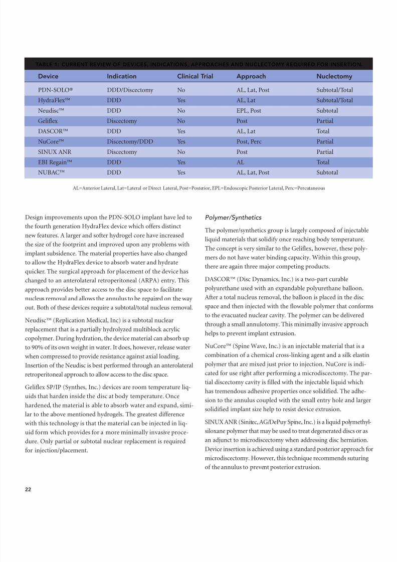
nucleus arthroplasty volume iii
TRANSCRIPT

8/9/2019 Nucleus Arthroplasty Volume III
http://slidepdf.com/reader/full/nucleus-arthroplasty-volume-iii 1/48
NucleusArthroplasty
Volume III: Surgical Technique& Technologies
Technology
in Spinal Care

8/9/2019 Nucleus Arthroplasty Volume III
http://slidepdf.com/reader/full/nucleus-arthroplasty-volume-iii 2/48
Table of Contents
This monograph series is a groundbreaking project in therapidly emerging field of non-fusion spinal surgery. The
full range of nucleus replacement technologies is examined
with discussion on surgical techniques, detailed informationon each cutting-edge device technology, indications, andpatient selection criteria.
Nucleus Arthroplasty™ Technology in Spinal Care ispublished for the medical profession by Raymedica, LLC,Minneapolis, MN 55431.
The views expressed in this series are those of the authorsand do not necessarily represent those of Raymedica, LLC.
ACKNOWLEDGEMENTWe, Raymedica, LLC, and the authors of this volume, wishto acknowledge our debt of gratitude for the importantcontribution of John Grabowski, Developmental Editor.His guidance has added a great deal to the teaching valueof this volume.
Copyright © 2006 and 2007 Raymedica, LLC. All rightsreserved. Printed in the U.S.A.
1 Introduction
2 Deputy Editorial Board
C H A P T E R 1 4 3 History of Motion Preservation Surgery of the Lumbar Spine
C H A P T E R 1 5 12 Examination, Radiologic and Diagnostic Evaluation, and Patient Indications
C H A P T E R 1 6 18 The Basis for Nucleus Replacement Surgery: Current Techniques
C H A P T E R 1 7 24 Intervertebral Disc Space Preparation for Nucleus Arthroplasty™ Technologies
C H A P T E R 1 8 29 Nucleus Arthroplasty™ Technology: Approach-Related Considerations
C H A P T E R 1 9 33 Clinical Outcomes Assessment of Nucleus Pulposus ReplacementC H A P T E R 2 0 39 Nucleus Replacement Complications and Salvage Procedures
44 Conclusion
www.nucleusarthroplasty.com

8/9/2019 Nucleus Arthroplasty Volume III
http://slidepdf.com/reader/full/nucleus-arthroplasty-volume-iii 3/48
Introduction
1
The first documented works describing the diagnosis andtreatment of the spine, spinal disorders, and spinal instability
date back to 1900-2500 B.C. Interestingly, the documents recom-mended against the treatment of spinal cord injury. The develop-ment of therapeutic treatments has a long history starting withthe cane, the first load-sharing device. Today, our efforts toimprove therapies to treat spine disease persist. We continue torecognize problems, identify issues, and define variables in aneffort to better understand spinal degeneration and to develop
innovative solutions that utilize a wide array of materials andtechnologies. Our field has had a rich history of advancements,accomplishments, and inventiveness. We owe a great debt to thepioneers who, armed with little more than a detailed knowledgeof anatomy, heralded in the era of spinal surgery. Their trials,errors, innovations, and teachings have guided our efforts toultimately improve clinical outcomes.
Early on, it was recognized that the disc played a vital role in overallspine health.With great effort and ingenuity, the unique anatomical,biomechanical, and physiological properties of the disc were eluci-dated and incorporated into elegant treatment algorithms. We now have access to an almost overwhelming flow of information aboutlumbar disc arthroplasty from countless sources. Central to the evo-lution of therapies is a better appreciation of the complexities of thelumbar disc. By combining knowledge gleaned from anatomical dis-section, biochemical processes, and resultant physiology with a disci-plined foundation in biomechanics, we have created a fabric of understanding never before enjoyed. Spine arthroplasty is now animportant and evolving area within the treatment of spinal disor-ders. This sub-discipline represents the coalescence of many areas of study focused on the development of new and exciting solutions toaddress clinical problems.
These significant advances in our understanding of the spine rep-resent a culmination of efforts occurring across many fronts. Ourincreased understanding of the biological factors at work in discdisease has been a driving force in the development and emer-gence of new materials and delivery methods. The critical rolethat advanced biocompatible alloys, polymers, and viscoelastichydrogels play in the innovation of disc arthroplasty technologiescannot be over emphasized.
Technological advancements have played a vital role in supportingand expanding our knowledge of motion preserving disc technolo-gies. The latest imaging technologies allow a much more detailedappreciation of pathological processes, such as disc degeneration,and provide the ability to monitor the results of an intervention.Computerized finite element analysis offers a risk-free environment
in which to test hypotheses and predict clinical impact. Biochemicaladvancements yield an intimate understanding of the chemical envi-ronment including chemical mediators and potential interventionportals. This wealth of knowledge can be used to great advantagewhen developing disc arthroplasty technologies.
Not to be overlooked, the socioeconomic challenges involved in thedevelopment of new technologies, such as the Nucleus Arthroplasty™motion preservation system, have also become more apparent.The all important variable of proper patient selection continues torequire constant reassessment and vigilance. Increasingly, third-party payers control access to care and treatment choice to an alarmingdegree. Such considerations can no longer be ignored in the questfor ideal patient management methods.
This publication has been constructed to provide an overview of the current Surgical Techniques & Technologies in NucleusArthroplasty™ technology. Key elements include an overview of the History of Motion Preservation Surgery in the Lumbar Spine,Current Techniques in Nucleus Replacement Surgery, ClinicalOutcomes Assessment, and Complications and Salvage Procedures.In addition, Volume III of this series will provide insight into thepotential market and the current players working in the forefront
of Nucleus Arthroplasty technology development activities. This isan incredibly exciting field as technologies focused on the repairand replacement of the diseased disc nucleus will catapult us farbeyond the treatment options we have available today.
In conclusion, we can say that the spine arthroplasty specialistof today is well prepared to deliver the most advanced solutionsto the clinical puzzle of disc disease with technologies based ona rich tradition of innovation and compassion coupled with atremendous wealth of physiological knowledge and assessmenttools. As spine surgery evolves from mechanical solutions totherapeutic solutions both surgeons and patients will benefit.We hope you will find this series on Nucleus Arthroplasty
technology to be a valuable asset.
Reginald J. Davis, MD, FACSCHIEF OF NEUROSURGERY
Baltimore Neurosurgical Associates, PA
Baltimore, MD 21204
Federico P. Girardi, MDASSISTANT PROFESSOR
OF ORTHOPEDIC SURGERY
Hospital for Special Surgery New York, NY 10021
Frank P. Cammisa, Jr., MD, FACSASSOCIATE PROFESSOR
OF CLINICAL SURGERY
Hospital for Special Surgery New York, NY 10021
Federico P. Girardi, MDReginald J. Davis, MD, FACS
Frank P. Cammisa, Jr., MD, FACS

8/9/2019 Nucleus Arthroplasty Volume III
http://slidepdf.com/reader/full/nucleus-arthroplasty-volume-iii 4/48

8/9/2019 Nucleus Arthroplasty Volume III
http://slidepdf.com/reader/full/nucleus-arthroplasty-volume-iii 5/48

8/9/2019 Nucleus Arthroplasty Volume III
http://slidepdf.com/reader/full/nucleus-arthroplasty-volume-iii 6/48
spinal disc height, integrity of the ligamentous structures, degreeof disc degeneration, and condition of the facet joints, using CTand MR imaging modalities (Figure 1). The goal of such analy-ses is to identify the presence of abnormal features or conditions
that impact the ability to perform normal motions.A careful analysis of the spinal elements is critical as it providesthe information necessary for evaluating treatment options ordeveloping a treatment plan to stabilize the spinal segment by recreating the proper tension in the annulus and surroundingligaments. This, in effect, will rebalance the spine and restore theappropriate lumbar curvature and corresponding sagittal bal-ance. Re-establishing the sagittal balance, and correspondingrange of motion (flexion-extension, lateral bending, rotation)is integral to this process as it represents the key to maintaininglong-term motion preservation, while reducing the potentialfor “adjacent level” disease.
The spinal elements under consideration include the spinal disc,vertebral bodies, posterior facets, and ligamentous structures.
These elements work in concert with the surrounding tissues tomaintain spinal lordosis and balance. The spinal disc is basically a self-contained viscoelastic system with multiple degrees of freedom in which translations and rotations are limited by theannulus fibrosus. The facets and posterior ligamentous struc-tures provide additional support in controlling spinal segmentrotation and translational motion that occur during flexion andextension activities. In addition, the anterior (ALL) and poste-rior (PLL) longitudinal ligaments assist in stabilizing the disc.Damage to these structures can alter the dynamic behavior of the spinal segment resulting in a change in the center of rotation
and corresponding motion pattern.
The key to “motion preservation” is to analyze and treat elemen-tal deficiencies. In fact, many of the device designs have beendeveloped to act as substitutes to address specific spinal patholo-gies or degenerative processes. In general terms, the availabledevice technologies can be classified into three major groupsthat include spinal nucleus replacements, total disc replacementsand posterior stabilization devices.
SPINAL NUCLEUS REPLACEMENTS
Spinal nucleus replacements seek to replace the nucleus portionof the spinal disc, while retaining the annulus and vertebral end-plates. In the late 1950’s, Dr. Ulf Fernström 1 implanted a SKF ballbearing in the disc space to reproduce the tension band across theadjacent vertebral segments, while attempting to maintain jointmotion. The results of his use of this metallic nucleus replace-ment core were published in 1964. This early design showed sig-nificant issues with vertebral endplate subsidence that resultedin a progressive loss in disc height.
While the success rate with the Fernström approach was rela-
tively low, this technique inspired the development of severalearly nucleus replacement devices including technologies thatutilized materials such as PMMA, 2 CoCr spheres, 3 and silicone. 4
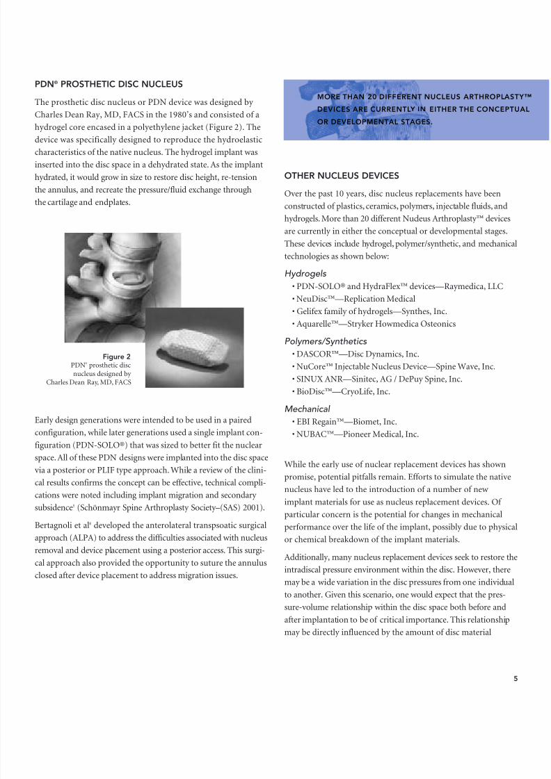
This initial work also served as the foundation for the develop-ment of the PDN® prosthetic disc nucleus device and Charité™total disc replacement.
4
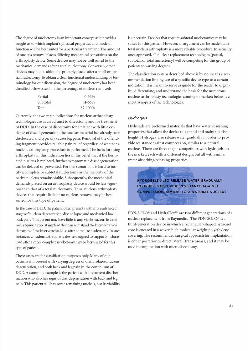
Figure 1Magnetic resonance imaging is one of the main improvementsin degenerative disc knowledge and pathology.
SPINAL NUCLEUS REPLACEMENTS SEEK TO REPLACETHE NUCLEUS PORTION OF THE SPINAL DISC, WHILERETAINING THE ANNULUS AND VERTEBRAL ENDPLATES.
8/9/2019 Nucleus Arthroplasty Volume III
http://slidepdf.com/reader/full/nucleus-arthroplasty-volume-iii 7/48

8/9/2019 Nucleus Arthroplasty Volume III
http://slidepdf.com/reader/full/nucleus-arthroplasty-volume-iii 8/48
removed and the subsequent volume of the implant utilized toreplace the nucleus. A post-implantation intradiscal pressure thatis too low may not adequately restore the disc height, while anintradiscal pressure that is too high may influence implant migra-tion or subsidence. Thus, it may be challenging to strike a balancethat can adequately address the specific needs of each individual.
Currently, there are no FDA-approved nucleus arthroplasty devices. Any device placed in the nucleus space where the intentis not to fuse the spine is considered an off-label use. As of April2007, four companies are in the process of conducting five U.S.Investigational Device Exemption (IDE) pilot clinical trials usingNucleus Arthroplasty™ technologies.
TOTAL DISC REPLACEMENT
Total disc replacements are designed to reproduce the motionand/or shock absorbing capabilities of the native disc. Thesedevices involve replacement of the disc nucleus, a portion of theannulus, and also often require modification of the vertebral end-plates to achieve long-term implant fixation. Over time, therehave been a number of total disc design concepts incorporatingthe use of different materials. While some of these designs utilizepolymer technologies, the majority of the current total discdesigns are mechanical in nature often incorporating a ball andsocket style design.
Acroflex®
The Acroflex is a polymeric disc prosthesis originally designed by Dr. Art Steffee in 1988 (Figure 3). This one-piece disc was com-posed of an elastomeric core bonded to titanium alloy baseplates.Cone shaped posts were incorporated into the baseplates to pro-vide short-term fixation. For long-term fixation, the baseplateswere coated with sintered titanium beads for bone ingrowth.
Despite extensive bench testing, early designs implanted inhumans had limited success due to failure of the rubber core. Thecore failures of this fully constrained implant design were likely aresult of the complex motion of the functional spinal unit which
undergoes both translation and rotation during flexion-extensionactivities. This creates a dynamic center of rotation that canimpose combined axial rotation and shear forces on the implantcore. The development of an appropriate elastomeric material thatcan accommodate such forces and resulting motions has not yetbeen resolved.
Additionally, a number of other polymeric disc designs havealso been studied. Urbaniak et al 7 designed a device with a sili-con core sandwiched between layers of Dacron mesh, whileEdeland 8 proposed the use of a silicon core bounded by poly-ethylene end caps. Lee et al9 designed a disc with a softer centercore (polysiloxane) surrounded by a stiffer matrix material(polyurethane). Early animal studies of such devices showedissues with infection, resorption, and dislodgement.
6
Figure 3Acroflex ® implant created and implantedby Dr. Art Steffee.
TOTAL DISC REPLACEMENTS ARE DESIGNED TO REPRODUCE THE MOTION AND/ORSHOCK ABSORBING CAPABILITIES OF THE NATIVE DISC. THESE DEVICES INVOLVEREPLACEMENT OF THE DISC NUCLEUS, A PORTION OF THE ANNULUS, AND ALSO OFTENREQUIRE MODIFICATION OF THE VERTEBRAL ENDPLATES TO ACHIEVE LONG-TERMIMPLANT FIXATION.

8/9/2019 Nucleus Arthroplasty Volume III
http://slidepdf.com/reader/full/nucleus-arthroplasty-volume-iii 9/48

8/9/2019 Nucleus Arthroplasty Volume III
http://slidepdf.com/reader/full/nucleus-arthroplasty-volume-iii 10/48
Maverick™
The Maverick was designed by Drs. Kenneth Pettine and RichardSalib in 1993. This is a two-piece metal-on-metal articulatingdisc design that incorporates a more posterior center of rotation.The device has a large central keel for alignment/guidance andinitial device stability. The metal-on-metal articulating surfaceis constrained and asymmetrically located on the baseplate sur-faces. As with other metal-on-metal designs in orthopedics,
long-term scientific data will be important to determine thepotential for metal ion release. A multicenter U.S. clinical study using the Maverick disc was initiated in the spring of 2003; thedevice is not currently FDA approved.
FlexiCore ®
The FlexiCore was designed by Dr. Thomas Errico in 2001. Thisis a two-piece metal-on-metal articulating disc design that isinserted as a single unit. The superior and inferior componentsare linked by a captured ball-and-socket joint. This prosthesis is
implanted through an anterior approach and inserted en bloc .The unique dome-shaped baseplate surfaces are designed toapproximate the concavities of the vertebral endplates for initialstability. The surfaces are coated with a titanium plasma spray toenhance bone ingrowth. Future issues with the metal-on-metalarticulation are similar to those discussed above. This technol-ogy is currently under U.S. clinical investigation; the device isnot currently FDA approved.
Other Total Disc Replacements
At this time, there are more than 20 different total disc designsin the conceptual or development stage. These designs vary inmaterial choice, shape, surgical technique, and implantation phi-losophy. In addition, there are currently a number of lumbartotal disc replacements in clinical study (Activ-L™, Kineflex™,Mobidisc®). In general, the basic operating principles of thesedevices are similar to the technologies presented above.
POSTERIOR TECHNOLOGIES
Dynamic stabilization is a term applied to devices that providenon-fusion support to the spine, while still allowing motion atthe affected levels. Such implants were first developed in Franceduring the 1980’s and were initially referred to as “ligamento-plasty,” as the early concepts all used various tethering methodsto stabilize posterior segments. Two different philosophies of pos-terior stabilization evolved based on different patient populationsand device biomechanics: 1) the use of adjunct support struc-
tures/ligaments used in combination with pedicle screw fixation 14
and 2) the use of spinous process attachments. 15 These conceptsare discussed in the following sections.
8
Prodisc® implantmotion preserva-
tion in L5-S1.
Figure 5a
Figure 5b

8/9/2019 Nucleus Arthroplasty Volume III
http://slidepdf.com/reader/full/nucleus-arthroplasty-volume-iii 11/48
Pedicle Screw-Based Systems
The Graf Ligament was designed by Dr. Henri Graf. It was oneof the earliest pedicle screw-based devices developed in Francein the late 1980’s. This system uses braided polyester ligamentsthat are looped around the screws to provide stability while stillallowing motion. The device was used in over 10,000 patientsduring the early 1990’s. However, long-term results were notpromising as the ligament loosened, causing failure in the tension
band and/or the pedicle screw.The Dynesys® system was invented by Drs. Gilles Dubois andOtmar Schwarzenbach. The device consists of pedicle screws,cords/ligaments and polyurethane spacers (Figure 6). This sys-tem is implanted and tensioned to provide spinal support suchthat the cord provides support and limits flexion, while thespacer limits extension. Thus, the Dynesys is designed to restabi-lize spinal segments that show symptoms of stenosis or spondy-lolisthesis. When used without bone graft, it is designed topreserve the natural function of the spine by allowing motionand sharing in-load transmission.
At this time, there are more than 30 pedicle screw-based poste-rior dynamic systems in development or undergoing clinical tri-als. However, regardless of the system, it is difficult to determineif posterior dynamic stabilization will be capable of maintaining
long-term segmental motion, while providing adequate spinalstabilization to slow the progression of disc disease.
Interspinous Process Spacers
The Wallis™ System was invented in 1984 by Dr. Jacques SenegasThis interspinous process spacer incorporates a tethering systemto stabilize discectomy patients and restore motion. The first-generation implant for non-rigid stabilization of lumbar seg-ments was developed in 1986. The device consisted of a titaniuminterspinous blocker and an artificial ligament made of Dacron;the latest generation incorporates a PEEK blocker. Functionally,the device restrains flexion and blocks extension; thus, increasingthe rigidity of destabilized segments. The overall implant has nopermanent fixation in the vertebral bone which is intended toavoid the risk of device loosening.
The coflex™ Interspinous Implant, previously known as theinterspinous“U,” was developed by Dr. Jacques Samani in 1994(Figure 7). The device is made of a single machined piece of tita-
nium alloy with no articulating parts. It was designed to be usedfor patients with moderate to severe lumbar spinal stenosis withconcomitant low back pain and neurogenic claudication. Unlikethe other interspinous spacers, the coflex Interspinous Implant isfunctionally dynamic allowing both flexion and extension. The
9
DYNAMIC STABILIZATION IS A TERM APPLIED TO DEVICES THAT PRO
NON-FUSION SUPPORT TO THE SPINE, WHILE STILL ALLOWING MOTIAT THE AFFECTED LEVELS.
Figure 6Dynesys® pedicle screwsand ligament system withpolyurethane spacers.

8/9/2019 Nucleus Arthroplasty Volume III
http://slidepdf.com/reader/full/nucleus-arthroplasty-volume-iii 12/48
10
device has been implanted in more than 20,000 patients worldwidewith up to 12 years of follow-up data demonstrating its safety. Thecoflex is currently in a U.S. FDA-approved study.
The X-STOP™ implant was developed by Drs. James Zuchermanand Kenneth Hsu. The device is used in the treatment of patientswith mild lumbar spinal stenosis and neurogenic claudication.The implant is designed as a two-piece system comprising a tita-nium alloy extender and a PEEK Optima oval spacer. It functionsby opening the lumbar space through permanent distraction of the posterior spinous process. This unloads the facets, opens theneuroforamen, and widens the spinal canal, removing the pressureon the nerve roots. Functionally, the X-STOP allows flexion, butblocks or restrains extension activities. The X-STOP has receivedFDA approval for commercial use in the U.S.
The DIAM™, or Device for Intervertebral Assisted Motion, wasdeveloped by Dr. Jean Taylor in 1997. The implant acts as ashock absorber that reduces loads on the surrounding vertebraeand restores the natural function at the affect level. Functionally,the DIAM system is designed to restrain flexion and allow exten-sion. The core of the DIAM system is made of silicone, while theouter mesh and tethers are made of medical-grade polyester. Theflexible properties of the DIAM system materials may also pro-tect the integrity of the spin-ous process. More than20,000 surgeries have been
successfully performed inEurope, Asia and LatinAmerica using this device.The DIAM is currently in aninvestigational U.S. FDA-approved study.
In general, dynamic stabilization systems represent an attractiveoption for patients that would otherwise undergo fusion proce-dures. As with any spinal care treatment, there are concerns asso-ciated with long-term performance. As a principle of their action,
the use of posterior stabilization implants may induce kyphosis atthe operative level. The major concern is that creating kyphosis atthe affected level will increase potential for hyperextension at theadjacent levels. Another concern, particularly for devices thatblock extension by bearing load on the spinous processes, is thepotential for progressive bony erosion resulting in a loss in effec-tiveness and an increased potential for bone fractures. This may beproblematic as the indications for use of such devices is generally segmental stenosis that often occurs at the age when osteoporosisis also a factor.
BIOTECHNOLOGIES
Currently, there is great deal of research being conducted todevelop biological or regeneration solutions to spinal care.Techniques such as the transplantation of nucleus cells 16 and cellculturing with subsequent reimplantation are being evaluatedfor use after disc herniation. Additional work is underway inves-tigating our ability to block the factors associated with apoptosisin nucleus cells. Such research will greatly influence future treat-ment options and redefine how we go about preserving motionwhen addressing degenerative disc disease. While many of thesestechnologies remain in their infancy, such solutions will ulti-mately displace the “mechanical repair” methods that representour current treatments and standard of care.
Figure 7Coflex™ Interspinous
Dynamic System
UNLIKE THE OTHER INTERSPINOUS SPACERS, THE COFLEX INTERSPINOUS IMPLANTIS FUNCTIONALLY DYNAMIC ALLOWING BOTH FLEXION AND EXTENSION.

8/9/2019 Nucleus Arthroplasty Volume III
http://slidepdf.com/reader/full/nucleus-arthroplasty-volume-iii 13/48
11
CONCLUSION
Our understanding and development of motion preservationtechnologies in the spine is only just beginning with initial solu-tions dating back over 50 years. Indeed some of the technologies
that are now clearing the necessary regulatory hurdles and gainingacceptance in clinical use have been in a continuum of develop-ment and refinement for more than 15 years. As in many fields, itis likely that only a few of these current technologies will survivethe tests of time and rigors associated with long-term follow up.
Regardless of form, the developers of current and future motionpreserving concepts share the same attributes: an original con-cept, a strong knowledge of pathology, an in-depth understand-ing of disc physiology/anatomy, and an ability to share theirpassion with structured development teams. In the future, restassured that we will continue to discover new and innovativesolutions to preserve spinal motion. These solutions will nodoubt expand our available treatment options to address allstages of degenerative disc disease and related spinal pathologies;thus, broadening our continuum of care.
REFERENCES1. Fernström U : Arthroplasty with intercorporal endoprothesis in herniated disc
and in painful disc. Acta Chir Scand Suppl. 1966; 357:154-9.
2. Cleveland D: Interspace reconstruction and spinal stabilization after disk removal. J Lancet. 1956 Oct; 76(10):327-31.
3. Harmon PH: Subtotal anterior lumbar disc excision and vertebral body fusion.III. Application to complicated and recurrent multilevel degenerations. Am JSurg. 1959 May; 97(5):649-59.
4. Fassio B, Ginestie JF: Discal prosthesis made of silicone: experimental study and 1st clinical cases. Nouv Presse Med. 1978 Jan 21; 7(3):207.
5. Shim CS, Lee SH, Park CW, Choi WC, Choi G, Choi WG, Lim SR, Lee HY:Partial disc replacement with the PDN prosthetic disc nucleus device: early clinical results. J Spinal Disord Tech. 2003 Aug;16(4):324-30.
6. Bertagnoli R, Vazquez RJ: The Anterolateral TransPsoatic Approach (ALPA): anew technique for implanting prosthetic disc-nucleus devices. J Spinal DisordTech. 2003 Aug;16(4):398-404.
7. Urnbaniak JR, Bright DS, Hopkins JE: Replacement of intervertebral discs inchimpanzees by silicone-dacron implants: a preliminary report. J BiomedMater Res. 1973;7(3):165-86.
8. Edeland HG: Suggestions for a total elasto-dynamic intervertebral disc pros-thesis. Biomater Med Devices Artif Organs. 1981; 9(1):65-72.
9. Langrana NA, Parsons JR, Lee CK et al. Materials and design concepts for anintervertebral disc spacer. I. Fiber reinforced composite design. J ApplBiomater. 1994; 5:125-32.
10. Karin Büttner KJ, Schellnack K, Zippel H: Biomechanics of the SB Charitélumbar intervertebral disc endoprosthesis. Int Orthop. 1989;13(3):173-6.
11. Lemaire JP, Skalli W, Lavaste F, Templier A, Mendes F, Diop A, Sauty V,Laloux E: Intervertebral disc prosthesis. Results and prospects for the year
2000. Clin Orthop Relat Res. 1997 Apr;(337):64-76.12. Kurtz SM, van Ooij A, Ross R, de Waal Malefijt J, Peloza J, Ciccarelli L,
Villarraga ML: Polyethylene wear and rim fracture in total disc arthroplasty.Spine J. 2007 Jan-Feb; 7(1):12-2.
13. Tropiano P, Huang RC, Girardi FP, Cammisa FP Jr, Marnay T: Lumbar totaldisc replacement. Seven to eleven-year follow-up. J Bone Joint Surg Am. 2005Mar; 87(3):490-6.
14. Graf H: Lumbar instability. Surgical treatment without fusion. Rachis 1992;412:123-37.
15. Senegas J, Etchevers P, Vital JM, Baulny D, Grenier F: Widening of the lumbarvertebral canal as an alternative to laminectomy in the treatment of lumbarstenosis. Rev chir orthop repar appar mot 74:15-22, 1988 (Fr).
16. Iwashina T, Mochida J, Sakai D, Yamamoto Y, Miyazaki T, Ando K, Hotta T:Feasibility of using a human nucleus pulposus cell line as a cell source in celltransplantation therapy for intervertebral disc degeneration. Spine. 2006May 15; 31(11):1177-86.
THESE SOLUTIONS WILL NO DOUBT EXPAND OUR AVAILABLE TREATMENT OPTIONSTO ADDRESS ALL STAGES OF DEGENERATIVE DISC DISEASE AND RELATED SPINAL
PATHOLOGIES; THUS, BROADENING OUR CONTINUUM OF CARE.
8/9/2019 Nucleus Arthroplasty Volume III
http://slidepdf.com/reader/full/nucleus-arthroplasty-volume-iii 14/48
Chapter 15 Examination, Radiologicand Diagnostic Evaluation,and Patient Indications
KEYPOINTS
• The continued introduction of new treatment alternativesemphasizes the need for better methods to examine andevaluate patients.
• A well-documented patient history is the foundation to under-standing the etiology of the patient’s pain.
• Advanced imaging modalities (CT, MRI) are of significant valuein visualizing the spine and associated soft tissue structures.
• The combination of examination and diagnostic tools is criticalto the definition of potential treatment options.
Andrew A. Sama, MDASSISTANT PROFESSOR OF ORTHOPEDIC SURGERY Weill Medical College of Cornell University Hospital for Special Surgery New York, NY 10021
Dr. med. univ. Rudolf BertagnoliFOUNDER
Pro-Spine Medical ConsultingStraubing, Germany 94315
Federico P. Girardi, MDASSISTANT PROFESSOR OF ORTHOPEDIC SURGERY
Hospital for Special Surgery New York, NY 10021
12

8/9/2019 Nucleus Arthroplasty Volume III
http://slidepdf.com/reader/full/nucleus-arthroplasty-volume-iii 15/48

8/9/2019 Nucleus Arthroplasty Volume III
http://slidepdf.com/reader/full/nucleus-arthroplasty-volume-iii 16/48
In addition, pain and function associated with range of motionstudies of the hips and knees, and the condition of the sacroiliac joints should be assessed. Assessment of the sacroiliac joints isachieved by applying manual pressure over the joints and per-
forming the Patrick’s flexion, abduction, external rotation maneu-ver. Finally, a vascular examination including an assessment of pulses, edema, and trophic changes should be performed.
INTERVERTEBRAL DISC ASSESSMENT
The intervertebral discs constitute roughly one third of the ver-tebral column length. Thus, changes that occur in the disc, suchas a loss in height or herniation, can have a profound effect onother spinal structures including the facet joints, paraspinousmuscles, and exiting nerves resulting in pain. Degeneration of
the disc itself, can also be a pain generator due to innervation of the annulus. 4
Degenerative disc disease has been classified in stages.5 The radi-ographic onset of the disease usually occurs in patients between20 and 60 years of age, but the clinical manifestation of thiscondition to the actual onset of pain is difficult to predict. 6 Someauthors have also reported that disc degeneration in certainindividuals may be a result of genetic predisposition. 7
In a normal disc, the properly hydrated nucleus pulposus acceptscompressive load and distributes it, via hydrostatic pressure, to theannulus fibrosus. As the disc degenerates, the nucleus loses water
content resulting in a reduced intradiscal pressure and correspon-ding loss in height, altering the biomechanics of the spine.
Consequently, the annulus assumes the role of distributing theexcess compressive load resulting in stress induced morphologi-cal changes. Such morphological changes can be evaluated usingseveral diagnostic methods to assist in the identification of thepain source and aid in developing a suitable therapeutic strategy.
DIAGNOSTIC EVALUATION
The most common spine imaging modalities include plain x-rays,computed tomography (CT) and magnetic resonance imaging(MRI). Additional imaging studies, such as discography or myel-ography, may also be requested by spine specialists. Many of theseimaging modalities have established grading systems that can beused to describe the stage of disc degeneration. 8
RADIOGRAPHY
X-ray is the most commonly performed spinal imaging study as it is readily available in primary care settings. AP and lateralplain films provide key information in regard to spinal align-ment, scoliosis, spondylolisthesis, bone quality/density, andpotential fractures. Films taken in flexion and extension posi-tions may also help identify normal spinal motion or potentialinstabilities. In addition, properly scaled films allow for themeasurement of vertebral body and disc dimensions that may be used in surgical planning (Figures 1 & 2).
A more detailed review of plain films can be used to identify variations in endplate morphology, such as Schmorl´s nodules,
which represent protrusions of the nucleus pulposus into thevertebral body. The presence of Schmorl´s nodes, may indicatethat degenerative processes are underway and may be advancedin the affected spinal segments. 9
While x-ray has many apparent advantages, there are a few well-recognized disadvantages that include the two dimensionalnature of the information, exposure to radiation, and an inabil-ity to directly assess the condition of the cartilaginous endplateand surrounding soft tissue structures.
14
Figure 1 Figure 2
THE RADIOGRAPHIC ONSET OF DEGENERATIVE DISCDISEASE USUALLY OCCURS IN PATIENTS BETWEEN20 AND 60 YEARS OF AGE, BUT THE CLINICALMANIFESTATION OF THIS CONDITION TO THE ACTUALONSET OF PAIN IS DIFFICULT TO PREDICT.

8/9/2019 Nucleus Arthroplasty Volume III
http://slidepdf.com/reader/full/nucleus-arthroplasty-volume-iii 17/48
COMPUTED TOMOGRAPHY
Computed tomography (CT) represents an advanced radiographictechnique that provides three dimensional imaging for analysis.This imaging method offers improved resolution and providesbetter visualization of the bony structures of the spine in com-
parison to plain radiography.CT provides a more detailed view that can be utilized to assessfacet integrity and health, disc space height, endplate sclerosis,and the presence of vacuum phenomena. When imaging is beingperformed to evaluate bone-related issues, CT is preferable toother methods as itallows direct visualiza-tion of the corticalbone. Thus, abnormali-ties such as parsdefects, bony foraminalstenosis, and calcifica-tion of disc herniationscan readily be assessed(Figures 3 a, b & c).
MRI
Magnetic resonance imaging (MRI) is a powerful imaging modality that can provide extraordinary detail. By using different relaxationtechniques (T1, T2, T1 ρ ), MRI can be utilized to obtain high con-trast images of the bone and soft tissue structures. In fact, this imag-ing method is sensitive enough to differentiate between differentsoft tissue structures such as the annulus fibrosus and the nucleuspulposus. For diagnostic purposes, the use of MRI can assist inidentifying the presence of high intensity zones, annular fissures,and disc herniations allowing resultant neurologic compression tobe quantified. MRI has also been used to characterize signal changesin the bone marrow adjacent to the vertebral endplates; a three-stage classification has been described by Modic. 10
When imaging the intervertebral disc, the major advantages of MRI are best recognized by the use of T2 weighted, sagittal imag-ing. With the T2 technique, the hydration of the disc nucleus canbe easily visualized and compared from one level to the nextallowing classification into degenerative stages (Figure 4).
Additional diagnostic information can be obtained using T1 ρ
spin lock relaxation. This processing technique aids in assessingthe proteoglycan content of the nucleus which directly correlateswith the water binding capacity. The T1 ρ technique also has thepotential to detect degeneration of the disc at a significantly ear-lier point in comparison to the more commonly used MRI tech-niques. 11 This becomes relevant for the application of treatmentstrategies such as biological repair enhancement 12 or nucleuspulposus replacement.
While MRI is well-recognized as an excellent tool for visualization of the vertebral bodies, spinal discs, nerves and surrounding structures,the diagnostic value of MRI to predict or identify potential paingenerators in the spine remains controversial. 13,14,15,16,17
15
Figure 3a
Figure 4
AS THE DISC DEGENERATES, THE NUCLEUS LOSESWATER CONTENT RESULTING IN A REDUCEDINTRADISCAL PRESSURE AND CORRESPONDINGLOSS IN HEIGHT, ALTERING THE BIOMECHANICSOF THE SPINE.
Figure 3b
Figure 3c

8/9/2019 Nucleus Arthroplasty Volume III
http://slidepdf.com/reader/full/nucleus-arthroplasty-volume-iii 18/48

8/9/2019 Nucleus Arthroplasty Volume III
http://slidepdf.com/reader/full/nucleus-arthroplasty-volume-iii 19/48
By using this information in combination with the anticipated phys-iological and biomechanical demands, a general outline of patientselection criteria can be developed to improve the potential forachieving good long-term clinical outcomes. Below is a general list
of inclusion and exclusion criteria for the use of nucleus arthroplasty technologies that is based on available literature. 2,3,19,20, 21
Inclusion
• Mild to moderate DDD• Back pain and/or leg pain (L2-S1)• Skeletally mature• Failed conservative care (6+ months)• Loss in disc height less than 50%, based on
adjacent normal disc
• Reasonable physical condition and weight (BMI < 30)• Documented pain/impact on quality of life
(VAS, ODI, SF-12/36)
Exclusion
• Allergies to device materials• Congenital bony or spinal abnormalities• Spondylolisthesis• Spinal stenosis (severe)• Spinal segment instability
• Facet degeneration• Schmorl´s nodules or endplate irregularities• Osteoporosis• Infection or malignancy • Significant emotional or psychological issues
As noted previously, this represents a general list of criteria sug-gested for the application of current nucleus arthroplasty tech-nologies. As the medical community continues to gain moreclinical experience with these technologies, the criteria will cer-
tainly change. However, in the early stages, the ability to strictly adhere to the inclusion and exclusion criteria defined for a par-ticular technology will be of the utmost importance in obtainingencouraging clinical results.
REFERENCES1. Latchaw Jr JP. A historical note on sciatia—Chp 1. In: Hardy RW editors:
Lumbar disc d isease. NY. 1982. Raven Press.
2. Bertagnoli R. Review of modern treatment options for degenerative disc dis-ease. In: Kaech DL and Jinkins JR, editors: Spinal restabilization procedures—Diagnostic and therapeutic aspects of intervertebral fusion cages, artificialdiscs and mobile implants. 2002. Elsevier BV.
3. Goins ML, Wimberley DW, Yuan PS, Fitzhenry LN, Vaccaro AR. Nucleus pul-posus replacement: an emerging technology. The Spine J. 2005; 5:317S–324S.
4. Coppes MH, Marani E, Thomeer RT, Groen G. Innervation of “painful” lum-bar discs. Spine. 1997; 22 (20):2342–2349.
5. Kirkaldy-Willis WH. Managing low back pain. NY. 1983. Churchill Livingstone.
6. Urban JPG, Roberts S and Ralphs JR. The nucleus of the intervertebral discfrom development to degeneraion, Amer.Zool. 2000; 40:53-61.
7. Chan D, Song Y, Sham P, Cheung K. Genetics of disc generation. Eur Spine J.2006; 15:S317-325 (2006).
8. Kettler A, Wilke H-J. Review of existing grading systems for cervical or lumbardisc and facet joint degeneration. Eur Spine J. 2006; 15:705–718.
9. Moore RJ. The vertebral endplate: disc degeneration, disc regeneration. EurSpine J. 2006; 15(S3):S333–S337.
10. Modic MT, Steinberg PM, Ross JS, Masaryk TJ, Carter JR. Degenerative discdisease assessment of changes in vertebrae with MR imaging. Radiology.1988; 166:193–199.
11. Auerbach JD, Johannessen W, Borthakur A, Wheaton AJ, Dolinskas CA,Balderston RA, Ravinder R, Elliot DM. In vivo quantification of human lum-bar disc degeneration using T1 ρ -weighted magnetic resonance imaging. Eur.Spine J. 2006: 15(S3):S338–344.
12. Yoon ST, Patel NM. Molecular therapy of the intervertebral disc. Eur Spine J.2006; 15(S3):379–388.
13. Aprill C, Bogduk N. High—intensity zone: A diagnostic sign of painful lum-bar disc on magnetic resonance imaging. Br J Radiol. 1992; 65:361–369.
14. Peng B, Hou S, Wu W, Zhang C, Yang Y. The pathogenesis and clinical significance of a high-intensity zone (HIZ) of lumbar intervertebral disc on MR imaging in the patient with discogenic low back pain. Eur Spine J. 2006;15:583–587.
15. Schellhas KP, Pollei SR, Gundry CR, Heithoff KB. Lumbar disc high-intensity zone: Correlation of magnetic resonance imaging and discography. Spine.1996; 21:79–86.
16. Carragee EJ, Paragiou SJ, Khurana S. 2000 Volvo award winner in clinicalstudies: lumbar high-intensity zone and discography in subjects without low back problems. Spine. 2000; 25(23):2987–92.
17. Rankine JJ, Gill KP, Hutchinson CE, Ross ER, Williamson JB. The clinical sig-nificance of the high-intensity zone on lumbar spine magnetic resonanceimaging, Spine. 1999; 24(18):1913-20.
18. Peh WGC. Provocative discography: current status. Biomed Imaging Interv J.1(1): e2.
19. Ray CD. The PDN prosthetic disc nucleus device. Eur Spine J. 2002;11(S2):S137–S142.
20. Shim CS, Lee SH, Park CW, Choi WC, Choi G, Choi WG, Lim SR, Lee HY.Partial disc replacement with the PDN prosthetic disc nucleus device: early clinical results. J Spinal Disord Tech. Aug 2003; 16(4):324-330.
21. Bertagnoli R, Karg A, Voigt A. Lumbar partial disc replacement. Orthop ClinN. Am. 2005; 36:341–347.
17
8/9/2019 Nucleus Arthroplasty Volume III
http://slidepdf.com/reader/full/nucleus-arthroplasty-volume-iii 20/48
KEYPOINTS
• Degeneration of the spine may be due to familial inheritance,age-related changes, or traumatic physical loading.
• The use of Nucleus Arthroplasty™ technology has been indi-cated for early stage degenerative disc disease and preventionof disc degeneration progression status post discectomy.
• The degree of nuclectomy is an important consideration when
determining the optimal nucleus arthroplasty treatment system.• The amount of nucleus removal places differing mechanical
constraints on a nucleus arthroplasty device.
• A vast array of nucleus arthroplasty implants are currently available including hydrogels, polymer/synthetics, andmechanical devices.
Chapter 16 The Basis for NucleusReplacement Surgery:Current Techniques
Eddie H. Chung, MDCLINICAL FELLOWThe Spine Institute, Santa MonicaLos Angeles, CA 90404
Ben P. Pradhan, MD, MSDIRECTOR OF CLINICAL RESEARCH
The Spine Institute, Santa MonicaLos Angeles, CA 90404
Hyun W. Bae, MDDIRECTOR OF RESEARCH
The Spine Institute, Santa MonicaLos Angeles, CA 90404
18
8/9/2019 Nucleus Arthroplasty Volume III
http://slidepdf.com/reader/full/nucleus-arthroplasty-volume-iii 21/48
INTRODUCTION
The goal of this chapter is to discuss the basis for nucleusreplacement surgery. Key elements of this discussion are the
intervertebral disc construct and degenerative process. As such, abrief review of the intervertebral disc structure and pathophysi-ology of disc degeneration is provided to establish a basis forunderstanding the current nucleus arthroplasty concepts andcorresponding strategies.
DISC STRUCTURE
The intervertebral disc is composed of three concentrically arranged sets of tissue. The outermost layer is a thick fibrousring of dense, highly organized Type I collagen called the annu-lus fibrosus. The cells found in the annulus have an ellipsoidalmorphology, similar to fibroblasts that produce Type I collagenin response to deformation. The second layer is a fibrocartilagi-nous layer that is larger in size, however, less organized than theannulus fibrosus, consisting predominantly of Type II collagen.Cells found in this middle layer are a mixture of annular andnuclear cells. The innermost layer, the nucleus pulposus, is alsodominated by Type II collagen arranged in a more random fash-ion. The cells found in the nucleus are initially notochordal inorigin, but are replaced by chondrocyte-like rounded cells duringearly adulthood. These cells produce fine Type II collagen fibrils
and proteoglycans in response to large hydrostatic and osmoticpressures. The high concentration of proteoglycans within thenucleus provides for great strength in axial compression due totheir high water binding properties. 1
DISC DEGENERATION
Currently, there are multiple hypotheses to explain the process of disc degeneration. A growing body of evidence through twin studiessupports genetics as the predominant factor in disc degeneration. 2
Degeneration through the simple wear and tear of aging has been
identified as a cause less frequently, and a very small percentage of discs degenerate through physical loading or trauma. 3
The pathophysiology of disc degeneration may be independentof the epidemiological cause, but has also been shown to bemultifactorial in nature. A decline in nutritional and wastetransport seem to be the most critical events that occur withinthe disc. This may be associated with the age-related increase indisc size as well as the decreasing number of peripheral arteries. 4
Other changes that occur during degeneration include cell senes-cence, tissue dehydration, modification of the matrix proteins,loss of aggregating proteoglycans, accumulation of degradedmacromolecules and apoptotic debris, increases in the degrada-
tive enzyme activity, decreases in the viable cell concentration,and fatigue failure of the matrix. 5
Currently, the most widely accepted theory of intervertebral discdegeneration pathophysiology is a three-stage approach describedby Kirkaldy-Willis. Stage I describes the acute pain of an initialinsult occurring in the early 20 to 30 years of life. This is the begin-ning of what Kirkaldy-Willis described as the “degenerative cas-cade.”6 Repetitive microtrauma to the vertebral endplates andmotion segment result in ischemic events that compromise thenutritional and metabolite transport to the disc.Microtrauma may also be a possible cause for proteoglycan fragmentation that hasbeen shown to begin as early as childhood. This decrease in theamount of nuclear aggregating proteoglycans in turn, decreases dischydration and resiliency. Type II collagen fibrils are replaced by Type I collagen in the inner annulus, as the annulus encroaches onthe nucleus. 7 This gradual advancement of the annulus, in additionto its increase in load bearing, may be responsible for the annulartears known to begin during this first stage. 8 Clinically, pain in thisstage is usually intermittent and self-limiting.
Stage II, or the instability stage, is a progression from Stage I withcontinued disc degeneration, dehydration, and loss of disc height.The disc space collapse occurs from trabecular microdamage of
the vertebral endplate and subsequent structural collapse. Thisallows the nucleus to migrate or “decompress” into the endplate,increasing the compressive load borne by the annulus. 9 This lossof hydrostatic nuclear pressure causes the annulus to bulge radi-ally both outward and inward, therefore decreasing annular andoverall disc height.7 The loss of height and continued annulartearing act as contributing factors that promote instability of themotion segment. This stage of degeneration may carry on into thefifth decade of life. Clinically, it presents as more severe episodesof low back pain with longer periods of duration.
19
CURRENTLY, THERE ARE MULTIPLE HYPOTHESES EXPLAIN THE PROCESS OF DISC DEGENERATION.GROWING BODY OF EVIDENCE THROUGH TWIN STSUPPORTS GENETICS AS THE PREDOMINANT FACTIN DISC DEGENERATION.

8/9/2019 Nucleus Arthroplasty Volume III
http://slidepdf.com/reader/full/nucleus-arthroplasty-volume-iii 22/48
Stage III, known as the stabilization stage, usually occurs in the60 and older population. Destruction of the vertebral endplatesand gross fissuring of the annulus have now reversed the previ-ous ischemic insult and caused the blood and nerve supply tothe disc to increase. 10 This increase in nutritional and metabolitetransport increases the presence of catabolic cells, metallopro-
teinases, and cytokines resulting in resorption of the nucleus.11
The damage to the annular and nuclear cells, in addition to thetremendously increased compressive mechanical environment,makes any attempt at repair impossible. 12 Disc resorption con-tinues on to its final phases leading to endstage disc collapse,endplate destruction, disc fibrosis, and osteophyte formation.Clinically, during this stage back pain typically decreases, whileleg pain increases due to the narrowing and collapse of theneural foramen and lateral recesses.
This degenerative cascade occurs in a continuum with all threestages blending into one another and possibly occurring simulta-neously. Depending on the causative nature of the degenerativeprocess, there may also be a much earlier or delayed shift in thetimeframe at which these stages present. 6
TREATMENT FOR DEGENERATIVEDISC DISEASE (DDD)
The search continues for an optimal treatment option for degen-erative disc disease (DDD), particularly in younger patients andpatients status post discectomy procedures. The ideal treatment
would be some form of biologic therapy that can stimulate tissueregeneration or repair. Protein (BMP’s, TGF-B, IL-1), intradiscalgene, and cell therapies (autologous or allograft stem cells) are allcurrently under investigation and appear to be future possibilities. 13
Traditionally, treatment of degenerative disc disease has beenlimited to some form of fusion procedure. However, the successrates of fusion techniques to address discogenic pain have beenhighly variable in the literature. 14 The increase in stiffness of thefused segment also increases stress transfer to adjacent levels,
often times resulting in the need for further surgical intervern-tion. 15 The term “motion preservation” has been the catch-allphrase to describe different non-fusion treatment options thatcould potentially avoid the risk of adjacent segment degenera-tion and subsequent surgery. Total disc arthroplasty, facetarthroplasty, motion stabilization, and nucleus arthroplasty are
all technologies that fall within this “motion preservation” cate-gory. The remainder of this chapter will focus on the basis fornuclear replacement technologies.
NUCLEUS REPLACEMENT
Nucleus arthroplasty has been indicated for the treatment of early stage degenerative disc disease with an intact annulus orstatus post microdiscectomy procedures where nuclear materialhas been removed. Currently, the available and developingdevices are either implantable or injectable materials that replacea portion of the nucleus pulposus. Their purpose is to help pre-serve the geometry of the intervertebral disc and maintain themotion of the disc space. 16
Early nucleus arthroplasty device concepts, dating back to the1950’s, were focused on replacement of the nucleus with stain-less steel ball bearings, silicone rubber, and polymethylmethacry-late (PMMA) cement. 17, 18, 19 These devices failed mainly due toour poor understanding of disc biomechanics, subsidence issues,and implant/bone material properties. Significant advances inour understanding of past issues and observation of device fail-
ure modes have brought us to a recent growth of new investiga-tional nuclear implants and therapies. Today’s new generationof product offerings include hydrogels, polymers/synthetics, andmechanical forms. These arthroplasty systems vary in theirapplication depending on whether treatment is for degenerativedisease or as an adjunct to discectomy. In addition, there are alsosurgical approach-related and nucleus removal or “degree of nuclectomy” considerations that must be evaluated to determinean optimal treatment system.
20
NUCLEUS ARTHROPLASTY HAS BEEN INDICATED FOR THE TREATMENT OFEARLY STAGE DEGENERATIVE DISC DISEASE WITH AN INTACT ANNULUS ORSTATUS POST MICRODISCECTOMY PROCEDURES WHERE NUCLEAR MATERIAL
HAS BEEN REMOVED.
8/9/2019 Nucleus Arthroplasty Volume III
http://slidepdf.com/reader/full/nucleus-arthroplasty-volume-iii 23/48
8/9/2019 Nucleus Arthroplasty Volume III
http://slidepdf.com/reader/full/nucleus-arthroplasty-volume-iii 24/48

8/9/2019 Nucleus Arthroplasty Volume III
http://slidepdf.com/reader/full/nucleus-arthroplasty-volume-iii 25/48
Mechanical Implants
Mechanical implants consist of single or multi-componentdevices made of hard materials or metals.
EBI Regain™ (Biomet, Inc.) is a one-piece, anatomically conform-ing device composed of pyrolytic carbon. The modulus of elastic-ity of the implant material is equivalent to bone. In addition, theimplant surface is highly polished to prevent damage to the carti-laginous endplate during motion. Total nucleus removal is requiredto accommodate placement of the device via an anterolateralretroperitoneal or direct lateral approach.
NUBAC™ (Pioneer Surgical Technology) is a two-piece implantconstruct made from PEEK-OPTIMA LT1. The concave superiorendplate and convex inferior endplate form a ball and socket joint. The device is designed to allow uniform stress distribution
under different loading conditions, while minimizing potentialsubsidence and extrusion risks. Placement of the device requiressubtotal nuclear removal and can be accomplished using an ante-rior, anterolateral or even posterior approach. Retention of asmuch annular tissue as possible is recommended.
CONCLUSION
Degeneration of the spine may be due to familial inheritance,age-related changes, or traumatic physical loading. Independentof the cause, the pathophysiology of degenerative disc diseasehas been shown to be irreversible if left untreated. Recently,“motion preservation” technologies have begun to emerge in anattempt to advance the continuum of care and offer alternativesto treatment using fusion. Of these emerging technologies,nucleus arthroplasty has been indicated for early-stage degenera-tive disc disease and prevention of disc degeneration progressionstatus post discectomy.
While initial attempts to replace the nucleus failed, significantadvances in our understanding of spinal biomechanics, implantmaterial, and implant failure modes have led to the development
of a multitude of new nucleus arthroplasty technologies. Theability to understand and classify the degree of nuclectomy required for the optimal use of such devices will be an importantfactor. Currently, a number of FDA clinical trials are underway toinvestigate the safety and efficacy of these potentially preventiveand adjunctive treatment devices.
REFERENCES1. Buckwalter JA, Mow VC, Boden SD, Eyre DR, Weidenbaum M. Intervertebral
disc structure, composition, and mechanical function. In: Buckwalter JA,Einhorn TA, Simon SR, editors. Orthopaedic Basic Science—Biology and bio-mechancis for the musculoskeletal system. 2nd ed. Rosemont: American
Academy of Orthopaedic Surgeons, 2002:548-55.2. Battie MC, Videman T. Lumbar disc degeneration: epidemiology and genetics.
J Bone Joint Surg Am. 2006 Apr;88 Suppl 2:3-9.
3. Battie MC, Videman T, Gibbons LE, Fisher LD, Manninen H, Gill K. 1995Volvo Award in clinical sciences. Determinants of lumbar disc degeneration. Astudy relating lifetime exposures and magnetic resonance imaging findings inidentical twins. Spine. 1995 Dec 15;20(24):2601-12.
4. Urban JP, Smith S, Fairbank JC. Nutrition of the intervertebral disc. Spine.2004 Dec 1;29(23):2700-9.
5. Setton LA, Chen J. Cell mechanics and mechanobiology in the intervertebraldisc. Spine. 2004 Dec 1;29(23):2710-23.
6. Yong-Hing K, Kirkaldy-Willis WH. The pathophysiology of degenerative dis-ease of the lumbar spine. Orthop Clin North Am. 1983 Jul;14(3):491-504.
7. Brinckmann P, Grootenboer H. Change of disc height, radial disc bulge, andintradiscal pressure from discectomy. An in vitro investigation on humanlumbar discs. Spine. 1991 Jun;16(6):641-6.
8. Boos N, Weissbach S, Rohrbach H, Weiler C, Spratt KF, Nerlich AG.Classification of age-related changes in lumbar intervertebral discs: 2002 VolvoAward in basic science. Spine. 2002 Dec 1;27(23):2631-44.
9. Adams MA, McNally DS, Dolan P. Stress distributions inside intervertebraldiscs. The effects of age and degeneration. J Bone Joint Surg Br. 1996Nov;78(6):965-72.
10. Melrose J, Roberts S, Smith S, Menage J, Ghosh P. Increased nerve and bloodvessel ingrowth associated with proteoglycan depletion in an ovine anular lesionmodel of experimental disc degeneration. Spine. 2002 Jun 15;27(12):1278-85.
11. Nerlich AG, Schleicher ED, Boos N. 1997 Volvo Award winner in basic sciencestudies. Immunohistologic markers for age-related changes of human lumbarintervertebral discs. Spine. 1997 Dec 15;22(24):2781-95.
12. Setton LA, Chen J. Mechanobiology of the intervertebral disc and relevanceto disc degeneration. J Bone Joint Surg Am. 2006 Apr;88 Suppl 2:52-7.
13. Evans C. Potential biologic therapies for the intervertebral disc. J Bone JointSurg Am. 2006 Apr;88 Suppl 2:95-8.
14. Hanley EN Jr, David SM. Lumbar arthrodesis for the treatment of back pain.J Bone Joint Surg Am. 1999 May;81(5):716-30.
15. Gillet P. The fate of the adjacent motion segments after lumbar fusion. J SpinalDisord Tech. 2003 Aug;16(4):338-45.
16. Sieber AN, Kostuik JP. Concepts in nuclear replacement. Spine J. 2004
Nov-Dec;4(6 Suppl):322S-324S.17. Mckenzie AH, Fernström V. Intervertebral disc arthroplasty: a long-term
evaluation. Orthop Int 1995;3:313-24.
18. Hamby WB, Glaser HT. Replacement of spinal intervertebral discs with locally polymerizing methylmethacrylate: experimental study of effects upon tissuesand report of a small clinical series. J Neurosurg. 1959 May;16(3):311-3.
19. Hou TS, Tu KY, Xu YK, Li ZB, Cai AH, Wang HC. Lumbar intervertebral discprosthesis. An experimental study. Chin Med J (Engl). 1991 May;104(5):381-6.
23

8/9/2019 Nucleus Arthroplasty Volume III
http://slidepdf.com/reader/full/nucleus-arthroplasty-volume-iii 26/48

8/9/2019 Nucleus Arthroplasty Volume III
http://slidepdf.com/reader/full/nucleus-arthroplasty-volume-iii 27/48
INTRODUCTION
Disc space preparation has historically been guided by place-ment of interbody fusion devices and more recently, total
disc replacements (TDR). The hallmark disc preparation forthese devices is destruction of the cartilaginous endplate tostimulate bony integration with little attention paid to annularintegrity. In the case of interbody fusion, the intended resultis bony ingrowth with subsequent elimination of motion. 1
Conversely, while bony ingrowth is promoted to fix the endplatecomponents of the TDR, the intent is for motion to be main-tained. With the introduction of nucleus arthroplasty, carefulattention to annulus sparing techniques and maintenance of thecartilaginous endplate are paramount to a successful outcome.Accordingly, surgical techniques have been tested and developedto accomplish these goals. This chapter is intended to informthe reader on the latest developments in disc space preparationfor nucleus arthroplasty.
The process of preparing the disc space for the introductionof a nucleus replacement device can be simply divided into thefollowing five steps:
1) Annulotomy
2) Nucleus Pulposus Evacuation
3) Cartilaginous Endplate Protection
4) Verification of Nucleus Evacuation
5) Assessment of the Evacuated Disc Space
1) ANNULOTOMY
It is well known that producing a transverse annulotomy inci-sion can result in a propagation of a radial tear with resultantinstability, while the use of a vertical incision has a significantly reduced impact on overall annular stability and disc function. 2
Some surgical techniques for nucleus arthroplasty require thecreation of an annular flap. The annular incision should be care-fully planned and positioned to maximize later repair and main-tain overall integrity of the annulus. Ideally, the incision shouldbe created to allow adequate access to the disc space to ensureremoval of the greatest volume of degenerated tissue, whilemaintaining a reasonable volume of viable annular tissue forsuture closure to enable more rapid healing and restore normalsegmental stability.
Posterior Approach:
When approaching the annulus posteriorly, direct repair is diffi-cult, if not impossible. Annular repair devices for use in suchinstances are being developed. But in the interim, it is desirableto make a small incision that can then be carefully dilated toallow for nucleus removal and subsequent device placement.There is evidence indicating that this technique may allow theannular tissue to fibrose forming organized scar tissue that actslike a plug to aid in preventing device extrusion through theannulotomy site. Device extrusion can also be minimized by ensuring that the final location is not directly in line with theaxis of implantation.
It is also very important to minimize the removal and/or disrup-tion of posterior ligamentous and bony structures. Thus, the
amount of laminar bone removal, facet removal, and musculardissection should be kept to a minimum to reduce posteriorinstability. Preservation of the posterior spinous and inter-spinous ligaments also helps to achieve this goal. A totallaminectomy can cause significant segmental instability whencompared to a carefully performed laminotomy. 3
Anterior Approach:
The anterior approach offers several advantages with regard toannular integrity when compared to the posterior approach. The
annulus tends to be thicker, more robust and free of herniations,tears or defects. The tissue quality in this region allows the cre-ation of an annular flap that can be later sutured closed versusthe tissue removal associated with a box style annulotomy.Ideally, the flap is created by making a through cut laterally atthe border of the psoas muscle and then projecting the outer six to ten layers of the annulus medially such that the attached orhinged portion is located at the border of the anterior longitudi-nal ligament (ALL). This produces a flap that is roughly 5-7mmin thickness with a length of 17-18mm laterally. Creating the flapin this manner maintains the structural integrity of the ALL and
allows for subsequent suture reinforcement to form a physicalbarrier to device expulsion. An “I” shaped incision is then madethrough the remaining layers to access the disc space. Care mustbe taken to protect the nerve when making the through cut atthe psoas due to the close proximity of the nerve root (~5mm).
25

8/9/2019 Nucleus Arthroplasty Volume III
http://slidepdf.com/reader/full/nucleus-arthroplasty-volume-iii 28/48
2) NUCLEUS PULPOSUS EVACUATION
Nucleus arthroplasty, unlike a simple discectomy, requires thatvarying amounts of nucleus pulposus be removed based on thedevice being considered. Further, unlike preparation for inter-body fusion or total disc arthroplasty, this nuclectomy must beaccomplished without disrupting the cartilaginous endplates orfurther damaging the fibrous annulus. Historically, nucleus evac-uation has been achieved by a variety of means. Automated per-cutaneous lumbar discectomy (APLD) is one such technique.When performing APLD, the patient is placed under IV seda-tion. Fluoroscopic guidance is then used to guide a 2mm tubulardissector into the center of the nucleus pulposus. The instru-mentation removes the disc material by combining suction withan enclosed guillotine cutting blade that is positioned at the tip.Once an initial passage has been created, more aggressive dissec-tors of larger diameter can be used; however, the degree of nucleus removal with this technique is currently not satisfactory for nucleus arthroplasty procedures.
Similarly, minimally invasive and percutaneous removal tech-niques have been employed using laser ablation and bi-portalmechanical removal. Enzymatic dissolution with chymopapainhas also been employed in certain instances, but is not currently available in the United States. While all these techniques requireonly minor disruption of the annulus, these approaches are alsoquite limited in ability to provide adequate nucleus removal.
Currently, the application of mechanical resection modalitiesremains the standard. The use of 5mm pituitary rongeurs witha straight, upbite and downbite orientation provides the mostconsistent nucleus cleanout. Removal is initiated in a straightlongitudinal fashion and subsequently towards each quadrantwithin the nuclear space. Special attention must be given to thearea immediately adjacent to the annulotomy which is the mostdifficult to reach. The most common finding in the degenerateddisc is desiccated nucleus material that can be difficult to distin-guish from annulus fibrosus when using the rongeur. As it iscritically important to avoid damaging the annulus to preservethe nuclear container, caution must be exercised. Of significantbenefit is the injection of saline into the disc space. The saline israpidly absorbed by the remaining nucleus which then swells,becoming easier to grasp and remove. When using a posteriorapproach, advanced imaging should be used to identify the loca-tion of any potential herniations or extruded fragments so thesurgery can be performed while maintaining as much annularintegrity as possible. If a lateral approach (ALPA) is used,
care must be taken to ensure that there are no breaches in theposterior annulus or extruded fragments in the canal.
Other technologies for more automated tissue resection are evolvingand may eventually be appropriate for intradiscal use. These includethe Spine-Jet™ (HydroCision®—Billerica,MA, USA) and the MDS®FlexTip® tissue shaver (Endius®—Plainville, MA, USA).
HydroCision: When used for nucleus arthroplasty, the propri-etary fluidjet technology of the SpineJet MicroResectors will
selectively hold, cut and remove the nucleus pulposus quickly and safely without damaging the surrounding annulus. Itincorporates a retrograde-flowing fluidjet directed into anevacuation tube forming a precise 1.5mm tissue-cutting win-dow that will only consume tissue entering it. The high velocity fluidjet (600 mph) creates a Venturi effect that simultaneously pulverizes and removes the tissue away from the surgical site(Figures 1 & 2).
26
Figure 2
Figure 1
UNLIKE PREPARATION FOR INTERBODY FUSION TOTAL DISC ARTHROPLASTY, THIS NUCLECTOMYMUST BE ACCOMPLISHED WITHOUT DISRUPTING
CARTILAGINOUS ENDPLATES OR FURTHER DAMATHE FIBROUS ANNULUS.

8/9/2019 Nucleus Arthroplasty Volume III
http://slidepdf.com/reader/full/nucleus-arthroplasty-volume-iii 29/48

8/9/2019 Nucleus Arthroplasty Volume III
http://slidepdf.com/reader/full/nucleus-arthroplasty-volume-iii 30/48
28
patient positioning that may not allow retention of the mediumwithin the disc space, or other issues such as the potential forallergic reaction. As an alternative, a ball end probe can be usedin conjunction with fluoroscopic imaging to define the borders of the evacuated space. The use of the probe has the advantage of providing the surgeon with the tactile feel necessary to detect dif-
ferences in tissue quality along the borders of the disc space.While the probe is easy to visualize in anterior-posterior and lat-eral views, the limited size of the probe tip and its relatively small contact area make it difficult to positively confirm com-plete evacuation of the volumetric space. An ideal methodwould be to visualize the disc space directly using endoscopicmethods to ensure nucleus evacuation while assessing theintegrity of the annulus fibrosus. However, the use of an opentechnique may be more complicated and overly burdensomecompared to the simple and practical methods described above.
5) ASSESSMENT OF THE EVACUATED DISC SPACE
Ideally, the patient’s spinal condition and disc space could befully characterized by the use of imaging prior to surgery.While the use of magnetic resonance imaging (MRI) is integralto this process, the resolution of such systems can be limiting;slice thicknesses are commonly on the order of 4mm. The useof plain film radiographs (anterior-posterior and lateral) canbe useful in such analyses, but radiographic images are alsoprone to a certain margin of error (2.3 to 4.5%). In order toobtain a true assessment of the disc space after nucleus evacua-tion, it is best to measure the space intra-operatively, withattention given to assessing the disc space in its natural lordoticstate. Thus, the introduction of a distractor or other mechani-cal means to gain access to the disc space must be avoided toeliminate measurement inaccuracies. An appropriate device
should be capable of being inserted through the establishedannulotomy and provide accurate intradiscal measurements of the central and lateral portions of the disc space. The resultingdisc space measurements should be repeatable among differentusers to ensure accurate sizing and selection of an appropriatenucleus replacement device. The ability to measure the disc
space intra-operatively would eliminate the need for the inser-tion/removal of trial components, thereby reducing potentialdamage to the endplates that may result in future subsidenceof the prosthesis.
REFERENCES1. Patwardhan AG, Carandang G, Ghanayem AJ, Havey RM, Cunningham B,
Voronov LI, Phillips FM. Compressive preload improves the stability of ante-rior lumbar interbody fusion cage constructs, J Bone Joint Surg Am. 2003 Sep;85-A (9):1749-56.
2. Lu WW, Luk KD, Ruan DK, Fei ZQ, Leong JC. Stability of the whole lumbarspine after multilevel fenestration and discectomy. Spine. 1999 Jul 1;24(13):1277-82.
3. Natarajan RN, Andersson GB, Patwardhan AG, Verma S. Effect of annular inci-sion type on the change in biomechanical properties in a herniated lumbarintervertebral disc. J Biomech Eng. 2002 Apr; 124(2):229-36.
4. Smith LJ, Fazzalari NL. Regional variations in the density and arrangement of elastic fibers in the annulus fibrosus of the human lumbar disc. J Anat. 2006Sep; 209(3):359-67.
5. Rao RD, Wang M, Singhal P, McGrady LM, Rao S. Intradiscal pressure andkinematic behavior of the lumbar spine after bilateral laminotomy andlaminectomy. Spine J. 2002 Sep-Oct; 2(5):320-6.
6. Frobin W, Brinckmann P, Biggemann M, Tillotson M, Burton K. Precisionmeasurement of disc height, vertebral height and sagittal plane displacement
from lateral radiographic views of the lumbar spine. Clin Biomech (Bristol,Avon). 1997; 12 Supp 1:S1-S63.
IN ORDER TO OBTAIN A TRUE ASSESSMENT OF THE DISC SPACE AFTERNUCLEUS EVACUATION, IT IS BEST TO MEASURE THE SPACE INTRA-OPERATIVELY, WITH ATTENTION GIVEN TO ASSESSING THE DISC SPACE
IN ITS NATURAL LORDOTIC STATE.

8/9/2019 Nucleus Arthroplasty Volume III
http://slidepdf.com/reader/full/nucleus-arthroplasty-volume-iii 31/48
29
KEY POINTS
• Anterolateral retroperitoneal approach (ARPA) with the patient inthe supine position permits access for nucleus arthroplasty proce-dures from L2 to S1, through a limited, muscle-splitting incision.
• Annular exposure of only 20mm, from one o’clock to threeo’clock, is all that is required to permit an annulotomy of approximately 17-18mm.
• In contrast to total disc arthroplasty and anterior spinal fusion,vascular mobilization is necessary only on a de minimus basis atL5-S1 and L4-L5; no mobilization is required at L3-L4 or L2-L3
• In terms of revision surgery, the risk profile of nucleus arthro-
plasty procedures will be more favorable than those associatedwith total disc arthroplasty and anterior spinal fusion, due tominimal vascular mobilization.
• Exposure for nucleus arthroplasty procedures preserves optionsfor subsequent anterior spinal surgical procedures by avoidinganterior exposure of the spine from eleven o’clock to one o’clock.
Chapter 18 Nucleus Arthroplasty ™ TechnologyApproach-Related Considerations
Gary A. Fantini, MD, FACSCLINICAL ASSOCIATE PROFESSOR OF SURGERY (VASCULAR)Weill Medical College of Cornell University New York, NY 10021
Salvador A. Brau, MD, FACSCLINICAL INSTRUCTOR OF SURGERY
Geffen School of Medicine—UCLALos Angeles, CA 90095

8/9/2019 Nucleus Arthroplasty Volume III
http://slidepdf.com/reader/full/nucleus-arthroplasty-volume-iii 32/48
INTRODUCTION
The 21st century has witnessed the mainstream introduction of non-fusion technologies for the treatment of low back pain. 1,2
Many of these new technologies use surgical approach alternativesthat avoid disruption of the posterior muscles and ligamentousstructures that stabilize the spine.
Procedures such as total disc arthroplasty utilize a direct anteriorapproach orthogonal to the midline of the vertebral body anddemand unfettered anterior spinal exposure, as precise midlineplacement is necessary for optimal device function and long-termmotion preservation. Extensive mobilization of major vascularstructures is necessary in order to achieve this, thus complicatingany scenario in which anterior revision surgery is required. 3,4
Additionally, there is significant disruption of the anterior and
posterior annular elements.In contrast, nucleus arthroplasty takes a de minimus approach tomobilization of the major vascular structures and requires only alimited annulotomy. An anterolateral retroperitoneal approach(ARPA) for nucleus arthroplasty has been developed.
ANTERIOR RETROPERITONEAL APPROACH (ARPA)FOR NUCLEUS ARTHROPLASTY
Bertagnoli has described an anterolateral transpsoatic approach(ALPA) for implantation of PDN® prosthetic disc nucleus
devices.5 While ARPA incorporates many of the techniques set forthin ALPA5, there are important differences, principally in regard topositioning of the patient and in the ability to access the L5-S1 discspace. The patient is positioned in the supine position on the oper-ating table,with the arms in abduction. The operating table mustaccommodate the use of C-arm fluoroscopy in the antero-posterior
and lateral planes. The target disc level is localized through bi-pla-nar fluoroscopy.An inflatable bladder may be placed beneath thelevel of interest, to permit addition of lordosis as necessary. A smalltransverse incision (4-6cm) is utilized, the location of which isbased upon the target level (Figure 1). Incisions for access to L2-L3,L3-L4 and L4-L5 are located on the left, while the incision for accessto L5-S1 is located on the right. In all instances, the medial bound-ary of the incision is the lateral aspect of the rectus sheath, and thelateral border approximates the anterior axillary line. Once throughthe skin and subcutaneous tissue, the fascia of the external obliquemuscle is opened along the direction of its fibers, and the muscle issplit. The internal oblique and transversus abdominis muscles aresplit along the direction of their fibers as well. Complete musclerelaxation by the anesthesiologist greatly facilitates the process of anterior spinal exposure.
The presence of retroperitoneal fat typically signals entrance intothe retroperitoneal space. This potential space is developed by
gently mobilizing the peritoneal sac away from the overlyingtransversalis fascia through blunt dissection with a peanutsponge or spongestick, and proceeding initially in a posteriordirection, then medial and finally anterior, in a plane along theanterior aspect of the psoas muscle (Figure 2). Failure to moveanteriorly along the psoas muscle, and instead continuing in aposterior and medial direction, will result in unwanted dissec-
tion posterior to the psoas mus-cle into the area of thequadratus lumborum muscle.The psoas tendon is a useful
landmark in this regard, indicat-ing the proper tissue plane. Theureter is swept medially, alongwith the visceral sac.
30
Figure 1The position of incisions used duringanterolateral retroperitoneal approach(ARPA) for nucleus arthroplasty.
IN CONTRAST TO TOTAL DISC ARTHROPLASTY AANTERIOR SPINAL FUSION, VASCULAR MOBILIZAIS NECESSARY ONLY ON A DE MINIMUS BASISAT L5-S1 AND L4-L5; NO MOBILIZATION IS REQUAT L3-L4 OR L2-L3.
Figure 2Retroperitonealdissection plane.

8/9/2019 Nucleus Arthroplasty Volume III
http://slidepdf.com/reader/full/nucleus-arthroplasty-volume-iii 33/48

8/9/2019 Nucleus Arthroplasty Volume III
http://slidepdf.com/reader/full/nucleus-arthroplasty-volume-iii 34/48
remain undisturbed. In the event that subsequent surgical re-intervention is required, such as for total disc arthroplasty oranterior interbody fusion, a left-sided retroperitoneal approachthrough virginal tissue planes will have been preserved. Similar to
the circumstance at the L4-L5 level, the exiting nerve root at theL5-S1 level takes a more ventral course than at L3-L4 and L2-L3.Therefore, care must be taken not to proceed too dorsally withstripping of the psoas muscle away from the spine. The oneo’clock to three o’clock orientation should be maintained.
In approximately 50% of individuals, there will be a branchemanating from the posterolateral aspect of the right commoniliac vein. This branch is similar to, but smaller than, the ileo-lumbar vein on the left side. Leaving the right common iliac veinat a right angle, this branch courses posteriorly, and must beclipped and divided in order to rotate the vessels medially andobtain the proper exposure.
APPROACH RELATED COMPLICATIONS
The reported incidence of major venous injury during anteriorspinal surgery is in the two to four percent range. The vastmajority of venous injuries involve the left common iliac vein,and occur during attempts to expose the L4-L5 and L5-S1 discspaces when performing anterior lumbar interbody fusion ortotal disc arthroplasty procedures. 7
The course of the left common iliac vessels runs obliquely acrossthe anterior aspect of the L5 vertebral body, traversing the L4-L5and L5-S1 disc spaces. Retraction over distances of several centime-ters is routinely necessary to permit anterior lumbar interbody fusion or total disc arthroplasty. 8
In contrast, the exposure required for nucleus arthroplasty takesa decidedly more de minimus approach to mobilization of themajor vascular structures. There is essentially no vascular mobi-lization required to gain access at L2-L3 or L3-L4, and only mini-mal mobilization required at L4-L5 and L5-S1. Therefore, theincidence of vascular injury should be correspondingly lowerwhen establishing a surgical window for a nucleus arthroplasty procedure. Management of iliac venous injury includes extensionof the incision to improve exposure, use of Trendelenburg’s posi-tion, and establishing proximal and distal control through man-ual pressure with spongesticks or Kitner peanut dissectors. Many venous injuries can be successfully managed in sutureless fashion,solely by application of topical hemostatic agents. 9 Failing this,vascular repair by lateral venorrhaphy 9 or endoluminal stentgrafting10 will be necessary.
Nerve root injury can nearly always be avoided by maintaining aplane of dissection ventral to the three o’clock axis. The ilioin-guinal and iliohypogastric nerves, arising from the ventral ramusof L1, should remain lateral to the plane of dissection, as they
course along or through the psoas muscle. Incidence of injury tothe hypogastric plexus, resulting in retrograde ejaculation, canbe minimized by allowing these sympathetic nerve fibers, whichare adherent to the peritoneal sac, to elevate and rotate en bloc with the peritoneal sac away from the sacrum and the L5-S1 discspace. Similarly, the ureter should also be allowed to elevate androtate en bloc with the peritoneal sac, thus avoiding disruption of segmental blood supply. Traction or section of the lumbar sym-pathetic chain may result in leg warmth and/or anhidrosis,which is typically benign. Abdominal asymmetry, due to dener-vation and subsequent weakness of the oblique muscles, should
not occur with this muscle-splitting approach.
REFERENCES1. Guyer RD, McAfee PC, Hochshuler SH, et al. Prospective randomized study of
the Charité artificial disc: Data from two investigational centers. Spine J. 2004;4(6 suppl):252-59.
2. Delamarter RB, Bae HW, Pradhan BB. Clinical results of ProDisc-II lumbartotal disc replacement: Report from the United States clinical trial. OrthopClin North Am. 2005; 36(3):301-13.
3. Bertagnoli R, Zigler J, Karg A, et al. Complications and strategies for revisionsurgery in total disc replacement. Orthop Clin North Am. 2005; 36(3):389-95.
4. Wagner WH, Regan JJ, Leary SP, et al. Access strategies for revision or explan-tation of the Charité lumbar artificial disc replacement. J Vasc Surg. 2006;44:1266-72.
5. Bertagnoli R, Vazquez RJ. The anterolateral transpsoatic approach (ALPA): Anew technique for implanting prosthetic disc-nucleus devices. J Spinal DisordTech. 2003; 16(4):398-404.
6. Brau SA. Mini-open approach to the spine for anterior lumbar interbody fusion:description of the procedure, results and complications. Spine J. 2002; 2:216-23.
7. Brau SA, Delamarter RB, Schiffman ML, et al. Vascular injury during anteriorlumbar surgery. Spine J. 2004; 4:409-12.
8. Cammisa Jr FP, Girardi FP, Fantini GA, Huang RC, Marnay T. Lumbar totaldisc replacement. Video J Orthopaed. Dec 2005; Video # 8018.
9. Fantini GA, Pappou IP, Girardi FP, Sandhu HS, Cammisa Jr FP. Iliac venousinjury complicating anterior spinal surgery: Incidence, predisposing factors andmanagement. Oral Presentation. SRS Annual Meeting, Monterey, CA, 2006.
10. Schneider JR, Alonzo MJ, Hahn D. Successful endovascular management of an acute iliac venous injury during lumbar discectomy and anterior spinalfusion. J Vasc Surg. 2006; 44:1353-6.
32

8/9/2019 Nucleus Arthroplasty Volume III
http://slidepdf.com/reader/full/nucleus-arthroplasty-volume-iii 35/48
33
KEY POINTS
• Nucleus pulposus replacement is a new technology of whichthere is a paucity of clinical data currently available.
• Both preformed and in situ curing polymers have been proposedas nucleus pulposus devices and tested to varying degrees.
• Clinical success, with improved indices on several functionalscales, and radiographic success, with maintenance of disc spaceheight and motion, has been reported at follow-up intervals of upto 10 years for the most widely used device, the PDN® prosthetic
disc nucleus.• Some authors have reported an increase in endplate sclerosis
and Modic changes associated with nucleus pulposus implants,the significance of which is unknown.
• Longer-term follow-up and randomized controlled trials areneeded to fully determine the role of nucleus replacement inthe treatment of lumbar degenerative disease.
Chapter 19 Clinical Outcomes Assessment ofNucleus Pulposus Replacement
Mike Finn, MDRESIDENT NEUROSURGERY Department of Neurosurgery University of UtahSalt Lake City, UT 84132
Daniel R. Fassett, MDSPINE FELLOW
Thomas Jefferson University Philadelphia, PA 19107
Alexander R. Vaccaro, MDPROFESSORDepartment of Orthopaedics
and Neurosurgery Co-Chief, Division of Spine Surgery Co-Spine Fellowship DirectorCo-Director Delaware Valley Regional
Spinal Cord Injury Center andThe Rothman InstituteDepartment of Orthopaedic Surgery Philadelphia, PA 19107

8/9/2019 Nucleus Arthroplasty Volume III
http://slidepdf.com/reader/full/nucleus-arthroplasty-volume-iii 36/48
INTRODUCTION
Lumbar disc degeneration is a natural occurrence of the nor-mal aging process resulting in low back pain and radiculopa-
thy. On occasion, the degenerative process can be a contributingfactor associated with symptomatic lumbar stenosis and neuro-genic claudication. The annulus fibrosus and the nucleus pulpo-sus of the intervertebral disc work in concert to providebiomechanical stability at each spinal segment, with the nucleussupporting compressive loads and the annulus, together with thefacet joints, resisting shear forces.
The nucleus pulposus comprises a sparse cell population that pro-duces an extracellular matrix (ECM) rich in proteoglycans thatbind water molecules to provide compressible properties to thenucleus. The ECM of the intervertebral disc naturally undergoes
continuous remodeling, but generally maintains a fine homeosta-sis between ECM production and degradation. A number of fac-tors, including those of genetic, metabolic, and mechanical origin,can alter the homeostatic balance of the ECM, resulting indestruction of proteoglycans and changes in collagen production.As the ECM degrades, loss of water content within the disc resultsin loss of disc height, altered biomechanics, and eventually advanced degeneration with endplate changes. Disc degenerationcan alter the regional biomechanics in the spine placing additionalstress on the facet joints or accelerating circumferential degenera-tive changes. Back pain can result from isolated disc degeneration,
secondary to inflammation and irritation of the nerves within theouter annulus, or from circumferential degeneration involving thedisc space and posterior facets. 1
SURGICAL TREATMENT
Surgical treatment of degenerative disc disease (DDD) has tradition-ally relied on two techniques, discectomy or fusion, but new tech-nologies like total disc replacement (TDR), nucleus arthroplasty,and posterior motion sparing dynamization are gaining popularity.Discectomy is performed when a portion of the nucleus has herni-
ated into, or through, the annulus and is causing nerve rootcompression and chemical irritation resulting in radicular pain,numbness, and weakness. In patients that fail to improve withconservative therapies, discectomy is a highly effective procedurefor alleviating radicular symptoms but is largely ineffective intreating back pain. Furthermore, discectomy may lead to addi-tional loss of disc height, contributing to hypermobility and bio-mechanical derangement of the segment. This has the potentialto hasten arthritic degeneration and increase low back pain. 2
The use of surgical fusion has been advocated for the treatment of discogenic back pain, but the clinical success of this procedure ishighly variable and often dependent on complex psychosocial fac-tors. In addition to involving a much more difficult patient selec-tion, fusion entails a greater surgical risk because of complicationsrelated to the more complex surgical approach, hardware failure,autograft donor-site morbidity, and potential for nonunion. Theelimination of a motion segment with fusion also may predisposethe patient to the degeneration of adjacent segments.
Total disc replacement, in which a mechanical artificial jointdevice is placed within the intervertebral disc space after totalremoval of the nucleus and some of the annulus, is being studiedextensively. The hope is that TDR will remove the pain genera-tor, while maintaining motion, to reduce the risk of adjacent-segment degeneration. TDR has been used in Europe for morethan a decade, but use within the United States has been limitedto clinical studies and a low usership during the last few years.Early clinical studies have shown that TDR has at least equiva-
lent outcomes to fusion procedures, but its use is still controver-sial and further study is needed to establish long-term safety andefficacy. Other concerns about TDR include the cost andlongevity of the artificial joint devices and the complexity of revision procedures due to the proximity of the greater vessels.
Nucleus Arthroplasty™ technology is receiving interest in thetreatment of lumbar disc disease as a means of restoring normalbiomechanical function to the degenerative spine. The goal of such technologies is to preserve motion at the index level by simu-lating the biomechanical properties of the native nucleus and, intheory, maintaining or restoring disc height. By replacing only thenucleus, these devices largely preserve the annulus and cartilagi-nous endplates, while re-establishing annular and ligamentoustension, and subsequent biomechanical function. 3 Additionally, by restoring disc height and near normal motion, nucleus replace-ment may delay or prevent facet degeneration after discectomy,and adjacent segment degeneration after fusion. Advantages of nucleus replacement in comparison with TDR include the possi-bility for less and/or minimally invasive placement, less complex revision, and more natural biomechanics with preservation of most of the annulus.
34
THE ELIMINATION OF A MOTION SEGMENT WITH FUALSO MAY PREDISPOSE THE PATIENT TO THE DEGEN
OF ADJACENT SEGMENTS.

8/9/2019 Nucleus Arthroplasty Volume III
http://slidepdf.com/reader/full/nucleus-arthroplasty-volume-iii 37/48
A broad array of nucleus replacement devices have been createdand tested biomechanically and in vivo using animal models.There are basically two main types of devices: preformed implants,which are implanted into the nucleus space, and in situ formed
implants, which are injected into the nucleus space. Preformedimplants have the advantage of providing more uniform implantmaterial characteristics and superior biocompatibility. In situ polymers, on the other hand, are designed to be injected througha smaller annular window and cured within the nuclear cavity to improve implant conformity and stress distribution, whiledecreasing the incidence of dislodgement. Only a few implants ineither group, however, have been tested in humans. This chapterwill review the currently available clinical results regarding nucleuspulposus implantation.
CLINICAL EVALUATION OF NUCLEUS REPLACEMENT
As the new generation of nucleus replacement devices is devel-oped, the rigorous assessment of clinical outcomes will be para-mount in proving superiority to existing technology. Widely accepted standards, including the Oswestry Disability Index (ODI), Prolo Scale, Visual Analog Scale (VAS), and SF-12 or SF-36(Table 1), have been used to assess outcomes and provide a basisfrom which to compare the results of these procedures to theresults of other treatment strategies. The real clinical utility of these devices will be answered in long-term studies evaluatingmotion preservation and the prevalence of adjacent and same-segment disc disease. In the near term, however, surrogates of these endpoints can be assessed with imaging, specifically by theexamination of the maintenance of disc space height, range of motion, and facet and endplate integrity.
EXISTING CLINICAL DATA
PDN ® —Prosthetic Disc Nucleus(Raymedica, LLC, Minneapolis, MN)
The PDN prosthetic disc nucleus has the most clinical datareported to date. The PDN implant consists of a hydrogel coreencased in a polyethylene jacket. The hydrogel core can absorb upto 80% of its weight in water, which allows the device to expandand maintain or restore disc space height. The implant jacket pre-vents overexpansion and subsequent overdistraction of the discspace that could cause endplate fracture.
The PDN device has tra-ditionally been placedthrough a posteriorapproach following an
annulotomy. Early ver-sions of the design wereused in a paired configu-ration, while later ver-sions used a single PDN(PDN-SOLO®), with thelong axis of the implantoriented in the coronalplane (Figure 1).
A Phase I clinical feasibility trial using the PDN paired design was
initiated in 1996. Two-year results were reported by Schönmayr,et al,4 in 1999. In this study, 11 patients were treated with an early,rectangular-shaped implant. Improvement was noted in meanProlo and Oswestry scores, and eight of ten patients were consid-ered to have an “excellent” result. One patient required reoperation
TABLE 1: OUTCOME MEASURES USED IN THE TREATMENT OF LUMBAR SPINAL DISORDERS.
Outcome Measure
Oswestry Disability Questionnaire
Prolo Scale
Visual Analog Scale
SF-12/SF-36
Description
Widely used back-specific questionnaire considered the“gold standard,” the questionnaire assesses functional
ability in ten categories including pain, sitting, standing,and walking tolerance, and social and sex life
Ten-point scale consisting of only two questions evaluatingthe functional and economic status of the patient
Simple ten-point rating scale for pain
Validated survey assessing eight components of generalhealth including psychological, physical, and social function
Scale
Ability rated on a 100 point scale,with 100 being the best
Scores of 9 and 10 are excellent whilescore of <4 is poor
0=no pain; 10=worst imaginable pain
Each category rated on a 100-pointscale with higher scores being better
35
Figure 1Dehydrated PDN-SOLO®
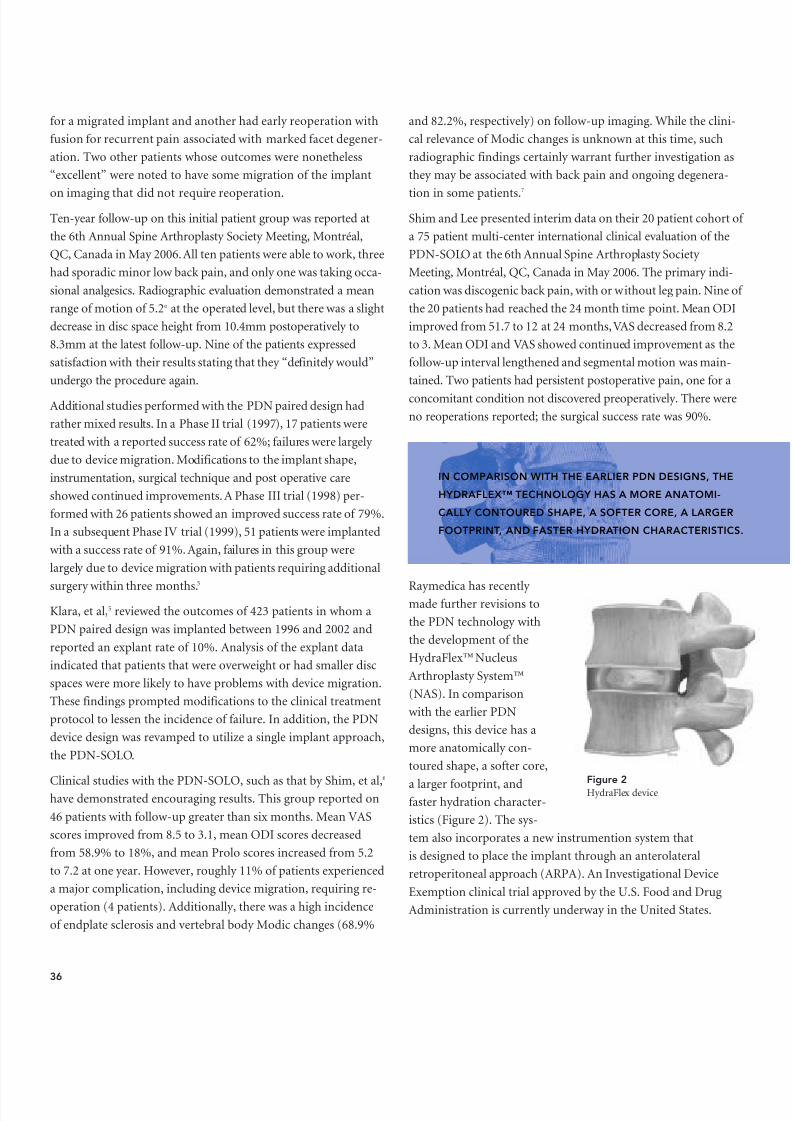
8/9/2019 Nucleus Arthroplasty Volume III
http://slidepdf.com/reader/full/nucleus-arthroplasty-volume-iii 38/48
for a migrated implant and another had early reoperation withfusion for recurrent pain associated with marked facet degener-ation. Two other patients whose outcomes were nonetheless“excellent” were noted to have some migration of the implant
on imaging that did not require reoperation.Ten-year follow-up on this initial patient group was reported atthe 6th Annual Spine Arthroplasty Society Meeting, Montréal,QC, Canada in May 2006.All ten patients were able to work, threehad sporadic minor low back pain, and only one was taking occa-sional analgesics. Radiographic evaluation demonstrated a meanrange of motion of 5.2 o at the operated level, but there was a slightdecrease in disc space height from 10.4mm postoperatively to8.3mm at the latest follow-up. Nine of the patients expressedsatisfaction with their results stating that they “definitely would”undergo the procedure again.
Additional studies performed with the PDN paired design hadrather mixed results. In a Phase II trial (1997), 17 patients weretreated with a reported success rate of 62%; failures were largely due to device migration. Modifications to the implant shape,instrumentation, surgical technique and post operative careshowed continued improvements. A Phase III trial (1998) per-formed with 26 patients showed an improved success rate of 79%.In a subsequent Phase IV trial (1999), 51 patients were implantedwith a success rate of 91%. Again, failures in this group werelargely due to device migration with patients requiring additional
surgery within three months.5
Klara, et al,5 reviewed the outcomes of 423 patients in whom aPDN paired design was implanted between 1996 and 2002 andreported an explant rate of 10%. Analysis of the explant dataindicated that patients that were overweight or had smaller discspaces were more likely to have problems with device migration.These findings prompted modifications to the clinical treatmentprotocol to lessen the incidence of failure. In addition, the PDNdevice design was revamped to utilize a single implant approach,the PDN-SOLO.
Clinical studies with the PDN-SOLO, such as that by Shim, et al,6
have demonstrated encouraging results. This group reported on46 patients with follow-up greater than six months. Mean VASscores improved from 8.5 to 3.1, mean ODI scores decreasedfrom 58.9% to 18%, and mean Prolo scores increased from 5.2to 7.2 at one year. However, roughly 11% of patients experienceda major complication, including device migration, requiring re-operation (4 patients). Additionally, there was a high incidenceof endplate sclerosis and vertebral body Modic changes (68.9%
and 82.2%, respectively) on follow-up imaging. While the clini-cal relevance of Modic changes is unknown at this time, suchradiographic findings certainly warrant further investigation asthey may be associated with back pain and ongoing degenera-
tion in some patients.7
Shim and Lee presented interim data on their 20 patient cohort of a 75 patient multi-center international clinical evaluation of thePDN-SOLO at the 6th Annual Spine Arthroplasty Society Meeting, Montréal, QC, Canada in May 2006. The primary indi-cation was discogenic back pain, with or without leg pain. Nine of the 20 patients had reached the 24 month time point. Mean ODIimproved from 51.7 to 12 at 24 months,VAS decreased from 8.2to 3. Mean ODI and VAS showed continued improvement as thefollow-up interval lengthened and segmental motion was main-tained. Two patients had persistent postoperative pain, one for aconcomitant condition not discovered preoperatively. There wereno reoperations reported; the surgical success rate was 90%.
Raymedica has recently made further revisions tothe PDN technology withthe development of theHydraFlex™ NucleusArthroplasty System™(NAS). In comparisonwith the earlier PDNdesigns, this device has amore anatomically con-toured shape, a softer core,
a larger footprint, andfaster hydration character-istics (Figure 2). The sys-tem also incorporates a new instrumention system thatis designed to place the implant through an anterolateralretroperitoneal approach (ARPA). An Investigational DeviceExemption clinical trial approved by the U.S. Food and DrugAdministration is currently underway in the United States.
Figure 2HydraFlex device
36
IN COMPARISON WITH THE EARLIER PDN DESIGNSHYDRAFLEX™ TECHNOLOGY HAS A MORE ANATOMCALLY CONTOURED SHAPE, A SOFTER CORE, A LAFOOTPRINT, AND FASTER HYDRATION CHARACTER

8/9/2019 Nucleus Arthroplasty Volume III
http://slidepdf.com/reader/full/nucleus-arthroplasty-volume-iii 39/48
Newcleus (Zimmer, Inc., Warsaw, IN)
The Newcleus implant is composed of polycarbonate urethane(PCU) and has a unique spiral shape derived from memory coil-ing (Figure 3), which enables it to be implanted through a very small annular incision and recoil into its original shape. The
unique shape also eliminates any fixed axis of the device, whichallows for easy positioning, as rotation or movement within thedisc space is not an issue. Clinical results on five patients withan average follow-up of 23.6 months (range 6–64 months) havebeen reported. 9 Results of this very limited study were promis-ing, with all patients experiencing an improvement in Oswestry score and all being satisfied with their outcome. There were nodevice dislodgements, neurologic deficit creations, or reopera-tions reported. Disc height was maintained during follow-upexaminations and flexion/extension radiographs demonstratedretained motion over the operated segments. Rotational CT
scanning demonstrated normal function of the facets withoutarthropathy. All patients demonstrated vertebral body signalchanges (Modic changes) on magnetic resonance imaging, afinding of questionable significance. An international multicen-ter study is currently underway to assess the long-term efficacy of this implant.
NuCore (Spine Wave, Inc., Shelton, CT)
The NuCore injectable nucleus is an in situ curing polymer com-posed of a recombinant protein hydrogel (Figure 4). Early resultson 12 patients have recently been reported. Four of these havebeen followed for a year and five have achieved 6-monthfollow-up. Thus far, all have experienced good pain relief andmaintenance of disc height. There have been no device extrusionor device-related complications. 10
BioDisc (CryoLife, Inc., Kennesaw, GA)
BioDisc is a protein hydrogel composed of a mix of bovine albu-min and glutaraldehyde (Figure 5). Nine patients with radiculopa-thy have been enrolled in a clinical trial of this device and early results are encouraging, with patients demonstrating significant
improvements in Oswestry score (mean 50.6 to 9.1),VAS score (mean 5.9 to1.6), and SF-36 physicalcomponent (mean 28.5 to48.1) after three months of follow-up. 11 No surgicalcomplications were notedand magnetic resonanceimaging at follow-up exam-inations demonstrated nomigration of implants.
Although these early results are encouraging, it is unclear fromthe data reported how many of these patients were treated foronly radicular symptoms and thus might have enjoyed similarimprovement for the discectomy portion of the procedure alone.
Figure 3NewCleus
Figure 4NuCore
37
AS THE NEW GENERATION OF NUCLEUS REPLACEMENTDEVICES IS DEVELOPED, THE RIGOROUS ASSESSMENTOF CLINICAL OUTCOMES WILL BE PARAMOUNT INPROVING SUPERIORITY TO EXISTING TECHNOLOGY.
Figure 5BioDisc

8/9/2019 Nucleus Arthroplasty Volume III
http://slidepdf.com/reader/full/nucleus-arthroplasty-volume-iii 40/48
DASCOR (Disc Dynamics, Inc., Eden Prairie, MN)
The DASCOR device con-sists of an injectablepolyurethane that poly-merizes minutes afterbeing injected into a bal-loon inserted into the discspace (Figure 6). Europeanand U.S. trials are cur-rently underway. Datafrom the first 16 patientsenrolled has beenpresented as promising. 1
Nubac (Pioneer Surgical Technology, Marquette, MI)
The Nubac device is an articulating PEEK-on-PEEK nucleusreplacement implant (Figure 7). Nubac received the CE mark approval in 2005 and a U.S. FDA conditional approval for clinicaltrials was granted in 2006.
Regain (EBI Biomet, Warsaw, IN)
The Regain nucleus replacement is a one-piece pyrocarbondevice (Figure 8). Pyrolytic carbon has a significant use history inlong-term implantable devices thus biocompatibility is not con-sidered and issue. The Regain is in clinical trials in Europe; it isnot commercially available in the United States.
CONCLUSION
Nucleus arthroplasty in the lumbar spine is a promising tech-nology that may prove to be effective in treating back pain andpreventing same and adjacent-level degeneration. Clinical expe-rience with these devices is limited, and a large experience hasonly been reported with one, the PDN device. Thus far, resultshave been promising, with improvements in numerous func-tional indices and maintenance of segmental motion and discspace height being reported in most studies. Of concern is thefrequency of device migration and the increases in vertebralModic changes and endplate sclerosis. Longer-term follow-upand randomized controlled trials will be needed to conclusively determine whether this technology is more beneficial thansimple discectomy or fusion procedures.
REFERENCES1. Di Martino A, Vaccaro AR, Lee JY, Denaro V, Lim MR. Nucleus pulposus
replacement: basic science and indications for clinical use. Spine. Aug 152005;30(16 Suppl):S16-22.
2. Hoffman RM, Wheeler KJ, Deyo RA. Surgery for herniated lumbar discs: aliterature synthesis. J Gen Intern Med. Sep 1993;8(9):487-496.
3. Bao QB, McCullen GM, Higham PA, Dumbleton JH, Yuan HA. The artificialdisc: theory, design and materials. Biomaterials. Jun 1996;17(12):1157-1167.
4. Schonmayr R, Busch C, Lotz C, Lotz-Metz G. Prosthetic disc nucleusimplants: the Wiesbaden feasibility study. 2 years follow-up in ten patients.Rive Neuroradiol. 1999;12 (Suppl 1):163-170.
5. Klara PM, Ray CD. Artificial nucleus replacement: clinical experience. Spine.Jun 15 2002;27(12):1374-1377.
6. Shim CS, Lee SH, Park CW, et al. Partial disc replacement with the PDN pros-thetic disc nucleus device: early clinical results. J Spinal Disord Tech. Aug2003;16(4):324-330.
7. Kuisma M, Karppinen J, Niinimaki J, et al. A three-year follow-up of lumbarspine endplate (Modic) changes. Spine. Jul 1 2006;31(15):1714-1718.
8. Bertagnoli R, Vazquez RJ. The Anterolateral TransPsoatic Approach (ALPA): anew technique for implanting prosthetic disc-nucleus devices. J Spinal DisordTech. Aug 2003;16(4):398-404.
9. Husson JL, Korge A, Polard JL, Nydegger T, Kneubuhler S, Mayer HM. Amemory coiling spiral as nucleus pulposus prosthesis: concept, specifications,bench testing, and first clinical results. J Spinal Disord Tech. Aug
2003;16(4):405-411.10. Berlemann U, Schwarzenbach O, Etter C, Kitchell S. Clinical evaluation of an
injectable, in situ curing nucleus replacement. Europ Cells Mater. 2006;11(1 Suppl):24.
11. Singhal V, MacEachern C, Craig N, Wardlaw D. Early Clinical results of anin situ polymerizing protein hydrogel nuclear repair system. Available at:http://www.cryolife.com/pdf/Britspine_poster.pdf.
38
Figure 7NuBac
Figure 6DASCOR
Figure 8Regain

8/9/2019 Nucleus Arthroplasty Volume III
http://slidepdf.com/reader/full/nucleus-arthroplasty-volume-iii 41/48
KEYPOINTS
• The surgical approach selected for implantation of nucleusreplacement technologies can influence initial placementand performance.
• Important aspects of the surgical technique include disc spacepreparation and subsequent sizing of the selected implant tothe available space.
• An additional complication associated with the use of nucleusreplacement technologies is the potential for allergic reaction.
• Nucleus replacement is less invasive than fusion or TDR tech-nologies that tend to be more destructive to the surroundingtissues and bone.
• Revision surgery for nucleus replacement devices offers many advantages over other non-fusion alternatives that are utilizedlater in the continuum of care.
Chapter 20 Nucleus ReplacementComplications andSalvage Procedures
Dr. med. univ. Rudolf BertagnoliFOUNDERPro-Spine Medical ConsultingStraubing, Germany 94315
Federico P. Girardi, MDASSISTANT PROFESSOR
OF ORTHOPEDIC SURGERY
Hospital for Special Surgery New York, NY 10021
Reginald J. Davis, MD, FACSCHIEF OF NEUROSURGERY
Baltimore Neurosurgical Associates, PA
Baltimore, MD 21204
39

8/9/2019 Nucleus Arthroplasty Volume III
http://slidepdf.com/reader/full/nucleus-arthroplasty-volume-iii 42/48
INTRODUCTION
Degenerative disc disease (DDD) is one of the most commonetiologies associated with low back pain (LBP). Currently,
low back pain is addressed using treatment modalities that incor-porate the use of non-operative or operative care. In general, sur-
gical treatment for low back pain occurring as a result of DDD isconsidered only after prolonged failure of non-surgical treatmentoptions. Often in such instances, the patient’s quality of life is soprofoundly affected, that surgery is the only viable treatment.
The main goals in treating lumbar DDD are to identify the paingenerator(s) and restore function. For many years, this goal hasbeen accomplished using spinal fusion with interbody devicesand/or rigid posterior instrumentation. More recently, the use of motion preserving alternatives, such as total disc replacement(TDR), are now being employed. In addition, the use of Nucleus
Arthroplasty™ or nucleus replacement therapies has moved tothe forefront. These technologies are very promising as the intentis to replace only the damaged or diseased nucleus, while seekingto restore function, maintain disc height, and preserve motion.Thus, nucleus replacement is less invasive than fusion or TDR technologies that tend to be more destructive to the surroundingtissues and bone.
Currently, there are a number of different design concepts indevelopment and evaluation. 1,2 In general, nucleus replacement canbe simply classified as preformed devices, which include hydrogelsor mechanical solutions, and in situ devices, which involve compo-nent mixing and in situ cure. The ability to successfully implement
nuclear replacement technologies is exciting as such solutions seek to expand the available surgical treatment options and potentially redefine the current continuum of care.
Paramount to the success of any surgical procedure is the properidentification of the treatment indications and selection of thecorresponding patient population. Obviously, each surgical pro-cedure has its own associated risks, complications, and potentialbenefits that must be assessed prior to treatment. One of themost common problems with nucleus replacement technologiesis their use in patients with advanced degenerative disc disease,where the window of opportunity for this type of treatment isfar gone. Thus, prerequisites for positive and predictable surgicaloutcomes involve adequate surgeon training in combinationwith the application of strict inclusion and exclusion criteria. 3
For the purposes of this chapter, the discussion associated withcomplications will be divided into specific areas including bio-mechanical, surgical approach, surgical technique, patientselection, and other concerns. This will be followed by a brief discussion of salvage procedures related specifically to nucleusreplacement technologies.
40
n=0%
n=100%
Conservativetreatment
PercutaneousDisc surgeries
OpenDisc surgeries
Fusionsurgeries
P o t e n t i a l T r e a t m e n t P o p u
l a t i o n
I n c r e a s i n
g t r e a t m e n t s i z e
, r i s k a n d
c o l l a t e r a
l d a m a g
e
D i s c S u r g e
r y / 3 C l a s
s i c a l S u r g
i c a l S t e p
s
Figure 1Bertagnoli 10

8/9/2019 Nucleus Arthroplasty Volume III
http://slidepdf.com/reader/full/nucleus-arthroplasty-volume-iii 43/48
Figure 2Bertagnoli 10
BIOMECHANICAL ISSUES
Biomechanical performance issues associated with nucleusreplacement devices are similar to those of other non-fusion,motion preserving devices and include wear, fracture/failure, dis-location, migration, subsidence, and loss of motion. Based on
the existing clinical information, the majority of these complica-tions are rare with the exception of device migration and subsi-dence. While migration tends to be related to the surgicalapproach and device configuration, subsidence, and resultingendplate damage, is more directly related to device design. Theability to determine the appropriate device stiffness has provedto be a challenging endeavor as it represents a balance betweenthe load carrying capacity of the device and the structuralintegrity of the vertebral endplates. If the device load carryingcapacity is too low, disc height may not be maintained, while aload carrying capacity that is too high may result in endplateremodeling and/or fracture. Extensive testing has been performedto further investigate the effects of implant stiffness, sizing andconformity in an effort to strike an appropriate balance.
APPROACH RELATED ISSUES
Until recently, the most common surgical approach used in con- junction with the placement of spinal devices has been an openposterior hemilaminotomy. This approach has the advantage of
allowing the surgeon to address posterior pathologies, such asherniations. However, depending on the extent of the pathology,access to the disc space requires significant dissection of the pos-terior musculature and associated osteoligamentous structures,and may require a partial facetectomy, leading to spinal instabil-
ity. Spinal instability is more problematic with nucleus replace-ments devices, primarily due to the lack of attachment to bony structures, making device migration an issue.
The use of a posterior approach also poses challenges to thenucleus removal process. The limited surgical window coupledwith the anatomy of the spine make it difficult to adequately remove nucleus from the contralateral regions within the disc.Inadequate nucleus removal can inhibit proper device placementand subsequently influence implant motion. Intraoperatively, theuse of contrast medium can be helpful to assess the extent of thenucleus evacuation prior to device placement.
Bertagnoli, et al,4 have described the use of an anterior lateraltranspsoatic approach (ALPA) for nucleus replacement. In thisprocedure, the disc space is accessed laterally by way of theretroperitoneum, and involves longitudinal splitting of the psoasmuscle. The approach utilizes a safe anatomical zone that allowsfor sparing of the posterior osteoligamentous structures. WithALPA, a larger section of the annulus can be exposed, simplifyingthe nucleus evacuation and implant insertion processes.
41
n=0%
n=100%I
II
III
IV
V
VI
VII
Percutaneous
Disc surgeries
OpenDisc surgeries
Anthroplasysurgeries:Nucleusreplacements
InterspinousImplantsWallis, Diam,Flexicore
Anthroplasysurgeries:Total Discreplacements
Post.Dynamicstabilization:Dynesys…
Fusionsurgeries
P o t e n t i a l T r e a t m e n t P o p u l a t i o n
I n c r e a s
i n g t r e a
t m e n t s i z e
, r i s k a n d
c o l l a t e
r a l d a m
a g e
M o d e r n
D i s c S u r g
e r y / 7 S u r g
i c a l S t e p
s

8/9/2019 Nucleus Arthroplasty Volume III
http://slidepdf.com/reader/full/nucleus-arthroplasty-volume-iii 44/48

8/9/2019 Nucleus Arthroplasty Volume III
http://slidepdf.com/reader/full/nucleus-arthroplasty-volume-iii 45/48
present with significant disc herniations would be undesirablecandidates. Currently, the ability to extend or broaden the use of this technology for such applications is still not clear.
OTHER MISCELLANEOUS COMPLICATIONS
The goal of a nucleus replacement device is to replace the nativenucleus material. Technologies developed to achieve this goal areutilizing a host of new materials in comparison to those used instandard orthopedic implants. Thus, additional complicationsinclude the potential for allergic reaction to the implant materialsor contrast media used during the implant sizing process. In addi-tion, depending on the implant technology, the ability to ade-quately view the implant or surrounding tissues post-operatively may be problematic using standard imaging modalities, (X-ray,
MRI, CT).
Based on existing clinical data, the risk of heterotopic ossificationhas not been shown to be a significant problem with nucleusreplacements. This can be a problem in more invasive proceduresthat require more involved dissections and/or preparation of theendplates, such as in total disc replacement procedures.
SALVAGE
The removal of failed nucleus replacement devices will largely be
accomplished via mechanical means. Consequently, the difficul-ties associated with removal will be closely related to the devicetype. Preformed and mechanical devices may be easier to reviseas their inherent design facilitates removal en bloc . In addition,such technologies possess a known shape quality allowing visualinspection to be utilized to ensure complete device removal.Revision of in situ curable designs may pose more of a challengeas the implant size and shape will vary for each application. Inaddition, some implants of this type are not contained within abarrier, and may potentially integrate with the surrounding disctissue. In such instances, the ability to ensure complete removal
may be challenging as the devices may not be viewable usingimaging modalities. Regardless of the device type, the tissuedestruction required to complete the removal process may limitthe surgeon’s revision options.
In general, revision surgery for nucleus replacement devices hasmany advantages over revision surgeries for other non-fusion alter-natives, such as total disc replacement, that are performed later inthe continuum of care. This is largely due to the fact that the initialsurgery to place a nuclear implant is much less destructive to the
vertebral bodies and surrounding tissue. This preservation of theannulus and posterior anatomy, enables the use of surgical revisionstrategies that utilize additional motion preservation technologies(interspinous spacers, TDR, dynamic stabilization).
In essence, the successful introduction of nucleus replacementsand other motion sparing technologies will significantly expandthe treatment options currently utilized today 10 (Figures 1 & 2).This potential shift in the continuum of spinal care will providethe surgeon flexibility in selecting an appropriate initial or sal-vage procedure that is focused on maintaining spinal motionand slowing the progression of the degenerative cascade.
REFERENCES1. Goins ML, Wimberley DW, Yuan PS, Fitzhenry LN, Vaccaro AR. Nucleus pul-
posus replacement: an emerging technology. The Spine J. Nov-Dec Suppl 2005;5:317S–324S.
2. Ray CD. The Raymedica prosthetic disc nucleus (PDN): stabilizing the degen-erated lumbar vertebral segment without fusion or total disc replacement. In.Eds. Kim DH, Camissa FP, Fessler RG. Dynamic reconstruction of the spine,2006; Chp 14:105–114.
3. Triano JJ, Bougie J, Rogers C, Scaringe J, Sorrels K, Skogsbergh D, Mior S.Procedural skills in spinal manipulation: do prerequisites matter? The SpineJ. Sep-Oct 2004; 4:557–563.
4. Bertagnoli R, Vazquez RJ. The anterolateral transpsoatic approach (ALPA):a new technique for implanting prosthetic disc—nucleus devices. J SpinalDisord Tech. Aug 2003; 16(4):398-404.
5. Shim CS, Lee SH, Park CW, Choi WC, Choi G, Choi WG, Lim SR, Lee HY.Partial disc replacement with the PDN prosthetic disc nucleus device: early clinical results. J Spinal Disord Tech. Aug 2003; 16(4):324-330.
6. Cragg A, Carl A, Castaneda F, Dickman C, Guterman L, Oliveira C. New per-cutaneous access method for minimally anterior lumbosacral surgery. J SpinalDisord Tech. 2004; 17(1):21–28.
7. Jin D, Qu D, Zhao L, Chen J, Jiang J. Prosthetic disc nucleus (PDN) replacementfor lumbar disc herniation. J Spinal Disord Tech.Aug 2003; 16(4):331–337.
8. Bhattacharyya T, Blyler C, Shenaq D. The natural history of new orthopeadicdevices. Clin Orthop Relat Res. Oct 2006; 451:263–266.
9. Bertagnoli R, Schönmayr R. Surgical and clinical results with the PDNprosthetic disc-nucleus device. Eur Spine J. 2002; 11(S2):S143–S148.
10. Bertagnoli R. Disc surgery in motion. Spine Line. 2004; 11/12:23–28.
43
REVISION SURGERY FOR NUCLEUS REPLACEMENDEVICES HAS MANY ADVANTAGES OVER REVISIO
GERIES FOR OTHER NON-FUSION ALTERNATIVES, AS TOTAL DISC REPLACEMENT, THAT ARE PERFORLATER IN THE CONTINUUM OF CARE.

8/9/2019 Nucleus Arthroplasty Volume III
http://slidepdf.com/reader/full/nucleus-arthroplasty-volume-iii 46/48
Although much remains to be learned about degenerativedisc disease (DDD), we are on the precipice of a new treat-
ment stratagem for this problem effecting a wide-ranging patientpopulation which has been inadequately treated with available
treatment options. Nucleus Arthroplasty™ is poised to addressmild to moderate DDD and may be a successful treatment optionfor later stage disease as well.
Pioneering spine surgeons have successfully used nucleusreplacement to treat DDD. However, nucleus arthroplasty takesthis promising therapy one step further. Nucleus arthroplasty isa total system approach that encompasses patient selection, indi-cations, device sizing, surgical technique and approach, andpost-operative patient care. By adopting this system approach,nucleus arthroplasty is expected to offer an effective treatmentoption for patients who are currently left untreated.
FUTURE PUBLICATIONS ONNUCLEUS ARTHROPLASTY™ TECHNOLOGY
We hope this monograph, Volume III— Surgical Techniques & Technologies , provides valuable guidance to spine surgeons asthey seek to provide more effective and less invasive options forthe treatment of DDD. This volume is part of a continuing serieson nucleus arthroplasty that can be used individually or collec-tively. A number of well-known authors will be represented in
future monographs on nucleus arthroplasty.Upcoming scheduled publication dates are as follows:
• Emerging Technology is scheduled to be released at theNorth American Spine Society (NASS) meeting in Austin,Texas, October 2007.
Please note that each one of these volumes will be available at: www.nucleusarthroplasty.com
Conclusion
Reginald J. Davis, MD, FACSCHIEF OF NEUROSURGERY
Baltimore Neurosurgical Associates, PABaltimore, MD 21204
Federico P. Girardi, MDASSISTANT PROFESSOR
OF ORTHOPEDIC SURGERY
Hospital for Special Surgery New York, NY 10021
Frank P. Cammisa, Jr., MD, FACSASSOCIATE PROFESSOR OF CLINICAL SURGERY
Hospital for Special Surgery New York, NY 10021
44
WE ARE ON THE PRECIPICE OF A NEW TREATMENTSTRATAGEM FOR THIS PROBLEM EFFECTING AWIDE-RANGING PATIENT POPULATION WHICH HASBEEN INADEQUATELY TREATED WITH AVAILABLETREATMENT OPTIONS.

8/9/2019 Nucleus Arthroplasty Volume III
http://slidepdf.com/reader/full/nucleus-arthroplasty-volume-iii 47/48www.nucleusarthroplasty.com

8/9/2019 Nucleus Arthroplasty Volume III
http://slidepdf.com/reader/full/nucleus-arthroplasty-volume-iii 48/48