nucleus saccharomyces: cytologicaljb.asm.org/content/68/5/514.full.pdf · ploited in yeast cytology...
TRANSCRIPT

THE NUCLEUS OF SACCHAROMYCES: A CYTOLOGICAL STUDY OF AFROZEN-DRIED POLYPLOID SERIES
BALAJI D. MUNDKUR
521-A, Matunga, Bombay, India
Received for publication Mlarch 30, 1954
Identification of the yeast nucleus, and par-ticularly a resolution of its mitotic cycle, canscarcely be considered to have yielded unanimousagreement. In spite of a voluminous cytologicalliterature, little has been established with cer-tainty in regard to the identity, morphology, ornumbers of chromosomes, or in regard to theorientation of the dividing nucleus with respectto the centrosomes during budding. Incertitudeand divergences of opinion on these questionshave resulted partly from difficulties inherentin obtaining suitable preparations of organismsso small as yeast for cytological analysis, andpartly from the fact that cytological studieswere undertaken in almost all instances on cul-tures unknown genetically as to the number oflinkage groups and the degree of ploidy.
Nagel (1946), DeLamater (1950), Lindegrenand Rafalko (1950), Lietz (1951), and Winge(1951a) have considered various interpretationsof yeast cytology at length; hence we shall omita detailed historical discussion. The purpose ofthe present report, based on cytological pro-cedures hitherto not used for yeast, is to describesingle and dividing cells and offer evidence fora vesicular nucleus as well as to consider in-adequacies apparent in some current viewpoints.
It is sufficient to indicate here two major,opposing ideas: According to Lindegren (1949,1952), following Wager and Peniston (1910),the chromosomes are located within the vacuole(the "nuclear vacuole") and exhibit a pattern of"mitotic" behavior thus far unknown in otherorganisms. This view is opposed by the majorityof yeast cytologists who hold that the true nu-cleus is that structure identified by Lindegrenas the centrosome; this is a Feulgen positive,sphere-like body of relatively massive proportionsdisposed adjacent to and outside the vacuolarwall. A few workers are in accord with the secondview (Guilliermond, 1920; Beams et al., 1940;Sinoto and Yuasa, 1941) and believe the divisionof this body to be amitotic. However, well es-
tablished genetical information is currentlyavailable to render this belief unwarranted. Con-clusive proof that this extravacuolar body is thenucleus derives from the fact that its total volumeand mean diameter increase in accord with thetheoretical expectation for polyploids (Mundkur,1953).This reinvestigation of a much-debated sub-
ject was undertaken in view of the availabilityof a polyploid series whose haploid, diploid,triploid, and tetraploid constitutions were clearlyestablished genetically with the aid of 6-11diagnostic markers by Lindegren and Lindegren(1951). Furthermore, a reexamination of theproblem with the aid of the Altmann-Gershfreezing-drying technique, hitherto not ex-ploited in yeast cytology (or perhaps in bacterialcytology), was deemed desirable in view of itsmany unambiguous qualities. Advantages of thistechnique are considered in detail by Gersh(1932, 1948) and recently by Bell (1952) andDanielli (1953). Suffice it to indicate here that(1) cessation of metabolic activity is almost in-stantaneous, (2) shifting of diffusible cell con-stituents is inappreciable, (3) there is littlepossibility of chemical alterations in cells leadingto morphological artifacts or pseudostaining re-actions such as those sometimes ensuing from theuse of chemical fixatives, (4) shrinkage of cellsis absent or negligible, (5) there is a greaterpreservation of cell inclusions than is possiblewhen fixing solutions are used, and (6) directinfiltration of dehydrated cells with celloidin orparaffin is possible.
MATERIAL AND METHODS
The cultures used include no. 13942 (haploid),11296 (diploid), 13894/11296 (triploid), and11296/11294 (tetraploid). The haploid anddiploid cultures are nonsporulating. Pure culturesof the triploid and tetraploid were derived bypicking single, large isolated colonies fromseparate plates very lightly seeded with samples
514
on July 5, 2018 by guesthttp://jb.asm
.org/D
ownloaded from
NUCLEUS OF SACCHAROMYCES
of mating mixtures involving, respectively,haploid X diploid and diploid X diploid cells.The rapid growth rates of triploid and tetraploidsingle cell colonies on plates (relative to haploidor diploid cells) as well as their unmistakablylarge cell sizes eliminate the possibility of in-cluding unmated haploids or diploid cells andinsure that the triploids and tetraploids, pickedas well isolated colonies, are pure. Such singlecolony isolates were used as inocula for obtaininglarger quantities of material for freeze-drying.Under conditions of active growth the stocks didnot sporulate on the liquid yeast extract-peptone-glucose medium used for their cultivation. Sam-ples of all cultures were examined microscopicallyimmediately prior to freezing to insure the ab-sence of spores and to check on homogeneity incell sizes which, within limits, is distinct for eachclass in the polyploid series (Mundkur, 1953).The cultures were raised (1) aerobically on
agar slants, (2) aerobically in liquid mediumwith sterile air forced vigorously through themedium from aerating stones, and (3) in un-shaken, actively fermenting liquid medium;yeast growing after the addition of fresh nutrientto medium spent by prior fermentation was alsoharvested.Yeast harvested for freezing-drying was
washed quickly in phosphate buffer (pH 4.5),centrifuged, and spread in pellets (approx. 2 mmthick) with a loop over narrow strips of slightlymoistened filter paper. The object of wetting thestrips was to prevent desiccation of the cells.The strips bearing yeast were then immediatelyimmersed in isopentane cooled to about -150 Cin liquid nitrogen, and the frozen material wassubsequently dehydrated in vacuo at about -30 Cover phosphorus pentoxide for three days. Dur-ing this time the submicroscopic ice crystalsformed in the cells on immersion in isopentanesublime without displacement of cell constituents.On complete dehydration, the cell pellets werequickly scraped off the paper strips into chilledabsolute alcohol, the alcohol drained out andreplaced with 15 per cent nitrocellulose inether-alcohol, and later with 30 per cent nitro-cellulose. Sections were cut at 3, 6, and 9 ,u,conforming roughly to the dimensions of cells ofincreasing degree of ploidy. This enables theinclusion of whole as well as sectioned cells in allpossible orientations in the celloidin section, thusinsuring an extremely favorable means of study-
ing stained preparations. This advantage isdenied those studying smears, especially whenelongate cells are concerned.
In some instances, the fixed cells were treatedwith amylase in 10 per cent alcohol at pH vary-ing from 2 to 4.5 so as to digest any glycogengranules that might obscure cytological detail,but the advantage gained by this treatment wasnot appreciable.
After removal of celloidin, the fixed materialwas stained with Heidenhain's hematoxylin andpyronin or eosin, thionin, or the Feulgen methodwith light green as counterstain. ConventionalFeulgen procedures as well as the Rafalko (1946)modification were used. Except for the centrioles,which are Feulgen negative, the results in allcases were identical. Perchloric acid (10 per cent)hydrolysis at 25 C for 2 to 3 hr yielded excellentresults and was used almost exclusively bothwith or without precipitation of the desoxy-ribonucleic acid as the lanthanum salt.
Living as well as fixed material was examinedunder the phase microscope; toluidine bluestaining of living and fixed cells was effected asrecommended by Lindegren (1949). Preparationsof material fixed with fluids, such as Bouin's,Schaudinn's, Helly's, Carnoy's, and with osmicfumes were also examined for comparison.
Digestion of ribonucleic acid in frozen-driedcells with ribonuclease was carried out for thepurpose of locating sites of desoxyribonucleic acidwith the ultraviolet microscope. The digestionswere effected with 0.1 per cent ribonuclease(Armour) in distilled water (adjusted to pH 6with NaOH) for 2 hr at 37 C (Kaufmann et al.,1951). The treated sections were then coatedwith glycerin, covered with quartz coverslips,and examined for absorption areas on a fluores-cent screen at 2652 A.
RESULTS AND OBSERVATIONS
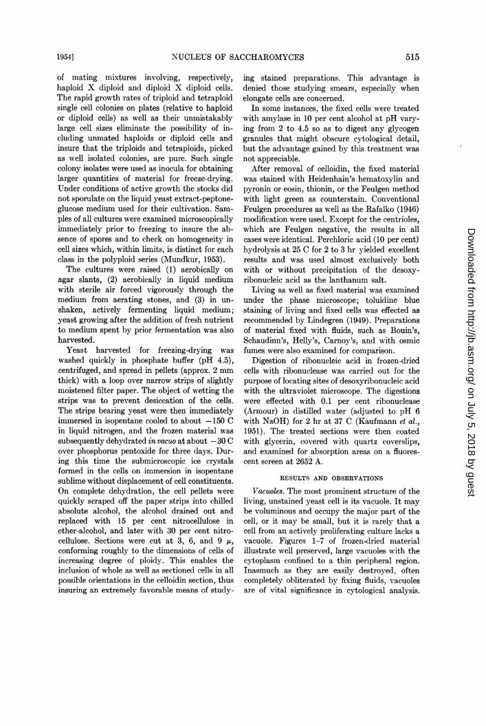
Vacuoles. The most prominent structure of theliving, unstained yeast cell is its vacuole. It maybe voluminous and occupy the major part of thecell, or it may be small, but it is rarely that acell from an actively proliferating culture lacks avacuole. Figures 1-7 of frozen-dried materialillustrate well preserved, large vacuoles with thecytoplasm confined to a thin peripheral region.Inasmuch as they are easily destroyed, oftencompletely obliterated by fixing fluids, vacuolesare of vital significance in cytological analysis.
5151954]
on July 5, 2018 by guesthttp://jb.asm
.org/D
ownloaded from

....1..... .*
2 1-b * ,
*...~~~4
Figures2: 6(diloi.tiplidanterapoidcels,resectvey) otelare,elp
_.acni rb.,:
I.~~~~~~~~~~~~~~~~~~~~~~~~~~~~~~~~~~~~~~~~~~~
.. :~ .4
Figure 3. Triploid. Exceptional instance of an intravacuolar nucleus. Hematoxylin-pyronin, 3,600 X.Figure 4. Group of sectioned as well as wxhole triploid cells in various orientations. Elementary na-
ture of nuclear division during budding is seen at a. A sectioned nucleus is seen at b. Note well pre-served vacuoles. Aerobically active cells. Hematoxylin-pyronin, 2,600 X.
Figure 5. Phase contrast image of cell at a in figure 4, focused on the nuclear constriction at thepoint of bud attachment. Vesicular nature of the nucleus is clearly apparent and is seen as a zone ofuniformly reduced density. The vacuole is structurally empty, and there is a faint centriole in thebud. The dark zone next to the nucleus is not Lindegren's "heterochromatin" but is merely an opticaleffect at this particular focus. It and the similar dark zone on the other side of the vacuole disappearon slight change of focus.
Figure 7. Group of diploid cells with typical spherical nuclei. Feulgen-light green, 3,600 X.Figure 8. Haploid cells from fermenting culture. Hematoxylin-eosin, 3,600 X.
516
on July 5, 2018 by guesthttp://jb.asm
.org/D
ownloaded from

NUCLEUS OF SACCHAROMYCES
Their partial shrinkage under chemical fixationfrequently results in a narrow, canal-like struc-ture. Because this canal sometimes terminates ateither end with a small spherical body and isflanked by two Feulgen positive structures, it isliable to be erroneously identified as the spindle,with the spherical dots (in reality, vacuolarinclusions) being termed "centrioles" and theFeulgen bodies (in reality, daughter nuclei) beingtermed "chromosomes". Photograph no. 2 andfig. 2 by Subramaniam (1952) are reminiscentof this condition. The possession by the vacuoleof variable numbers of basophilic structuresnecessitates that the vacuole be maintained intactafter fixation so that the relation of these struc-tures (the "chromosomes" of Wager and Penis-ton, and Lindegren) to the rest of the cell, aswell as their morphology, be amenable to un-obscured observation.The vacuoles generally occur one per cell and
may be simple or lobed. Occasionally two, butrarely more than two, independent vacuolesoccur. Multiple vacuoles have not been observedin the present study. In all cases, vacuoles havesmooth surfaces and contain a hyaline sap whichexhibits a much less intense, often negative,affinity for stains such as pyronin or eosin whichthe cytoplasm takes on deeply.The vacuolar contents are morphologically
highly variable. They are effectively stainedwith toluidine blue by Lindegren's method. Liv-ing cells stained with toluidine blue retain theirvacuoles intact and permit a clear observation ofintravacuolar bodies. These, however, are notdetectable in all cells in the mount-only a smallpercentage of cells exhibit them in a given mount-but their frequency can be increased by alter-ing the cultural conditions, principally the pH.According to Lindegren, these bodies show apattern of behavior involving end to end fusionsof short elements into thick, paired complexeswhich later split longitudinally into Y shapes,followed eventually by migration of one fullysplit complex (chromosomes) into the bud. Thepresent work does not corroborate such a se-quence either in frozen-dried material or infresh cells stained with Lindegren's method. Thefollowing observations indicate that these in-travacuolar bodies are not chromosomes and,further, that the "sequence" is fortuitous ratherthan a mitotic design:
(1) There is no constancy in the number ofthese inclusions in cells of single-cell derivation.
(2) In the polyploid series investigated, in-crements in their numbers are neither obligatorynor consistent with progression in ploidy.
(3) A mother cell may possess a large thickrod together wvith tiny fragments while itsattached bud may possess only a thick rod.
(4) Vacuoles in mother cell and attachedimmature bud may possess complexes not inaccord with the postulated sequence; for in-stance, the bud may occasionally have tinyfragments while the mother cell vacuole has aY shaped or a single rod.
(5) They are Feulgen negative.(6) Their appearance in cells is controlled by
cultural conditions, and unlike true chromosomes,the inclusions are not universally visible in alldividing cells.
Figures 1, 2, 4, 6, and 7 of cells from vigorouslyaerated cultures are typical of aerobically activecells. Their vacuoles are no less prominent thanthose in fermenting cells (figures 8, 9, 14).These observations do not corroborate Subra-maniam's (1952) view that a basic differencebetween aerobic and anaerobic cells is that theformer are totally devoid of vacuoles while thelatter possess prominent ones.
The Nucleus
As far as cytochemical criteria are concerned,the chromatinic nature of the spherical extra-vacuolar region has been abundantly demon-strated by the majority of yeast cytologists.Opinion is divided, however, as to whether theentire structure is Feulgen positive. Some, forinstance Lietz (1951), hold that a crescent-likesegment of a sphere is Feulgen positive and,depending on the angle from which it is viewed,makes the sphere appear partially or completelystained. A similar view is held by Lindegren whoterms the segment "centrochromatin" and de-scribes it as plastered externally on his "cen-trosome" in contrast to Lietz who illustrates itas a structure within the nucleus.
Proliferating, frozen-dried yeast cells examinedunder phase contrast lack such crescents (figure 5)whereas they are occasionally seen in materialfixed with Carnoy's or Helly's or in fresh cellsmounted in weak (5-10 per cent) acetic acid.These observations together with the fact that
5F)'1719541
on July 5, 2018 by guesthttp://jb.asm
.org/D
ownloaded from

ws 9V
10sto
.
It
a.
I,.:I
i :
Iql,...
I Id
12
16
B:
_.4w.
..r
13 14
19
20
Figure 9. Cuneiform nucleus, triploid, in cell with two independent vacuoles. Hematoxylin-pyronin,3,600 X.
Figures 10, 11, 12, 13 (haploid, diploid, triploid, and tetraploid cells, respectively). The maximumnumber of stainable bodies is two per cell, regardless of ploidy. Nucleus in the haploid cell is in theprocess of division. Figure 11 is Feulgen-light green, 3,300 X; the others, hematoxylin-pyronin, 3,600 X.
Figure 14. Tetraploid cell from fermenting culture. Centriole in clear space beneath vacuole. Smallcell at lower right is not a bud of the larger cell but merely a sectioned individual in its vicinity. He-matoxylin-pyronin, 3,600 X.
Figures 15, 16, 17 (triploid, diploid, diploid). Cells with single centrioles. Fermenting cultures. Hema-toxylin-eosin, 3,600 X.
Figures 18, 19, 20. Diploids with paired centrioles. Part of the dividing nucleus in figure 19 com-
pletely fills an incipient bud. Hematoxylin-pyronin, 3,600 X.
518
L-
lb
9
A.
.^:.E..r.
.:::4.: 4
Ai"a
::.....:
'.i..g: 1..,.'J"..Jw,:,:
on July 5, 2018 by guesthttp://jb.asm
.org/D
ownloaded from

NUCLEUS OF SACCHAROMYCES
crescentic bodies are not visible in nuclei offrozen-dried yeast establish that their presenceis due to diffusion currents and the consequentperipheral accumulation of coagulated chromatinin the nuclei of chemically fixed material.
Pseudocrescents can be frequently seen, how-ever, in frozen-dried, stained material, but theirresemblance to crescents in conventionally fixedcells is deceptive. Diagram 1 shows how a nucleussituated eccentrically to the long axis in a frozen-dried cell with a well preserved vacuole can createan illusory crescent. Diagram 1 a and b representtwo parallel planes of foci in a cell viewed from thesame direction. The stippled area of the nucleusat a, being out of focus and partially obscured bythe light counterstain in the vacuole, appearsfaintly stained when the cell is examined withthe microscope. The dark crescent is that part ofthe nucleus which is in focus and which is notobscured by the vacuole. When the plane offocus is lowered as at b, the stippled area is nowclearly seen and appears darker while the cres-cent fades out of view. c explaining these situa-tions represents a lateral view of the same cell,that is, after it has been "rotated" through anangle of 900 from orientation a or b. Thesesituations cannot be satisfactorily photographedsince the slight difference in intensity of colora-tion between "crescent" and the body of thenucleus does not register properly.A further difference between the crescents in
frozen-dried yeast and in chemically fixed cellsis that in the former the surface of its concavityadjoins the vacuole, whereas in the latter itsorientation to the vacuole (if this is intact) isvariable. As Lietz correctly points out, detectionof the crescents in chemically fixed cells dependson critical differentiation. In the present study,controlled destaining of hematoxylin preparationsat 12 minute intervals, ranging up to 12 minutesin weak iron-alum solution, revealed that thenucleus destained uniformly until the dye wascompletely washed out. Crescents were not seenin Feulgen preparations. Furthermore, with theultraviolet microscope only homogeneous ab-sorption areas were visible.
Nuclei in the polyploid series. Progressive in-crements in total volumes of interphase nucleiconcomitant with progression in ploidy are wellknown in higher organisms. Mundkur (1953)reported the occurrence of a similar relationshipin yeast, thus extending the phenomenon to
microorganisms. Figures 1 to 33 show thesedifferences in nuclear sizes, while the ranges invariation of nuclear diameter in anaerobicallycultured members of this series are showngraphically in diagram 2. Each dot represents themean of one hundred diameters.
Table 1 summarizes mean nuclear diametersand the corresponding, calculated mean volumesin aerobically and anaerobically grown cells.Each mean is based on at least one hundredmeasurements of spherical or near-sphericalnuclei in whole, randomly selected nonbuddingcells in stained preparations.
In table 2, one hundred and two cases involv-ing a relation of the total volume of the cell tothat of its nucleus are analyzed to determinewhether a definite proportionality-the nucleo-cytoplasmic ratio-exists between the two. A pre-paration of aerobically grown haploids was usedfor these measurements. Haploid cells and nucleiare almost perfectly spherical and permit pre-cise measurements of volume. The mean nucleardiameter and volume obtained from these meas-urements are 0.86 ,u and 0.331 A, respectively,and agree closely with those for anaerobic cells.The frequency distribution in table 2 indicates
a tendency for large cells to possess large nuclei,but cases of equally large nuclei occurring inrelatively small cells also exist. For instance, anucleus of diameter 1.25 Au (vol = 1.02 M3) iscorrelated in equal frequency with cell volumesof either extreme. No case of the smallest sizednucleus associated with the largest sized cell wasdetected. The nucleo-cytoplasmic ratio is not con-stant but decreases as the nuclear volume in-creases, indicating that increase in cell volumeis not linear with respect to nuclear volume.A similar conclusion can be drawn in regard
to cells in the diplophase (table 3). These cellsalso were aerobically cultivated, but their meannuclear diameter and volume are the same asthose of fermenting cells (table 1). One hundred
at) bc()CDiagram 1
19543 519
on July 5, 2018 by guesthttp://jb.asm
.org/D
ownloaded from

BALAJI D. MIUNDKUR
Zs ..=..
._>.
.
o.'mo ::
'^':i'. io '.2@
..^..*ffi:wt!i
.. :::.:.
:....: :::i
_['
*.w.*X._.
...2_::s_.._.r,g,lllt_....
ASSk:
.,_ .i._:_ . . :. . be. :
...,...e_..i.:!: jo.
_._._.... .
}::'.'}:.i .''9:'' .*:_l_F,_r
._
24
waSi~~
I.*:: ::
i.::.: 2T 2 8
30 3'
i
32
im:.s4=
rl
Figures 21, 22. Paired centrioles in triploid cells. Note constriction developing in the nucleus infigure 22; one of the centrioles in this cell is insufficiently differentiated. Hematoxylin-eosin, 3,600 X.
Figure 23. Early stage in budding showing centriole in the incipient bud. Aerobic triploid. Hema-toxylin-pyronin, 3,600 X.
Figure 24. Bud having a centriole is almost as large as the mother cell, but nucleus in the latter hasyet to begin division. Diploid, 3,600 X.
29
.xl
OR
*.' :..
a
33 .w~
520 [VOL. 68
4 r
on July 5, 2018 by guesthttp://jb.asm
.org/D
ownloaded from

NUCLEUS OF SACCHAROMYCES
measurements each of nonbudding cells andtheir respective nuclei are represented in table 3.The total volume of each cell was calculatedfrom length and breadth measurements usingthe formula for ellipsoids. It will be noted thatthere is one case where the smallest sized nucleus(diam = 1 A; vol = 0.5236 l3) is associated with acell falling in the extreme, large cell class. Thevolume of this particular cell was 65.47 A3. Sinceonly one cell (vol = 37.71 M3) among the hundredmeasured possessed a nucleus of diameter 2.0 ,u,credence cannot be placed on the calculated ratioof 9.002 as being truly representative.An examination of any yeast culture of single
cell origin invariably reveals a fraction of cellswhich are appreciably much smaller than themajority, and whose presence in the culture canbe accounted for on the basis of the specialpeculiarities of the budding process. Buddinginvolves the progressive increase in volume of anoutgrowth from the mother cell without ap-preciable diminution of the latter's volume.Small cells occurring in a culture may be eitherimmature buds severed from the mother cell ormay be products of breakdown in ploidy inpopulations other than a haploid. It is im-probable that the latter possibility exists in theclones used in the present study, at least underthe conditions employed for their growth. It isapparent from the graph relating to fermentingcells that there is no overlap of the range ofvariation of haploid nuclei over those of higherdegrees of ploidy, indicating that a breakdown ofnuclei to the haploid level does not occur andthat Subramaniam's view that fermenting cellsbecome endopolyploid cannot be substantiated.The morphological and cytological attributes ofeach class in the series are, as a rule, maintainedwithin each preparation.
Tables 2 and 3 indicate that there is no relationbetween haploids and diploids in regard to nu-
clear and cell sizes. Both haploid and diploid cellspossess nuclei of diameter 1.0 ,u; but the meancell volumes associated with nuclei of this diam-eter are widely discrepant in each case, being9.41 /i3 for haploid cells and 18.73 .i for diploids.The mean cell volumes of haploid and diploid cellscorrelated with nuclei of diameter 1.25 ,u arealmost similar. It will be noted, however, thatthe diploid mean cell volume correlated withnuclei of this diameter is less than that of cellshaving nuclei 1.0 At in diameter.
Tables 4 and 5 represent similar karyoplasmicrelations in triploid and tetraploid cells,respectively.The data in tables 2 to 5 are consistent with
the view that nuclei in the "resting" state ex-hibit volume fluctuations which are associatedwith their stage of growth, and which may beeither rhythmic or continuous (Hughes, 1952);they indicate, moreover, that a constant nucleo-cytoplasmic ratio prevails in none of the culturesstudied.
Structure of the nucleus. Genetic analyses ofextensive pedigrees, of which the currently in-vestigated series is a member, have revealed at
rErRAPLOID
TRIPLOID
DIPLOID
HAPLOID
0.5 X0 1.5 2.0 2.5NUCLEAR DIAMErER
Diagram 2
30jl
Figure 25. Early stage in nuclear division showing the nucleus projecting a blunt, beak-like processinto the bud. Diploid, 3,600 X.
Figure 26. Tetraploid cell obliquely oriented in the section with only the bud and part of the nu-cleus in good focus; note centriole in bud and entry of nucleus into the bud. Hematoxylin-eosin, 3,600 X.
Figure 27. Tetraploid cell with mature bud and dividing nucleus. Thionin-light green, 4,000 X.Figure 28. Dividing nuclei. Feulgen-light green, 3,600 X.Figure 29. Diploid with bud and undivided nucleus, 3,600 X.Figure 30. Triploid. Nucleus in mother cell is in the process of division after having supplied the
bud with a daughter nucleus from a previous division. Hematoxylin-pyronin, 3,600 X.Figures 31 to 33. Various division figures. 3,600 X.
1954] 521
on July 5, 2018 by guesthttp://jb.asm
.org/D
ownloaded from

BALAJI D. MUNDKUR
TABLE 1
Mean nuclear diameters (,u) and volumes (.u) in aerobically and anaerobically grown members of thepolyploid series
TABLE 2Nucleo-cytoplasmic ratios: haploid
Classes of Cell VolumesDiam- Ratioeter 0 8.1 16.1 24.1 32.1 40.1 48.1 56.1 64.1 Total Mean Cell Mean Cell Volumeof to to to to to to to to to Volume Nuclear Volume
Nucleus 8 16 24 32 40 48 56 64 72)A3 IA3 JA3 A3 JA3 A3 A3 IA3 _
Frequenctesc3
4.640.5 10 6 0 0 0 0 0 0 0 16 4.64 \ 4 = 70.96
0.0654
7.660.75 11 17 0 0 0 0 0 0 0 28 7.66 = 34.71
0.2208
9.411.0 16 29 3 0 1 0 0 0 0 49 9.41 = 17.98
0.5236
14.421.25 1 2 3 1 1 0 0 0 1 9 14.42 = 14.1
1.02
TABLE 3Nucleo-cytoplasmic ratios: diploid
Classes of Cell VolumesDiam- Ratio
eter 0 8.1 16.1 24.1 32.1 40.1 48.1 56.1 64.1 Total Mean Cell Mean Cell Volumeof to to to to to to to to to Volume Nuclear Volume
Nucleus 8 16 24 32 40 48 56 64 72L3 A3 3 3 3 U3 Is3 A3 JA3
M Frequencies 3
1.0 3 9 11 0 1 1 0 0 1 26 18.73 18.73 = 35.7710.5236
13.651.25 2 14 5 4 1 0 1 0 0 27- 13.65 = 13.3481.0226
14.411.5 0 11 18 5 4 0 2 0 0 40 14.41 1 =7668= 8.155
17.781.75 0 2 0 2 0 0 1 1 0 6 17.78 = 6.3362.8060
2.0 0 0 0 0 1 0 0 0 0 1 37.71 =771 9.0021 ~~~~~~~~~~~~~~4.1888
522 [VOL. 68
on July 5, 2018 by guesthttp://jb.asm
.org/D
ownloaded from

NUCLEUS OF SACCHAROMYCES
TABLE 4Nucleo-cytoplasmic ratios: triploid
Classes of Cell VolumesDiam- Mean Ratioeter 0 8.1 16.1 24.1 32.1 40.1 48.1 56.1 64.1 72.1 80.1 Total Cell Mean Cell Volumeof to to to to to to to to to to to Volume Nuclear VolumeNucleus 8 16 24 32 40 48 56 64 72 80 92
p3 JA p3 p3 p3 p5 p3 p3 p3 p3 p3
p Frequencies pA3
19.381.5 0 7 6 3 1 0 0 0 0 0 0 17 19.38 = 10.96
1.7668
36.431.75 0 1 9 4 7 2 3 4 1 0 0 31 36.43 = 12.98
2.8060
2.0 0 2 6 4 5 7 10 5 0 3 1 43 43.47 = 10.374. 1888
48.012.25 0 0 0 0 1 1 4 0 0 0 0 6 48.01 =8.04
5.964
least four linkage groups (Lindegren, 1949). A yeast does not bt"eak down at any time in vegeta-minimum of 4, 8, 12, and 16 chromosomes could tive division, nor does the nucleus enlarge ap-therefore be expected in haploid, diploid, triploid, preciably during division.and tetraploid nuclei, respectively. It is obvious Yeast nuclei in stained preparations of frozen-that in interphase nuclei so small as those of dried material are clearly vesicular. Regardlesshaploid yeast (mean nuclear volume = approx. of the degree of ploidy and the size of the nucleus,0.32 ,U3) unambiguous resolution of the con- the nuclear contents appear as a homogeneouslystituent elements and their behavior during stained material. The most meticulous observa-division would be rather difficult. In common tion under the optimal conditions of K6hlerwith most ascomycetes, the nuclear wall in illumination as well as phase contrast failed to
TABLE 5Nucleo-cytoplasmic ratios: tetraploid
Classes of Cell VolumesDiam- - - - - _ - _-_ - -Mean Ratioeter 0 8.1 16.1 24.1 32.1 40.1 48.1 56.1 64.1 72.1 80.1 100.1 Total Cell Mean Cell Volumeof to to to to to to to to to to to to Volume Nuclear Volume
Nucleus 8 16 24 32 40 48 56 64 72 80 100 132p3 p3 p3 p3 p3 pA3 p3 JA A3 pA3 pA3 p3
p Frequencies p3
24.0991.5 0 3 4 3 1 0 0 0 0 0 0 0 11 24.099 1 = 13.64
1.7668
39 .6991.75 0 0 3 4 4 2 3 0 1 0 1 0 18 39.699 28060=14.175
2.0 0 0 3 12 10 6 5 4 3 1 4 2 50 47.39 4.39=11.314.1888
43.72.25 0 0 2 2 31 2 4 1 0 0 0 15 43.7 = 7.32
5.964
2.5 0 0 0 0 32 0 0 0 0 0 1 6 53.8 53.8 -=6.578. 1812
52319541
on July 5, 2018 by guesthttp://jb.asm
.org/D
ownloaded from

BALAJI D. MUNDKUR
reveal any discernible structural entities withinthe interphase nucleus. The possibility thathomogeneous staining of the interphase nucleusderives from the telophasic dispersion of themajor part of chromosomal nucleoprotein canbe dismissed since even dividing nuclei are devoidof visible chromatinic threads.
It might be argued that homogeneous Feulgencoloration of the nucleus could be due to thediffusion of desoxyribonucleic acid from thechromosomes into the nuclear sap during hy-drolysis, thus resulting in a uniform stainingmaking the chromosomes indistinguishable fromthe chromosomin in the nuclear sap (Stedmanand Stedman, 1947). That such cannot be thecase is indicated by the facts that (1) no chro-matin threads or granules are visible with stainssuch as hematoxylin or thionin, the applicationof which requires no previous hydrolysis. Con-trolled differentiation of hematoxylin stainednuclei, involving removal of preparations afterincreasing periods (fi minute intervals) in weakiron-alum solutions, results only in uniformdestaining and ultimately complete removal ofthe dye. No suggestions of chromosome-likestructures are detectable at any stage. (2) Phasemicroscopy of living, unstained yeast shows nodifference in refractivity at different points withinthe nucleus. The nucleus under phase contrastappears as a zone of uniformly reduced density.(3) With the ultraviolet microscope the nucleusappears as a uniformly dark, spherical region ineach cell. These observations, together with theconsideration that freeze-drying affords a de-pendable means of studying cytological detail,strengthen the view that the yeast nucleus ismorphologically vesicular and optically empty,showing no observable structural differentiation,and that the desoxyribonucleic acid exists in auniformly dispersed state throughout nucleardivision-at least at levels of resolution reachedwith the light microscope.There are no appreciable variations in in-
tensity of Feulgen coloration suggestive of cyclicfluctuations in desoxyribonucleic acid contentin dividing nuclei. Regardless of whether theyare single or in the process of division, the ex-tremely uniform intensity of Feulgen colorationin every cell in a preparation is striking; nor doesFeulgen intensity appreciably increase fromhaploid to tetraploid nuclei, at least as far ascan be visually determined. However, Ogur et al.
(1952) working with the same material used inthe present study found the desoxyribonucleicacid content per cell to be consistent with anintegral ratio in the polyploid series.
Nucleoli have not been observed in frozen-dried yeast. AIodern cytological analysis hasabundantly established their origin from specificsites on the chromosome, and they are believedto function in the transfer of ribonucleic acid tothe cytoplasm. The homogeneous and undiffer-entiated nature of yeast nuclei suggests thatribonucleic acid may be elaborated at nonspecificsites within the nucleus, its possible transfer tothe cytoplasm involving perhaps simple, con-tinuous diffusion. It is well known that in higherorganisms release of ribonucleic acid from thenucleolus is a cyNclic phenomenon correlated withmitosis and that the desoxyribonucleic acidcontent of the nucleus exceeds the quantity ofribonucleic acid in the whole cell. In yeast, onthe other hand, the ratio of desoxyribonucleicacid:ribonucleic acid is reversed, the quantity ofthe latter being greatly in excess. It is suggestedthat if these great quantities of ribonucleic acidare synthesized in the yeast nucleus and liberatedinto the cytoplasm, this process takes place con-tinuously rather than cyclically.
Nuclear division. The typical yeast nucleus isspherical or near-spherical (figures 7. 8). Some-times it is so closely applied to the vacuole thata smooth dent occurs on its surface in contactwith the vacuole (figures 1, 6). In extreme, in-frequent cases the nucleus may be so firmlyappressed to the vacuole that it is considerablyflattened (figure 2). In either case the vacuolarwall retains a rigid, smooth surface and is notaffected by the apparent pressure of the nucleus.In exceptional instances, the nucleus occurswithin the vacuole (figure 3) or is cuneiform(figure 9).The pattern of nuclear division in vegetative
cells is identical in all members of the polyploidseries, whether grown aerobically or anaerobically,and is an extremely simple process. The "mitosis"is intranuclear as in ascomycetes generally andoccurs without the formation of a spindle and thedissolution of the nuclear wall. The single,spherical nucleus merely elongates into an hour-glass form and splits into two by a medial con-striction.
Division of the nucleus generally proceedsconcomitantly with the budding process, but the
524 [VOL. 68
on July 5, 2018 by guesthttp://jb.asm
.org/D
ownloaded from

NUCLEUS OF SACCHAROMYCES
two are not necessarily synchronous. For in-stance, figures 24 and 29 illustrate how large thebud may grow before the nucleus in the mothercell even commences division. Less frequently,the entire division of the nucleus takes place ina single, nonbudding cell (figures 10, 11, 12, 13),or a daughter nucleus may begin dividing im-mediately after a bud (still in attachment to themother cell) has received its sister nucleus(figure 30). The elementary nature of the divisionof a nucleus, the vesicular structure of which isconstantly maintained, is particularly evident infigure 4 at a and in figure 5 showing the samecell in bright field and phase contrast, respec-tively. Figures 23 to 26 represent early stages ofdivision.
In preparations stained with hematoxylin oneor two small, spherical bodies are frequentlyfound associated with the interphase or dividingnucleus (figures 15 to 26). These bodies are uni-form in size and are Feulgen negative. Theirdetection in hematoxylin stained cells dependson critical differentiation since insufficient de-staining in iron alum makes them indistinguish-able owing to dye in the surrounding cytoplasm,while even slightly excessive differentiationdestains them completely. It is possible also thatthese minute bodies are not invariably present inall cells since they were not seen at any time inmany cells whose progressive destaining wasspecifically observed at short intervals in at-tempts to differentiate possible chromosomes inthe nucleus. When they do occur, they are foundsingly or in pairs in the cytoplasm. Their occur-rence in a cytoplasmic clear space is infrequent(figure 14). They are roughly the size of yeastmitochondria, with which they cannot be con-fused since the latter occur in great numbers(especially in triploids and tetraploids) and areeasily destroyed by ribonuclease or perchloricacid while these paired or single bodies are un-affected. In view of their occurrence in pairs inassociation with the nucleus and an exhibitionof what is seemingly an orientating influence onthe division of the latter, I have identified thesebodies tentatively as centrioles.
Figure 23 shows a single centriole entering anincipient bud; this is the earliest stage prior tothe commencement of nuclear division involvingelongation of the nucleus in the mother cell andthe projection of a blunt, beak-like nuclear pro-cess into the bud (figures 25, 26). Paired centrioles
are sometimes seen in such close proximity thattheir origin by division of a single body seemslikely. Their positions with respect to the nucleusare variable. Whether single or paired, the cen-trioles are most usually located across the vacuoleopposite the nucleus. It is difficult to detectpredictably consistent sequences in the behaviorof the centrioles during nuclear division, butthere can be little doubt that these bodies areassociated with nuclear activity in those cellswhere they are detectable.
Nuclei or chromosomes? The use of geneticallyknown yeast stocks is clearly an advantage in thecytological analysis of an organism whose nuclearstructure has been so variously interpreted.Knowledge of the number of linkage groupsenables us to expect the members of a polyploidseries to exhibit appropriate increases in chromo-some numbers consonant with the minimum re-quired by genetical theory. Lack of this controlby previous workers inevitably resulted inerroneously terming the daughter nuclei in asingle cell as chromosomes. Winge (1951a) ob-served single cells with two, spherical Feulgenpositive areas (his figures 7 to 11) and identifiedthem as chromosomes or chromosome-like bodies.Subramaniam (1952) likewise considered the twoadjacent, Feulgen positive or hematoxylinstained rounded structures in single cells aschromosomes.The present study reveals that the maximum
number of such spherical, stainable bodies in asingle cell, regardless of its degree of ploidy, is twobut that their volumes are ploidy-dependent and,furthermore, that they are the products ofdivision of a single, structurally undifferentiatedFeulgen positive sphere. This proves that thesebodies are daughter nuclei and not chromosomessince, otherwise, at least four bodies in haploidsand sixteen in tetraploids would be expected inkeeping with the minimum of four linkagegroups known in the stocks investigated. Figures10 to 14, and 30 illustrate typical cases of cellsof differing ploidy with two nuclei.
DISCUSSION
Shortcomings in chemical fixation of suchcytologically difficult material as yeast becomeapparent on comparison with preparations offrozen-dried cells. The commonly used fixingfluids exert a harsh effect on protoplasmowing to their strong oxidative, reducing, pre-
5251954]
on July 5, 2018 by guesthttp://jb.asm
.org/D
ownloaded from

BALAJI D. MUNDKUR
cipitating or cross-linking properties and causeconsiderable shrinkage and morphological dis-tortion; diffusion gradients and the formation ofminute aggregates are frequent aftermaths ofchemical fixation. Ross (1953) in a statisticalstudy indicated the drastic shrinking effects ofsimple fixatives and mixed fluids on Helixprimary spermatocytes. Part of the final shrink-ages observed resulted during embedding inparaffin, but the wide discrepancies in percent-age of shrinkage ensuing from the use of differentfluids indicate that fixation undoubtedly con-tributes to reduction of total volume to the majorextent. Ross estimated average linear shrinkageto about 67 per cent and a volume shrinkage toabout 30 per cent of the original dimensions ofboth cell and nucleus. These are no small dis-advantages and must be considered in inter-preting details of cell structure. The occurrenceof spurious staining resulting from drastic chem-ical changes in protoplasm should also be con-sidered. This is especially important for a cyto-chemical reaction such as the Feulgen. Thus, it isnecessary to avoid formaldehyde or chromic ormercuric fixatives since formaldehyde wouldclearly interfere with a true localization of desoxy-ribonucleic acid while chromic or other strongfixatives are liable to split gluco-proteins andliberate aldehyde groups on surfaces not normallypossessing them.The deleterious influence of chemical fixation
on the yeast cell has been commented on by mostworkers, but chromosome counts and elaboratedescriptions of mitotic figures have, neverthe-less, been attempted. Lietz (1951) stated thateven in his best fixed preparations the uniformappearance of definite mitotic stages in everycell in the preparation was not distinct and thatthe "chromosomes" (which approached the limitof microscopic visibility) lie so close to each otherthat the insignificant space between them washard to observe. Insufficient swelling of thesebodies or the shrinking of the Nwhole nucleus pre-vented recognition of detail, and stages in nucleardivision were recognizable only at particularoptical angles. He reported 3 chromosome inhaploids of Saccharomyces priorianus and 6 indiploids.
Levan (1947) stated that fixation and stainingwere "always whimsical" and that not even in thebest metaphase plates was it possible to determinethe exact chromosome number, there being a
"strong tendency for chromosomes at all stagesto stick together into one or two bodies". Never-theless, he reported chromatids and their move-ments as well as centromeres of "more or lessterminal position".Beams et al. (1940) reported that in many
cells the nucleus was associated with a vacuole-like structure (not the central vacuole) and thatit was difficult to determine whether this appear-ance was due to shrinkage or to the presence of anactual vacuole. All of their fixed cells, they report,showed some shrinkage. They also stated that thenucleus undergoes a typical amitosis duringbudding. In the sense that amitosis involves adistribution of unequal chromosome comple-ments to daughter nuclei, their view is easilydisproved by information currently availablefrom yeast genetics.Bartholomew and Mittwer (1952) described
chromosomes after using perhaps the mostdrastic methods. They irradiated dry smears forprolonged periods (72 hr) with ultraviolet, thusprobably causing depolymerization of nucleicacids and other severe cytological changes, andthen observed the cells under the electron micro-scope. Similarly irradiated controls were stainedby Rafalko's Feulgen or Lindegren's method andexamined under the light microscope. Theystated that the structure as revealed by thesemethods fits best into the morphological conceptsof Lindegren, but expressed uncertainty regardingthe identity of chromosomes when the Feulgenmethod was used. They showed no pictures ofstained cells, and their electron micrographs re-veal nothing in the vacuole suggestive of Linde-gren's chromosomes. In a later paper (Bartholo-mew and Mittwer, 1953) electron microscopepictures are shown of cells fixed in Bouin'sfluid or osmic vapor and then irradiated for 96hr. These cells have their vacuoles completelyobliterated but show hour-glass shaped darkzones resembling those we have described asdividing nuclei.DeLamater (1950) described nuclear division
in vegetative diploids of Saccharomyces cerevisiaereporting in considerable detail the prophase totelophase configuration of chromosomes and theirrelation to what was believed to be a spindle. Hestated that "the chromosome number in thisstrain of this organism is probably four, althoughfurther study may increase this number astechniques are improved". DeLamater used
526 [VOL. 68
on July 5, 2018 by guesthttp://jb.asm
.org/D
ownloaded from

NUCLEUS OF SACCHAROMYCES
Schaudinn's fixative at 60 C and 1 per centformaldehyde prior to staining with the Schiffreagent. Owing to the use of aldehyde-mordantedmaterial, it is perhaps not surprising that DeLamater obtained "Feulgen-positive" centrioles-structures generally known to be devoid ofdesoxyribonucleic acid. DeLamater's centriolesare identical with those observed in hematoxylin(but not Feulgen) preparations in the presentwork.
Winge's (1951a) photomicrographs show thathis material was poorly fixed. The cytoplasm ishighly granular or bubble-like. The sphericalstructures which he calls chromosomes or chro-mosome-like bodies are identical with the paireddaughter nuclei seen in the present study, for thereasons already noted. However, Winge showsthree other pictures where the nuclei seem to havetwo to four long, parallel rods which also are
termed chromosomes. Winge did not describeFeulgen positive rods though he stated (p. 87)that Feulgen staining results in better differentia-tion. He also observed that he did not "feeljustified in coming to any definite conclusionsregarding the cytology of yeasts, based upon thepreparations at my disposal" and that "theinterpretation of the whole subject appeared tome to be dubious". His results, he remarks,however, agree partially with those of Badianin that Winge observed "2 chromatin-like bodiesin both the diploid and the haploid cells of Saccharo-myces, although in some instances 4 chromatin-like bodies were seen in the diplophase" (italicsmine). It is these latter, parallel rod-like struc-tures that are shown in his figures 12, 13, and 14,but the number claimed for them (four) is notconvincingly apparent in the last two illustra-tions.Some inadequacies in the views of Subra-
maniam and co-workers have been mentionedabove and have also been analyzed at length byWinge (1951a, b). In the present author'sopinion, these criticisms have not been con-
vincingly refuted (Subramaniam, 1951). Repre-sentative conclusions and the results of theirmethods will be found in Subramaniam (1952),Prahlada and Subramaniam (1953), and Durai-swami (1953).The foregoing considerations indicate that an
appraisal of previous cytological interpretationsof yeast necessitates attention to possible inade-quacies in technique. An estimate of the varying
severity of damage to cells during fixation isperhaps best provided by the divergent descrip-tions of chromosomes and nuclear division byworkers who used chemical fixatives. The bodiesidentified as chromosomes range in size from thebarely visible, such as those described by Lietz,to the large spheres or rods described by Wingeand Subramaniam. Levan's report of terminalcentromeres in his relatively minute chromosomesis markedly at variance with Subramaniam's(1952) statement that "one can only surmise thatthe centromere is of a diffuse nature".
In view of the small sizes of yeast nuclei, re-duced further by fixing fluids, descriptions ofdetail may lack precision, and the identificationof various structures as chromosomes, spindles,etc. must be judged in consideration of possibleartifactual aggregates, pseudostaining of vacuolarinclusions, and general alteration in the morphol-ogy of the cell. It can scarcely be said that thesepossibilities were eliminated in the past; con-sistently uniform patterns of mitosis were notdetectable in a given smear and various explana-tions ("endopolyploidy", "chromosome lagging","vagabond chromosomes") were invoked. Thepossibility of artifactual aggregates of desoxyri-bonucleic acid occurring after the use of chemi-cal fixatives is particularly likely in a more orless homogeneously fluid nucleus. The state-ments of Nagel, Lietz, and Levan on technicaldifficulties in fixation and observation of stainedmaterial for chromosome counts indicate therelevance of these considerations.A knowledge of the number of linkage groups
in the material investigated is an advantage inmaking chromosome counts. Establishment ofthe minimum number of chromosomes in ahaploid by genetical means provides a check whenthey are searched cytologically so that should thenumber of stained bodies observed be less thanthat indicated by genetical theory it becomesobvious that they cannot be the chromosomes.Thus, Lietz reported 3 chromosomes in haploidand 6 in diploid cells of Saccharomyces priorianus,and DeLamater tentatively reported 4 chromo-somes in diploid Saccharomyces cerevisiae whereasit is known from genetical analysis that at leastfour chromosomes exist in haplophase.The present study has combined reliable cyto-
logical techniques with adequate genetical controiand reveals, perhaps with the least ambiguitythus far, that the yeast nucleus contains no
5271954]
on July 5, 2018 by guesthttp://jb.asm
.org/D
ownloaded from

BALAJI D. MUNDKUR
observable chromosomes but is of the homoge-neous, "diffuse" type, having the chromatinuniformly dispersed as submicroscopic particlesin the nuclear fluid. These nuclei were observed aswholes and as sections in all orientations, unlikein smears where the examination of elongatecells is obligate. The various methods usedindependently establish the optical emptiness ofyeast nuclei. The possibility that submicroscopic,"standard" chromosomes may exist is, of course,to be recognized. That the genetic function ofthe nucleus is served by an essentially homoge-neous chromatinic body is equally worthy ofrecognition. Sufficient genetical informationconsistent with Mendelian theory is currentlyavailable from bacteria (Lederberg, 1951;Lederberg and Tatum, 1953) in which the exist-ence of chromosomes has not been indubitablyestablished (Chapman and Hillier, 1953; Brad-field, 1954); and from the viruses (Hershey andRotman, 1949; Luria and Dulbeeco, 1949) wherechromosomes certainly do not exist as morpho-logical entities. A mechanism insuring geneticalcontinuity in yeast by simple partitioning of avesicular nucleus does not, therefore, seemunlikely.
ACKNOWLEDGMENTS
Part of this work was done in the Departmentof Anatomy, University of Chicago, in the labora -tory of Dr. I. Gersh, whose encouragement andadvice I gratefully acknowledge. I am indebtedto Dr. and Mrs. C. C. Lindegren for placing theyeast stocks used in this investigation at mydisposal.
SUMMARY
Frozen-dried cytological preparations of agenetically established, polyploid series of yeastwere observed under phase contrast and bright-field illumination, and with the ultravioletmicroscope. A study of sectioned as well as wholecells revealed that:The nucleus is an extravacuolar, optically
"empty" vesicle having no detectable chromo-somes at any period. The Feulgen positivechromatin particles are submicroscopic and inuniform dispersion in the nucleus. Nucleoli arenot recognizable.
Nuclear division is not necessarily in synchronywith the budding process; a complete divisionmay occur in a single cell. Nuclear division is anelementary process involving no breakdown of
the nuclear wall but merely an elongation of thevesicle into hour-glass form and simple closureof the medial constriction. The maximum numberof Feulgen positive bodies in any single cell,regardless of the degree of ploidy, is two-thesebeing the products of a nuclear division.A spindle or spindle-like organization could
not be observed.Centrioles may be associated with nuclei and
exhibit positions suggestive of an orientatinginfluence during nuclear division. Their role,however, is not predictably consistent, and theirpresence in dividing cells is not obligatory.
Single nuclei as well as cells in the polyploidseries exhibit expected increments in volume.Statistical analysis revealed that a constantnucleo-cytoplasmic ratio does not obtain in anymember of the series.There are no cytological differences observable
in aerobically or anaerobically grown cells.Some prevalent concepts of nuclear structure
in yeast are examined in the light of the presentstudy. It is concluded that previous interpreta-tions must be judged on the basis of inadequategenetical control and of artifacts accruing fromthe use of fixing fluids.
REFERENCES
BADIAN, J. 1937 Sur la cytologie des levures.Bull. intern. acad. polon. sci., B, 1, 61-87.
BARTHOLOMEW, J. W., AND MITTWER, T. 1952Cellular structure as revealed by ultravioletphotolysis and the electron microscope. J.Bacteriol., 64, 1-8.
BARTHOLOMEW, J. W., AND MITTWER, T. 1953Demonstration of yeast bud scars with theelectron microscope. J. Bacteriol., 65, 272-275.
BEAMS, H. W., ZELL, L. W., AND SULKIN, N. H.1940 Cytological studies on yeast cells withspecial reference to the budding process.Cytologia (Tokyo), 11, 30-35.
BELL, L. G. E. 1952 The application of freezingand drying techniques in cytology. In In-ternational review of cytology. Vol. 1. Aca-demic Press, New York.
BRADFIELD, J. R. G. 1954 Electron microscopicobservations on bacterial nuclei. Nature,173, 184-186.
CHAPMAN, G. B., AND HILLIER, J. 1953 Elec-tron microscopy of ultra-thin sections ofbacteria. I. Cellular divisions in Bacilluscereus. J. Bacteriol., 66, 362-373.
DANIELLI, J. 1953 Cytochemistry. John Wileyand Sons, Inc., New York.
528 [VOI,. 68
on July 5, 2018 by guesthttp://jb.asm
.org/D
ownloaded from

NUCLEUS OF SACCHAROMYCES
DELAMATER, E. D. 1950 The nuclear cytologyof the vegetative diplophase of Saccharomycescerevisiae. J. Bacteriol., 60, 321-332.
DURAISWAMI, S. 1953 Studies on the cytologyof yeasts. 8. Karyokinesis and cytokinesisin a diploid brewery yeast. La Cellule, 55,381-395.
GERSH, I. 1932 The Altmann technique forfixation by drying while freezing. Anat.Record., 53, 309-337.
GERSH, I. 1948 Application in pathology of themethod of fixation by freezing and drying oftissues. Bull. Intern. Assoc. Med. Mus., 28,179-185.
GUILLIERMOND, A. 1920 The yeasts. Blakiston,New York.
HERSHEY, A. D., AND ROTMAN, R. 1949 Geneticrecombination between host-range andplaque-type mutants of bacteriophage insingle bacterial cells. Genetics, 34, 44-71.
HUGHES, A. 1952 The mitotic cycle. Butter-worths Scientific Publications, London,England.
KAUFMANN, B. P., GAY, H., AND McDONALD, M.R. 1951 Enzymatic degradation of ribonu-cleoproteins. Am. J. Botany, 38, 268-275.
LEDERBERG, J. 1951 Genetic studies with bac-teria. In Genetics in the twentieth century.MacMillan Co., New York.
LEDERBERG, J., AND TATUM, E. L. 1953 Sex inbacteria: Genetic studies 1945-1952. Science,118, 169-175.
LEVAN, A. 1947 Studies on the camphor reac-tion of yeast. Hereditas, 33, 457-514.
LIETZ, K. 1951 Beitrag zur Hefecytologie.Arch. Mikrobiol., 16, 275-302.
LINDEGREN, C. C. 1949 The yeast cell. Itsgenetics and cytology. Educational Pub-lishers Inc., St. Louis, Mo.
LINDEGREN, C. C. 1952 The structure of theyeast cell. Symposia Soc. Exptl. Biol., 6,276-289.
LINDEGREN, C. C., AND LINDEGREN, G. 1951Tetraploid Saccharomyces. J. Gen. Micro-biol., 5, 885-893.
LINDEGREN, C. C., AND RAFALKO, M. M. 1950The structure of the nucleus of Saccharomycesbayanus. Exptl. Cell Research, 1, 169-187.
LURIA, S. E., AND DULBECCO, R. 1949 Geneticrecombinations leading to production ofactive bacteriophage from ultra-violet in-activated bacteriophage particles. Genetics,34, 93-112.
MUNDKUR, B. D. 1953 Interphase nuclei andcell sizes in a polyploid series of Saccharomy-ces. Experientia, 9, 373-374.
NAGEL, L. 1946 A cytological study of yeast(Saccharomyces cerevisiae). Ann. MissouriBotan. Garden, 33, 249-288.
OGUR, M., MINCKLER, S., LINDEGREN, G., ANDLINDEGREN, C. C. 1952 The nucleic acidsin a polyploid series of Saccharomyces. Arch.Biochem. and Biophys., 40, 175-184.
PRAHLADA RAO, L. S., AND SUBRAMANIAM, M. K.1953 Studies on the cytology of yeasts. 7.Nuclear phenomena in cells from 24 houragar slants. Proc. Indian Acad. Sci., B, 37,72-81.
RAFALKO, J. S. 1946 A modified Feulgen tech-nique for small and diffuse chromatin ele-ments. Stain Technol., 21, 91-93.
Ross, K. F. A. 1953 Cell shrinkage caused byfixatives and paraffin-wax embedding inordinary cytological preparations. Quart.J. Microscop. Sci., 94, 125-139.
SINOTO, Y., AND YUASA, A. 1941 Karyologicalstudies in Saccharomyces cerevisiae. Cy-tologia, 11, 464-472.
STEDMAN, E., AND STEDMAN, E. 1947 Thefunction of desoxyribose-nucleic acid in thecell nucleus. Symposia Soc. Exptl. Biol., 1,232-251.
SUBRAMANIAM, M. K. 1951 Whither yeast ge-netics? Current Sci. (India), 20, 257-259.
SUBRAMANIAM, M. K. 1952 On the identifica-tion of various structures in the yeast cell.J. Indian Inst. Sci., 34, 11-23.
WAGER, H., AND PENISTON, A. 1910 Cytologicalobservations on the yeast plant. Ann.Botany, (London), 24, 45-84.
WINGE, 0. 1951a The relation between yeastcytology and genetics. A critique. Compt.rend. trav. lab. Carlsberg, Sdr. physiol., 25,85-99.
WINGE, 0. 1951b Comments on the abovenote. Current Sci. (India), 20, 236.
5291954]
on July 5, 2018 by guesthttp://jb.asm
.org/D
ownloaded from