nursing care of the child with a gastrointestinal disorder
TRANSCRIPT
Nursing Care of the Child with a Gastrointestinal Disorder
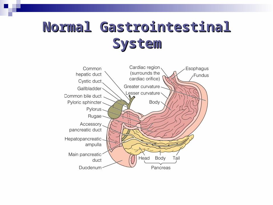
Normal Gastrointestinal Normal Gastrointestinal SystemSystem
Disorders of
Development
Cleft Lip and Cleft PalateCleft Lip and Cleft Palate
Etiology- Failure of maxillary and median nasal processes to fuse during embryonic development
Remember the psycho-social implications for these children and families
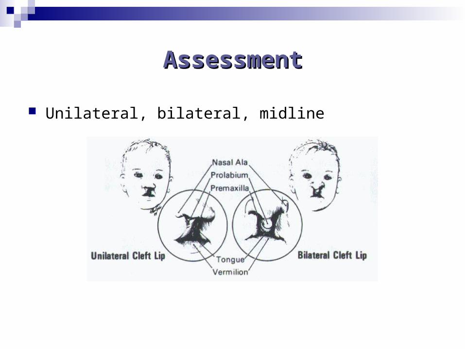
AssessmentAssessment
Unilateral, bilateral, midline
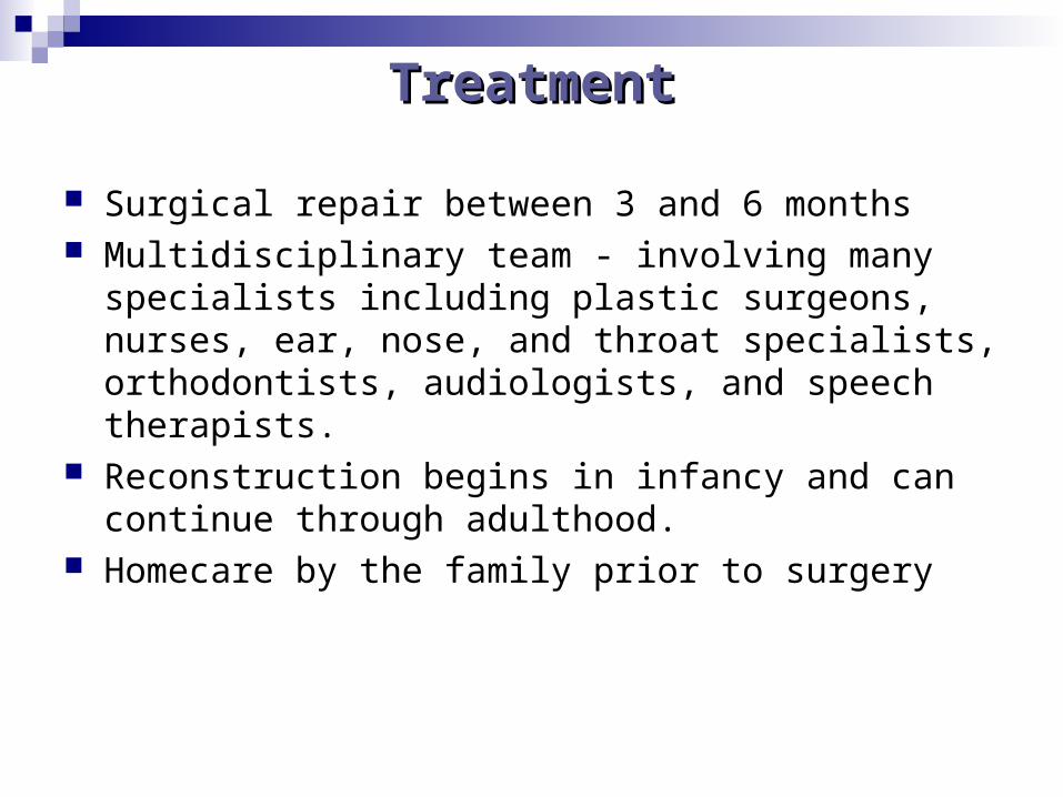
TreatmentTreatment
Surgical repair between 3 and 6 months Multidisciplinary team - involving many specialists
including plastic surgeons, nurses, ear, nose, and throat specialists, orthodontists, audiologists, and speech therapists.
Reconstruction begins in infancy and can continue through adulthood.
Homecare by the family prior to surgery
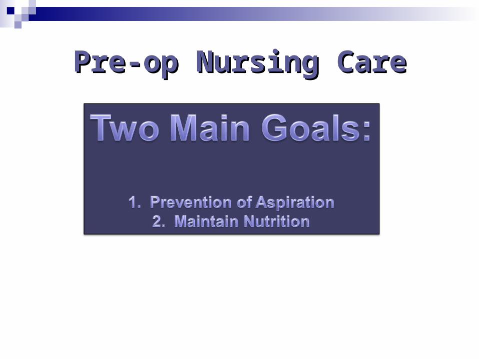
Pre-op Nursing CarePre-op Nursing Care
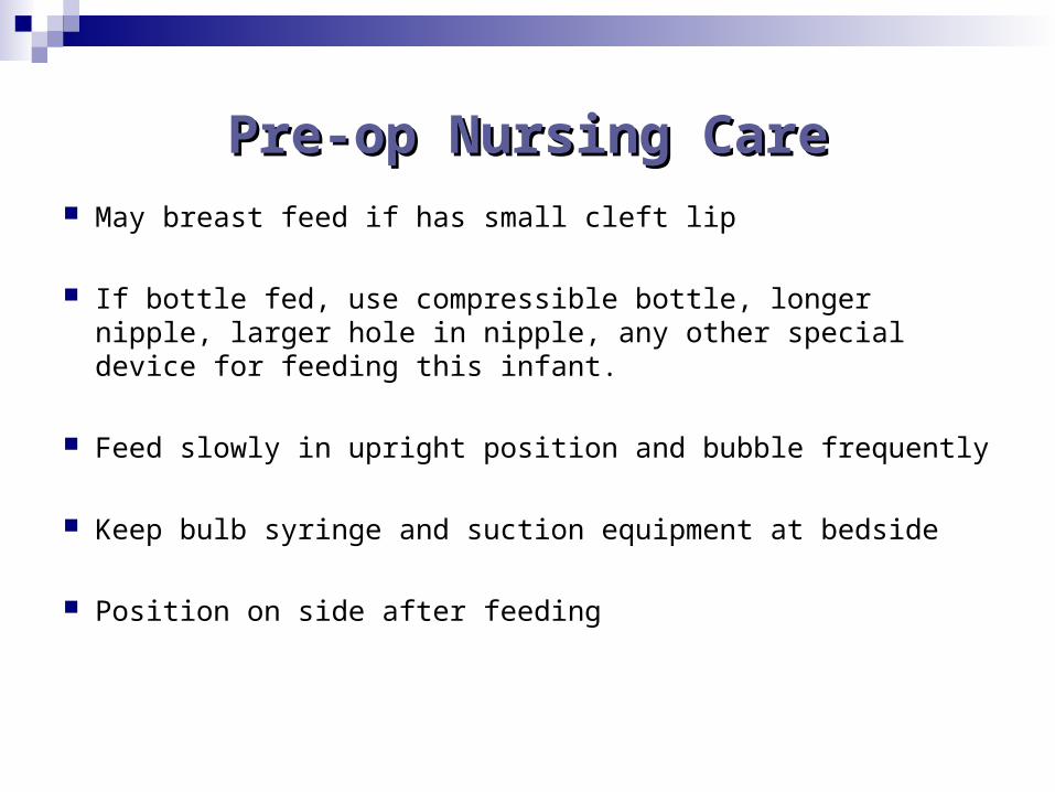
Pre-op Nursing CarePre-op Nursing Care May breast feed if has small cleft lip
If bottle fed, use compressible bottle, longer nipple, larger hole in nipple, any other special device for feeding this infant.
Feed slowly in upright position and bubble frequently
Keep bulb syringe and suction equipment at bedside
Position on side after feeding
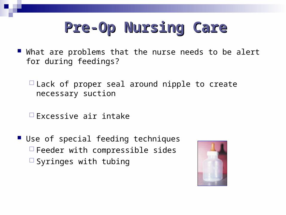
Pre-Op Nursing CarePre-Op Nursing Care
What are problems that the nurse needs to be alert for during feedings?
Lack of proper seal around nipple to create necessary suction
Excessive air intake
Use of special feeding techniques Feeder with compressible sides Syringes with tubing
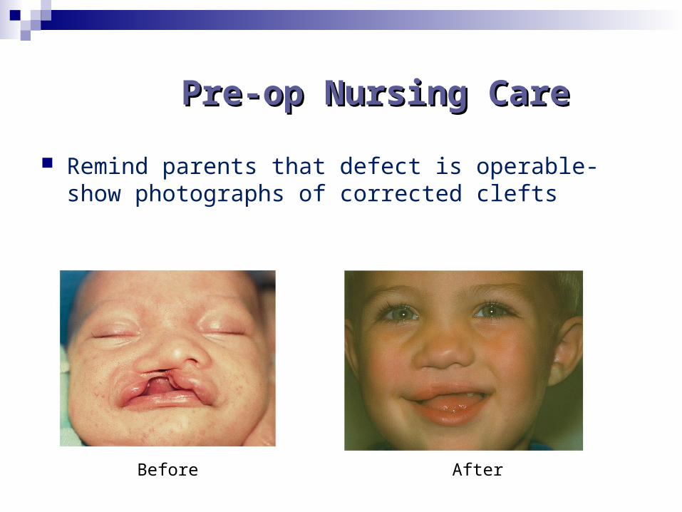
Pre-op Nursing CarePre-op Nursing Care
Remind parents that defect is operable- show photographs of corrected clefts
Before After
Therapeutic Management
Surgical Correction
A number of professionals are involved including surgeons, nurses, ear, nose, and throat specialists, audiologists, speech therapist, orthodontists, and
plastic surgeons.
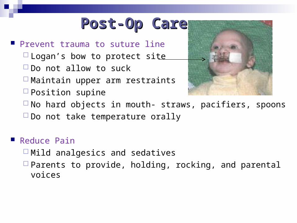
Post-Op CarePost-Op Care Prevent trauma to suture line
Logan’s bow to protect site Do not allow to suck Maintain upper arm restraints Position supine No hard objects in mouth- straws, pacifiers, spoons Do not take temperature orally
Reduce Pain Mild analgesics and sedatives Parents to provide, holding, rocking, and parental voices
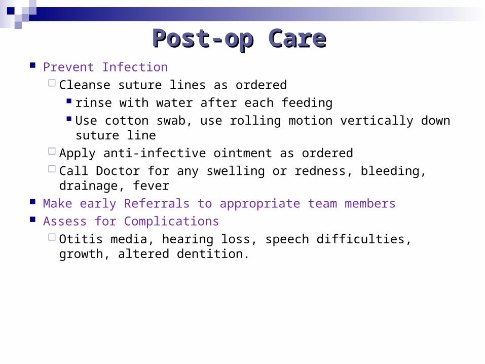
Post-op CarePost-op Care Prevent Infection
Cleanse suture lines as ordered rinse with water after each feeding Use cotton swab, use rolling motion vertically down suture
line Apply anti-infective ointment as ordered Call Doctor for any swelling or redness, bleeding, drainage, fever
Make early Referrals to appropriate team members Assess for Complications
Otitis media, hearing loss, speech difficulties, growth, altered dentition.
Esophageal Atresia
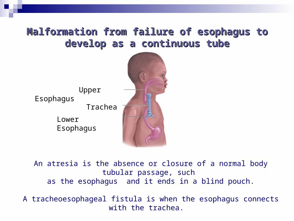
Malformation from failure of esophagus to Malformation from failure of esophagus to develop as a continuous tubedevelop as a continuous tube
Upper Esophagus
Trachea
Lower Esophagus
An atresia is the absence or closure of a normal body tubular passage, such as the esophagus and it ends in a blind pouch.
A tracheoesophageal fistula is when the esophagus connects with the trachea.
Signs and SymptomsSigns and Symptoms
Excessive amounts of salivation / mucus, frothy bubbles in the mouth and sometimes nose
Three “C’s” - Coughing, choking, and cyanosis when fed, overflow may be aspirated
Food may be expelled through the nose immediately
following the feeding Rattling respirations and frequent respiratory problems such as
aspiration pneumonia Gastric distention, if fistula History of polyhydramnios during pregnancy can suggest a high
gastrointestinal obstruction
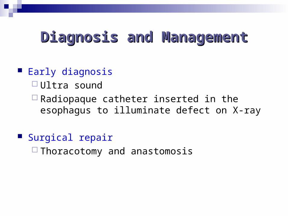
Diagnosis and ManagementDiagnosis and Management
Early diagnosis Ultra sound Radiopaque catheter inserted in the esophagus to
illuminate defect on X-ray
Surgical repair Thoracotomy and anastomosis
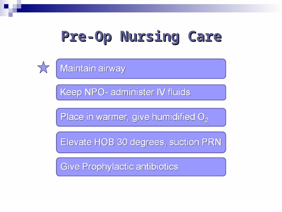
Pre-Op Nursing CarePre-Op Nursing Care
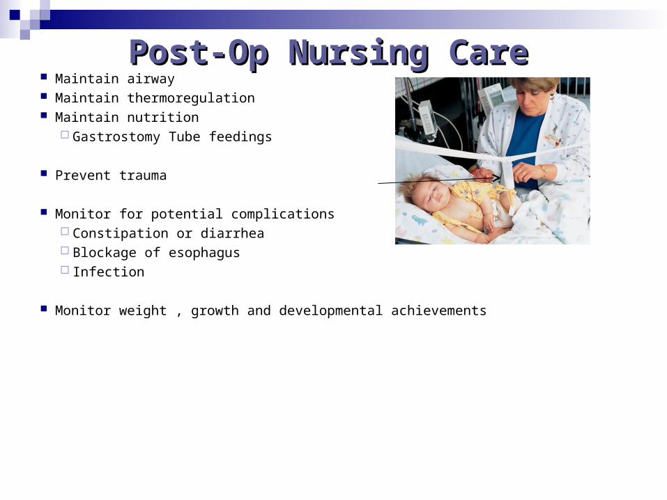
Post-Op Nursing CarePost-Op Nursing Care Maintain airway Maintain thermoregulation Maintain nutrition
Gastrostomy Tube feedings
Prevent trauma
Monitor for potential complications Constipation or diarrhea Blockage of esophagus Infection
Monitor weight , growth and developmental achievements
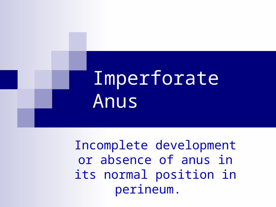
Imperforate Anus
Incomplete development or absence of anus in its normal
position in perineum.
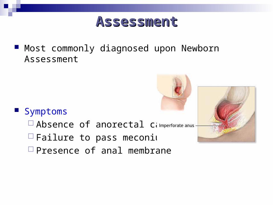
AssessmentAssessment
Most commonly diagnosed upon Newborn Assessment
Symptoms Absence of anorectal canal Failure to pass meconium Presence of anal membrane
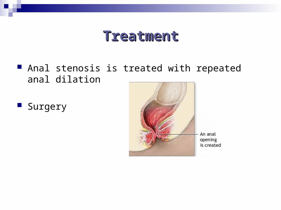
TreatmentTreatment
Anal stenosis is treated with repeated anal dilation
Surgery
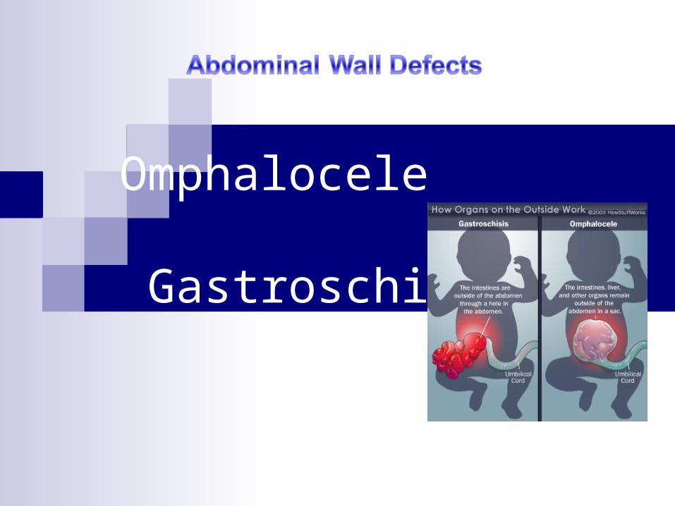
Omphalocele
Gastroschisis
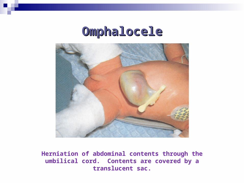
OmphaloceleOmphalocele
Herniation of abdominal contents through the umbilical cord. Contents are covered by a translucent sac.
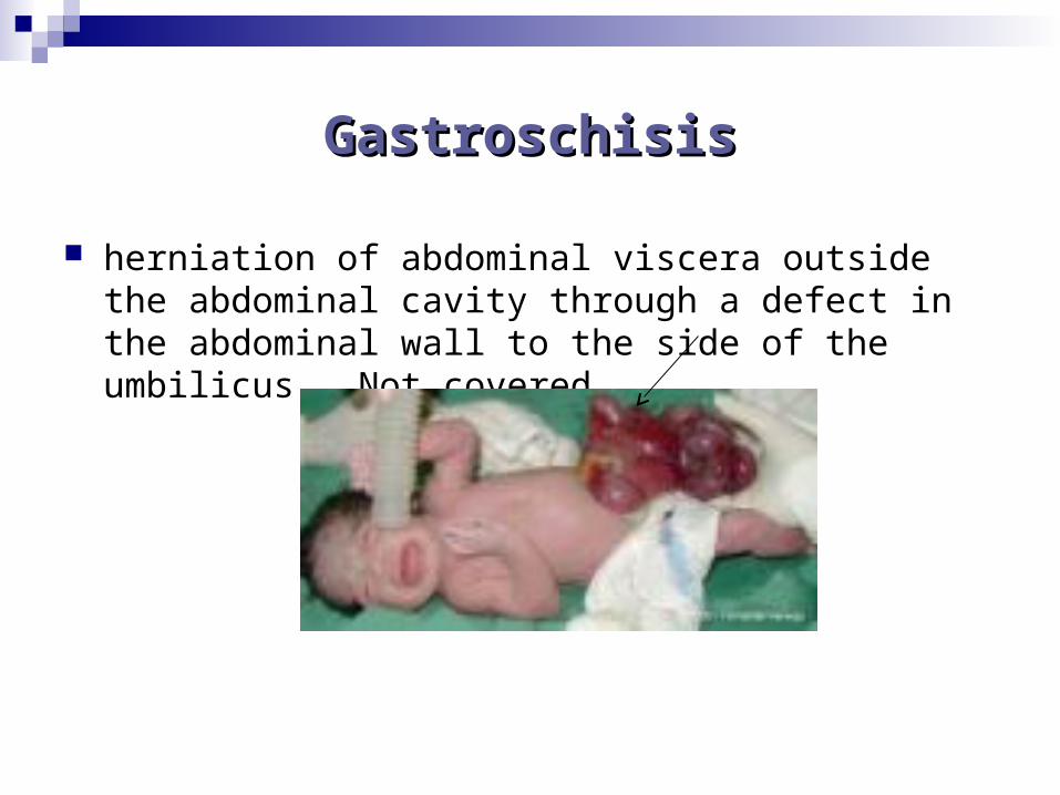
GastroschisisGastroschisis
herniation of abdominal viscera outside the abdominal cavity through a defect in the abdominal wall to the side of the umbilicus. Not covered.
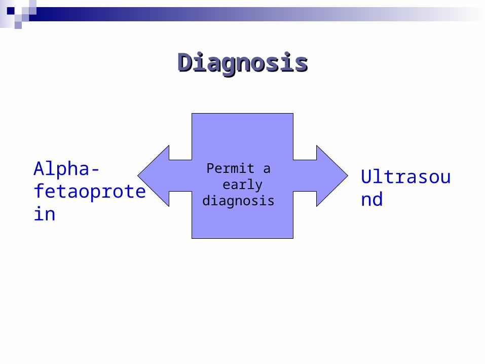
DiagnosisDiagnosis
Alpha-fetaoprotein
Permit a early diagnosis Ultrasound
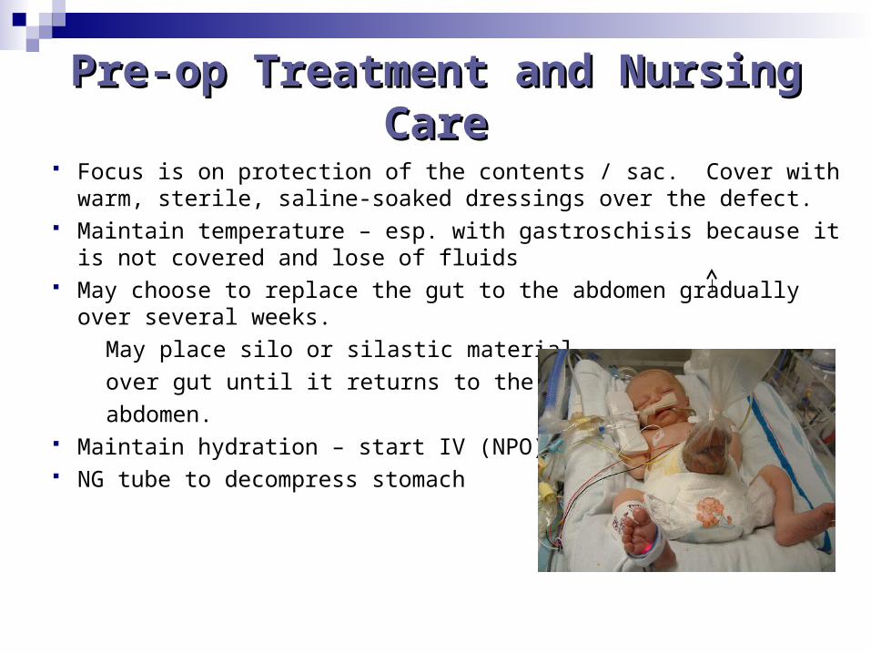
Pre-op Treatment and Nursing Pre-op Treatment and Nursing CareCare
Focus is on protection of the contents / sac. Cover with warm, sterile, saline-soaked dressings over the defect.
Maintain temperature – esp. with gastroschisis because it is not covered and lose of fluids
May choose to replace the gut to the abdomen gradually over several weeks.
May place silo or silastic material
over gut until it returns to the
abdomen. Maintain hydration – start IV (NPO) NG tube to decompress stomach
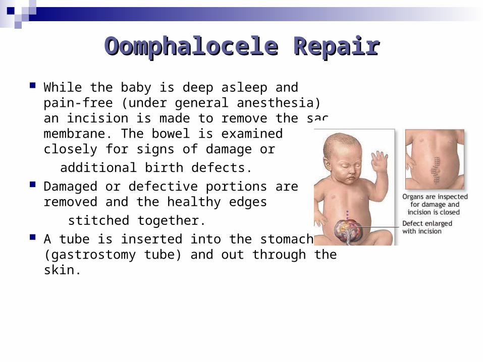
Oomphalocele RepairOomphalocele Repair
While the baby is deep asleep and pain-free (under general anesthesia) an incision is made to remove the sac membrane. The bowel is examined closely for signs of damage or
additional birth defects. Damaged or defective portions are removed
and the healthy edges
stitched together. A tube is inserted into the stomach
(gastrostomy tube) and out through the skin.

Gastroschisis RepairGastroschisis Repair
Surgical repair of abdominal wall defects
involves replacing the abdomen through
the abdominal wall defect, repairing the
defect if possible, or creating abdominal
organs back into the a sterile pouch to
protect the intestines while they are
gradually pushed back into the abdomen.
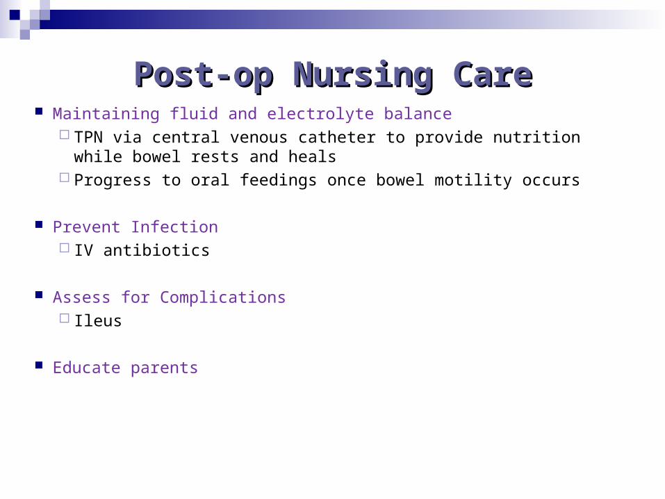
Post-op Nursing CarePost-op Nursing Care Maintaining fluid and electrolyte balance
TPN via central venous catheter to provide nutrition while bowel rests and heals
Progress to oral feedings once bowel motility occurs
Prevent Infection IV antibiotics
Assess for Complications Ileus
Educate parents
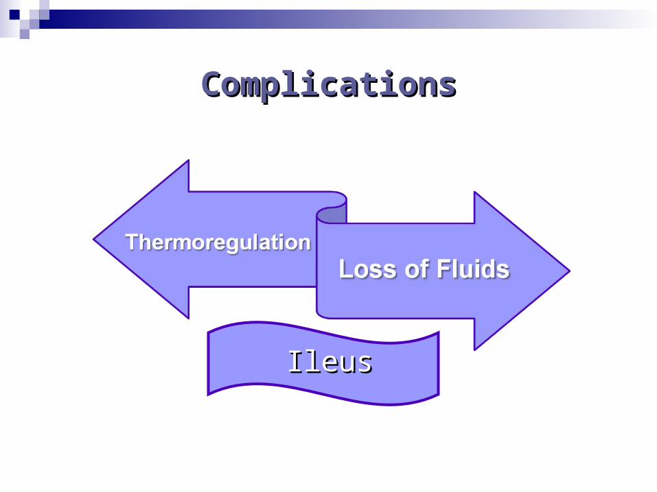
ComplicationsComplications
IleusIleus
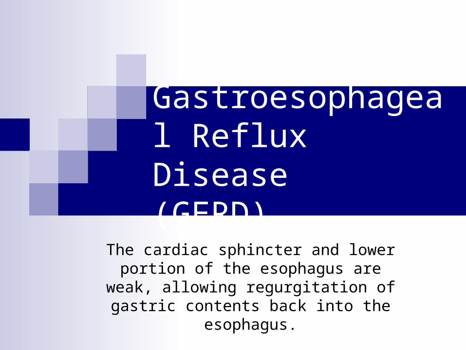
Gastroesophageal Reflux Disease(GERD)
The cardiac sphincter and lower portion of the esophagus are weak, allowing
regurgitation of gastric contents back into the esophagus.
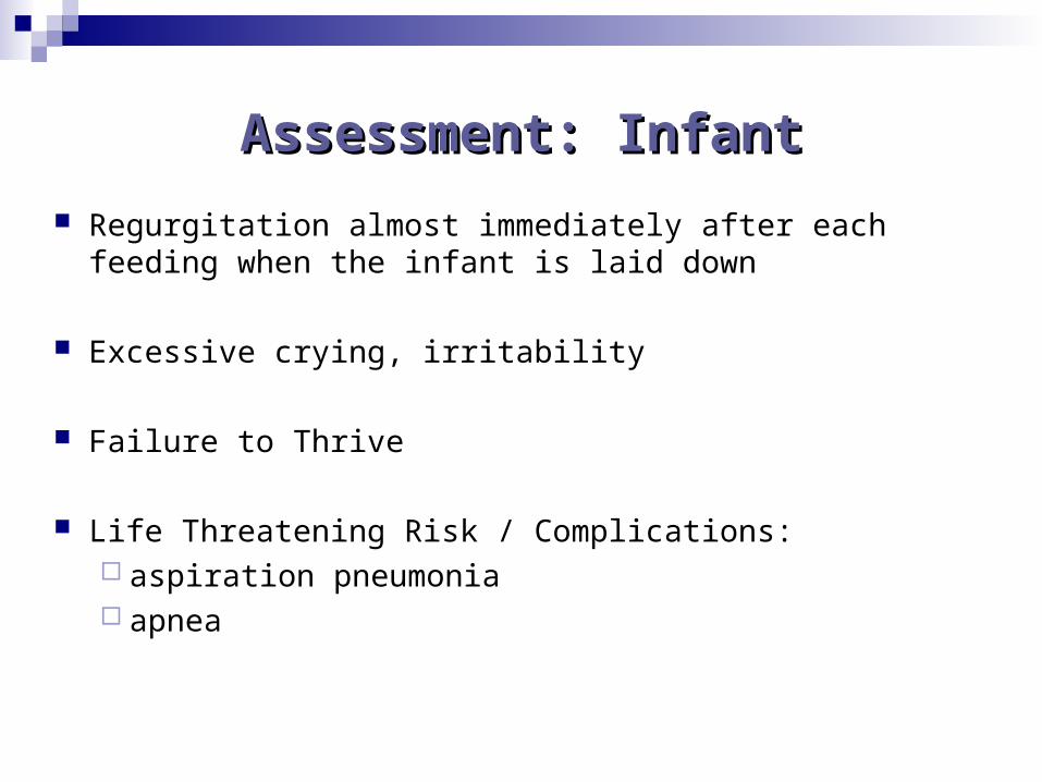
Assessment: InfantAssessment: Infant
Regurgitation almost immediately after each feeding when the infant is laid down
Excessive crying, irritability
Failure to Thrive
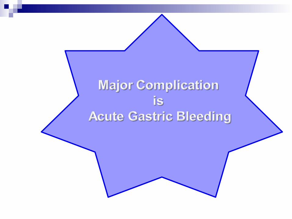
Life Threatening Risk / Complications: aspiration pneumonia apnea
Assessment: ChildAssessment: Child
Heartburn
Abdominal pain
Cough, recurrent pneumonia
Dysphagia
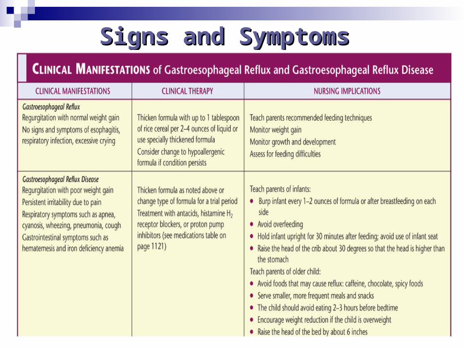
Signs and SymptomsSigns and Symptoms
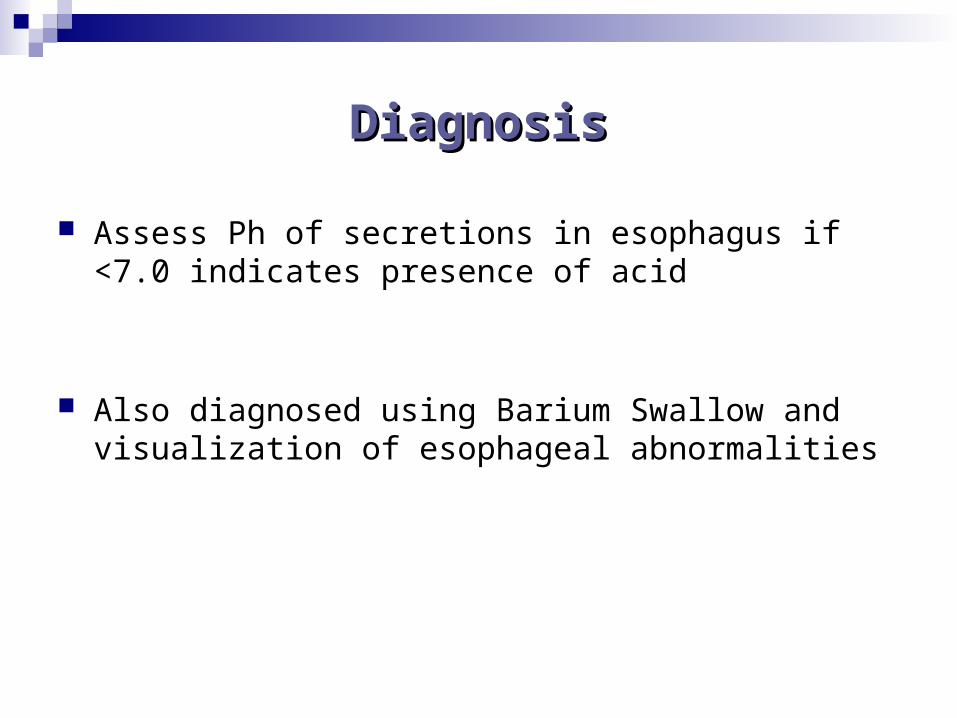
DiagnosisDiagnosis
Assess Ph of secretions in esophagus if <7.0 indicates presence of acid
Also diagnosed using Barium Swallow and visualization of esophageal abnormalities
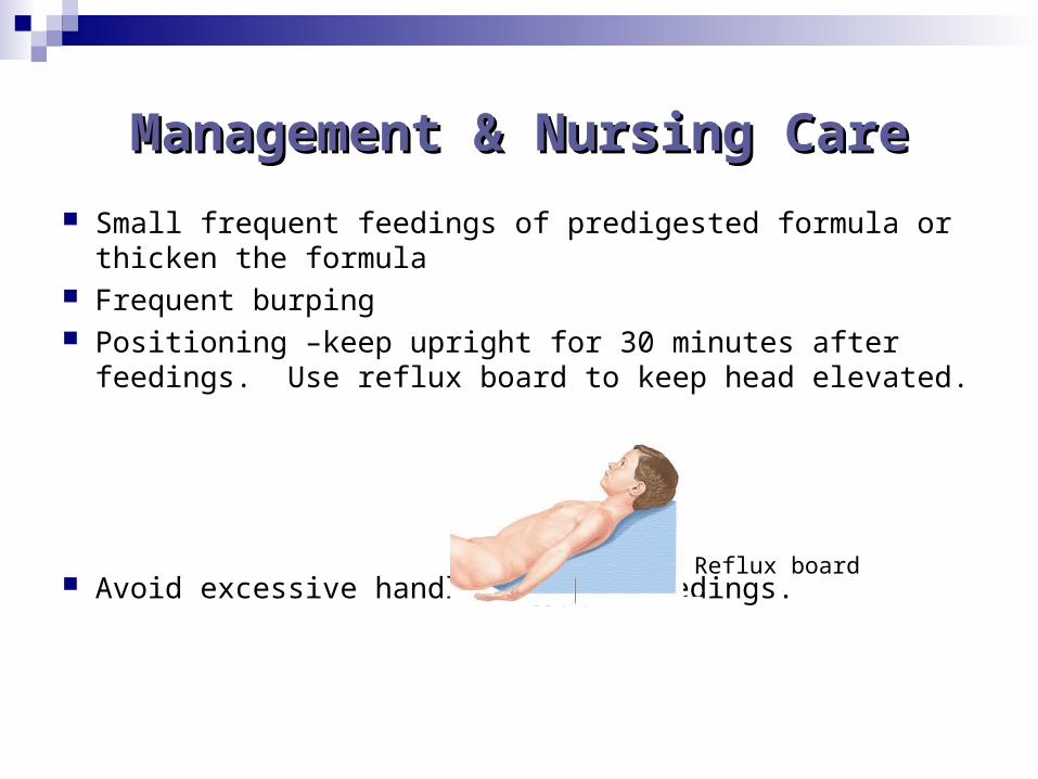
Management & Nursing CareManagement & Nursing Care
Small frequent feedings of predigested formula or thicken the formula
Frequent burping Positioning –keep upright for 30 minutes after feedings. Use
reflux board to keep head elevated.
Avoid excessive handling after feedings.
Reflux board
MedicationsMedications H2 Histamine receptor antagonists – reduce gastric acidity
Zantac and Pepcid Proton-pump inhibitors
Prevacid Prilosec
Gastric emptying Reglan
Antacids Gaviscon
**be sure to study nursing implications and side effects
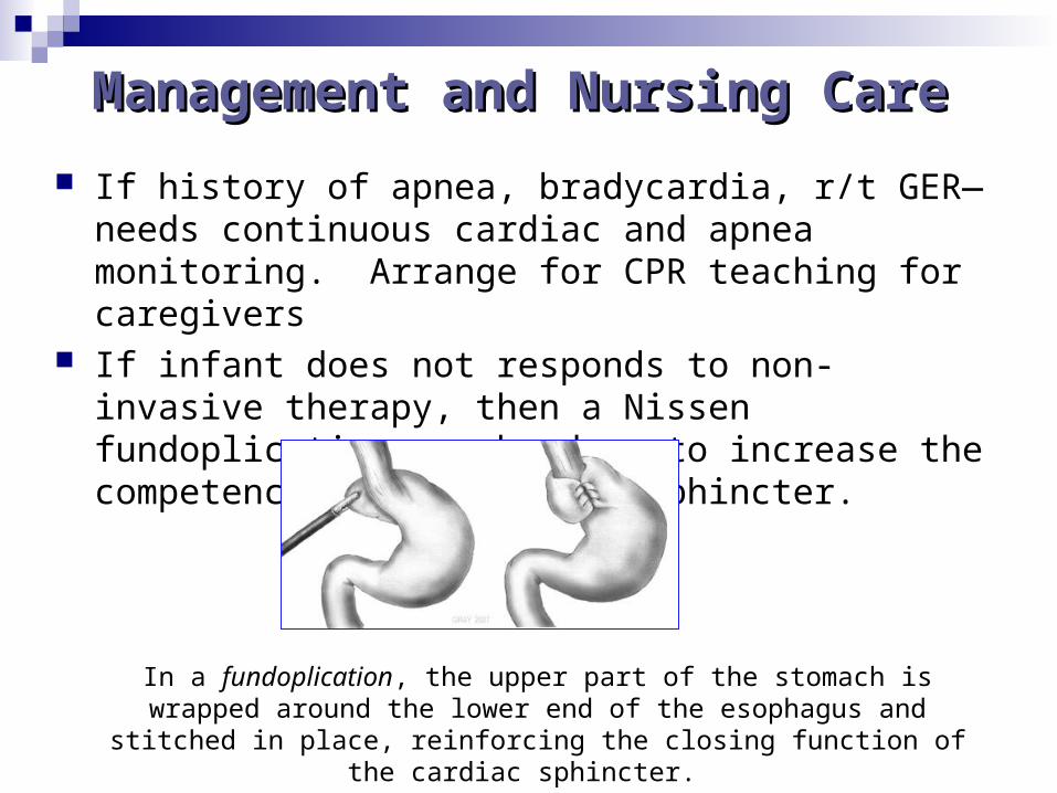
Management and Nursing CareManagement and Nursing Care
If history of apnea, bradycardia, r/t GER—needs continuous cardiac and apnea monitoring. Arrange for CPR teaching for caregivers
If infant does not responds to non-invasive therapy, then a Nissen fundoplication may be done to increase the competence of the cardiac sphincter.
In a fundoplication, the upper part of the stomach is wrapped around the lower end of the esophagus and stitched in place, reinforcing the closing
function of the cardiac sphincter.
Post-op Nursing CarePost-op Nursing Care
Assess for pain, abdominal distention, and return of bowel sounds.
Teach parents about gastrostomy tube feedings
Diarrhea
Infectious Gastroenteritis
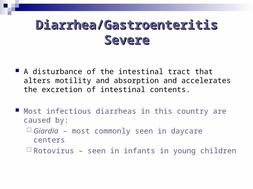
Diarrhea/GastroenteritisDiarrhea/GastroenteritisSevereSevere
A disturbance of the intestinal tract that alters motility and absorption and accelerates the excretion of intestinal contents.
Most infectious diarrheas in this country are caused by:Giardia – most commonly seen in daycare centers Rotovirus – seen in infants in young children
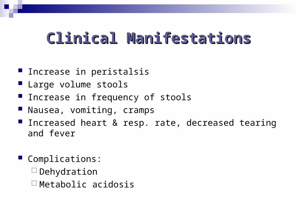
Clinical ManifestationsClinical Manifestations
Increase in peristalsis Large volume stools Increase in frequency of stools Nausea, vomiting, cramps Increased heart & resp. rate, decreased tearing and fever
Complications: Dehydration Metabolic acidosis
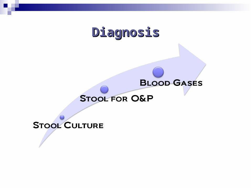
DiagnosisDiagnosis
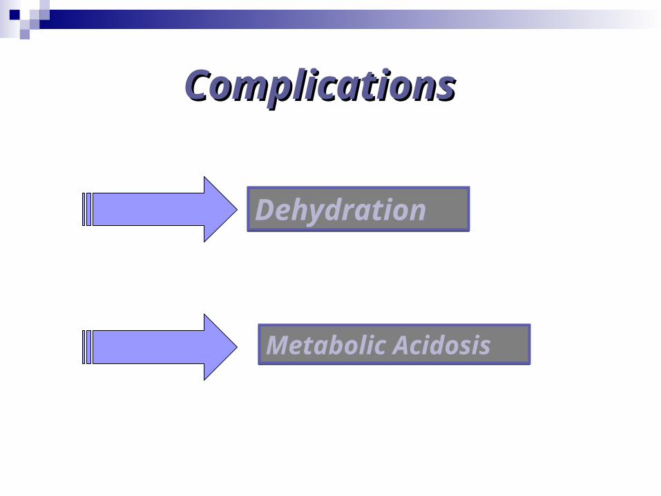
ComplicatioComplicationsns
DehydrationDehydration
Metabolic AcidosisMetabolic Acidosis
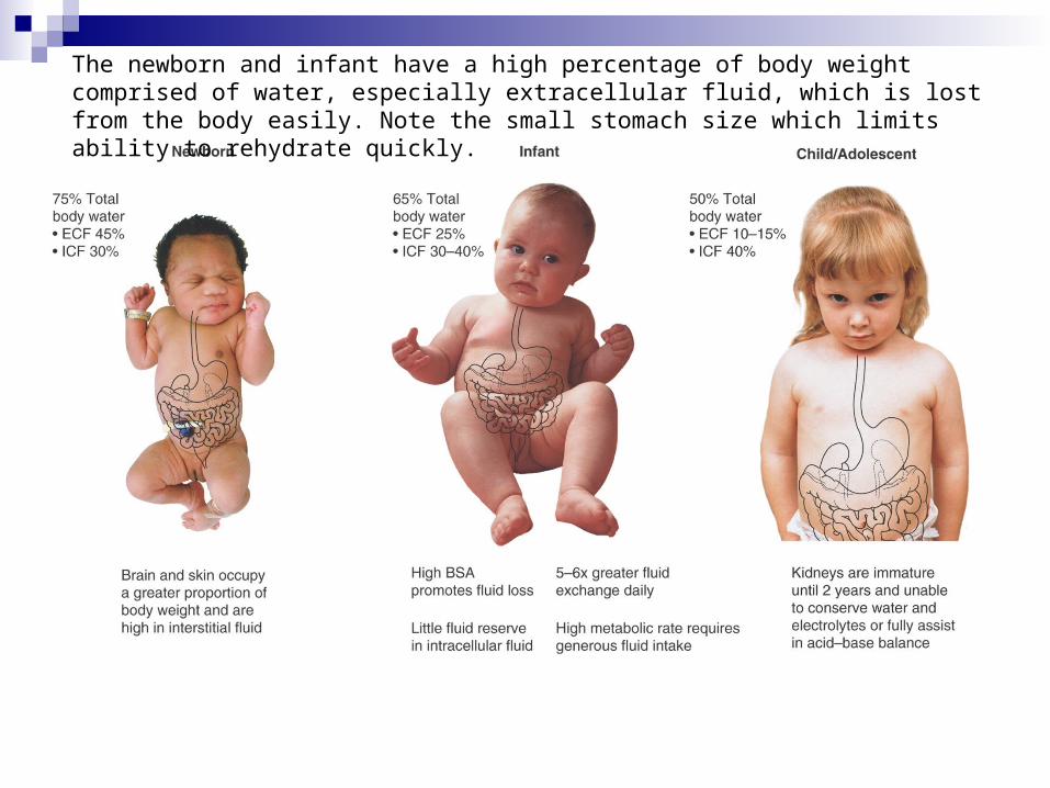
The newborn and infant have a high percentage of body weight comprised of water, especially extracellular fluid, which is lost from the body easily. Note the small stomach size which limits ability to rehydrate quickly.

Dehydration
Infant Depressed fontanels Sunken eye orbits Fussy, Irritable Thirsty Fewer wet diapers
Child Decreased tear production Skin non-elastic Decreased urinary output Thirsty Restless
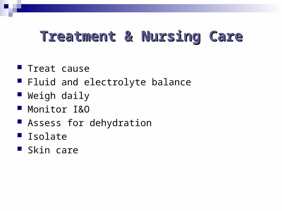
Treatment & Nursing CareTreatment & Nursing Care
Treat cause Fluid and electrolyte balance Weigh daily Monitor I&O Assess for dehydration Isolate Skin care
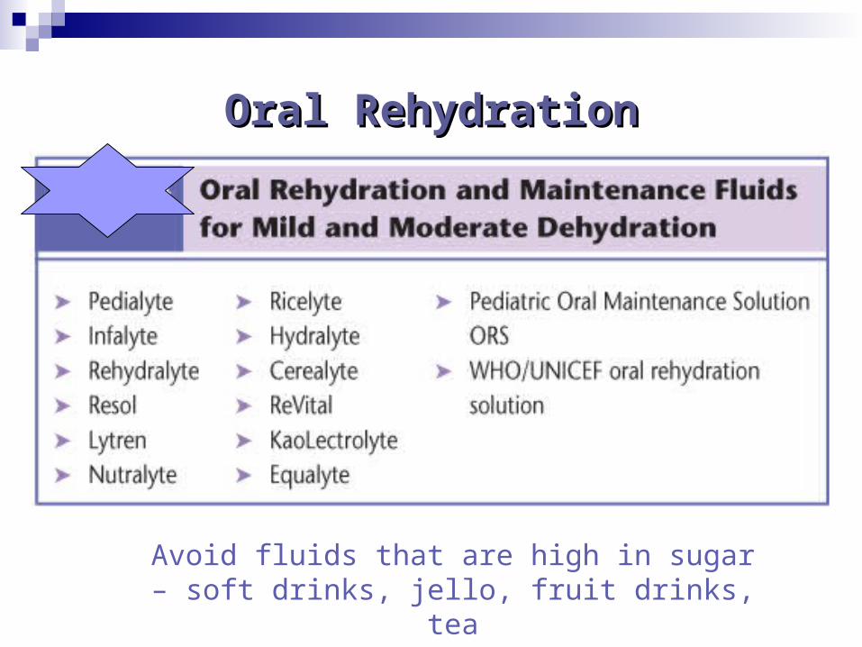
Oral RehydrationOral Rehydration
Avoid fluids that are high in sugar – soft drinks, jello, fruit drinks, tea
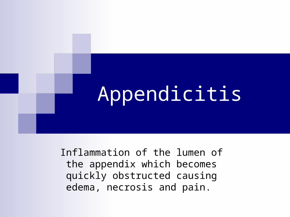
Appendicitis
Inflammation of the lumen of the appendix which becomes quickly obstructed causing
edema, necrosis and pain.
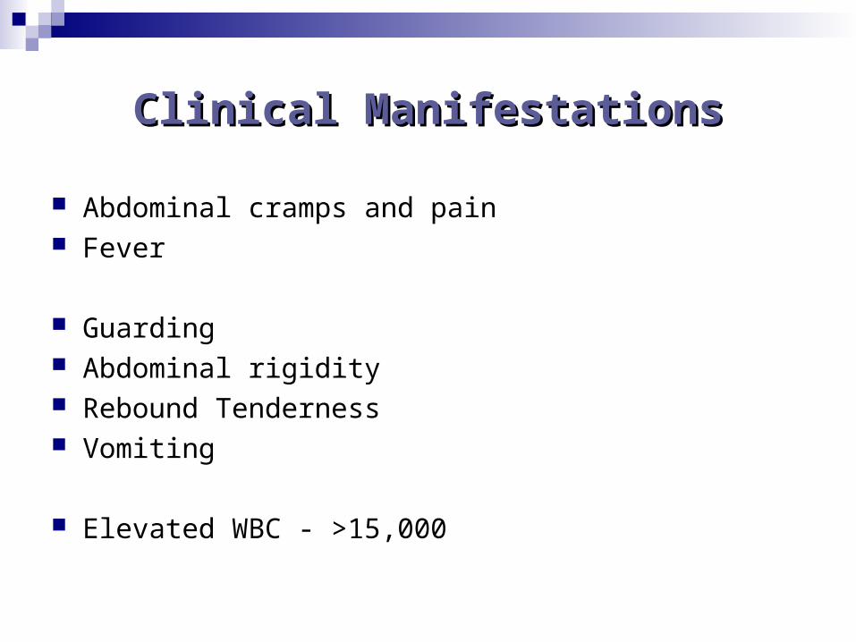
Clinical ManifestationsClinical Manifestations
Abdominal cramps and pain Fever
Guarding Abdominal rigidity Rebound Tenderness Vomiting
Elevated WBC - >15,000
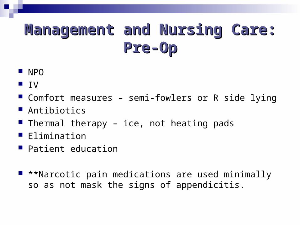
Management and Nursing Management and Nursing Care: Pre-OpCare: Pre-Op
NPO IV Comfort measures – semi-fowlers or R side lying Antibiotics Thermal therapy – ice, not heating pads Elimination Patient education
**Narcotic pain medications are used minimally so as not mask the signs of appendicitis.
AppendicitisAppendicitis
What is the most common symptom indicating that the appendix may have ruptured?
Management and Nursing Management and Nursing Care: Post-OpCare: Post-Op
NPO Antibiotics Analgesia Patient teaching
Pyloric Stenosis
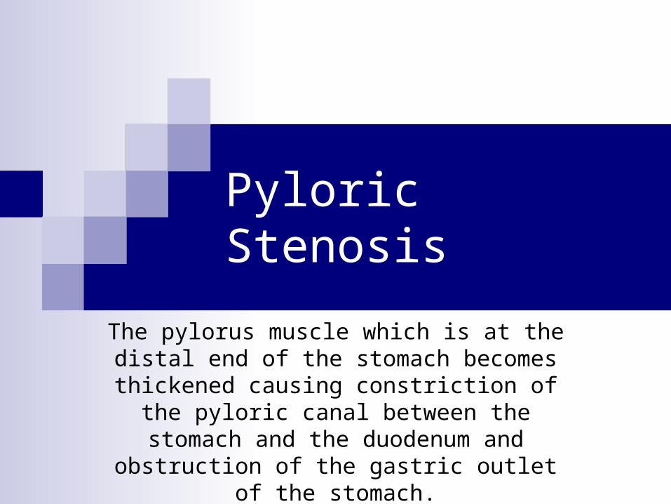
The pylorus muscle which is at the distal end of the stomach becomes thickened causing
constriction of the pyloric canal between the stomach and the duodenum and obstruction of
the gastric outlet of the stomach.
Pyloric StenosisPyloric Stenosis
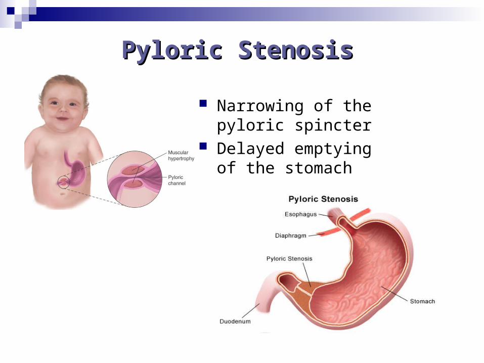
Narrowing of the pyloric spincter
Delayed emptying of the stomach
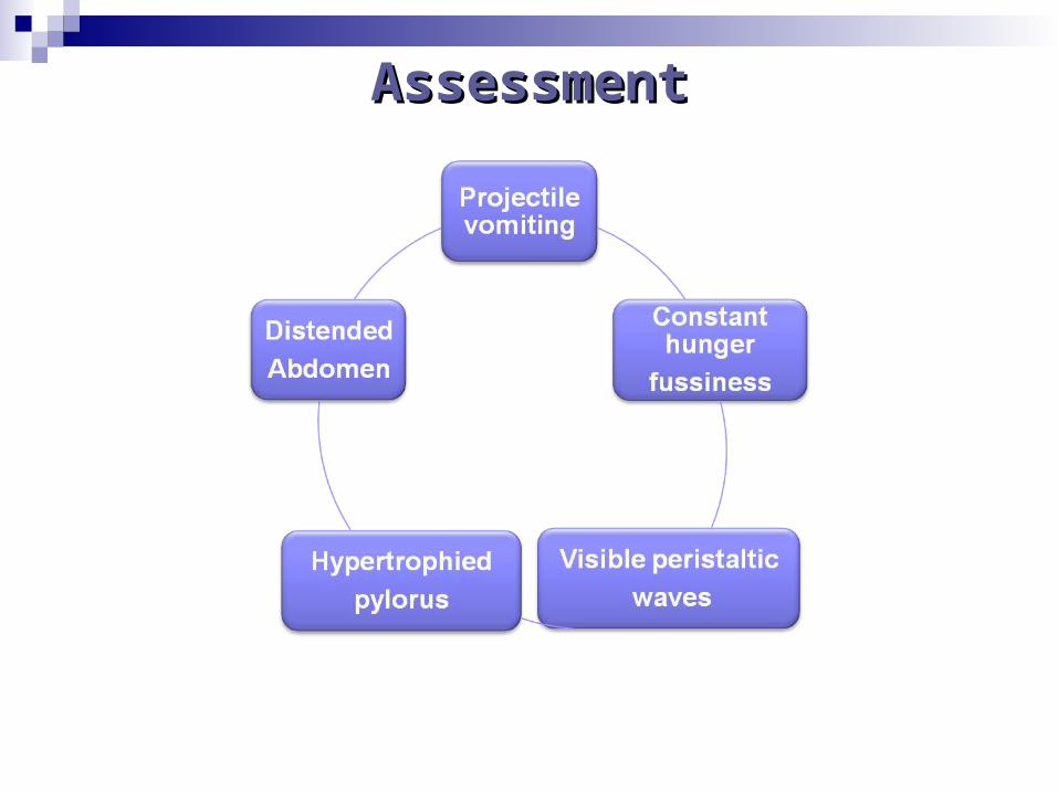
AssessmentAssessment
Treatment and Nursing CareTreatment and Nursing Care
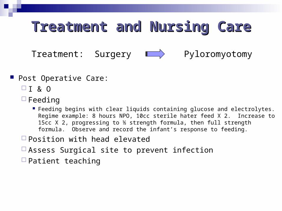
Treatment: Surgery Pyloromyotomy
Post Operative Care: I & O Feeding
Feeding begins with clear liquids containing glucose and electrolytes. Regime example: 8 hours NPO, 10cc sterile hater feed X 2. Increase to 15cc X 2, progressing to ½ strength formula, then full strength formula. Observe and record the infant’s response to feeding.
Position with head elevated Assess Surgical site to prevent infection Patient teaching
Critical ThinkingCritical Thinking
A 4 week old infant with a history of vomiting after feeding has been hospitalized with a tentative diagnosis of pyloric stenosis. Which of these actions is priority for the nurse? Begin an intravenous infusion Measure abdominal circumference Orient family to unit Weigh infant
Intussuception
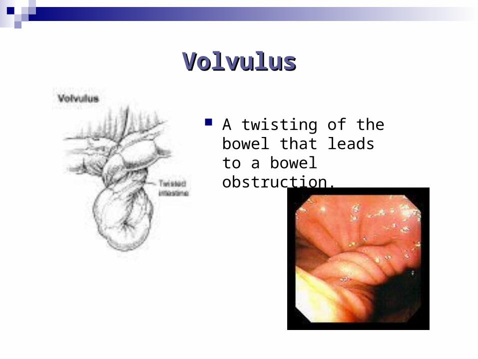
Volvulus
Both are forms of bowel obstruction
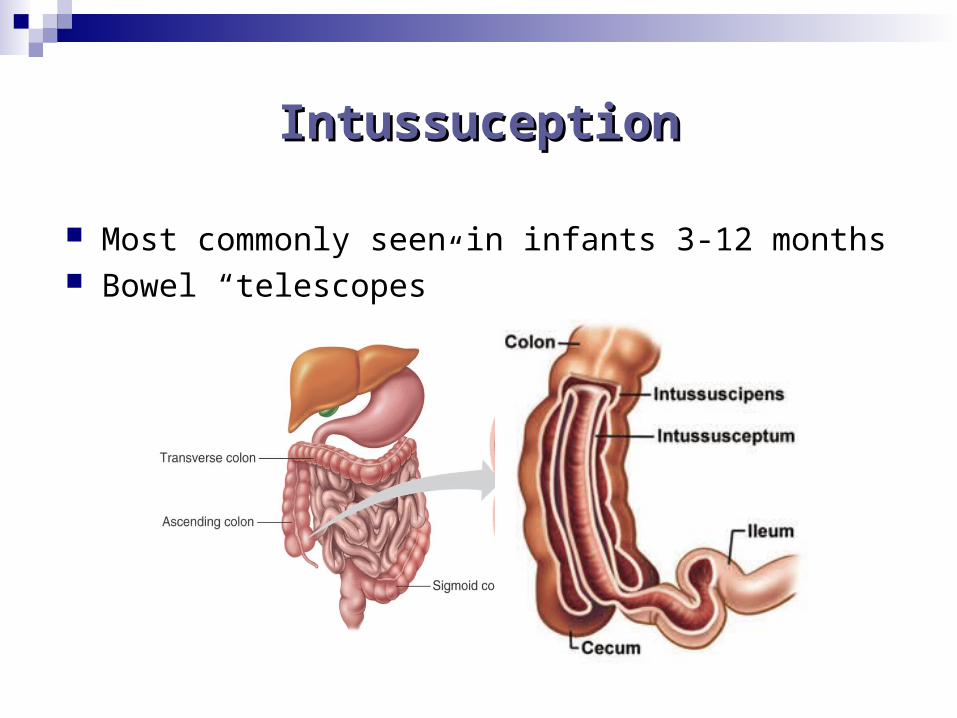
IntussuceptionIntussuception
Most commonly seen in infants 3-12 months Bowel “telescopes”
within itself
VolvulusVolvulus
A twisting of the bowel that leads to a bowel obstruction.
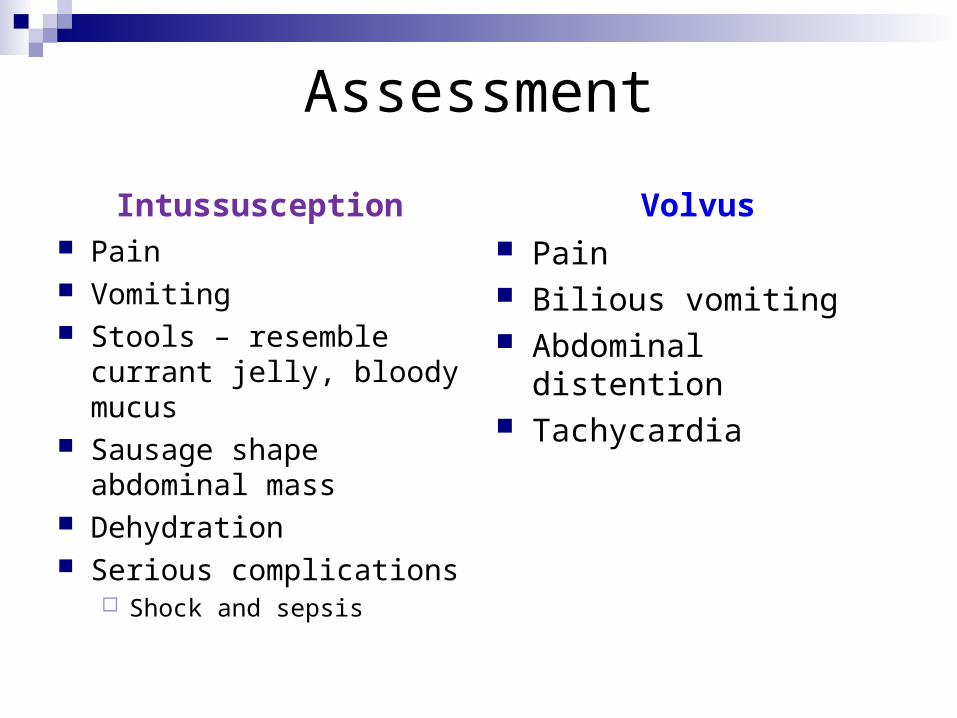
Assessment
Intussusception Pain Vomiting Stools – resemble currant
jelly, bloody mucus Sausage shape
abdominal mass Dehydration Serious complications
Shock and sepsis
Volvus Pain Bilious vomiting Abdominal distention Tachycardia
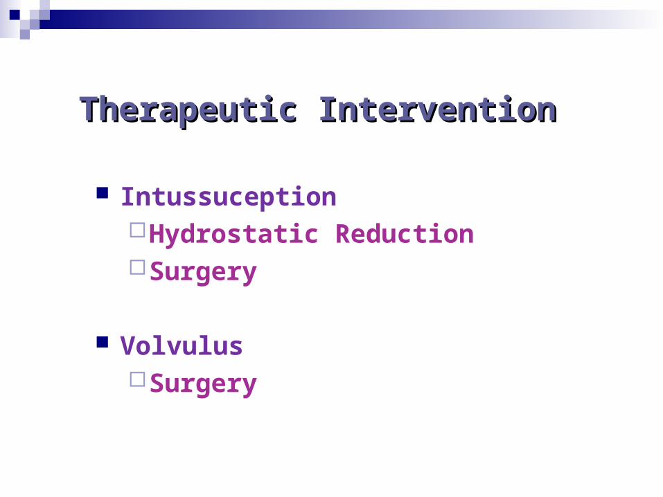
Therapeutic Intervention Therapeutic Intervention
IntussuceptionHydrostatic ReductionSurgery
VolvulusSurgery
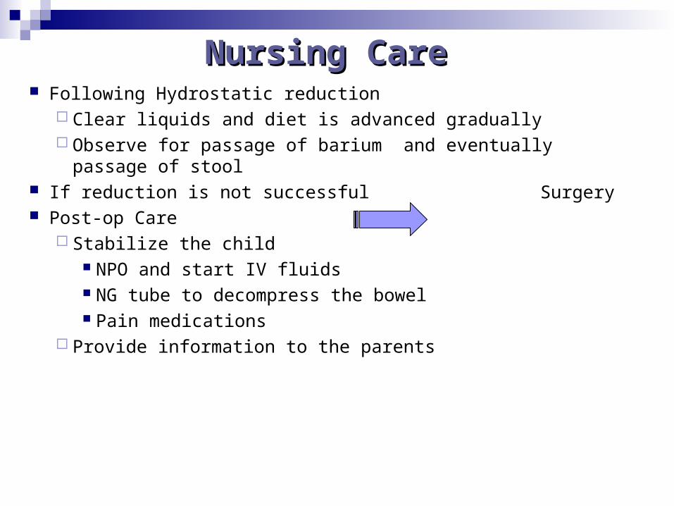
Nursing CareNursing Care Following Hydrostatic reduction
Clear liquids and diet is advanced gradually Observe for passage of barium and eventually passage of stool
If reduction is not successful Surgery Post-op Care
Stabilize the child NPO and start IV fluids NG tube to decompress the bowel Pain medications
Provide information to the parents
Hirschsprung's Disease
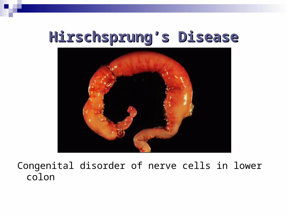
Hirschsprung’s DiseaseHirschsprung’s Disease
Congenital disorder of nerve cells in lower colon
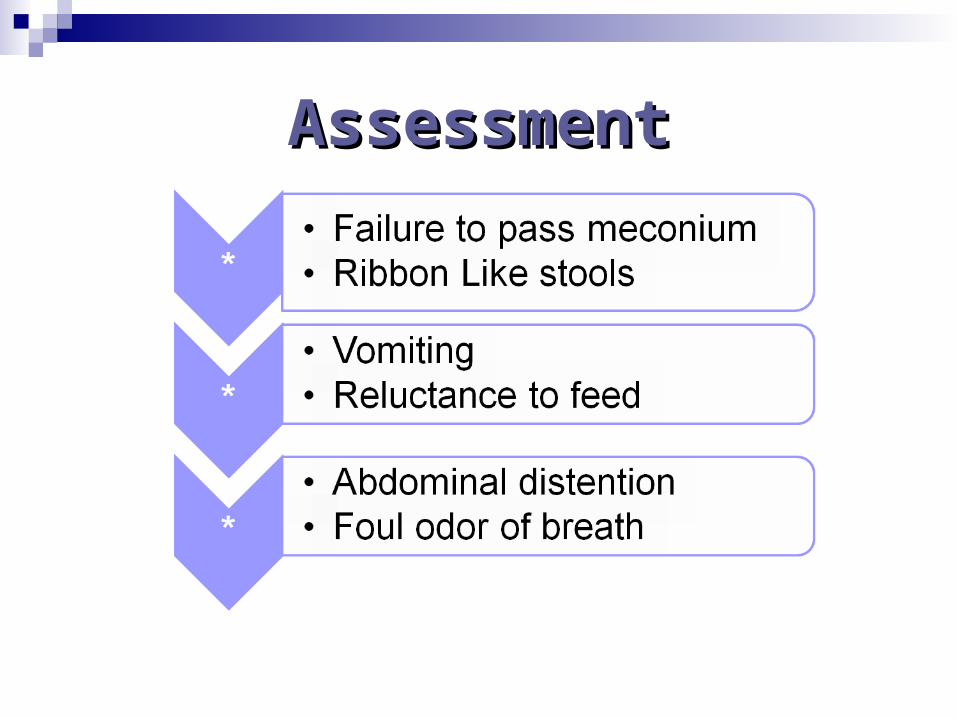
AssessmentAssessment
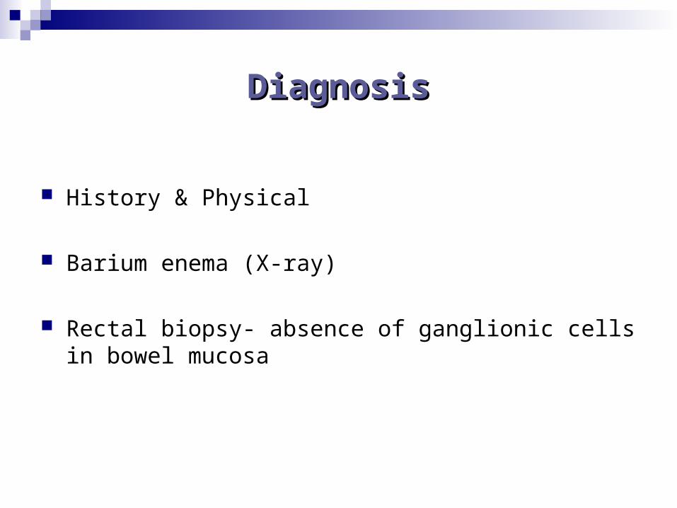
DiagnosisDiagnosis
History & Physical
Barium enema (X-ray)
Rectal biopsy- absence of ganglionic cells in bowel mucosa
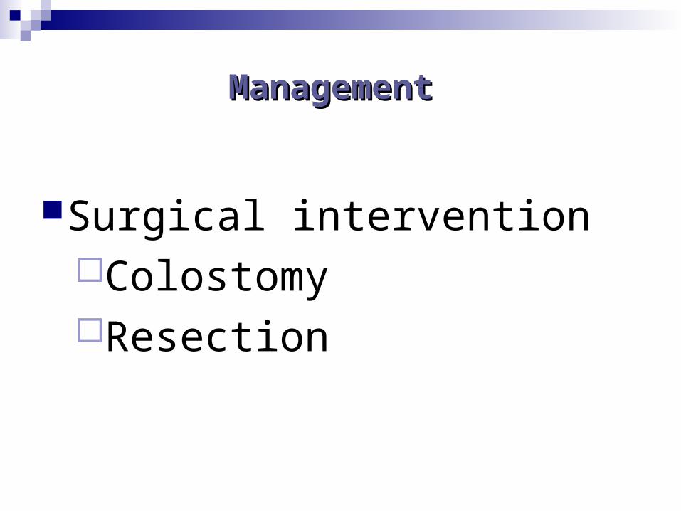
Management Management
Surgical intervention ColostomyResection
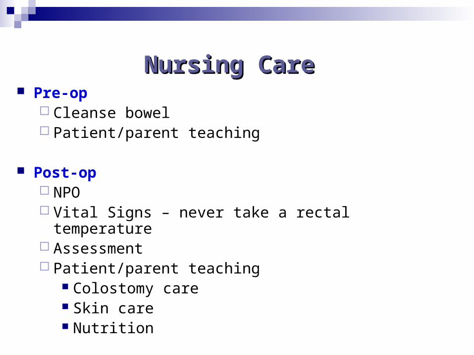
Nursing CareNursing Care Pre-op
Cleanse bowel Patient/parent teaching
Post-op NPO Vital Signs – never take a rectal temperature Assessment Patient/parent teaching
Colostomy care Skin care Nutrition
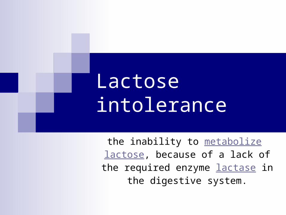
Lactose intolerance
the inability to metabolize lactose, because of a lack of the required enzyme lactase in
the digestive system.
Lactose IntoleranceLactose Intolerance
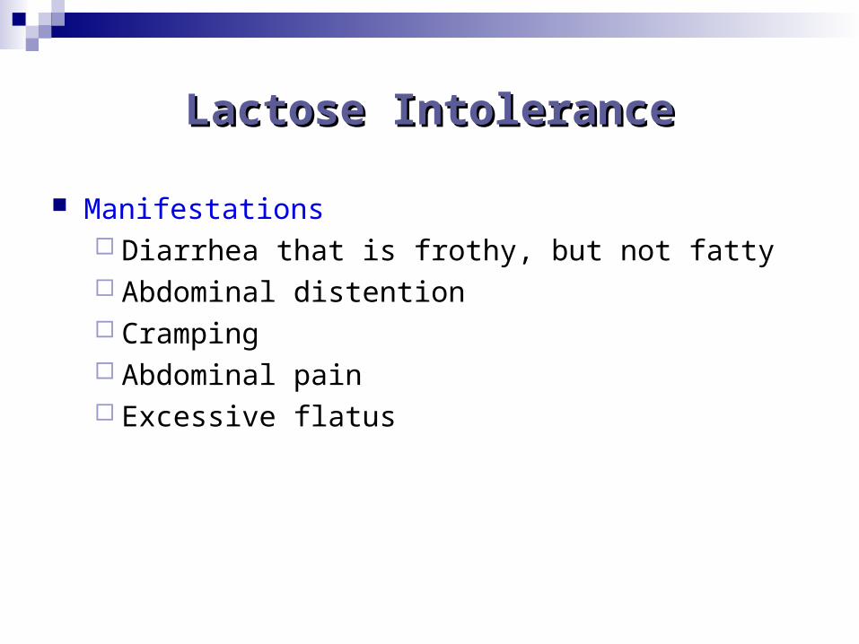
Manifestations Diarrhea that is frothy, but not fatty Abdominal distention Cramping Abdominal pain Excessive flatus
Lactose IntoleranceLactose Intolerance
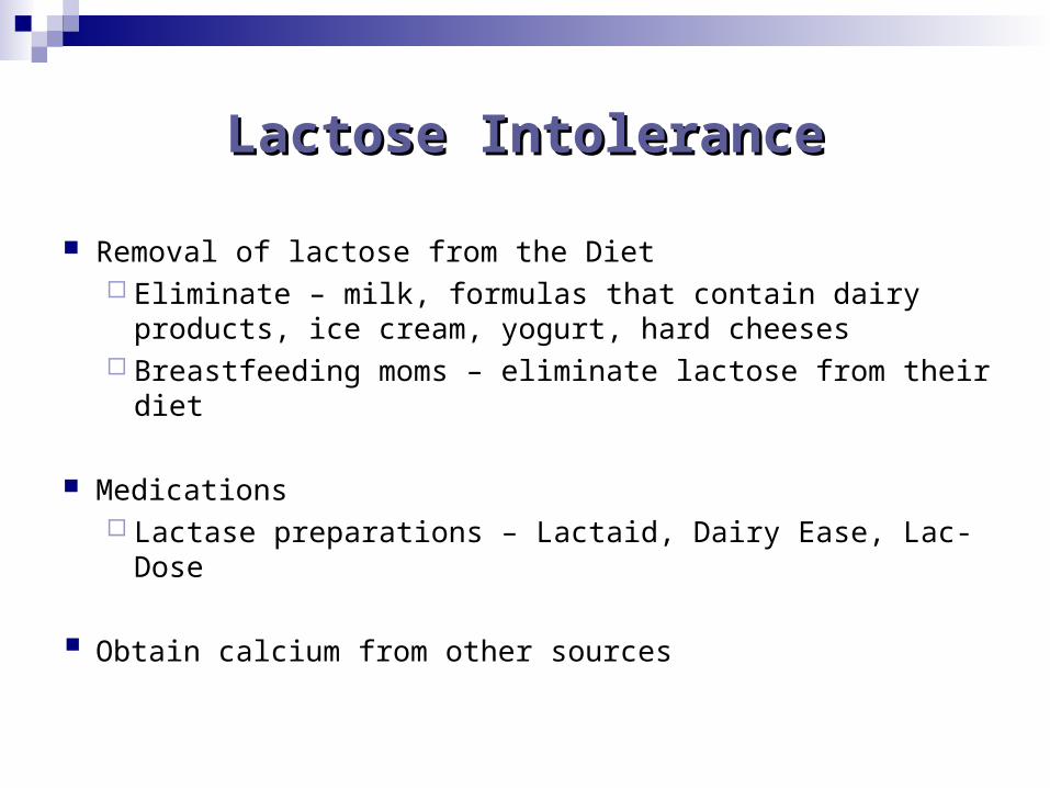
Removal of lactose from the Diet Eliminate – milk, formulas that contain dairy products,
ice cream, yogurt, hard cheeses Breastfeeding moms – eliminate lactose from their diet
Medications Lactase preparations – Lactaid, Dairy Ease, Lac-Dose
Obtain calcium from other sources

Celiac Disease
inability to digest gliadin which is a
by-product of gluten breakdown.
Signs and Symptoms Signs and Symptoms
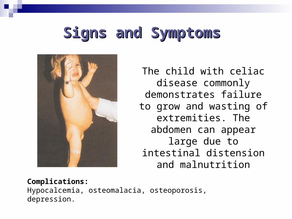
The child with celiac disease commonly demonstrates
failure to grow and wasting of extremities. The abdomen can appear large due to intestinal distension and malnutrition
Complications: Hypocalcemia, osteomalacia, osteoporosis, depression.
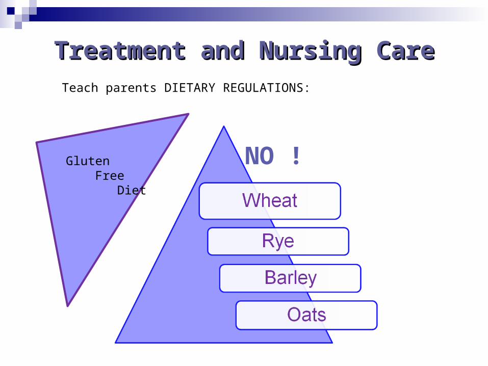
Treatment and Nursing CareTreatment and Nursing CareTeach parents DIETARY REGULATIONS:
Gluten Free Diet
NO !