observation of pure spin transport in a diamond spin wire · pdf fileobservation of pure spin...
TRANSCRIPT

Observation of Pure Spin Transport in a Diamond Spin Wire
J. Cardellino†,1, N. Scozzaro†,1, M. R. Herman1, A. Berger1, C. Zhang1, K.C. Fong1,C. Jayaprakash1, D.V. Pelekhov1, P.C. Hammel1,∗
January 10, 2014
Spin transport electronics – spintronics – focuseson utilizing electron spin as a state variable forquantum and classical information processing andstorage [1]. Some insulating materials, such as di-amond, offer defect centers whose associated spinsare well-isolated from their environment givingthem long coherence times [2–4]; however, spininteractions are important for transport [5], en-tanglement [6], and read-out [7]. Here, we reportdirect measurement of pure spin transport – freeof any charge motion – within a nanoscale quasi1D ‘spin wire’, and find a spin diffusion length∼ 700 nm. We exploit the statistical fluctuationsof a small number of spins [8] (
√N < 100 net spins)
which are in thermal equilibrium and have no im-posed polarization gradient. The spin transportproceeds by means of magnetic dipole interactionsthat induce flip-flop transitions [9], a mechanismthat can enable highly efficient, even reversible[10], pure spin currents. To further study the dy-namics within the spin wire, we implement a mag-netic resonance protocol that improves spatial res-olution and provides nanoscale spectroscopic in-formation which confirms the observed spin trans-port. This spectroscopic tool opens a potentialroute for spatially encoding spin information inlong-lived nuclear spin states. Our measurementsprobe intrinsic spin dynamics at the nanometrescale, providing detailed insight needed for prac-tical devices which seek to control spin.
† These authors contributed equally to this work1 Department of Physics, Ohio State University, Columbus,
Ohio 43210, USA.∗ Corresponding Author: [email protected]
Understanding and controlling spin transport is a cen-tral challenge in spintronics and has been extensively stud-ied in systems where the spin density is large and treatedas a continuum variable [11]. However, an understand-ing at the few spin level is needed for miniaturization andquantum applications. Appropriate and well-controlledcoupling between spins can allow for efficient spin trans-port, while preserving long spin lifetimes. For example,two-spin flip-flop transitions – the simultaneous flippingof a pair of anti-aligned neighbouring spins due to theirdipolar interaction – conserve magnetization, and succes-sive flip-flops can result in a pure, diffusive spin transport[9, 12], as reported here. In contrast to non-equilibriumexperiments [13, 14] which measure thermal polarizationrecovery, we monitor the spin noise [8, 15], which arisesfrom statistical fluctuations. This allows us to observethe intrinsic thermal equilibrium spin dynamics of the en-semble [16].
In macroscopic systems containing a large number ofparticles, Fick’s law of diffusion describes the time evolu-tion of particles moving from regions of high to low con-centration, resulting in a smooth evolution toward equilib-rium. By contrast, in few-spin ensembles fluctuations canlead to transient polarizations much larger than the Boltz-mann (thermal) polarization, and even flow “against” thepolarization gradient [17]. Our measurements focus noton the polarization itself, but on the time auto-correlationof the polarization in a nanoscale spin detection volume.In our experiments, these correlations are lost as a con-sequence of spin transport out of our measurement vol-ume. We model this process using numerical simulationsthat incorporate this inherent randomness of few-spin,statistically-polarized ensembles, and our model corrob-orates our measurements.
In order to study flip-flop-dominated spin dynamics, we
1
arX
iv:1
309.
3199
v3 [
cond
-mat
.mes
-hal
l] 8
Jan
201
4
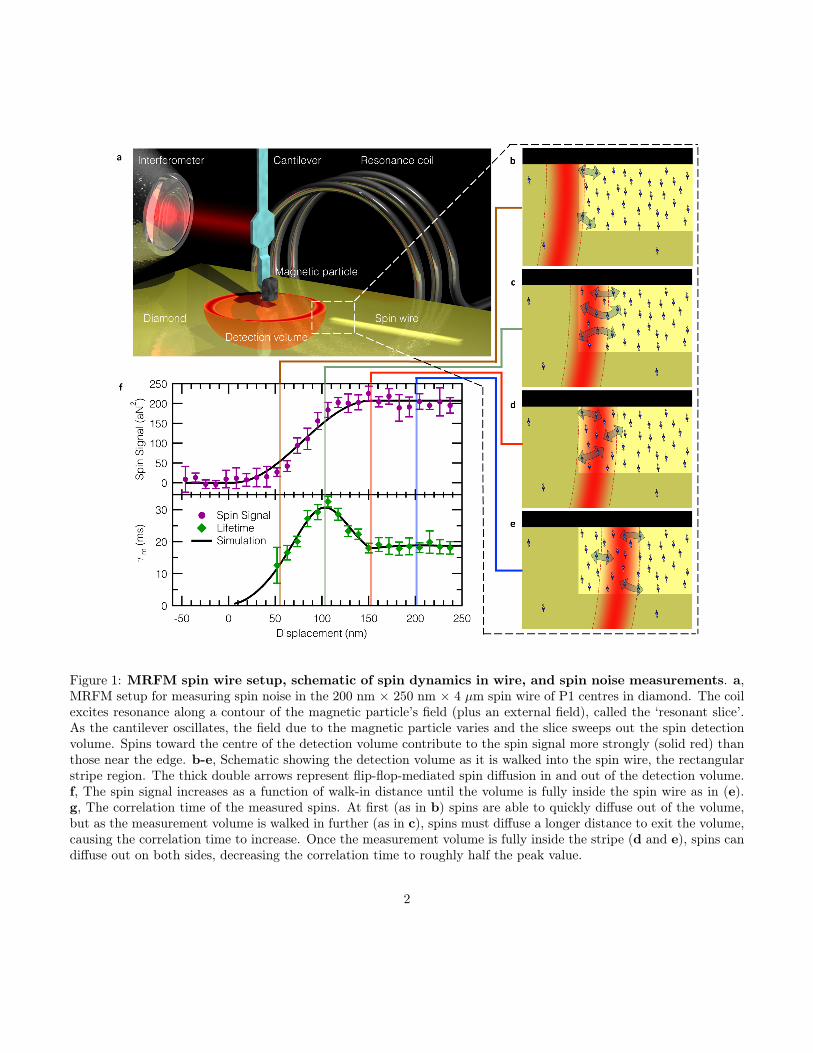
Figure 1: MRFM spin wire setup, schematic of spin dynamics in wire, and spin noise measurements. a,MRFM setup for measuring spin noise in the 200 nm × 250 nm × 4 µm spin wire of P1 centres in diamond. The coilexcites resonance along a contour of the magnetic particle’s field (plus an external field), called the ‘resonant slice’.As the cantilever oscillates, the field due to the magnetic particle varies and the slice sweeps out the spin detectionvolume. Spins toward the centre of the detection volume contribute to the spin signal more strongly (solid red) thanthose near the edge. b-e, Schematic showing the detection volume as it is walked into the spin wire, the rectangularstripe region. The thick double arrows represent flip-flop-mediated spin diffusion in and out of the detection volume.f, The spin signal increases as a function of walk-in distance until the volume is fully inside the spin wire as in (e).g, The correlation time of the measured spins. At first (as in b) spins are able to quickly diffuse out of the volume,but as the measurement volume is walked in further (as in c), spins must diffuse a longer distance to exit the volume,causing the correlation time to increase. Once the measurement volume is fully inside the stripe (d and e), spins candiffuse out on both sides, decreasing the correlation time to roughly half the peak value.
2

fabricated a nanoscale channel of implanted nitrogen (P1)centres in diamond at a density of 6 ppm. At this den-sity the strength of the dipolar coupling between spins isstrong, leading to a flip-flop time, Tff ∼ 10−4 s, severalorders of magnitude shorter than T1 ∼ 1 s. The densityoutside the spin wire (0.3 ppm) is sufficiently small suchthat flip-flop transitions out of the wire are very rare, thusconfining the spin transport to the wire.
We use magnetic resonance force microscopy (MRFM)to probe the spins within the wire (Fig. 1a). The forcedetector is an IBM-style ultra-soft cantilever [18] with aSmCo5 magnetic particle glued onto the tip. To reso-nantly detect spins, we implement the iOSCAR timingprotocol [8, 19]. A superconducting niobium coil is usedto drive the spin resonance at a frequency of ωrf = 2.18GHz. This frequency defines a ‘resonant slice’ where thetotal magnetic field (tip field plus external field) experi-enced by sample spins is equal to ωrf
γ = 778 G. By drivingthe cantilever at its self-determined resonant frequency toan amplitude of 150 nm, the resonant slice sweeps out avolume of this width, hereafter termed the detection vol-ume.
We scan the detection volume into the spin wire (Fig.1b-e) and measure the force exerted on the cantilever bythe selected spins. From this measurement we can ex-tract two quantities: spin signal (Fig. 1f, top), and theforce correlation time τm (Fig. 1f, bottom). The spin sig-nal is obtained from the variance in the time record ofthe force signal and is directly related to the number ofmeasured spins [19]. τm describes the characteristic timefor the net moment of the detected spins to decorrelate.This can be viewed as the time needed to transport spinout of the detection volume via a random-walk diffusionprocesses, with each step taking an average timeTff . Oncethe detection volume enters the spin wire, the signal growswith the number of detected spins, eventually reaching aplateau when completely within the wire. The correlationtime shows a complex behaviour as the detection volumemoves into the wire. These results can be understoodwithin the framework of flip-flop-mediated spin transport:ensemble correlation is lost as spins diffuse into or out ofthe detection volume. Just inside a displacement of 0 nm(Fig. 1b), the spins inside the volume easily and rapidlyinteract with nearby outside neighbours, resulting in arelatively short correlation time. With increasing over-lap of the detection volume and the spin wire, spins must
diffuse further to exit the detection volume and changethe overall magnetization of the ensemble, thus increasingτm. The correlation time peaks when approximately 68%of the detection volume has entered the spin wire (Fig.1c). This is attributed to the force sensitivity profile ofthe detection volume: spins in the middle of the volumeare most sensitively detected (solid red indicates highestsensitivity in Fig. 1), while the edges are least sensitive(see SI). As the detection volume approaches 100% over-lap (Fig. 1d), spins are able to diffuse out either side ofthe most sensitive region, reducing the correlation timeby roughly a factor of 2. Scanning deeper into the spinwire results in no further change, since the measurementgeometry becomes translationally invariant (Fig. 1e).
A global measurement of the spin polarization, en-compassing the entire spin wire, would not uncover thismagnetization-preserving spin transport process. In theopposite limit of measuring a single spin, T1-type spin re-laxation becomes indistinguishable from flip-flop inducedpolarization changes. By systematically varying the sizeof the overlap of the detection volume with the spin wire,and hence the number of detected spins, our measure-ment of τm reveals the observed signature of spin trans-port. This is similar to measurements of spin lifetime byelectrically- or optically-detected Hanle signals [11,20,21],where diffusion of spins away from the detection volume(electrical contact or optical probe spot) can influence spindephasing.
Since the average polarization gradient vanishes atthermal equilibrium, the conventional model of diffusiondriven by polarization gradient predicts that τm will be in-dependent of detection volume position within our ther-mal equilibrium wire. Such a model neglects the domi-nant role spin fluctuations play in nanoscale ensembles.In order to fit the measured data in Fig. 1f, we use aMonte Carlo simulation which models the flip-flops be-tween individual spins as a Markov process (see SI). Fromthis fit we can extract the flip-flop time, Tff = 0.21 ms,and the corresponding diffusion constant of D = a2
Tff=
4.6 × 10⁻⁹ cm2
s , where a = n−1/3 = 9.82 nm is the aver-age nearest-neighbour separation of spins with a densityn = 1.06 × 1018 cm−3 (6 ppm). We find good agree-ment comparing this to the theoretical flip-flop time [12]Tff,th = 30T2 = 0.36 ms, where we have used T2 = 11.9µs, which arises from dipolar interactions (at a densityof 6 ppm) and has been experimentally verified [22]. We
3

Figure 2: Hyperfine spectrum measurement for spin wire. a, Energy level diagrams for three hyperfine-splitspin populations, which are simultaneously on resonance, yet spatially separated by the gradient. b, Schematic of thethree volumes which are walked into the spin wire. c, The spin signal increases in a step-like fashion as each volumeenters the wire. d, A nanovolume EPR spectrum can be obtained by deconvolving the spin signal with the knownforce sensitivity (see SI) and the three hyperfine peaks are resolved.
4
furthermore find a diffusion length L =√DT1 = 720 nm,
which is significantly larger than the lateral dimensionsof the wire (depth 250 nm, width 200 nm, and length 4µm). This diffusion length is competitive with metallicspin transport devices [23].
The above expression for Tff,th assumes zero magneticfield gradient for the system, but in our experiment wehave a strong gradient (∼ 1.3 G/nm). In the presenceof a field gradient, neighbouring spins experience a fielddifference ∆B, and thus different Zeeman energy split-tings. This can suppress flip-flops because they no longerconserve energy [24–26]. However, inhomogeneous linebroadening, if on the order of ∆B, can make up this en-ergy difference and allow flip-flops to proceed. Since themeasured flip-flop time Tff agrees closely to the expectedzero-gradient value Tff,th, we expect a spectral linewidth∆B & 6.5 G.
To measure the EPR spectrum of the spin wire, we im-plemented a modified iOSCAR protocol which improvesspatial resolution and therefore provides the capability toresolve spectral features (since spatial and spectral resolu-tion are directly related in magnetic resonance imaging).A conventional iOSCAR measurement utilizes the entiredetection volume swept out by the cantilever oscillation,as this couples to the largest number of spins and cre-ates the largest force signal. By truncating the detectionvolume to the most-sensitive portion, we couple only tospins in this smaller region while maintaining high forcesensitivity. Utilizing this technique (further details in SI),which we call partially-interrupted OSCAR (piOSCAR),we again scan the detection volume into the spin wire.The improved resolution enables us to resolve a staircasestructure with three steps (Fig. 2c), corresponding to thethree peaks of the P1 center’s hyperfine spectrum as dis-cussed below.
The P1 centre is a substitutional nitrogen defect withan unpaired spin- 1
2 electron hyperfine coupled to the spin-1 nitrogen nucleus. The hyperfine interaction results in atriplet of peaks in the EPR spectrum (Fig. 2a) corre-sponding to the three nuclear spin states, mI = −1, 0, 1.The hyperfine splitting for our diamond crystal orienta-tion relative to the external field is 33 G (see SI andref. [27]). In the magnetic field gradient of our probe mag-net, this splitting results in three spatially distinct spin de-tection volumes, with each volume defined by the contourof magnetic field which satisfies the resonance condition
for a particular hyperfine transition (Fig. 2b). We ob-serve a step-like increase in the spin signal as each volumeenters the wire and the cantilever becomes coupled to anadditional hyperfine transition (Fig. 2c). The spacing be-tween steps (s = 25 nm) provides an accurate means ofmeasuring the probe field gradient, because the spacing isset by the ratio of the known hyperfine splitting (33 G)to the gradient, which we find to be 1.3 G
nm. This is con-sistent with our estimations of gradient obtained using astandard technique [28]. The staircase structure in figure2c is a convolution of the implanted spin density (approx-imately a step function), our force sensitivity profile, andthe EPR spectrum of spins in the sample; deconvolutionreveals the EPR spectrum shown in Fig. 2d (details in SI).From the spectrum we find an average linewidth of 7.6 G,which agrees with the expected linewidth from above, andcorroborates the transport we measure in the spin wire.
In conclusion, we have measured flip-flop-mediated spintransport in an insulating diamond spin wire in the com-plete absence of charge transport. This transport arisesfrom the intrinsic spin dynamics of dipole-coupled P1 cen-tres at thermal equilibrium, which we observe by measur-ing spin noise without imposing a polarization gradient.By spatially resolving the three electron spin populations(corresponding to the three spin states of the hyperfine-coupled nitrogen nucleus) we obtained the EPR spectrumof less than 100 net spins in the spin wire. The resultinglinewidth confirms that flip-flop mediated spin diffusionis responsible for the observed spin transport. The spec-trum also enables accurate measurement of the appliedmagnetic field gradient. These measurements provide in-sight into the mechanisms of spin transport relevant forthe development of nanoscale spin device elements.Methods The ultra-soft silicon cantilever has a spring
constant of 100 µN/m, resonance frequency of about 3kHz, and approximate dimensions of 90 microns in length,1 micron width, and 100 nm thickness. Displacement ofthe cantilever is measured through a 1550 nm laser inter-ferometer. The MRFM measurements were taken at tem-peratures of 4.2 K. The measured moment of the SmCo5
magnetic particle was 3.6× 10⁻¹² J/T. The niobium res-onator coil had about 2.5 turns and a 300 micron diam-eter, and the resonance bowl had a diameter of about3 microns. The diamond sample studied in this experi-ment had a background nitrogen concentration of 0.3 ppm(5.27× 1016 cm−3). To create the high spin-density wire,
5

the sample was first prepared using electron beam pat-terning and then exposed to nitrogen ion implantation ata variety of energies to create a uniform spin density. Fi-nally the sample was annealed to yield an approximatelyuniform, channel (6 ppm or 1.06×1018 cm−3) with width,depth, and length of 200 nm, 250 nm, and 4 microns, re-spectively. The sample was purchased commercially fromElement Six, grown via chemical vapor deposition, andthe nitrogen ion implantation was performed by LeonardKroko Inc.Acknowledgements The research presented in this
manuscript was financially supported by the Army Re-search Office (grant number W911NF-09-1-0147), the Na-tional Science Foundation (grant number DMR-0807093),and the Center for Emergent Materials (CEM), an NSFfunded MRSEC (grant number DMR-0820414). Techni-cal support was provided by the NanoSystems Laboratoryat the Ohio State University. The authors would like tothank R. J. Furnshahl for valuable discussions related tothe simulations.
References
[1] Wolf, S. et al. Spintronics: A spin-based electronicsvision for the future. Science 294, 1488–1495 (2001).
[2] Hanson, R., Dobrovitski, V. V., Feiguin, A. E.,Gywat, O. & Awschalom, D. D. Coherent dy-namics of a single spin interacting with an ad-justable spin bath. Science 320, 352–355 (2008).URL http://www.sciencemag.org/content/320/5874/352.full.pdf.
[3] Balasubramanian, G. et al. Ultralong spin coher-ence time in isotopically engineered diamond. NatureMaterials 8, 383–387 (2009). URL http://dx.doi.org/10.1038/nmat2420.
[4] Takahashi, S., Hanson, R., van Tol, J., Sherwin, M. S.& Awschalom, D. D. Quenching spin decoherence indiamond through spin bath polarization. Phys. Rev.Lett. 101, 047601 (2008). URL http://link.aps.org/doi/10.1103/PhysRevLett.101.047601.
[5] Heinrich, B. et al. Dynamic exchange coupling inmagnetic bilayers. Physical review letters 90, 187601(2003).
[6] Pfaff, W. et al. Demonstration of entanglement-by-measurement of solid-state qubits. Nat Phys 9,29–33 (2013). URL http://dx.doi.org/10.1038/nphys2444.
[7] Grinolds, M. et al. Nanoscale magnetic imaging of asingle electron spin under ambient conditions. NaturePhysics (2013).
[8] Mamin, H. J., Budakian, R., Chui, B. W. & Rugar,D. Detection and manipulation of statistical polar-ization in small spin ensembles. Phys. Rev. Lett. 91,207604 (2003). URL http://link.aps.org/doi/10.1103/PhysRevLett.91.207604.
[9] Bloembergen, N. On the interaction of nuclear spinsin a crystalline lattice. Physica 15, 386–426 (1949).
[10] Zhang, S., Meier, B. H. & Ernst, R. R. Polariza-tion echoes in nmr. Phys. Rev. Lett. 69, 2149–2151(1992). URL http://link.aps.org/doi/10.1103/PhysRevLett.69.2149.
[11] Lou, X. et al. Electrical detection of spin transport inlateral ferromagnet–semiconductor devices. NaturePhysics 3, 197–202 (2007).
[12] Abragam, A. The Principles of Nuclear Magnetism(Clarendon, Oxford, 1961).
[13] Vinante, A., Wijts, G., Usenko, O., Schinkelshoek,L. & Oosterkamp, T. Magnetic resonance force mi-croscopy of paramagnetic electron spins at millikelvintemperatures. Nature communications 2, 572 (2011).
[14] Eberhardt, K. W., Mouaziz, S., Boero, G., Brugger,J. & Meier, B. H. Direct observation of nuclear spindiffusion in real space. Physical review letters 99,227603 (2007).
[15] Fong, K. C., Herman, M. R., Banerjee, P., Pelekhov,D. V. & Hammel, P. C. Spin lifetime in small en-sembles of electron spins measured by magnetic res-onance force microscopy. Phys. Rev. B 84, 220405(2011). URL http://link.aps.org/doi/10.1103/PhysRevB.84.220405.
[16] Muller, G. M. et al. Spin noise spectroscopy in gaas(110) quantum wells: Access to intrinsic spin life-times and equilibrium electron dynamics. Phys. Rev.
6
Lett. 101, 206601 (2008). URL http://link.aps.org/doi/10.1103/PhysRevLett.101.206601.
[17] Seitaridou, E., Inamdar, M. M., Phillips, R., Ghosh,K. & Dill, K. Measuring flux distributions for dif-fusion in the small-numbers limit. The Journal ofPhysical Chemistry B 111, 2288–2292 (2007).
[18] Chui, B. W. et al. Mass-loaded cantilevers with sup-pressed higher-order modes for magnetic resonanceforce microscopy. In TRANSDUCERS, Solid-StateSensors, Actuators and Microsystems, 12th Interna-tional Conference on, 2003, vol. 2, 1120–1123 (IEEE,2003).
[19] Rugar, D., Budakian, R., Mamin, H. J. & Chui,B. W. Single spin detection by magnetic resonanceforce microscopy. Nature 430, 329–332 (2004).
[20] Huang, B. & Appelbaum, I. Time-of-flight spec-troscopy via spin precession: The larmor clock andanomalous spin dephasing in silicon. Physical ReviewB 82, 241202 (2010).
[21] Furis, M. et al. Local hanle-effect studies of spin driftand diffusion in n: Gaas epilayers and spin-transportdevices. New Journal of Physics 9, 347 (2007).
[22] Van Wyk, J., Reynhardt, E., High, G. & Kiflawi,I. The dependences of esr line widths and spin-spinrelaxation times of single nitrogen defects on the con-centration of nitrogen defects in diamond. Journal ofPhysics D: Applied Physics 30, 1790 (1997).
[23] Jedema, F., Heersche, H., Filip, A., Baselmans, J. &Van Wees, B. Electrical detection of spin precessionin a metallic mesoscopic spin valve. Nature 416, 713–716 (2002).
[24] Budakian, R., Mamin, H. J. & Rugar, D. Sup-pression of spin diffusion near a micron-size fer-romagnet. Physical Review Letters 92, 037205(2004). URL http://link.aps.org/abstract/PRL/v92/e037205.
[25] Genack, A. Z. & Redfield, A. G. Theory of nuclearspin diffusion in a spatially varying magnetic field.Phys. Rev. B 12, 78–87 (1975). URL http://link.aps.org/doi/10.1103/PhysRevB.12.78.
[26] Tyryshkin, A. M. et al. Electron spin coherence ex-ceeding seconds in high-purity silicon. Nature mate-rials 11, 143–147 (2011).
[27] Cook, R. &Whiffen, D. Electron nuclear double reso-nance study of a nitrogen centre in diamond. Proceed-ings of the Royal Society of London. Series A. Math-ematical and Physical Sciences 295, 99–106 (1966).
[28] Stipe, B. et al. Electron spin relaxation near amicron-size ferromagnet. Physical review letters 87,277602 (2001).
7