observation of substrate diffusion and ligand binding in
TRANSCRIPT

research papers
878 https://doi.org/10.1107/S2052252521008125 IUCrJ (2021). 8, 878–895
IUCrJISSN 2052-2525
BIOLOGYjMEDICINE
Received 21 June 2021
Accepted 6 August 2021
Edited by K. Moffat, University of Chicago, USA
In memoriam of John Spence.
Keywords: substrate diffusion in crystals;
antibiotic resistance; �-lactamases; enzyme
kinetics; irreversible inhibition; mix-and-inject
serial crystallography; serial femtosecond
crystallography; European X-ray Free-Electron
Laser; megahertz pulse-repetition rate; protein
structure determination; drug discovery;
ceftriaxone; sulbactam; X-ray crystallography;
enzyme mechanisms.
PDB references: BlaC, unmixed, 7k8l; mixed
with ceftriaxone, 5 ms, 7k8e; 10 ms, 7k8f;
50 ms, 7k8h; mixed with sulbactam, 66 ms,
7k8k
Supporting information: this article has
supporting information at www.iucrj.org
Observation of substrate diffusion and ligandbinding in enzyme crystals using high-repetition-rate mix-and-inject serial crystallography
Suraj Pandey,a George Calvey,b Andrea M. Katz,b Tek Narsingh Malla,a Faisal H. M.
Koua,c Jose M. Martin-Garcia,d,e Ishwor Poudyal,a Jay-How Yang,d Mohammad
Vakili,f Oleksandr Yefanov,c Kara A. Zielinski,b Sasa Bajt,g,h Salah Awel,c Katarina
Doerner,f Matthias Frank,i Luca Gelisio,c Rebecca Jernigan,d Henry Kirkwood,f
Marco Kloos,f Jayanath Koliyadu,f Valerio Mariani,c,j Mitchell D. Miller,k Grant
Mills,f Garrett Nelson,l Jose L. Olmos Jr,k,m Alireza Sadri,c Tokushi Sato,f Alexandra
Tolstikova,c Weijun Xu,k Abbas Ourmazd,a John C. H. Spence,l Peter Schwander,a
Anton Barty,g Henry N. Chapman,c,h,n Petra Fromme,d Adrian P. Mancuso,f,o
George N. Phillips Jr,k,p Richard Bean,f Lois Pollackb and Marius Schmidta*
aPhysics Department, University of Wisconsin-Milwaukee, 3135 North Maryland Avenue, Milwaukee, WI 53211, USA,bSchool of Applied and Engineering Physics, Cornell University, 254 Clark Hall, Ithaca, NY 14853, USA, cCenter for
Free-Electron Laser Science, Deutsches Elektronen-Synchrotron DESY, Notkestrasse 85, 22607 Hamburg, Germany,dSchool of Molecular Sciences and Biodesign Center for Applied Structural Discovery, Arizona State University, Tempe,
AZ 85287-1604, USA, eInstitute of Physical Chemistry Rocasolano, Spanish National Research Council, Calle de Serrano
119, 28006 Madrid, Spain, fEuropean XFEL, Holzkoppel 4, 22869 Schenefeld, Germany, gDeutsches Elektronen-
Synchrotron DESY, Notkestrasse 85, 22607 Hamburg, Germany, hThe Hamburg Centre for Ultrafast Imaging, Luruper
Chaussee 149, 22761 Hamburg, Germany, iLawrence Livermore National Laboratory, 7000 East Avenue, Livermore,
CA 94550, USA, jSLAC National Accelerator Laboratory, 2575 Sand Hill Rd, Menlo Park, California 94025, USA,kDepartment of BioSciences, Rice University, 6100 Main Street, Houston, TX 77005, USA, lDepartment of Physics,
Arizona State University, Tempe, AZ 85287, USA, mDepartment of Bioengineering and Therapeutic Sciences, University
of California San Francisco, San Francisco, CA 94158, USA, nDepartment of Physics, Universitat Hamburg, Luruper
Chaussee 149, 22761 Hamburg, Germany, oDepartment of Chemistry and Physics, La Trobe Institute for Molecular
Science, La Trobe University, Melbourne, Victoria 3086, Australia, and pDepartment of Chemistry, Rice University, 6100
Main Street, Houston, TX 77005, USA. *Correspondence e-mail: [email protected]
Here, we illustrate what happens inside the catalytic cleft of an enzyme when
substrate or ligand binds on single-millisecond timescales. The initial phase of
the enzymatic cycle is observed with near-atomic resolution using the most
advanced X-ray source currently available: the European XFEL (EuXFEL).
The high repetition rate of the EuXFEL combined with our mix-and-
inject technology enables the initial phase of ceftriaxone binding to the
Mycobacterium tuberculosis �-lactamase to be followed using time-resolved
crystallography in real time. It is shown how a diffusion coefficient in enzyme
crystals can be derived directly from the X-ray data, enabling the determination
of ligand and enzyme–ligand concentrations at any position in the crystal
volume as a function of time. In addition, the structure of the irreversible
inhibitor sulbactam bound to the enzyme at a 66 ms time delay after mixing is
described. This demonstrates that the EuXFEL can be used as an important tool
for biomedically relevant research.
1. Introduction
Combatting the rise of infectious diseases requires a colla-
borative and interdisciplinary approach. Structural biologists
can contribute by investigating the reaction mechanisms of
biomedically significant enzymes as a structural basis to
develop cures for diseases. Bacterial infections with strains
that are resistant to currently available antibiotics are on the
rise (Cassini et al., 2019). A study sponsored by the British
government projected that in the near future more people will
die from untreatable bacterial infections than from cancer
(https://amr-review.org/). Bacterial enzymes that inactivate
currently available drugs are central to antibiotic resistance
(Fair & Tor, 2014), and unraveling the catalytic mechanism of
these enzymes will be beneficial for the development of novel
antibiotics (Imming et al., 2006). �-Lactamases such as the
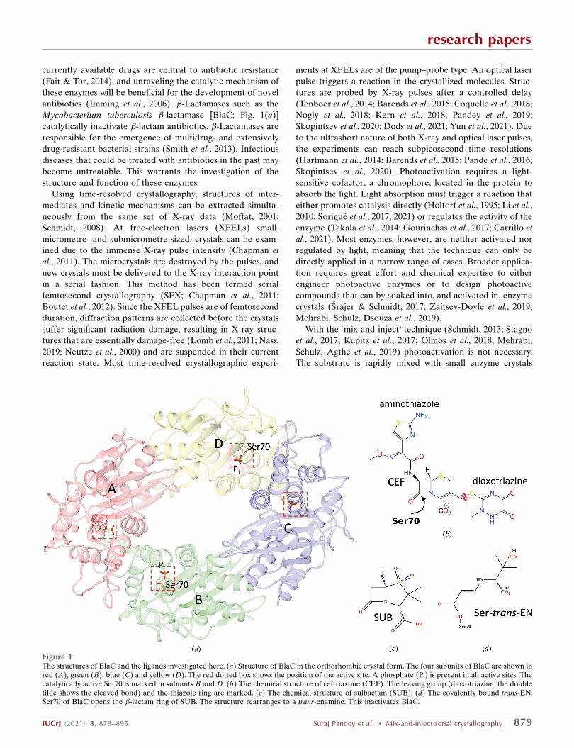
Mycobacterium tuberculosis �-lactamase [BlaC; Fig. 1(a)]
catalytically inactivate �-lactam antibiotics. �-Lactamases are
responsible for the emergence of multidrug- and extensively
drug-resistant bacterial strains (Smith et al., 2013). Infectious
diseases that could be treated with antibiotics in the past may
become untreatable. This warrants the investigation of the
structure and function of these enzymes.
Using time-resolved crystallography, structures of inter-
mediates and kinetic mechanisms can be extracted simulta-
neously from the same set of X-ray data (Moffat, 2001;
Schmidt, 2008). At free-electron lasers (XFELs) small,
micrometre- and submicrometre-sized, crystals can be exam-
ined due to the immense X-ray pulse intensity (Chapman et
al., 2011). The microcrystals are destroyed by the pulses, and
new crystals must be delivered to the X-ray interaction point
in a serial fashion. This method has been termed serial
femtosecond crystallography (SFX; Chapman et al., 2011;
Boutet et al., 2012). Since the XFEL pulses are of femtosecond
duration, diffraction patterns are collected before the crystals
suffer significant radiation damage, resulting in X-ray struc-
tures that are essentially damage-free (Lomb et al., 2011; Nass,
2019; Neutze et al., 2000) and are suspended in their current
reaction state. Most time-resolved crystallographic experi-
ments at XFELs are of the pump–probe type. An optical laser
pulse triggers a reaction in the crystallized molecules. Struc-
tures are probed by X-ray pulses after a controlled delay
(Tenboer et al., 2014; Barends et al., 2015; Coquelle et al., 2018;
Nogly et al., 2018; Kern et al., 2018; Pandey et al., 2019;
Skopintsev et al., 2020; Dods et al., 2021; Yun et al., 2021). Due
to the ultrashort nature of both X-ray and optical laser pulses,
the experiments can reach subpicosecond time resolutions
(Hartmann et al., 2014; Barends et al., 2015; Pande et al., 2016;
Skopintsev et al., 2020). Photoactivation requires a light-
sensitive cofactor, a chromophore, located in the protein to
absorb the light. Light absorption must trigger a reaction that
either promotes catalysis directly (Holtorf et al., 1995; Li et al.,
2010; Sorigue et al., 2017, 2021) or regulates the activity of the
enzyme (Takala et al., 2014; Gourinchas et al., 2017; Carrillo et
al., 2021). Most enzymes, however, are neither activated nor
regulated by light, meaning that the technique can only be
directly applied in a narrow range of cases. Broader applica-
tion requires great effort and chemical expertise to either
engineer photoactive enzymes or to design photoactive
compounds that can by soaked into, and activated in, enzyme
crystals (Srajer & Schmidt, 2017; Zaitsev-Doyle et al., 2019;
Mehrabi, Schulz, Dsouza et al., 2019).
With the ‘mix-and-inject’ technique (Schmidt, 2013; Stagno
et al., 2017; Kupitz et al., 2017; Olmos et al., 2018; Mehrabi,
Schulz, Agthe et al., 2019) photoactivation is not necessary.
The substrate is rapidly mixed with small enzyme crystals
research papers
IUCrJ (2021). 8, 878–895 Suraj Pandey et al. � Mix-and-inject serial crystallography 879
Figure 1The structures of BlaC and the ligands investigated here. (a) Structure of BlaC in the orthorhombic crystal form. The four subunits of BlaC are shown inred (A), green (B), blue (C) and yellow (D). The red dotted box shows the position of the active site. A phosphate (Pi) is present in all active sites. Thecatalytically active Ser70 is marked in subunits B and D. (b) The chemical structure of ceftriaxone (CEF). The leaving group (dioxotriazine; the doubletilde shows the cleaved bond) and the thiazole ring are marked. (c) The chemical structure of sulbactam (SUB). (d) The covalently bound trans-EN.Ser70 of BlaC opens the �-lactam ring of SUB. The structure rearranges to a trans-enamine. This inactivates BlaC.
during sample delivery (Calvey et al., 2019). Mixing occurs at a
well controlled location ‘en route’ to the X-ray beam. During
the time delay �tm that occurs between mixing and injection,
the substrate diffuses into the crystals and binds to the
enzyme. The complex formed by the substrate and the enzyme
then initiates the enzymatic cycle. Variation of �tm allows the
measurement of rate coefficients together with atomic reso-
lution structures which can be associated with intermediate
states of the protein reactions (Kupitz et al., 2017; Olmos et al.,
2018; Mehrabi, Schulz, Agthe et al., 2019). This can reveal the
mechanism of enzyme action at the molecular level or the
binding of a drug molecule. The combination of serial
femtosecond crystallography with mixing before injection has
been denoted ‘mix-and-inject serial crystallography’ (MISC;
Kupitz et al., 2017; Olmos et al., 2018). The feasibility of MISC
has previously been demonstrated with BlaC on longer
millisecond timescales (Kupitz et al., 2017; Olmos et al., 2018).
The observation of intermediate-state structures, and the
maximization of the potential time resolution in both photo-
activation and mix-and-inject techniques, relies on an accu-
rately gauged start time of the reaction inside the crystals. In
photoactivation experiments this requires a sufficient pene-
tration of optical laser light into the crystal to ensure that a
reaction is simultaneously triggered in a significant fraction of
the molecules. In mix-and-inject experiments, the diffusion
time of the substrate into the crystal may limit the ability to
discriminate diffusion and kinetics, including substrate
binding. To overcome this limitation, micrometre or sub-
micrometre crystal sizes are required that ensure that the
substrate diffuses rapidly into the crystals and the reaction is
triggered swiftly and much faster than the lifetime of the
reaction intermediates of interest (Schmidt, 2013).
The reaction of the cephalosporin antibiotic ceftriaxone
[CEF; Fig. 1(b)] with BlaC is an excellent candidate for
exploration with MISC. Previously, this reaction was investi-
gated for �tm of longer than 30 ms (Kupitz et al., 2017; Olmos
et al., 2018). At 30 ms, however, the CEF binding sites in BlaC
were essentially fully occupied (Olmos et al., 2018), a state also
reached on similar timescales for other proteins and enzymes
(Stagno et al., 2017; Mehrabi, Schulz, Agthe et al., 2019; Ishi-
gami et al., 2019). The substrate-binding phase and the
formation of the enzyme–substrate complex, however, remain
elusive. Here, we aim to characterize the early phase of
substrate binding with single-millisecond time delays by using
the megahertz X-ray pulse-repetition rate of the European
XFEL (EuXFEL; Decking et al., 2020).
In addition, we aim to investigate the reaction of BlaC with
an inhibitor, sulbactam [SUB; Fig. 1(c)], on a millisecond
timescale. The biochemistry of SUB and its application in
combination with �-lactam antibiotics have been described in
detail elsewhere (Totir et al., 2007). SUB binds to the active
site of BlaC and reacts with the catalytically active serine of
�-lactamases to form a covalently bound species. Most abun-
dant is the so-called trans-enamine (trans-EN) species
[Fig. 1(d)] that inhibits �-lactamases and helps to eliminate
�-lactamase-induced antibiotic resistance. Static structures of
trans-ENs with �-lactamases, including BlaC, have recently
been characterized (Cheng et al., 2020; Tassoni et al., 2019), but
structures of the early species that form during SUB binding
remain elusive.
2. Methods
2.1. BlaC crystals
Platelet-shaped crystals of BlaC with approximate dimen-
sions of 10 � 10 � 2 mm (Appendix A) were produced by a
stirring method on site in the XBI facility of the EuXFEL
(Han et al., 2021) using ammonium phosphate (AP) as
described by Olmos et al. (2018). The crystals belonged to
space group P21 (Table 1), with four BlaC subunits in the
asymmetric unit [Fig. 1(a)] (Olmos et al., 2018). Only two
subunits bind CEF in their catalytic cleft, as demonstrated
previously (Olmos et al., 2018). The concentration of BlaC
subunits in the crystals is 15.5 mM, so that the concentration of
active subunits is 7.8 mM. When this concentration is matched
by substrate, the substrate concentration is called ‘stoichio-
metric’ in the following description.
2.2. Data collection at the EuXFEL
The platelets were mixed with ceftriaxone [CEF; Fig. 1(b);
molecular mass 554.6 g mol�1; 200 mM in 0.8 M AP] or
research papers
880 Suraj Pandey et al. � Mix-and-inject serial crystallography IUCrJ (2021). 8, 878–895
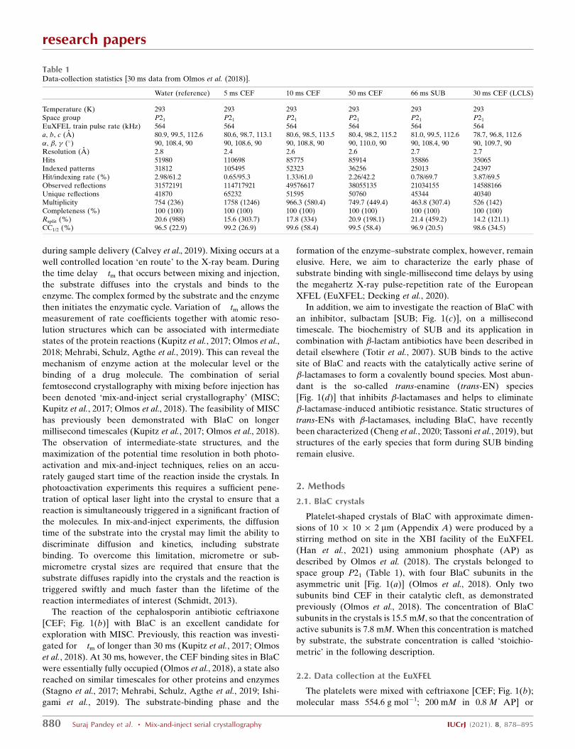
Table 1Data-collection statistics [30 ms data from Olmos et al. (2018)].
Water (reference) 5 ms CEF 10 ms CEF 50 ms CEF 66 ms SUB 30 ms CEF (LCLS)
Temperature (K) 293 293 293 293 293 293Space group P21 P21 P21 P21 P21 P21
EuXFEL train pulse rate (kHz) 564 564 564 564 564 564a, b, c (A) 80.9, 99.5, 112.6 80.6, 98.7, 113.1 80.6, 98.5, 113.5 80.4, 98.2, 115.2 81.0, 99.5, 112.6 78.7, 96.8, 112.6�, �, � (�) 90, 108.4, 90 90, 108.6, 90 90, 108.8, 90 90, 110.0, 90 90, 108.4, 90 90, 109.7, 90Resolution (A) 2.8 2.4 2.6 2.6 2.7 2.7Hits 51980 110698 85775 85914 35886 35065Indexed patterns 31812 105495 52323 36256 25013 24397Hit/indexing rate (%) 2.98/61.2 0.65/95.3 1.33/61.0 2.26/42.2 0.78/69.7 3.87/69.5Observed reflections 31572191 114717921 49576617 38055135 21034155 14588166Unique reflections 41870 65232 51595 50760 45344 40340Multiplicity 754 (236) 1758 (1246) 966.3 (580.4) 749.7 (449.4) 463.8 (307.4) 526 (142)Completeness (%) 100 (100) 100 (100) 100 (100) 100 (100) 100 (100) 100 (100)Rsplit (%) 20.6 (988) 15.6 (303.7) 17.8 (334) 20.9 (198.1) 21.4 (459.2) 14.2 (121.1)CC1/2 (%) 96.5 (22.9) 99.2 (26.9) 99.6 (58.4) 99.5 (58.4) 96.9 (20.5) 98.6 (34.5)
sulbactam (SUB) inhibitor [Fig. 1(c), molecular mass
223.2 g mol�1, 100 mM in 0.8 M AP] using optimized mixing
injectors (Calvey et al., 2019) which were adapted to operate at
the SPB/SFX instrument (Mancuso, 2019) of the EuXFEL.
Flow rates and mixer geometries are shown in Table 2. The
mixture was intercepted after a delay �tm by X-ray pulses
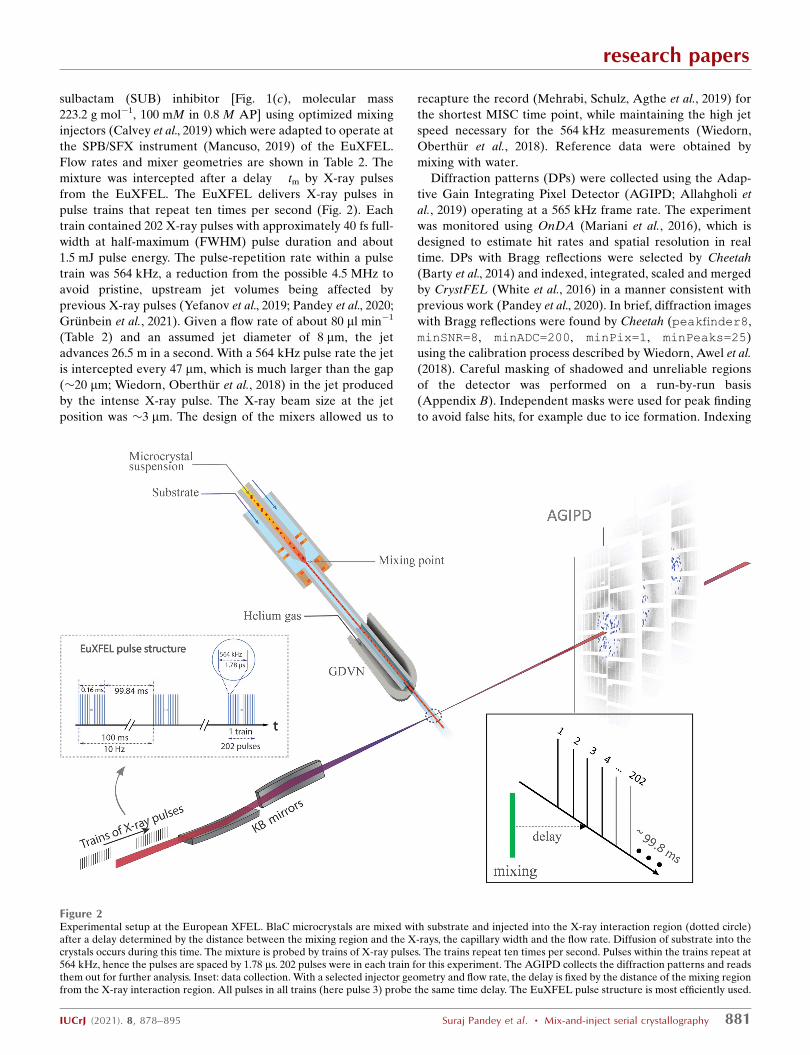
from the EuXFEL. The EuXFEL delivers X-ray pulses in
pulse trains that repeat ten times per second (Fig. 2). Each
train contained 202 X-ray pulses with approximately 40 fs full-
width at half-maximum (FWHM) pulse duration and about
1.5 mJ pulse energy. The pulse-repetition rate within a pulse
train was 564 kHz, a reduction from the possible 4.5 MHz to
avoid pristine, upstream jet volumes being affected by
previous X-ray pulses (Yefanov et al., 2019; Pandey et al., 2020;
Grunbein et al., 2021). Given a flow rate of about 80 ml min�1
(Table 2) and an assumed jet diameter of 8 mm, the jet
advances 26.5 m in a second. With a 564 kHz pulse rate the jet
is intercepted every 47 mm, which is much larger than the gap
(�20 mm; Wiedorn, Oberthur et al., 2018) in the jet produced
by the intense X-ray pulse. The X-ray beam size at the jet
position was �3 mm. The design of the mixers allowed us to
recapture the record (Mehrabi, Schulz, Agthe et al., 2019) for
the shortest MISC time point, while maintaining the high jet
speed necessary for the 564 kHz measurements (Wiedorn,
Oberthur et al., 2018). Reference data were obtained by
mixing with water.
Diffraction patterns (DPs) were collected using the Adap-
tive Gain Integrating Pixel Detector (AGIPD; Allahgholi et
al., 2019) operating at a 565 kHz frame rate. The experiment
was monitored using OnDA (Mariani et al., 2016), which is
designed to estimate hit rates and spatial resolution in real
time. DPs with Bragg reflections were selected by Cheetah
(Barty et al., 2014) and indexed, integrated, scaled and merged
by CrystFEL (White et al., 2016) in a manner consistent with
previous work (Pandey et al., 2020). In brief, diffraction images
with Bragg reflections were found by Cheetah (peakfinder8,
minSNR=8, minADC=200, minPix=1, minPeaks=25)
using the calibration process described by Wiedorn, Awel et al.
(2018). Careful masking of shadowed and unreliable regions
of the detector was performed on a run-by-run basis
(Appendix B). Independent masks were used for peak finding
to avoid false hits, for example due to ice formation. Indexing
research papers
IUCrJ (2021). 8, 878–895 Suraj Pandey et al. � Mix-and-inject serial crystallography 881
Figure 2Experimental setup at the European XFEL. BlaC microcrystals are mixed with substrate and injected into the X-ray interaction region (dotted circle)after a delay determined by the distance between the mixing region and the X-rays, the capillary width and the flow rate. Diffusion of substrate into thecrystals occurs during this time. The mixture is probed by trains of X-ray pulses. The trains repeat ten times per second. Pulses within the trains repeat at564 kHz, hence the pulses are spaced by 1.78 ms. 202 pulses were in each train for this experiment. The AGIPD collects the diffraction patterns and readsthem out for further analysis. Inset: data collection. With a selected injector geometry and flow rate, the delay is fixed by the distance of the mixing regionfrom the X-ray interaction region. All pulses in all trains (here pulse 3) probe the same time delay. The EuXFEL pulse structure is most efficiently used.

was performed with CrystFEL (version 0.9.0) using the
indexing package XGANDALF (Gevorkov et al., 2019) with
the following parameters: peaks=peakfinder8, Min-
SNR=5, Min-pixel-count=1, Threshold=400. The
detector geometry was refined using geoptimiser (Yefanov et
al., 2015). Merging and scaling of the Bragg peak intensities
were performed using the partialator program from CrystFEL.
To avoid the integration of noise for weakly scattering
patterns, reflections were included up to 1.0 nm�1 above a
conservative resolution estimate for each crystal (--push-
res=1.0). Hit rates and indexing rates were stable in the
order of 1.0% and 70%, respectively, irrespective of the pulse
index in the train (Appendix A). The lower hit rate is a
consequence of diluting the crystalline slurry with the ligand/
substrate. It has been shown that the X-ray pulse position in
the train has no effect on the structure (Yefanov et al., 2019).
Structure-factor amplitudes were generated from the
measured intensities using programs from the CCP4 software
suite (Winn et al., 2011). Data-collection statistics are shown in
Table 1.
As a control, and to investigate the result of the complete
reaction of BlaC with SUB in the platelet crystal form,
macroscopic crystals were grown in sitting drops (10 ml BlaC at
45 mg ml�1 mixed in a 1:1 ratio with 2.1 M AP pH 4.1).
Crystals grew within three days. The crystals were soaked for
3 h in a cryobuffer consisting of 2 M AP, 20% glycerol and
100 mM SUB. The crystals were cooled in liquid nitrogen.
Data were collected on beamline ID-19 of the Structural
Biology Center, Advanced Photon Source, Argonne National
Laboratory. Data were processed to 2.7 A using HKL-3000
(Minor et al., 2006). Details will be presented elsewhere.
2.3. Difference-map calculation and structure determination
The structures of BlaC and the BlaC–CEF complexes were
determined as described previously (Kupitz et al., 2017; Olmos
et al., 2018). Since the unit-cell constants change substantially
after mixing (Table 1), isomorphous difference maps cannot
be calculated and OMIT difference maps (DEDomit) were
used. An initial BlaC model, PDB (Berman et al., 2002) entry
6b5x (Olmos et al., 2018), was refined against the structure-
factor amplitudes |Fo(t)|CEF collected at a particular �tm. The
content of the active sites (water and phosphate) was removed
during the refinement. From the refined model amplitudes, |Fc|
were determined. From the amplitudes (and the phases
obtained from the refined model), weighted m|Fo(t)|CEF �
D|Fc| OMIT maps (DEDomit) were calculated. Polder differ-
ence maps (Liebschner et al., 2017; DEDPolder) were calculated
to display weak difference electron-density features. The CEF
was modeled in the polder maps. To refine the structure and to
determine the fractional concentration of both Pi and CEF,
grouped occupancy refinement was performed using Phenix
(Liebschner et al., 2019). CEF was flagged together with Pi as a
pair of molecules occupying the same space. The positions and
B factors of all atoms as well as the occupancies of Pi and CEF
were refined simultaneously. As a check, the sum of the
occupancies of the flagged molecules should not deviate too
much from unity.
Initial structures of the BlaC–SUB complexes were deter-
mined by inspecting both isomorphous and OMIT difference
maps. The OMIT map was calculated in a similar way as
described above except that amplitudes |Fo(t)|SUB were used.
A weighted DEDiso map was calculated from difference
amplitudes w[|Fo(t)|SUB � |Fo|WAT], where the reference
amplitudes |Fo|WAT were obtained by mixing with water. The
weighting factor was calculated as described previously for
photoactive yellow protein (Ren et al., 2001; Pandey et al.,
2020) and for the needle crystal form of BlaC (Olmos et al.,
2018). The trans-EN and SUB molecules were inserted into
the OMIT map. The positions and orientations were cross-
examined to agree with the DEDiso map. The complexes were
refined using REFMAC (Murshudov et al., 2011) against the
|Fo(t)|SUB amplitudes. The refinement statistics for the BlaC–
CEF and BlaC–SUB complexes are shown in Table 3.
2.4. Binding kinetics of CEF
Refined occupancies are fitted by functions that account for
saturation of CEF and decline of Pi,
CCEFðtÞ ¼CS;CEF � t
t1=2 þ t; ð1Þ
and
CPiðtÞ ¼
Cini;Pi� t1=2
t1=2 þ t; ð2Þ
research papers
882 Suraj Pandey et al. � Mix-and-inject serial crystallography IUCrJ (2021). 8, 878–895
Table 2Parameters for the mix-and-inject experiments.
Concentrations of ceftriaxone (CEF) and sulbactam (SUB) are shown as were flowed through the outer capillary line of the mixing injectors. Time delays areachieved after mixing in a constriction as per Calvey et al. (2019).
Water SUB CEF CEF CEF
�tm (ms) (10) 66 5 10 50Ligand concentration (mM) — 100 200 200 200Ligand buffer — 0.8 M AP pH 4.6 0.8 M AP pH 4.6 0.8 M AP pH 4.6 0.8 M AP pH 4.6Ligand flow (ml min�1) 74.5 54.5 76.7 74.5 71.8Crystal flow (ml min�1) 5.5 11.6 3.3 5.5 8.2Mixing injector capillary internal diameter (mm) 50 75 50 50 75Constriction length (mm) 17.8 36.1 9.3 17.8 36.1Timing uncertainty (ms) — 9.3 1.8 3.0 10.4Experimental time to collect the data set (min) 50 56 138 250 32
respectively. CS;CEF and Cini;Piare the occupancy of CEF after a
long time (at saturation) and the initial occupancy of Pi,
respectively. The constant t1/2 is either the time taken to reach
half saturation of CEF or denotes the time when Pi has
declined to half its initial concentration. The initial occupancy
of Pi was set as 1.0, but the final occupancy of CEF was not
constrained to 1.0 to account for a more realistic scenario.
2.5. The diffusion coefficient of CEF in the BlaC platelets
The occupancy of CEF bound noncovalently to the active
center of BlaC was calculated with typical sized 10 � 10 �
2 mm (platelet-like) crystals (Appendix A) by varying the
diffusion coefficient D for the CEF in crystals until agreement
with the experiment was achieved. The crystal was divided
into 20 voxels along each direction and 20 time intervals were
used. For each time interval, the concentration of the CEF
substrate (CCEF) in each of the 8000 voxels in the crystal was
determined using the known solution to Fick’s second law for
a rectangular volume, represented by the first 20 modes
(Schmidt, 2013, 2020; Carslaw & Jaeger, 1959),
CCEFðx; y; z; tÞ ¼
C0;CEF
�1�
64
�3
P19
l¼0
P19
m¼0
P19
n¼0
ð�1Þlþmþn
ð2l þ 1Þð2mþ 1Þð2nþ 1Þ
� cosð2l þ 1Þ�x
2acosð2mþ 1Þ�y
2bcosð2nþ 1Þ�z
2c
� exp �D�2
4
ð2l þ 1Þ2
a2þð2mþ 1Þ2
b2þð2nþ 1Þ2
c2
� �t
� ��;
ð3Þ
where l, m, n are integer numbers that define the modes. x, y
and z are coordinates within the crystal that extend from�a to
+a,�b to +b and�c to c, where a, b, c are the half edge lengths
of the platelet-shaped BlaC crystals. D is the diffusion coef-
ficient, t is the time after mixing and C0,CEF is the outside CEF
concentration, which was set to 150 mM. This analytical
approach to diffusion is strictly speaking only valid in the
absence of substrate binding. However, here the substrate
concentration (�150 mM) is much higher than the concen-
tration of the subunits that bind the CEF (7.8 mM). The
concentration of substrate in the crystals increases rapidly to
values much higher than the stoichiometric concentration. At
saturation, the ES concentration is only 5.2% of that of the
substrate. In addition, the speed (rate) of substrate binding is
low until sufficient substrate is present. Accordingly, substrate
binding is a small perturbation of the free CEF concentration
on all but the very shortest timescales.
On the timescales employed here, the formation of later
intermediates and the catalytic turnover of BlaC with CEF do
not play a role (Boyd & Lunn, 1979; Hugonnet & Blanchard,
2007; Tremblay et al., 2010; Olmos et al., 2018). Both processes
unfold over much longer timescales (Fig. 3) than the time
delays examined here.
CEF binding to the active sites of BlaC is dependent on the
free BlaC concentration in the crystal and the rate coefficients
that describe the binding kinetics (Fig. 3). Here, the rate
coefficient kon of 3.2 M�1 s�1 as estimated by Olmos and
coworkers was used. The koff rate coefficient (dashed arrow in
Fig. 3) was assumed to be negligible relative to the on-rate
coefficient. There is only one free parameter, the diffusion
coefficient D, which can be inferred by matching calculated
occupancies to the refined occupancies observed at �tm. (3)
provides substrate (CEF) concentrations in each individual
voxel (at each position in the crystal) at any particular time t.
CEF binding to BlaC was calculated for each voxel by
numerically integrating the rate equation with time intervals
dt,
d½ES� ¼ ½EðtiÞ�free � ½CCEFðtiÞ� � kon dt;
½ESðtiþ1Þ� ¼ ½ESðtiÞ� þ d½ES�;
½Eðtiþ1Þ�free ¼ ½EðtiÞ�free � d½ES�;
tiþ1 ¼ ti þ dt: ð4Þ
research papers
IUCrJ (2021). 8, 878–895 Suraj Pandey et al. � Mix-and-inject serial crystallography 883
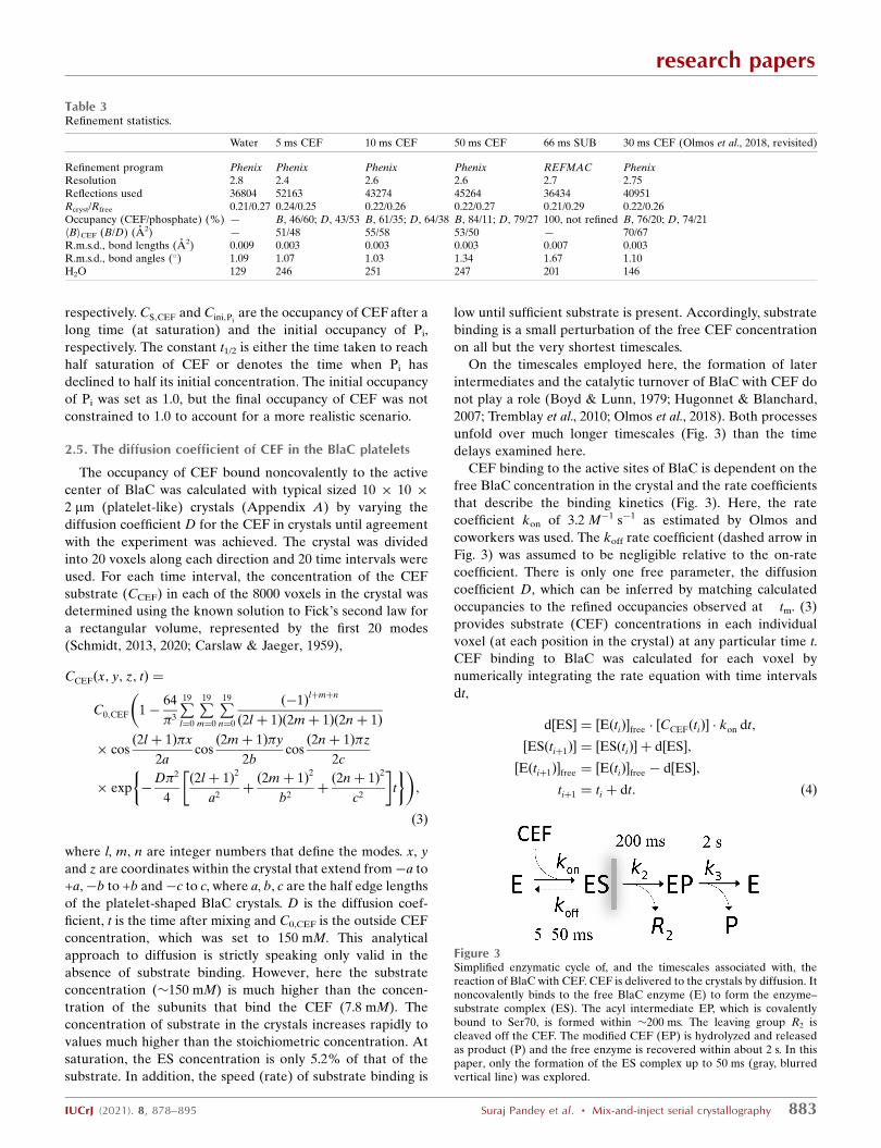
Figure 3Simplified enzymatic cycle of, and the timescales associated with, thereaction of BlaC with CEF. CEF is delivered to the crystals by diffusion. Itnoncovalently binds to the free BlaC enzyme (E) to form the enzyme–substrate complex (ES). The acyl intermediate EP, which is covalentlybound to Ser70, is formed within �200 ms. The leaving group R2 iscleaved off the CEF. The modified CEF (EP) is hydrolyzed and releasedas product (P) and the free enzyme is recovered within about 2 s. In thispaper, only the formation of the ES complex up to 50 ms (gray, blurredvertical line) was explored.
Table 3Refinement statistics.
Water 5 ms CEF 10 ms CEF 50 ms CEF 66 ms SUB 30 ms CEF (Olmos et al., 2018, revisited)
Refinement program Phenix Phenix Phenix Phenix REFMAC PhenixResolution 2.8 2.4 2.6 2.6 2.7 2.75Reflections used 36804 52163 43274 45264 36434 40951Rcryst/Rfree 0.21/0.27 0.24/0.25 0.22/0.26 0.22/0.27 0.21/0.29 0.22/0.26Occupancy (CEF/phosphate) (%) — B, 46/60; D, 43/53 B, 61/35; D, 64/38 B, 84/11; D, 79/27 100, not refined B, 76/20; D, 74/21hBiCEF (B/D) (A2) — 51/48 55/58 53/50 — 70/67R.m.s.d., bond lengths (A2) 0.009 0.003 0.003 0.003 0.007 0.003R.m.s.d., bond angles (�) 1.09 1.07 1.03 1.34 1.67 1.10H2O 129 246 251 247 201 146

In (4), d[ES] is the change in concentration of the BlaC–CEF
complex ([ES]) given the free enzyme ([E]) and free CEF
([CCEF]) concentrations at time ti and the kon rate. The free-
enzyme concentration [E] decreases and that of the BlaC–
CEF complex increases with each time step. (4) is repeated by
increasing ti by dt until ti is larger than a given delay time, for
example 10 ms. The substrate [CCEF] is provided everywhere
by diffusion (3) and its concentration is also dependent on ti.
The goal was to reproduce the approximate 50% BlaC–CEF
occupancy in the B and D subunits (occobs) which was
observed in the experiment at around 5 ms. The calculated
BlaC–CEF occupancy (occcalc) is the average of the calculated
occupancies found in all voxels of the crystal. Occcalc can then
be compared with occobs and adjusted by varying the diffusion
coefficient of CEF.
3. Results and discussion
3.1. Formation of the enzyme–substrate complex
The EuXFEL 564 kHz pulse structure was used to measure
the binding of the large CEF substrate to BlaC at a �tm of 5,
10 and 50 ms. The �tm of 5 ms is about an order of magnitude
faster than the earliest (30 ms) time point collected previously
(Olmos et al., 2018). The �2 mm thin, platelet-shaped BlaC
microcrystals allow fast diffusion times across the thin
dimension. Furthermore, diffusion is facilitated by large
channels in the crystals (Olmos et al., 2018). Therefore, these
crystals are ideally suited for mix-and-inject investigations on
fast timescales.
As observed in the previous studies at longer �tm, CEF only
binds to BlaC subunits B and D. In Figs. 4(b)–4(d) polder
difference electron-density maps (Liebschner et al., 2017;
DEDPolder) are shown in the active site of subunit B. On early
timescales (5 and 10 ms after mixing) we simultaneously
observe electron densities for CEF and the close-by phosphate
(Pi) molecule. Since CEF and Pi occupy the same space, their
presence is mutually exclusive and the electron density reflects
an average over sites occupied by Pi and others occupied by
CEF. Pi is also found near the CEF binding site in the un-
liganded (unmixed) form [Fig. 4(a)]. At �tm = 5 ms, the Pi and
CEF occupancies are both approximately 50%. The available
catalytic sites in subunits B and D are equally occupied either
by a CEF or by a Pi. At �tm = 50 ms, the Pi density vanishes.
Nevertheless, the Pi occupancy refines to 19% and that of CEF
to 82% (Table 3). Here, an electron-rich compound (Pi) is
refined in conjunction with CEF, occupying equivalent spaces
in different unit cells. This may result in an overestimate of the
occupancy of Pi. As there is no indication of phosphate-shaped
electron density at 50 ms [Fig. 4(d)], we consider this to be the
error margin of our occupancy refinement.
In agreement with previous work (Olmos et al., 2018), an
additional CEF molecule is identified close to each active site
that weakly interacts (stacks) with the CEF already bound
there (Fig. 5). The stacking sites seem to be only transiently
visited by CEF molecules until the active sites are fully
occupied. The unit-cell parameter changes roughly follow
CEF binding and Pi release [Fig. 6(a), inset; Table 1]. When
sulbactam, which is about 2.5 times smaller, binds the Pi is not
replaced and the unit-cell parameters do not change (see
below and Table 1). We postulate that the displacement of the
strongly negatively charged Pi, as well as the occupancy of the
stacking site, may contribute to the unit-cell changes observed
when CEF is mixed in. The needle crystal form described
earlier (Olmos et al., 2018) does not show unit-cell changes.
There, neither the Pi nor the stacking site is present. In our
BlaC platelets the CEF occupancy can be very heterogenous,
in particular at 5 ms, which should result in different unit-cell
parameters near the surface and in the center, respectively.
However, the Bragg reflections are not split, which is in
accordance with observations by others (Ramakrishnan et al.,
2021; Stagno et al., 2017; Kupitz et al., 2014). This may be a
consequence of the fully coherent illumination of the entire
microcrystal volume by the XFEL radiation or may be due to
the plasticity of microcrystals that even supports phase tran-
sitions and space-group changes (Ramakrishnan et al., 2021).
research papers
884 Suraj Pandey et al. � Mix-and-inject serial crystallography IUCrJ (2021). 8, 878–895
Figure 4Polder difference electron density, contour level 3�, in the active center of BlaC subunit B. (a) The CEF ligand has not yet diffused in; the phosphate (Pi)from the crystallization buffer is dominant in the active site. (b) 5 ms after mixing: the phosphate is beginning to be displaced by CEF. (c) 10 ms aftermixing: the phosphate is no longer dominant. (d) 50 ms after mixing: little evidence of the phosphate remains and the density only has features of theantibiotic compound. Nearby amino acids are marked in (a).
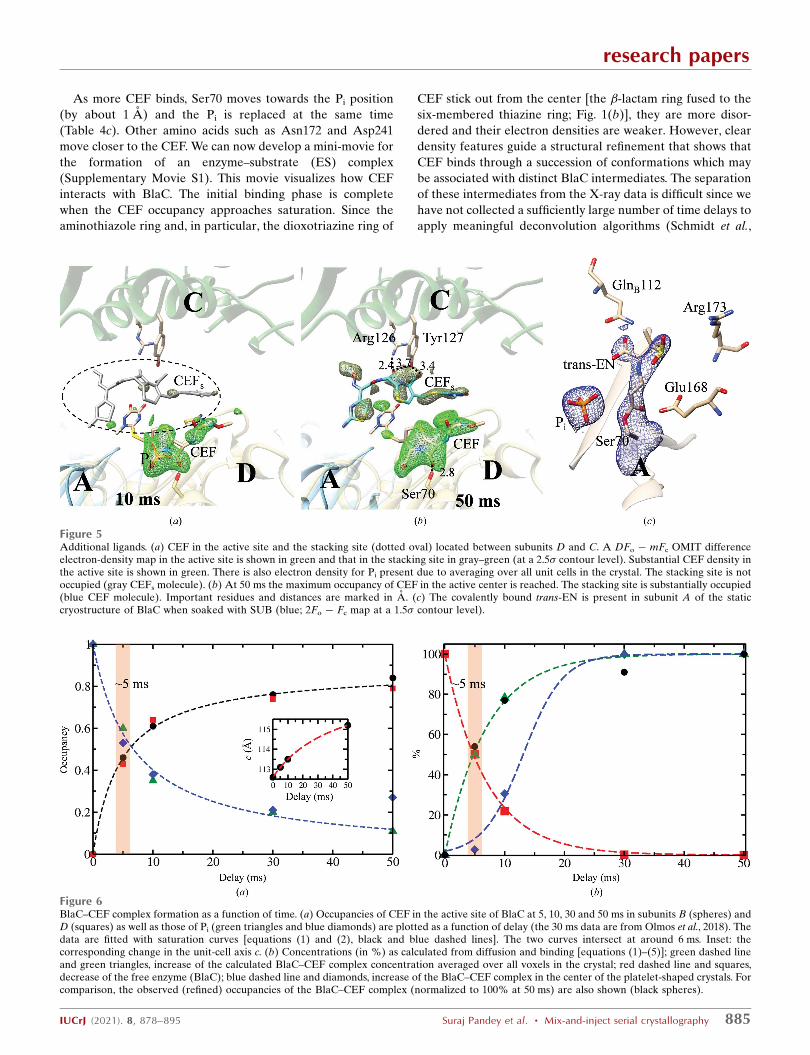
As more CEF binds, Ser70 moves towards the Pi position
(by about 1 A) and the Pi is replaced at the same time
(Table 4c). Other amino acids such as Asn172 and Asp241
move closer to the CEF. We can now develop a mini-movie for
the formation of an enzyme–substrate (ES) complex
(Supplementary Movie S1). This movie visualizes how CEF
interacts with BlaC. The initial binding phase is complete
when the CEF occupancy approaches saturation. Since the
aminothiazole ring and, in particular, the dioxotriazine ring of
CEF stick out from the center [the �-lactam ring fused to the
six-membered thiazine ring; Fig. 1(b)], they are more disor-
dered and their electron densities are weaker. However, clear
density features guide a structural refinement that shows that
CEF binds through a succession of conformations which may
be associated with distinct BlaC intermediates. The separation
of these intermediates from the X-ray data is difficult since we
have not collected a sufficiently large number of time delays to
apply meaningful deconvolution algorithms (Schmidt et al.,
research papers
IUCrJ (2021). 8, 878–895 Suraj Pandey et al. � Mix-and-inject serial crystallography 885
Figure 6BlaC–CEF complex formation as a function of time. (a) Occupancies of CEF in the active site of BlaC at 5, 10, 30 and 50 ms in subunits B (spheres) andD (squares) as well as those of Pi (green triangles and blue diamonds) are plotted as a function of delay (the 30 ms data are from Olmos et al., 2018). Thedata are fitted with saturation curves [equations (1) and (2), black and blue dashed lines]. The two curves intersect at around 6 ms. Inset: thecorresponding change in the unit-cell axis c. (b) Concentrations (in %) as calculated from diffusion and binding [equations (1)–(5)]; green dashed lineand green triangles, increase of the calculated BlaC–CEF complex concentration averaged over all voxels in the crystal; red dashed line and squares,decrease of the free enzyme (BlaC); blue dashed line and diamonds, increase of the BlaC–CEF complex in the center of the platelet-shaped crystals. Forcomparison, the observed (refined) occupancies of the BlaC–CEF complex (normalized to 100% at 50 ms) are also shown (black spheres).
Figure 5Additional ligands. (a) CEF in the active site and the stacking site (dotted oval) located between subunits D and C. A DFo � mFc OMIT differenceelectron-density map in the active site is shown in green and that in the stacking site in gray–green (at a 2.5� contour level). Substantial CEF density inthe active site is shown in green. There is also electron density for Pi present due to averaging over all unit cells in the crystal. The stacking site is notoccupied (gray CEFs molecule). (b) At 50 ms the maximum occupancy of CEF in the active center is reached. The stacking site is substantially occupied(blue CEF molecule). Important residues and distances are marked in A. (c) The covalently bound trans-EN is present in subunit A of the staticcryostructure of BlaC when soaked with SUB (blue; 2Fo � Fc map at a 1.5� contour level).

2003; Kostov & Moffat, 2011). The high X-ray pulse-repetition
rate of the EuXFEL may make this possible since it allows the
fast collection of data sets at tightly spaced delays. Given the
resolution of our X-ray data (Table 1), it is challenging to
make a distinction between ligand binding being supported by
conformational disorder (Tzeng & Kalodimos, 2012) or by
adaptation of the structure to a changing energy landscape,
which would resemble an ‘induced fit’ (Changeux & Edelstein,
2011). Both scenarios (Vogt & Di Cera, 2012) would most
likely result in the same (or a very similar) crystallographic
signal. We hypothesize that both mechanisms are involved to
some degree, which might be unraveled by single-particle
experiments, as recently demonstrated for an unrelated
biological system (Dashti et al., 2020). However, the structures
of BlaC as well as of CEF change [Table 4(c), Supplementary
Movie S1], which might be interpreted as the signature of an
induced fit after the initial binding event.
Formation of the ES complex is most important since it
triggers the enzymatic cycle. Hence, it determines the time
resolution of the MISC method. The ES complex consists of
CEF noncovalently bound in the active site of BlaC (Fig. 4).
CEF is delivered by diffusion into the crystals. The crystals
must be small enough to enable short enough diffusion times
so that the binding kinetics can be observed. However, MISC
does not measure the free substrate
concentration in the crystals, and
therefore diffusion is rather observed
indirectly through the increase in the
occupancies of well ordered substrate
molecules in the active centers of BlaC.
When the diffusion times are very short,
occupancies may accumulate on a
timescale longer than the diffusion time,
as they are governed by the binding
kinetics. The formation of the ES
complex, and therefore the time reso-
lution of the MISC method, is therefore
not only dependent on the ligand
concentration delivered by diffusion but
also on the magnitude of the rate coef-
ficients that characterize the kinetic
mechanism.
3.2. Inhibitor binding
The structure of the BlaC–SUB
complex was probed at �tm = 66 ms.
Strong positive DEDiso shows SUB
binding to all four subunits of BlaC,
which is in stark contrast to CEF, which
only binds to subunits B and D. The
absence of negative DEDiso at the Pi
position [Figs. 7(a) and 7(b)] shows that
the Pi does not move and stays in the
active site. At the time delay of 66 ms
sulbactam binding to BlaC is hetero-
geneous. In subunits B and D the
DEDiso is elongated and stretches outwards from Ser70
[Fig. 7(b)]. In subunits A and C the DEDiso is pillow-like and is
more distant from Ser70 [Fig. 7(a)]. The elongated DEDiso in
subunits B and D [Fig. 7(b)] can be explained by a covalently
bound trans-EN as a result of the reaction of the sulbactam
with the catalytic Ser70. The diffusion time is fast enough that
66 ms after mixing all B and D subunits contain trans-EN, the
position of which is stabilized by a network of BlaC residues
[Fig. 7 and Table 4(b)]. This is quite unexpected, as it was
suggested that it would take minutes for the enamine to form
after binding of SUB to BlaC (Totir et al., 2007; Cheng et al.,
2020; Tassoni et al., 2019). In subunits B and D Arg173 displays
a stretched, open conformation, allowing the SUB to orient
correctly towards Ser70 and to react swiftly to the trans-EN,
which then irreversibly inhibits BlaC (Tassoni et al., 2019). The
nearby Pi, which is displaced when the much larger ligand CEF
is present, stays in place in all subunits and is likely to add to
the stability of both complexes.
The pillow-like DEDiso in a region more distant from Ser70
in subunits A and C [Fig. 7(a)] can be explained by an intact
sulbactam molecule that is noncovalently bound to the active
site. The SUB is oriented so that the ring sulfur dioxide points
towards Ser70, with the �-lactam ring pointing away from
Ser70. We hypothesize that this ‘upside-down’ orientation is
research papers
886 Suraj Pandey et al. � Mix-and-inject serial crystallography IUCrJ (2021). 8, 878–895
Table 4Important distances in the active centers of BlaC.
s, strong hydrogen bond up to 2.5 A; h, hydrogen bond from 2.6 to 3.2 A; w, weak interaction >3.2 A(Jeffrey, 1997).
(a) Distances in (A) in subunits A and C to the sulbactam (0RN).
Subunit A Subunit C
Ser70 OG to Pi O 3.2 (h) 2.6 (h)Ser70 OG to 0RN OAF 3.1 (h) 3.2 (h)Gln112 OE1 to 0RN OX Gln from B, 2.8 (h) Gln from D, 3.3 (w)Asn172 ND2 to 0RN OAF 3.2 (h) 3.1 (h)Glu168 O2 to 0RN OAF 3.0 (h) 2.8 (h)Arg173 NH1 to 0RN O 2.5 (s) 3.9 (w)Thr239 O to 0RN OAO 3.9 (w) 2.9 (h)
(b) Distances in (A) in subunits B and D to the Ser70 trans-enamine (TSS).
Subunit B Subunit D
Lys73 NZ to TSS O13 2.8 (h) 3.0 (h)Gln109 OE1 to TSS O12 Gln from A, 2.7 (h) Gln from C, 2.9 (h)Thr239 O to TSS 08 2.9 (h) 2.9 (h)Asp241 OD2 to TSS O11 6.4 4.1
(c) Distances in (A) during ceftriaxone binding (CEF is only bound to subunits B and D).
Subunit B Subunit D
0 ms 5 ms 10 ms 50 ms 0 ms 5 ms 10 ms 50 ms
Ser70 OG to H2O 3.1 (h) 2.8 (h) 3.3 (w) 2.8 (h) 3.5 (w) 2.4† (s) 3.1 (h) 3.2 (h)Ser70 OG to Pi O4 3.6 (w) 3.5 (w) 3.4 (w) 2.6 (s) 2.7 (h) 3.7 (w) 3.9 (w) 2.7 (h)Ser70 OG to CEF O na‡ 3.1 (h) 2.9 (h) 2.9 (h) na 2.9 (h) 3.0 (h) 2.8 (h)Ser128 OG to CEF OAD na 2.4 (s) 2.4 (s) 2.5 (s) na 2.3 (s) 2.4 (s) 2.6 (h)Asn172 ND2 to CEF OAR na 2.7 (h) 3.1 (h) 3.2 (h) na 2.8 (h) 3.0 (h) 3.1 (h)Thr237 OG1 to CEF OA1 na 2.7 (h) 3.1 (h) 3.0 (h) na 2.7 (h) 2.6 (h) 3.1 (h)Thr239 OG2 to CEF OA1 na 3.3 (w) 3.0 (h) 3.0 (h) na 3.4 (w) 3.4 (w) 3.1 (h)Asp241 OD1 to CEF NAC na 4.7 4.3 3.2 (h) na 4.4 4.2 3.6 (w)
† Weak OMIT difference electron density. ‡ Not applicable.
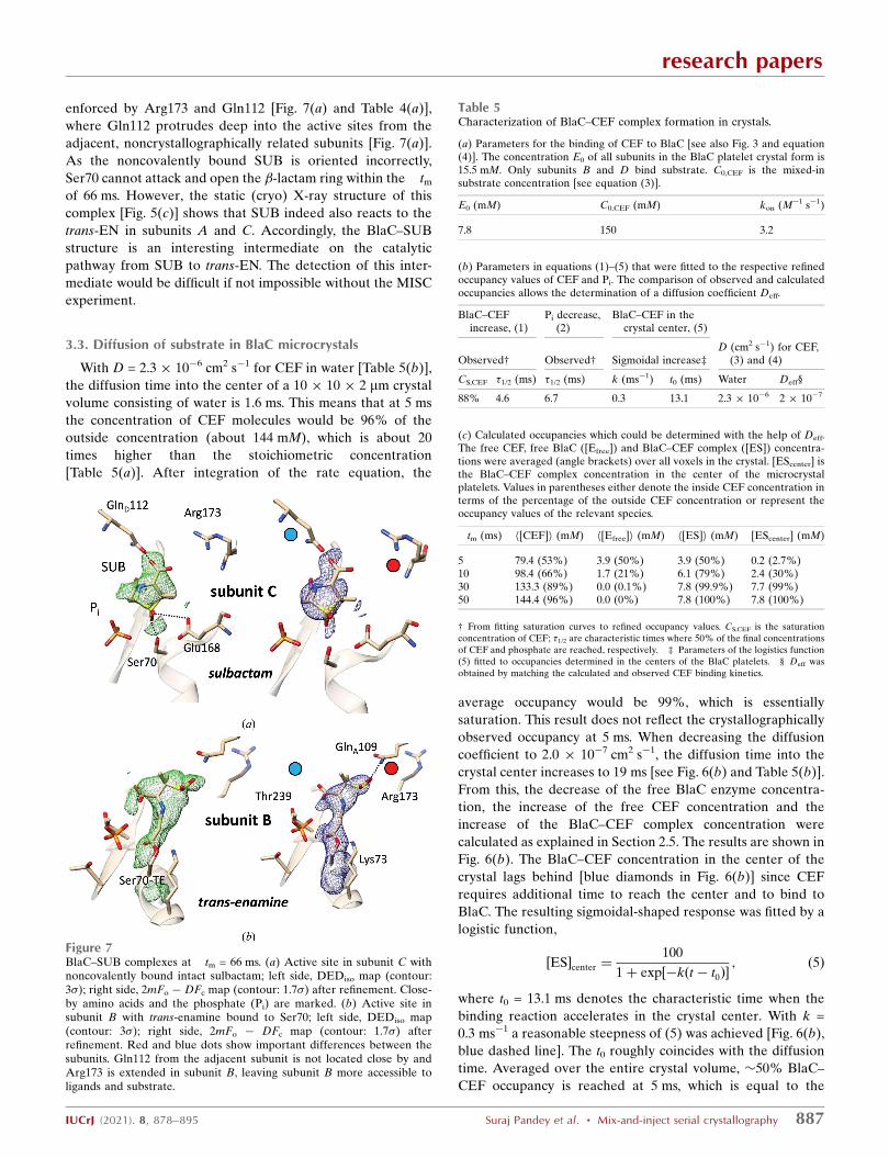
enforced by Arg173 and Gln112 [Fig. 7(a) and Table 4(a)],
where Gln112 protrudes deep into the active sites from the
adjacent, noncrystallographically related subunits [Fig. 7(a)].
As the noncovalently bound SUB is oriented incorrectly,
Ser70 cannot attack and open the �-lactam ring within the �tmof 66 ms. However, the static (cryo) X-ray structure of this
complex [Fig. 5(c)] shows that SUB indeed also reacts to the
trans-EN in subunits A and C. Accordingly, the BlaC–SUB
structure is an interesting intermediate on the catalytic
pathway from SUB to trans-EN. The detection of this inter-
mediate would be difficult if not impossible without the MISC
experiment.
3.3. Diffusion of substrate in BlaC microcrystals
With D = 2.3 � 10�6 cm2 s�1 for CEF in water [Table 5(b)],
the diffusion time into the center of a 10 � 10 � 2 mm crystal
volume consisting of water is 1.6 ms. This means that at 5 ms
the concentration of CEF molecules would be 96% of the
outside concentration (about 144 mM), which is about 20
times higher than the stoichiometric concentration
[Table 5(a)]. After integration of the rate equation, the
average occupancy would be 99%, which is essentially
saturation. This result does not reflect the crystallographically
observed occupancy at 5 ms. When decreasing the diffusion
coefficient to 2.0 � 10�7 cm2 s�1, the diffusion time into the
crystal center increases to 19 ms [see Fig. 6(b) and Table 5(b)].
From this, the decrease of the free BlaC enzyme concentra-
tion, the increase of the free CEF concentration and the
increase of the BlaC–CEF complex concentration were
calculated as explained in Section 2.5. The results are shown in
Fig. 6(b). The BlaC–CEF concentration in the center of the
crystal lags behind [blue diamonds in Fig. 6(b)] since CEF
requires additional time to reach the center and to bind to
BlaC. The resulting sigmoidal-shaped response was fitted by a
logistic function,
½ES�center ¼100
1þ exp½�kðt � t0Þ�; ð5Þ
where t0 = 13.1 ms denotes the characteristic time when the
binding reaction accelerates in the crystal center. With k =
0.3 ms�1 a reasonable steepness of (5) was achieved [Fig. 6(b),
blue dashed line]. The t0 roughly coincides with the diffusion
time. Averaged over the entire crystal volume, �50% BlaC–
CEF occupancy is reached at 5 ms, which is equal to the
research papers
IUCrJ (2021). 8, 878–895 Suraj Pandey et al. � Mix-and-inject serial crystallography 887
Figure 7BlaC–SUB complexes at �tm = 66 ms. (a) Active site in subunit C withnoncovalently bound intact sulbactam; left side, DEDiso map (contour:3�); right side, 2mFo � DFc map (contour: 1.7�) after refinement. Close-by amino acids and the phosphate (Pi) are marked. (b) Active site insubunit B with trans-enamine bound to Ser70; left side, DEDiso map(contour: 3�); right side, 2mFo � DFc map (contour: 1.7�) afterrefinement. Red and blue dots show important differences between thesubunits. Gln112 from the adjacent subunit is not located close by andArg173 is extended in subunit B, leaving subunit B more accessible toligands and substrate.
Table 5Characterization of BlaC–CEF complex formation in crystals.
(a) Parameters for the binding of CEF to BlaC [see also Fig. 3 and equation(4)]. The concentration E0 of all subunits in the BlaC platelet crystal form is15.5 mM. Only subunits B and D bind substrate. C0,CEF is the mixed-insubstrate concentration [see equation (3)].
E0 (mM) C0,CEF (mM) kon (M�1 s�1)
7.8 150 3.2
(b) Parameters in equations (1)–(5) that were fitted to the respective refinedoccupancy values of CEF and Pi. The comparison of observed and calculatedoccupancies allows the determination of a diffusion coefficient Deff.
BlaC–CEFincrease, (1)
Pi decrease,(2)
BlaC–CEF in thecrystal center, (5)
Observed† Observed† Sigmoidal increase‡D (cm2 s�1) for CEF,
(3) and (4)
CS,CEF �1/2 (ms) �1/2 (ms) k (ms�1) t0 (ms) Water Deff§
88% 4.6 6.7 0.3 13.1 2.3 � 10�6 2 � 10�7
(c) Calculated occupancies which could be determined with the help of Deff.The free CEF, free BlaC ([Efree]) and BlaC–CEF complex ([ES]) concentra-tions were averaged (angle brackets) over all voxels in the crystal. [EScenter] isthe BlaC–CEF complex concentration in the center of the microcrystalplatelets. Values in parentheses either denote the inside CEF concentration interms of the percentage of the outside CEF concentration or represent theoccupancy values of the relevant species.
�tm (ms) h[CEF]i (mM) h[Efree]i (mM) h[ES]i (mM) [EScenter] (mM)
5 79.4 (53%) 3.9 (50%) 3.9 (50%) 0.2 (2.7%)10 98.4 (66%) 1.7 (21%) 6.1 (79%) 2.4 (30%)30 133.3 (89%) 0.0 (0.1%) 7.8 (99.9%) 7.7 (99%)50 144.4 (96%) 0.0 (0%) 7.8 (100%) 7.8 (100%)
† From fitting saturation curves to refined occupancy values. CS,CEF is the saturationconcentration of CEF; �1/2 are characteristic times where 50% of the final concentrationsof CEF and phosphate are reached, respectively. ‡ Parameters of the logistics function(5) fitted to occupancies determined in the centers of the BlaC platelets. § Deff wasobtained by matching the calculated and observed CEF binding kinetics.

occupancy determined experimentally at �tm = 5 ms [compare
the green dashed line in Fig. 6(b) with Fig. 6(a)]. Fig. 8 shows a
heatmap that plots BlaC–CEF occupancies through the center
of half a BlaC crystal (see also Fig. 9 for a 3D representation).
The concentrations of the BlaC–CEF complex are depicted
with various colors (see the scale bar on the right). At the edge
of the crystal, almost all active BlaC subunits (B and D) are
already bound to CEF at 5 ms. In the center the BlaC–CEF
complex concentration is 0.21 mM (2.7% of the total
concentration of B and D subunits in the crystal), although the
CEF concentration delivered by diffusion is already 35 mM
(which is the mentioned 23% of the outside CEF concentra-
tion but is 5.5 times the stoichiometric concentration). The
situation changes completely at 30 ms, where almost 100%
occupancy is reached everywhere in the crystal, which is in
accordance with earlier results (Olmos et al., 2018) and with
the occupancy at �tm = 50 ms reported here (see also
Supplementary Movie S2). As also
discussed earlier, the variation of occu-
pancy across microcrystals at faster mix-
and-inject delays does not affect the
enzyme kinetics, as the nucleophilic
attack of Ser70 of BlaC on the �-lactam
ring happens long after the crystals
establish full CEF occupancy. It needs to
be pointed out here that the simple
model of CEF diffusion into BlaC
microcrystals and the binding of CEF to
BlaC molecules can be augmented by
taking into account, for example, the
exclusion volume occupied by reacting
and nonreacting BlaC subunits
(Geremia et al., 2006), by the mentioned
pointwise depletion of the free CEF
concentration in each voxel by binding
to BlaC active centers, or the diffusion
of substrate directly through protein
molecules facilitated by protein
dynamics. Since the exact mechanism of
diffusion through protein crystals
(Geremia et al., 2006) is difficult to
determine, the unknown parameters are tied up in the effec-
tive diffusion coefficient (Deff) determined here.
3.4. Reaction initiation by diffusion
CEF diffusion is about a factor of 12 slower in the BlaC
crystals than in water, with a Deff of about �2 � 10�7 cm2 s�1
[Table 5(b)]. This slowdown is in agreement with findings that
were previously obtained from simulations on substrate
diffusion in enzyme crystals (Geremia et al., 2006). Estimates
of enzyme–ligand occupancies can now be directly deduced
from time-resolved X-ray crystallography everywhere in a
crystal after mixing. Not surprisingly, at 5 ms the occupancy is
high (>90%) only near the crystal surface [Fig. 8(a)], where
sufficient substrate is present to promote ES formation at a
high rate. In the center of the crystals the ES complex
concentration is initially small [Table 5(c), Figs. 8(a) and 9(a)].
The binding rate is not sufficiently high to generate significant
occupancy. After �tm = 10 ms the binding rate increases, until
at 30 ms full occupancy of the BlaC–CEF complex is reached
everywhere (Olmos et al., 2018) [Fig. 6(b), green dashed line,
Table 5(c)]. With the rapid diffusion of CEF into small BlaC
crystals, we are now able to quantify variations of substrate,
enzyme and ES concentrations across the enzyme crystal
volume at any time [Table 5(c), Figs. 8 and 9]. The remarkable
speed of ES accumulation shows that the mix-and-inject
technique can be used to characterize enzymes with turnover
times much faster than that of BlaC. The direct observation of
the important initial ligand- and substrate-binding phase in
biomedically relevant enzymes is possible at the EuXFEL.
Since the ES complex (here the BlaC–CEF complex) trig-
gers the enzymatic cycle, accurate kinetics can be extracted to
the point where the time required to accumulate sufficient ES
research papers
888 Suraj Pandey et al. � Mix-and-inject serial crystallography IUCrJ (2021). 8, 878–895
Figure 8Concentrations of the BlaC–CEF complex in 10 � 10 � 2 mm platelet-shaped crystals (a) 5 ms, (b)10 ms and (c) 30 ms after mixing with 200 mM ceftriaxone (150 mM final concentration assumed).The concentrations are shown in different colors (see the scale bar on the right) in central crosssections through half the width of the crystals. The drawings are not to scale, since the sectionsdisplayed are 5 mm horizontally (width) and 2 mm vertically (thickness). The enlargement along theshort 2 mm axis allows the display of the nuanced occupancy differences.
Figure 93D representation of CEF occupancy values in the BlaC catalytic cleft5 ms (a) and 10 ms (b) after mixing in a typical BlaC microcrystal platelet.Dark blue colors denote low occupancies and lighter hues denote highoccupancies.

complex approaches the lifetime of the next intermediate in
the catalytic cycle (Schmidt, 2013). This finding holds for any
other technique (Srajer & Schmidt, 2017) which aims to
trigger enzymatic reactions, even in noncrystalline samples.
Not only is it required to bring sufficient substrate into the
vicinity of the enzyme, but the binding kinetics also need to be
taken into account. With microcrystals below a certain crystal
size, the binding of substrate, and not the diffusion of substrate
into the crystal volume, may become rate-limiting. As a
consequence, for BlaC crystals of a size of about 1 mm the
speed of ES complex formation is not substantially different
from that in solution. The Deff determined here suggests that
accurate measurements of the substrate-binding kinetics
would not be possible with significantly larger crystals.
Enzymes with turnover times faster than that of BlaC will
usually also display faster substrate-binding kinetics with
larger kon rate coefficients. In such cases, the crystal sizes (and
their size distributions) or perhaps the temperature must be
adjusted appropriately to ensure that the diffusion times can
catch up with the substrate-binding rates.
Diffusion is an effective way to initiate reactions. Given a
sufficient substrate concentration, and appropriately small
crystals, all of the crystal volume is already infused with a
multiple of the stoichiometric substrate concentration after a
few milliseconds [Table 5(c)]. This is very important for fast
reaction initiation since the rate (speed) of enzyme–substrate
complex formation, and therefore the time resolution of the
method, depends decisively, and primarily, on the concentra-
tion of the substrate, and of course also on the free enzyme
concentration and the kinetic rate coefficients. Others
(Mehrabi, Schulz, Dsouza et al., 2019) have reported signifi-
cant substrate occupancy in the active site only 30 ms after the
activation of a caged substrate that is even located close by.
This slow occupancy increase may be a result of (sub-)stoi-
chiometric substrate concentrations in the unit cell, which
strengthens our point of view. BlaC is not a fast enzyme. Apart
from the possibility of investigating the initial substrate-
binding phase(s) potentially on submillisecond timescales, the
benefits of XFEL-based mix-and-inject approaches may come
to light once faster enzymes with turnover times of <50 ms are
investigated. These experiments require small crystals.
Exploration of how to investigate these small crystals using
either XFELs or synchrotrons, perhaps after upgrade to an
advanced accelerator lattice (Eriksson, 2016; Wanzenberg et
al., 2019), remains to be performed.
4. Outlook
In order to further investigate CEF and SUB binding and their
reactions with Ser70, a time series should be collected that
consists of data sets at multiple �tm values that span from a
few milliseconds to seconds. To achieve this, the EuXFEL
pulse structure must be exploited most efficiently. Every X-ray
pulse in all pulse trains provides observations of the same time
delay, and our experiments took maximum advantage of the
high pulse rate (Fig. 2, inset). This is in contrast to optical
pump–X-ray probe experiments, which require appropriate
waiting times between laser excitations to guarantee that the
laser-excited volume exits the X-ray interaction region, so that
multiple laser activations can be avoided (Pandey et al., 2020).
We showed that diffraction data sufficient for good-quality
structure determination can be collected in about half an hour,
as demonstrated for the 50 ms CEF time point. This time can
be reduced substantially by limiting the number of diffraction
patterns per data set (around 25 000 is appropriate for this
space group; Olmos et al., 2018) and by optimizing the crystal
density that flows through the mixing device. High crystal
density will lead to higher hit rates, but might also cause
frequent interruptions caused by injector clogging. In our
experiments, a fine balance between crystal size and crystal
density was found so that the mix-and-inject experiments with
CEF and SUB could be completed successfully with accep-
table hit rates (Table 1) given the high X-ray pulse-repetition
rate at the EuXFEL. Previous experiments have shown that
research papers
IUCrJ (2021). 8, 878–895 Suraj Pandey et al. � Mix-and-inject serial crystallography 889
Figure 10Crystals, hit and indexing rates. (a) Microscopic image of the platelet crystal form of BlaC and (b) exemplary hit (red squares) and indexing (blackspheres) rates from BlaC/CEF mixing as a function of the pulse ID in the train. Both rates are stable across the entire pulse train.
the collection of sufficient patterns for structure determination
should be possible in less than 20 min at the detector-limited
repetition rate of the EuXFEL (Yefanov et al., 2019; Pandey et
al., 2020). This provides the tantalizing possibility of directly
characterizing kinetic processes in biomolecules from single-
digit millisecond to longer timescales within relatively short
experimental times. The kinetics can rapidly change when
environmental conditions are varied. It may be possible, for
example, to control the temperature in the mixing injector
delay line to determine barriers of activation from the
resulting X-ray data (Schmidt et al., 2013). The full analysis of
such a multidimensional data set requires the development
and deployment of user-friendly classification algorithms to
separate mixtures into their pure components (Schmidt et al.,
2003) and to derive kinetics and energetics (Schmidt et al.,
2013) consistent with the electron-density maps and the
structures of intermediate states along the reaction pathway.
5. Summary
Our experiments permitted a real-time view into the active
sites of an enzyme during substrate binding. They facilitate
more mix-and-inject experiments at the EuXFEL with
unprecedented data-collection rates, allowing more structures
to be determined per allocated experimental time. This
capability will become an important tool for biomedically
relevant research in years to come.
APPENDIX AMicrocrystals and data collection
Fig. 10(a) shows a microscopic image of the BlaC platelets
used for the experiments. They appear to be uniform in size
and shape. In Fig. 10(b) the hit and indexing rates achieved
with these crystals in our MISC experiments are displayed
across all of the pulses in the pulse train of the EuXFEL. A
decay in the hit rate following pulse 1 is not observed. This is
an indication that the jet is fast enough that a fresh jet volume
is intercepted by the following X-ray pulses (Wiedorn, Ober-
thur et al., 2018; Yefanov et al., 2019; Pandey et al., 2020).
APPENDIX BDiffraction patterns and masks
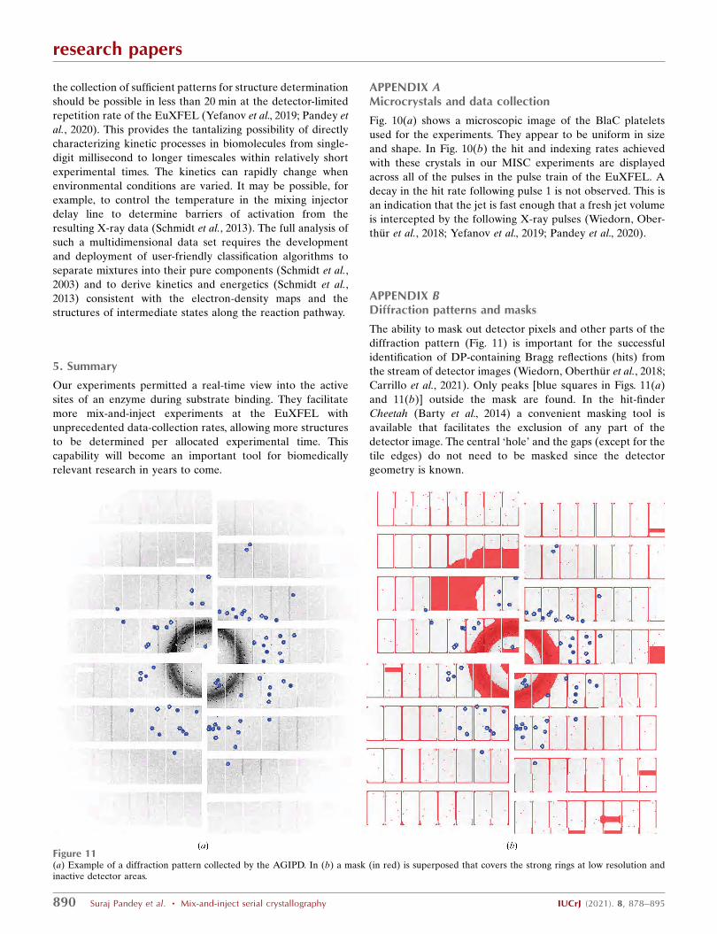
The ability to mask out detector pixels and other parts of the
diffraction pattern (Fig. 11) is important for the successful
identification of DP-containing Bragg reflections (hits) from
the stream of detector images (Wiedorn, Oberthur et al., 2018;
Carrillo et al., 2021). Only peaks [blue squares in Figs. 11(a)
and 11(b)] outside the mask are found. In the hit-finder
Cheetah (Barty et al., 2014) a convenient masking tool is
available that facilitates the exclusion of any part of the
detector image. The central ‘hole’ and the gaps (except for the
tile edges) do not need to be masked since the detector
geometry is known.
research papers
890 Suraj Pandey et al. � Mix-and-inject serial crystallography IUCrJ (2021). 8, 878–895
Figure 11(a) Example of a diffraction pattern collected by the AGIPD. In (b) a mask (in red) is superposed that covers the strong rings at low resolution andinactive detector areas.
Acknowledgements
We are very grateful to those who supported this experiment
by being present in person at the European XFEL during the
onset of the COVID-19 pandemic in March 2020. We
acknowledge the European XFEL in Schenefeld, Germany
for the provision of X-ray free-electron laser beamtime on the
SPB/SFX scientific instrument and would like to thank the
staff for their assistance. We acknowledge the use of the XBI
facility at the European XFEL. The SFX stream files were
deposited in the CXIDB with code 170. Individual contribu-
tions were as follows. SP, TM, JM-G, JH-Y, FK, IP, MM, RJ,
MS, WX and JO expressed, purified and crystallized the
protein. RB, KD, HK, MK, JK, GM, TS and MV operated the
SPB/SFX instrument. LP, AMK, GC and KAZ designed and
provided injector nozzles. MV, FK and MK assembled and
operated the nozzles. FK, JM-G, J-HY, LG and PS collected
the data. SP, IP, OY, VM, PS, AT and AB processed the data.
SP, IP, TM, GP and MS analyzed the data. IP, MF, AS, FK, PS
and MS logged the experiment. RB, APM, MS and GP
designed the experiment. SP, PF, APM, RB, GP and MS wrote
the manuscript with input from all other authors.
Funding information
This work was supported by the National Science Foundation
Science and Technology Center ‘BioXFEL’ through award
STC-1231306, and in part by the US Department of Energy,
Office of Science, Basic Energy Sciences under contract DE-
SC0002164 (AO, algorithm design and development) and by
the National Science Foundation under contract Nos. 1551489
(AO, underlying analytical models) and DBI-2029533 (AO,
functional conformations). This material is based upon work
supported by the National Science Foundation Graduate
Research Fellowship Program under Grant No. 1450681 to
JLO. The work was also supported by funds from the National
Institutes of Health grant R01 GM117342-0404. Funding and
support are also acknowledged from the National Institutes of
Health grant R01 GM095583, from the Biodesign Center for
Applied Structural Discovery at ASU, from National Science
Foundation award No. 1565180 and the US Department of
Energy through Lawrence Livermore National Laboratory
under contract DE-AC52-07NA27344. KAZ was supported by
the Cornell Molecular Biophysics Training Program (NIH
T32-GM008267). This work was also supported by the Cluster
of Excellence ‘CUI: Advanced Imaging of Matter’ of the
Deutsche Forschungsgemeinschaft (DFG), EXC 2056, project
ID 390715994. CFEL is supported by the Gottfried Wilhelm
Leibniz Program of the DFG, the ‘X-probe’ project funded by
the European Union 2020 Research and Innovation Program
under Marie Sklodowska-Curie grant agreement 637295, the
European Research Council, ‘Frontiers in Attosecond X-ray
Science: Imaging and Spectroscopy (AXSIS)’, ERC-2013-SyG
609920, and the Human Frontiers Science Program grant
RGP0010 2017. This work is also supported by the AXSIS
project funded by the European Research Council under the
European Union Seventh Framework Program (FP/2007-
2013)/ERC Grant Agreement No. 609920.
References
Allahgholi, A., Becker, J., Delfs, A., Dinapoli, R., Goettlicher, P.,Greiffenberg, D., Henrich, B., Hirsemann, H., Kuhn, M., Klanner,R., Klyuev, A., Krueger, H., Lange, S., Laurus, T., Marras, A.,Mezza, D., Mozzanica, A., Niemann, M., Poehlsen, J., Schwandt, J.,Sheviakov, I., Shi, X., Smoljanin, S., Steffen, L., Sztuk-Dambietz, J.,Trunk, U., Xia, Q., Zeribi, M., Zhang, J., Zimmer, M., Schmitt, B. &Graafsma, H. (2019). J. Synchrotron Rad. 26, 74–82.
Barends, T. R. M., Foucar, L., Ardevol, A., Nass, K., Aquila, A.,Botha, S., Doak, R. B., Falahati, K., Hartmann, E., Hilpert, M.,Heinz, M., Hoffmann, M. C., Kofinger, J., Koglin, J. E., Kovacsova,G., Liang, M., Milathianaki, D., Lemke, H. T., Reinstein, J., Roome,C. M., Shoeman, R. L., Williams, G. J., Burghardt, I., Hummer, G.,Boutet, S. & Schlichting, I. (2015). Science, 350, 445–450.
Barty, A., Kirian, R. A., Maia, F. R. N. C., Hantke, M., Yoon, C. H.,White, T. A. & Chapman, H. (2014). J. Appl. Cryst. 47, 1118–1131.
Berman, H. M., Battistuz, T., Bhat, T. N., Bluhm, W. F., Bourne, P. E.,Burkhardt, K., Feng, Z., Gilliland, G. L., Iype, L., Jain, S., Fagan, P.,Marvin, J., Padilla, D., Ravichandran, V., Schneider, B., Thanki, N.,Weissig, H., Westbrook, J. D. & Zardecki, C. (2002). Acta Cryst.D58, 899–907.
Boutet, S., Lomb, L., Williams, G. J., Barends, T. R., Aquila, A., Doak,R. B., Weierstall, U., DePonte, D. P., Steinbrener, J., Shoeman,R. L., Messerschmidt, M., Barty, A., White, T. A., Kassemeyer, S.,Kirian, R. A., Seibert, M. M., Montanez, P. A., Kenney, C., Herbst,R., Hart, P., Pines, J., Haller, G., Gruner, S. M., Philipp, H. T., Tate,M. W., Hromalik, M., Koerner, L. J., van Bakel, N., Morse, J.,Ghonsalves, W., Arnlund, D., Bogan, M. J., Caleman, C., Fromme,R., Hampton, C. Y., Hunter, M. S., Johansson, L. C., Katona, G.,Kupitz, C., Liang, M., Martin, A. V., Nass, K., Redecke, L., Stellato,F., Timneanu, N., Wang, D., Zatsepin, N. A., Schafer, D., Defever, J.,Neutze, R., Fromme, P., Spence, J. C. H., Chapman, H. N. &Schlichting, I. (2012). Science, 337, 362–364.
Boyd, D. B. & Lunn, W. H. (1979). J. Med. Chem. 22, 778–784.Calvey, G. D., Katz, A. M. & Pollack, L. (2019). Anal. Chem. 91, 7139–
7144.Carrillo, M., Pandey, S., Sanchez, J., Noda, M., Poudyal, I., Aldama,
L., Malla, T. N., Claesson, E., Wahlgren, W. Y., Feliz, D., Srajer, V.,Maj, M., Castillon, L., Iwata, S., Nango, E., Tanaka, R., Tanaka, T.,Fangjia, L., Tono, K., Owada, S., Westenhoff, S., Stojkovic, E. A. &Schmidt, M. (2021). Structure, 29, 743–754.
Carslaw, H. S. & Jaeger, J. C. (1959). Conduction Heat in Solids, 2nded. Oxford: Clarendon Press.
Cassini, A., Hogberg, L. D., Plachouras, D., Quattrocchi, A., Hoxha,A., Simonsen, G. S., Colomb-Cotinat, M., Kretzschmar, M. E.,Devleesschauwer, B., Cecchini, M., Ouakrim, D. A., Oliveira, T. C.,Struelens, M. J., Suetens, C., Monnet, D. L., Strauss, R., Mertens, K.,Struyf, T., Catry, B., Latour, K., Ivanov, I. N., Dobreva, E. G.,Tambic-Andrasevic, A., Soprek, S., Budimir, A., Paphitou, N.,Zemlickova, H., Schytte Olsen, S., Wolff Sonksen, U., Martin, P.,Ivanova, M., Lyytikainen, O., Jalava, J., Coignard, B., Eckmanns, T.,Abu Sin, M., Haller, S., Daikos, G. L., Gikas, A., Tsiodras, S.,Kontopidou, F., Toth, A., Hajdu, A., Guolaugsson, O., Kristinsson,K. G., Murchan, S., Burns, K., Pezzotti, P., Gagliotti, C., Dumpis, U.,Liuimiene, A., Perrin, M., Borg, M. A., de Greeff, S. C., Monen,J. C., Koek, M. B., Elstrøm, P., Zabicka, D., Deptula, A.,Hryniewicz, W., Canica, M., Nogueira, P. J., Fernandes, P. A.,Manageiro, V., Popescu, G. A., Serban, R. I., Schreterova, E.,Litvova, S., Stefkovicova, M., Kolman, J., Klavs, I., Korosec, A.,Aracil, B., Asensio, A., Perez-Vazquez, M., Billstrom, H., Larsson,S., Reilly, J. S., Johnson, A. & Hopkins, S. (2019). Lancet Infect. Dis.19, 56–66.
Changeux, J.-P. & Edelstein, S. (2011). F1000 Biol. Rep. 3, 19.Chapman, H. N., Fromme, P., Barty, A., White, T. A., Kirian, R. A.,
Aquila, A., Hunter, M. S., Schulz, J., DePonte, D. P., Weierstall, U.,Doak, R. B., Maia, F. R. N. C., Martin, A. V., Schlichting, I., Lomb,L., Coppola, N., Shoeman, R. L., Epp, S. W., Hartmann, R., Rolles,
research papers
IUCrJ (2021). 8, 878–895 Suraj Pandey et al. � Mix-and-inject serial crystallography 891

D., Rudenko, A., Foucar, L., Kimmel, N., Weidenspointner, G.,Holl, P., Liang, M., Barthelmess, M., Caleman, C., Boutet, S.,Bogan, M. J., Krzywinski, J., Bostedt, C., Bajt, S., Gumprecht, L.,Rudek, B., Erk, B., Schmidt, C., Homke, A., Reich, C., Pietschner,D., Struder, L., Hauser, G., Gorke, H., Ullrich, J., Herrmann, S.,Schaller, G., Schopper, F., Soltau, H., Kuhnel, K.-U., Messer-schmidt, M., Bozek, J. D., Hau-Riege, S. P., Frank, M., Hampton,C. Y., Sierra, R. G., Starodub, D., Williams, G. J., Hajdu, J.,Timneanu, N., Seibert, M. M., Andreasson, J., Rocker, A., Jonsson,O., Svenda, M., Stern, S., Nass, K., Andritschke, R., Schroter, C.-D.,Krasniqi, F., Bott, M., Schmidt, K. E., Wang, X., Grotjohann, I.,Holton, J. M., Barends, T. R. M., Neutze, R., Marchesini, S.,Fromme, R., Schorb, S., Rupp, D., Adolph, M., Gorkhover, T.,Andersson, I., Hirsemann, H., Potdevin, G., Graafsma, H., Nilsson,B. & Spence, J. C. H. (2011). Nature, 470, 73–77.
Cheng, Q., Xu, C., Chai, J., Zhang, R., Wai Chi Chan, E. & Chen, S.(2020). ACS Infect. Dis. 6, 577–587.
Coquelle, N., Sliwa, M., Woodhouse, J., Schiro, G., Adam, V., Aquila,A., Barends, T. R. M., Boutet, S., Byrdin, M., Carbajo, S., De laMora, E., Doak, R. B., Feliks, M., Fieschi, F., Foucar, L., Guillon, V.,Hilpert, M., Hunter, M. S., Jakobs, S., Koglin, J. E., Kovacsova, G.,Lane, T. J., Levy, B., Liang, M. N., Nass, K., Ridard, J., Robinson,J. S., Roome, C. M., Ruckebusch, C., Seaberg, M., Thepaut, M.,Cammarata, M., Demachy, I., Field, M., Shoeman, R. L., Bourgeois,D., Colletier, J.-P., Schlichting, I. & Weik, M. (2018). Nat. Chem. 10,31–37.
Dashti, A., Mashayekhi, G., Shekhar, M., Ben Hail, D., Salah, S.,Schwander, P., des Georges, A., Singharoy, A., Frank, J. &Ourmazd, A. (2020). Nat. Commun. 11, 4734.
Decking, W., Abeghyan, S., Abramian, P., Abramsky, A., Aguirre, A.,Albrecht, C., Alou, P., Altarelli, M., Altmann, P., Amyan, K.,Anashin, V., Apostolov, E., Appel, K., Auguste, D., Ayvazyan, V.,Baark, S., Babies, F., Baboi, N., Bak, P., Balandin, V., Baldinger, R.,Baranasic, B., Barbanotti, S., Belikov, O., Belokurov, V., Belova, L.,Belyakov, V., Berry, S., Bertucci, M., Beutner, B., Block, A.,Blocher, M., Bockmann, T., Bohm, C., Bohnert, M., Bondar, V.,Bondarchuk, E., Bonezzi, M., Borowiec, P., Bosch, C., Bosenberg,U., Bosotti, A., Bospflug, R., Bousonville, M., Boyd, E., Bozhko, Y.,Brand, A., Branlard, J., Briechle, S., Brinker, F., Brinker, S.,Brinkmann, R., Brockhauser, S., Brovko, O., Bruck, H., Brudgam,A., Butkowski, L., Buttner, T., Calero, J., Castro-Carballo, E.,Cattalanotto, G., Charrier, J., Chen, J., Cherepenko, A., Cheskidov,V., Chiodini, M., Chong, A., Choroba, S., Chorowski, M., Churanov,D., Cichalewski, W., Clausen, M., Clement, W., Cloue, C., Cobos,J. A., Coppola, N., Cunis, S., Czuba, K., Czwalinna, M., D’Almagne,B., Dammann, J., Danared, H., Wagner, A. D., Delfs, A., Delfs, T.,Dietrich, F., Dietrich, T., Dohlus, M., Dommach, M., Donat, A.,Dong, X., Doynikov, N., Dressel, M., Duda, M., Duda, P., Eckoldt,H., Ehsan, W., Eidam, J., Eints, F., Engling, C., Englisch, U.,Ermakov, A., Escherich, K., Eschke, J., Saldin, E., Faesing, M.,Fallou, A., Felber, M., Fenner, M., Fernandes, B., Fernandez, J. M.,Feuker, S., Filippakopoulos, K., Floettmann, K., Fogel, V., Fontaine,M., Frances, A., Martin, I. F., Freund, W., Freyermuth, T., Friedland,M., Frohlich, L., Fusetti, M., Fydrych, J., Gallas, A., Garcia, O.,Garcia-Tabares, L., Geloni, G., Gerasimova, N., Gerth, C., Gessler,P., Gharibyan, V., Gloor, M., Glowinkowski, J., Goessel, A.,Golebiewski, Z., Golubeva, N., Grabowski, W., Graeff, W.,Grebentsov, A., Grecki, M., Grevsmuehl, T., Gross, M., Grosse-Wortmann, U., Grunert, J., Grunewald, S., Grzegory, P., Feng, G.,Guler, H., Gusev, G., Gutierrez, J. L., Hagge, L., Hamberg, M.,Hanneken, R., Harms, E., Hartl, I., Hauberg, A., Hauf, S.,Hauschildt, J., Hauser, J., Havlicek, J., Hedqvist, A., Heidbrook,N., Hellberg, F., Henning, D., Hensler, O., Hermann, T., Hidvegi,A., Hierholzer, M., Hintz, H., Hoffmann, F., Hoffmann, M.,Hoffmann, M., Holler, Y., Huning, M., Ignatenko, A., Ilchen, M.,Iluk, A., Iversen, J., Iversen, J., Izquierdo, M., Jachmann, L., Jardon,N., Jastrow, U., Jensch, K., Jensen, J., Dotabek, M. J. O., Jidda, M.,Jin, H., Johansson, N., Jonas, R., Kaabi, W., Kaefer, D., Kammering,
R., Kapitza, H., Karabekyan, S., Karstensen, S., Kasprzak, K.,Katalev, V., Keese, D., Keil, B., Kholopov, M., Killenberger, M.,Kitaev, B., Klimchenko, Y., Klos, R., Knebel, L., Koch, A., Koepke,M., Kohler, S., Kohler, W., Kohlstrunk, N., Konopkova, Z.,Konstantinov, A., Kook, W., Koprek, W., Korfer, M., Korth, O.,Kosarev, A., Kosinski, K., Kostin, D., Kot, Y., Kotarba, A., Kozak,T., Kozak, V., Kramert, R., Krasilnikov, M., Krasnov, A., Krause, B.,Kravchuk, L., Krebs, O., Kretschmer, R., Kreutzkamp, J., Kroplin,O., Krzysik, K., Kube, G., Kuehn, H., Kujala, N., Kulikov, V.,Kuzminych, V., La Civita, D., Lacroix, M., Lamb, T., Lancetov, A.,Larsson, M., Le Pinvidic, D., Lederer, S., Lensch, T., Lenz, D.,Leuschner, A., Levenhagen, F., Li, Y., Liebing, J., Lilje, L., Limberg,T., Lipka, D., List, B., Liu, J., Liu, S., Lorbeer, B., Lorkiewicz, J., Lu,H. H., Ludwig, F., Machau, K., Maciocha, W., Madec, C., Magueur,C., Maiano, C., Maksimova, I., Malcher, K., Maltezopoulos, T.,Mamoshkina, E., Manschwetus, B., Marcellini, F., Marinkovic, G.,Martinez, T., Martirosyan, H., Maschmann, W., Maslov, M.,Matheisen, A., Mavric, U., Meissner, J., Meissner, K., Messer-schmidt, M., Meyners, N., Michalski, G., Michelato, P., Mildner, N.,Moe, M., Moglia, F., Mohr, C., Mohr, S., Moller, W., Mommerz, M.,Monaco, L., Montiel, C., Moretti, M., Morozov, I., Morozov, P.,Mross, D., Mueller, J., Muller, C., Muller, J., Muller, K., Munilla, J.,Munnich, A., Muratov, V., Napoly, O., Naser, B., Nefedov, N.,Neumann, R., Neumann, R., Ngada, N., Noelle, D., Obier, F.,Okunev, I., Oliver, J. A., Omet, M., Oppelt, A., Ottmar, A.,Oublaid, M., Pagani, C., Paparella, R., Paramonov, V., Peitzmann,C., Penning, J., Perus, A., Peters, F., Petersen, B., Petrov, A., Petrov,I., Pfeiffer, S., Pfluger, J., Philipp, S., Pienaud, Y., Pierini, P.,Pivovarov, S., Planas, M., Plawski, E., Pohl, M., Polinski, J., Popov,V., Prat, S., Prenting, J., Priebe, G., Pryschelski, H., Przygoda, K.,Pyata, E., Racky, B., Rathjen, A., Ratuschni, W., Regnaud-Campderros, S., Rehlich, K., Reschke, D., Robson, C., Roever, J.,Roggli, M., Rothenburg, J., Rusinski, E., Rybaniec, R., Sahling, H.,Salmani, M., Samoylova, L., Sanzone, D., Saretzki, F., Sawlanski,O., Schaffran, J., Schlarb, H., Schlosser, M., Schlott, V., Schmidt, C.,Schmidt-Foehre, F., Schmitz, M., Schmokel, M., Schnautz, T.,Schneidmiller, E., Scholz, M., Schoneburg, B., Schultze, J., Schulz,C., Schwarz, A., Sekutowicz, J., Sellmann, D., Semenov, E., Serkez,S., Sertore, D., Shehzad, N., Shemarykin, P., Shi, L., Sienkiewicz, M.,Sikora, D., Sikorski, M., Silenzi, A., Simon, C., Singer, W., Singer,X., Sinn, H., Sinram, K., Skvorodnev, N., Smirnow, P., Sommer, T.,Sorokin, A., Stadler, M., Steckel, M., Steffen, B., Steinhau-Kuhl, N.,Stephan, F., Stodulski, M., Stolper, M., Sulimov, A., Susen, R.,Swierblewski, J., Sydlo, C., Syresin, E., Sytchev, V., Szuba, J., Tesch,N., Thie, J., Thiebault, A., Tiedtke, K., Tischhauser, D., Tolkiehn, J.,Tomin, S., Tonisch, F., Toral, F., Torbin, I., Trapp, A., Treyer, D.,Trowitzsch, G., Trublet, T., Tschentscher, T., Ullrich, F., Vannoni,M., Varela, P., Varghese, G., Vashchenko, G., Vasic, M., Vazquez-Velez, C., Verguet, A., Vilcins-Czvitkovits, S., Villanueva, R.,Visentin, B., Viti, M., Vogel, E., Volobuev, E., Wagner, R., Walker,N., Wamsat, T., Weddig, H., Weichert, G., Weise, H., Wenndorf, R.,Werner, M., Wichmann, R., Wiebers, C., Wiencek, M., Wilksen, T.,Will, I., Winkelmann, L., Winkowski, M., Wittenburg, K., Witzig,A., Wlk, P., Wohlenberg, T., Wojciechowski, M., Wolff-Fabris, F.,Wrochna, G., Wrona, K., Yakopov, M., Yang, B., Yang, F., Yurkov,M., Zagorodnov, I., Zalden, P., Zavadtsev, A., Zavadtsev, D.,Zhirnov, A., Zhukov, A., Ziemann, V., Zolotov, A., Zolotukhina,N., Zummack, F. & Zybin, D. (2020). Nat. Photonics, 14, 391–397.
Dods, R., Bath, P., Morozov, D., Gagner, V. A., Arnlund, D., Luk,H. L., Kubel, J., Maj, M., Vallejos, A., Wickstrand, C., Bosman, R.,Beyerlein, K. R., Nelson, G., Liang, M. N., Milathianaki, D.,Robinson, J., Harimoorthy, R., Berntsen, P., Malmerberg, E.,Johansson, L., Andersson, R., Carbajo, S., Claesson, E., Conrad,C. E., Dahl, P., Hammarin, G., Hunter, M. S., Li, C. F., Lisova, S.,Royant, A., Safari, C., Sharma, A., Williams, G. J., Yefanov, O.,Westenhoff, S., Davidsson, J., DePonte, D. P., Boutet, S., Barty, A.,Katona, G., Groenhof, G., Branden, G. & Neutze, R. (2021). Nature,589, 310–314.
research papers
892 Suraj Pandey et al. � Mix-and-inject serial crystallography IUCrJ (2021). 8, 878–895

Eriksson, M. (2016). AIP Conf. Proc. 1741, 020001.Fair, R. J. & Tor, Y. (2014). Perspect. Medicin. Chem. 6, PMC.S14459.Geremia, S., Campagnolo, M., Demitri, N. & Johnson, L. N. (2006).
Structure, 14, 393–400.Gevorkov, Y., Yefanov, O., Barty, A., White, T. A., Mariani, V.,
Brehm, W., Tolstikova, A., Grigat, R.-R. & Chapman, H. N. (2019).Acta Cryst. A75, 694–704.
Gourinchas, G., Etzl, S., Gobl, C., Vide, U., Madl, T. & Winkler, A.(2017). Sci. Adv. 3, e1602498.
Grunbein, M. L., Gorel, A., Foucar, L., Carbajo, S., Colocho, W.,Gilevich, S., Hartmann, E., Hilpert, M., Hunter, M., Kloos, M.,Koglin, J. E., Lane, T. J., Lewandowski, J., Lutman, A., Nass, K.,Nass Kovacs, G., Roome, C. M., Sheppard, J., Shoeman, R. L.,Stricker, M., van Driel, T., Vetter, S., Doak, R. B., Boutet, S.,Aquila, A., Decker, F. J., Barends, T. R. M., Stan, C. A. &Schlichting, I. (2021). Nat. Commun. 12, 1672.
Han, H., Round, E., Schubert, R., Gul, Y., Makroczyova, J., Meza, D.,Heuser, P., Aepfelbacher, M., Barak, I., Betzel, C., Fromme, P.,Kursula, I., Nissen, P., Tereschenko, E., Schulz, J., Uetrecht, C.,Ulicny, J., Wilmanns, M., Hajdu, J., Lamzin, V. S. & Lorenzen, K.(2021). J. Appl. Cryst. 54, 7–21.
Hartmann, N., Helml, W., Galler, A., Bionta, M. R., Grunert, J. L.,Molodtsov, S., Ferguson, K. R., Schorb, S., Swiggers, M. L., Carron,S., Bostedt, C., Castagna, J., Bozek, J., Glownia, J. M., Kane, D. J.,Fry, A. R., White, W. E., Hauri, C. P., Feurer, T. & Coffee, R. N.(2014). Nat. Photonics, 8, 706–709.
Holtorf, H., Reinbothe, S., Reinbothe, C., Bereza, B. & Apel, K.(1995). Proc. Natl Acad. Sci. USA, 92, 3254–3258.
Hugonnet, J. E. & Blanchard, J. S. (2007). Biochemistry, 46, 11998–12004.
Imming, P., Sinning, C. & Meyer, A. (2006). Nat. Rev. Drug Discov. 5,821–834.
Ishigami, I., Lewis-Ballester, A., Echelmeier, A., Brehm, G.,Zatsepin, N. A., Grant, T. D., Coe, J. D., Lisova, S., Nelson, G.,Zhang, S., Dobson, Z. F., Boutet, S., Sierra, R. G., Batyuk, A.,Fromme, P., Fromme, R., Spence, J. C. H., Ros, A., Yeh, S. R. &Rousseau, D. L. (2019). Proc. Natl Acad. Sci. USA, 116, 3572–3577.
Jeffrey, G. A. (1997). An Introduction to Hydrogen Bonding. OxfordUniversity Press.
Kern, J., Chatterjee, R., Young, I. D., Fuller, F. D., Lassalle, L.,Ibrahim, M., Gul, S., Fransson, T., Brewster, A. S., Alonso-Mori, R.,Hussein, R., Zhang, M., Douthit, L., de Lichtenberg, C., Cheah,M. H., Shevela, D., Wersig, J., Seuffert, I., Sokaras, D., Pastor, E.,Weninger, C., Kroll, T., Sierra, R. G., Aller, P., Butryn, A., Orville,A. M., Liang, M., Batyuk, A., Koglin, J. E., Carbajo, S., Boutet, S.,Moriarty, N. W., Holton, J. M., Dobbek, H., Adams, P. D.,Bergmann, U., Sauter, N. K., Zouni, A., Messinger, J., Yano, J. &Yachandra, V. K. (2018). Nature, 563, 421–425.
Kostov, K. S. & Moffat, K. (2011). Biophys. J. 100, 440–449.Kupitz, C., Basu, S., Grotjohann, I., Fromme, R., Zatsepin, N. A.,
Rendek, K. N., Hunter, M. S., Shoeman, R. L., White, T. A., Wang,D., James, D., Yang, J.-H., Cobb, D. E., Reeder, B., Sierra, R. G.,Liu, H., Barty, A., Aquila, A. L., Deponte, D., Kirian, R. A., Bari,S., Bergkamp, J. J., Beyerlein, K. R., Bogan, M. J., Caleman, C.,Chao, T.-C., Conrad, C. E., Davis, K. M., Fleckenstein, H., Galli, L.,Hau-Riege, S. P., Kassemeyer, S., Laksmono, H., Liang, M., Lomb,L., Marchesini, S., Martin, A. V., Messerschmidt, M., Milathianaki,D., Nass, K., Ros, A., Roy-Chowdhury, S., Schmidt, K., Seibert, M.,Steinbrener, J., Stellato, F., Yan, L., Yoon, C., Moore, T. A., Moore,A. L., Pushkar, Y., Williams, G. J., Boutet, S., Doak, R. B.,Weierstall, U., Frank, M., Chapman, H. N., Spence, J. C. H. &Fromme, P. (2014). Nature, 513, 261–265.
Kupitz, C., Olmos, J. L. Jr, Holl, M., Tremblay, L., Pande, K., Pandey,S., Oberthur, D., Hunter, M., Liang, M., Aquila, A., Tenboer, J.,Calvey, G., Katz, A., Chen, Y., Wiedorn, M. O., Knoska, J., Meents,A., Majriani, V., Norwood, T., Poudyal, I., Grant, T., Miller, M. D.,Xu, W., Tolstikova, A., Morgan, A., Metz, M., Martin-Garcia, J. M.,Zook, J. D., Roy-Chowdhury, S., Coe, J., Nagaratnam, N., Meza, D.,
Fromme, R., Basu, S., Frank, M., White, T., Barty, A., Bajt, S.,Yefanov, O., Chapman, H. N., Zatsepin, N., Nelson, G., Weierstall,U., Spence, J., Schwander, P., Pollack, L., Fromme, P., Ourmazd, A.,Phillips, G. N. Jr & Schmidt, M. (2017). Struct. Dyn. 4, 044003.
Li, J., Liu, Z., Tan, C., Guo, X., Wang, L., Sancar, A. & Zhong, D.(2010). Nature, 466, 887–890.
Liebschner, D., Afonine, P. V., Baker, M. L., Bunkoczi, G., Chen,V. B., Croll, T. I., Hintze, B., Hung, L.-W., Jain, S., McCoy, A. J.,Moriarty, N. W., Oeffner, R. D., Poon, B. K., Prisant, M. G., Read,R. J., Richardson, J. S., Richardson, D. C., Sammito, M. D., Sobolev,O. V., Stockwell, D. H., Terwilliger, T. C., Urzhumtsev, A. G.,Videau, L. L., Williams, C. J. & Adams, P. D. (2019). Acta Cryst.D75, 861–877.
Liebschner, D., Afonine, P. V., Moriarty, N. W., Poon, B. K., Sobolev,O. V., Terwilliger, T. C. & Adams, P. D. (2017). Acta Cryst. D73,148–157.
Lomb, L., Barends, T. R. M., Kassemeyer, S., Aquila, A., Epp, S. W.,Erk, B., Foucar, L., Hartmann, R., Rudek, B., Rolles, D., Rudenko,A., Shoeman, R. L., Andreasson, J., Bajt, S., Barthelmess, M., Barty,A., Bogan, M. J., Bostedt, C., Bozek, J. D., Caleman, C., Coffee, R.,Coppola, N., DePonte, D. P., Doak, R. B., Ekeberg, T., Fleckenstein,H., Fromme, P., Gebhardt, M., Graafsma, H., Gumprecht, L.,Hampton, C. Y., Hartmann, A., Hauser, G., Hirsemann, H., Holl, P.,Holton, J. M., Hunter, M. S., Kabsch, W., Kimmel, N., Kirian, R. A.,Liang, M., Maia, F. R. N. C., Meinhart, A., Marchesini, S., Martin,A. V., Nass, K., Reich, C., Schulz, J., Seibert, M. M., Sierra, R.,Soltau, H., Spence, J. C. H., Steinbrener, J., Stellato, F., Stern, S.,Timneanu, N., Wang, X., Weidenspointner, G., Weierstall, U.,White, T. A., Wunderer, C., Chapman, H. N., Ullrich, J., Struder, L.& Schlichting, I. (2011). Phys. Rev. B, 84, 214111.
Mancuso, A. P., Aquila, A., Batchelor, L., Bean, R. J., Bielecki, J.,Borchers, G., Doerner, K., Giewekemeyer, K., Graceffa, R., Kelsey,O. D., Kim, Y., Kirkwood, H. J., Legrand, A., Letrun, R., Manning,B., Lopez Morillo, L., Messerschmidt, M., Mills, G., Raabe, S.,Reimers, N., Round, A., Sato, T., Schulz, J., Signe Takem, C.,Sikorski, M., Stern, S., Thute, P., Vagovic, P., Weinhausen, B. &Tschentscher, T. (2019). J Synchrotron Rad, 26, 660–676.
Mariani, V., Morgan, A., Yoon, C. H., Lane, T. J., White, T. A.,O’Grady, C., Kuhn, M., Aplin, S., Koglin, J., Barty, A. & Chapman,H. N. (2016). J. Appl. Cryst. 49, 1073–1080.
Mehrabi, P., Schulz, E. C., Agthe, M., Horrell, S., Bourenkov, G., vonStetten, D., Leimkohl, J. P., Schikora, H., Schneider, T. R., Pearson,A. R., Tellkamp, F. & Miller, R. J. D. (2019). Nat. Methods, 16, 979–982.
Mehrabi, P., Schulz, E. C., Dsouza, R., Muller-Werkmeister, H. M.,Tellkamp, F., Miller, R. J. D. & Pai, E. F. (2019). Science, 365, 1167–1170.
Minor, W., Cymborowski, M., Otwinowski, Z. & Chruszcz, M. (2006).Acta Cryst. D62, 859–866.
Moffat, K. (2001). Chem. Rev. 101, 1569–1582.Murshudov, G. N., Skubak, P., Lebedev, A. A., Pannu, N. S., Steiner,
R. A., Nicholls, R. A., Winn, M. D., Long, F. & Vagin, A. A. (2011).Acta Cryst. D67, 355–367.
Nass, K. (2019). Acta Cryst. D75, 211–218.Neutze, R., Wouts, R., van der Spoel, D., Weckert, E. & Hajdu, J.
(2000). Nature, 406, 752–757.Nogly, P., Weinert, T., James, D., Carbajo, S., Ozerov, D., Furrer, A.,
Gashi, D., Borin, V., Skopintsev, P., Jaeger, K., Nass, K., Bath, P.,Bosman, R., Koglin, J., Seaberg, M., Lane, T., Kekilli, D., Brunle, S.,Tanaka, T., Wu, W., Milne, C., White, T., Barty, A., Weierstall, U.,Panneels, V., Nango, E., Iwata, S., Hunter, M., Schapiro, I.,Schertler, G., Neutze, R. & Standfuss, J. (2018). Science, 361,eaat0094.
Olmos, J. L. Jr, Pandey, S., Martin-Garcia, J. M., Calvey, G., Katz, A.,Knoska, J., Kupitz, C., Hunter, M. S., Liang, M., Oberthuer, D.,Yefanov, O., Wiedorn, M., Heyman, M., Holl, M., Pande, K., Barty,A., Miller, M. D., Stern, S., Roy-Chowdhury, S., Coe, J.,Nagaratnam, N., Zook, J., Verburgt, J., Norwood, T., Poudyal, I.,
research papers
IUCrJ (2021). 8, 878–895 Suraj Pandey et al. � Mix-and-inject serial crystallography 893
Xu, D., Koglin, J., Seaberg, M. H., Zhao, Y., Bajt, S., Grant, T.,Mariani, V., Nelson, G., Subramanian, G., Bae, E., Fromme, R.,Fung, R., Schwander, P., Frank, M., White, T. A., Weierstall, U.,Zatsepin, N., Spence, J., Fromme, P., Chapman, H. N., Pollack, L.,Tremblay, L., Ourmazd, A., Phillips, G. N. Jr & Schmidt, M. (2018).BMC Biol. 16, 59.
Pande, K., Hutchison, C. D. M., Groenhof, G., Aquila, A., Robinson,J. S., Tenboer, J., Basu, S., Boutet, S., DePonte, D., Liang, M., White,T., Zatsepin, N., Yefanov, O., Morozov, D., Oberthuer, D., Gati, C.,Subramanian, G., James, D., Zhao, Y., Koralek, J., Brayshaw, J.,Kupitz, C., Conrad, C., Roy-Chowdhury, S., Coe, J. D., Metz, M.,Xavier, P. L., Grant, T. D., Koglin, J. E., Ketawala, G., Fromme, R.,Srajer, V., Henning, R., Spence, J. C. H., Ourmazd, A., Schwander,P., Weierstall, U., Frank, M., Fromme, P., Barty, A., Chapman, H. N.,Moffat, K., van Thor, J. J. & Schmidt, M. (2016). Science, 352, 725–729.
Pandey, S., Bean, R., Sato, T., Poudyal, I., Bielecki, J., Cruz Villarreal,J., Yefanov, O., Mariani, V., White, T. A., Kupitz, C., Hunter, M.,Abdellatif, M. H., Bajt, S., Bondar, V., Echelmeier, A., Doppler, D.,Emons, M., Frank, M., Fromme, R., Gevorkov, Y., Giovanetti, G.,Jiang, M., Kim, D., Kim, Y., Kirkwood, H., Klimovskaia, A.,Knoska, J., Koua, F. H. M., Letrun, R., Lisova, S., Maia, L.,Mazalova, V., Meza, D., Michelat, T., Ourmazd, A., Palmer, G.,Ramilli, M., Schubert, R., Schwander, P., Silenzi, A., Sztuk-Dambietz, J., Tolstikova, A., Chapman, H. N., Ros, A., Barty, A.,Fromme, P., Mancuso, A. P. & Schmidt, M. (2020). Nat. Methods, 17,73–78.
Ramakrishnan, S., Stagno, J. R., Conrad, C. E., Ding, J., Yu, P.,Bhandari, Y. R., Lee, Y. T., Pauly, G., Yefanov, O., Wiedorn, M. O.,Knoska, J., Oberthur, D., White, T. A., Barty, A., Mariani, V., Li, C.,Brehm, W., Heinz, W. F., Magidson, V., Lockett, S., Hunter, M. S.,Boutet, S., Zatsepin, N. A., Zuo, X., Grant, T. D., Pandey, S.,Schmidt, M., Spence, J. C. H., Chapman, H. N. & Wang, Y.-X.(2021). Nat. Commun. 12, 1762.
Ren, Z., Perman, B., Srajer, V., Teng, T.-Y., Pradervand, C.,Bourgeois, D., Schotte, F., Ursby, T., Kort, R., Wulff, M. & Moffat,K. (2001). Biochemistry, 40, 13788–13801.
Schmidt, M. (2008). Ultrashort Laser Pulses in Medicine and Biology,edited by M. Braun, P. Gilch & W. Zinth, pp. 201–241. Berlin,Heidelberg, New York: Springer
Schmidt, M. (2013). Adv. Condens. Matter Phys. 2013, 1–10.
Schmidt, M. (2020). Crystals, 10, 116.
Schmidt, M., Rajagopal, S., Ren, Z. & Moffat, K. (2003). Biophys. J.84, 2112–2129.
Schmidt, M., Srajer, V., Henning, R., Ihee, H., Purwar, N., Tenboer, J.& Tripathi, S. (2013). Acta Cryst. D69, 2534–2542.
Skopintsev, P., Ehrenberg, D., Weinert, T., James, D., Kar, R. K.,Johnson, P. J. M., Ozerov, D., Furrer, A., Martiel, I., Dworkowski,F., Nass, K., Knopp, G., Cirelli, C., Arrell, C., Gashi, D., Mous, S.,Wranik, M., Gruhl, T., Kekilli, D., Brunle, S., Deupi, X., Schertler,G. F. X., Benoit, R. M., Panneels, V., Nogly, P., Schapiro, I., Milne,C., Heberle, J. & Standfuss, J. (2020). Nature, 583, 314–318.
Smith, T., Wolff, K. A. & Nguyen, L. (2013). Curr. Top. Microbiol.Immunol. 374, 53–80.
Sorigue, D., Hadjidemetriou, K., Blangy, S., Gotthard, G., Bonvalet,A., Coquelle, N., Samire, P., Aleksandrov, A., Antonucci, L.,Benachir, A., Boutet, S., Byrdin, M., Cammarata, M., Carbajo, S.,Cuine, S., Doak, R. B., Foucar, L., Gorel, A., Grunbein, M.,Hartmann, E., Hienerwadel, R., Hilpert, M., Kloos, M., Lane, T. J.,Legeret, B., Legrand, P., Li-Beisson, Y., Moulin, S. L. Y., Nurizzo,D., Peltier, G., Schiro, G., Shoeman, R. L., Sliwa, M., Solinas, X.,Zhuang, B., Barends, T. R. M., Colletier, J., Joffre, M., Royant, A.,Berthomieu, C., Weik, M., Domratcheva, T., Brettel, K., Vos, M. H.,Schlichting, I., Arnoux, P., Muller, P. & Beisson, F. (2021). Science,372, eabd5687.
Sorigue, D., Legeret, B., Cuine, S., Blangy, S., Moulin, S., Billon, E.,Richaud, P., Brugiere, S., Coute, Y., Nurizzo, D., Muller, P., Brettel,
K., Pignol, D., Arnoux, P., Li-Beisson, Y., Peltier, G. & Beisson, F.(2017). Science, 357, 903–907.
Srajer, V. & Schmidt, V. (2017). J. Phys. D Appl. Phys. 50, 373001.Stagno, J. R., Liu, Y., Bhandari, Y. R., Conrad, C. E., Panja, S., Swain,
M., Fan, L., Nelson, G., Li, C., Wendel, D. R., White, T. A., Coe,J. D., Wiedorn, M. O., Knoska, J., Oberthuer, D., Tuckey, R. A., Yu,P., Dyba, M., Tarasov, S. G., Weierstall, U., Grant, T. D., Schwieters,C. D., Zhang, J., Ferre-D’Amare, A. R., Fromme, P., Draper, D. E.,Liang, M., Hunter, M. S., Boutet, S., Tan, K., Zuo, X., Ji, X., Barty,A., Zatsepin, N. A., Chapman, H. N., Spence, J. C. H., Woodson,S. A. & Wang, Y.-X. (2017). Nature, 541, 242–246.
Takala, H., Bjorling, A., Berntsson, O., Lehtivuori, H., Niebling, S.,Hoernke, M., Kosheleva, I., Henning, R., Menzel, A., Ihalainen,J. A. & Westenhoff, S. (2014). Nature, 509, 245–248.
Tassoni, R., Blok, A., Pannu, N. S. & Ubbink, M. (2019).Biochemistry, 58, 997–1009.
Tenboer, J., Basu, S., Zatsepin, N., Pande, K., Milathianaki, D., Frank,M., Hunter, M., Boutet, S., Williams, G. J., Koglin, J. E., Oberthuer,D., Heymann, M., Kupitz, C., Conrad, C., Coe, J., Roy-Chowdhury,S., Weierstall, U., James, D., Wang, D., Grant, T., Barty, A., Yefanov,O., Scales, J., Gati, C., Seuring, C., Srajer, V., Henning, R.,Schwander, P., Fromme, R., Ourmazd, A., Moffat, K., Van Thor,J. J., Spence, J. C. H., Fromme, P., Chapman, H. N. & Schmidt, M.(2014). Science, 346, 1242–1246.
Totir, M. A., Helfand, M. S., Carey, M. P., Sheri, A., Buynak, J. D.,Bonomo, R. A. & Carey, P. R. (2007). Biochemistry, 46, 8980–8987.
Tremblay, L. W., Xu, H. & Blanchard, J. S. (2010). Biochemistry, 49,9685–9687.
Tzeng, S. R. & Kalodimos, C. G. (2012). Nature, 488, 236–240.Vogt, A. D. & Di Cera, E. (2012). Biochemistry, 51, 5894–5902.Wanzenberg, R., Agapov, I., Brefeld, W., Brinkmann, R., Chae, Y.,
Chao, H., Keil, J., Gavalda, X. N., Rohlsberger, R., Schroer, C. G.,Tischer, M. & Weckert, E. (2019). AIP Conf. Proc. 2054, 030002.
White, T. A., Mariani, V., Brehm, W., Yefanov, O., Barty, A.,Beyerlein, K. R., Chervinskii, F., Galli, L., Gati, C., Nakane, T.,Tolstikova, A., Yamashita, K., Yoon, C. H., Diederichs, K. &Chapman, H. N. (2016). J. Appl. Cryst. 49, 680–689.
Wiedorn, M. O., Awel, S., Morgan, A. J., Ayyer, K., Gevorkov, Y.,Fleckenstein, H., Roth, N., Adriano, L., Bean, R., Beyerlein, K. R.,Chen, J., Coe, J., Cruz-Mazo, F., Ekeberg, T., Graceffa, R.,Heymann, M., Horke, D. A., Knoska, J., Mariani, V., Nazari, R.,Oberthur, D., Samanta, A. K., Sierra, R. G., Stan, C. A., Yefanov,O., Rompotis, D., Correa, J., Erk, B., Treusch, R., Schulz, J., Hogue,B. G., Ganan-Calvo, A. M., Fromme, P., Kupper, J., Rode, A. V.,Bajt, S., Kirian, R. A. & Chapman, H. N. (2018). IUCrJ, 5, 574–584.
Wiedorn, M. O., Oberthur, D., Bean, R., Schubert, R., Werner, N.,Abbey, B., Aepfelbacher, M., Adriano, L., Allahgholi, A., Al-Qudami, N., Andreasson, J., Aplin, S., Awel, S., Ayyer, K., Bajt, S.,Barak, I., Bari, S., Bielecki, J., Botha, S., Boukhelef, D., Brehm, W.,Brockhauser, S., Cheviakov, I., Coleman, M. A., Cruz-Mazo, F.,Danilevski, C., Darmanin, C., Doak, R. B., Domaracky, M., Dorner,K., Du, Y., Fangohr, H., Fleckenstein, H., Frank, M., Fromme, P.,Ganan-Calvo, A. M., Gevorkov, Y., Giewekemeyer, K., Ginn,H. M., Graafsma, H., Graceffa, R., Greiffenberg, D., Gumprecht,L., Gottlicher, P., Hajdu, J., Hauf, S., Heymann, M., Holmes, S.,Horke, D. A., Hunter, M. S., Imlau, S., Kaukher, A., Kim, Y.,Klyuev, A., Knoska, J., Kobe, B., Kuhn, M., Kupitz, C., Kupper, J.,Lahey-Rudolph, J. M., Laurus, T., Le Cong, K., Letrun, R., Xavier,P. L., Maia, L., Maia, F., Mariani, V., Messerschmidt, M., Metz, M.,Mezza, D., Michelat, T., Mills, G., Monteiro, D. C. F., Morgan, A.,Muhlig, K., Munke, A., Munnich, A., Nette, J., Nugent, K. A.,Nuguid, T., Orville, A. M., Pandey, S., Pena, G., Villanueva-Perez,P., Poehlsen, J., Previtali, G., Redecke, L., Riekehr, W. M., Rohde,H., Round, A., Safenreiter, T., Sarrou, I., Sato, T., Schmidt, M.,Schmitt, B., Schonherr, R., Schulz, J., Sellberg, J. A., Seibert, M. M.,Seuring, C., Shelby, M. L., Shoeman, R. L., Sikorski, M., Silenzi, A.,Stan, C. A., Shi, X., Stern, S., Sztuk-Dambietz, J., Szuba, J.,Tolstikova, A., Trebbin, M., Trunk, U., Vagovic, P., Ve, T.,
research papers
894 Suraj Pandey et al. � Mix-and-inject serial crystallography IUCrJ (2021). 8, 878–895
Weinhausen, B., White, T. A., Wrona, K., Xu, C., Yefanov, O.,Zatsepin, N., Zhang, J., Perbandt, M., Mancuso, A. P., Betzel, C.,Chapman, H. & Barty, A. (2018). Nat. Commun. 9, 4025.
Winn, M. D., Ballard, C. C., Cowtan, K. D., Dodson, E. J., Emsley, P.,Evans, P. R., Keegan, R. M., Krissinel, E. B., Leslie, A. G. W.,McCoy, A., McNicholas, S. J., Murshudov, G. N., Pannu, N. S.,Potterton, E. A., Powell, H. R., Read, R. J., Vagin, A. & Wilson,K. S. (2011). Acta Cryst. D67, 235–242.
Yefanov, O., Mariani, V., Gati, C., White, T. A., Chapman, H. N. &Barty, A. (2015). Opt. Express, 23, 28459–28470.
Yefanov, O., Oberthur, D., Bean, R., Wiedorn, M. O., Knoska, J.,Pena, G., Awel, S., Gumprecht, L., Domaracky, M., Sarrou, I.,Lourdu Xavier, P., Metz, M., Bajt, S., Mariani, V., Gevorkov, Y.,White, T. A., Tolstikova, A., Villanueva-Perez, P., Seuring, C.,Aplin, S., Estillore, A. D., Kupper, J., Klyuev, A., Kuhn, M., Laurus,
T., Graafsma, H., Monteiro, D. C. F., Trebbin, M., Maia, F. R. N. C.,Cruz-Mazo, F., Ganan-Calvo, A. M., Heymann, M., Darmanin, C.,Abbey, B., Schmidt, M., Fromme, P., Giewekemeyer, K., Sikorski,M., Graceffa, R., Vagovic, P., Kluyver, T., Bergemann, M., Fangohr,H., Sztuk-Dambietz, J., Hauf, S., Raab, N., Bondar, V., Mancuso, A.P., Chapman, H. & Barty, A. (2019). Struct. Dyn. 6, 064702.
Yun, J.-H., Li, X., Yue, J., Park, J.-H., Jin, Z., Li, C., Hu, H., Shi, Y.,Pandey, S., Carbajo, S., Boutet, S., Hunter, M. S., Liang, M., Sierra,R. G., Lane, T. J., Zhou, L., Weierstall, U., Zatsepin, N. A., Ohki,M., Tame, J. R. H., Park, S. Y., Spence, J. C. H., Zhang, W., Schmidt,M., Lee, W. & Liu, H. (2021). Proc. Natl Acad. Sci. USA, 118,e2020486118.
Zaitsev-Doyle, J. J., Puchert, A., Pfeifer, Y., Yan, H., Yorke, B. A.,Muller-Werkmeister, H. M., Uetrecht, C., Rehbein, J., Huse, N.,Pearson, A. R. & Sans, M. (2019). RSC Adv. 9, 8695–8699.
research papers
IUCrJ (2021). 8, 878–895 Suraj Pandey et al. � Mix-and-inject serial crystallography 895