observation of the embryonic development in pseudoplatystoma
TRANSCRIPT

Zygote 16 (November), pp. 333–342. C© 2008 Cambridge University Pressdoi:10.1017/S0967199408004838 First Published Online 11 July 2008 Printed in the United Kingdom
Observation of the embryonic development in Pseudoplatystomacoruscans (Siluriformes: Pimelodidae) under light and scanningelectron microscopy
Camila Marques1,2,3
, Laura Satiko Okada Nakaghi2,3
, Francine Faustino2, Luciana Nakaghi Ganeco
2
and Jose Augusto Senhorini4
Aquaculture Center of UNESP, CAUNESP and Departamento de Morfologia e Fisiologia Animal, UNESP – Sao Paulo StateUniversity, Jaboticabal; and Centro de Pesquisa e Gestao de Recursos Pesqueiros Continentais – CEPTA/IBAMA,Pirassununga, SP, Brazil
Date submitted: 10.10.07. Date accepted: 20.02.08
Summary
Pseudoplatystoma coruscans is a very popular species for tropical fish culture as it has boneless meatof delicate taste and firm texture. Few studies on fish reproductive biology refer to the morphologicalfeatures of eggs. The goal, therefore, of this present work was to perform a structural and ultrastructuralanalysis of fertilization and embryonic development in P. coruscans. The incubation period, fromfertilization to hatching, lasts 13 h at 28/29 ◦C and 18 h at 27 ◦C. The oocytes had a mean diameter of0.95 mm and hatched larvae were 2.55 mm in diameter. Analysing their development, we observed round,yellow oocytes that bore a double chorion membrane and a single micropyle. At 10 s after fertilization,several spermatozoa were detected attached to the oocyte surface. After 1 min of development, afertilization cone that obstructed the micropyle could be observed. Segmentation started between 20and 30 min after fertilization, when the egg cell was then formed. The first cleavage occurred between30 and 45 min after fertilization, prior to reaching the morula stage (75 and 90 min after fertilization).The epiboly movement started at 120 and 180 min after fertilization and ended at 360 and 480 min afterfertilization. Differentiation between cephalic and caudal region was detected after 420 and 600 min afterfertilization and larvae hatched between 780 and 1080 min after fertilization. Seven main embryonicdevelopment stages were identified: egg cell, cleavage, morula, blastula, gastrula, segmentation withdifferentiation between cephalic and caudal regions, and hatching.
Keywords: Eggs, Embryology, Morphology, Pseudoplatystoma coruscans, SEM
1 All correspondence to: C. Marques. Departamento de Mor-fologia e Fisiologia Animal, UNESP – Sao Paulo State Univer-sity, Via de Acesso Prof. Paulo Donato Castellane, s/n, CEP14884–900, Jaboticabal, SP, Brazil. Tel:/Fax: +55 16 3209-2654(r. 232). e-mail: [email protected] Aquaculture Center of UNESP, CAUNESP, Via de AcessoProf. Paulo Donato Castellane, s/n, CEP 14884-900, Jaboti-cabal, SP, Brazil.3 Departamento de Morfologia e Fisiologia Animal, UNESP –Sao Paulo State University, Via de Acesso Prof. Paulo DonatoCastellane, s/n, CEP 14884-900, Jaboticabal, SP, Brazil.4 Centro de Pesquisa e Gestao de Recursos Pesqueiros Conti-nentais – CEPTA/IBAMA, Rod. SP 201, Km 6.5, P.O. Box 64,CEP 13630-970, Pirassununga, SP, Brazil.
Introduction
The fish Pseudoplatystoma coruscans is a large exampleof the species, characterized by a dark-spotted body,and is popularly known as spotted sorubim. This fishis found in the Amazon, Sao Francisco and Pratta riverbasins (Nakatani et al., 2001) and is a very popular fishin Brazilian culture because of its high quality meat(Kubitza et al., 1998).
P. coruscans exhibits full spawning, externalfertilization and parental care (Vazzoler, 1996). As thisspecies undergoes reproductive migration, changes inthe exogenous features, which trigger both gonadaldevelopment and reproduction directly, affect survivalrates and, along with overfishing, these changes seemto be the major cause of population breakdown in

334 C. Marques et al.
P. coruscans in rivers in the state of Sao Paulo, Brazil(Vazzoler, 1996).
Despite technological advances in artificial reproduc-tion and incubation, some basic aspects that are relatedto the embryonic stages in these fish, from fertilizationto hatching, are still poorly evaluated (Shardo, 1995).The study of the initial life cycle phases is essentialfor elucidating both taxonomic and ecological issues(Sanches et al., 1999).
Embryonic development is a complex processthat needs an understanding of fish ontogeny. Thisunderstanding is also useful for biotechnologicalexperiments or as environmental bioindicators andto evaluate the effects of toxic substances on aquaticfauna. Thus, embryos can be used in multifunctionalapproaches (Flores et al., 2002; Botero et al., 2004;Ninhaus-Silveira et al., 2006). Information on theembryonic development of P. coruscans is scarce andis restricted to reports by Cardoso et al. (1995)and Landines et al. (2003). The goal of the presentwork, therefore, was to perform a structural andultrastructural analysis of the morphological eventsduring fertilization and embryonic development inP. coruscans.
Materials and methods
Three oocyte and egg collections were carried out at theCentro de Pesquisa e Gestao de Recursos PesqueirosContinentais – CEPTA/IBAMA, in Pirassununga – SP,during the reproductive season. The first collectionwas performed in January, while the other collectionswere performed in December 2004. The inducedreproduction of P. coruscans and dry fertilization werecarried out according to the method described byWoynarovich & Horvath (1983). Adults were inducedusing carp hypophysis extract (CHE). Two doses(0.5 mg/kg and 5.0 mg/kg, respectively) were appliedto females at 10 h intervals. A single 1 mg/kg injectionwas applied to males, as the same time as the secondfemale injection.
Specimens of P. coruscans were collected duringoocyte extrusion, just when oocytes and sperm weremixed (fertilization = time 0), at 10 and 30 s afterfertilization, 1, 2, 5, 7, 10, 15, 20, 30 and 45 minafter fertilization and at each 15 min up to 2 h afterfertilization as well as each hour until hatching.
After extrusion, oocytes were kept in a bowl andsperm were added and gently homogenized. Egghydration was performed 1 min after fertilization.The eggs were transferred to 60 l incubators at anestimated rate of 1000 eggs/l (60 000 eggs/incubator).The water flow during the first 5 h of developmentwas 5 l/min and, from final incubation to hatching,
flow was increased to 10 l/min. Fertilization rate wascalculated 5 h after mixing male and female gametes(fertilization time).
Sample processing and analysis was performedin the Histology and Embryology Laboratory atthe Morphology and Animal Physiology Departmentand Electronic Microscopy Laboratory, FCAV–UNESP,Jaboticabal campus.
Samples were fixed in Karnovsky’s fixative solution(2.5% glutaraldehyde + 2.5% paraformaldehyde) for24 h, rinsed in 0.1 M sodium cacodylate buffer (pH 7.4)and stored in 70% alcohol for further analysis accordingto the following methodologies:
Stereomicroscopic analysis and morphometry
The oocyte/egg diameter at each sampling periodwas measured using a ZEISS stereomicroscope witha micrometer (optical lens scale = 1 mm). Then, the oo-cytes and eggs were selected and photomicrographedin a LEICA stereomicroscope.
Light microscopy (LM)
Samples of oocytes and eggs that had been selected forlight microscopy analysis were embedded in paraffinand historesin and cut into 3 to 5 μm diameter slices,mounted in slides, stained with haematoxylin–eosin(HE) (Tolosa et al., 2003) and photomicrographed in anAXIOSKOP-ZEISS photomicroscope.
Scanning electron microscopy
After washing in sodium cacodylate buffer, the sampleswere post-fixed in 1% osmium tetroxide for 2 h, washedagain, dehydrated in ethanol series, critical point driedin liquid CO2 drier (BALTEC), mounted in coppergrid, metalized in gold–palladium and observed in ascanning electron microscopy (SEM) (JEOL-JSM 5410).
Results
The embryonic period of Pseudoplatystoma coruscans,from fertilization to hatching, lasted 18 h at 27 ◦C in thefirst collection, 13 h at 28 ◦C in the second one and 13 hat 29 ◦C in the latter.
Fertilization rate was considered high in the firstcollection (88% average). In the other collections, thefertilization rates were low (a mean value of 13%).
During extrusion, the oocytes sampled in the threecollections, had a mean diameter of 0.95 mm, whichincreased prior to larval hatching, then reaching a meandiameter of 2.55 mm.
Table 1 shows the main egg development stagesand their features, with times for the three collections.It is noteworthy that development times were

Embryonic development of P. coruscans 335
Table 1 Description of development stages in P. coruscans eggs observed in stereomicroscopes at each collection moment.
Post-fertilization time (min)
Stages Collection 1 Collection 2 Collection 3 Description
Egg cell or blastodisc 30 20 20 Animal pole (PA) will initiate segmentationCleavage 45 30 30 1st cleavage – 2 blastomeres; 2nd cleavage – 4
blastomeresCleavage 60 45 45 3rd cleavage – 8 blastomeres; 4th cleavage –
16 blastomeresMorula 90 75 75 AP bears more than 64 blastomeresBlastula 180 120 120 Cell boundaries are not definedGastrula 300 180 180 EpibolyGastrula–yolk plug 480 360 360 Yolk not covered by embryonic cells, end of
gastrula stageSegmentation 600 420 420 Presence of vesicles (optical and Kuppfer’s)
and somites. Development of caudal andcephalic regions
Hatching 1080 780 780 Total chorion rupture
not coincident in the collections, as they were notsynchronic.
Stereomicroscope analysis
P. coruscans had round yellowish eggs that bore adouble chorion membrane with a gelatinous layer overthe chorion, which provided\ some adherence to eggs(Fig. 1a).
The phases mentioned in Table 1 are representedin Fig. 1 (b–k). The egg cell was identified at 30 minafter fertilization in the first collection and 20 min afterfertilization in the second and third collections. Thisphase is the beginning of segmentation, also referredto as blastodisc. Cleavage follows, when the blastodiscdivides into 2, 4, 8, 16, 32 and 64 blastomeres, this stagestarted 45 min after fertilization in the first collectionand 30 min after fertilization in the second and thirdcollections.
The presence of more than 64 blastomeres,characterstic of the morula stage, was observed 90 minafter fertilization in the first collection; while, in theother collections, this stage was detected after 75 min.The blastula stage, when cell boundaries could nolonger be defined, was observed after 120 min inthe two latter collections and after 180 min afterfertilization in the first collection.
The gastrula stage, characterized by epibolymovement, started 300 min after fertilization in the firstcollection and after 180 min in the others. At 360 minafter fertilization in the second and third collectionsand at 480 min after fertilization in the first collection,the yolk was nearly all covered by embryonic cells,forming the yolk plug, characterizing the end of thegastrula stage.
After 600 min, the cephalic embryo region couldbe differentiated from the caudal region in the firstcollection. This stage was observed after 420 minin the second and third collections. The somitesand the optical vesicle were detected 660 min afterfertilization in the first collection and after 480 and 540min in the second and third collections, respectively(Fig. 1j).
After 720 min in the first collection and at 540 minafter fertilization in the others, the tail becamemore elongated. Hatching began after 1020 min ofdevelopment and lasted up to full chorion ruptureat 1080 min after fertilization (Fig. 1k) in the firstcollection and after 720 min in the second and thirdcollections.
In the present study, P. coruscans larvae were shownto have an elongated and transparent body, free ofpigmentation.
Scanning electron microscopy and light microscopyanalysis
The presence of a micropyle, i.e. a small and specializedopening for sperm penetration during fertilization, wasrevealed by SEM analysis (Fig. 2a). The micropylarapparatus of P. coruscans, comprising the vestibuleand the micropylar canal, was characterized bya conical vestibule, smooth surface, without anyspecial features and a narrow micropylar canal.The micropylar canal was surrounded by a thickmargin.
Under light microscopy (LM), it was possible toobserve movement of the cytoplasm (arrow) towardsthe micropyle region in the oocytes collected at themoment of fertilization, characterizing the animal pole

336 C. Marques et al.
Figure 1 (a) Photomicrographs of P. coruscans oocytes – presence of a double chorion membrane (∗); (b–i) egg of P. coruscans;(b) egg cell or blastodisc; (c) presence of 2 blastomeres; (d) 4 blastomeres; (e) 8 blastomeres, (f) 16 blastomeres; (g) morula stage;(h) blastula stage; (i) gastrula stage, formation of yolk plug (arrow); (j, k) embryo; (j) differentiation of cephalic and caudalregion, presence of somites (arrow) and optical vesicle (op); (k) newly hatched larva.
(ap) (Fig. 3a). At time 0 (mixing of sperm and oocytes),the initial formation of animal and vegetative poleswas detected. The vegetative pole was composed of
the yolk and the animal pole was formed by the fusionbetween masculine and feminine pronuclei plus thedisplaced cytoplasm. The animal pole was basophilic

Embryonic development of P. coruscans 337
Figure 2 Scanning electron micrographs of the oocyte/egg of P. coruscans. (a) micropyle region in the oocyte at the extrusion;(b) presence of several spermatozoa at the entrance of oocyte micropylar region; (c) visualization of a spermatozoon at themicropyle entrance; (d) egg during the formation of fertilization cone (arrow); (e) formation of egg cell, when the animal poleis about to begin the segmentation; (f–k) presence of 2, 4, 8, 16, 32 and 64 blastomeres, respectively; (l) morula stage.
and homogeneous. The vegetative pole was acidophilicand presented yolk grains, which became smaller andscarcer when located close to the animal pole (Fig. 3b).The largest and more abundant granules, located in the
cytoplasm opposite to animal pole, were derived fromthe coalescence of yolk grains (Fig. 3c).
Several cortical alveoli, aligned through the entireoocyte margin, were observed in the cortical cytoplasm

338 C. Marques et al.
Figure 3 Photomicrographs of the eggs/embryo of P. coruscans. (a) during the fertilization, where the cytoplasmatic movement(arrow) can be observed towards the animal pole (ap); (b) a few cortical alveoli and small yolk grains in the animal pole;(c) a high number of cortical alveoli and enlarging of yolk grains in the vegetative pole; ps: perivitelline space; (d) formation ofegg–cell; (e,f) presence of 2 and 8 blastomeres, respectively; (g) morula stage; (h) blastula stage; (i) gastrula stage; (j) visualizationof prosencephalon (pro) and optical vesicle (op); (k) newly hatched larva (HE).
during fertilization, composing a thin basophilic layer.The number of cortical alveoli layers present in theoocyte margin had decreased after the fertilization ofP. coruscans oocytes.
The rupture of cortical alveoli triggered chorionelevation and the consequent enlargement of theperivitelline space, leading to a significant increase inthe egg diameter, from 7 min after fertilization on,due to egg hydration. After 10 min after fertilization,cortical alveoli were absent.
The formation of perivitelline space in the spottedsorubim (Fig. 3c) and its enlargement resulted in theseparation of chorion and egg membrane.
Several spermatozoa were observed at the micropyleentrance and a single spermatozoon was detectedmoving towards the micropyle canal after ‘10 s’,in SEM analysis, indicating a probable fertilization(Fig. 2b, c respectively). Sperm penetration promotedfertilization, as confirmed by the formation of a cone30 s after fertilization (Fig. 2d), a spherical structure that
Embryonic development of P. coruscans 339
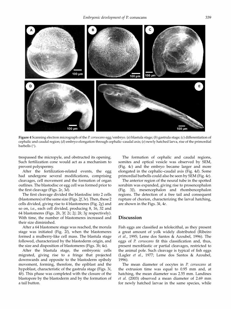
Figure 4 Scanning electron micrograph of the P. coruscans egg/embryo. (a) blastula stage; (b) gastrula stage; (c) differentiation ofcephalic and caudal region; (d) embryo elongation through cephalic–caudal axis; (e) newly hatched larva, rise of the primordialbarbells (∗).
trespassed the micropyle, and obstructed its opening.Such fertilization cone would act as a mechanism toprevent polyspermy.
After the fertilization-related events, the egghad undergone several modifications, comprisingcleavages, cell movement and the formation of organoutlines. The blastodisc or egg cell was formed prior tothe first cleavage (Figs. 2e, 3d).
The first cleavage divided the blastodisc into 2 cells(blastomeres) of the same size (Figs. 2f, 3e). Then, these 2cells divided, giving rise to 4 blastomeres (Fig. 2g) andso on, i.e., each cell divided, producing 8, 16, 32 and64 blastomeres (Figs. 2h, 3f; 2i; 2j; 2k; 3g respectively).With time, the number of blastomeres increased andtheir size diminished.
After a 64 blastomere stage was reached, the morulastage was initiated (Fig. 2l), when the blastomeresformed a mulberry-like cell mass. The blastula stagefollowed, characterized by the blastoderm origin, andthe size and disposition of blastomeres (Figs. 3h; 4a).
After the blastula stage, the embryonic cellsmigrated, giving rise to a fringe that projecteddownwards and opposite to the blastoderm epibolymovement, forming, therefore, the epiblast and thehypoblast, characteristic of the gastrula stage (Figs. 3i,4b). This phase was completed with the closure of theblastopore by the blastoderm and by the formation ofa tail button.
The formation of cephalic and caudal regions,somites and optical vesicle was observed by SEM,(Fig. 4c) and the embryo became larger and moreelongated in the cephalic–caudal axis (Fig. 4d). Someprimordial barbells could also be seen by SEM (Fig. 4e).
The anterior region of the neural tube in the spottedsorubim was expanded, giving rise to prosencephalon(Fig. 3J), mesencephalon and rhombencephalonregions. The detection of a free tail and consequentrupture of chorion, characterizing the larval hatching,are shown in the Figs. 3k, 4e.
Discussion
Fish eggs are classified as telolecithal, as they presenta great amount of yolk widely distributed (Ribeiroet al., 1995; Leme dos Santos & Azoubel, 1996). Theeggs of P. coruscans fit this classification and, thus,present meroblastic or partial cleavages, restricted tothe animal pole. Such cleavage is typical of fish eggs(Lagler et al., 1977; Leme dos Santos & Azoubel,1996).
The mean diameter of oocytes in P. coruscans atthe extrusion time was equal to 0.95 mm and, athatching, the mean diameter was 2.55 mm. Landineset al. (2003) observed a mean diameter of 2.69 mmfor newly hatched larvae in the same species, while

340 C. Marques et al.
Faustino et al. (2007) reported a mean diameter of1.14 mm for oocytes and 3.28 mm for larvae in thesorubim hybrid (P. coruscans × P. fasciatum). Oocytediameter is likely to be related to the fertilizationmode of each species; animals presenting externalfertilization usually produce oocytes of a smalldiameter (Sanches et al., 1999), whereas those that useinternal fertilization often present larger oocytes. Forinstance, Parauchenipterus galeatus is a species withinternal fertilization, whose mean oocyte diameterreaches 1.62 mm (Vazzoler, 1996).
Moreover, Suzuki (1992) showed that some species,such as Hoplias malabaricus and Serrasalmus marginatus(piranha), which present parental care, producedoocytes that were more than 2.00 mm in diameter. Theegg diameter also influences the incubation period, i.e.,larger eggs have a longer incubation time (Sargent et al.,1987).
Such studies corroborate the present data, as, lackingparental care and presenting external fertilization, theP. coruscans produced eggs of a small diameter andpresented a short incubation period.
Temperature is another feature that plays a roleon incubation. At higher temperatures, the incubationtime is shortened, while, at lower temperatures, thisperiod is increased (Leme dos Santos & Azoubel, 1996;Ninhaus-Silveira et al., 2006), as reported in the presentstudy.
The presence of a double chorion membrane wasalso observed in the oocytes and eggs prior cleavages.The same structure was also reported by Cardoso et al.(1995) in P. coruscans and by Sanches et al. (1999) in P.galeatus.
The development times in each collection weredistinct, as they were not simultaneous. Morrisonet al. (2001) suggested that variations in the embryonicdevelopment rate and in the embryonic development(asynchrony and malformation) are related to theincubation period and breeder’s age. Kimmel et al.(1995), Morrison et al. (2001) and Ninhaus-Silveiraet al. (2006) showed that, even within a fertilizedspawning incubated at optimal conditions, there issome asynchrony in the embryonic development time.
Analysing the micropylar apparatus in the oocytes,a conical vestibule, with a smooth surface and narrowmicropylar canal, was observed. This micropylarcanal is surrounded by a thick and gelatinous layer.According to Ganeco & Nakaghi (2003), the micropyleis a concave region located in the radiate zone orchorion, composed of a continuum vestibule, with aninternal canal that narrows towards to the plasmaticmembrane of the egg.
Such micropyle shape allows the entry of a singlespermatozoon, according to Kobayashi & Yamamoto(1981) and Rizzo & Bazzoli (1993). Other ways ofpreventing polyspermy include the formation of a
fertilization cone, which obstructs the micropylar canal,as well as the releasing of cortical alveoli, whichincreases the perivitelline space after the fertilizingspermatozoon has entered (Ganeco, 2003).
At the moment of fertilization, cytoplasmaticmovement towards the micropyle begins in theoocytes, which characterizes the animal pole. Similarobservations were reported by Kimmel et al. (1995)and Leme dos Santos & Azoubel (1996). Brasilet al. (2002) and Ganeco (2003), however, observedcytoplasmatic movement in eggs collected during theoocyte extrusion prior to fertilization. At the sametime, the basophilic and homogeneous animal pole andthe acidophilic vegetative pole were formed. Similarfeatures were observed by Matkovic et al. (1985) inRhamdia sapo.
The yolk grains were smaller and scarcer close tothe vegetative pole and larger and more numerousclose to the animal pole, as a result of the coalescencebetween yolk grains. Such a phenomenon was alsoobserved in Prochilodus lineatus (Brasil et al., 2002)and Byrcon orbignyanus (Ganeco, 2003). The samerelationship between number and size was alsodetected in the cortical alveoli: they were larger andmore abundant in the vegetative pole and they becamescarce and smaller in the animal pole. Few corticalalveoli at the animal pole region were also seenin Fundulus heteroclitus (Brummett & Dumont, 1979)Oryzias latipes (Iwamatsu & Ohta, 1981), Oncorhynchusketa (Kobayashi & Yamamoto, 1981) and Prochiloduslineatus (Brasil et al., 2002). Ohta (1985) and Hart (1990)assumed that the small amount of cortical alveoli inthe animal pole would be preventive, putting off thestart of perivitelline space formation and favouring theentry of the fertilizing spermatozoon.
Laale (1980) observed a similar feature, pointingout that during exocytosis of cortical alveoli contents,Ca2+ ions are absorbed and they would be involvedin chorion hardening, which would act as a mechanicprotection for the developing embryo (Laale, 1980;Lonning et al., 1984).
Embryonic development encompassed all stagesfrom fertilization to hatching, including cleavage,gastrulation and the beginning of organogenesis(Shardo, 1995).
The first cleavage divided the blastodisc into twoblastomeres of the same size. A similar observation wasreported in other species of the genus Cynolebias (Carter& Wourms, 1991), in zebrafish (Kimmel et al., 1995)and in Prochilodus lineatus (Ninhaus-Silveira et al., 2006).Cleavage started from the centre towards the blastodiscmargin (Matkovic et al., 1985; Shardo, 1995). Successivecleavage took place up to the 65 blastomere stage andas long as the number of blastomeres increased, theirsize decreased (Wourms & Evans, 1974; Castellani et al.,1994; Ninhaus-Silveira et al., 2006).

Embryonic development of P. coruscans 341
We observed that morula stage in the P. coruscans isfollowed by the blastula stage, which is characterizedby the blastoderm formation and, then, by the gastrulastage, when epiboly movements begin (Leme dosSantos & Azoubel, 1996), which extends up to theclosure of the blastopore by the blastoderm andformation of the tail button (Kimmel et al., 1995). Similarobservations were reported by Ninhaus-Silveira et al.(2006) in Prochilodus lineatus and by Ganeco (2003) inByrcon orbignyanus.
The anterior region of the neural tube in thestudied species expanded to form the prosencephalon,mesencephalon and rhombencephalon regions, similarto the patterns described in Brachydanio rerio (Kimmelet al., 1995), Rhamdia sapo (Cussac et al., 1985), Byrconorbignyanus (Ganeco, (2003) and Prochilodus lineatus(Ninhaus-Silveira et al., 2006).
The present data, therefore, showed that the studyof embryonic development is essential to a betterknowledge of some biological features of a species,helpful to elucidate issues related to fish rearing at thisstage, being also useful to further taxonomic, ecologicaland conservational studies in the P. coruscans.
Acknowledgements
The authors would like to thank CEPTA/IBAMA,especially Dr Jose Augusto Senhorini, for providingthe samples and collection assistance; FAPESP, for thefinancial support (scientific initiation grant Proc. N03/13112–0); and Mr Orandi Mateus for helping toprocess the material.
References
Botero, M., Fresneda, A., Montoya, A.F. & Angel, M.O. (2004).Descripcion Del desarrollo embrionario de zigotos hıbridosobtenidos por el cruce de machos de Cachama Blanca(Piaractus brachypomus) y hembras de Cachama Negra(Colossoma macropomum). Rev. Col. Cienc. Pec. 17, 38–45.
Brasil, D.F., Nakaghi, L.S.O., Leme dos Santos, H.S., QuagioGrassioto, I. & Foresti, F. (2002). Estudo morfologico dosprimeiros momentos da fertilizacao em curimbat Pro-chilodus lineatus (Valenciennes, 1836). [online], CIVA 2002.http://www.civa2002.org/, pp. 733–47.
Brummett, A.R. & Dumont, J.N. (1979). Initial stages of spermpenetration into the egg of Fundulus heteroclitus. J. Exp. Zool.201, 417–34.
Cardoso, E.L., Alves, M.S.D., Ferreira, R.M.A. & Godinho,H.P. (1995). Embryogenesis of the neotropical freshwaterSiluriformes Pseudoplatystoma coruscans. Aquat. Living Res.8, 343–6.
Carter, C.A. & Wourms, J.P. (1991). Cell behavior during earlydevelopment in the South American annual fishes of thegenus Cynolebias. J. Morphol. 210, 247–66.
Castellani, L.R., Tse, H.G., Leme dos Santos, H.S., Faria, R.H.S.& Santos, M.L.S. (1994). Desenvolvimento embrionariodo curimbata Prochilodus lineatus (VALENCIENNES, 1836)(Cypriniformes, Prochidontidae). Rev. Bras. Cienc. Morf. 11,99–105.
Cussac, V.E., Matkovic, M.V. & Maggese, M.C. (1985). De-sarrollo embrionario de Rhamdia sapo (Valenciennes, 1840)Eigenmann Y Eigenmann, 1888 (Pisces, Pimelodidae), I.Organogenesis media organogenesis tardia y eclosion. Rev.Bras. Biol. 45, 149–60.
Faustino, F., Nakaghi, L.S.O., Marques, C., Makino, L. &Senhorini, J.A. (2007) Fertilizacao e desenvolvimento em-brionario: morfometria e analise estereomicroscopica dosovos dos hıbridos de surubins (pintado, Pseudoplatystomacorruscans × cachara, Pseudoplatystoma fasciatum). Acta Sci.29, 49–55.
Flores, J.C.B., Araiza, M.A.F. & Valle, M.R.G. (2002).Desarrollo embrionario de Ctenopharyngodon idellus (Carpaherbıvora). [online], CIVA 2002. http://www.civa2002.org/,pp. 792–7.
Ganeco, L.N. (2003). Analise dos ovos de piracanjuba, Byrconorbignyanus (Valenciennes, 1894), durante a fertilizacaoe o desenvolvimento embrionario, sob condicoes dereproducao induzida. Masters degree, UniversidadeEstadual Paulista, Jaboticabal.
Ganeco, L.N. & Nakaghi, L.S.O. (2003). Morfologia damicropila e da superfıcie dos ovocitos de piracanjuba,Byrcon orbignyanus (Osteichthyes, Characidae), sobmicroscopia eletronica de varredura. Acta Sci. 25, 227–31.
Hart, N.H. (1990). Fertilization in teleost fishes: mechanismsof sperm–egg interactions. Int. Rev. Cytol. 121, 1–66.
Iwamatsu, T. & Ohta, T. (1981). Scanning electron microscopicobservation on sperm penetration in teleostean fish. J. Exp.Zool. 218, 261–77.
Kimmel, C.B., Ballard, W.W., Kimmel, S.R & Ullmann, B.(1995). Stages of embryonic development of the zebrafish.Dev. Dyn. 203, 253–310.
Kobayashi, W. & Yamamoto, T. (1981). Fine structure ofthe micropylar apparatus of the chum salmon egg, witha discussion of the mechanism for blocking polyspermy.J. Exp. Zool. 217, 265–75.
Kubitza, F., Campos, J.L. & Brum, J.A. (1998). Producaointensiva de surubins no projeto Pacu. Ltda. e AgropeixeLtda. In Anais da Aquicultura, Recife, Pernambuco 1, 393–407.
Laale, W.H. (1980). The perivitelline space and egg envelopesof bony fishes: a review. Copeia 2, 210–26.
Lagler, K.F., Bardach, J.E., Miller, R.R. & Passino, D.R.M.(1977). Ichthyology, 2nd edn, New York: John Wiley & Sons,Inc.
Landines, M.A., Senhorini, J.A., Sanabria, A.I. & Urbinati, E.C.(2003). Desenvolvimento Embrionario do Pintado (Pseudo-platystoma coruscans Agassiz, 1829). Bol. Tec. Cepta 6, 1–13.
Leme dos Santos, H.S. & Azoubel, R. (1996). Embriologiacomparada. Jaboticabal: FUNEP.
Lonning, S., Kjorsvik, E. & Davenport, J. (1984). Thehardening process of the chorion of the cod, Gadus morhuaL. and lampsucker, Cyclopterous lumpus L. J. Fish Biol. 24,505–22.
Matkovic, M.V., Cussac, V.E. & Cukier, M. (1985). Desarrolloembrionario de Rhamdia sapo (Valenciennes, 1840)
342 C. Marques et al.
Eigenmann Y Eigenmann, 1888 (Pisces, Pimelodidae). I.Segmentacion, morfogenesis y organogenesis temprana.Rev. Bras. Biol. 45, 39–50.
Morrison, C.M., Miyake, T. & Wright, J. Jr (2001). Histologicalstudy of the development of the embryo and early ofOreochromis niloticus (Pisces, Cichlidae). J. Morphol. 247,172–95.
Ninhaus-Silveira, A., Foresti, F. & Azevedo, A. (2006).Structural and ultrastructural analysis of embryonicdevelopment of Prochilodus lineatus (Valenciennes, 1836)(Characiformes, Prochilodontidae). Zygote 14, 217–29.
Nakatani, K., Agostinho, A.A., Baumgartner, G., Bialetzki,A., Sanches, P.V. & Cavicchioli, M. (2001). Ovos e larvas depeixes de agua doce, desenvolvimento e manual de identificacao.Maringa: UEM, Nupelia.
Ohta, T. (1985). Electron microscopy observations on spermentry and pronuclear formation in naked eggs of the rosebitterling in polyspermic fertilization. J. Exp. Zool. 234, 273–81.
Ribeiro, C.R., Leme dos Santos, H.S. & Bolzan, A.A.(1995). Estudo comparativo da embriogenese de peixesosseos (Pacu, Piaractus mesopotamicus, Tambaqui, Colossomamacropomum e hıbrido Tambacu). Rev. Bras. Biol. 55, 65–78.
Rizzo, E. & Bazzoli, N. (1993). Oogenesis, oocyte surface andmicropylar apparatus of Prochilodus affinis Reinhardt, 1874(Pisces Characiformes). Eur. Arch. Biol. 104, 1–6.
Sanches, P.V., Nakatani, K. & Bialetzki, A. (1999).Morphological description of the developmental stagesof Parauchenipterus galeatus (Linnaeus, 1766) (Silurifores,Auchenipteridae) on the floodplain of the upper ParanaRiver. Rev. Bras. Biol. 59, 429–38.
Sargent, R.C., Taylor, P.D. & Gross, M.R. (1987). Parental careand evolution of egg size in fishes. Am. Nat. 121, 32–46.
Shardo, J.D. (1995). Comparative embryology of teleosteanfishes. I. Development and staging of the American Shad,Alosa sapidissima (Wilson, 1811). J. Morphol. 225, 125–67.
Suzuki, H.I. (1992). Variacoes na morfologia ovariana e nodesenvolvimento do folıculo de peixes teleosteos da baciado rio Parana no trecho entre a foz do rio Paranapanema ea do rio Iguacu. Masters Degree, Universidade Federal doParana, Curitiba.
Tolosa, E.M.C., Behmer, O.A. & Freitas-Neto, A.G. (2003).Manual de tecnicas para histologia normal e patologica. Barueri–SP: Manole.
Vazzoler, A.E.A.M. (1996). Biologia da reproducao de peixesteleosteos: teoria e pratica. NUPELIA. Maringa: EDUEM.
Wourms, J.P. & Evans, D. (1974). The embryonic developmentof the black prickleback, Xiphister atropurpureus, a PacificCoast blennioid fish. Can. J. Zool. 52, 879–87.
Woynarovich, E. & Hovart, L. (1983). A propagacao artificialde peixes de aguas tropicais. Brasılia, DF: FAO/CODEVASF–CNPq, (Manual de Extensao, 5).