obstetrical doppler: how, when and why? lifelong learning/meetings/asm2013... · obstetrical...
TRANSCRIPT

Obstetrical Doppler: How, when and why?
François Audibert Dept of Obstetrics
and Gynecology
Disclosure statement l I do not have an affiliation, financial or otherwise, with a pharmaceutical company, medical device or communications organization.
l I have no conflicts of interest to disclose (i.e. no industry funding received or other commercial relationships).
l I have no financial relationship or advisory role with pharmaceutical or device-making companies, or CME provider.
Objectives 1. To discuss the indications of obstetrical Doppler
testing in uterine, umbilical, and cerebral arteries 2. To describe the results and limitations of obstetrical
Doppler in screening for fetal growth restriction and preeclampsia
3. To expose the methods of Doppler evaluation of fetal well-being for growth-restricted fetuses


Question 1
In case of fetal growth restriction due to severe placental insufficiency, the following Doppler pattern is more likely:
A- Increased cerebral and umbilical pulsatility B- Increased cerebral velocity C- Decreased cerebral and increased umbilical pulsatility D- Reverse flow in cerebral artery E- Reverse flow in umbilical vein (answer: C)

Question 2
In case of severe fetal anemia, the following pattern is more likely:
A- Increased cerebral and umbilical pulsatility B- Increased cerebral velocity C- Decreased cerebral and increased umbilical pulsatility D- Reverse flow in cerebral artery E- Reverse flow in umbilical vein
(answer: B)

Question 3
At 18 weeks, a bilateral notch in the uterine artery Doppler can be:
A- a normal finding B- associated with an increased risk of preeclampsia
or IUGR C- Should be repeated at 24-26 weeks D- Requires an antihypertensive treatment E- A+B+C are correct (answer: E)

How to obtain a good Doppler signal?

Effect of angle
Figure 3: Effect of the Doppler angle in the sonogram. (A) higher-frequency Doppler signal is obtained if the beam is aligned more to the direction of flow. In the diagram, beam (A) is more aligned than (B) and produces higher-frequency Doppler signals. The beam/flow angle at (C) is almost 90° and there is a very poor Doppler signal. The flow at (D) is away from the beam and there is a negative signal.
Simple general rule : keep angle ≤ 45° In some cases, as close as 0°as possible

Basic settings
l Gate size: adapt to vessel size l Gain: adjust to obtain good signal/noise ratio l PRF (Pulse repetition frequency) or scale:
adjust to flow velocity

Wall filter
l Set too low: noise l Set too high: you lose information or you
create absent end-diastolic flow

Resistance index, pulsatility index, ratios ???
Figure 16: Flow velocity indices
Indices make you independent from insonation angles PI gives the maximum of hemodynamic information

PI, RI

Role of Doppler in management of placental insufficiency
I. Prediction IUGR/preeclampsia (long term) • Uterine arteries (notch/increased resistance) • Objective : population screening / IUGR etiology
II. Diagnosis/surveillance (mid term) • Umbilical arteries • Objective : adjust frequency of monitoring
III. Fetal well-being (short term) • Umbilical arteries
Cerebral arteries • Other fetal Dopplers
• Ductus Venosus, IVC • Aortic isthmus etc…
• Objective : decide when to deliver a fetus with IUGR
http://www.fetalmedicine.com/fmf/online-education/03-doppler/

Uterine arteries
• Screening test
• 20-24 weeks
• PI or RI increased
• Protodiastolic NOTCH
• 60-90% sensitivity
• PPV 20-30%

Uterine arteries

Pulsatility index in the uterine artery with gestation (mean 95th and 5th centiles)

Normal uterine artery
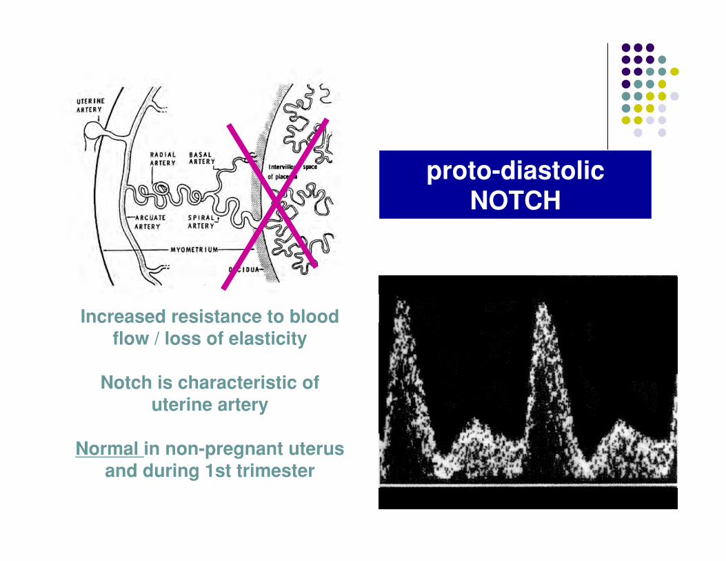
proto-diastolic NOTCH
Increased resistance to blood flow / loss of elasticity
Notch is characteristic of
uterine artery
Normal in non-pregnant uterus and during 1st trimester
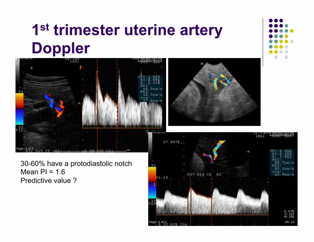
1st trimester uterine artery Doppler
30-60% have a protodiastolic notch Mean PI = 1.6 Predictive value ?
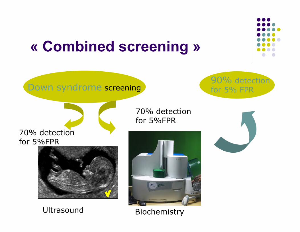
« Combined screening »
Down syndrome screening
70% detection for 5%FPR
70% detection for 5%FPR
90% detection for 5% FPR
Ultrasound Biochemistry

« Combined screening »
Preeclampsia screening ?
40% detection for 10%FPR?
50% detection for 10%FPR?
X% detection for Y% FPR ?
Doppler Biochemistry

Fetal surveillance

Umbilical Doppler
Umbilical artery
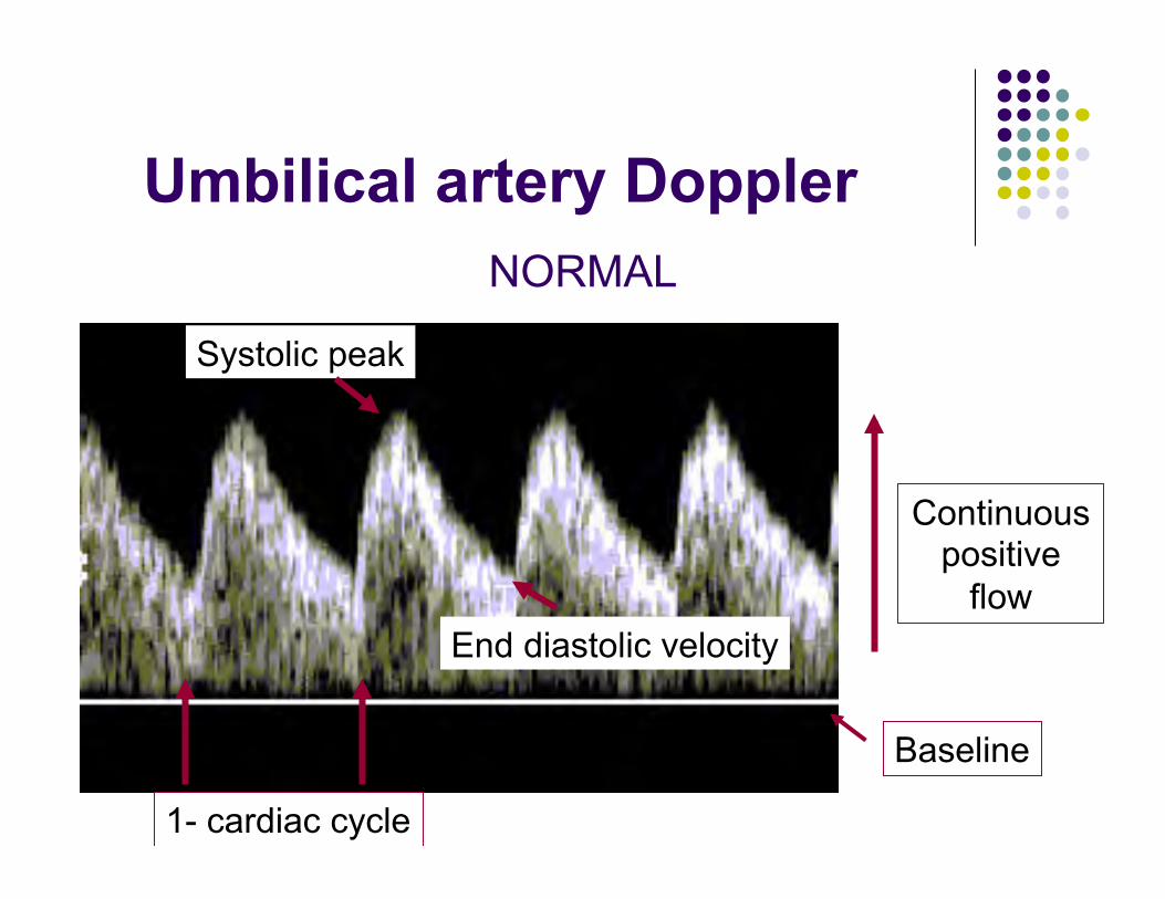
Umbilical artery Doppler NORMAL
Baseline
Continuous positive
flow
1- cardiac cycle
Systolic peak
End diastolic velocity

Progression of Abnormal Doppler
Waveform of Umbilical Artery
Normal diastolic flow
Decreased diastolic flow
Absent diastolic flow
Reverse flow
BAD
VERY BAD
AWFUL

Middle Cerebral Artery (MCA)
• Main branch of circle of Willis • Carries 80% of blood supply • Easy to identify • Easy to repeat

Brain sparing effect
Fetal adaptation / response to hypoxemia BRAIN, heart, adrenals
Normal
Vasodilatation
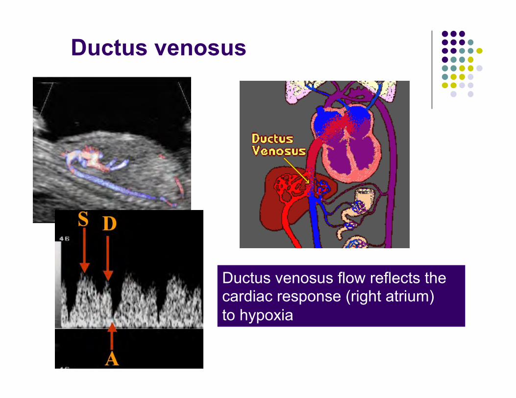
Ductus venosus
Ductus venosus flow reflects the cardiac response (right atrium) to hypoxia

Ductus venosus: « a » wave

Ductus venosus
Normal
Abnormal
Critical

Doppler evolution
Baschat AA, Ultrasound Obstet Gynecol 2011
Early IUGR (22-34 weeks)
Late IUGR (34-40 weeks)
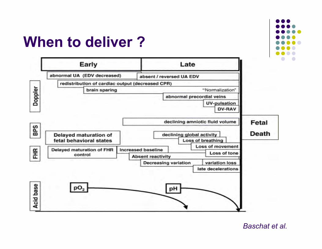
When to deliver ?
Baschat et al.

When to deliver IUGR? Aortic isthmus??
Aortic isthmus
JC Fouron Brain sparing effect

Aortic isthmus
JC Fouron Am J Obstet Gynecol 2005
Abnormal isthmus flow predicts suboptimal neurodevelopment

Middle cerebral artery in Fetal Anemia: increased velocity
Ø Vmax >1.5 MoM Ø Se=100% NPV=100% Ø FP=12% Ø PPV=65%
Angle=0°
Indications: maternal antibodies, fetal hydrops, parvovirus exposure, etc…


Doppler ultrasound for the fetal assessment in high-risk pregnancies
ü Reduction in perinatal deaths RR=0.71 ü Fewer inductions of labour ü Fewer admissions to hospital ü No report of adverse effects ü No difference for fetal distress in labour ü No difference in caesarean delivery ü No benefit proven in low-risk women
Cochrane Collaboration 2010
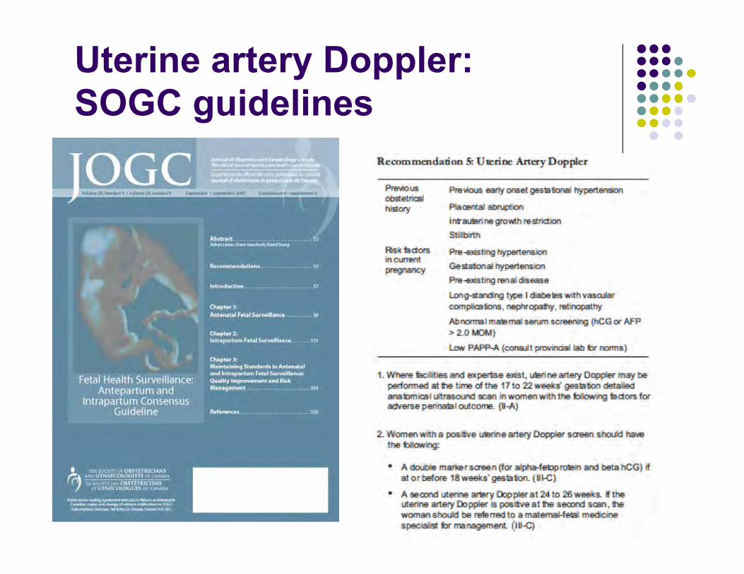
Uterine artery Doppler: SOGC guidelines
Umbilical artery Doppler: SOGC guidelines
1. Should not be used as a screening tool in low-risk women
2. Should be performed in suspected IUGR or placental pathology (preeclampsia)
3. Abnormal Doppler is an indication for increased fetal surveillance or delivery, depending on other clinical factors