ocular emergency blow out fracture - dos times · www. dosonline.org l 19 ocular emergency...
TRANSCRIPT
www. dosonline.org l 17
Ocular Emergency
Nitin Vichare MS, DNB,FAICO
Dept. of Ophthalmology, Command Hospital, (Southern Command), Pune, Maharashtra
Blow Out Fracture
Ocular Emergency
Nitin VichareMS, DNB,FAICO
Orbital injury forms an important aspect of ocular trauma. The blowout fracture is the most common type
of orbital fracture that confronts the ophthalmic surgeon. The phenomenon of isolated orbital wall fracture was first recognized by MacKenzie in 1844. In 1957, Smith and Regan described inferior rectus entrapment with decreased ocular motility in the setting of an orbital floor fracture and used the term “blow-out fracture”1. It is not uncommon to have a patient presenting with injury by a blunt object like fist or a ball which have caused bony injury sparing the eyeball. Evaluation and management of such cases forms integral part of ocular trauma management.
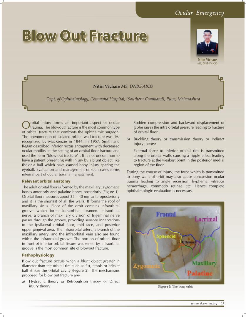
Relevant orbital anatomyThe adult orbital floor is formed by the maxillary, zygomatic bones anteriorly and palatine bones posteriorly (Figure 1). Orbital floor measures about 35 – 40 mm anteroposteriorly and it is the shortest of all the walls. It forms the roof of maxillary sinus. Floor of the orbit contains infraorbital groove which forms infraorbital foramen. Infraorbital nerve, a branch of maxillary division of trigeminal nerve passes through the groove, providing sensory innervations to the ipsilateral orbital floor, mid face, and posterior upper gingival area. The infraorbital artery, a branch of the maxillary artery, and the infraorbital vein also are found within the infraorbital groove. The portion of orbital floor in front of inferior orbital fissure weakened by infraorbital groove is the most common site of blowout fracture.
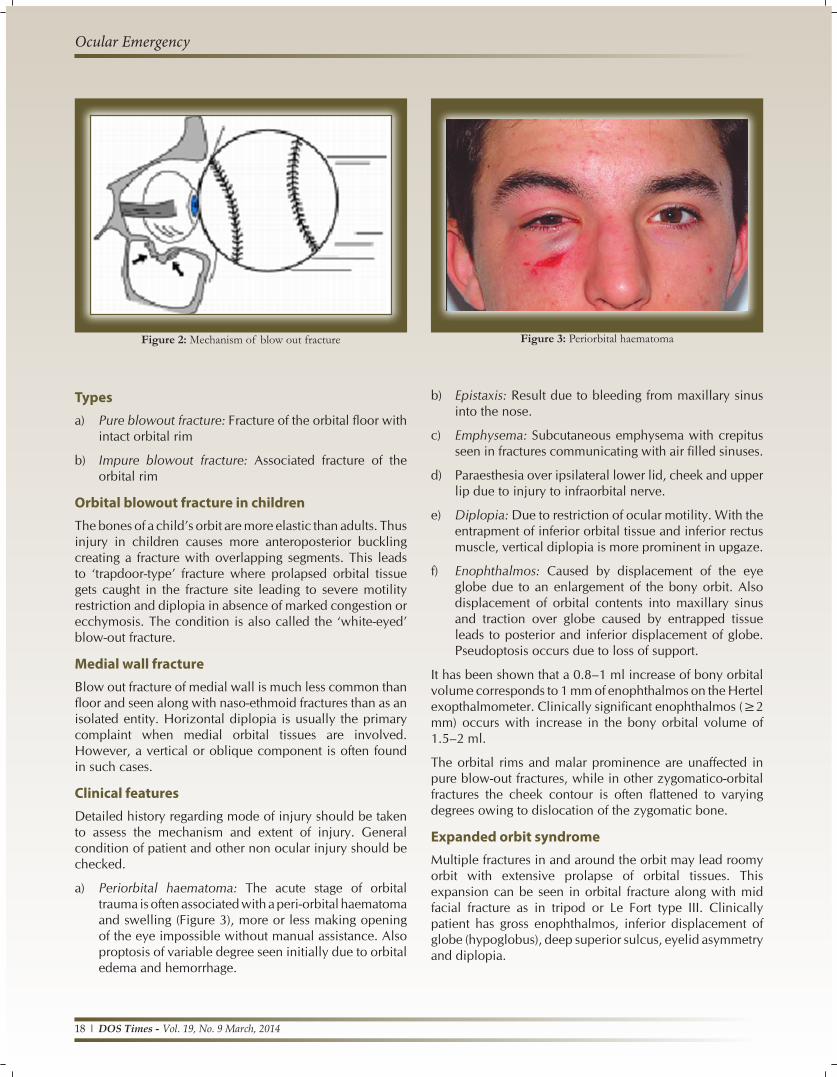
PathophysiologyBlow out fracture occurs when a blunt object greater in diameter than the orbital rim such as fist, tennis or cricket ball strikes the orbital cavity (Figure 2). The mechanisms proposed for blow out fracture are-
a) Hydraulic theory or Retropulsion theory or Direct injury theory:
Sudden compression and backward displacement of globe raises the intra orbital pressure leading to fracture of orbital floor.
b) Buckling theory or transmission theory or Indirect injury theory:
External force to inferior orbital rim is transmitted along the orbital walls causing a ripple effect leading to fracture at the weakest point in the posterior medial region of the floor.
During the course of injury, the force which is transmitted to bony walls of orbit may also cause concussion ocular trauma leading to angle recession, hyphema, vitreous hemorrhage, commotio retinae etc. Hence complete ophthalmologic evaluation is necessary.
Figure 1: The bony orbit
18 l DOS Times - Vol. 19, No. 9 March, 2014
Ocular Emergency
Typesa) Pure blowout fracture: Fracture of the orbital floor with
intact orbital rim
b) Impure blowout fracture: Associated fracture of the orbital rim
Orbital blowout fracture in childrenThe bones of a child’s orbit are more elastic than adults. Thus injury in children causes more anteroposterior buckling creating a fracture with overlapping segments. This leads to ‘trapdoor-type’ fracture where prolapsed orbital tissue gets caught in the fracture site leading to severe motility restriction and diplopia in absence of marked congestion or ecchymosis. The condition is also called the ‘white-eyed’ blow-out fracture.
Medial wall fractureBlow out fracture of medial wall is much less common than floor and seen along with naso-ethmoid fractures than as an isolated entity. Horizontal diplopia is usually the primary complaint when medial orbital tissues are involved. However, a vertical or oblique component is often found in such cases.
Clinical featuresDetailed history regarding mode of injury should be taken to assess the mechanism and extent of injury. General condition of patient and other non ocular injury should be checked.
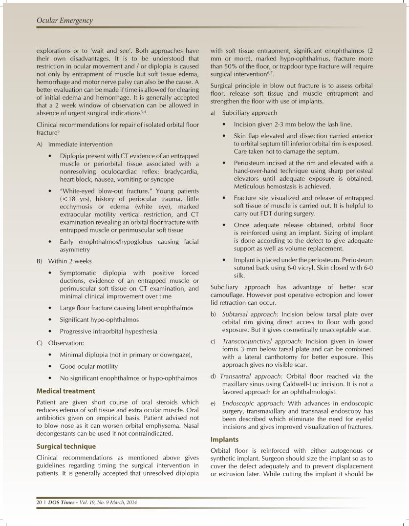
a) Periorbital haematoma: The acute stage of orbital trauma is often associated with a peri-orbital haematoma and swelling (Figure 3), more or less making opening of the eye impossible without manual assistance. Also proptosis of variable degree seen initially due to orbital edema and hemorrhage.
b) Epistaxis: Result due to bleeding from maxillary sinus into the nose.
c) Emphysema: Subcutaneous emphysema with crepitus seen in fractures communicating with air filled sinuses.
d) Paraesthesia over ipsilateral lower lid, cheek and upper lip due to injury to infraorbital nerve.
e) Diplopia: Due to restriction of ocular motility. With the entrapment of inferior orbital tissue and inferior rectus muscle, vertical diplopia is more prominent in upgaze.
f) Enophthalmos: Caused by displacement of the eye globe due to an enlargement of the bony orbit. Also displacement of orbital contents into maxillary sinus and traction over globe caused by entrapped tissue leads to posterior and inferior displacement of globe. Pseudoptosis occurs due to loss of support.
It has been shown that a 0.8–1 ml increase of bony orbital volume corresponds to 1 mm of enophthalmos on the Hertel exopthalmometer. Clinically significant enophthalmos (≥2 mm) occurs with increase in the bony orbital volume of 1.5–2 ml.
The orbital rims and malar prominence are unaffected in pure blow-out fractures, while in other zygomatico-orbital fractures the cheek contour is often flattened to varying degrees owing to dislocation of the zygomatic bone.
Expanded orbit syndromeMultiple fractures in and around the orbit may lead roomy orbit with extensive prolapse of orbital tissues. This expansion can be seen in orbital fracture along with mid facial fracture as in tripod or Le Fort type III. Clinically patient has gross enophthalmos, inferior displacement of globe (hypoglobus), deep superior sulcus, eyelid asymmetry and diplopia.
Figure 2: Mechanism of blow out fracture Figure 3: Periorbital haematoma

www. dosonline.org l 19
Ocular Emergency
EvaluationExternal force causing blow out fracture can also cause concussion injury to eyeball leading to extensive damage. Hence thorough ocular examination is necessary to record ocular status and manage patient accordingly.
Recording of visual acuity at presentation has medico-legal importance in ocular trauma cases. If required eyelids can be gently separated to allow patient to read the chart. Palpate orbital rim to look for deformity and crepitus. Slit lamp evaluation of cornea and anterior segment should be performed. Pupillary reflex should be checked as presence of RAPD points towards optic nerve injury. Fundus evaluation should be done to note for Berlin’s oedema which can be the cause of unexplained diminution of vision.
Ocular motility: Entrapment of orbital contents and inferior rectus muscle leads to motility restriction especially in upgaze (Figure 4).
Hertel exopthalmometer: To document enophthalmos. With passage of time and absorption of orbital fat over period can lead to increase in enophthalmos.
Force Duction Test (FDT): FDT is useful in determining whether dysmotility is restrictive or paralytic. In blow out fracture with inferior rectus entrapment FDT is ‘positive’ indicating mechanical cause.
Force Generation Test (FGT): In testing force generation, the muscle insertion is grasped and the patient is asked to look into the muscle’s field of action. A paretic muscle will feel weak when compared with the fellow eye.
Diplopia charting: With red green glass, diplopia charting with streak light shows diplopia worsening in upgaze
Hess screen or Lee screen test can be done.
Imaginga) Plain X-rays: Easily available and cost effective imaging
modality. Water’s view is the most useful projection for
detecting an orbital floor fracture (Figure 5). X ray shows bony discontinuity in orbital floor with herniation of soft tissue in maxillary antrum seen as ‘hanging drop’ sign2.
b) Computerized tomography (CT) scanning: CT gives detailed visualization of bony and soft tissue injury where entrapment of muscle can be appreciated. Coronal sections are particularly useful (Figure 6).
c) Magnetic resonance imaging (MRI): Can be utilized when there is need for greater soft tissue evaluation. MRI is insufficient in assessing the bony structures and therefore needs to be combined with CT.
Management protocolsIn case of isolated orbital floor fracture the two main strategies have been either to perform early surgical
Figure 6: Coronal CT showing floor fracture
Figure 4: restriction in upgaze Figure 5: Water’s view
20 l DOS Times - Vol. 19, No. 9 March, 2014
Ocular Emergency
explorations or to ‘wait and see’. Both approaches have their own disadvantages. It is to be understood that restriction in ocular movement and / or diplopia is caused not only by entrapment of muscle but soft tissue edema, hemorrhage and motor nerve palsy can also be the cause. A better evaluation can be made if time is allowed for clearing of initial edema and hemorrhage. It is generally accepted that a 2 week window of observation can be allowed in absence of urgent surgical indications3,4.
Clinical recommendations for repair of isolated orbital floor fracture5
A) Immediate intervention
• Diplopia present with CT evidence of an entrapped muscle or periorbital tissue associated with a nonresolving oculocardiac reflex: bradycardia, heart block, nausea, vomiting or syncope
• “White-eyed blow-out fracture.” Young patients (<18 yrs), history of periocular trauma, little ecchymosis or edema (white eye), marked extraocular motility vertical restriction, and CT examination revealing an orbital floor fracture with entrapped muscle or perimuscular soft tissue
• Early enophthalmos/hypoglobus causing facial asymmetry
B) Within 2 weeks
• Symptomatic diplopia with positive forced ductions, evidence of an entrapped muscle or perimuscular soft tissue on CT examination, and minimal clinical improvement over time
• Large floor fracture causing latent enophthalmos
• Significant hypo-ophthalmos
• Progressive infraorbital hypesthesia
C) Observation:
• Minimal diplopia (not in primary or downgaze),
• Good ocular motility
• No significant enophthalmos or hypo-ophthalmos
Medical treatmentPatient are given short course of oral steroids which reduces edema of soft tissue and extra ocular muscle. Oral antibiotics given on empirical basis. Patient advised not to blow nose as it can worsen orbital emphysema. Nasal decongestants can be used if not contraindicated.
Surgical techniqueClinical recommendations as mentioned above gives guidelines regarding timing the surgical intervention in patients. It is generally accepted that unresolved diplopia
with soft tissue entrapment, significant enophthalmos (2 mm or more), marked hypo-ophthalmus, fracture more than 50% of the floor, or trapdoor type fracture will require surgical intervention6,7.
Surgical principle in blow out fracture is to assess orbital floor, release soft tissue and muscle entrapment and strengthen the floor with use of implants.
a) Subciliary approach
• Incision given 2-3 mm below the lash line.
• Skin flap elevated and dissection carried anterior to orbital septum till inferior orbital rim is exposed. Care taken not to damage the septum.
• Periosteum incised at the rim and elevated with a hand-over-hand technique using sharp periosteal elevators until adequate exposure is obtained. Meticulous hemostasis is achieved.
• Fracture site visualized and release of entrapped soft tissue of muscle is carried out. It is helpful to carry out FDT during surgery.
• Once adequate release obtained, orbital floor is reinforced using an implant. Sizing of implant is done according to the defect to give adequate support as well as volume replacement.
• Implant is placed under the periosteum. Periosteum sutured back using 6-0 vicryl. Skin closed with 6-0 silk.
Subciliary approach has advantage of better scar camouflage. However post operative ectropion and lower lid retraction can occur.
b) Subtarsal approach: Incision below tarsal plate over orbital rim giving direct access to floor with good exposure. But it gives cosmetically unacceptable scar.
c) Transconjunctival approach: Incision given in lower fornix 3 mm below tarsal plate and can be combined with a lateral canthotomy for better exposure. This approach gives no visible scar.
d) Transantral approach: Orbital floor reached via the maxillary sinus using Caldwell-Luc incision. It is not a favored approach for an ophthalmologist.
e) Endoscopic approach: With advances in endoscopic surgery, transmaxillary and transnasal endoscopy has been described which eliminate the need for eyelid incisions and gives improved visualization of fractures.
ImplantsOrbital floor is reinforced with either autogenous or synthetic implant. Surgeon should size the implant so as to cover the defect adequately and to prevent displacement or extrusion later. While cutting the implant it should be

www. dosonline.org l 21
Ocular Emergency
tapered posteriorly so as to fit orbital floor configuration. Table 1 enumerates various implants.
Complications of surgery• Intra operative bleeding
• Residual or new onset diplopia
• Extra ocular muscle dysfunction
• Post operative neuralgia
• Residual enophthalmos
• Implant extrusion
• Possible loss of vision
Although the surgery may be a complete success in the eyes of the surgeon, the patient may view the outcome as unsatisfactory. To minimize this, the surgeon and patient should be in mutual agreement regarding the realistic outcome of the repair.
Treatment of persistent visually handicapping diplopiaFew patients will have persistent diplopia even after adequate surgical repair of floor fracture. Diplopia in primary gaze and in down gaze (functional gaze) are more troublesome. Such cases will require muscle surgery. To correct diplopia in down gaze ‘Reverse Knapp procedure’ performed placing medial and lateral recti behind inferior rectus muscle. Fresnel prisms can be employed in selective cases.
Late treatment of cosmetically unacceptable enophthalmosResurgery with adequate size orbital implant if downward sinking of eye along with enophthalmos is unacceptable to patient. Correction of psudoptosis done with mullerectomy which will increase palpebral height.
References1. Smith B, Regan WF Jr. Blow-out fracture of the orbit; mechanism
and correction of internal orbital fracture. Am J Ophthalmol. 1957;44(6):733-739.
2. NgP,ChuC,YoungN,SooM. Imagingoforbitalfloor fractures.Australas Radiol. 1996;40(3):264-268.
3. Egbert JE,May K, Kersten RC, Kulwin DR. Pediatric orbital floorfracture : direct extraocular muscle involvement. Ophthalmology 2000;107(10):1875-1879.
4. Courtney DJ, Thomas S, Whitfield PH. Isolated orbital blowoutfractures: survey and review. Br J Oral Maxillofac Surg. 2000; 38 (5):496-504.
5. Michael A Burnstine. Clinical Recommendations for Repair of Isolated Orbital Floor Fractures, An Evidence-based Analysis. Ophthalmology 2002; 109: 1207-1210.
6. Prashant Yadav, Neelam Pushker, Mandeep Bajaj, Mahesh Chandra,Dinesh Shrey, Pawan Lohiya. Orbital Blow out Fracture. DOS Times - 2008; (8) Vol. 14, No.2.
7. Putterman AM, Smith BC, Lisman RD. Blow out fracture. In: Smith’s Ophthalmic plastic reconstructive surgery. 2nd edn, Mosby, 1998, 209-223.
Table 1: Examples of implant materials used in orbital floor repair
Implant material Advantage DisadvantageMembranous bone Autogenous - Morbidity at donor site
- Extended operation time- Resorption unpredictable
Cartilage Autogenous - Morbidity at donor site- Extended operation time- Resorption unpredictable
Titanium mesh Biocompatible Stable - Foreign material that remains in the body- Combination with bone recommended
Porous polyethylene(Medpore)sheets
Easy to shape and handleBiocompatible Stable
Foreign material that remains in the body
Silicon sheet Easy to handle cheap Extrusion rates higher
Silastic sheet (Teflon) Easy to shape and handle Foreign body reaction and extrusion common