of grape seeds proanthocyanidins bisphenol a in male rats
TRANSCRIPT

Page 1/25
Neurotoxicity and Neuroin�ammatory Effects ofBisphenol A in Male Rats. The Neuroprotective Roleof Grape Seeds ProanthocyanidinsSaber Mohamed Eweda ( [email protected] )
Taibah University https://orcid.org/0000-0002-5151-0137Heba M. Abdou
Alexandria UniversityHeba-Tallah Abd Elrahim Abd Elkader
Alexandria UniversityAmel H. El-Gendy
Alexandria University
Research Article
Keywords: Bisphenol A, Grape seed proanthocyanidins, oxidative stress, Na/K-ATPase, TNF-α, COX-2
Posted Date: June 8th, 2021
DOI: https://doi.org/10.21203/rs.3.rs-544366/v1
License: This work is licensed under a Creative Commons Attribution 4.0 International License. Read Full License

Page 2/25
AbstractExposure to bisphenol A (BPA) contributes to neurological disorders, but the underlying mechanisms arestill not completely understood. We studied the neurotoxic effect of BPA and how it promotesin�ammation and alteration in the neurotransmission synthesis, release and transmission. This studywas also designed to investigate the neuroprotective effect of grape seed proanthocyanidins (GSPE)against BPA-induced neurotoxicity in rats. Rats were equally divided into 4 group with 7 rats in each asfollows: control group, BPA group, GSPE + BPA group and GSPE group. Rats were orally treated with theirrespective doses (50 mg BPA/kg BW and/or 200 mg GSPE/kg BW) daily for 70 days. BPA elicitssigni�cant elevation in malondialdehyde (MDA) and nitric oxide (NO) associated with signi�cantreduction in glutathione (GSH), total thiols, glutathione peroxidase (GPx), superoxide dismutase (SOD)and glutathione-S-transferase (GST). BPA exposure result in increased dopamine and serotonin levels,elevation in acetylcholine esterase (AchE) activity and reduction in Na/K-ATPase and total ATPaseactivities in the brain. Also, BPA induces upregulation in the gene expression of the in�ammatory markers;tumor necrosis factor-α (TNF-α) and cyclooxygenase-2 (COX-2), and in the tumor suppressor and pro-oxidant p53 protein. The pretreatment with GSPE attenuate or ameliorate all the oxidative and neurotoxicparameters induced by BPA. Our results suggest that GSPE has a promising role in modulating BPA-induced neuroin�ammation and neurotoxicity and its antioxidant and free radical scavenging activitiesmay in part be responsible for such effects.
IntroductionThe exposure to neurotoxic compounds induce over production of reactive oxygen species (ROS) andreactive nitrogen species (RNS) which play a crucial role in neurodegenerative disorders by oxidizingproteins, DNA and lipids of neuronal cells leading to pathways for cell death (Zhao et al. 2019). Thechronic exposure to neurotoxic substances from environment or from ingestion of food triggerin�ammatory process in the brain contributing to cognitive impairment and neurodegenerative diseases,including, Alzheimer's disease (AD) and Parkinson's disease (PD) (Niranjan 2018). Exposure of humans tobisphenol A (BPA) has been a matter of concern due to its increased usage in the products of day to dayuse. High amounts of BPA used in the production of polycarbonate plastics and epoxy resins which usedin food and beverages packaging, electronic devices, optical storage discs, construction material, dentalsealants and thermal printing paper (Raisuddin &Sharma 2017). BPA monomers can leach-out fromthese products, when subjected to washing processes, heating or contact with acidic or basic pH, andenter human body through occupational and food contact (Musachio et al. 2020). BPA has been linkedwith multiple neuropsychological disorders and neurobehavioral disturbances due to its ability to passthrough blood-brain barrier (Resnik &Elliott 2015). It has been reported that BPA causes neurologicalinjury by damaging protein and lipid structures through free radicals mediated mechanisms (Agarwal etal. 2016). The prenatal exposure to BPA was related to adverse impacts of behaviours and cognitiveabilities at children's early life, such as social response and working memory (Braun et al. 2017).
Page 3/25
The bioactive compounds from natural sources have been attracting growing attention in recent decades.Phytochemicals, as polyphenols, in fruit, vegetables and medicinal plants have a wide range of health-protective effects on heart, kidney, liver and brain (Nassiri-Asl &Hosseinzadeh 2009). Grape seeds are oneof the richest sources of polyphenols; monomeric �avan-3-ols, phenolic acids and oligomericproanthocyanidins (Prasain et al. 2009). Proanthocyanidins from grape seeds contain complex pool ofpolyphenolic compounds mainly catechin monomers, or their dimers, trimers, and oligomers, which areotherwise known as tannins (Zhang et al. 2015). Grape seeds proanthocyanidins (GSPE) are superiorradical’s scavenger over vitamins C, E and carotene. It protects liver and brain against lipid peroxidationand DNA damage, has antidiabetic, antibacterial, anticancer and antiin�ammatory effects (Shan et al.2010). It has been revealed that GSPE induce protective effects against brain damage via their chelatingability and antioxidant properties (Chu et al. 2014). GSPE protect against depression and age-relatedmental deterioration by inducing hypothalamic-pituitary-adrenal axis action and hippocampalneurogenesis (Sutcliffe et al. 2017).
Despite the role of BPA in neurotoxicity and neuronal damage have been targeted by several researchesover the years, the underlying mechanisms by which BPA induces neurotoxicity and neurodegenerativedisorders still need to be further elucidated. The present study aimed to investigate the neurotoxic effectsof BPA on brain tissue that may be a cause for neuronal damage and neurodegenerative diseases. Also,the study investigates the possible neuroprotective effects of GSPE on BPA-induced oxidative stress,neuroin�ammation and neurotoxicity in rats’ brain.
Materials And Methods
ReagentsBisphenol A 98% purity in the form of white crystals was purchased from Loba Chemie for laboratoryreagents and �ne chemicals (India). Grape seed proanthocyanidins was purchased from Arab companyfor pharmaceuticals and medicinal plants (MEPACO-MEDIFOOD, Enshas- El Raml, Sharkeya, EGYPT).Reduced glutathione (GSH), glutathione reductase (GR), 2-thiobarbituric acid, pyrogallol, NADPH, bovineserum albumin and 1,1,3,3-tetraethoxypropane, 5,5'-dithiobis-(2-nitrobenzoic acid) (DTNB) werepurchased from Sigma-Aldrich (Merck KGaA, USA). All other chemicals and kits were of analytical grade.
Animals and experimental designTwenty-eight adult male Wister albino rats (weight, 120–130 g) were obtained from the animal house ofFaculty of Medicine, Alexandria University, Alexandria, Egypt. Rats were housed in stainless steel cagesand provided with a basal diet and tap water ad libitum. Rats were maintained under 50–60% relativehumidity and 25 ± 5˚C with a 12 h light/dark cycle during the experimental period. After two weeks ofacclimatization, rats were randomly and equally divided into four equal groups with 7 rats in each asfollows: control group received 0.5 ml distilled water and 0.5 corn oil, BPA-group was treated with 0.5 mlBPA at a dose of 50 mg/kg BW/day dissolved in corn oil (Tyl et al. 2002), GSPE + BPA group received 0.5

Page 4/25
ml GSPE at a dose 200 mg/kg BW/day dissolved in distilled water (Yousef et al. 2009) and 0.5 ml BPA ata dose 50 mg/kg BW/day, and GSPE-group which treated with 200 mg/kg BW/day GSPE dissolved in 0.5distilled water. During the experimental period, GSPE was given 30 min before BPA administration anddoses were given once daily via gavage for 70 consecutive days. The design and experimentaltechniques of the current study were approved by the Institutional Animal Care and Use Committee(IACUC) of Alexandria University in accordance with the guidelines of the National Institutes of HealthGuide for the Care and Use of Laboratory Animals. All efforts were made to minimise the suffering of ratsduring the experimental period.
Blood sampling and tissue preparationAt the end of experimental period, rats were deeply euthanized via an intraperitoneal injection of 100mg/kg ketamine and 20 mg/kg xylazine after a starvation period of 12 h. four ml of blood was obtainedthrough cardiac puncture using a sterile syringe. Death was subsequently con�rmed by the inhalation ofCO2 followed by cervical dislocation. Serum was obtained by centrifugation of clotted blood at 1000 x gand 25˚C for 10 min and kept at 20˚C for lipid pro�ling tests. Brain was immediately excised, washedusing chilled saline solution and the adhering fat and connective tissues were removed. Parts of the braintissues were immediately
removed and kept at -80°C for molecular analysis. Another parts of brain were minced and homogenized(10%, w/v) in ice-cold phosphate buffer (0.01 M, pH 7.4) containing 1.15% KCl. The homogenates werecentrifuged at 10000×g for 20 min at 4°C (Hitachi Ltd.; model, EBA 12R). Supernatants were stored inaliquots of 1ml at 80˚C for subsequent biochemical analysis.
Determination of oxidative stress biomarkers in brain tissueLipid peroxidation end product (malondialdehyde, MDA) was measured in brain tissue as thiobarbituricacid reactive substances (TBARS) at 532 nm using 2-thiobarbituric acid and expressed as nmol MDA/gtissue (Draper &Hadley 1990). Nitric oxide (NO) concentration in brain supernatant was measured bycommercial kit purchased from Biodiagnostic Co. Cairo, Egypt. The reaction based on simple Griessreaction according to the method of (Guevara et al. 1998).
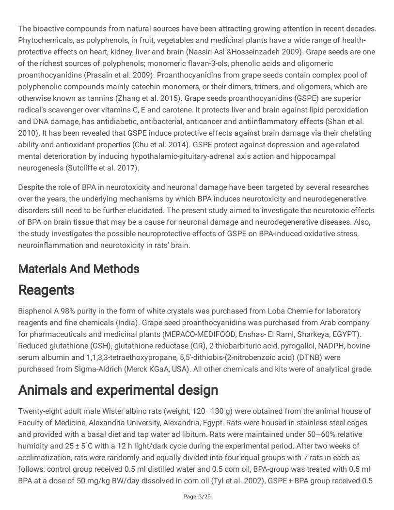
Determination of enzymatic antioxidants and detoxi�cantsactivities in brain tissuesTotal glutathione peroxidase (GPx) activity was measured using cumene hydroperoxide as a substrate bythe method of (Flohe &Günzler 1984). The assay method based on monitoring the generation of GSHfrom GSSG by the action of glutathione reductase in presence of NADPH. The activity of superoxidedismutase (SOD) was determined according to the method of (Marklund &Marklund 1974). The methoddepends on inhibition of spontaneous autoxidation of pyrogallol at alkaline pH by removal of superoxideanion radicals using SOD in the sample. Autoxidation was monitored as an increase in absorbance at

Page 5/25
420 nm. The total activity of glutathione-S-transferase (GST) was assayed by the method of (Habig et al.1974). Optical density was measured at 340 nm.
Determination of non-enzymatic antioxidants in braintissuesReduced GSH was determined in supernatant of brain tissue using NADPH as a reducing agent anddependent on oxidation of GSH by 5,5 - dithiobis (2-nitrobenzoic acid) (DTNB) as previously described(Gri�th 1980). Total thiol content of brain tissue supernatant was carried out as described previously(Sedlak &Lindsay 1968).
Measurement of serum lipid pro�leThe serum levels of triglycerides (TG), total cholesterol (TC), low density lipoprotein cholesterol (LDL) andhigh-density lipoprotein cholesterol (HDL-C) were measured by commercially available diagnostic kitsfrom Biosystems S.A (Biosystems S.A. Costa Brava 30, Barcelona, Spain).
Determination of protein contentThe protein content of brain tissue supernatant was determined by the method of Lowry using folin-ciocalteu reagent and bovine serum albumin as a standard (Lowry et al. 1951).
Determination of neuro-speci�c biomarkersAcetyl cholinesterase (AChE) activity was assayed in brain tissue using the method of (Ellman et al.1961). The activity of AChE was calculated as nmoles of hydrolyzed acetylthiocholine iodide/min/mgprotein using molar extinction coe�cient of 1.36 × 10⁴ M− 1 cm− 1. The speci�c activity of total ATPaseactivity in brain supernatant was assayed using ELISA kit purchased from ImmunoWay BiotechnologyCompany (USA). Brain Na+-K+/ATPase speci�c activity was assayed according to the manualinstructions of ELISA kit purchased from MyBioSource (USA). The activities of both total APTase andNa+/K+-ATPase were expressed as µmol/min/mg protein. Dopamine (DA) and serotonin levels in brainwere measured using sandwich technique of Solid phase ELISA kits for the in vitro quantitativemeasurement purchased from Gen Way Biotech (San Diego, USA).
Molecular analysis and gene expression of p53, COX-2 andTNF-αParts of brain tissues were kept in RNA later for immediate stabilization. Total RNA was extracted frombrain tissues using Biozol RNA Isolation Kit. The RNA extract was re-suspended in 100 𝜇L RNase-freeH2O. The purity of the RNA preparation was estimated to be 1.8–2.0 according to the ratio of absorbanceat 260 nm and 280 nm [Abs 260/Abs 280]. RNA concentration was determined using the followingequation: 1 absorbance unit at 260 nm corresponds to approximate concentration of 40 𝜇g/mL of single-stranded RNA (Chomczynski &Sacchi 2006). Alteration in the neuro-in�ammatory markers; tumournecrosis factor-α (TNF-α), cyolooxygenase-2 (COX-2) and p53 mRNAs gene expression levels were

Page 6/25
determined using reverse-transcriptase PCR analysis. One-step Maxime RT-PCR premix Kit reaction wasused for the cDNA synthesis and for ampli�cation of target genes using the following speci�c primersets;
TNF-α (Accession number: NM_012675.3);
Forward: 5′-GCACAGAAAGCATGATCCGA-3'
Reverse: 5'-CCTGGTATGAAGTGGCAAAT-3'
COX-2 (Accession number: S67722);
Forward: 5'-TGATGACTGCCCAACTCCCATG-3'
Reverse: 5'-AATGTTGAAGGTGTCCGGCAGC-3'
p53 (Accession number: NM_030989);
Forward: 5'-GTC GGC TCC GAC TAT ACC ACT ATC-3'
Reverse: 5'-CTC TCT TTG CAC TCC CTG GGG-3'
Β-actin (Accession number: NM_031144.3);
Forward: 5'-TGTGATGGTGGGAATGGGTCAG-3'
Reverse: 5'-TTTGATGTCACGCACGATTTCC-3'.
The annealing temperature and time were optimized for each primer/template combination.
The ampli�ed RT-PCR products (10𝜇L) were mixed with 2𝜇L of sample loading dye, electrophoresed on1.5% agarose gel (1.5 g/100 mL 0.5x TBE) containing 10 𝜇g/mL ethidium bromide (EtBr) dye, andvisualized with gel documentation system. The intensity of bands was calculated by using UVI band maxsoftware program. The relative expression of TNF-α, COX-2 and p53 transcripts were quanti�ed andnormalized to that of β-actin.
Statistical analysisStatistical Package for the Social Sciences software (SPSS, version 16.0) was used for statisticalanalysis. Data were expressed as mean ± standard error of the mean. Differences among groups weredetermined using one-way analysis of variance (ANOVA). Means were then statistically compared usingLeast Signi�cant Difference (LSD) test. P < 0.05 was considered to indicate a statistically signi�cantdifference.
Results

Page 7/25
Effect of BPA and/or GSPE on antioxidant and oxidativestress status of rat’s brainsBPA-treated rats showed a signi�cant increase (P < 0.05) in MDA and Nitric oxide levels in brain comparedto control group. While, levels of GSH and total thiol and the activities of the antioxidant enzymes GPx,SOD and GST in the rat brain were found to be signi�cantly increased (P < 0.05) in BPA-treatment groupas compared to control group. Pre-treatment of rats in (GSPE + BPA)-group with GSPE attenuate theoxidative stress and improve the antioxidant status as observed by the signi�cant reduction (P < 0.05) inMDA and NO levels and signi�cant increase (P < 0.05) in GSH, total thiol, GPx, SOD and GST in brain ofrats as compared to BPA-group. The treatment with GSPE alone return all of the above parameters backto the levels of control or improve the antioxidant status over the control values (Tables 1 and 2).
Table (1)
Changes in the levels of oxidative stress and non-enzymatic antioxidant parameters and total proteincontent in brain extract of different experimental groups.
Parameters Experimental Groups
Control BPA GSPE+BPA GSPE
MDA
(nmol/g tissue)
6.80±0.63 13.95±1.10a 9.74±0.74ab 6.05±0.40b
NO
(µmol/g tissue)
0.34±0.02 0.64±0.05a 0.34±0.02b 0.25±0.02b
GSH
(μmol/g tissue)
90.00±2.59 63.00±0.49a 72.33±0.08ab 90.49±3.31b
Total Thiol
(µmol/g tissue)
2.60±0.04 2.05±0.05a 2.26±0.07ab 2.62±0.10b
Total protein content
(mg/g tissue)
74.20±0.78 65.12±2.10a 77.12±3.12b 80.50±4.47b
Values are expressed as means ± SE.
a The mean values are signi�cantly different in comparison with the control group at 𝑝 ≤ 0.05.
b The mean values are signi�cantly different in comparison with the BPA-group at 𝑝 ≤ 0.05.
Page 8/25
Table (2)
Changes in the levels of enzymatic antioxidants and detoxi�cants activities in brain extract of differentexperimental groups.
Parameters Experimental Groups
Control BPA GSPE+BPA GSPE
SOD
(mU/mg tissue)
2.62±0.02 2.02±0.03a 2.59±0.01b 2.72±0.02b
GPx
(mU/mg tissue)
360.07±19.94 218.76±6.51a 276.36±4.80ab 472.71±7.08ab
GST
(μmol/hr/g tissue)
1.72±0.02 1.19±0.01a 1.69±0.05b 1.78±0.08b
Values are expressed as means ± SE.
a The mean values are signi�cantly different in comparison with control group at 𝑝 ≤ 0.05.
b The mean values are signi�cantly different in comparison with the BPA-group at 𝑝 ≤ 0.05.
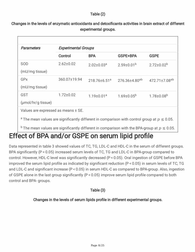
Effect of BPA and/or GSPE on serum lipid pro�leData represented in table 3 showed values of TC, TG, LDL-C and HDL-C in the serum of different groups.BPA signi�cantly (P < 0.05) increased serum levels of TC, TG and LDL-C in BPA-group compared tocontrol. However, HDL-C level was signi�cantly decreased (P < 0.05). Oral ingestion of GSPE before BPAimproved the serum lipid pro�le as indicated by signi�cant reduction (P < 0.05) in serum levels of TC, TGand LDL-C and signi�cant increase (P < 0.05) in serum HDL-C as compared to BPA-group. Also, ingestionof GSPE alone in the last group signi�cantly (P < 0.05) improve serum lipid pro�le compared to bothcontrol and BPA- groups.
Table (3)
Changes in the levels of serum lipids pro�le in different experimental groups.
Page 9/25
Parameters Experimental Groups
Control BPA GSPE+BPA GSPE
TC
(mg/dl)
156.32±2.26 290.51±1.65a 193.53±2.04ab 175.40±1.77ab
TG
(mg/dl)
145.19±3.15 236.80±9.34a 187.42±6.93ab 132.92±4.32ab
LDL-C
(mg/dl)
92.50±4.52 145.00±3.43a 110.61±2.02ab 72.35±2.31ab
HDL-C
(mg/dl)
55.64±1.62 31.22±2.02a 45.41±1.32ab 64.92±2.03ab
Values are expressed as means ± SE.
a The mean values are signi�cantly different in comparison with the control group at 𝑝 ≤ 0.05.
b The mean values are signi�cantly different in comparison with the BPA-group at 𝑝 ≤ 0.05.
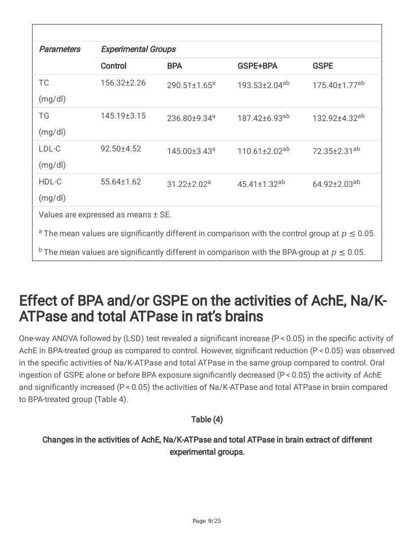
Effect of BPA and/or GSPE on the activities of AchE, Na/K-ATPase and total ATPase in rat’s brainsOne-way ANOVA followed by (LSD) test revealed a signi�cant increase (P < 0.05) in the speci�c activity ofAchE in BPA-treated group as compared to control. However, signi�cant reduction (P < 0.05) was observedin the speci�c activities of Na/K-ATPase and total ATPase in the same group compared to control. Oralingestion of GSPE alone or before BPA exposure signi�cantly decreased (P < 0.05) the activity of AchEand signi�cantly increased (P < 0.05) the activities of Na/K-ATPase and total ATPase in brain comparedto BPA-treated group (Table 4).
Table (4)
Changes in the activities of AchE, Na/K-ATPase and total ATPase in brain extract of differentexperimental groups.
Page 10/25
Parameters Experimental Groups
Control BPA GSPE+BPA GSPE
AchE
(nmol/min/mg protein)
87.15±1.33 101.65±4.07a 90.89±2.30b 81.18±3.88b
Na/K-ATPase
(µmol/min/mg protein)
3.81±0.12 1.52±0.02a 2.79±0.02ab 4.40±0.08ab
Total ATPase
(μmol/min/mg protein)
2.01±0.02 1.20±0.02a 1.79±0.01ab 2.80±0.02ab
Values are expressed as means ± SE.
a The mean values are signi�cantly different in comparison with the control group at 𝑝 ≤ 0.05.
b The mean values are signi�cantly different in comparison with the BPA-group at 𝑝 ≤ 0.05.
Effect of BPA and/or GSPE on dopamine and serotoninlevels in rat’s brainsThe data represented in Figs. 1 and 2 summarized the levels of dopamine and serotonin in the brain ofexperimentally studied animals. The results indicated that the brain levels of dopamine and serotoninwere signi�cantly increased (P < 0.05) in rats treated with BPA compared to control. On the contrary,treatment with GSPE before BPA in (GSPE + BPA)-treated group signi�cantly decreased (P < 0.05) levels ofdopamine and serotonin in the brain compared to BPA-group. Non-signi�cant differences were observedin the levels of dopamine and serotonin in brains of GSPE-group compared with that of control group.
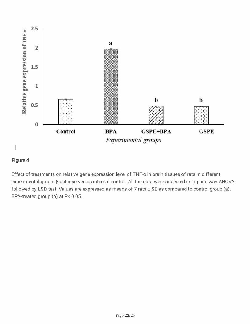
Effect of BPA and/or GSPE on gene expression levels ofTNF-α, COX-2 and p53 in rat’s brainsEffect of chronic exposure to BPA alone or after ingestion of GSPE on gene expression of TNF-α, COX-2and p53 in brain cells of different treated group was measured relative to β-actin and represented inFigs. 3–6. Results indicated that treatment with BPA signi�cantly induced (P < 0.05) expression of TNF-α,COX-2 and p53 genes (lane 2) in brain as compared to that of control group (lane 1). Oral ingestion ofGSPE alone (GSPE-group) or before BPA ingestion (GSPE + BPA-group) caused signi�cant repression (P < 0.05) in TNF-α, COX-2 and p53 mRNA expressions in brain (lanes 3 and 4) as compared to BPA-treatedgroup (lane 2). The mRNA expressions of TNF-α, COX-2 and p53 were barely detectable in GSPE + BPAand GSPE-treated groups and were non-signi�cantly different from that of control group.

Page 11/25
Discussion
Neurotoxic effects of bisphenol AThe brain is highly susceptible to oxidative damage because it uses high amounts of oxygen, has highquantities of polyunsaturated fatty acids and has relatively low antioxidant defence system (Li et al.2013). The present study revealed that BPA induced oxidative stress in brain tissues of rats as indicatedby increased nitric oxide and MDA, reduction of glutathione and total thiol levels and inhibition ofantioxidant enzymes; GPx, SOD and GST compared to control group. Thus, at the current dose of BPA, theantioxidant system of brain was unable to provide complete protection against the induced free radicaldamage. These results are in line with other study reported that BPA can increase levels of MDA and ROS,and reduce antioxidant defence system such as glutathione, catalase, and SOD (Hassani et al. 2017).The decreased activities of SOD and GPx in our study re�ected the increased ROS and lipid peroxidationlevels. BPA converted into reactive species by phase I enzymes system. The reduction in GST activityleads to non-neutralization of these reactive species and contribute to oxidative stress at the brain(Chahine &O'Donnell 2011). It has been reported that BPA accumulated in mitochondrial membrane andlead to inhibition of complex I of the respiratory chain, interference with electron transport and producemore ROS (Khan et al. 2016). The decreased brain GSH level in this study could be due to consumption ofGSH in large amount to combat BPA- induced free radical damage. The elevated LPO contents in thebrain tissues of BPA exposed rats in the present study could lead to acute damage of the brain cells. NO,at high concentration, and its metabolite ONOO and ROS are key mechanisms in both necrotic andapoptotic cell death which may implicated in destruction of neuronal cells, severe in�ammatory disordersand neurodegenerative diseases (Paul et al. 2018).
The chronic exposure to BPA in this study induced dyslipidimic state in the blood of experimental rats.This dyslipidimic state was con�rmed by increased levels of serum TC, TG and LDL-C accompanied todecreased level of serum HDL-C. The current results are matched with that of Eweda et al. (2020) whoreported that oral ingestion of 30 mg BPA/kg BW daily for 6 weeks induced disruption in serum lipidpro�le associated with accumulation of TG and cholesterol in liver tissues of rats (Eweda et al. 2020). Ithas been demonstrated that BPA disrupts the neuroendocrine mechanisms that regulate food intake andenergy expenditure which chronically may leads to obesity and dyslipidmic state (Stoker et al. 2020).BPA-induced hypercholesterolemia could increase the overproduction of ROS by increasing mitochondrialrespiration and decreasing the e�ciency of antioxidant system (Thiruchenduran et al. 2011).
Alterations in the neurotoxicity biomarkers as AChE, Na+/K+-ATPase, dopamine and serotonin have beenused to measure the neurotoxicity of environmental chemicals (Wu &Li 2015). AChE is a robust andreliable diagnostic and prognostic markers of neurotoxicity and neuro-in�ammation (Anand &Singh2013). Our results revealed an increase in the activity of AchE in BPA-treated group compared to control.The increased activity of AChE may suggest the neurotoxic effect of BPA at the dose administered. Onthe other hand, the chronic exposure to high dose of BPA reduced the activities of total and Na+/K+-ATPase in the brain cells of the same group. The reduction in N+/ K+-ATPase activity alters neuronal

Page 12/25
excitability and negatively affect sodium-calcium exchangers resulting in intracellular acidi�cation,calcium overload and neuronal damage (Shrivastava et al. 2020). Also, the decreased activity of total andNa+/K+-ATPase in brain may be attributed to the oxidative stress induced by the chronic exposure to BPAas illustrated in this study.
Our results revealed that treatment with BPA resulted in elevated dopamine and serotonin levels in brain.These results are in consistence with a study reported that subcutaneous administration of BPAincreased dopamine levels in brain tissue of pregnant mice ((Michałowicz 2014). Also, signi�cant effectswere observed in the levels of dopamine and serotonin in rat offspring orally administered BPA at highconcentrations (Honma et al. 2006). The increased dopamine and serotonin concentrations may resultfrom effects of BPA on dopamine re-uptake, monoamine oxidase (MAO) and serotonin release. Theelevated dopamine may be subjected to metabolism by monoamine oxidase or autoxidized generatingtoxic products such as hydrogen peroxide, oxygen‐derived radicals, semiquinones, and quinones whichmay exert neurotoxic effects (Ben‐Shachar et al. 1995). Also, the reduction of serotonin metabolites afteradministration of BPA suggesting a reduction in the activity of metabolic enzymes, such as MAO, andsubsequent elevation of serotonin in the brain (Negishi et al. 2004).
Damaged or diseased brain cells express and release high levels of the pro-in�ammatory cytokine, TNF-α(Montgomery &Bowers 2012). Also, COX-2 enzymes actively participate in in�ammation reactions andassociated in pathological mechanisms of aging and neurodegeneration (Lacy et al. 2016). Moreover,under stress or pathological conditions, p53 activate pro-oxidant genes, repress antioxidant genes andpromoting more severe pro-oxidant state (Chatoo et al. 2011). At the molecular level, our study revealedthat the chronic exposure to BPA increased gene expression levels of TNF-α, COX-2 and p53 in brain.These �ndings are in accordance with other results revealed that the sub-chronic exposure to BPAsigni�cantly increased gene expression of pro-in�ammatory cytokine TNF-α, IL-17, and IL-6 in male micewhich indicate on the neuroin�ammation and neurotoxicity (Khan et al. 2019). Also, it has been shownthat BPA exposure induces the expression of COX-2 and exacerbates in�ammation (Song et al. 2017).The over production of ROS and RNS, as in the present study, can induce p53 expression and productionof high pro-in�ammatory cytokines such as IL-6 and TNF-α (Solleiro-Villavicencio &Rivas-Arancibia2018). Moreover, it has been reported that TNF-α rapidly stimulate over production of damaging freeradicals via upregulation of COX-2 which may contribute in necrotic neuronal cell death (Phillis et al.2006). In addition, when the anti-oxidative capacity is greatly surpassed, p53 exhibits pro-oxidativeactivity that ends in cell death (Liu &Xu 2011). All these events ultimately contribute to widespreadneuronal death and the possibility of neurodegeneration disorders.
Protective effect of grape seeds proanthocyanidins againstBPA-neurotoxicityNatural products containing �avonoids, tannins and polyphenols (as Proanthocyanidins) have the abilityto protect against oxidative stress and could represent important tool for decreasing the progression of

Page 13/25
neurodegenerative diseases in animal and humans (Davinelli et al. 2016). The current study revealed theneuroprotective effects of GSPE against BPA-induced brain toxicity. Treatment of rats with GSPE beforeexposure to BPA resulted in signi�cant reduction in the brain oxidative stress as indicated by reduction ofMDA and NO to a levels comparable to that of control. Also, GSPE increased the levels of GSH and totalthiol contents and the activities of SOD, GPx and GST in brain cells compared to control group. Previousstudy showed that grape seed extract had a protective effect on brain by decreasing levels of MDA,scavenging of ROS and upregulating of antioxidant enzymes (Jiao et al. 2017). The anti-oxidativemechanisms of GSPE include radical scavenging action, singlet oxygen quenching action and chelatingaction. GSPE played a role in decreasing the iron levels, which participate in Fenton reaction, through theiriron chelation with their o-diphenol groups (Yun et al. 2020). The increased GSH content afteradministration of GSPE may be attributed to enhancement of its synthesis via increasing the expressionof γ-glutamyl-cysteine synthetase (Bagchi et al. 2002). The decreased NO level may be result fromreduced gene expression of iNOS mRNA by GSPE (Harbeoui et al. 2019). Also, the ability of GSPE torestore the normal levels of vitamin E and C could account for the reduction of free radical and oxidativestress in the brain of BPA-treated rats (Balu et al. 2006).
The pre-treatment of BPA group with GSPE attenuate the dyslipidemic state in these rats throughreduction of serum TC, TG and LDL-C associated with elevation of HDL-C. These results are in line withother research revealed that oral administration of GSPE for two months ameliorate the levels of totallipids, TC, TG, LDL-C and HDL-C in gibberellic acid-treated male rats (Hassan &Al-Rawi 2013). GSPE wasreported to develop lipid homeostasis by enhancing cholesterol removal in bile, decreased plasma TGand LDL-C levels and increased plasma HDL-C (Del Bas et al. 2005). GSPE-rich foods also inhibitdigestive enzymes and suppress fat and glucose absorption from the gut via inhibition of lipase andamylase (Salvadó et al. 2015). The feeding of HFD-rats with grape seed �our augmented fatty acidoxidation, increased lipolysis and decreased fatty acid synthesis in these rats which could explain theattenuation of dyslipidemic state induced by BPA (Zhou et al. 2019).
The neuroprotective effects of GSPE in the current study were observed in the restoration of the activitiesof AchE, Na/K-ATPase and total ATPase in brain tissue to levels comparable with the control group. Ourresults were in consistent with other study showed that the oral ingestion of GSPE increased theconcentration of acetylcholine with a moderate reduction in the activity of AchE in adult rats andminimizing age-related loss of cognition in older rats (Devi et al. 2006). The inhibition of AchE activityhas been described to boost brain cholinergic function and amend memory de�cit associated withneurodegenerative diseases (Adefeghaa et al. 2015). Also, Na+/K+-ATPase was suggested as a target fornatural compounds, such as GSPE (Almaliti et al. 2013). It has been reported that the neuroprotective roleof curcumin was attributed to the elevation in Na/K-ATPase activity in regions of rats’ brain (Vajraguptaet al. 2003). GSPE may attenuate or reduced neurotoxicity by its ability to increase Na+/K+-ATPaseactivity. On the other hand, the reduction in dopamine and serotonin levels were observed in the brain ofGSPE+BPA and GSPE-groups. The decreased levels of dopamine and serotonin may be resulted from

Page 14/25
counteraction of GSPE to BPA-induced inhibition of MAO and to the reduction in neurotransmitterreuptake.
The pre-treatment of BPA-group with GSPE downregulate the gene expression of TNF-α, COX-2 and p53.Our results are consistence with previous study reported that GSPE suppress gene expression of TNF-αand COX-2 (Chu et al. 2016). The anti-in�ammatory effect of GSPE could attributed to upregulation ofperoxisome proliferator-activated receptor-γ (PPAR-γ) expression and subsequent downregulation of COX-2 expression (Liu et al. 2020). The decreased BPA-induced neuroin�ammation by GSPE may be due toreduction of NO level via down-regulation of iNOS expression (Zhou et al. 2011). Also, p53 depletionattenuate neuronal death and protect neurons against apoptotic stimuli via decreased p53 DNA-bindingactivity, inhibition of caspases and prevention of mitochondrial dysfunction (Jembrek et al. 2018). Thus,the administration of GSPE might represent a promising approach for treatment of neuroin�ammation,neurotoxicity and several neurodegenerative conditions.
ConclusionOur research revealed that the chronic exposure to BPA was associated with brain oxidative stress,neuroin�ammation and injury in neural tissue which may lead to degenerative diseases. The BPA-inducedneuronal oxidative stress leads to alteration in neuro-speci�c enzymes; AchE, Na/K-ATPase and totalATPase and in neurotransmitters; dopamine and serotonin. These neuronal changes result inneuroin�ammation, neurotoxicity and might neuronal death as indicated by overexpression of TNF-α,COX-2 enzyme and p53 protein. Also, the study demonstrated the protective effects of GSPE against theabove mentioned BPA-induced neurotoxicity as indicated by the attenuation or amelioration of alloxidative and neurotoxic parameters induced by BPA. Our observations add to the body of evidence thatGSPE may be a novel therapeutic agent for treating neurodegenerative disorders.
Declarations
Author contributionsHeba Mohamed Abdou and Heba-Tallah Abd Elkader conceived, designed the study and collect the data.Heba Mohamed Abdou, Heba-Tallah Abd Elkader, Saber M. Eweda and Amel H. El-Gendy Performed theexperiments, analyzed the data, prepared �gures and/or tables. Saber M. Eweda wrote the manuscript. Allauthors read, authored and approved the �nal manuscript submitted for publication.
FundingThis research did not receive any speci�c grant from funding agencies in the public, commercial, or not-for-pro�t sectors.

Page 15/25
Availability of data and materials All the data and material were available. The data of this article are included within the article and itsadditional �les.
Ethics approval and consent to participate All the experimental and animal handling procedures were performed according to the guidelinesapproved by Alexandria University Institutional Animal Care and Use Committee (ALEXU-IACUC), amember of the International Council for Laboratory Animal Science (ICLAS) (Approval number:AU04200125201).
Consent for publicationAll co-authors agree to publish this manuscript in the Journal (Environmental pollution and research)
Con�ict of InterestThe authors declare that they have no con�icts of interest.
References1. Adefeghaa S, Oboha G, Odubanjoa T, Ogunsuyia O (2015) A comparative study on the antioxidative
activities, anticholinesterase properties and essential oil composition of clove (Syzygiumaromaticum) bud and Ethiopian pepper (Xylopia aethiopica). Riv. Ital. Sost. Gr 92: 257-268
2. Agarwal S, Yadav A, Tiwari SK, Seth B, Chauhan LKS, Khare P, Ray RS, Chaturvedi RK (2016)Dynamin-related protein 1 inhibition mitigates bisphenol A-mediated alterations in mitochondrialdynamics and neural stem cell proliferation and differentiation. Journal of Biological Chemistry 291:15923-15939
3. Almaliti J, Nada SE, Carter B, Shah ZA, Tillekeratne LV (2013) Natural products inspired synthesis ofneuroprotective agents against H2O2-induced cell death. Bioorganic & medicinal chemistry letters23: 1232-1237
4. Anand P, Singh B (2013) A review on cholinesterase inhibitors for Alzheimer’s disease. Archives ofpharmacal research 36: 375-399
5. Bagchi D, Bagchi M, Stohs SJ, Ray SD, Sen CK, Preuss HG (2002) Cellular protection withproanthocyanidins derived from grape seeds. Annals of the New York Academy of Sciences 957:260-270
Page 16/25
�. Balu M, Sangeetha P, Murali G, Panneerselvam C (2006) Modulatory role of grape seed extract onage-related oxidative DNA damage in central nervous system of rats. Brain research bulletin 68: 469-473
7. Ben‐Shachar D, Zuk R, Glinka Y (1995) Dopamine neurotoxicity: inhibition of mitochondrialrespiration. Journal of neurochemistry 64: 718-723
�. Braun JM, Muckle G, Arbuckle T, Bouchard MF, Fraser WD, Ouellet E, Séguin JR, Oulhote Y, WebsterGM, Lanphear BP (2017) Associations of prenatal urinary bisphenol A concentrations with childbehaviors and cognitive abilities. Environmental health perspectives 125: 067008
9. Chahine S, O'Donnell MJ (2011) Interactions between detoxi�cation mechanisms and excretion inMalpighian tubules of Drosophila melanogaster. Journal of Experimental Biology 214: 462-468
10. Chatoo W, Abdouh M, Bernier G (2011) p53 pro-oxidant activity in the central nervous system:implication in aging and neurodegenerative diseases. Antioxidants & redox signaling 15: 1729-1737
11. Chomczynski P, Sacchi N (2006) The single-step method of RNA isolation by acid guanidiniumthiocyanate–phenol–chloroform extraction: twenty-something years on. Nature protocols 1: 581-585
12. Chu H, Tang Q, Huang H, Hao W, Wei X (2016) Grape-seed proanthocyanidins inhibit thelipopolysaccharide-induced in�ammatory mediator expression in RAW264. 7 macrophages bysuppressing MAPK and NF-κb signal pathways. Environmental toxicology and pharmacology 41:159-166
13. Chu KO, Chan S-O, Pang CP, Wang CC (2014) Pro-oxidative and antioxidative controls and signalingmodi�cation of polyphenolic phytochemicals: contribution to health promotion and diseaseprevention? Journal of agricultural and food chemistry 62: 4026-4038
14. Davinelli S, Maes M, Corbi G, Zarrelli A, Willcox DC, Scapagnini G (2016) Dietary phytochemicals andneuro-in�ammaging: from mechanistic insights to translational challenges. Immunity & Ageing 13:16
15. Del Bas JM, Fernández-Larrea J, Blay M, Ardèvol A, Salvadó MJ, Arola L, Bladé C (2005) Grape seedprocyanidins improve atherosclerotic risk index and induce liver CYP7A1 and SHP expression inhealthy rats. The FASEB journal 19: 1-24
1�. Devi A, Jolitha AB, Ishii N (2006) Grape Seed Proanthocyanidin Extract (GSPE) and antioxidantdefense in the brain of adult rats. Medical Science Monitor 12: BR124-BR129
17. Draper H, Hadley M (1990) [43] Malondialdehyde determination as index of lipid Peroxidation,Methods in enzymology. Elsevier, pp. 421-431
1�. Ellman GL, Courtney KD, Andres Jr V, Featherstone RM (1961) A new and rapid colorimetricdetermination of acetylcholinesterase activity. Biochemical pharmacology 7: 88-95
19. Eweda SM, Newairy ASA, Abdou HM, Gaber AS (2020) Bisphenol A‐induced oxidative damage in thehepatic and cardiac tissues of rats: The modulatory role of sesame lignans. Experimental andTherapeutic Medicine 19: 33-44
20. Flohe L, Günzler WA (1984) [12] Assays of glutathione peroxidase. Methods in enzymology 105: 114-120

Page 17/25
21. Gri�th OW (1980) Determination of glutathione and glutathione disul�de using glutathionereductase and 2-vinylpyridine. Analytical biochemistry 106: 207-212
22. Guevara I, Iwanejko J, Dembińska-Kieć A, Pankiewicz J, Wanat A, Anna P, Gołąbek I, Bartuś S,Malczewska-Malec M, Szczudlik A (1998) Determination of nitrite/nitrate in human biologicalmaterial by the simple Griess reaction. Clinica Chimica Acta 274: 177-188
23. Habig WH, Pabst MJ, Jakoby WB (1974) Glutathione S-transferases the �rst enzymatic step inmercapturic acid formation. Journal of biological Chemistry 249: 7130-7139
24. Harbeoui H, Hichami A, Wannes WA, Lemput J, Tounsi MS, Khan N (2019) Anti-in�ammatory effectof grape (Vitis vinifera L.) seed extract through the downregulation of NF-κB and MAPK pathways inLPS-induced RAW264. 7 macrophages. South African Journal of Botany 125: 1-8
25. Hassan HA, Al-Rawi MM (2013) Grape seeds proanthocyanidin extract as a hepatic-reno-protectiveagent against gibberellic acid induced oxidative stress and cellular alterations. Cytotechnology 65:567-576
2�. Hassani FV, Mehri S, Abnous K, Birner-Gruenberger R, Hosseinzadeh H (2017) Protective effect ofcrocin on BPA-induced liver toxicity in rats through inhibition of oxidative stress and downregulationof MAPK and MAPKAP signaling pathway and miRNA-122 expression. Food and ChemicalToxicology 107: 395-405
27. Honma T, Miyagawa M, Suda M, Wang R-S, Kobayashi K, Sekiguchi S (2006) Effects of perinatalexposure to bisphenol A on brain neurotransmitters in female rat offspring. Industrial health 44: 510-524
2�. Jembrek MJ, Slade N, Hof PR, Šimić G (2018): The interactions of p53 with tau and Aß as potentialtherapeutic targets for Alzheimer’s disease. Progress in neurobiology 168: 104-127
29. Jiao J, Wei Y, Chen J, Chen X, Zhang Y (2017): Anti-aging and redox state regulation effects of A-typeproanthocyanidins-rich cranberry concentrate and its comparison with grape seed extract in mice.Journal of Functional Foods 30: 63-73
30. Khan J, Salhotra S, Goswami P, Akhter J, Jahan S, Gupta S, Sharma S, Banerjee BD, Parvez S, GuptaS (2019): Bisphenol A triggers axonal injury and myelin degeneration with concomitantneurobehavioral toxicity in C57BL/6J male mice. Toxicology 428: 152299
31. Khan S, Beigh S, Chaudhari BP, Sharma S, Aliul Hasan Abdi S, Ahmad S, Ahmad F, Parvez S,Raisuddin S (2016) Mitochondrial dysfunction induced by Bisphenol A is a factor of itshepatotoxicity in rats. Environmental toxicology 31: 1922-1934
32. Lacy SH, Woeller CF, Thatcher TH, Maddipati KR, Honn KV, Sime PJ, Phipps RP (2016) Human lung�broblasts produce proresolving peroxisome proliferator-activated receptor-γ ligands in acyclooxygenase-2-dependent manner. American Journal of Physiology-Lung Cellular and MolecularPhysiology 311: L855-L867
33. Li J, Li W, Jiang Z-G, Ghanbari HA (2013) Oxidative stress and neurodegenerative disorders.International journal of molecular sciences 14: 24438-24475
34. Liu D, Xu Y (2011): p53, oxidative stress, and aging. Antioxidants & redox signaling 15: 1669-1678

Page 18/25
35. Liu J, Hu S, Zhu B, Shao S, Yuan L (2020) Grape seed procyanidin suppresses in�ammation incigarette smoke-exposed pulmonary arterial hypertension rats by the PPAR-γ/COX-2 pathway.Nutrition, Metabolism and Cardiovascular Diseases 30: 347-354
3�. Lowry OH, Rosebrough NJ, Farr AL, Randall RJ (1951) Protein measurement with the Folin phenolreagent. Journal of biological chemistry 193: 265-275
37. Marklund S, Marklund G (1974) Involvement of the superoxide anion radical in the autoxidation ofpyrogallol and a convenient assay for superoxide dismutase. European journal of biochemistry 47:469-474
3�. Michałowicz J (2014) Bisphenol A–sources, toxicity and biotransformation. Environmentaltoxicology and pharmacology 37: 738-758
39. Montgomery SL, Bowers WJ (2012) Tumor necrosis factor-alpha and the roles it plays inhomeostatic and degenerative processes within the central nervous system. Journal ofneuroimmune pharmacology 7: 42-59
40. Musachio EAS, Araujo SM, Bortolotto VC, de Freitas Couto S, Dahleh MMM, Poetini MR, Jardim EF,Meichtry LB, Ramborger BP, Roehrs R (2020) Bisphenol A exposure is involved in the development ofParkinson like disease in Drosophila melanogaster. Food and Chemical Toxicology 137: 111128
41. Nassiri‐Asl M, Hosseinzadeh H (2009) Review of the pharmacological effects of Vitis vinifera (Grape)and its bioactive compounds. Phytotherapy Research: An International Journal Devoted toPharmacological and Toxicological Evaluation of Natural Product Derivatives 23: 1197-1204
42. Negishi T, Kawasaki K, Suzaki S, Maeda H, Ishii Y, Kyuwa S, Kuroda Y, Yoshikawa Y (2004) Behavioralalterations in response to fear-provoking stimuli and tranylcypromine induced by perinatal exposureto bisphenol A and nonylphenol in male rats. Environmental health perspectives 112: 1159-1164
43. Niranjan R (2018) Recent advances in the mechanisms of neuroin�ammation and their roles inneurodegeneration. Neurochemistry international 120: 13-20
44. Paul R, Phukan BC, Thenmozhi AJ, Manivasagam T, Bhattacharya P, Borah A (2018) Melatoninprotects against behavioral de�cits, dopamine loss and oxidative stress in homocysteine model ofParkinson's disease. Life sciences 192: 238-245
45. Phillis JW, Horrocks LA, Farooqui AA (2006) Cyclooxygenases, lipoxygenases, and epoxygenases inCNS: their role and involvement in neurological disorders. Brain research reviews 52: 201-243
4�. Prasain JK, Peng N, Dai Y, Moore R, Arabshahi A, Wilson L, Barnes S, Wyss JM, Kim H, Watts RL(2009) Liquid chromatography tandem mass spectrometry identi�cation of proanthocyanidins in ratplasma after oral administration of grape seed extract. Phytomedicine 16: 233-243
47. Raisuddin S, Sharma S (2017) Endocrine-disrupting chemicals in food and their toxicologicalimplications, Food Toxicology. Apple Academic Press, pp. 199-250
4�. Resnik DB, Elliott KC (2015) Bisphenol A and risk management ethics. Bioethics 29: 182-189
49. Salvadó MJ, Casanova E, Fernández-Iglesias A, Arola L, Bladé C (2015) Roles of proanthocyanidinrich extracts in obesity. Food & function 6: 1053-1071
Page 19/25
50. Sedlak J, Lindsay RH (1968) Estimation of total, protein-bound, and nonprotein sulfhydryl groups intissue with Ellman's reagent. Analytical biochemistry 25: 192-205
51. Shan Y, Ye X-h, Xin H (2010) Effect of the grape seed proanthocyanidin extract on the free radical andenergy metabolism indicators during the movement. Scienti�c Research and Essays 5: 148-153
52. Shrivastava AN, Triller A, Melki R (2020) Cell biology and dynamics of Neuronal Na+/K+-ATPase inhealth and diseases. Neuropharmacology 169: 107461
53. Solleiro-Villavicencio H, Rivas-Arancibia S (2018) Effect of chronic oxidative stress onneuroin�ammatory response mediated by CD4+ T cells in neurodegenerative diseases. Frontiers incellular neuroscience 12: 114
54. Song H, Park J, Bui PT, Choi K, Gye MC, Hong Y-C, Kim JH, Lee YJ (2017) Bisphenol A induces COX-2through the mitogen-activated protein kinase pathway and is associated with levels of in�ammation-related markers in elderly populations. Environmental research 158: 490-498
55. Stoker C, Andreoli MF, Kass L, Bosquiazzo VL, Rossetti MF, Canesini G, Luque EH, Ramos JG (2020)Perinatal exposure to bisphenol A (BPA) impairs neuroendocrine mechanisms regulating food intakeand kisspetin system in adult male rats. Evidences of metabolic disruptor hypothesis. Molecular andcellular endocrinology 499: 110614
5�. Sutcliffe TC, Winter AN, Punessen NC, Linseman DA (2017) Procyanidin B2 protects neurons fromoxidative, nitrosative, and excitotoxic stress. Antioxidants 6: 77
57. Thiruchenduran M, Vijayan NA, Sawaminathan JK, Devaraj SN (2011) Protective effect of grape seedproanthocyanidins against cholesterol cholic acid diet-induced hypercholesterolemia in rats.Cardiovascular Pathology 20: 361-368
5�. Tyl R, Myers C, Marr M, Thomas B, Keimowitz A, Brine D, Veselica M, Fail P, Chang T, Seely J (2002)Three-generation reproductive toxicity study of dietary bisphenol A in CD Sprague-Dawley rats.Toxicological Sciences 68: 121-146
59. Vajragupta O, Boonchoong P, Watanabe H, Tohda M, Kummasud N, Sumanont Y (2003) Manganesecomplexes of curcumin and its derivatives: evaluation for the radical scavenging ability andneuroprotective activity. Free Radical Biology and Medicine 35: 1632-1644
�0. Wu J-P, Li M-H (2015) Inhibitory effects of pain relief drugs on neurological enzymes: implications ontheir potential neurotoxicity to aquatic animals. Environmental toxicology and pharmacology 39:898-905
�1. Yousef M, Saad A, El-Shennawy L (2009) Protective effect of grape seed proanthocyanidin extractagainst oxidative stress induced by cisplatin in rats. Food and Chemical Toxicology 47: 1176-1183
�2. Yun S, Chu D, He X, Zhang W, Feng C (2020) Protective effects of grape seed proanthocyanidinsagainst iron overload-induced renal oxidative damage in rats. Journal of Trace Elements in Medicineand Biology 57: 126407
�3. Zhang S, Cui Y, Li L, Li Y, Zhou P, Luo L, Sun B (2015) Preparative HSCCC isolation ofphloroglucinolysis products from grape seed polymeric proanthocyanidins as new powerfulantioxidants. Food chemistry 188: 422-429

Page 20/25
�4. Zhao J, Bi W, Xiao S, Lan X, Cheng X, Zhang J, Lu D, Wei W, Wang Y, Li H (2019) Neuroin�ammationinduced by lipopolysaccharide causes cognitive impairment in mice. Scienti�c reports 9: 1-12
�5. Zhou D-y, Du Q, Li R-r, Huang M, Zhang Q, Wei G-z (2011) Grape seed proanthocyanidin extractattenuates airway in�ammation and hyperresponsiveness in a murine model of asthma bydownregulating inducible nitric oxide synthase. Planta medica 77: 1575-1581
��. Zhou F, Yin M, Liu Y, Han X, Guo J, Ren C, Wang W, Huang W, Zhan J, You Y (2019) Grape seed �ourintake decreases adiposity gain in high-fat-diet induced obese mice by activating thermogenesis.Journal of Functional Foods 62: 103509
Figures
Figure 1
Effect of treatments on dopamine levels in brain tissues of rats in different experimental group. All thedata were analyzed using one-way ANOVA followed by LSD test. Values are expressed as means of 7 rats± SE as compared to control group (a), BPA-treated group (b) at P< 0.05.

Page 21/25
Figure 2
Effect of treatments on serotonin levels in brain tissues of rats in different experimental group. All thedata were analyzed using one-way ANOVA followed by LSD test. Values are expressed as means of 7 rats± SE as compared to control group (a), BPA-treated group (b) at P< 0.05.

Page 22/25
Figure 3
Effect of treatments on reverse-transcriptase PCR (RT-PCR) analysis of p53, COX-2 and TNF-α mRNAexpression in brain tissues of rats in different experimental group. β-actin serves as the loading control.
Page 23/25
Figure 4
Effect of treatments on relative gene expression level of TNF-α in brain tissues of rats in differentexperimental group. β-actin serves as internal control. All the data were analyzed using one-way ANOVAfollowed by LSD test. Values are expressed as means of 7 rats ± SE as compared to control group (a),BPA-treated group (b) at P< 0.05.

Page 24/25
Figure 5
Effect of treatments on relative gene expression level of COX-2 in brain tissues of rats in differentexperimental group. β-actin serves as internal control. All the data were analyzed using one-way ANOVAfollowed by LSD test. Values are expressed as means of 7 rats ± SE as compared to control group (a),BPA-treated group (b) at P< 0.05.

Page 25/25
Figure 6
Effect of treatments on relative gene expression level of p53 in brain tissues of rats in differentexperimental group. β-actin serves as the internal control. All the data were analyzed using one-wayANOVA followed by LSD test. Values are expressed as means of 7 rats ± SE as compared to control group(a), BPA-treated group (b) at P< 0.05.
Supplementary Files
This is a list of supplementary �les associated with this preprint. Click to download.
Graphicalabstarct.jpg