of laparoscopic diagnosis upper tract infection
TRANSCRIPT
Infectious Diseases in Obstetrics and Gynecology 5:291-296 (I 997)(C) 1998 Wiley-Liss, Inc.
Performance of Clinical and Laparoscopic Criteriafor the Diagnosis of Upper Genital Tract Infection
Jeffrey F. Peipert,* Lori A. Boardman, 1 and C. James Sung21Division ofAmbulatory Care, Department of Obstetrics and Gynecology, Women and Infants’
Hospital, Broach Universily School ofMedMne, Providence, RIeDepartment of Pathology, Women and Infants’ Hospital, Broavn Universit School ofMedicine,
Providence, RI
ABSTRACT
Objective: The purpose of this study was to validate the standard minimal clinical criteria and thelaparoscopic triad of tubal edema, erythema, and purulent exudate used to diagnose acute uppergenital tract infection.
Methods: Subjects included women who either met the Centers for Disease Control and Pre-vention’s (CDC) minimal criteria for acute pelvic inflammatory disease or had other signs of uppergenital tract infection (i.e., atypical pelvic pain, abnormal uterine bleeding, or cervicitis). The sub-jects were evaluated with a baseline interview, comprehensive laboratory testing, and either anendometrial biopsy or laparoscopy with endometrial and fimbrial biopsies for definitive diagnosis ofupper genital tract infection. Patients were considered positive for upper genital tract infection ifthey had any of the following findings: 1) histologic evidence of endometritis or salpingitis; 2)laparoscopic visualization of purulent exudate in the pelvis without another source; or 3) positivetesting for Neisseria gonorrhoeae or Chlamydia trachomatis from the endometrium, fallopian tubes, orpelvis.
Results: One hundred twenty-nine women with adequate endometrial samples were evaluatedbetween August 1993 and September 1997, and 62 had complete laparoscopic evaluations. Thesensitivities of the CDC’s minimal clinical criteria for pelvic inflammatory disease and the laparo-scopic triad of edema, erythema, and purulent exudate were 65% and 60%, respectively.
Conclusions: Commonly used minimal clinical criteria for pelvic inflammatory disease and thelaparoscopic triad of tubal edema, erythema, and purulent exudate have limited sensitivity withcorrespondingly high false negative rates. Infect. Dis. Obstet. Gynecol. 5:291-296, 1997.(C) 1998 Wiley-Liss, Inc.
KEY WORDS
adnexitis; endometritis; salpingitis; pelvic inflammatory disease; diagnosis
pper genital tract infection, commonly re-
ferred to as pelvic inflammatory disease(PID), is often a difficult diagnosis to make. Accu-rate diagnosis is extremely important for the man-
agement of the acute disease and the prevention of
long-term morbidity. The Centers for DiseaseControl and Prevention (CDC) suggests that em-
piric treatment should be instituted on the basis ofthe presence of abdominal tenderness, adenxaltenderness, and cervical motion tenderness. Only
Presented in part at the Annual Meeting of the Infectious Diseases Society in Obstetrics and Gynecology, Monterey, CA,1994.Contract grant sponsor: American Association for AIDS Research; Contract grant number: 02110-15-RG; Contract grantsponsor: Rhode Island Foundation.
*Correspondence to: Dr. Jeffrey F. Peipert, Department of Obstetrics and Gynecology, Women and Infants’ Hospital, 101Dudley Street, Providence, RI 02905. E-mail: jpeipert@wihri
Received 18 April 1997Clinical Study Accepted 22 September 1997
CRITERIA FOR UGTI PEIPERT ETAL.
recently has an attempt been made to validate theminimal criteria, and in this report the sensitivity ofthese criteria was quite low (33%).z
Laparoscopic visualization of tubal edema, ery-thema, and purulent exudate is considered the"gold standard" for the diagnosis of acute PID.3’4
However, the accuracy of laparoscopy for the diag-nosis of acute PID has been challenged. Sellors et
al. 5 demonstrated that laparoscopic visualizationhas a sensitivity of 50% and a specificity of approxi-mately 85% compared to diagnosis by fimbrial
minibiopsy. These findings have resulted in some
investigators questioning whether laparoscopyshould still be considered the "gold standard" forthe diagnosis of PID.The purpose of this study is to prospectively
validate the CDC’s minimal clinical criteria for thediagnosis of PID and the laparoscopic triad of tubaledema, erythema, and purulent exudate comparedto predefined objective criteria for upper genitaltract infection. Diagnostic test characteristics are
calculated for the minimal clinical criteria, inde-
pendent clinical signs, individual laparoscopic find-
ings, and laparoscopic triad of edema, erythema,and purulent exudate.
While infection of the uterus, fallopian tubes,ovaries, and pelvis is commonly referred to as acute
PID, others consider acute PID only synonymouswith acute salpingitis. To avoid this controversy,
the term upper genital tract infection will be usedthroughout this manuscript.
SUBJECTS AND METHODS
A cross-sectional study was performed in order to
evaluate currently available diagnostic testing andcriteria for the diagnosis of acute upper genital tract
infection. The methodology for this study has beenreported previously.6 Informed consent was ob-tained from women who chose to participate in thestudy if they presented with either "classic" or
"non-classic" signs of upper genital tract infection
(defined below). We invited consecutive women
identified with signs or symptoms of upper genitaltract infection to participate. Patients were re-
cruited from an urgent care unit of a large urbanwomen’s hospital and referred from adjacent clinicsand practices in the hospital vicinity.
Patients with "classic" signs of upper genitaltract infection presented with pelvic pain (less than30 days duration) and were found to have the three
CDC stated criteria of abdominal, cervical motion,and adnexal tenderness. The second group in-
cluded women presenting with "non-classic" signsof upper genital tract infection. These signs in-cluded atypical pelvic pain (i.e., pain less than 30
days duration that did not meet the CDC’s mini-mal criteria for acute PID), abnormal uterine
bleeding of unclear etiology (in women less thanthe age of 40 years to avoid those who were peri-menopausal) or cervicitis as demonstrated by a mu-
copurulent cervical discharge or a positive cervical
microbiologic test for N. gonorrhoeae and/or C. tra-
chomatis. The choice of this broad range of signs of
upper genital tract infection was deliberate: the in-
tention was to include a full spectrum of cases re-
quired to evaluate the chosen diagnostic tests. 7,8
All participants were between the ages of 16 and45 years; women less than age 18 years were in-
cluded if they were legally able to consent or ifinformed consent could be obtained from a legalguardian. Exclusion criteria included pregnancy,delivery, or operative procedure within the preced-ing 4 weeks, inability to give informed consent, or
a history of hysterectomy or bilateral salpingec-tomy. Women with tubal ligation were not ex-
cluded.Evaluation began with a comprehensive enroll-
ment interview reviewing past medical, obstetrical,and gynecological and sexual history. Physical ex-
amination and laboratory testing including com-
plete blood count, erythrocyte sedimentation rate,
and C-reactive protein were then performed. Ab-dominal and pelvic tenderness were recorded andgraded based on a modification of the McCormacket al.9 scale. Vaginal secretions were screened forwhite blood cells and evaluated for evidence of
vaginitis as documented by the presence of clue
cells, trichomonads, and pseudohyphae, or spores.Cervical cultures were obtained for N. gonor-
rhoeae and C. trachomatis. Polymerase chain reaction
(PCR) testing was also obtained for C. trachomatis.Endometrial biopsies were sent for the same cul-tures as well as for aerobic and anaerobic bacteria.All participants underwent either endometrial bi-
opsy alone or a combination of endometrial biopsyand laparoscopy in order to obtain objective evi-
dence of acute upper genital tract infection. Thechoice of either a laparoscopic evaluation or an en-
dometrial biopsy alone was made by the patient.While all participants with pain or tenderness were
292 INFECTIOUS DISEASES IN OBSTETRICS AND GYNECOLOGY

CRITERIA FOR UGTI PEIPERTETAL.
offered a laparoscopic evaluation, subjects withoutthese findings were not offered this option as a
laparoscopy was not felt to be clinically indicated.Although we recognized that this two-tiered evalu-ation process could introduce a selection or detec-tion bias, we felt it was necessary to capture thewide spectrum of upper tract infections.
If laparoscopy was performed, tubal and perito-neal cultures along with a fimbrial minibiopsy wereadded to the above microbiologic studies. If thefallopian tubes were occluded and appeared dis-tended, then an aspiration of the tubes was per-formed to identify purulent material and to obtaina specimen for microbiologic evaluation.We used objective criteria for our outcome of
upper genital tract infection. These criteria in-eluded any of the following: 1) histologic evidenceof acute endometritis or salpingitis; 2) laparoscopicevidence of salpingitis as demonstrated’ by puru-lent exudate in the pelvis with no other source; or
3) microbiologic evidence of N. gonorrhoeae or C.trachomatis from the endometrium, fallopian tubes,or pelvis. For acute endometritis to be verified,neutrophils had to be present in one or more of thefollowing areas in the absence of tissue disintegra-tion such as menstrual changes: the surface epithe-lium, the subepithelial stroma, and/or the endome-trial glands. Although not part of our objective cri-teria for acute upper genital tract infection, we alsoevaluated whether using chronic endometritiswould change our results. Chronic endometritiswas defined by the presence of plasma cells with or
without stromal fibrosis. Acute or chronic salpingi-tis was defined as the presence of neutrophils or
plasma cells in the lamina propria of the tubal mu-cosa. The mere presence of inflammatory cells inengorged blood vessels would be considered nega-tive.1
In addition to our objective definition of uppergenital tract infection, we performed additionalanalyses of the subset ofwomen who were found to
have laparoscopic or histologic evidence of salpin-gitis. In this subgroup, we include women with thelaparoscopic triad of edema, erythema, and puru-lent exudate, visualization of a tubo-ovarian ab-scess, or histologic evidence of acute or chronicsalpingitis.The study protocol was approved by the
Women and Infants’ Hospital Institutional ReviewBoard prior to initiation of the research. Statistical
analysis was performed using Xz amd Fisher exact
tests for categorical variables, unpaired t-tests forcontinuous variables, and non-parametric tests forcontinuous variables that were not normally distrib-uted. P < 0.05 was considered statistically signifi-cant. Sensitivity and specificity were calculated.Since predictive values are so dependent on theprevalence of the disease in the population, we
used Bayes’ theorem to calculate predictive valuesand based these calculations on prevalence rates of30% and 60%. Separate analyses were performedincluding women with chronic endometritis to see
if the results would vary. Statistical Analysis Soft-ware (SAS Institute, Cary, NC) was used for theanalysis.
RESULTSBetween August 1993 and September 1997, 132women were evaluated for evidence of upper geni-tal tract infection. Three women were excludeddue to inadequate endometrial samples. Sixty-fiveof the 129 patients (52%) underwent an endome-trial biopsy and cultures, while 62 (48%) had cul-tures, endometrial biopsy, and laparoscopy.The demographic characteristics of the entire
study population were as follows: median age 24years (range 16-45 years), median gravidity 2(range 0-12), median years of education 12 (range3-16 years). The median number of sexual partnersin the 6 months prior to study inclusion was witha range of 0-5, and the median number of lifetimesexual partners was 4 with a range from to >100.There were no significant differences in age, gra-vidity, education, race, insurance or marital status,
smoking history, or history of sexually transmitteddiseases or PID between patients who underwentlaparoscopic evaluations and those who were evalu-ated with endometrial biopsy alone. As expected,participants who met the minimal clinical criteriafor the diagnosis of PID were more likely to un-
dergo laparoscopic evaluation than women with"non-classic" presentations (P 0.001).
Of the 70 women who met the minimal criteriafor PID, 40% were found to have objective evi-dence of acute upper genital tract infection. Of the43 participants with objective evidence of uppergenital tract infection, 28 met the minimal criteriafor PID (sensitivity 65%). Of the 59 women with"non-classic" signs of upper genital tract infection,25% were found to have objective evidence of up-
INFECTIOUS DISEASES IN OBSTETRICS AND GYNECOLOGY 293

CRITERIA FOR UGTI PEIPERT ETAL.
TABLE I. Objective evidence of upper genital tractinfection (UGTI) by indication for evaluation
IndicationNo. No. %
evaluated positive Positive
Minimal CDC criteria for acute
PID (i.e., "classic" UGTI) 70 28 40"Non-classic" UGTI 59 14 25
Atypical pain 32 7 22Cervicitis 12 4 33Abnormal bleeding 13 2 15Abnormal bleeding
and cervicitis 2 2 100
per genital tract infection. Table 1 shows the re-
suits of the evaluations by indication for enroll-ment. Six of the 14 women (43%) with evidence ofmucopurulent cervical discharge or a positive cer-vical test for N. gonorrhoeae or C. trachomatis, butwho did not meet the CDC’s minimal criteria forPID, were found to have objective evidence of up-per genital tract infection.The diagnostic test characteristics and predic-
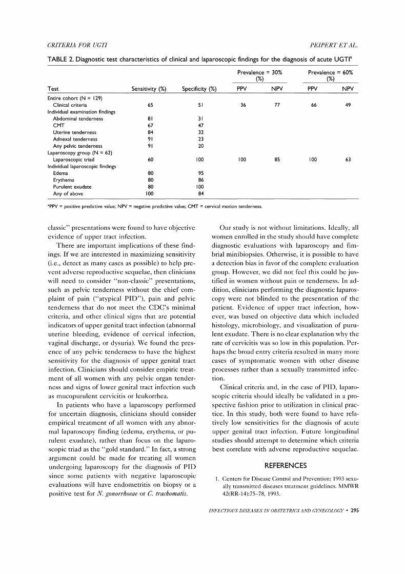
tive values of the clinical and laparoscopic findingsbased on prevalences of 30% and 60% were calcu-lated (Table 2). The most sensitive physical exami-nation finding was the presence of any pelvic ten-
derness (91%). The minimal clinical criteria for thediagnosis of PID had a sensitivity of 65% and speci-ficity of 51%. In the 62 evaluable patients withcomplete laparoscopic evaluation, the laparoscopictriad of edema, erythema, and purulent exudatehad a sensitivity of 60% and a specificity of 100%.If any one of these findings was noted at the timeof the laparoscopy, the sensitivity was 100% andspecificity was 84%. Separate analyses were per-formed using chronic endometritis as a positive in-dicator of upper genital tract infection. The resultsvaried only slightly (_+6%).
There were 48 patients who had a laparoscopicevaluation, but did not have the classic triad ofedema, erythema, and purulent exudate. Eight(17%) were found to have histologic evidence ofacute or chronic endometritis. Two of these 48 pa-tients (4%) had microbiologic evidence of chlamyd-ial infection in the upper genital tract. Forty of the48 patients had an adequate fimbrial biopsy. Fourof 40 patients (10%) without the laparoscopic triadhad histologic evidence of salpingitis.
Of the 18 patients with either histologic or lap-aroscopic evidence of salpingitis (laparoscopic triador tubo-ovarian abscess), 6 (33%) had normal en-
dometrial biopsies. Four of 9 patients with histo-
logic evidence of salpingitis did not have the classic
laparoscopic triad of edema, erythema, and puru-lent exudate. Of these 4 patients, 2 had erythemaalone, had edema and erythema but no purulentexudate, and had erythema and purulent exudatebut no tubal edema.
DISCUSSION
Clinical accuracy in the diagnosis of such uppergenital tract infections is currently imprecise; nu-
merous reports comparing clinical and laparoscopicdiagnoses of pelvic inflammatory disease havedemonstrated the lack of correlation between thetwo methods.9,11-,lz In this study, we have at-
tempted to validate the accepted clinical criteria
and the classic laparoscopic criteria for PID. A clini-
cal research dilemma is how to validate what is
currently accepted as the "gold standard" (i.e., lap-aroscopic visualization). We did this by creating ob-jective criteria that would be maximally sensitiveand reflect several measures of infection: histology,microbiology, and visualization of infected struc-
tures (laparoscopy).We found the sensitivities of both the accepted
clinical criteria and the triad of laparoscopy visual-ization of edema, erythema, and purulent exudateto be disappointingly low. Our findings are in
agreement with those of Sellors et al.s and Korn et
al. z In a study of 95 women who presented to pri-mary care physicians with pelvic pain, Sellors et al. s
noted that clinicians had a 41-61% sensitivity forthe diagnosis of PID. Laparoscopic diagnosis ofPID had a sensitivity of 48-50%. Korn and col-leaguesz noted a 33% sensitivity for the CDC’sminimal criteria. Korn et al.,z however, used plasmacell endometritis as the objective criterion for up-per genital tract infection. It is clear that manycases of upper tract infection will be missed (falsenegative) if we rely on the strict clinical criteria or
the laparoscopic triad of edema, erythema, and pu-rulent exudate.
Our study is one of a few in the medical litera-
ture to evaluate "non-classic" signs of PID (atypi-cal pain, abnormal bleeding, positive tests forN. gonorrhoeae or C. trachomatis, or evidence ofmucopurulent cervicitis) in the inclusion criteria.
Twenty-five percent of these women with "non-
294 INFECTIOUS DISEASES IN OBSTETRICS AND GYNECOLOGY
CRITERIA FOR UGTI PEIPERT ETAL.
TABLE 2. Diagnostic test characteristics of clinical and laparoscopic findings for the diagnosis of acute UGTI
Prevalence 30% Prevalence 60%(Y.) (%)
Test Sensitivity (%) Specificity (%) PPV NPV PPV NPVEntire cohort (N 129)
Clinical criteria 65 51Individual examination findingsAbdominal tenderness 81 3CMT 67 47Uterine tenderness 84 32Adnexal tenderness 91 23Any pelvic tenderness 91 20
Laparoscopy group (N 62)Laparoscopic triad 60 100
Individual laparoscopic findingsEdema 80 95Erythema 80 86Purulent exudate 80 100Any of above 100 84
36 77 66 49
100 85 100 63
aPPV positive predictive value; NPV negative predictive value; CMT cervical motion tenderness.
classic" presentations were found to have objectiveevidence of upper tract infection.
There are important implications of these find-
ings. If we are interested in maximizing sensitivity(i.e., detect as many cases as possible) to help pre-vent adverse reproductive sequelae, then clinicianswill need to consider "non-classic" presentations,such as pelvic tenderness without the chief com-
plaint of pain ("atypical PID"), pain and pelvictenderness that do not meet the CDC’s minimalcriteria, and other clinical signs that are potentialindicators of upper genital tract infection (abnormaluterine bleeding, evidence of cervical infection,vaginal discharge, or dysuria). We found the pres-ence of any pelvic tenderness to have the highestsensitivity for the diagnosis of upper genital tract
infection. Clinicians should consider empiric treat-
ment of all women with any pelvic organ tender-ness and signs of lower genital tract infection suchas mucopurulent cervicitis or leukorrhea.
In patients who have a laparoscopy performedfor uncertain diagnosis, clinicians should consider
empirical treatment of all women with any abnor-mal laparoscopy finding (edema, erythema, or pu-rulent exudate), rather than focus on the laparo-scopic triad as the "gold standard." In fact, a strong
argument could be made for treating all womenundergoing laparoscopy for the diagnosis of PIDsince some patients with negative laparoscopicevaluations will have endometritis on biopsy or a
positive test for N. gonorrhoeae or C. trachomatis.
Our study is not without limitations. Ideally, allwomen enrolled in the study should have completediagnostic evaluations with laparoscopy and tim-brial minibiopsies. Otherwise, it is possible to havea detection bias in favor of the complete evaluationgroup. However, we did not feel this could be jus-tified in women without pain or tenderness. In ad-dition, clinicians performing the diagnostic laparos-copy were not blinded to the presentation of thepatient. Evidence of upper tract infection, how-ever, was based on objective data which includedhistology, microbiology, and visualization of puru-lent exudate. There is no clear explanation why therate of cervicitis was so low in this population. Per-haps the broad entry criteria resulted in many more
cases of symptomatic women with other disease
processes rather than a sexually transmitted infec-tion.
Clinical criteria and, in the case of PID, laparo-scopic criteria should ideally be validated in a pro-spective fashion prior to utilization in clinical prac-tice. In this study, both were found to have rela-tively low sensitivities for the diagnosis of acute
upper genital tract infection. Future longitudinalstudies should attempt to determine which criteriabest correlate with adverse reproductive sequelae.
REFERENCES
1. Centers for Disease Control and Prevention: 1993 sexu-
ally transmitted diseases treatment guidelines. MMWR42(RR-14):75-78, 1993.
INFECTIOUS DISEASES 1N OBSTETRICS AND GYNECOLOGY 295

CRITERIA FOR UGTI PEIPERT ETAL.
2. Korn AP, Hessol N, Padian N, Bolan G, Muzsnai D,Donegan E, et al.: Commonly used diagnostic criteriafor pelvic inflammatory disease have poor sensitivity forplasma cell endometritis. Sex Transm Dis 22:335-341,1995.
3. Jacobson L, Westrom L: Objectivized diagnosis of acutepelvic inflammatory disease. Am J Obstet Gynecol 105:1088-1098, 1969.
4. Method MW: Laparoscopy in the diagnosis of pelvicinflammatory disease: Selection criteria. Reprod Med33:901-906, 1988.
5. Sellors J, Mahony J, Goldsmith C, et al.: The accuracy ofclinical findings and laparoscopy in pelvic inflammatorydisease. Am J Obstet Gynecol 164:113-120, 1991.
6. Peipert JF, Boardman L, Hogan JW, Sung J, Mayer KH:Laboratory evaluation of acute upper genital tract infec-tion. Obstet Gynecol 87:730-736, 1996.
7. Ransohoff DF, Feinstein AR: Problems of spectrum andbias in evaluating the efficacy of diagnostic tests. NEngl J Med 199:926-930, 1978.
8. Peipert JF, Sweeney PJ: Diagnostic testing in obstetricsand gynecology: A clinician’s guide. Obstet Gynecol 82:619-623, 1993.
9. McCormack WM, Nowroozi K, Alpert (3, et al.: Acutepelvic inflammatory disease: Characteristics of patientswith gonococcal and non-gonococcal infection andevaluation of their response to treatment with aqueouspenicillin (3 and spectinomycin hydrochloride. SexTransm Dis 4:125-131, 1977.
10. Kurman R: Blaustein’s Pathology of the Female GenitalTract. 4th ed. New York: Springer-Verlag, pp 534-538,1994.
11. Jacobson L: Laparoscopy in the diagnosis of acute
salpingitis. Acta Obstet (3ynecol Scand 43:160-174,1964.
12. Hadgu A, Westrom L, Brooks CA, Reynolds (3H,Thompson SE: Predicting acute pelvic inflammatorydisease: A multivariate analysis. Am J Obstet Gynecol155:954-960, 1986.
296 INFECTIOUS DISEASES IN OBSTETRICS AND GYNECOLOGY

Submit your manuscripts athttp://www.hindawi.com
Stem CellsInternational
Hindawi Publishing Corporationhttp://www.hindawi.com Volume 2014
Hindawi Publishing Corporationhttp://www.hindawi.com Volume 2014
MEDIATORSINFLAMMATION
of
Hindawi Publishing Corporationhttp://www.hindawi.com Volume 2014
Behavioural Neurology
EndocrinologyInternational Journal of
Hindawi Publishing Corporationhttp://www.hindawi.com Volume 2014
Hindawi Publishing Corporationhttp://www.hindawi.com Volume 2014
Disease Markers
Hindawi Publishing Corporationhttp://www.hindawi.com Volume 2014
BioMed Research International
OncologyJournal of
Hindawi Publishing Corporationhttp://www.hindawi.com Volume 2014
Hindawi Publishing Corporationhttp://www.hindawi.com Volume 2014
Oxidative Medicine and Cellular Longevity
Hindawi Publishing Corporationhttp://www.hindawi.com Volume 2014
PPAR Research
The Scientific World JournalHindawi Publishing Corporation http://www.hindawi.com Volume 2014
Immunology ResearchHindawi Publishing Corporationhttp://www.hindawi.com Volume 2014
Journal of
ObesityJournal of
Hindawi Publishing Corporationhttp://www.hindawi.com Volume 2014
Hindawi Publishing Corporationhttp://www.hindawi.com Volume 2014
Computational and Mathematical Methods in Medicine
OphthalmologyJournal of
Hindawi Publishing Corporationhttp://www.hindawi.com Volume 2014
Diabetes ResearchJournal of
Hindawi Publishing Corporationhttp://www.hindawi.com Volume 2014
Hindawi Publishing Corporationhttp://www.hindawi.com Volume 2014
Research and TreatmentAIDS
Hindawi Publishing Corporationhttp://www.hindawi.com Volume 2014
Gastroenterology Research and Practice
Hindawi Publishing Corporationhttp://www.hindawi.com Volume 2014
Parkinson’s Disease
Evidence-Based Complementary and Alternative Medicine
Volume 2014Hindawi Publishing Corporationhttp://www.hindawi.com