oligodendrocyte differentiation and signaling after transferrin internalization: a mechanism of...
TRANSCRIPT

Experimental Neurology 248 (2013) 262–274
Contents lists available at ScienceDirect
Experimental Neurology
j ourna l homepage: www.e lsev ie r .com/ locate /yexnr
Oligodendrocyte differentiation and signaling after transferrininternalization: A mechanism of action
María Julia Pérez a, Natalia Fernandez b, Juana María Pasquini a,⁎a Cátedra de Química Biológica Patológica, Departamento de Química Biológica, Facultad de Farmacia y Bioquímica (FFyB), Universidad de Buenos Aires (UBA),Instituto de Química y Fisicoquímica Biológica “Prof. Alejandro C. Paladini” (IQUIFIB), UBA-Consejo Nacional de Investigaciones Científicas y Técnicas (CONICET),Junin 965, CABA, Buenos Aires, Argentinab Laboratorio de Farmacología de Receptores, Cátedra de Química Medicinal, Departamento de Farmacología, Facultad de Farmacia y Bioquímica (FFyB),Universidad de Buenos Aires (UBA), Junin 965, CABA, Buenos Aires, Argentina
Abbreviations: Ab-TfR, mouse antibody against Tf reapotransferrin; Cyt B, cytochalasin B; DIII, pEGFP-Cdominant negative mutant; DIIIΔ2, pEGFP-C2-Eps15 DImutant; EH29, pEGFP-C2-Eps15 EH29 plasmid, Eps15 dF3-contactin; Fyn, Fyn tyrosine kinase; GAPDH, glyceraldenase; LY, LY294002, PI3 kinase inhibitor; MBP, myelinamine monodansylcadaverin; NG2, nerve/glia antigen 2;godendrocyte progenitor cell; PD, PD98059, MEK1/2 inhibgrowth factor α; PI3K, phosphatidylinositol 3-kinase; PP2inhibitor; Tf, transferrin; TfR, transferrin receptor; Tf-TR,U0126, MEK1/2 inhibitor; WM, wortmannin, PI3 kinase in⁎ Corresponding author at: Cátedra de Química Bioló
Biológica, FFyB, UBA, Junin 965, CABA, Buenos Aires,8287/88.
E-mail addresses: [email protected] (M.J. Pé[email protected] (N. Fernandez), jpasquin@
0014-4886/$ – see front matter © 2013 Elsevier Inc. Allhttp://dx.doi.org/10.1016/j.expneurol.2013.06.014
a b s t r a c t
a r t i c l e i n f oArticle history:Received 10 January 2013Revised 12 June 2013Accepted 14 June 2013Available online 21 June 2013
Keywords:Fyn tyrosine kinasePI3K/Akt pathwayMEK/ERK pathwaysOligodendrocyte differentiation andtransferrin
Oligodendrocytes are the cells producing the myelin membrane around the axons in the central nervoussystem and, although apotransferrin (aTf) is required for oligodendrocyte differentiation, the underlyingmechanisms are not fully understood.Fyn tyrosine kinase, a member of the Src family of proteins, has been shown to play an important role inmyelination by up-regulating the expression of myelin basic protein; however, a molecular link betweenaTf and Fyn kinase signaling pathway during oligodendrocytes differentiation has not been established yet.Our aim was to investigate whether Fyn kinase, MEK/ERK and PI3K/Akt signaling pathways are requiredfor aTf-stimulation of oligodendrocyte differentiation and also to determine if the transferrin receptor isinvolved in these mechanisms.Treatment of primary cultures of oligodendroglial precursor cells with aTf leads to Fyn kinase activation by amechanism that involves transferrin receptor. In turn, Fyn kinase activation promotes MEK-mediated transientphosphorylation of ERK1/2. On the other hand, transferrin receptor internalization also produces rapid andsustained activation of Akt, which involves phosphatidylinositol 3-kinase (PI3K) activation. Finally, aTf incorpo-rated through clathrin-mediated endocytosis increasesmyelin basic protein, F3-contactin and β-tubulin throughFyn/MEK/ERK pathways, as well as an activation of the PI3K/Akt pathway. Our results also demonstrate that theactivation of the pathways necessary for oligodendroglial precursor cell maturation is dependent on AP2 recruit-ment onto the plasma membrane for clathrin-mediated endocytosis of transferrin receptor.
© 2013 Elsevier Inc. All rights reserved.
Introduction
Oligodendrocytes (OL) are responsible for myelination around axonsin the central nervous system. However, the molecular mechanisms
ceptor (Ab-2) clone 42/6; aTf,2-Eps15 DIII plasmid, Eps15IIΔ2 plasmid, Eps15 irrelevantominant negative mutant; F3,hyde 3-phosphate dehydroge-basic protein; MDC, primary
OL, oligodendrocytes; OPC, oli-itor; PDGFR-α, platelet-derived, Src family of tyrosine kinasesTexas Red-labeled transferrin;hibitor; β-tub, β-tubulin.gica Patológica, Dpto. QuímicaArgentina. Fax: +54 11 4964
z),qb.ffyb.uba.ar (J.M. Pasquini).
rights reserved.
underlying oligodendrocyte progenitor cell (OPC) differentiation andmyelinogenesis are complex and still remain under study.
Transferrin (Tf) is a glycoprotein synthesized in the liver andwhose principal function is to act as an iron transport protein.Many investigators have demonstrated that Tf is also necessary forcellular growth (Kawabata et al., 2000; Suzuki et al., 2006) and hasbacteriostatic properties (Artini et al., 2012; Wally and Buchanan,2007).
Tf accumulation by OL is associated with myelin production(Espinosa de los Monteros et al., 1999). Moreover, Tf is synthesizedby OL, which also express abundant Tf mRNA (Bartlett et al., 1991;Bloch et al., 1985; Rouault and Cooperman, 2006) and can secrete Tfwhen cultured (Espinosa de los Monteros et al., 1990). It has alsobeen reported that apotransferrin (aTf) accelerates the myelinationprocess in vivo (Escobar Cabrera et al, 1994, 1997; Marta et al.,2000; Saleh et al., 2003). aTf also prevents hypomyelination producedby iron deficiency in rats (Badaracco et al., 2008), reduces hypoxic/ischaemic white matter injury in rats (Guardia Clausi et al., 2012)and stimulates remyelination in cuprizone-induced demyelination,an animal model of multiple sclerosis (Adamo et al., 2006). Similarly,

263M.J. Pérez et al. / Experimental Neurology 248 (2013) 262–274
aTf induces OPC differentiation in vitro (Garcia et al., 2004; Paez et al.,2002).
Fyn tyrosine kinase (Fyn) is a protein belonging to the Src-familyof non-receptor tyrosine kinases, among which Fyn, Lyn and Src areexpressed by OL (Colognato et al., 2004; Umemori et al., 1992). Fynexpression and kinase activity have been identified as mediators ofdifferent OL processes such as migration, differentiation, axonal con-tact and myelination start-up (Baer et al., 2009; Krämer et al., 1999;Osterhout et al., 1999; Umemori et al., 1994). In addition, previousreports have shown the relevance of MEK/ERK (Fyffe-Maricich et al.,2011) and PI3K/Akt signaling cascades in OPC differentiation(Bibollet-Bahena and Almazan, 2009; Cui et al., 2005; Flores et al.,2008).
The aim of our study was to investigate the molecular mechanismsand signaling pathways by which aTf promote OPC differentiation.We observed that (i) aTf treatment of OPC activates Fyn; (ii) aTf stim-ulates a transient phosphorylation of ERK downstream Fyn activation,as well as rapid and sustained Akt phosphorylation independent ofFyn activation; (iii) the activation of these signaling pathwaysdepends on Tf receptor (TfR) internalization by clathrin-coated pits;(iv) aTf induction of the expression of proteins involved in themyelination process, such as myelin basic protein (MBP), β-tubulinand F3-contactin, is mediated by Fyn/MEK/ERK and PI3K/Aktpathways.
Materials and methods
Materials
Human apotransferrin (aTf), cytochalasin B (Cyt B), paraformalde-hyde (PFA), serum albumin, poly-L-lysine, triiodothyronine (T3),Triton X-100, Hoechst (bis-Benzamide H 33258), DMSO andwortmannin were obtained from Sigma-Aldrich (St Louis, MO).DMEM/F12, Lipofectamine 2000, human transferrin conjugated toTexas Red (Tf-TR) and TMB Single Solution (3,3′,5,5′-TetraMethyl-Benzidine) were from Life Technologies (Argentina). Fetal calfserum was from Natocor (Argentina). Mowiol and PP2 (4-Amino-5-(4-chlorophenyl)-7-(t-butyl) pyrazolo [3,4-d]pyrimidine) werefrom Calbiochem (Nottingham, UK). PD 98059 (2′-amino-3′-methoxiflavone) and U0126 (1,4-diamino-2,3-dicyano-1,4-bis(2-aminophenyl-thio)butadiene) were from Promega (Madison, USA).LY 294002 [2-(4-Morpholinyl)-8-phenyl-4H-1-benzopyran-4-one]was purchased from Cell Signaling Technology (Danvers, MA).Immobilon-Psq (PVDF transfer membrane) was from Millipore(Temecula, CA), while Hyperfim ECL and ECL Plus Western BlottingDetection Reagents were purchased from GE Healthcare (Bucking-hamshire, UK). Human platelet-derived growth factor-AA (PDGF)and basic fibroblast growth factor (bFGF) were purchased fromPeprotech (Mexico City, Mexico). Antibodies used were as follows,mouse anti-Active Src (dephosphorylated-Tyr 529) (Invitrogen);rabbit anti-total Fyn, rabbit anti-ERK1/2 (Santa Cruz Biotechnology);rabbit anti-phospho-ERK1/2 (p-Thr202/p-Tyr204), rabbit anti-phospho-Akt (p-Ser 473), rabbit anti-Akt (Cell Signaling Technology);mouse anti-β-tubulin (Chemicon International); goat anti-Tf (ICNBiomedicals); mouse anti-GAPDH (Abcam); rabbit anti-myelin basicprotein (MBP) and mouse anti-O4 (generous gift from A. Campagnoni-UCLA); mouse anti-GFAP and anti-neurofilament NF200 (Sigma-Aldrich); rabbit anti-NG2 Chondroitinsulfate proteoglycan (Millipore,Temecula, CA), goat anti-PDGFR α (Neuromics); mouse anti-CD71OX-26 transferrin receptor (BD Biosciences Pharmingen); rabbit anti-F3-contactin (generous gift fromDr.Watanabe) andmouse anti-Tf recep-tor (Ab-2) clone 42/6 (Calbiochem, Nottingham, UK). Horseradishperoxidase, Cy2, DyLyght 488, Cy3 and DyLight 549-conjugated second-ary antibodies used for immunoblotting and immunocytochemistrywere obtained from Jackson ImmunoResearch Laboratories (WestGrove, PA). All other chemicals were analytical grade reagents.
Oligodendrocyte progenitor cell primary culture
Primary cultures of OPC from newborn Wistar rats of either sexwere performed according to McCarthy and de Vellis (1980). After re-moving the meningeal membranes, newborn rat cerebral hemi-spheres were mechanically dissociated by gentle repetitive pipettingin a mixture of DMEM/F12 (1, 1 v/v) containing 5 g/ml streptomycinand 5 U/ml penicillin, supplemented with 10% fetal calf serum. Thecell suspensions were seeded in poly-L-lysine-coated 75 cm2 tissueculture flasks and incubated at 37 °C in 5% CO2, with changes ofmedium every 4 days. After 14 days in culture, when cells reachedconfluence, the subpopulation of OPC was obtained by using adifferential cell adhesion protocol. After first shake at 140 rpm/minduring 1 h, the medium containing microglia was discarded. Thencultures were shaken overnight at 240 rpm/min. The cell suspensionobtained was filtered through a 15 μm mesh filter and plated on bac-terial grade Petri dishes for 1 h. Astrocytes and microglia were at-tached to the plastic surface while OPC remained in suspension.Then OPC were centrifuged at 1500 rpm during 10 min and the pelletwas resuspended in glial defined medium (GDM) (Casaccia-Bonnefilet al., 1996), without the addition of aTf. OPC in suspension wereseeded either on 12-mm poly-L-lysine-coated coverslips or 30-mmpoly-L-lysine-coated Petri dishes with GDM in the presence of PDGF(10 ng/mL) and bFGF (10 ng/mL) during 24 h. Oligodendroglialcell cultures were evaluated quantitatively with anti-O4, anti-neurofilaments NF200 and anti-GFAP antibodies were 95% pure(Pasquini et al., 2003). For experiments involving intracellular signal-ing, OPC medium was changed to GDM without PDGF and bFGFduring 4 h before aTf (100 μg/mL) treatment in order to initiate a mi-togen starvation condition. Then, for experiments with kinase inhibi-tors, cells were pretreated with DMSO, 5 μM PP2 (Src family oftyrosine kinases inhibitor, Lck and Fyn) (Hanke et al., 1996), 10 μMU0126 and 2.5 μM PD 98059 (MEK1/2 inhibitors), 1 μM wortmanninand 50 μM LY 294002 (PI3 kinase inhibitors) for 30 min before theaddition of aTf (100 μg/mL) during different times.
SDS-PAGE and Western blot analysis
After treatment, cells were harvested in 150 μL of ice-cold lysisbuffer which contained 20 mmol/L Tris-HCl (pH 8), 1% NonidetP-40, 10% glycerol, 137 mmol/L NaCl, 1 mmol/L PMSF, 1 mmol/Laprotinin, 0.1 mmol/L sodium vanadate, and 20 mmol/L NaF. Proteincontent of cell lysates was determined with the BIO-RAD ProteinAssay Kit and the samples were adjusted with loading buffercontaining 2% sodium dodecyl sulfate (SDS), 5% glycerol, 5%b-mercaptoethanol and 0.01% bromophenol blue and boiled for5 min. Aliquots containing 20 μg of protein were resolved by SDSpolyacrylamide gel electrophoresis (SDS-PAGE) and transferred toPVDF membranes. Membranes were blocked in 5% non-fat driedmilk in 0.1% Tween 20 in TBS for 1 h at room temperature andincubated with an appropriate primary antibody overnight at 4 °C[mouse anti-Active Src (Tyr 529), rabbit anti-Fyn, rabbit anti-phospho-ERK1/2 (Thr202/Tyr204), rabbit anti-ERK1/2, rabbit anti-phospho-Akt (Ser 473), rabbit anti-Akt, goat anti-Tf, rabbit anti-MBP,rabbit anti-F3-contactin, mouse anti-β-tubulin, mouse anti-GAPDH].After being washed, membranes were incubated in the correspondinghorseradish peroxidase-conjugated secondary antibody. Bands werevisualized by chemiluminescence with ECL Western Blotting DetectionKit on autoradiographic film. Films were scanned and quantified usingScion Image® software from National Institutes of Health (NIH). Tonormalize for sample loading and protein transfer, membrane-boundproteins were first exposed to anti-active or phospho-protein antibody,then stripped at 60 °C during 1 h, and finally incubated with anti-totalantibody. The active or phospho-protein/total protein ratio was usedto evaluate signaling activation.

264 M.J. Pérez et al. / Experimental Neurology 248 (2013) 262–274
Transferrin uptake assay
OPC were incubated with Tf–TR or human aTf (100 μg/mL) fordifferent time periods (0–60 min) at 37 °C. After being washed,some cells were fixed and other cells were lysated to quantitate Tfincorporation by fluorescence microscopy or by Western blot (WB),respectively. Two controls of endocytosis were used: the assay wasperformed at 4 °C to calculate specific cell surface binding and actinpolymerization was inhibited with 1 h treatment of 1 μMCyt B beforethe addition of Tf-TR for 15 min.
Fig. 1. OPC cultures incorporate Tf in vitro. A, Cultures were treated with Tf-TR during diffetitation of Texas-Red fluorescence of images shown in A after normalizing the red IOD to thtimes to aTf using anti-human Tf antibodies and anti-GAPDH to detect immunopositive baratio. E, Microscope imaging of Tf-TR incorporation at different temperatures (4 °C, 37 °C)the Tf-TR (37 °C) condition is shown in E at higher magnification. F, Quantitative analysis oof each condition. Differences among the different controls are not significant. G, QuantitatiHoechst nuclear dye is shown in blue (A and E). Scale bar in A equals 200 μm for all imagesthe high magnification inset e. Bars in B, D and F represent the mean ± SEM belongingComparison Test were used in B, D and G; and followed by Neuman–Keuls Multiple Compans non-significant, symbols above the bar indicate significance compared to correspondin3-phosphate dehydrogenase; IOD: integrated optical density; PDGFR-α: platelet-derived gr
Plasmids and transient transfections
The pEGFP-C2-Eps15 EH29, pEGFP-C2-Eps15 DIII, and pEGFP-C2-Eps15 DIIIΔ2 constructs were generous gifts from Dr. Benmerah(Universite' Paris 5, Institut Cochin, Departement de MaladiesInfectieuses, Paris, France). For transient transfection, OPC wereplated in GDM supplemented with PDGF and bFGF (10 ng/mL each)at 2.5 × 10⁵ cells per well in six-well poly-L-lysine-coated culturedishes 1 d before transfection. The transfection protocol wasoptimized upon suppliers' (Invitrogen) recommendation as follows,
rent time frames and Tf-TR uptake was evaluated by Texas Red fluorescence. B, Quan-e total nuclei in each image. C, WB analysis of total cell lysates after different exposurends. D, Quantitative analysis of Tf uptake shown in C is represented as a Tf(h)/GAPDHor in the presence of an actin polymerization inhibitor (1 μM Cyt. B, 37 °C). Inset inf images is shown in E as a red fluorescence IOD/nuclei ratio and compared to the Ctlon of red IOD of PDGFR-α+ cells in each image at 15 and 30 min after Tf-TR exposure.in the panel. Scale bar in E represents 100 μm for all images of the panel and 50 μm forto three independent experiments. One-way ANOVA followed by Dunnett's Multiplerison Test were used in F to determine statistical significance; ***p b 0.001, *p b 0.05,g control. aTf: human apotransferrin; Cyt. B: Cytochalasin B; GAPDH: Glyceraldehydeowth factor α; Tf-TR: Texas Red-labeled transferrin.

265M.J. Pérez et al. / Experimental Neurology 248 (2013) 262–274
cells in 1.5 mL of culture medium received 2.7 μg of the specific Eps15construct and 2.7 μg of mock in 300 μL of GDM with 12 μL ofLipofectAmine 2000 per well. After 6 h of incubation, the mediumwas replaced. 48 h after transfection, cells were stimulated during15 min with aTf (100 μg/mL) and total protein samples wereobtained and analyzed by WB. The expression of the EGFP-Epsconstruct was confirmed by fluorescence microscopy.
Stimulation of transferrin receptor
TfR stimulation was performed by the crosslinking of TfR withmouse anti-Tf receptor (Ab-2) clone 42/6 (Ab-TfR) (Calbiochem,Nottingham, UK). Half an hour before aTf (100 μg/mL) treatment,OPC were incubated with 2 μg/mL of anti-Ab-TfR at 37 °C during30 min. Non-treated OPC were used as controls. To stop stimulation,cells were cooled to 4 °C, washed with PBS and total protein contentof cell lysates analyzed by WB.
TfR2 stimulation was evaluated with bovine Tf (100 μg/mL). OPCwere treated with human aTf or bovine aTf during 15 min and totalprotein samples were analyzed by WB.
Fig. 2. OPC incorporate Tf-TR and express Tf receptor 1. A–C, Tf-TR (red) immunocytochePDGFR-α (B) and Tf receptor (CD71, C) are shown in green. D, PDGFR-α positive cells arenuclear dye is shown in blue. The insets in A–D are shown at higher magnification on theand D. Scale bar in panel C equals 75 μm. Scale bars equal 25 μm for a, b and d images.Red-labeled transferrin; PDGFR-α: platelet-derived growth factor α.
Pharmacological studies
OPC were pre-treated with 50 μM monodansylcadaverin (MDC)or DMSO during 10 min at 37 °C previous to the addition of Tf-TRfor 15 min. MDC specifically inhibits the membrane-bound enzymetransglutaminase type I and interferes with clathrin-mediated vesicleformation. Cells were fixed to quantitate Tf incorporation by fluores-cence microscopy. Total protein lysates were used to analyze signal-ing activation by WB.
Immunocytochemistry
Multiwells containing cultured OPC were kept for 1 d at 37 °C inGDM. Cells were fixed with 4% PFA in PBS for 30 min at room temper-ature. Fixed cells were permeabilized by incubation with 0.01% TritonX-100 in PBS for 15 min, blocked with 5% fetal calf serum in PBS for2 h, and incubated overnight at 4 °C with the following primary anti-bodies, goat anti-Tf(h); rabbit anti-MBP; rabbit anti-NG2; goatanti-PDGFR α; and mouse anti-CD71. Then, coverslips were rinsedand incubated for 2 h at room temperature with the appropriate
mical analysis in cultured cells treated with Tf-TR (100 μg/ml) for 15 min. NG2 (A),shown in green and Tf receptor 1 immunodetection (CD71) is shown in red. Hoechstright side of each panel (a–d). Scale bars equal 100 μm for all images in panels A, B
Scale bar in c equals 20 μm. NG2: Nerve/glia antigen 2; Tf: Transferrin; Tf-TR: Texas

266 M.J. Pérez et al. / Experimental Neurology 248 (2013) 262–274
fluorescent-conjugated secondary antibody. Finally, coverslips weremounted with Mowiol solution.
Microscopy image processing
Image acquisition was examined by epifluorescence microscopy(Olympus BX50) or confocal microscopy (Olympus FV1000).
Epifluorescent images were analyzed with Image-Pro Plus Software(Media Cybernetics) and confocal images were analyzed using theFluoview 2.1 software (Olympus).
Statistical analysis
Three independent experiments were performed for statisticalanalysis using GraphPad Prism 5.00 software. Data were representedas the mean ± SEM. Two-tailed Student's t-test and one-way ANOVAfollowed by Neuman–Keuls or Dunnett's Multiple ComparisonTests were used to determine statistical significance; ***p b 0.001,**p b 0.01, *p b 0.05, and ns non-significant.
Results
We previously analyzed the differentiation process of OPC culturein the presence of aTf, especially the increase in MBP expression (Paezet al., 2002) and the cytoskeletal reorganization (Perez et al., 2009). Inorder to describe the molecular mechanisms involved in these effects,we first focused on the analysis of Tf incorporation in the OPC culture.
aTf is internalized by oligodendrocyte cultures
Kinetic studies of (Tf-TR) uptake were done in cultured OPC.Analysis of Tf-TR incorporation showed a significant increase in theintegrated optical density (IOD) values of Texas Red fluorescence asfrom 15 min of exposure and up to 30 min related to control (Figs. 1Aand B). Similar results were obtained when aTf incorporation wasmeasured by WB analysis. In this case, aTf incorporation was detectedearlier (at 5 min) and remained with no significant differences up to30 min of treatment (Figs. 1C and D). To evaluate whether aTf incorpo-ration was due to endocytic mechanisms, we evaluated Tf-TRincorporation at 4 °C, to calculate specific cell surface binding, and inthe presence of Cyt B, an actin polymerization inhibitor which actsas general endocytosis inhibitor. A significant decrease in Tf-TRincorporation was observed when cells were incubated at 4 °C or inthe presence of Cyt B, which suggests the involvement of the endocyticmachinery in aTf incorporation by OPC (Figs. 1E and F).
To confirm that Tf-TR-incorporating cells belong to the oligoden-droglial lineage, we evaluated Tf-TR incorporation of PDGFR-α+. Theanalysis at 15 min and 30 min after Tf-TR exposure showed a signifi-cant increase in IOD values with respect to control (Fig. 1G). Moreover,we showed the expression of OPC markers by immunocytochemistry.NG2+ (Fig. 2A) and PDGFR-α+ (Fig. 2B) cells showed Tf-TR incorpora-tionwith intense puncta in the cell soma, asmagnified in Figs. 2A and B.Immunodetection for TfR (CD71) showed that the receptor has asubcellular distribution pattern that is coincident, in some points, withTf-TR signal (Fig. 2C). Higher magnification images in Fig. 2C indicatethat both TfR and Tf-TR have punctate patterns, which suggests that Tfinternalization occurs through canonical-receptor-mediated endocyto-sis. Finally, immunodetection of PDGFR-α and CD71 in the same cells(Fig. 2D) confirmed that OPC express the TfR.
Fig. 3. aTf increases Fyn activation andphosphorylation of ERK andAkt in a time-dependentmanner. Whole cell lysates were analyzed byWB at different time points after aTf additionto the culture medium. A, OPC cell lysate was immunoblotted by anti-Tyr 529 to indirectlymeasure the levels of active Fyn kinase. Fyn activation was analyzed as an active-Fyn/totalFyn ratio. B, ERK phosphorylation was evaluated as a p-ERK/ERK ratio. C, Akt phosphoryla-tion was analyzed as a p-Akt/Akt ratio. The GAPDH immunopositive bands were used assample loading. The bars in all graphs represent the mean ± SEM belonging to three inde-pendent experiments. One-way ANOVA followed by Dunnett's Multiple Comparison Testwas used to determine statistical significance and the symbols above each bar representthe significance compared to the control condition (white bar); ***p b 0.001, **p b 0.01,*p b 0.05, ns non-significant. aTf: human apotransferrin; Fyn: Fyn tyrosine kinase;GAPDH: Glyceraldehyde 3-phosphate dehydrogenase.

267M.J. Pérez et al. / Experimental Neurology 248 (2013) 262–274
aTf signaling involves Fyn/MEK/ERK and PI3K/Akt pathways through itsreceptor
To investigate the involvement of Fyn, MEK/ERK and PI3K/Akt sig-naling pathways in mediating aTf effect, we assessed the activationlevels of Fyn, ERK 1/2 and Akt by WB with specific antibodies.
It has been reported that dephosphorylation of Tyr 529 andauto-phosphorylation of Tyr 418 of Fyn are the consecutive andcrucial steps involved in Fyn activation (Cole et al., 2003). We useda monoclonal antibody against dephosphorylated Tyr 529 to indirect-ly measure the levels of active Fyn. Kinetic studies showed maximalstimulation of Fyn at 15 min of treatment with aTf (Fig. 3A). ERK1/2phosphorylation was maximal at 15 min with a sustained patternup to 180 min (Fig. 3B). Finally, Akt activation occurred faster(5 min), reaching its maximal levels at 30 min of treatment withaTf and decreasing afterwards. GAPDH expression remained with nosignificant differences up to 180 min of treatment. Moreover, whenwe assessed the activation levels of Fyn, ERK 1/2 and Akt related toGAPDH immunobands the activation pattern was similar than theanalysis of phosphorylated protein/total protein ratio.
With the aim of evaluating whether the modulation of thesesignaling pathways depends on TfR, OPC were pre-incubated during30 min with a specific antibody against TfR (Ab–TfR), which hasbeen reported to block the binding of transferrin to its receptor(Lesley and Schulte, 1985; Trowbridge and Lopez, 1982).
In our hands, pre-treatment of OPC with the antibody before theaddition of aTf showed no inhibition of aTf incorporation (Fig. 4A).By confocal microscopy, we observed that fluorescence of Tf-TR wasincorporated into the cells in the presence of Ab-TfR (data notshown). Moreover, we did not find differences in the magnitude ofFyn activation by aTf, although we observed an increase in the basallevels of active Fyn after treatment with the antibody (Fig. 4B).
Consistently, neither Akt phosphorylation nor ERK1/2 phosphoryla-tion were blocked by the monoclonal antibody. Again, pre-treatmentwith anti-TfR antibody led to an increase in the basal phosphorylatedlevels of ERK1/2 (Figs. 4C and D). These results agree with observationsmade by other authors, where anti-TfR antibody was shown to stimu-late transferrin and iron uptake in rat reticulocytes by facilitating theformation of coated pits and, in consequence, by increasing the rate ofturnover of TfR (McArdle and Morgan, 1984).
Kawabata et al. (2004) reported that bovine Tf could interact onlywith TfR2 but not with TfR1. Taking this advantage into considerationand in order to investigate whether TfR2 stimulation might lead toERK activation in OPC cultures, we performed OPC treatment withbovine aTf in the same conditions as used with human aTf. BovineaTf and human aTf produced similar effects on Fyn activation andERK phosphorylation (Figs. 4E and F). On the other hand, bovine aTfwas not able to stimulate Akt phosphorylation (Fig. 4G).
Confocal analysis of Tf-TR and TfR1 expression (Figs. 4H–J) showedsome Tf-TR+/TfR1+ intracellular vesicles (arrowheads) indicating Tf in-ternalization through the canonical pathway. However, we detectedTf-TR+ vesicles lacking TfR1 signal located in OPC processes (Fig. 4H,arrows). It was reasonable to think that Tf, as well as Tf-TR, could beincorporated into cells through both receptors.
Fyn, ERK1/2 and Akt activation was then studied in the presence ofspecific inhibitors to further assess the role of these kinases and thesequence of activation events in the signaling pathways evoked byaTf stimulation. The inhibitor of the Src family of tyrosine kinases(PP2), PI3K inhibitors (LY294002 and wortmannin) or MEK inhibitors(PD98059 and U0126) were added to OPC cultures 30 min before aTftreatment and the phosphorylation levels of the kinases were exam-ined by WB.
Pre-treatment of OPC with PP2 completely decreased Fyn activationby aTf (Fig. 5A), while MEK and PI3K inhibitors did not modifyaTf-induced Fyn activation (Figs. 5B and C). Surprisingly, pre-treatmentof cells with PD98059, led to an increase in basal phosphorylation of
Fyn, which points at the existence of a feedback regulatory mechanismof Fyn mediated by ERK (Fig. 5B).
When ERK phosphorylation promoted by aTf was evaluated, weobserved that PP2 inhibitor of Fyn tyrosine kinase, as well asPD98059 and U0126 MEK inhibitors, completely blocked ERK phos-phorylation, which suggests that it depends on Fyn-MEK activity(Figs. 6A–C). Pre-treatment of cells with the PI3K inhibitors,LY294002 and wortmannin had no effect on aTf-stimulated phos-phorylation of ERK1/2 (Figs. 6B and C). Finally, Akt activation wasblocked by the PI3K inhibitors, which indicates that PI3K mediatesAkt phosphorylation (Figs. 7B and C). PP2 inhibitor reduced basallevel of Akt phosphorylation; however, had no effect on Akt phos-phorylation by aTf stimulation. This result indicates that PI3K/Aktpathway is independent of Fyn activation (Fig. 7A).
Fyn activation by aTf requires clathrin-mediated endocytosis of TfR
To address the functional role of TfR internalization for intracellu-lar signal distribution, we evaluated aTf-promoted Fyn activation inthe presence of the MDC, which has been described as an endocytosisinhibitor (Ray and Samanta, 1996). We found that Tf-TR internaliza-tion was significantly inhibited in the presence of MDC (Figs. 8Aand B). Moreover, Fyn activation by aTf was completely blocked incells treated with this inhibitor (Fig. 8C). This result indicates thatTfR endocytosis is necessary for Fyn stimulation and confirmed ourprevious observations suggesting that TfR internalization wasinvolved in mediating aTf action on OPC.
It has been previously reported that disruption of functional AP2adaptor complex in HeLa cells eliminates endocytic clathrin-coatedstructures and blocks Tf uptake. To further investigate clathrininvolvement in the activation of intracellular pathways by aTf, weused two dominant-negative mutants of Eps15 protein, one of themembers of AP2 complex, which is specifically required for thecorrect assembly of functional clathrin-coated pits (Rappoport et al.,2004).
DIII and EH29, Eps15 dominant negative mutants, significantlyreduced aTf-induced Fyn activation (Fig. 8D), which indicates thatdisruption of clathrin-coated-pit-mediated endocytosis (Benmerahet al., 1999) abolished aTf-stimulated Fyn activation. Moreover, theinhibition of the pathway was most noticeable at the level of ERKand Akt modulation, where the Eps15 dominant negative mutantssignificantly abolished ERK and Akt activation (Figs. 8E and F).Transfection with an irrelevant mutant (DIIIΔ2), used as a negativecontrol, showed no effect on signaling activation by aTf. These resultsclearly showed that, when aTf was added to the culture medium,clathrin-mediated internalization was necessary for activation ofFyn, ERK and Akt signaling.
aTf induced OL maturation is mediated by Fyn/MEK/ERK and PI3K/Aktpathways
Since our results indicated that aTf treatment of OPC led to PI3K/Akt and Fyn/MEK/ERK pathway stimulation, we evaluated the in-volvement of these signaling cascades in OL maturation. Umemoriet al. (1999) described that Fyn induces MBP gene transcriptionduring myelination. In connection with this, we investigated whetherthe regulation of OPC maturation by aTf involves Fyn activity, by eval-uating MBP, β-tubulin and F3-contactin levels after aTf treatmentwith or without PP2 inhibitor.
Immunocytochemical analysis showed that 4-day treatment ofOPC with aTf resulted in an increase in MBP immunoreaction. This in-crease was abolished by the Fyn kinase inhibitor PP2 (Figs. 9A and B).Similarly, MBP analysis by WB showed that aTf induction of MBP wasdecreased by the presence of PP2 (Fig. 9C). Furthermore, the increasein β-tubulin levels after aTf treatment was blocked by PP2. Similarly,PP2 inhibitor also diminished the basal levels of this protein (Fig. 9D).

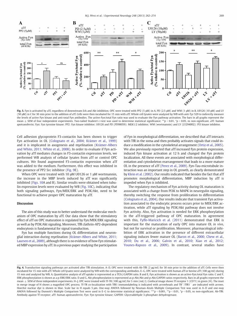
Fig. 5. Fyn is activated by aTf, regardless of downstream Erk and Akt inhibition. OPC were treated with PP2 (5 μM) in A, PD (2.5 μM) and WM (1 μM) in B, U0126 (10 μM) and LY(50 μM) in C for 30 min prior to the addition of aTf. Cells were then incubated for 15 min with aTf. Whole cell lysates were analyzed by WB with anti-Tyr 529 to indirectly measurethe levels of active Fyn kinase and anti-total Fyn antibodies. The active-Fyn/total Fyn ratio was used to evaluate the Fyn pathway activation. The bars in all graphs represent themean ± SEM of four independent experiments. Two-tailed Student's t-test was used to determine statistical significance; **p b 0.01, *p b 0.05, ns non-significant. aTf: humanapotransferrin; Fyn: Fyn tyrosine kinase; PP2: Fyn kinase inhibitor; U0126 and PD (PD98059): MEK1/2 inhibitor; WM (wortmannin) and LY (LY294002): PI3 kinase inhibitor.
269M.J. Pérez et al. / Experimental Neurology 248 (2013) 262–274
Cell adhesion glycoprotein F3-contactin has been shown to triggerFyn activation in OL (Colognato et al., 2004; Krämer et al., 1999)and it is implicated in axogenesis and myelination (Krämer-Albersand White, 2011; White et al., 2008). In order to evaluate if Fyn acti-vation by aTf mediates changes in F3-contactin expression levels, weperformed WB analysis of cellular lysates from aTf or control OPCcultures. We found augmented F3-contactin expression when aTfwas added to the medium. Furthermore, this effect was inhibited inthe presence of PP2 Src inhibitor (Fig. 9E).
When OPC were treated with 10 μM U0126 or 1 μM wortmannin,the increase in the MBP levels induced by aTf was significantlyinhibited (Figs. 10A and B). Similar results were obtained when tubu-lin expression levels were evaluated by WB (Fig. 10C), indicating thatboth signaling pathways, Fyn/MEK/ERK and PI3K/Akt, need to befunctional to achieve proper OPC maturation by aTf.
Discussion
The aim of this study was to better understand themolecular mech-anism of OPC maturation by aTf. Our data show that the stimulatoryeffect of aTf on OPC maturation is regulated by Fyn/MEK/ERK signalingaswell as by PI3K/Akt signaling. Moreover, TfR clathrin-AP2-dependentendocytosis is fundamental for signal transduction.
Fyn has multiple functions during OL differentiation and neuron-glial interaction during myelination (Krämer-Albers and White, 2011;Laursen et al., 2009), although there is no evidence of how Fyn stimulat-edMBPexpression by aTf. In a previous paper studying theparticipation
Fig. 4. Transduction signaling pathways are activated after TfR stimulation. A–D, OPC wereincubated for 15 min with aTf. Whole cell lysates were analyzed byWB with the correspond15 min and analyzed by WB. A, Quantitative analysis of aTf uptake is represented as a Tf(h)ERK phosphorylation is shown as a p-ERK/ERK ratio. D and G, Akt phosphorylation is represemean ± SEM of three independent experiments. H–J, OPC were treated with Tf-TR (100 μg/min merge image of H shows a magnified OPC process. Tf-TR co-localization with TfR1 immHoechst nuclear dye is shown in blue. Scale bar in H equals 5 μm. One-way ANOVA foANOVA followed by Dunnett's Multiple Comparison Test were used in E–G to determineAntibody against Tf receptor; aTf: human apotransferrin; Fyn: Fyn tyrosine kinase; GAPDH
of Fyn in morphological differentiation, we described that aTf interactswith TfR in the soma and then probably activates signals that could in-duce a modification in the cytoskeletal arrangement (Ortiz et al., 2005).We also previously reported that aTf increased Fyn protein expression,induced Fyn kinase activation at 12 h and changed the Fyn proteinlocalization. All these events are associated with morphological differ-entiation and cytoskeleton rearrangement that leads to a more matureOL in the presence of aTf (Perez et al., 2009). Fyn-Tau-microtubule in-teraction was an important step in OL growth, as clearly demonstratedby Klein et al. (2002). Our results indicated that besides the fact that aTfpromotes morphological differentiation, MBP induction by aTf isimpeded when Fyn is inhibited.
The regulatory mechanism of Fyn activity during OL maturation isassociated with a change from PI3K to MAPK in neuregulin signaling,thereby switching the response from proliferation to differentiation(Colognato et al., 2004). Our results indicate that transient Fyn activa-tion associated to the endocytic process occurs prior to MEK/ERK ac-tivation, while aTf signaling by PI3K/Akt pathway does not involveFyn activity. Also, Fyn activation is needed for ERK phosphorylationin the aTf-triggered pathway of OPC maturation. In agreementwith this, Fyffe-Maricich et al. (2011) demonstrated that ERK isimportant for the maturation of early OPC into mature OL in vivo,but not for survival or proliferation. Moreover, pharmacological inhi-bition of ERK activation in the presence of different extracellularsignaling induces fewer mature OL (Baron et al., 2000; Chew et al.,2010; Du et al., 2006; Galvin et al., 2010; Xiao et al., 2012;Younes-Rapozo et al., 2009). In contrast, several studies have
treated with Ab-TfR (2 μg/ml) for 30 min prior to the addition of aTf. Cells were thening antibodies. E–G, OPC were treated with human aTf or bovine aTf (100 μg/ml) during/GAPDH ratio. B and E, Fyn activation is shown as an active-Fyn/total Fyn ratio. C and F,nted as p-Akt/Akt and p-Akt/GAPDH ratios respectively. Bars in all graphs represent thel) for 5 min (red, I). Confocal image shows Tf receptor 1 (CD71) in green (H). The insetunolabeling is indicated with arrowheads and TR+/TfR1− are indicated with arrows.llowed by Neuman–Keuls Multiple Comparison Test was used in A–D and one-waystatistical significance; ***p b 0.001, **p b 0.01, *p b 0.05, ns non-significant. Ab-TfR:: Glyceraldehyde 3-phosphate dehydrogenase.

Fig. 6. aTf activates ERK through Fyn. OPC were treated with PP2 (5 μM) in A, U0126 (10 μM) andWM (1 μM) in B, PD (2.5 μM) and LY (50 μM) in C for 30 min prior to the additionof aTf. Cells were then incubated for 15 min with aTf. Whole cell lysates were analyzed byWB with anti-phospho ERK and anti-ERK1/2 antibodies. The p-ERK/ERK ratio was used toevaluate ERK pathway activation. Bars in all graphs represent the mean ± SEM of four independent experiments. Two-tailed Student's t-test was used to determine statistical sig-nificance; *p b 0.05, ns non-significant. aTf: human apotransferrin; PP2: Fyn kinase inhibitor; U0126 and PD (PD98059): MEK1/2 inhibitor; WM (wortmannin) and LY (LY294002):PI3 kinase inhibitor.
270 M.J. Pérez et al. / Experimental Neurology 248 (2013) 262–274
suggested that MAPK signaling is important for OL proliferation (Cuiand Almazan, 2007; Okada et al., 2010; Stariha and Kim, 2001).
There are two membrane receptors of Tf that have different func-tions and expression patterns. TfR1 is expressed in all cells andmediatesthe cellular uptake of transferrin-bound iron. TfR2 is only expressed insome cells and acts as an iron sensor to regulate hepcidin expression.In the central nervous system, TfR1 is expressed in normal brain andin brain tumors (Han et al., 2003; Recht et al., 1990) and TfR2 ispredominantly expressed in hepatocytes, although Moos et al. (1998)report their expression in neurons. Hänninen et al. (2009) describeTfR2 expression in human brain and brain tumors and Calzolari et al.(2010) do so in astrocytomas. Furthermore, ERK signaling activation is
Fig. 7. PI3K/Akt pathway is independent of Fyn and ERK activation. OPC were treated with Pfor 30 min prior to the addition of aTf. Cells were then incubated for 15 min with aTf. Wholep-Akt/Akt ratio was used to evaluate Akt pathway activation. Bars in all graphs represent thedetermine statistical significance; ***p b 0.001, **p b 0.01, *p b 0.05, ns non-significant. aTfinhibitor; WM (wortmannin) and LY (LY294002): PI3 kinase inhibitor.
associated with TfR2 in different cells regulating iron metabolism(Calzolari et al., 2006; Poli et al., 2010; Ramey et al., 2009). Our experi-ments showed that the signaling pathway of human aTf is differentfrom that of bovine aTf, which suggests that PI3K/Akt is independentfrom Fyn/MEK/ERK pathway. Thus the induction of ERK phosphoryla-tion triggered by aTf seems to be linked to TfR2. It was probably moreinteresting to observe that theMEK/ERK pathwaywas directly associat-ed with MBP expression protein. The precise identification and the roleof TfR in glial cells remain unexplored. However, the fact that aTf hasmaturational effects in a temporal window when TfR is present in OPCgives support to our hypothesis that differentiation is due to receptorinternalization and that the activation of the signal transduction
P2 (5 μM) in A, PD (2.5 μM) and WM (1 μM) in B, U0126 (10 μM) and LY (50 μM) in Ccell lysates were analyzed by WB with anti-phospho Akt and anti-total Akt antibodies.mean ± SEM of four independent experiments. Two-tailed Student's t-test was used to: human apotransferrin; PP2: Fyn kinase inhibitor; U0126 and PD (PD98059): MEK1/2

Fig. 8. Signaling activation by aTf requires clathrin-mediated endocytosis of TfR. A, Cultures were treated with MDC (50 μM) during 10 min previous to the addition of Tf-TR. Cellswere then incubated for 15 min with Tf-TR. Scale bar equals 50 μm for all panels. Tf-TR fluorescence is shown in red and Hoechst nuclear dye is shown in blue. B, The red fluores-cence shown in A is quantitated and shown as a red fluorescence/nuclei ratio. C, OPC were treated with MDC (50 μM) for 10 min prior to the addition of aTf. Cells were thenincubated for 15 min with aTf. Whole cell lysates were analyzed by WB. The active-Fyn/total Fyn ratio was used to evaluate Fyn activation. D-F, OPC were transfected withMock (Ctl and aTf), EH29, DIII, and DIIIΔ2 plasmid as described in the Materials and methods section, followed by treatment with aTf for 15 min. Whole cell lysates were analyzedby WB. D, Fyn activation was analyzed as an active-Fyn/total Fyn ratio. The GAPDH immunopositive bands were used as sample loading. E, ERK phosphorylation was evaluated as ap-ERK/ERK ratio. F, Akt phosphorylation was analyzed as a p-Akt/Akt ratio. The bars in all graphs represent the mean ± SEM of three independent experiments. Two-tailedStudent's t-test was used to determine statistical significance; ***p b 0.001, **p b 0.01, *p b 0.05, ns non-significant. aTf: human apotransferrin; Fyn: Fyn tyrosine kinase;GAPDH: Glyceraldehyde 3-phosphate dehydrogenase; IOD: integrated optical density; MDC: primary amine monodansylcadaverin; Tf-TR: Texas Red-labeled transferrin; EH29:Eps15 dominant negative mutant -pEGFP-C2-Eps15 EH29 plasmid-; DIII: Eps15 dominant negative mutant -pEGFP-C2-Eps15 DIII plasmid-; DIIIΔ2: Eps15 irrelevant mutant-pEGFP-C2-Eps15 DIIIΔ2 plasmid-.
271M.J. Pérez et al. / Experimental Neurology 248 (2013) 262–274
cascade is triggered by this phenomenon. Although mature OL do notexpress TfR, Todorich et al. (2008) suggested that Tim-2, an alternativeiron delivery mechanism, exists in these cells.
Depending on the stage of OL development, a growth factor canmediate cell proliferation, survival or progression of differentiation
through the PI3K pathway (Baron et al., 2000; Cui and Almazan,2007; Flores et al., 2008; Guardiola-Diaz et al., 2012; Xu et al.,2012). It is possible to speculate that enhancement of OPC maturationby aTf is due to Akt phosphorylation. The PI3K/Akt and the MAPK sig-naling pathways have been implicated in different processes such as
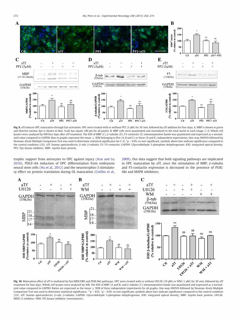
Fig. 9. aTf induces OPC maturation through Fyn activation. OPC were treated with or without PP2 (5 μM) for 30 min, followed by aTf addition for four days. A, MBP is shown in greenand Hoechst nuclear dye is shown in blue. Scale bar equals 100 μm for all panels. B, MBP cells were quantitated and normalized to the total nuclei in each image. C–E, Whole celllysates were analyzed by WB four days after aTf treatment. The IOD of MBP (C), β-tubulin (D), F3-contactin (E) immunopositive bands was quantitated and expressed as a normal-ized value compared to GAPDH. Bars in graphs represent the mean ± SEM belonging to five (A, B and C) or three (D and E) independent experiments. One-way ANOVA followed byNeuman–Keuls Multiple Comparison Test was used to determine statistical significance for C–E; *p b 0.05, ns non-significant, symbols above bars indicate significance compared tothe control condition (Ctl). aTf: human apotransferrin; β-tub: β-tubulin; F3: F3-contactin; GAPDH: Glyceraldehyde 3-phosphate dehydrogenase; IOD: integrated optical density;PP2: Fyn kinase inhibitor; MBP: myelin basic protein.
272 M.J. Pérez et al. / Experimental Neurology 248 (2013) 262–274
trophic support from astrocytes to OPC against injury (Arai and Lo,2010), PDGF-AA induction of OPC differentiation from embryonicneural stem cells (Hu et al., 2012) and the neurotrophin-3 stimulato-ry effect on protein translation during OL maturation (Coelho et al.,
Fig. 10. Maturation effect of aTf is mediated by Fyn/MEK/ERK and PI3K/Akt pathways. OPC wtreatment for four days. Whole cell lysates were analyzed by WB. The IOD of MBP (A and B)ized value compared to GAPDH. Ratios are expressed as the mean ± SEM of three independComparison Test was used to determine statistical significance; **p b 0.01, *p b 0.05, ns non(Ctl). aTf: human apotransferrin; β-tub: β-tubulin; GAPDH: Glyceraldehyde 3-phosphateMEK1/2 inhibitor; WM: PI3 kinase inhibitor (wortmannin).
2009). Our data suggest that both signaling pathways are implicatedin OPC maturation by aTf, since the stimulation of MBP, β-tubulinand F3-contactin expression is decreased in the presence of PI3K/Akt and MAPK inhibitors.
ere treated with or without U0126 (10 μM) or WM (1 μM) for 30 min, followed by aTfand β-tubulin (C) immunopositive bands was quantitated and expressed as a normal-ent experiments for all graphs. One-way ANOVA followed by Neuman–Keuls Multiple-significant, symbols above bars indicate significance compared to the control conditiondehydrogenase; IOD: integrated optical density; MBP: myelin basic protein; U0126:

273M.J. Pérez et al. / Experimental Neurology 248 (2013) 262–274
Clathrin-dependent endocytosis inhibitor MDC did not only inhib-it TfR internalization but also blocked aTf-stimulated signaling. Thisresult allows us to better understand the functional role of TfR endo-cytosis in intracellular signal transduction. The role for clathrin coatedvesicles in TfR recycling, as well as the involvement of AP2 complex inthe uptake of proteins with YXXϕ sorting signal, such as the TfR(Nesterov et al., 1999; Ohno et al, 1995) were also previously demon-strated. However, there is also evidence that alternative mechanismsmediated by caveolae could also lead to TfR internalization, whichsuggests the existence of mechanisms that may compensate fordefects in the clathrin-dependent uptake route (Puri, 2009). On theother hand, AP2 depletion was found to cause inhibition of Tf uptakeeven though AP2 knock-down did not diminish clathrin-coatedvesicle formation; this observation rules out the possibility of anAP2-independent clathrin-dependent internalization of TfR (Motleyet al., 2003).
In the present work we observed that lack of Fyn/MEK/ERK andPI3K/Akt activation in EH29 and DIII (dominant negative ofEps15)-transfected cells indicated that TfR signaling occurs in the ear-lier steps of receptor endocytosis and that it depends on the properformation of the AP2-Eps15 complex. Cao et al. (2010) demonstratedthat activated Src kinase regulated TfR endocytosis in epithelial cells.In this context, if there is an alternative mechanism for TfR internali-zation that could take place, it seems to be insufficient to evoke Fynactivation. These results demonstrated that the activation of the path-ways necessary for OPC maturation is dependent on AP2 recruitmentonto the plasma membrane for clathrin-mediated endocytosis of TfR.
Together, the data presented here demonstrate that endocytosis ofTfR accelerates OL differentiation. In addition, we provide evidenceindicating that this effect is exerted through Fyn/MEK/ERK andPI3K/Akt signaling pathways. In agreement with this, insulin-likegrowth factor-1 also stimulates protein synthesis through PI3K/mTOR/Akt and MEK/ERK pathways in OL (Bibollet-Bahena andAlmazan, 2009). The molecular mechanisms and signaling pathwaysby which aTf promotes OPC differentiation provide a key point todevelop new therapies to counteract the loss of OPC maturation indemyelinating disorders.
Acknowledgments
Funding sources: This work was supported by grants from theAgencia Nacional de Promoción Científica y Tecnológica (BID-PICT0791) and the University of Buenos Aires (UBA 20020100100395).
The authors are grateful to Dr. Paula Franco and Lucas Silvestrofffor insightful comments and helpful discussions on this manuscriptand to Dr. Benmerah for the kind gift of expression plasmids. Wewish to thank Dr. Ferrari Alejandro for the excellent artwork.
References
Adamo, A.M., Paez, P.M., Escobar Cabrera, O.E., Wolfson, M., Franco, P.G., Pasquini, J.M.,Soto, E.F., 2006. Remyelination after cuprizone-induced demyelination in the rat isstimulated by apotransferrin. Exp. Neurol. 198, 519–529.
Arai, K., Lo, E.H., 2010. Astrocytes protect oligodendrocyte precursor cells via MEK/ERKand PI3K/Akt signaling. J. Neurosci. Res. 88, 758–763.
Artini, M., Scoarughi, G.L., Cellini, A., Papa, R., Barbato, G., Selan, L., 2012. Holo and apo-transferrins interfere with adherence to abiotic surfaces and with adhesion/invasionto HeLa cells in Staphylococcus spp Biometals 25, 413–421.
Badaracco, M.E., Ortiz, E.H., Soto, E.F., Connor, J., Pasquini, J.M., 2008. Effect of transferrinon hypomyelination induced by iron deficiency. J. Neurosci. Res. 86, 2663–2673.
Baer, A.S., Syed, Y.A., Kang, S.U., Mitteregger, D., Vig, R., ffrench-Constant, C., Franklin,R.J.M., Altmann, F., Lubec, G., Kotter, M.R., 2009. Myelin-mediated inhibition of ol-igodendrocyte precursor differentiation can be overcome by pharmacologicalmodulation of Fyn-RhoA and protein kinase C signaling. Brain 132, 465–481.
Baron, W., Metz, B., Bansal, R., Hoekstra, D., de Vries, H., 2000. PDGF and FGF-2signaling in oligodendrocyte progenitor cells, regulation of proliferation anddifferentiation by multiple intracellular signaling pathways. Mol. Cell. Neurosci.15, 314–329.
Bartlett, W.P., Li, X.S., Connor, J.R., 1991. Expression of transferrin mRNA in the CNS ofnormal and jumpy mice. J. Neurochem. 57, 318–322.
Benmerah, A., Bayrou, M., Cerf-Bensussan, N., Dautry-Varsat, A., 1999. Inhibition ofclathrin-coated pit assembly by an Eps15 mutant. J. Cell Sci. 112, 1303–1311.
Bibollet-Bahena, O., Almazan, G., 2009. IGF-1-stimulated protein synthesis in oligoden-drocyte progenitors requires PI3K/mTOR/Akt and MEK/ERK pathways. J. Neurochem.109, 1440–1451.
Bloch, B., Popovici, T., Levin, M.J., Tuil, D., Kahn, A., 1985. Transferrin gene expressionvisualized in oligodendrocytes of the rat brain by using in situ hybridization andimmunohistochemistry. Proc. Natl. Acad. Sci. U. S. A. 82, 6706–6710.
Calzolari, A., Raggi, C., Deaglio, S., Sposi, N.M., Stafsnes, M., Fecchi, K., Parolini, I.,Malavasi, F., Peschle, C., Sargiacomo, M., Testa, U., 2006. TfR2 localizes in lipidraft domains and is released in exosomes to activate signal transduction alongthe MAPK pathway. J. Cell Sci. 119, 4486–4498.
Calzolari, A., Larocca, L.M., Deaglio, S., Finisguerra, V., Boe, A., Raggi, C., Ricci-Vitani, L.,Pierconti, F., Malavasi, F., De Maria, R., Testa, U., Pallini, R., 2010. Transferrinreceptor 2 is frequently and highly expressed in glioblastomas. Transl. Oncol. 3,123–134.
Cao, H., Chen, J., Krueger, E.W., McNiven, M.A., 2010. SRC-mediated phosphorylation ofdynamin and cortactin regulates the “constitutive” endocytosis of transferrin. Mol.Cell. Biol. 30, 781–792.
Casaccia-Bonnefil, P., Aibel, L., Chao, M.V., 1996. Central glial and neuronal populationsdisplay differential sensitivity to ceramide-dependent cell death. J. Neurosci. Res.43, 382–389.
Chew, L.J., Coley, W., Cheng, Y., Gallo, V., 2010. Mechanisms of regulation of oligodendrocytedevelopment by p38 mitogen-activated protein kinase. J. Neurosci. 30, 11011–11027.
Coelho, R.P., Yuelling, L.M., Fuss, B., Sato-Bigbee, C., 2009. Neurotrophin-3 targets thetranslational initiation machinery in oligodendrocytes. Glia 57, 1754–1764.
Cole, P.A., Shen, K., Qiao, Y., Wang, D., 2003. Protein tyrosine kinases Src and Csk, a tail'stale. Curr. Opin. Chem. Biol. 7, 580–585.
Colognato, H., Ramachandrappa, S., Olsen, I.M., ffrench-Constant, C., 2004. Integrins di-rect Src family kinases to regulate distinct phases of oligodendrocyte development.J. Cell Biol. 167, 365–375.
Cui, Q.L., Almazan, G., 2007. IGF-I induced oligodendrocyte progenitor proliferationrequires PI3K/Akt, MEK/ERK, and Src-like tyrosine kinases. J. Neurochem. 100,1480–1493.
Cui, Q.L., Zheng, W.H., Quirion, R., Almazan, G., 2005. Inhibition of Src-like kinasesreveals Akt-dependent and -independent pathways in insulin-like growth factorI-mediated oligodendrocyte progenitor survival. J. Biol. Chem. 280, 8918–8928.
Du, Y., Lercher, L.D., Zhou, R., Dreyfus, C.F., 2006. Mitogen-activated protein kinasepathway mediates effects of brain-derived neurotrophic factor on differentiationof basal forebrain oligodendrocytes. J. Neurosci. Res. 84, 1692–1702.
Escobar Cabrera, O.E., Bongarzone, E.R., Soto, E.F., Pasquini, J.M., 1994. Single intracere-bral injection of apotransferrin in young rats induces increased myelination. Dev.Neurosci. 16, 248–254.
Escobar Cabrera, O.E., Zakin, M.M., Soto, E.F., Pasquini, J.M., 1997. Single intracranial in-jection of apotransferrin in young rats increases the expression of specific myelinprotein mRNA. J. Neurosci. Res. 47, 603–608.
Espinosa de los Monteros, A., Kumar, S., Scully, S., Cole, R., de Vellis, J., 1990. Transferringene expression and secretion by rat brain cells in vitro. J. Neurosci. Res. 25,576–580.
Espinosa de los Monteros, A., Kumar, S., Zhao, P., Huang, C.J., Nazarian, R., Pan, T., Scully,S., Chang, R., de Vellis, J., 1999. Transferrin is an essential factor for myelination.Neurochem. Res. 24, 235–248.
Flores, A.I., Narayanan, S.P., Morse, E.N., Shick, H.E., Yin, X., Kidd, G., Avila, R.L.,Kirschner, D.A., Macklin, W.B., 2008. Constitutively active Akt induces enhancedmyelination in the CNS. J. Neurosci. 28, 7174–7183.
Fyffe-Maricich, S.L., Karlo, J.C., Landreth, G.E., Miller, R.H., 2011. The ERK2 mitogen-activated protein kinase regulates the timing of oligodendrocyte differentiation.J. Neurosci. 31, 843–850.
Galvin, J., Eyermann, C., Colognato, H., 2010. Dystroglycan modulates the ability ofinsulin-like growth factor-1 to promote oligodendrocyte differentiation. J. Neurosci.Res. 88, 3295–3307.
Garcia, C., Paez, P.M., Davio, C., Soto, E.F., Pasquini, J.M., 2004. Apotransferrin inducescAMP/CREB pathway and cell cycle exit in immature oligodendroglial cells.J. Neurosci. Res. 78, 338–346.
Guardia Clausi, M., Paez, P.M., Campagnoni, A.T., Pasquini, L.A., Pasquini, J.M., 2012.Intranasal administration of aTf protects and repairs the neonatal white matterafter a cerebral hypoxic-ischemic event. Glia 60, 1540–1554.
Guardiola-Diaz, H.M., Ishii, A., Bansal, R., 2012. Erk1/2 MAPK andmTOR signaling sequen-tially regulates progression through distinct stages ofoligodendrocyte differentiation.Glia 3, 476–486.
Han, J., Day, J.R., Connor, J.R., Beard, J.L., 2003. Gene expression of transferrin and trans-ferrin receptor in brains of control vs. iron-deficient rats. Nutr. Neurosci. 6, 1–10.
Hanke, J.H., Gardner, J.P., Dow, R.L., Changelian, P.S., Brissette, W.H., Weringer, E.J.,Pollok, B.A., Connelly, P.A., 1996. Discovery of a novel, potent, and Src family-selective tyrosine kinase inhibitor. Study of Lck- and FynT-dependent T cell activa-tion. J. Biol. Chem. 271, 695–701.
Hänninen, M.M., Haapasalo, J., Haapasalo, H., Fleming, R.E., Britton, R.S., Bacon, B.R.,Parkkila, S., 2009. Expression of iron-related genes in human brain and braintumors. BMC Neurosci. 10, 36.
Hu, J.G., Wang, Y.X., Wang, H.J., Bao, M.S., Wang, Z.H., Ge, X., Wang, F.C., Zhou, J.S., Lü,H.Z., 2012. PDGF-AA mediates B104 CM-induced oligodendrocyte precursor celldifferentiation of embryonic neural stem cells through Erk, PI3K, and p38 signaling.J. Mol. Neurosci. 46, 644–653.
Kawabata, H., Germain, R.S., Vuong, P.T., Nakamaki, T., Said, J.W., Koeffler, H.P., 2000.Transferrin receptor 2-alpha supports cell growth both in iron-chelated culturedcells and in vivo. J. Biol. Chem. 275, 16618–16625.

274 M.J. Pérez et al. / Experimental Neurology 248 (2013) 262–274
Kawabata, H., Tong, X., Kawanami, T., Wano, Y., Hirose, Y., Sugai, S., Koeffler, H.P., 2004.Analyses for binding of the transferrin family of proteins to the transferrin receptor2. Br. J. Haematol. 127, 464–473.
Klein, C., Kramer, E.M., Cardine, A.M., Schraven, B., Brandt, R., Trotter, J., 2002. Processoutgrowth of oligodendrocytes is promoted by interaction of Fyn kinase with thecytoskeletal protein tau. J. Neurosci. 22, 698–707.
Krämer, E.M., Klein, C., Koch, T., Boytinck, M., Trotter, J., 1999. Compartmentation of Fynkinase with glycosylphosphatidylinositol-anchored molecules in oligodendrocytesfacilitates kinase activation during myelination. J. Biol. Chem. 274, 29042–29049.
Krämer-Albers, E.M., White, R., 2011. From axon-glial signalling to myelination, theintegrating role of oligodendroglial Fyn kinase. Cell. Mol. Life Sci. 68, 2003–2012.
Laursen, L.S., Chan, C.W., ffrench-Constant, C., 2009. An integrin-contactin complexregulates CNS myelination by differential Fyn phosphorylation. J. Neurosci. 29,9174–9185.
Lesley, J.F., Schulte, R.J., 1985. Inhibition of cell growth by monoclonal anti-transferrinreceptor antibodies. Mol. Cell. Biol. 5, 1814–1821.
Marta, C.B., Escobar Cabrera, O.E., Garcia, C.I., Villar, M.J., Pasquini, J.M., Soto, E.F., 2000.Oligodendroglial cell differentiation in rat brain is accelerated by the intracranialinjection of apotransferrin. Cell. Mol. Biol. 46, 529–539.
McArdle, H.J., Morgan, E.H., 1984. The effect of monoclonal antibodies to the humantransferrin receptor on transferrin and iron uptake by rat and rabbit reticulocytes.J. Biol. Chem. 259, 1398–1400.
McCarthy, K.D., de Vellis, J., 1980. Preparation of separate astroglial and oligodendrog-lial cell cultures from rat cerebral tissue. J. Cell Biol. 85, 890–902.
Moos, T., Oates, P.S., Morgan, E.H., 1998. Expression of the neuronal transferrinreceptor is age dependent and susceptible to iron deficiency. J. Comp. Neurol.398, 420–430.
Motley, A., Bright, N.A., Seaman, M.N., Robinson, M.S., 2003. Clathrin-mediatedendocytosis in AP-2-depleted cells. J. Cell Biol. 162, 909–918.
Nesterov, A., Carter, R.E., Sorkina, T., Gill, G.N., Sorkin, A., 1999. Inhibition of thereceptor-binding function of clathrin adaptor protein AP-2 by dominant-negativemutant mu2 subunit and its effects on endocytosis. EMBO J. 18, 2489–2499.
Ohno, H., Stewart, J., Fournier, M.C., Bosshart, H., Rhee, I., Miyatake, S., Saito, T.,Gallusser, A., Kirchhausen, T., Bonifacino, J.S., 1995. Interaction of tyrosine-basedsorting signals with clathrin-associated proteins. Science 269, 1872–1875.
Okada, M., Makino, A., Nakajima, M., Okuyama, S., Furukawa, S., Furukawa, Y., 2010.Estrogen stimulates proliferation and differentiation of neural stem/progenitor cellsthrough different signal transduction pathways. Int. J. Mol. Sci. 11, 4114–4123.
Ortiz, E.H., Pasquini, L.A., Soto, E.F., Pasquini, J.M., 2005. Apotransferrin and thecytoskeleton of oligodendroglial cells. J. Neurosci. Res. 82, 822–830.
Osterhout, D.J., Wolven, A., Wolf, R.M., Resh, M.D., Chao, M.V., 1999. Morphologicaldifferentiation of oligodendrocytes requires activation of Fyn tyrosine kinase.J. Cell Biol. 145, 1209–1218.
Paez, P.M., Marta, C.B., Moreno, M.B., Soto, E.F., Pasquini, J.M., 2002. Apotransferrindecreases migration and enhances differentiation of oligodendroglial progenitorcells in an in vitro system. Dev. Neurosci. 24, 47–58.
Pasquini, L.A., Paez, P.M., Moreno, M.A., Pasquini, J.M., Soto, E.F., 2003. Inhibition of theproteasome by lactacystin enhances oligodendroglial cell differentiation. J. Neurosci.23, 4635–4644.
Perez, M.J., Ortiz, E.H., Roffé, M., Soto, E.F., Pasquini, J.M., 2009. Fyn kinase is involved inoligodendroglial cell differentiation induced by apotransferrin. J. Neurosci. Res. 15,3378–3389.
Poli, M., Luscieti, S., Gandini, V., Maccarinelli, F., Finazzi, D., Silvestri, L., Roetto, A., Arosio,P., 2010. Transferrin receptor 2 and HFE regulate furin expression via mitogen-activated protein kinase/extracellular signal-regulated kinase (MAPK/Erk) signaling.Implications for transferrin-dependent hepcidin regulation. Haematologica 95,1832–1840.
Puri, C., 2009. Loss of myosin VI no insert isoform (NoI) induces a defect in clathrin-mediated endocytosis and leads to caveolar endocytosis of transferrin receptor.J. Biol. Chem. 284, 34998–35014.
Ramey, G., Deschemin, J.C., Vaulont, S., 2009. Crosstalk between the mitogen activatedprotein kinase and bone morphogenetic protein/he mojuvelin pathways isrequired for the induction of hepcidin by holotransferrin in primary mouse hepa-tocytes. Haematologica 94, 765–772.
Rappoport, J.Z., Simon, S.M., Benmerah, A., 2004. Understanding living clathrin-coatedpits. Traffic 5, 327–337.
Ray, E., Samanta, A.K., 1996. Dansyl cadaverine regulates ligand induced endocytosis ofinterleukin-8 receptor in human polymorphonuclear neutrophils. FEBS Lett. 378,235–239.
Recht, L.D., Griffin, T.W., Raso, V., Salimi, A.R., 1990. Potent cytotoxicity of an antihumantransferrin receptor-ricin A-chain immunotoxin on human glioma cells in vitro.Cancer Res. 50, 6696–6700.
Rouault, T.A., Cooperman, S., 2006. Brain iron metabolism. Semin. Pediatr. Neurol. 13,142–148.
Saleh, M.C., Espinosa de los Monteros, A., de Arriba Zerpa, G.A., Fontaine, I., Piaud, O.,Djordjijevic, D., Baroukh, N., Garcia Otin, A.L., Ortiz, E., Lewis, S., Fiette, L.,Santambrogio, P., Belzung, C., Connor, J.R., de Vellis, J., Pasquini, J.M., Zakin, M.M.,Baron, B., Guillou, F., 2003. Myelination and motor coordination are increased intransferrin transgenic mice. J. Neurosci. Res. 72, 587–594.
Stariha, R.L., Kim, S.U., 2001. Protein kinase C and mitogen-activated protein kinasesignalling in oligodendrocytes. Microsc. Res. Tech. 6, 680–688.
Suzuki, A., Raya, A., Kawakami, Y., Morita, M., Matsui, T., Nakashima, K., Gage, F.H.,Rodríguez-Esteban, C., Izpisúa Belmonte, J.C., 2006. Maintenance of embryonicstem cell pluripotency by Nanog-mediated reversal of mesoderm specification.Nat. Clin. Pract. Cardiovasc. Med. (Suppl. 1), S114–S122.
Todorich, B., Zhang, X., Slagle-Webb, B., Seaman, W.E., Connor, J.R., 2008. Tim-2 is thereceptor for H-ferritin on oligodendrocytes. J. Neurochem. 107, 1495–1505.
Trowbridge, I.S., Lopez, F., 1982. Monoclonal antibody to transferrin receptor blockstransferrin binding and inhibits human tumor cell growth in vitro. Proc. Natl.Acad. Sci. U. S. A. 79, 1175–1179.
Umemori, H., Wanaka, A., Kato, H., Takeuchi, M., Tohyama, M., Yamamoto, T., 1992.Specific expressions of Fyn and Lyn, lymphocyte antigen receptor-associated tyro-sine kinases, in the central nervous system. Brain Res. Mol. Brain Res. 16, 303–310.
Umemori, H., Sato, S., Yagi, T., Aizawa, S., Yamamoto, T., 1994. Initial events ofmyelination involve Fyn tyrosine kinase signalling. Nature 367, 572–576.
Umemori, H., Kadowaki, Y., Hirosawa, K., Yoshida, Y., Hironaka, K., Okano, H.,Yamamoto, T., 1999. Stimulation of myelin basic protein gene transcription byFyn tyrosine kinase for myelination. J. Neurosci. 19, 1393–1397.
Wally, J., Buchanan, S.K., 2007. A structural comparison of human serum transferrinand human lactoferrin. Biometals 20, 249–262.
White, R., Gonsior, C., Krämer-Albers, E.M., Stöhr, N., Hüttelmaier, S., Trotter, J., 2008.Activation of oligodendroglial Fyn kinase enhances translation of mRNAstransported in hnRNP A2-dependent RNA granules. J. Cell Biol. 181, 579–586.
Xiao, L., Guo, D., Hu, C., Shen, W., Shan, L., Li, C., Liu, X., Yang, W., Zhang, W., He, C., 2012.Diosgenin promotes oligodendrocyte progenitor cell differentiation through estro-gen receptor-mediated ERK1/2 activation to accelerate remyelination. Glia 60,1037–1052.
Xu, C., Lv, L., Zheng, G., Li, B., Gao, L., Sun, Y., 2012. Neuregulin1β1 protects oligoden-drocyte progenitor cells from oxygen glucose deprivation injury induced apoptosisvia ErbB4-dependent activation of PI3-kinase/Akt. Brain Res. 1467, 104–112.
Younes-Rapozo, V., Felgueiras, L.O., Viana, N.L., Fierro, I.M., Barja-Fidalgo, C., Manhães,A.C., Barradas, P.C., 2009. A role for the MAPK/ERK pathway in oligodendroglial dif-ferentiation in vitro, stage specific effects on cell branching. Int. J. Dev. Neurosci. 27,757–768.