on october 20, 2016 by swets subscription service · on october 20, 2016 by swets subscription...
TRANSCRIPT

Interleukin-17 Is Required for Control of Chronic Lung InfectionCaused by Pseudomonas aeruginosa
Hannah K. Bayes, Neil D. Ritchie, Thomas J. Evans
Institute of Infection, Immunity and Inflammation, University of Glasgow, Glasgow, United Kingdom
Chronic pulmonary infection with Pseudomonas aeruginosa is a feature of cystic fibrosis (CF) and other chronic lung diseases.Cytokines of the interleukin-17 (IL-17) family have been proposed as important in the host response to P. aeruginosa infectionthrough their role in augmenting antibacterial immune responses, although their proinflammatory effect may contribute tolung damage that occurs as a result of chronic infection. We set out to explore the role of IL-17 in the host response to chronic P.aeruginosa infection. We used a murine model of chronic pulmonary infection with CF-related strains of P. aeruginosa. Wedemonstrate that IL-17 cytokine signaling is essential for mouse survival and prevention of chronic infection at 2 weeks postin-oculation using two different P. aeruginosa strains. Following infection, there was a marked expansion of cells within mediasti-nal lymph nodes, comprised mainly of innate lymphoid cells (ILCs); �90% of IL-17-producing (IL-17�) cells had markers con-sistent with group 3 ILCs. A smaller percentage of IL-17� cells had markers consistent with a B1 phenotype. In lunghomogenates harvested 14 days following infection, there was a significant expansion of IL-17� cells; about 50% of these wereCD3�, split equally between CD4� Th17 cells and �� T cells, while the CD3� IL-17� cells were almost exclusively group 3 ILCs.Further experiments with B cell-deficient mice showed that B cell production of IL-17 or natural antibodies did not provide anydefense against chronic P. aeruginosa infection. Thus, IL-17 rather than antibody is a key element in host defense againstchronic pulmonary infection with P. aeruginosa.
Some bacteria have evolved the ability to produce chronic in-fection of the respiratory tract. Mycobacterium tuberculosis is
perhaps the best known example, but the Gram-negative patho-gen Pseudomonas aeruginosa can also become persistent in thelower airways. This occurs most notably in patients with cysticfibrosis (CF) and bronchiectasis but is also increasingly recog-nized in other chronic lung diseases, such as chronic obstructivepulmonary disease (COPD). In CF, P. aeruginosa infections areinitially intermittent and can be eradicated by intensive antibiotictreatment (1). Transition to chronic P. aeruginosa airway infec-tion usually ensues, such that by age 20, 60 to 70% of CF patientsare chronically infected (2). The continuous presence of P. aerugi-nosa in the airways is accompanied by an inexorable decline inrespiratory function, leading to premature death or lung trans-plantation (3). Thus, this switch from intermittent to chronic in-fection is a key event in the progression of disease (1). Althoughantibiotics can delay this transition, better therapies aimed at pre-venting chronic infection could potentially significantly attenuatethe rate of decline in lung function in patients affected by CF, aswell in other chronic lung diseases in which chronic P. aeruginosainfection occurs.
Little is known of the mechanisms of host defense againstchronic P. aeruginosa infection. Cytokines of the interleukin-17(IL-17) family have been suggested as important in protectionagainst P. aeruginosa infection. IL-17 in the lung may be impor-tant in host defense against P. aeruginosa through its ability toorchestrate a neutrophil response and by the induction of a varietyof innate antimicrobial peptides (4). Increased levels of IL-17A(hereinafter referred to as IL-17) are found in sputum and bron-chial lavage specimens of patients with CF (5), produced by avariety of cells of the innate and acquired immune system, includ-ing T cells of the Th17 lineage (6–9). Other cells known to produceIL-17 include, inter alia, innate lymphoid cells (ILCs), �� T cells,and natural killer (NK) cells. Although such inflammatory re-
sponses can contribute to host defense, they also can potentiallycause tissue damage, as is well known for M. tuberculosis infection,where the host inflammatory response can result in significanttissue damage. Proinflammatory actions of IL-17 in P. aeruginosainfection could increase tissue damage through excess neutrophilaccumulation and induction of matrix metalloproteinases (10).Indeed, the inflammatory changes and subsequent bronchiectasisso typical of CF have been suggested to be driven by IL-17 cyto-kines. Although one study examined the role of IL-17 in acuteinfection (11), the specific role of IL-17 in chronic P. aeruginosainfection has not been addressed.
In the work presented here, we have defined the interactionsand effector functions of the IL-17 axis in the pathogenesis ofchronic pulmonary P. aeruginosa infection. Using a murinemodel, we show that IL-17 signaling is crucial in host defenseagainst chronic P. aeruginosa infection, protecting against chroniccolonization and death. Despite increased bacterial burdens, micelacking IL-17 signaling had less weight loss than controls. Weidentified a diverse range of cellular sources of IL-17 both in drain-ing mediastinal lymph nodes and in lungs following infection.
Received 16 August 2016 Returned for modification 12 September 2016Accepted 26 September 2016
Accepted manuscript posted online 3 October 2016
Citation Bayes HK, Ritchie ND, Evans TJ. 2016. Interleukin-17 is required for controlof chronic lung infection caused by Pseudomonas aeruginosa. Infect Immun 84:3507–3516. doi:10.1128/IAI.00717-16.
Editor: B. A. McCormick, The University of Massachusetts Medical School
Address correspondence to Thomas J. Evans, [email protected].
Supplemental material for this article may be found at http://dx.doi.org/10.1128/IAI.00717-16.
Copyright © 2016 Bayes et al. This is an open-access article distributed under theterms of the Creative Commons Attribution 4.0 International license.
crossmark
December 2016 Volume 84 Number 12 iai.asm.org 3507Infection and Immunity
on Decem
ber 21, 2016 by SW
ET
S S
UB
SC
RIP
TIO
N S
ER
VIC
Ehttp://iai.asm
.org/D
ownloaded from

MATERIALS AND METHODSAgar bead infection model. The infection model was adapted from theprotocol described by van Heeckeren and Schluchter (12) and modified asdescribed previously (13). Pseudomonas aeruginosa-laden agar beads wereprepared the day before inoculation and stored overnight at 4°C, and adifferent bead preparation was used for each experiment. P. aeruginosa-laden beads were stored on ice throughout the murine surgery. Followinginoculation of P. aeruginosa-laden beads, the inoculum administered wasconfirmed by homogenization and quantitative bacteriology of a furthertwo aliquots of beads. Sterile agar beads were stored at 4°C and used forseveral experiments. Sterile agar bead preparations were confirmed to besterile before and after each use. For experiments using knockout mu-tants, all animals (knockout mutants and wild-type [WT] controls) weretreated with P. aeruginosa-laden beads. In separate experiments, WT micewere treated with either P. aeruginosa-laden or sterile agar beads.
For inoculation with P. aeruginosa-laden beads, mice were anesthe-tized using isoflurane via nose cone, and the trachea was exposed andcannulated (22-gauge intravenous cannulae; BD Biosciences) under asep-tic conditions. An average inoculum of 1 � 106 CFU/50 �l per mouse wasdelivered. Animals were closely monitored postoperatively using a diseaseseverity scoring system (see Table S1 in the supplemental material). Ananimal reaching a moribund endpoint was euthanized.
Surviving animals at 14 days after infection were culled; lungs wereremoved from some and mechanically homogenized, and aliquots platedon bacteriological medium to determine the numbers of CFU of P. aerugi-nosa. Chronicity rates were defined as the percentages of animals at 14days after infection that had viable P. aeruginosa cells recovered from theirlungs using this method.
Flow cytometry. Antibodies to the following were used for flow cy-tometry: CD3e (145-2C11; eBioscience and BioLegend); CD19 (eBio1D3[eBioscience] and 6D5 [BioLegend]); CD4 (GK1.5), CD5 (53-7.3),CD11c (N418), CD23 (B3B4), CD43 (eBioR2/60), �� T cell receptor(��-TCR) (UC7-13D5), gamma interferon (IFN-�) (XMG1.2), IgD (11-26c), and IgM (II/41) (all from eBioscience); CD45R/B220 (RA3-6B2),granulocyte-macrophage colony-stimulating factor (GM-CSF) (MP1-22E9),Gr-1 (RB6-8C5), and IL-17A (TC11-18H10.1) (all from BioLegend); andIL-22 (3F11; Genentech). Isotype controls were used to confirm the spec-ificity of staining. For intracellular staining, cells were polyclonally stim-ulated with 50-ng/ml phorbol myristate acetate (PMA) and 500-ng/mlionomycin in the presence of brefeldin A (BD GolgiPlug at 1 �g/ml) at37°C for 5 h, fixed using 4% paraformaldehyde (Thermo Scientific) inphosphate-buffered saline (PBS) for 10 min at 4°C, and then washed influorescence-activated cell sorting (FACS) buffer (PBS, 2% fetal calf se-rum [FCS], 0.09% sodium azide [Sigma-Aldrich]). Cells were permeab-ilized using PermWash buffer (BD Biosciences) prior to staining. Deadcells were detected by using eFluor506 (eBioscience). For neutrophilquantification via flow cytometry, CountBright absolute counting beads(Invitrogen) were added prior to washing cells and used according to themanufacturer’s instructions. Stained cells were analyzed using a FACSAriainstrument (BD Biosciences) and FlowJo software (TreeStar).
P. aeruginosa strains. The clinical NH57388A strain was provided byN. Hoffmann (University of Copenhagen). This strain possesses a muta-tion in mucA that results in hyperproduction of alginate (14). The mucoidYH5 strain and nonmucoid GRI-1 strain were obtained locally, from apatient with CF and a patient with ventilator-associated pneumonia, re-spectively. P. aeruginosa strains were maintained in �80°C stocks untilrequired. Prior to use in cell culture, each strain was grown to mid-logphase in Luria-Bertani (LB) broth (Invitrogen) and bacterial concentra-tions at an optical density at 600 nm (OD600) of between 0.4 and 0.6 werequantified by serial dilution and plating to enumerate CFU (GeneQuantPro spectrophotometer; Amersham Biosciences). Heat-killed P. aerugi-nosa preparations were produced by heating a known concentration of P.aeruginosa in PBS to 95°C for 10 min.
Cytokine measurement. Murine IL-17A, IL-17F, IL-21, IL-22, andIFN-� were quantified by enzyme-linked immunosorbent assays (ELISAs)
(all eBioscience). The lower limits of detection were �4 pg/ml forIL-17A, �15pg/ml for IL-17F, �16 pg/ml for IL-21, �8 pg/ml forIL-22, and �15 pg/ml for IFN-�. Cytokine levels below the lowerlimits of detection of the assay were assigned a value of zero.
Immunochemistry and histology scoring. Lung sections were stainedwith brilliant violet 421 anti-B220 antibody (clone RA3-6B2; Biolegend)and anti-CD90.2 antibody (Thy1.2) (clone 53-2.1; Biolegend) conjugatedwith Alexa Fluor 488-streptavidin (both at a concentration of 1:50).SYTOX green nuclear stain (Invitrogen) was used at a concentration of1:10,000. The histology scoring system used is shown in Table S2 in thesupplemental material.
Animals. All mice were used between 12 and 16 weeks of age. IL-17Areceptor (IL-17RA) knockout mice (15) were from T. Mitchell, Universityof Glasgow, and originally supplied by Jay Kolls; �MT mice (16) werefrom R. Nibbs, University of Glasgow. Each of these lines was on aC57BL/6 background. C57BL/6 mice bred in-house were used as wild-type controls for knockout comparisons. The animal work was carried outunder a project license as required by United Kingdom Home Officeregulations, as well as scrutiny and approval by an institutional reviewboard.
Ethics. The animal studies were approved by the granting of a projectlicense from the United Kingdom Home Office, a ministerial governmentdepartment that oversees all experimental work with animals in theUnited Kingdom. The project license number is 60/4361. This work wasalso reviewed and approved by the University of Glasgow Animal Welfareand Ethical Review Board, under the same license number.
Mediastinal lymph node, splenocyte, and peritoneal B1a cell stimu-lation. Mediastinal lymph nodes and spleens were passed through 80-�mNitex mesh and red blood cells (RBCs) lysed (RBC lysis buffer; Sigma-Aldrich) to form a single-cell suspension. Cells were either left unstimu-lated or stimulated with heat-killed P. aeruginosa at a multiplicity of in-fection (MOI) of 30. Following 3 days of culture, 100 �l of supernatantwas removed for cytokine and P. aeruginosa-specific antibody quantifica-tion, and cells prepared for flow cytometry.
Lung homogenate, BAL fluid, and pleural wash samples. Lung tissuewas agitated for 1 h at 37°C with 10 �g/ml DNase (Roche) and 0.65units/ml Liberase (Roche), passed through 80-�m Nitex mesh, and RBCslysed to obtain a single-cell suspension. Bronchoalveolar lavage (BAL)fluid and pleural wash samples underwent RBC lysis. Cells were thenprepared for flow cytometry.
P. aeruginosa-specific immunoglobulin quantification. The ELISA-based method of detecting P. aeruginosa-specific IgM and IgG wasadapted from the method of Moser et al. (17). Bound antibody was quan-tified by detection of biotinylated goat anti-mouse IgM (mu chain spe-cific; Vector Laboratories) or anti-mouse IgG (Fc specific; Sigma-Aldrich).
Sequence analysis. Genomes were compared using progressive-Mauve.
Statistics. Results are presented as median values or, for technicalrepeats, mean values and standard errors of the means (SEM). Nonpara-metric statistical tests were used (Mann-Whitney, Kruskal-Wallis, andwhere appropriate, Dunn’s multiple-comparison tests). For parametrictesting, Student’s 2-sample t test was used. Comparisons of animal weightchanges were analyzed using repeated-measures ANOVA. Statistical anal-ysis was undertaken using Prism version 6.0 (GraphPad Software). A Pvalue of �0.05 was considered significant.
RESULTSEstablishment of a model of chronic pulmonary infection withP. aeruginosa. We adapted the well-established agar bead modelof P. aeruginosa infection in mice (18, 19). We used two differentclinical strains of P. aeruginosa from patients with CF: YH5, amucoid isolate, and NH57388A (14), a highly mucoid strain of P.aeruginosa that carries a mutation in the mucA gene, resulting inalginate overproduction, a common phenotypic change in P.
Bayes et al.
3508 iai.asm.org December 2016 Volume 84 Number 12Infection and Immunity
on Decem
ber 21, 2016 by SW
ET
S S
UB
SC
RIP
TIO
N S
ER
VIC
Ehttp://iai.asm
.org/D
ownloaded from
aeruginosa isolates following chronic infection of the airways inCF patients (20). We performed draft whole-genomic sequencingof the YH5 strain and compared its sequence with the publishedsequences of the type strain, PAO1 (21), and NH57388A (22).Comparison of the whole genomes (see Fig. S1A in the supple-mental material) shows the typical pattern of strain differences inthis species, with blocks of highly conserved sequences inter-spersed with regions of insertions or deletions; note that a segmentof the NH57388 genome is inverted relative to the sequence ofPAO1. Of the many differences, a notable one is the disruption inNH57388 of the mucA gene (see Fig. S1B), which encodes an anti-sigma factor that is responsible for the mucoid phenotype of thisstrain (14). This gene is retained in PAO1 and YH5. Another sig-nificant difference is the loss of some of the genes of the phenazinebiosynthesis pathway in YH5 (see Fig. S1C). This pathway is es-sential for the synthesis of pyocyanin, an important virulence fac-tor for P. aeruginosa (23). These differences are considered furtherin Discussion.
Following transtracheal delivery of P. aeruginosa-laden beads,virtually all animals recovered completely after anesthesia (4 fatal-ities within 1 h in 300 procedures). YH5-infected animals re-mained well for the 14-day period of each experiment, with nodifferences in clinical score between animals that were given P.aeruginosa-laden beads and those given sterile beads. However,weight loss following bead delivery was significantly greater inmice given the YH5 P. aeruginosa-laden beads than in those re-ceiving sterile beads (24). Following NH57388A inoculation,there were various amounts of early mortality (�4 days), between0 and 40%. Surviving animals remained well. No animals devel-oped bacteremia following pulmonary infection. At 2 weeks fol-lowing delivery of beads, NH57388A infection resulted in a meanchronicity rate of 43.4% (standard deviation [SD], 23.9%, andrange, 11.1 to 71.5%, for results from 5 experiments with 7 to 10surviving animals/group). The YH5 strain resulted in a meanchronicity rate of 18.82% (SD, 20.54%, and range, 0 to 37.5%, forresults from 6 experiments with 7 to 14 surviving animals/group)at 14 days postinoculation.
As a measure of the inflammatory response following infec-tion, we enumerated neutrophil numbers in BAL fluid samples 2weeks following infection (see Fig. S2 in the supplemental mate-rial) (13). These results showed that animals with viable bacteriawithin their lungs at 2 weeks following infection had significantlyelevated levels of neutrophils within their BAL fluid samples com-pared to the levels in BAL fluid samples from animals that hadreceived sterile beads or those that had cleared the infection.
The bacterial colony counts in chronically infected animals at 2weeks after infection were very similar for NH57388A and YH5(see Fig. S3a in the supplemental material); lung homogenatesfrom animals receiving sterile beads contained no organisms. Anotable feature of NH57388A P. aeruginosa colonies recoveredfrom the lungs of chronically infected animals was the appearanceof many small-colony variants (see Fig. S3b); this is a commonphenotypic variant found in clinical P. aeruginosa isolates and isassociated with greater biofilm formation and antibiotic resis-tance. These small-colony variants were present in over 80% ofchronically infected animals and could be seen as early as 4 daysafter infection. Small-colony variants were not seen following in-fection with the YH5 strain.
Use of the GRI-1 strain, recovered from a patient with ventila-tor-associated pneumonia, resulted in marked hemorrhagic
pneumonia with death of the infected animals with 24 h, and thus,this strain was not used further in our animal models of chronicinfection.
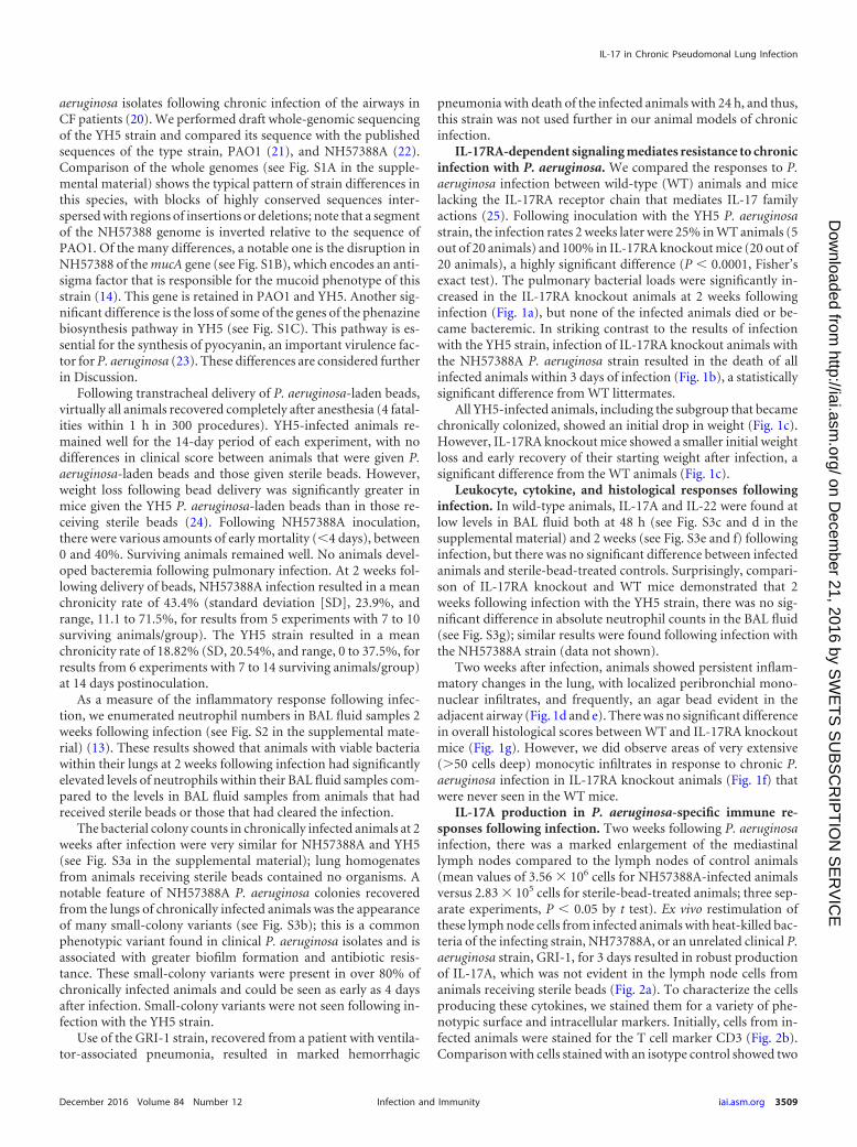
IL-17RA-dependent signaling mediates resistance to chronicinfection with P. aeruginosa. We compared the responses to P.aeruginosa infection between wild-type (WT) animals and micelacking the IL-17RA receptor chain that mediates IL-17 familyactions (25). Following inoculation with the YH5 P. aeruginosastrain, the infection rates 2 weeks later were 25% in WT animals (5out of 20 animals) and 100% in IL-17RA knockout mice (20 out of20 animals), a highly significant difference (P � 0.0001, Fisher’sexact test). The pulmonary bacterial loads were significantly in-creased in the IL-17RA knockout animals at 2 weeks followinginfection (Fig. 1a), but none of the infected animals died or be-came bacteremic. In striking contrast to the results of infectionwith the YH5 strain, infection of IL-17RA knockout animals withthe NH57388A P. aeruginosa strain resulted in the death of allinfected animals within 3 days of infection (Fig. 1b), a statisticallysignificant difference from WT littermates.
All YH5-infected animals, including the subgroup that becamechronically colonized, showed an initial drop in weight (Fig. 1c).However, IL-17RA knockout mice showed a smaller initial weightloss and early recovery of their starting weight after infection, asignificant difference from the WT animals (Fig. 1c).
Leukocyte, cytokine, and histological responses followinginfection. In wild-type animals, IL-17A and IL-22 were found atlow levels in BAL fluid both at 48 h (see Fig. S3c and d in thesupplemental material) and 2 weeks (see Fig. S3e and f) followinginfection, but there was no significant difference between infectedanimals and sterile-bead-treated controls. Surprisingly, compari-son of IL-17RA knockout and WT mice demonstrated that 2weeks following infection with the YH5 strain, there was no sig-nificant difference in absolute neutrophil counts in the BAL fluid(see Fig. S3g); similar results were found following infection withthe NH57388A strain (data not shown).
Two weeks after infection, animals showed persistent inflam-matory changes in the lung, with localized peribronchial mono-nuclear infiltrates, and frequently, an agar bead evident in theadjacent airway (Fig. 1d and e). There was no significant differencein overall histological scores between WT and IL-17RA knockoutmice (Fig. 1g). However, we did observe areas of very extensive(50 cells deep) monocytic infiltrates in response to chronic P.aeruginosa infection in IL-17RA knockout animals (Fig. 1f) thatwere never seen in the WT mice.
IL-17A production in P. aeruginosa-specific immune re-sponses following infection. Two weeks following P. aeruginosainfection, there was a marked enlargement of the mediastinallymph nodes compared to the lymph nodes of control animals(mean values of 3.56 � 106 cells for NH57388A-infected animalsversus 2.83 � 105 cells for sterile-bead-treated animals; three sep-arate experiments, P � 0.05 by t test). Ex vivo restimulation ofthese lymph node cells from infected animals with heat-killed bac-teria of the infecting strain, NH73788A, or an unrelated clinical P.aeruginosa strain, GRI-1, for 3 days resulted in robust productionof IL-17A, which was not evident in the lymph node cells fromanimals receiving sterile beads (Fig. 2a). To characterize the cellsproducing these cytokines, we stained them for a variety of phe-notypic surface and intracellular markers. Initially, cells from in-fected animals were stained for the T cell marker CD3 (Fig. 2b).Comparison with cells stained with an isotype control showed two
IL-17 in Chronic Pseudomonal Lung Infection
December 2016 Volume 84 Number 12 iai.asm.org 3509Infection and Immunity
on Decem
ber 21, 2016 by SW
ET
S S
UB
SC
RIP
TIO
N S
ER
VIC
Ehttp://iai.asm
.org/D
ownloaded from

populations: a very clear CD3 population with low side scatter(Fig. 2b, outlined in black) and a more diffuse population withlower expression of CD3 (Fig. 2b, outlined in red) that overlappedconsiderably with cells stained with the isotype control. We de-noted these populations as CD3 intermediate-high (CD3int-hi)and CD3 low-intermediate (CD3lo-int), respectively.
Following restimulation of cells from infected animals withNH57388A bacteria, there was a marked expansion of CD3lo-int
cells that was not seen in cells from animals that had receivedsterile beads (Fig. 2b). There was also an increase in the numbersof CD3int-hi cells in the nodes of infected animals; these too in-creased in numbers in response to restimulation, but to a lesser
FIG 1 Responses of WT and IL-17RA-knockout animals to pulmonary P. aeruginosa infection. (a) Pulmonary bacterial burdens in animals chronically infectedwith YH5 at 2 weeks postinfection. Lines indicate median values. P value was determined with the Mann-Whitney test from pooled results of two experiments,each with 10 mice per group. (b) Kaplan-Meir survival curves of WT (circles) or IL-17RA knockout (KO) animals (squares) infected with the NH57388A strain(n � 10 per group). Significant differences were determined by log rank test; P � 0.0092. (c) Weight changes in animals (n � 10) remaining chronically infectedat 14 days following infection with the YH5 strain; results are for WT (circles) or IL-17RA KO animals (squares). Each point represents the mean weight at thattime; error bars show SEM. Differences between the groups are significant at a P value of �0.0001 by repeated-measures ANOVA. (d, e) Hematoxylin and eosin(H&E)-stained lung sections from infected WT mice 2 weeks after infection with YH5 strain. Arrows show agar beads. Scale bars show 100 �m (d) or 50 �m (e).(f) Large (50 cells deep) monocytic accumulations were observed in lungs from IL-17RA KO mice. Scale bar shows 10 �m. (g) Histological scores for indicatedanimals 2 weeks after infection with YH5 strain. P � 0.7266 by Mann-Whitney test.
Bayes et al.
3510 iai.asm.org December 2016 Volume 84 Number 12Infection and Immunity
on Decem
ber 21, 2016 by SW
ET
S S
UB
SC
RIP
TIO
N S
ER
VIC
Ehttp://iai.asm
.org/D
ownloaded from

FIG 2 Immune responses following P. aeruginosa infection. Two weeks after infection, mediastinal lymph node cells from WT mice treated as indicated werestimulated ex vivo with heat-killed P. aeruginosa strains (MOI of 30) or left unstimulated for 3 days. (a) Levels of IL-17A secretion are shown; bars show meanvalues and error bars show SEM. **, significantly different from sterile beads at a P value of �0.01 by t test; Un, unstimulated; NH, NH57388A. Representativeresults from three separate experiments are shown. (b) Flow cytometry results for stimulated (3 days) mediastinal lymph node cells stained as shown. TheCD3lo-int populations are outlined in red, and the CD3int-hi populations are outlined in black. The total number of cells recovered in each gate is shown next tothe gate. SSC, side scatter. (c to e) Flow cytometry of lymph node cells as described in the legend to panel B except that cells were gated on the CD3int-hi population(c), the CD3lo-int population (d), or as indicated (e). The percentage of cells in each quadrant is shown. (f) Fold increases in P. aeruginosa-binding IgM and IgGproduced by mediastinal lymph node cells from infected animals restimulated with P. aeruginosa strains ex vivo compared to the levels produced by unstimulatedcells. Columns represent mean values for triplicate wells; error bars show SEM. Columns were compared to a theoretical mean of 1.0 by Student’s t test. NS,nonsignificant; *, P � 0.05; **, P � 0.01.
IL-17 in Chronic Pseudomonal Lung Infection
December 2016 Volume 84 Number 12 iai.asm.org 3511Infection and Immunity
on Decem
ber 21, 2016 by SW
ET
S S
UB
SC
RIP
TIO
N S
ER
VIC
Ehttp://iai.asm
.org/D
ownloaded from
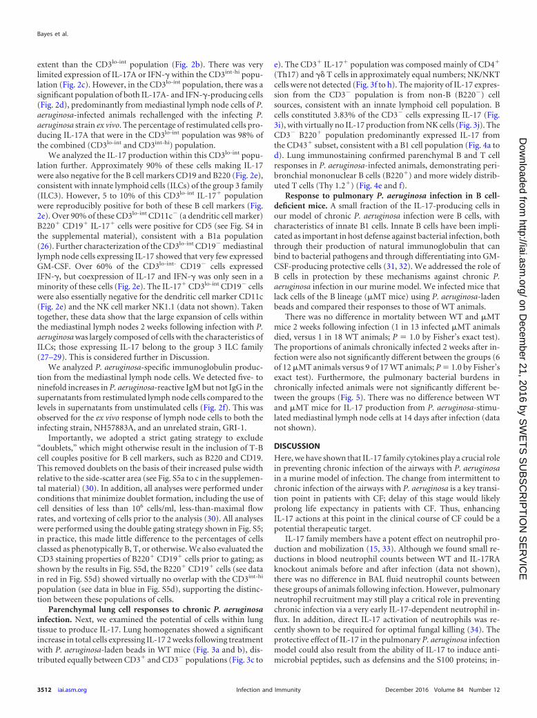
extent than the CD3lo-int population (Fig. 2b). There was verylimited expression of IL-17A or IFN-� within the CD3int-hi popu-lation (Fig. 2c). However, in the CD3lo-int population, there was asignificant population of both IL-17A- and IFN-�-producing cells(Fig. 2d), predominantly from mediastinal lymph node cells of P.aeruginosa-infected animals rechallenged with the infecting P.aeruginosa strain ex vivo. The percentage of restimulated cells pro-ducing IL-17A that were in the CD3lo-int population was 98% ofthe combined (CD3lo-int and CD3int-hi) population.
We analyzed the IL-17 production within this CD3lo-int popu-lation further. Approximately 90% of these cells making IL-17were also negative for the B cell markers CD19 and B220 (Fig. 2e),consistent with innate lymphoid cells (ILCs) of the group 3 family(ILC3). However, 5 to 10% of this CD3lo-int IL-17 populationwere reproducibly positive for both of these B cell markers (Fig.2e). Over 90% of these CD3lo-int CD11c� (a dendritic cell marker)B220 CD19 IL-17 cells were positive for CD5 (see Fig. S4 inthe supplemental material), consistent with a B1a population(26). Further characterization of the CD3lo-int CD19� mediastinallymph node cells expressing IL-17 showed that very few expressedGM-CSF. Over 60% of the CD3lo-int- CD19� cells expressedIFN-�, but coexpression of IL-17 and IFN-� was only seen in aminority of these cells (Fig. 2e). The IL-17 CD3lo-int CD19� cellswere also essentially negative for the dendritic cell marker CD11c(Fig. 2e) and the NK cell marker NK1.1 (data not shown). Takentogether, these data show that the large expansion of cells withinthe mediastinal lymph nodes 2 weeks following infection with P.aeruginosa was largely composed of cells with the characteristics ofILCs; those expressing IL-17 belong to the group 3 ILC family(27–29). This is considered further in Discussion.
We analyzed P. aeruginosa-specific immunoglobulin produc-tion from the mediastinal lymph node cells. We detected five- toninefold increases in P. aeruginosa-reactive IgM but not IgG in thesupernatants from restimulated lymph node cells compared to thelevels in supernatants from unstimulated cells (Fig. 2f). This wasobserved for the ex vivo response of lymph node cells to both theinfecting strain, NH57883A, and an unrelated strain, GRI-1.
Importantly, we adopted a strict gating strategy to exclude“doublets,” which might otherwise result in the inclusion of T-Bcell couples positive for B cell markers, such as B220 and CD19.This removed doublets on the basis of their increased pulse widthrelative to the side-scatter area (see Fig. S5a to c in the supplemen-tal material) (30). In addition, all analyses were performed underconditions that minimize doublet formation, including the use ofcell densities of less than 106 cells/ml, less-than-maximal flowrates, and vortexing of cells prior to the analysis (30). All analyseswere performed using the double gating strategy shown in Fig. S5;in practice, this made little difference to the percentages of cellsclassed as phenotypically B, T, or otherwise. We also evaluated theCD3 staining properties of B220 CD19 cells prior to gating; asshown by the results in Fig. S5d, the B220 CD19 cells (see datain red in Fig. S5d) showed virtually no overlap with the CD3int-hi
population (see data in blue in Fig. S5d), supporting the distinc-tion between these populations of cells.
Parenchymal lung cell responses to chronic P. aeruginosainfection. Next, we examined the potential of cells within lungtissue to produce IL-17. Lung homogenates showed a significantincrease in total cells expressing IL-17 2 weeks following treatmentwith P. aeruginosa-laden beads in WT mice (Fig. 3a and b), dis-tributed equally between CD3 and CD3� populations (Fig. 3c to
e). The CD3 IL-17 population was composed mainly of CD4
(Th17) and �� T cells in approximately equal numbers; NK/NKTcells were not detected (Fig. 3f to h). The majority of IL-17 expres-sion from the CD3� population is from non-B (B220�) cellsources, consistent with an innate lymphoid cell population. Bcells constituted 3.83% of the CD3� cells expressing IL-17 (Fig.3i), with virtually no IL-17 production from NK cells (Fig. 3j). TheCD3� B220 population predominantly expressed IL-17 fromthe CD43 subset, consistent with a B1 cell population (Fig. 4a tod). Lung immunostaining confirmed parenchymal B and T cellresponses in P. aeruginosa-infected animals, demonstrating peri-bronchial mononuclear B cells (B220) and more widely distrib-uted T cells (Thy 1.2) (Fig. 4e and f).
Response to pulmonary P. aeruginosa infection in B cell-deficient mice. A small fraction of the IL-17-producing cells inour model of chronic P. aeruginosa infection were B cells, withcharacteristics of innate B1 cells. Innate B cells have been impli-cated as important in host defense against bacterial infection, boththrough their production of natural immunoglobulin that canbind to bacterial pathogens and through differentiating into GM-CSF-producing protective cells (31, 32). We addressed the role ofB cells in protection by these mechanisms against chronic P.aeruginosa infection in our murine model. We infected mice thatlack cells of the B lineage (�MT mice) using P. aeruginosa-ladenbeads and compared their responses to those of WT animals.
There was no difference in mortality between WT and �MTmice 2 weeks following infection (1 in 13 infected �MT animalsdied, versus 1 in 18 WT animals; P � 1.0 by Fisher’s exact test).The proportions of animals chronically infected 2 weeks after in-fection were also not significantly different between the groups (6of 12 �MT animals versus 9 of 17 WT animals; P � 1.0 by Fisher’sexact test). Furthermore, the pulmonary bacterial burdens inchronically infected animals were not significantly different be-tween the groups (Fig. 5). There was no difference between WTand �MT mice for IL-17 production from P. aeruginosa-stimu-lated mediastinal lymph node cells at 14 days after infection (datanot shown).
DISCUSSION
Here, we have shown that IL-17 family cytokines play a crucial rolein preventing chronic infection of the airways with P. aeruginosain a murine model of infection. The change from intermittent tochronic infection of the airways with P. aeruginosa is a key transi-tion point in patients with CF; delay of this stage would likelyprolong life expectancy in patients with CF. Thus, enhancingIL-17 actions at this point in the clinical course of CF could be apotential therapeutic target.
IL-17 family members have a potent effect on neutrophil pro-duction and mobilization (15, 33). Although we found small re-ductions in blood neutrophil counts between WT and IL-17RAknockout animals before and after infection (data not shown),there was no difference in BAL fluid neutrophil counts betweenthese groups of animals following infection. However, pulmonaryneutrophil recruitment may still play a critical role in preventingchronic infection via a very early IL-17-dependent neutrophil in-flux. In addition, direct IL-17 activation of neutrophils was re-cently shown to be required for optimal fungal killing (34). Theprotective effect of IL-17 in the pulmonary P. aeruginosa infectionmodel could also result from the ability of IL-17 to induce anti-microbial peptides, such as defensins and the S100 proteins; in-
Bayes et al.
3512 iai.asm.org December 2016 Volume 84 Number 12Infection and Immunity
on Decem
ber 21, 2016 by SW
ET
S S
UB
SC
RIP
TIO
N S
ER
VIC
Ehttp://iai.asm
.org/D
ownloaded from

deed, in ocular infections, human �-defensin 2 plays an importantrole in host defense against P. aeruginosa (35). In addition, IL-17induces proinflammatory cytokines like IL-6 and tumor necrosisfactor alpha (TNF- ). The lack of weight loss seen in infectedIL-17RA knockout mice may reflect the lack of such inflammatorycytokine production, since these cytokines are associated with lossof body mass (36).
We identified a number of different cellular sources that pro-duce IL-17 in this model of infection. In the draining mediastinallymph nodes following infection, we found a considerable expan-sion of cells that predominantly had the characteristics of group 3ILCs. Specifically, given their production of IL-17, these are likely
to be lymphoid tissue inducer (LTi) cells. Further characterizationusing the ILC3 markers CD127 and ROR�t will be required toestablish the identity of these cells beyond doubt. ILC3/LTi cellshave been implicated in host resistance to extracellular bacteria,chronic inflammation, and tissue repair. Recently, group 3 ILCshave been shown to present antigen and to contribute to the con-trol of CD4 T cell responses to commensal bacteria (37, 38). Theexpansion of group 3 ILCs in the mediastinal lymph nodes hasbeen described previously in a helminth infection model (39).This study found that in mesenteric lymph nodes, the group 3ILCs migrated in a CCR7-dependent fashion from the intestine.The origin of the group 3 ILCs that migrate to mediastinal lymph
FIG 3 Lung parenchymal responses to P. aeruginosa infection. (a) Representative intracellular expression of IL-17A in lung parenchymal cells 2 weeks followinginstillation of sterile or P. aeruginosa-laden (strain NH57388A) beads. Numbers show percentages of total cells in ringed areas. (b) Percentages of total lungparenchymal cells expressing IL-17A 2 weeks after introduction of sterile or P. aeruginosa-laden beads. Each symbol represents an individual animal; lines showmedian values. P value was determined by Mann-Whitney test. (c) Representative expression of intracellular IL-17A in CD3 and CD3� populations in lungs 2weeks following P. aeruginosa infection as assessed by flow cytometry. (d, e) Data are as described in the legend to panel B but show the percentages of CD3 (d)and CD3� (e) cells expressing intracellular IL-17A. (f to h) Flow cytometry results for CD3 IL-17A cells expressing the indicated markers; boxed areas aredeemed positive relative to results for isotype controls. Figures show percentages of the total CD3 IL-17A population expressing the indicated markers. (i, j)Data are as described for panels F to H but with gating on CD3� IL-17A cells.
IL-17 in Chronic Pseudomonal Lung Infection
December 2016 Volume 84 Number 12 iai.asm.org 3513Infection and Immunity
on Decem
ber 21, 2016 by SW
ET
S S
UB
SC
RIP
TIO
N S
ER
VIC
Ehttp://iai.asm
.org/D
ownloaded from

nodes is not clear, especially as the lung has a very low number ofthese cells. The function of these cells in this location is also notclear. Neutrophils are recruited to regional lymph nodes in infec-tion and inflammation. In this location, they potentially fulfill anumber of roles, including limiting pathogen escape, modulatingsubcapsular sinus macrophage numbers, and influencing den-
dritic cell maturation and antigen presentation (reviewed in ref-erence 40). IL-17 family cytokines released by group 3 ILCs withindraining lymph nodes would thus be one mechanism wherebyneutrophils could be recruited to this site during an infection.Further work will be required to explore these possibilities.
We found a small but significant population of B cells thatproduced IL-17 in the mesenteric lymph nodes following infec-tion. B cell production of IL-17 has been found in a model ofTrypanosoma cruzi infection in mice (41). Our study suggests thatB1 cells are a source of this cytokine. B1 cells are the predominantB cell in pleural and peritoneal compartments and continuouslytraffic into these areas by a CXCL13-dependent pathway (42).Following activation, they migrate to regional lymph nodes andintestinal lamina propria (26, 43). The accumulation of IL-17-producing B1 cells in mediastinal lymph nodes following infectionreported here may reflect this activation-induced cell trafficking.Immature plasma cells (plasmablasts) may also express CCD19,B220, and CD43 and, thus, may also be the B cells producing IL-17within the mediastinal lymph nodes described here (44). Furtherwork with genetic marking of distinct lineages will be required toidentify these cells unequivocally. These mediastinal node cellsfrom P. aeruginosa-infected animals require further stimulationwith P. aeruginosa to expand the population of B1a cells and toproduce secretion of IL-17. This migration and subsequent divi-sion in response to P. aeruginosa is likely produced via Toll-likereceptor (TLR) stimulation with lipopolysaccharide (LPS) fromthe Gram-negative organisms, as has been shown previously (45).However, given that mice lacking B cells showed no defect in theincidence of chronic infection with P. aeruginosa, these B1 cells aredispensable as a source of protective IL-17.
Within lung parenchyma following infection, IL-17 was pro-duced from CD4 cells with properties of Th17 cells, as well as ��T cells. Both these cell types were increased in the lung followingchronic infection. The generation of P. aeruginosa-specific Th17cells by a pseudomonal vaccine may thus be of importance inproviding protection against chronic infection.
The P. aeruginosa strains used here were both derived from CFpatients but showed important differences in the model (Fig. 1).Apart from the known difference in alginate production in theNH57388A strain, genome sequencing revealed multiple genetic
FIG 4 Characteristics of B cells in lungs of animals instilled with P. aeruginosa-laden beads. (a to d) Two weeks after transtracheal instillation of P. aeruginosa-laden agar beads, the lungs were homogenized and cells polyclonally stim-ulated, extracellularly stained for CD3, B220, and CD43, and thenpermeabilized and stained for intracellular IL-17A, followed by flow cytom-etry. (a, b) Representative plots of the expression of B220 and IL-17A by liveCD3� cell populations. (b) Values above gates represent percentage of totalIL-17A cells in each gate. (c, d) Representative plots of expression of IL-17Aand CD43 by CD3� B220 cells. (d) Values in quadrants represent percentagesof total cells. Results shown are representative of two separate experiments. (e,f) Lung sections from animals 2 weeks following infection with NH57388Awere immunostained for B cells using B220 staining (e) or for T cells withThy1.2 staining (f). Immunostaining is indicated in red, and boxed areas areshown enlarged to the right. Nuclei are counterstained green.
FIG 5 Bacterial counts in WT and �MT mice following infection. Animalswere infected with the NH57388A strain. Each symbol represents the result foran individual animal; lines indicate median values. P value for comparisonbetween the groups was determined by Mann Whitney test.
Bayes et al.
3514 iai.asm.org December 2016 Volume 84 Number 12Infection and Immunity
on Decem
ber 21, 2016 by SW
ET
S S
UB
SC
RIP
TIO
N S
ER
VIC
Ehttp://iai.asm
.org/D
ownloaded from

differences between this strain and YH5 (see Fig. S1 in the supple-mental material). The alginate overproduction in NH57388A isdue to a deletion of part of the mucA gene, which encodes a MucAanti-sigma factor (22). Also of interest is that YH5 has lost thegenetic machinery necessary to synthesize pyocyanin. This couldaccount for its relative lack of virulence compared to that of theNH57388A strain in the IL-17RA knockout animals, but furtherwork will be required to determine which of these genetic differ-ences are most important.
Chronic P. aeruginosa infection is a significant problem notonly in CF but also in COPD (46) and non-CF bronchiectasis (47).Delaying the onset of chronic infection in such patient groupswould represent a very valuable therapeutic goal. Antibiotics candelay this progression but cannot prevent this transition. A num-ber of vaccines against P. aeruginosa have been developed to at-tempt to prevent chronic infection; as yet, none of these haveshown clinical efficacy (48). The work described here opens a newperspective on the prevention and treatment of such P. aeruginosainfection. Potential therapies based on augmenting IL-17 ac-tion—through vaccination or otherwise— could be of great valuein delaying or preventing chronic P. aeruginosa infection inchronic lung disease.
ACKNOWLEDGMENTS
The work was funded by the Wellcome Trust (grant number 094779) andthe Medical Research Council (grant number G1001998).
The funders had no role in study design, data collection and interpre-tation, or the decision to submit the work for publication.
FUNDING INFORMATIONThis work, including the efforts of Neil D. Ritchie and Tom J. Evans, wasfunded by Medical Research Council (MRC) (G1001998). This work, in-cluding the efforts of Hannah K. Bayes and Tom J. Evans, was funded byWellcome Trust (094779).
REFERENCES1. Hoiby N, Frederiksen B, Pressler T. 2005. Eradication of early Pseu-
domonas aeruginosa infection. J Cyst Fibros 4(Suppl 2):S49 –S54. http://dx.doi.org/10.1016/j.jcf.2005.05.018.
2. Folkesson A, Jelsbak L, Yang L, Johansen HK, Ciofu O, Hoiby N, MolinS. 2012. Adaptation of Pseudomonas aeruginosa to the cystic fibrosisairway: an evolutionary perspective. Nat Rev Microbiol 10:841– 851. http://dx.doi.org/10.1038/nrmicro2907.
3. Nichols D, Chmiel J, Berger M. 2008. Chronic inflammation in thecystic fibrosis lung: alterations in inter- and intracellular signaling.Clin Rev Allergy Immunol 34:146 –162. http://dx.doi.org/10.1007/s12016-007-8039-9.
4. Iwakura Y, Nakae S, Saijo S, Ishigame H. 2008. The roles of IL-17Ain inflammatory immune responses and host defense against patho-gens. Immunol Rev 226:57–79. http://dx.doi.org/10.1111/j.1600-065X.2008.00699.x.
5. Decraene A, Willems-Widyastuti A, Kasran A, De Boeck K, BullensDM, Dupont LJ. 2010. Elevated expression of both mRNA and proteinlevels of IL-17A in sputum of stable cystic fibrosis patients. Respir Res11:177. http://dx.doi.org/10.1186/1465-9921-11-177.
6. Priebe GP, Walsh RL, Cederroth TA, Kamei A, Coutinho-Sledge YS,Goldberg JB, Pier GB. 2008. IL-17 is a critical component of vaccine-induced protection against lung infection by lipopolysaccharide-heterologous strains of Pseudomonas aeruginosa. J Immunol 181:4965–4975. http://dx.doi.org/10.4049/jimmunol.181.7.4965.
7. Aujla SJ, Dubin PJ, Kolls JK. 2007. Th17 cells and mucosal host defense.Semin Immunol 19:377–382. http://dx.doi.org/10.1016/j.smim.2007.10.009.
8. Dubin PJ, Kolls JK. 2007. Pseudomonas aeruginosa and the host pulmo-nary immune response. Expert Rev Respir Med 1:121–137. http://dx.doi.org/10.1586/17476348.1.1.121.
9. Bayes HK, Bicknell S, MacGregor G, Evans TJ. 2014. T helper cell subsetsspecific for Pseudomonas aeruginosa in healthy individuals and patientswith cystic fibrosis. PLoS One 9:e90263. http://dx.doi.org/10.1371/journal.pone.0090263.
10. Dubin PJ, McAllister F, Kolls JK. 2007. Is cystic fibrosis a TH17 disease?Inflamm Res 56:221–227. http://dx.doi.org/10.1007/s00011-007-6187-2.
11. Liu J, Feng Y, Yang K, Li Q, Ye L, Han L, Wan H. 2011. Early productionof IL-17 protects against acute pulmonary Pseudomonas aeruginosa in-fection in mice. FEMS Immunol Med Microbiol 61:179 –188. http://dx.doi.org/10.1111/j.1574-695X.2010.00764.x.
12. van Heeckeren A, Schluchter M. 2002. Murine models of chronic Pseu-domonas aeruginosa lung infection. Lab Anim 36:291. http://dx.doi.org/10.1258/002367702320162405.
13. Bayes HK, Ritchie ND, Irvine S, Evans TJ. A murine model of earlyPseudomonas aeruginosa lung disease with transition to chronic infec-tion. Sci Rep, in press.
14. Hoffmann N, Rasmussen TB, Jensen PO, Stub C, Hentzer M, Molin S,Ciofu O, Givskov M, Johansen HK, Hoiby N. 2005. Novel mouse modelof chronic Pseudomonas aeruginosa lung infection mimicking cystic fi-brosis. Infect Immun 73:2504 –2514. http://dx.doi.org/10.1128/IAI.73.4.2504-2514.2005.
15. Ye P, Rodriguez FH, Kanaly S, Stocking KL, Schurr J, SchwarzenbergerP, Oliver P, Huang W, Zhang P, Zhang J, Shellito JE, Bagby GJ, NelsonS, Charrier K, Peschon JJ, Kolls JK. 2001. Requirement of interleukin 17receptor signaling for lung CXC chemokine and granulocyte colony-stimulating factor expression, neutrophil recruitment, and host defense. JExp Med 194:519 –527. http://dx.doi.org/10.1084/jem.194.4.519.
16. Kitamura D, Roes J, Kuhn R, Rajewsky K. 1991. A B cell-deficient mouseby targeted disruption of the membrane exon of the immunoglobulin muchain gene. Nature 350:423– 426. http://dx.doi.org/10.1038/350423a0.
17. Moser C, Jensen PO, Kobayashi O, Hougen HP, Song Z, Rygaard J,Kharazmi A, Høiby N. 2002. Improved outcome of chronic Pseudomo-nas aeruginosa lung infection is associated with induction of a Th1-dominated cytokine response. Clin Exp Immunol 127:206 –213. http://dx.doi.org/10.1046/j.1365-2249.2002.01731.x.
18. van Heeckeren AM, Schluchter MD. 2002. Murine models of chronicPseudomonas aeruginosa lung infection. Lab Anim 36:291–312. http://dx.doi.org/10.1258/002367702320162405.
19. Bragonzi A. 2010. Murine models of acute and chronic lung infectionwith cystic fibrosis pathogens. Int J Med Microbiol 300:584 –593. http://dx.doi.org/10.1016/j.ijmm.2010.08.012.
20. Koch C. 2002. Early infection and progression of cystic fibrosis lungdisease. Pediatr Pulmonol 34:232–236. http://dx.doi.org/10.1002/ppul.10135.
21. Stover CK, Pham XQ, Erwin AL, Mizoguchi SD, Warrener P, HickeyMJ, Brinkman FS, Hufnagle WO, Kowalik DJ, Lagrou M, Garber RL,Goltry L, Tolentino E, Westbrock-Wadman S, Yuan Y, Brody LL,Coulter SN, Folger KR, Kas A, Larbig K, Lim R, Smith K, Spencer D,Wong GK, Wu Z, Paulsen IT, Reizer J, Saier MH, Hancock RE, Lory S,Olson MV. 2000. Complete genome sequence of Pseudomonas aerugi-nosa PAO1, an opportunistic pathogen. Nature 406:959 –964. http://dx.doi.org/10.1038/35023079.
22. Norman A, Ciofu O, Amador CI, Hoiby N, Jelsbak L. 2016. Genomesequence of Pseudomonas aeruginosa strain DK1-NH57388A, a stablemucoid cystic fibrosis isolate. Genome Announc 4:e00008-16. http://dx.doi.org/10.1128/genomeA.00008-16.
23. Rada B, Leto TL. 2013. Pyocyanin effects on respiratory epithelium:relevance in Pseudomonas aeruginosa airway infections. Trends Micro-biol 21:73– 81. http://dx.doi.org/10.1016/j.tim.2012.10.004.
24. Bayes HK, Ritchie ND, Ward C, Corris PA, Brodlie M, Evans TJ. 30June 2016. IL-22 exacerbates weight loss in a murine model of chronicpulmonary Pseudomonas aeruginosa infection. J Cyst Fibros. http://dx.doi.org/10.1016/j.jcf.2016.06.008.
25. Gaffen SL. 2011. Recent advances in the IL-17 cytokine family. Curr OpinImmunol 23:613– 619. http://dx.doi.org/10.1016/j.coi.2011.07.006.
26. Baumgarth N. 2011. The double life of a B-1 cell: self-reactivity selects forprotective effector functions. Nat Rev Immunol 11:34 – 46. http://dx.doi.org/10.1038/nri2901.
27. Sonnenberg GF, Artis D. 2015. Innate lymphoid cells in the initiation,regulation and resolution of inflammation. Nat Med 21:698 –708. http://dx.doi.org/10.1038/nm.3892.
28. Artis D, Spits H. 2015. The biology of innate lymphoid cells. Nature517:293–301. http://dx.doi.org/10.1038/nature14189.
IL-17 in Chronic Pseudomonal Lung Infection
December 2016 Volume 84 Number 12 iai.asm.org 3515Infection and Immunity
on Decem
ber 21, 2016 by SW
ET
S S
UB
SC
RIP
TIO
N S
ER
VIC
Ehttp://iai.asm
.org/D
ownloaded from

29. Spits H, Artis D, Colonna M, Diefenbach A, Di Santo JP, Eberl G,Koyasu S, Locksley RM, McKenzie AN, Mebius RE, Powrie F, Vivier E.2013. Innate lymphoid cells—a proposal for uniform nomenclature. NatRev Immunol 13:145–149. http://dx.doi.org/10.1038/nri3365.
30. Griffin DO, Rothstein TL. 2012. Human B1 cell frequency: isolation andanalysis of human B1 cells. Front Immunol 3:122. http://dx.doi.org/10.3389/fimmu.2012.00122.
31. Weber GF, Chousterman BG, Hilgendorf I, Robbins CS, Theurl I,Gerhardt LM, Iwamoto Y, Quach TD, Ali M, Chen JW, Rothstein TL,Nahrendorf M, Weissleder R, Swirski FK. 2014. Pleural innate responseactivator B cells protect against pneumonia via a GM-CSF-IgM axis. J ExpMed 211:1243–1256. http://dx.doi.org/10.1084/jem.20131471.
32. Rauch PJ, Chudnovskiy A, Robbins CS, Weber GF, Etzrodt M, Hilgen-dorf I, Tiglao E, Figueiredo JL, Iwamoto Y, Theurl I, Gorbatov R,Waring MT, Chicoine AT, Mouded M, Pittet MJ, Nahrendorf M,Weissleder R, Swirski FK. 2012. Innate response activator B cells protectagainst microbial sepsis. Science 335:597– 601. http://dx.doi.org/10.1126/science.1215173.
33. Schwarzenberger P, Huang W, Ye P, Oliver P, Manuel M, Zhang Z,Bagby G, Nelson S, Kolls JK. 2000. Requirement of endogenous stem cellfactor and granulocyte-colony-stimulating factor for IL-17-mediatedgranulopoiesis. J Immunol 164:4783– 4789. http://dx.doi.org/10.4049/jimmunol.164.9.4783.
34. Taylor PR, Leal SM, Jr, Sun Y, Pearlman E. 2014. Aspergillus andFusarium corneal infections are regulated by Th17 cells and IL-17-producing neutrophils. J Immunol 192:3319 –3327. http://dx.doi.org/10.4049/jimmunol.1302235.
35. Augustin DK, Heimer SR, Tam C, Li WY, Le Due JM, Evans DJ, FleiszigSM. 2011. Role of defensins in corneal epithelial barrier function againstPseudomonas aeruginosa traversal. Infect Immun 79:595– 605. http://dx.doi.org/10.1128/IAI.00854-10.
36. Wallenius V, Wallenius K, Ahren B, Rudling M, Carlsten H, DicksonSL, Ohlsson C, Jansson JO. 2002. Interleukin-6-deficient mice de-velop mature-onset obesity. Nat Med 8:75–79. http://dx.doi.org/10.1038/nm0102-75.
37. von Burg N, Chappaz S, Baerenwaldt A, Horvath E, Bose Dasgupta S,Ashok D, Pieters J, Tacchini-Cottier F, Rolink A, Acha-Orbea H, FinkeD. 2014. Activated group 3 innate lymphoid cells promote T-cell-mediated immune responses. Proc Natl Acad Sci U S A 111:12835–12840.http://dx.doi.org/10.1073/pnas.1406908111.
38. Goc J, Hepworth MR, Sonnenberg GF. 2016. Group 3 innate lymphoidcells: regulating host-commensal bacteria interactions in inflammationand cancer. Int Immunol 28:43–52. http://dx.doi.org/10.1093/intimm/dxv056.
39. Mackley EC, Houston S, Marriott CL, Halford EE, Lucas B, Cerovic V,Filbey KJ, Maizels RM, Hepworth MR, Sonnenberg GF, Milling S,Withers DR. 2015. CCR7-dependent trafficking of RORgamma() ILCscreates a unique microenvironment within mucosal draining lymphnodes. Nat Commun 6:5862. http://dx.doi.org/10.1038/ncomms6862.
40. Nathan C. 2006. Neutrophils and immunity: challenges and opportuni-ties. Nat Rev Immunol 6:173–182. http://dx.doi.org/10.1038/nri1785.
41. Bermejo DA, Jackson SW, Gorosito-Serran M, Acosta-Rodriguez EV,Amezcua-Vesely MC, Sather BD, Singh AK, Khim S, Mucci J, Liggitt D,Campetella O, Oukka M, Gruppi A, Rawlings DJ. 2013. Trypanosomacruzi trans-sialidase initiates a program independent of the transcriptionfactors ROR�t and Ahr that leads to IL-17 production by activated B cells.Nat Immunol 14:514 –522. http://dx.doi.org/10.1038/ni.2569.
42. Ansel KM, Harris RB, Cyster JG. 2002. CXCL13 is required for B1 cellhoming, natural antibody production, and body cavity immunity. Immu-nity 16:67–76. http://dx.doi.org/10.1016/S1074-7613(01)00257-6.
43. Choi YS, Baumgarth N. 2008. Dual role for B-1a cells in immunity toinfluenza virus infection. J Exp Med 205:3053–3064. http://dx.doi.org/10.1084/jem.20080979.
44. Kallies A, Hasbold J, Tarlinton DM, Dietrich W, Corcoran LM, Hodg-kin PD, Nutt SL. 2004. Plasma cell ontogeny defined by quantitativechanges in blimp-1 expression. J Exp Med 200:967–977. http://dx.doi.org/10.1084/jem.20040973.
45. Ha SA, Tsuji M, Suzuki K, Meek B, Yasuda N, Kaisho T, Fagarasan S.2006. Regulation of B1 cell migration by signals through Toll-like recep-tors. J Exp Med 203:2541–2550. http://dx.doi.org/10.1084/jem.20061041.
46. Rakhimova E, Wiehlmann L, Brauer AL, Sethi S, Murphy TF, TummlerB. 2009. Pseudomonas aeruginosa population biology in chronic obstruc-tive pulmonary disease. J Infect Dis 200:1928 –1935. http://dx.doi.org/10.1086/648404.
47. Zoumot Z, Wilson R. 2010. Respiratory infection in noncystic fibrosisbronchiectasis. Curr Opin Infect Dis 23:165–170. http://dx.doi.org/10.1097/QCO.0b013e328335af91.
48. Johansen HK, Gotzsche PC. 2015. Vaccines for preventing infection withPseudomonas aeruginosa in cystic fibrosis. Cochrane Database Syst Rev2015(8):CD001399. http://dx.doi.org/10.1002/14651858.CD001399.pub4.
Bayes et al.
3516 iai.asm.org December 2016 Volume 84 Number 12Infection and Immunity
on Decem
ber 21, 2016 by SW
ET
S S
UB
SC
RIP
TIO
N S
ER
VIC
Ehttp://iai.asm
.org/D
ownloaded from