on the mechanism of protein fold-switching by a molecular
TRANSCRIPT
University of Massachusetts AmherstScholarWorks@UMass AmherstBiochemistry & Molecular Biology DepartmentFaculty Publication Series Biochemistry and Molecular Biology
2010
On the mechanism of protein fold-switching by amolecular sensorMargaret M. StrattonUniversity of Massachusetts Amherst
S N. LohState University of New York Upstate Medical University
Follow this and additional works at: https://scholarworks.umass.edu/biochem_faculty_pubs
Part of the Molecular Biology Commons
This Article is brought to you for free and open access by the Biochemistry and Molecular Biology at ScholarWorks@UMass Amherst. It has beenaccepted for inclusion in Biochemistry & Molecular Biology Department Faculty Publication Series by an authorized administrator ofScholarWorks@UMass Amherst. For more information, please contact [email protected].
Recommended CitationStratton, Margaret M. and Loh, S N., "On the mechanism of protein fold-switching by a molecular sensor" (2010). Proteins. 4.1002/prot.22833
On the mechanism of protein fold-switching by a molecularsensor
Margaret M. Stratton and Stewart N. Loh*
Department of Biochemistry and Molecular Biology, State University of New York Upstate MedicalUniversity, Syracuse New York 13210
AbstractAlternate frame folding (AFF) is a mechanism by which conformational change can be engineeredinto a protein. The protein structure switches from the wild-type fold (N) to a circularly-permutedfold (N′), or vice versa, in response to a signaling event such as ligand binding. Despite the factthat the two native states have similar structures, their interconversion involves folding andunfolding of large parts of the molecule. This rearrangement is reported by fluorescent groupswhose relative proximities change as a result of the order–disorder transition. The nature of theconformational change is expected to be similar from protein to protein; thus, it may be possible toemploy AFF as a general method to create optical biosensors. Toward that goal, we test basicaspects of the AFF mechanism using the AFF variant of calbindin D9k. A simple three-state modelfor fold switching holds that N and N′ interconvert through the unfolded state. This model predictsthat the fundamental properties of the switch—calcium binding affinity, signal response (i.e.,fluorescence change upon binding), and switching rate—can be controlled by altering the relativestabilities of N and N′. We find that selectively destabilizing N or N′ changes the equilibriumproperties of the switch (binding affinity and signal response) in accordance with the model.However, kinetic data indicate that the switching pathway does not require whole-moleculeunfolding. The rate is instead limited by unfolding of a portion of the protein, possibly in concertwith folding of a corresponding region.
Keywordsalternate frame folding; allostery; biosensor; intrinsic disorder; binding and folding; fluorescencereporting
INTRODUCTIONIn two earlier studies, we introduced the concept of alternate frame protein folding (AFF) asa potentially general mechanism for creating biosensors1 and artificial zymogens.2 Themethod was applied to the small protein calbindin D9k to convert it into calbindin-AFF, acalcium- driven molecular switch. Calbindin-AFF is able to fold in one of two frames withinthe same polypeptide chain. The first frame consists of the normal sequence of amino acidsand folding results in the wild-type (WT) structure (N) [Fig. 1(A)]. The second frame iscomprised of a permuted amino acid sequence and folding produces a circularly-permutedstructure (N′). The N and N′ conformations are mutually exclusive because they share a
©2010 Wiley-Liss, Inc.*Correspondence to: Stewart N. Loh, Department of Biochemistry and Molecular Biology, State University of New York UpstateMedical University, Syracuse New York 13210. [email protected] Supporting Information may be found in the online version of this article.
NIH Public AccessAuthor ManuscriptProteins. Author manuscript; available in PMC 2011 July 28.
Published in final edited form as:Proteins. 2010 December ; 78(16): 3260–3269. doi:10.1002/prot.22833.
NIH
-PA Author Manuscript
NIH
-PA Author Manuscript
NIH
-PA Author Manuscript

common stretch of amino acids. We previously suggested that the AFF mechanism isspecified by the same thermodynamic and kinetic considerations that govern protein foldingand ligand binding. The purpose of this study is to test that hypothesis.
The general approach for engineering the N ⇄N′ conformational change into a given proteinis to duplicate a segment from the C-terminus and append it to the N-terminus, or to copy asegment from the N-terminus and ligate it to the C-terminus. Which segment is chosendepends on the location of key functional residues. For the biosensor application describedhere, the replicated segment must contain at least one amino acid which, when mutated,eliminates ligand binding. Beyond that criterion, the length of the duplicate polypeptide isdictated by preference for the termini of the circularly permuted protein to be located at asurface loop of the WT structure. Choosing the duplication to begin at the loop closest insequence to the binding residue will minimize overall length and help to avoid potentialaggregation and/or degradation problems.
Figure 1(B) illustrates how we modified the amino acid sequence of WT calbindin (75amino acids) to generate calbindin-AFF. WT calbindin consists of two calcium-binding EF-hands: hand-1 (residues 1–43) and hand-2 (residues 44–75). Calbindin contains three surfaceloops. Two are used to coordinate calcium, leaving the loop connecting hand-1 and hand-2as the only possible site for circular permutation. Calbindin-AFF is generated by duplicatingresidues 44–75 and appending them to the N-terminus of WT calbindin. This peptideequates to hand-2, although for a typical protein the duplicate sequence need not correspondto a functional domain. The duplicate sequence is designated by prime superscripts (44′–75′)but is otherwise numbered identically to the parent sequence. Calbindin-AFF is thuscomprised of three EF-hands: hand-2′ (colored red in Fig. 1), hand-1 (green), and hand-2(blue). Finally, a six-amino acid linker is inserted between hand-2′ and hand-1 to bridge theoriginal N- and C-termini of the circularly permuted fold.
Conformation N is produced by folding of hand-1 and hand-2 [Fig. 1(A)]. N′ is generated byfolding of hand-2′ and hand-1. N and N′ cannot exist simultaneously because they competefor hand-1. We introduce mutations into each frame to: (i) drive the N → N′ and N′ → Nfold shifts using binding energy (E65Q calcium binding mutation), or (ii) to selectivelydestabilize one of the folds (F66W hydrophobic core mutation). To illustrate thenomenclature of mutants, Q65 indicates the E65Q mutation at position 65 in fold N, whileW66′ denotes the F66W mutation in position 66′ of fold N′.
We predict that the “orphan” regions of an AFF-modified protein, that is, one of the twoduplicate segments, will be disordered in either native conformation. For example, N willcontain a disordered N-terminal tail and N′ will contain a disordered C-terminal tail [Fig.1(A)]. This expectation is general and it should hold true for calbindin-AFF, because hand-2is largely unstructured in isolation.3 In that respect, calbindin-AFF is reminiscent ofintrinsically disordered proteins found in nature.4–6 Intrinsically disordered proteins arechiefly unfolded in the cell until they bind their ligands. Favorable interactions between theprotein and the substrate provide the energetic contributions necessary to drive folding.7Similarly, calcium binding is the driving force for a disorder–order transition within thecalbindin-AFF molecule. A notable difference is that calbindin-AFF is native in both boundand free forms, and binding-induced folding is coupled to unfolding of another region of themolecule. Thus, no net folding or unfolding takes place; rather, the structure is remodeled.
The goal of this study is to define basic thermodynamic and kinetic principles that underliethe AFF mechanism. By doing so we address the following questions: (i) Does the entirecalbindin-AFF molecule have to unfold to refold using a different set of amino acids? (ii)Are the N- and C-terminal tails folded or unfolded? (iii) Can we manipulate the populations
Stratton and Loh Page 2
Proteins. Author manuscript; available in PMC 2011 July 28.
NIH
-PA Author Manuscript
NIH
-PA Author Manuscript
NIH
-PA Author Manuscript

of N and N′ to control ligand binding affinity and the signal response of the switch? (iv) Canwe modulate the rate of switching between the two conformations? This information willprovide insight to this unique binding/folding/unfolding mechanism and help guide futuredesign of biosensors based on the AFF principle.
MATERIALS AND METHODSGene construction, protein expression, and dye labeling
Genes were constructed and proteins were expressed and purified as described.1 All geneswere fully sequenced. To label the proteins, we first removed residual β-mercaptoethanol(which was added during purification) by desalting lyophilized protein into 10 mM Tris (pH7.5) using a Sephadex G25 spin column. Protein concentration was measured by absorbance(ε278 =1400 M−1 cm−1) and samples were precipitated using 10% trichloroacetic acid.Pellets were resuspended in 7M guanidine hydrochloride (GdnHCl), 10 mM Tris (pH 8.0) toa final protein concentration of 1 mM. Cys groups were reduced by adding a 10-fold excessof Tris (2-carboxyethyl) phosphine HCl (TCEP) and incubating for 30 m at roomtemperature. Protein was refolded by rapid 20-fold dilution into 0.1M Tris (pH 8.0).BODIPY-FL maleimide (Molecular Probes) or N-1-pyrene maleimide (Anaspec) was addedin two-fold excess of Cys concentration and labeling was allowed to proceed for 1 h at roomtemperature and in the dark. Protein was desalted into 20 mM Tris (pH 7.5), 0.1M NaClusing a PD10 column (Bio-Rad). Fluorophore concentration was estimated using absorbance(BODIPY ε503 = 80,000 M−1 cm−1, pyrene ε345 =24,000 M−1 cm−1). Despite increasing thedye concentration and the duration of the labeling step, MALDI mass spectrometry (notshown) revealed that the labeling reactions were incomplete. A typical experiment resultedin ~30% double-labeled protein, ~60% single-labeled, and ~10% unlabeled.
Equilibrium denaturation and calcium binding experimentsAll data were recorded at 20°C. Protein samples for denaturation experiments were preparedby mixing a solution of protein (7–10 μM) in 20 mM Tris (pH 7.5), and either 200 μM CaCl2(holo samples) or 400 μM EDTA (apo samples) with an identical solution containing thehighest concentration of denaturant used in the experiment. Unlabeled protein samplescontained 0.15 mM TCEP. Dilutions were made using a Hamilto-nian Microlab 540Bdispenser and denaturant concentrations were measured by refractive index.8 Samples wereequilibrated overnight at 20°C. CD data were collected on an Aviv Model 202spectropolarimeter. BODIPY fluorescence data were recorded on a Horiba Jobin-YvonFluoromax-3 fluorimeter with excitation at 503 nm and emission from 507 to 600 nm.Equilibrium calcium binding experiments were performed as described.1 Pyrene excimerfluorescence was calculated by normalizing the spectra to the highest intensity peak (374nm), adding the excimer emission values from 460 to 500 nm, and dividing by monomerfluorescence at 383 nm.9 Calcium binding by calbindin can be specified by two macroscopicassociation constants which reflect binding of the first calcium to either hand, then bindingof the second ion to the remaining hand. These values are ~106 M−1 and 107 M−1,respectively, for WT calbindin.10 Our experiments do not permit these macroscopicconstants to be determined individually. We instead fit the data to the Hill equation (Y = A +B[Ca2+]n/([Ca2+]n + (1/Ka,obs)n)) where A is initial fluorescence, B is the amplitude, and n isthe cooperativity coefficient.
Rapid kinetic experimentsStopped-flow fluorescence data were recorded at 20°C using a Bio-Logic SFM4-Q/S rapidmixing device. All stopped-flow fluorescence experiments employed BOD-IPY-labeledproteins; excitation was at 485 nm and a 497-nm high-pass filter was employed on detection.Unfolding experiments were performed by mixing native (Q65+Q65′) (3–6 μM in 20 mM
Stratton and Loh Page 3
Proteins. Author manuscript; available in PMC 2011 July 28.
NIH
-PA Author Manuscript
NIH
-PA Author Manuscript
NIH
-PA Author Manuscript

Tris, pH 7.5) with 0.5 mM CaCl2 (1:1 ratio) in Mixer 1, then diluting the product with 9.8Murea in Mixers 2 and 3 to achieve the final urea concentration shown in the figures. ForCa2+-binding experiments, 50 μM EDTA was added to the protein samples to ensure apoconditions. Apo-proteins were first diluted with various volumes of 6M urea (up to 2.5 Mfinal concentration, using Mixer 1), then mixed with CaCl2 to a final concentration of 300mM.
Stopped-flow CD data were acquired on an Aviv model 400 spectropolarimeter. Calciumbinding experiments were performed as above, except unlabeled proteins were used. Datawere recorded at 222 nm.
RESULTSGeneral model of fold switch
The AFF mechanism can be represented using an allosteric coupling model described byHilser and Thompson (HT)11 and by ourselves.12 The HT model shares similarities with thelongstanding Monod-Wyman-Changeux13 and Koshland-Nemethy-Filmer14 models ofallostery; however, it is different in that the binding-incompetent conformations areunfolded. The HT formulation describes site-to-site communication within a single two-domain protein in which both domains are intrinsically disordered. Each folded domainrecognizes a separate ligand and binding drives folding in both cases. The effect of bindingto one domain on folding of the other is determined by an energy term (gint) which describesthe interaction between the two-folded domains. If gint is positive then the two domainsinteract favorably and binding to the first domain increases the probability that the secondwill fold. The model that we introduced previously, which describes the coupled folding–unfolding equilibrium between two protein domains that fold in a mutually exclusivefashion, is similar to the HT model except the coupling energy is always unfavorable.12
The AFF mechanism is a special case of the HT model in which gint is negative andeffectively infinite. In this case the two domains correspond to N and N′. Because N and N′compete for a shared sequence, folding of N precludes folding of N′ and vice versa. Theresulting mechanism contains only states N, N′, and U (the globally unfolded conformation)as described by Eq. (1):
where L is ligand, Ka is the association constant, and Kfold is the equilibrium constant forfolding. For N to convert to N′ (and vice versa), at least a portion of the molecule mustunfold and a corresponding portion must fold. The interconversion is not required to proceedthrough U (e.g., the shared region need not unfold); U is included simply to provide acommon reference point for free energy changes.
The three fundamental properties of the sensor are ligand binding affinity, output signalresponse (e.g., fluorescence change), and response rate. The thermodynamic stabilities of Nand N′ dictate the equilibrium aspects of the switch; namely, binding affinity andfluorescence change. Consider the case where a binding mutation is introduced into N′ so
that . The observed association constant (Ka,obsN) is given by Eq. (2):
Stratton and Loh Page 4
Proteins. Author manuscript; available in PMC 2011 July 28.
NIH
-PA Author Manuscript
NIH
-PA Author Manuscript
NIH
-PA Author Manuscript
Equation (2) can be rearranged to yield the following relationship between and :
If N is much more stable than N′ then . If the stability of N′ is comparable to orgreater than that of N, then a portion of binding energy is used to drive the N′→ Nconformational change and the observed association constant drops. The magnitude of theobserved fluorescence change is similarly determined by the extent to which the populationsof N and N′ change upon binding. Figure 2 illustrates how these properties can bemanipulated by selectively destabilizing one of the two possible native conformations. Theblack energy levels depict N and N′ as being equal in free energy; this is approximately true
for calbindin-AFF (vide infra). When N and N′ are isoenergetic then . Thefraction of N equals 0.5 and it can increase by as much as a factor of two upon binding.Introducing a destabilizing mutation into N (Δ ΔG = −2 kcal mol−1; gray energy levels)drops the observed binding affinity by 16-fold (from 0.5 ×Ka
N to 0.03 ×KaN), reflecting the
fact that the conversion of N′ to N is thermodynamically uphill. The fraction of N canincrease by a factor of 33 (from 0.03 to 1.0), which allows the fluorescence change toincrease almost to its maximum value (given by the ratio of the fluorescence intensities of Nand N′).
What determines the rate of switching? The three-state mechanism shown in Figure 2(B)postulates that the N →N′ and N′ → N rates are largely limited by global unfolding of N andN′ (kunf N and kunf N′, respectively). If so, then the observed rate of ligand binding can bemodulated as well. The height of the rate-limiting barrier in Figure 2 can be reduced by amutation (shown in gray) that raises the free energy of N′ without proportionally raising thefree energy of the N′ → U transition state. This type of behavior, which is categorized as φ <1.0 in the φ-analysis methodology of Fersht,15 is common among destabilizing mutations. Itshould be noted that both the N→N′ and N′→ N rates can in principle be accelerated withoutaffecting Ka,obs. This can be achieved by introducing a mutation into the shared region[colored green in Fig. 1(A)], which would be expected to destabilize N and N′ to similarextents.
Selection of mutants and method of fluorescence detectionWe previously determined that WT and circularly permuted calbindin are of similar stabilityin their calcium-free (apo) as well as calcium-bound (holo) states.1 Though WT andpermuted calbindin lack the N- and C-terminal extensions found in calbindin-AFF, theynevertheless serve as reasonable models of the N and N′ states of calbindin-AFF,respectively. Given this assumption, calbindin-AFF is expected to exist in a roughly 1:1ratio of the two native conformations. We chose the hydrophobic core mutation F66W toperturb that ratio. This mutation destabilizes WT apo-calbindin by 2.3 kcal mol−1.16
Interestingly, the F66W mutation increases the macroscopic association constant of bindingthe first Ca2+ to either site (by 10-fold) without affecting binding to the remaining site.17
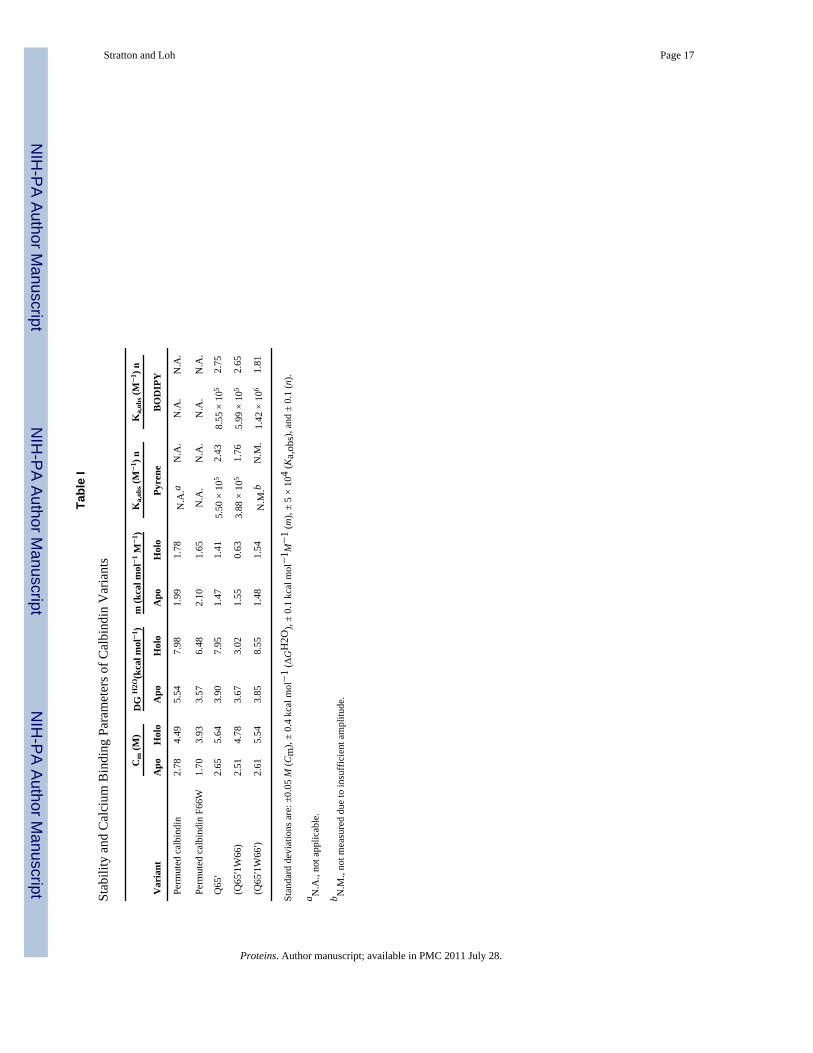
The net result is that overall calcium affinity is increased but cooperativity is decreased. Toestimate the effect of the F66W mutation on N′, we used circular dichroism (CD) to monitorGdnHCl-induced unfolding of circularly permuted calbindin (Supporting Information Fig.S1(A,B)]. Thermodynamic parameters are obtained by fitting the data to the two-state linearextrapolation equation ΔG = ΔGH2O–m[denaturant], where m is the cooperativity parameterand ΔGH2O is the folding free energy extrapolated to zero denaturant. Table I indicates that
Stratton and Loh Page 5
Proteins. Author manuscript; available in PMC 2011 July 28.
NIH
-PA Author Manuscript
NIH
-PA Author Manuscript
NIH
-PA Author Manuscript
the F66W mutation reduces stability of permuted apo-calbindin by 1.97 kcal mol−1 andpermuted holo-calbindin by 1.50 kcal mol−1.
Calbindin-AFF is comprised of three EF-hands: hand-2′, hand-1, and hand-2 (see Fig. 1).The E65Q mutation reduces binding affinity of hand-2 of WT calbindin by 105-fold.18 Weemployed this mutation to inactivate hand-2 of Q65 and hand-2′ of Q65′. Because the E65Qmutation does not dramatically perturb stability of WT calbindin (Δ ΔG = 0.36 kcal mol−1),1apo-Q65 and apo-Q65′ are still expected to consist in an ~1:1 ratio of N and N′. Binding oftwo calcium ions causes Q65 and Q65′ to fully adopt conformations N′ and N, respectively.It is important to note that both variants can coordinate one Ca2+ with hand-1. As a result,the driving force for the conformational change is provided by the free energy change ofligating the second calcium ion to hand-2 (or hand-2′) once the first has already bound tohand-1.
The calcium-induced fold shift of calbindin-AFF is monitored by placing either BODIPY-FL or pyrene groups at the two positions indicated in Figure 1(A). In conformation N′, thefluorophores adopt the equivalent of sequential positions in the surface loop and are hence inclose proximity. In conformation N they are separated by residues 44′–75′ [Fig. 1(A)] andare expected to be distant from one another. The distance increase is reported either as anincrease in BODIPY fluorescence at 513 nm (due to reduction of BODIPY self-quenching)or as a decrease in pyrene fluorescence from 460–500 nm (due to loss of pyrene excimerformation). Thus, calcium binding can be linked to an increase or a decrease in fluorescencedepending on the choice of calbindin-AFF variant (Q65 or Q65′) or selection offluorophores (BODIPY or pyrene). All calcium binding experiments in this study areperformed using Q65′ and mutants thereof. Since binding triggers the N′ → Nconformational change in Q65′, addition of calcium is predicted to increase and decreasefluorescence of the BODIPY- and pyrene-labeled proteins, respectively.
Equilibrium denaturation experimentsWe tested the mechanism outlined in Figure 2 by determining the effect of W66 and W66′on equilibrium unfolding of Q65′. If apo-Q65′ consists of comparable fractions of N and N′,as expected, then its GdnHCl unfolding curve will be the superposition of two closely-spaced transitions. The two transitions can be isolated, however, using the F66W mutation.W66 destabilizes N by ~2 kcal mol−1 but it has no effect on N′. This causes N′ to becomealmost fully populated, and the unfolding curve of apo-(Q65′1W66) approximates that of thepure N′ state. Similarly, the unfolding curve of apo-(Q65′+W66′) corresponds to that of thepure N state. Apo-(Q65′+W66) and apo-(Q65′+W66′) have similar ΔGH2O values andmidpoints of denaturation (Cm) (Table I) and their unfolding curves are nearlysuperimposable [Supporting Information Fig. S1(D,E)]. The two transitions that make up theobserved unfolding curve of apo-Q65′ should be almost superimposable as well, leading tothe prediction that all three apo-Q65′ variants will produce similar ΔGH2O and Cm values.Table I shows this to be the case. These findings confirm that N and N′ are approximatelyisoenergetic in apo-Q65′, as depicted in Figure 2.
We next employed the W66 and W66′ mutations to test whether calcium induces the foldshift. W66 strongly destabilizes holo-Q65′ (ΔΔG = −4.9 kcal mol−1, ΔCm = −0.86 MGdnHCl), suggesting that position 66 is structured in the calcium-bound state. By contrast,W66′ has little effect on holo-Q65′ (Δ ΔG = +0.6 kcal mol−1, ΔCm = −0.10 M GdnHCl),revealing that position 66′ is in an unstructured environment. These data indicate thatcalcium binding drives holo-Q65′ to adopt conformation N exclusively.
Stratton and Loh Page 6
Proteins. Author manuscript; available in PMC 2011 July 28.
NIH
-PA Author Manuscript
NIH
-PA Author Manuscript
NIH
-PA Author Manuscript

Residual structure of N- and C-terminal tailsThe magnitude of the fluorescence change between N and N′ is likely to depend on theextent to which the duplicated polypeptide is structured or disordered. In N′, thefluorophores are held in close proximity regardless of the structure of the C-terminal tail,because the two Cys residues adopt what would be consecutive positions in the WTstructure [Fig. 1(A)]. In conformation N, we anticipate that the two fluorophores will bemaximally distant from one another if the N-terminal tail is completely unstructured andflexible. CD studies find that isolated hand-2 is largely disordered in the absence ofcalcium,3 and thioldisulfide exchange experiments suggest that hand-2 is disordered in holo-Q65.1 As an additional test we measured fluorescence of BODIPY-labeled holo-Q65, holo-Q65′, and holo-(Q65+Q65′) as a function of urea. We employed urea (instead of GdnHCl)because all of the above species possess Cm values in excess of 7 M urea (not shown). Wecan therefore monitor structural changes over a wide range of urea concentrations in whichthe proteins remain native. Figure 3 indicates that fluorescence of holo-Q65′ increasessignificantly from 0 to 2 M urea while that of holo-Q65 remains relatively constant up to 6M urea. The holo-Q65′ data suggest that the hand-2′ is partially ordered in conformation N,causing fractional quenching of the BODIPY groups. The residual structure appears to meltgradually between 0 and 2 M urea. Hand-2 is presumably likewise partially ordered in N′ butits unfolding does not seem to alter the proximity of the fluorophores, consistent with thepredicted structure in Figure 1(A). The fluorescence change of holo-(Q65+Q65′) is inbetween that of holo-Q65 and holo-Q65′, reflecting the 31:1 ratio of N′ and N believed to bepresent in holo-(Q65+Q65′).
Equilibrium calcium binding experimentsWe next tested whether W66 and W66′ modulate calcium binding affinity and fluorescencechange of Q65′ in accordance with the model. Figure 4 shows results obtained for Q65′,(Q65′+W66), and (Q65′+W66′) labeled with pyrene [Fig. 4(A)] and BODIPY [Fig. 4(B)].Table I summarizes the fitted binding parameters. As forecasted by Figure 1(A), addition ofcalcium to Q65′ decreases pyrene fluorescence and increases BODIPY fluorescence.Equation (3) calculates that W66 will decrease Ka,obs of Q65′ by a factor of 16 and increasethe amplitudes of the fluorescence change. We observe that this mutation lowers Ka,obs by afactor of 1.4 for both pyrene and BODIPY-labeled Q65′. The discrepancy likely arises fromthe fact that the F66W mutation is known to increase the intrinsic calcium binding affinity ofWT calbindin by 10-fold.17 This effect may mask a larger than apparent reduction in Ka,obs.In agreement with the model, the W66 mutation increases the amplitudes of the pyrene andBODIPY fluorescence changes.
Equation (3) estimates that the W66′ mutation will increase Ka,obs of Q65′ by a factor of ~2(from 0.5 × Ka to ~Ka) and decrease the amplitude change. The pyrene fluorescence changeof (Q65′+W66′) diminishes to the point where Ka,obs cannot be determined accurately (datanot shown). Unexpectedly, the W66′ mutation inverts the sign of the BODIPY fluorescencechange [Fig. 4(B)]. The probable cause is a new quenching interaction formed betweenBODIPY and W66′ in conformation N. Residues 44′–75′ are principally disordered in N andTrp is known to quench BODIPY in unfolded proteins.19 Regardless of the nature of thefluorescence change, the W66′ mutation increases Ka,obs by a factor of 4.2.
Kinetic measurementsWe measured rates and amplitudes of calcium-induced fold switching using BODIPY-labeled calbindin-AFF variants. The rates are faster than those we reported earlier usingpyrene-labeled protein.1 The reason is not clear; it may be attributable to a kinetic barrierimposed by pyrene dimer formation. Figure 5(A) shows calcium binding data for BODIPY-labeled Q65 and Q65′ monitored by stopped-flow fluorescence. The data fit adequately to
Stratton and Loh Page 7
Proteins. Author manuscript; available in PMC 2011 July 28.
NIH
-PA Author Manuscript
NIH
-PA Author Manuscript
NIH
-PA Author Manuscript

one-exponential functions, and the amplitudes of Q65 and Q65′ exhibit the expectedinversion of sign as the switch is driven in opposite directions. The N →N′ rate is four-foldfaster than the N′ → N rate (6.85 and 1.29 s−1, respectively).
To test whether these rates are limited by whole-molecule unfolding, we measured theglobal unfolding rate of BODIPY-labeled holo-(Q65+Q65′) as a function of ureaconcentration [Fig. 5(B)]. Holo-(Q65+Q65′) contains a single calcium ion bound to hand-1[Fig. 1(A)]. Because hand-1 is folded in both conformations, and because calcium binds tothis site in folded calbindin extremely rapidly (k = 109 M−1 s−1),20 the singly-bound specieswill form within the mixing time of Ca2+-binding experiments. Thus, the observed signal inFigure 5(A) corresponds to folding/unfolding of hand-2/hand-2′ in the context of holo-(Q65+Q65′). Figure 5(B) reveals that these events do not require global unfolding. The urea-induced unfolding rate of holo-(Q65+Q65′) extrapolates to 0.13 s−1 in the absence ofdenaturant. This figure is 10-fold and 53-fold slower than the observed N′ → N and N →N′rates, respectively.
Although conformational switching of Q65 and Q65′ does not appear to proceed via globalunfolding, Figure 5(B) suggests that both transition states involve partial unfolding. The N′→ N and N → N′ rates both accelerate with increasing concentrations of urea. The slopes ofthe lines are similar to that of the global unfolding line, suggesting that the transition statesof all three reactions expose comparable amounts of additional surface area. Thecorresponding amplitudes of the N′ → N and N → N′ reactions are shown in the inset toFigure 5(B). In agreement with the sub-denaturing urea experiments (see Fig. 3), the Q65′amplitude increases with urea and the Q65 amplitude remains constant.
The W66 and W66′ mutations give insight into the nature of the N′ → N transition state ofQ65′. The conversion of N′ to N requires unfolding of hand-2′ and folding of hand-2 [Fig.1(A)]. If the N′ → N rate is limited by one of these processes, then W66 and W66′ maydecrease and increase the observed rate, respectively. Both mutations unexpectedly result inbi-phasic kinetics. For each variant, the two amplitudes are of the same sign and are similarin magnitude (Supporting Information Fig. S2). Figure 5(C) compares the rate constants ofQ65′, (Q65′+W66), and (Q65′+W66′). It can be seen that the slow rate of (Q65′+W66) isless than that of Q65′, and the slow rate of (Q65′+W66′) is greater than that of Q65′.Although the mechanism cannot be derived unambiguously from these data, a simpleinterpretation is that the faster phase corresponds to formation of an intermediate and theslower phase is the rate-limiting conversion of the intermediate to N. If so, then the W66mutation increases the transition state barrier slightly while the W66′ mutation lowers itsignificantly. This result is consistent with the physical picture of folding and unfolding.
Finally, we monitored calcium binding by stopped-flow CD to identify any intermediatesthat may accumulate during the fold switch. No change in ellipticity at 222 nm was observedfor either Q65 or Q65′ (data not shown). This result indicates that the conformational changeproceeds without populating species which contain either extra or missing α-helicalstructure.
DISCUSSIONIn this study we establish a framework for understanding basic thermodynamic and kineticaspects of the AFF switching mechanism. There are two main findings. The first is that theequilibrium properties of the switch—binding affinity and signal response—can bemanipulated using well-established principles of protein stability. The second is that theconformational change which underlies AFF appears to involve limited remodeling of thestructure rather than global unfolding/refolding. With respect to the first point, it is
Stratton and Loh Page 8
Proteins. Author manuscript; available in PMC 2011 July 28.
NIH
-PA Author Manuscript
NIH
-PA Author Manuscript
NIH
-PA Author Manuscript
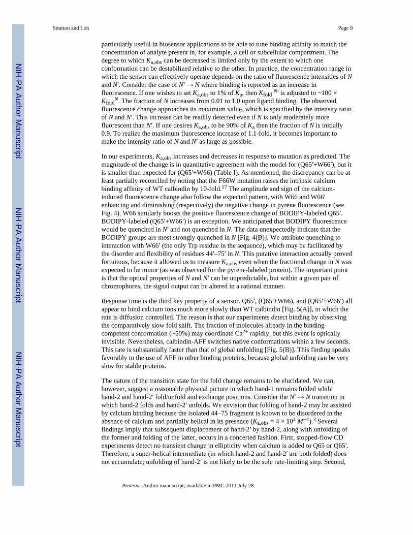
particularly useful in biosensor applications to be able to tune binding affinity to match theconcentration of analyte present in, for example, a cell or subcellular compartment. Thedegree to which Ka,obs can be decreased is limited only by the extent to which oneconformation can be destabilized relative to the other. In practice, the concentration range inwhich the sensor can effectively operate depends on the ratio of fluorescence intensities of Nand N′. Consider the case of N′ → N where binding is reported as an increase influorescence. If one wishes to set Ka,obs to 1% of Ka, then Kfold N′ is adjusted to ~100 ×Kfold
N. The fraction of N increases from 0.01 to 1.0 upon ligand binding. The observedfluorescence change approaches its maximum value, which is specified by the intensity ratioof N and N′. This increase can be readily detected even if N is only moderately morefluorescent than N′. If one desires Ka,obs to be 90% of Ka then the fraction of N is initially0.9. To realize the maximum fluorescence increase of 1.1-fold, it becomes important tomake the intensity ratio of N and N′ as large as possible.
In our experiments, Ka,obs increases and decreases in response to mutation as predicted. Themagnitude of the change is in quantitative agreement with the model for (Q65′+W66′), but itis smaller than expected for (Q65′+W66) (Table I). As mentioned, the discrepancy can be atleast partially reconciled by noting that the F66W mutation raises the intrinsic calciumbinding affinity of WT calbindin by 10-fold.17 The amplitude and sign of the calcium-induced fluorescence change also follow the expected pattern, with W66 and W66′enhancing and diminishing (respectively) the negative change in pyrene fluorescence (seeFig. 4). W66 similarly boosts the positive fluorescence change of BODIPY-labeled Q65′.BODIPY-labeled (Q65′+W66′) is an exception. We anticipated that BODIPY fluorescencewould be quenched in N′ and not quenched in N. The data unexpectedly indicate that theBODIPY groups are most strongly quenched in N [Fig. 4(B)]. We attribute quenching tointeraction with W66′ (the only Trp residue in the sequence), which may be facilitated bythe disorder and flexibility of residues 44′–75′ in N. This putative interaction actually provedfortuitous, because it allowed us to measure Ka,obs even when the fractional change in N wasexpected to be minor (as was observed for the pyrene-labeled protein). The important pointis that the optical properties of N and N′ can be unpredictable, but within a given pair ofchromophores, the signal output can be altered in a rational manner.
Response time is the third key property of a sensor. Q65′, (Q65′+W66), and (Q65′+W66′) allappear to bind calcium ions much more slowly than WT calbindin [Fig. 5(A)], in which therate is diffusion controlled. The reason is that our experiments detect binding by observingthe comparatively slow fold shift. The fraction of molecules already in the binding-competent conformation (~50%) may coordinate Ca2+ rapidly, but this event is opticallyinvisible. Nevertheless, calbindin-AFF switches native conformations within a few seconds.This rate is substantially faster than that of global unfolding [Fig. 5(B)]. This finding speaksfavorably to the use of AFF in other binding proteins, because global unfolding can be veryslow for stable proteins.
The nature of the transition state for the fold change remains to be elucidated. We can,however, suggest a reasonable physical picture in which hand-1 remains folded whilehand-2 and hand-2′ fold/unfold and exchange positions. Consider the N′ → N transition inwhich hand-2 folds and hand-2′ unfolds. We envision that folding of hand-2 may be assistedby calcium binding because the isolated 44–75 fragment is known to be disordered in theabsence of calcium and partially helical in its presence (Ka,obs = 4 × 10⁴ M−1).3 Severalfindings imply that subsequent displacement of hand-2′ by hand-2, along with unfolding ofthe former and folding of the latter, occurs in a concerted fashion. First, stopped-flow CDexperiments detect no transient change in ellipticity when calcium is added to Q65 or Q65′.Therefore, a super-helical intermediate (in which hand-2 and hand-2′ are both folded) doesnot accumulate; unfolding of hand-2′ is not likely to be the sole rate-limiting step. Second,
Stratton and Loh Page 9
Proteins. Author manuscript; available in PMC 2011 July 28.
NIH
-PA Author Manuscript
NIH
-PA Author Manuscript
NIH
-PA Author Manuscript

the W66 and W66′ mutations respectively decrease and increase the slow rate of the N′ → Nreaction [Fig. 5(C)]. This result is expected if folding of hand-2 as well as unfolding ofhand-2′ contribute to the rate-limiting barrier. The N′ → N and N → N′ rates increase withurea concentration [Fig. 5(B)], suggesting that both transition states are more unfolded thanthe ground states.
CONCLUSIONSAFF unites two aspects of protein folding. The first is the well-established linkage betweenfolding and binding. The second is a newer concept of coupling folding to unfolding ofanother portion of the molecule.2,12,21–24 AFF therefore constitutes a conformationalswitching mechanism that is driven by binding energy. Folding is itself a dramaticconformational change, and others have exploited binding-induced folding of unfoldedproteins to create optical biosensors.19 Our mechanism is unique in that the bound and freeproteins are both native; fold switching does not involve net folding or unfolding, onlystructural rearrangement. In this way, the globally unfolded state (which may presentproblems of solubility and degradation) is averted. Thus, binding affinity and switching ratecan be tuned by manipulating the relative stabilities of the two native forms and the heightof the energy barrier that separates them. This study demonstrates that it is possible to makesuch adjustments by varying the free energies of N and/or N′ according to establishedprinciples.
Supplementary MaterialRefer to Web version on PubMed Central for supplementary material.
AcknowledgmentsThe authors thank C. Robert Matthews for use of his stopped-flow CD instrument, Sagar Kathuria for assistancewith stopped-flow CD experiments, and Jeung-Hoi Ha for discussions.
References1. Stratton MM, Mitrea DM, Loh SN. A Ca2+-sensing molecular switch based on alternate frame
protein folding. ACS Chem Biol. 2008; 3:723–732. [PubMed: 18947182]2. Mitrea DM, Parsons L, Loh SN. Engineering an artificial zymogen by alternate frame protein
folding. Proc Natl Acad Sci USA. 2010; 107:2824–2829. [PubMed: 20133757]3. Julenius K, Robblee J, Thulin E, Finn BE, Fairman R, Linse S. Coupling of ligand binding and
dimerization of helix-loop-helix peptides: spectroscopic and sedimentation analyses of calbindinD9k EF-hands. Proteins. 2002; 47:323–333. [PubMed: 11948786]
4. Dyson HJ, Wright PE. Coupling of folding and binding for unstructured proteins. Curr Opin StructBiol. 2002; 12:54–60. [PubMed: 11839490]
5. Wright PE, Dyson HJ. Linking folding and binding. Curr Opin Struct Biol. 2009; 19:31–38.[PubMed: 19157855]
6. Galea CA, Pagala VR, Obenauer JC, Park CG, Slaughter CA, Kriwacki RW. Proteomic studies ofthe intrinsically unstructured mammalian proteome. J Proteome Res. 2006; 5:2839–2848. [PubMed:17022655]
7. Dyson HJ, Wright PE. Intrinsically unstructured proteins and their functions. Nat Rev Mol CellBiol. 2005; 6:197–208. [PubMed: 15738986]
8. Pace, CN.; Scholtz, JM., editors. Protein structure: a practical approach. 2. New York: OxfordUniversity Press; 1997.
9. Wenz JJ, Barrantes FJ. Resolution of complex fluorescence spectra of lipids and nicotinicacetylcholine receptor by multivariate analysis reveals protein-mediated effects on the receptor’simmediate lipid microenvironment. PMC Biophys. 2008; 1:6. [PubMed: 19351428]
Stratton and Loh Page 10
Proteins. Author manuscript; available in PMC 2011 July 28.
NIH
-PA Author Manuscript
NIH
-PA Author Manuscript
NIH
-PA Author Manuscript

10. Forsen S, Linse S. Cooperativity: over the Hill. Trends Biochem Sci. 1995; 20:495–497. [PubMed:8571449]
11. Hilser VJ, Thompson EB. Intrinsic disorder as a mechanism to optimize allosteric coupling inproteins. Proc Natl Acad Sci USA. 2007; 104:8311–8315. [PubMed: 17494761]
12. Cutler T, Loh SN. Thermodynamic analysis of an antagonistic folding-unfolding equilibriumbetween two protein domains. J Mol Biol. 2007; 371:308–316. [PubMed: 17572441]
13. Monod J, Wyman J, Changeux JP. On the nature of allosteric transitions: a plausible model. J MolBiol. 1965; 12:88–118. [PubMed: 14343300]
14. Koshland DE Jr, Nemethy G, Filmer D. Comparison of experimental binding data and theoreticalmodels in proteins containing subunits. Biochemistry. 1966; 5:365–385. [PubMed: 5938952]
15. Fersht AR, Sato S. Phi-value analysis and the nature of protein-folding transition states. Proc NatlAcad Sci USA. 2004; 101:7976–7981. [PubMed: 15150406]
16. Julenius K, Thulin E, Linse S, Finn BE. Hydrophobic core substitutions in calbindin D9k: effectson stability and structure. Biochemistry. 1998; 37:8915–8925. [PubMed: 9636033]
17. Kragelund BB, Jonsson M, Bifulco G, Chazin WJ, Nilsson H, Finn BE, Linse S. Hydrophobic coresubstitutions in calbindin D9k: effects on Ca2+ binding and dissociation. Biochemistry. 1998;37:8926–8937. [PubMed: 9636034]
18. Carlstrom G, Chazin WJ. Two-dimensional 1H nuclear magnetic resonance studies of the half-saturated (Ca2+)1 state of calbindin D9k: further implications for the molecular basis ofcooperative Ca2+ binding. J Mol Biol. 1993; 231:415–430. [PubMed: 8389885]
19. Kohn JE, Plaxco KW. Engineering a signal transduction mechanism for protein-based biosensors.Proc Natl Acad Sci USA. 2005; 102:10841–10845. [PubMed: 16046542]
20. Forsen S, Linse S, Thulin E, Lindegard B, Martin SR, Bayley PM, Brodin P, Grundstrom T.Kinetics of calcium binding to calbindin mutants. Eur J Biochem. 1988; 177:47–52. [PubMed:3181158]
21. Peng Q, Li H. Direct observation of tug-of-war during the folding of a mutually exclusive protein.J Am Chem Soc. 2009; 131:13347–13354. [PubMed: 19719116]
22. Radley TL, Markowska AI, Bettinger BT, Ha J-H, Loh SN. Allosteric switching by mutuallyexclusive folding of protein domains. J Mol Biol. 2003; 332:529–536. [PubMed: 12963365]
23. Cutler TA, Mills BM, Lubin DJ, Chong LT, Loh SN. Effect of inter-domain linker length on anantagonistic folding-unfolding equilibrium between two protein domains. J Mol Biol. 2009;386:854–868. [PubMed: 19038264]
24. Ha J-H, Butler JS, Mitrea DM, Loh SN. Modular enzyme design: regulation by mutually exclusiveprotein folding. J Mol Biol. 2006; 357:1058–1062. [PubMed: 16483603]
Stratton and Loh Page 11
Proteins. Author manuscript; available in PMC 2011 July 28.
NIH
-PA Author Manuscript
NIH
-PA Author Manuscript
NIH
-PA Author Manuscript

Figure 1.Structures and amino acid sequences of calbindin-AFF variants. (A) Predicted structures ofN and N′ conformations. The color scheme is as follows: red, hand-2′ (residues 44′–75′);green, hand-1 (residues 1–43); blue, hand-2 (residues 44–75). The side chain of E65′ isshown in light pink and the alpha carbon of Q65 is shown in pale cyan. Calcium ions aredepicted as gray spheres. The Q65 variant is shown in which calcium binding induces thered region to fold and the blue region to unfold. The stars mark the location of Cys residuesto which fluorophores are attached, to track the N–N′ conformational change. (B) Aminoacid sequences of calbindin-AFF variants showing the shared sequence in green and theduplicated sequences in red and blue, as in Panel A. The black segment is a six-amino acidlinker. Circles indicate viable calcium binding sites; crossed-out squares denote calciumbinding sites made nonfunctional by the presence of the Q65 or Q65′ mutation. The presenceof the F66W mutation is marked by W.
Stratton and Loh Page 12
Proteins. Author manuscript; available in PMC 2011 July 28.
NIH
-PA Author Manuscript
NIH
-PA Author Manuscript
NIH
-PA Author Manuscript
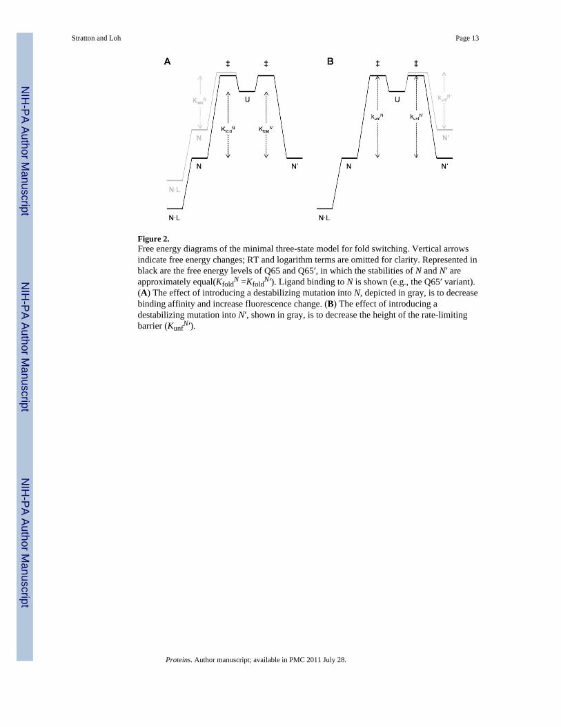
Figure 2.Free energy diagrams of the minimal three-state model for fold switching. Vertical arrowsindicate free energy changes; RT and logarithm terms are omitted for clarity. Represented inblack are the free energy levels of Q65 and Q65′, in which the stabilities of N and N′ areapproximately equal(Kfold
N =KfoldN′). Ligand binding to N is shown (e.g., the Q65′ variant).
(A) The effect of introducing a destabilizing mutation into N, depicted in gray, is to decreasebinding affinity and increase fluorescence change. (B) The effect of introducing adestabilizing mutation into N′, shown in gray, is to decrease the height of the rate-limitingbarrier (Kunf
N′).
Stratton and Loh Page 13
Proteins. Author manuscript; available in PMC 2011 July 28.
NIH
-PA Author Manuscript
NIH
-PA Author Manuscript
NIH
-PA Author Manuscript

Figure 3.Dependence of BODIPY fluorescence on subdenaturing concentrations of urea. Holo-Q65 isshown in triangles, holo-Q65′ in circles, and holo-(Q65+Q65′) in diamonds.
Stratton and Loh Page 14
Proteins. Author manuscript; available in PMC 2011 July 28.
NIH
-PA Author Manuscript
NIH
-PA Author Manuscript
NIH
-PA Author Manuscript

Figure 4.Equilibrium calcium binding of calbindin-AFF variants. (A) Binding of calcium to Q65′ (●)and (Q65′+W66) (○), detected by pyrene excimer fluorescence. (B) Binding of calcium toQ65′ (●), (Q65′+W66) (○), and (Q65′+W66′) (gray triangles) monitored by BODIPYfluorescence. Lines are best fits of the data to the Hill equation.
Stratton and Loh Page 15
Proteins. Author manuscript; available in PMC 2011 July 28.
NIH
-PA Author Manuscript
NIH
-PA Author Manuscript
NIH
-PA Author Manuscript

Figure 5.Rates and amplitudes of calcium-induced fold switching monitored by BODIPYfluorescence. (A) Representative stopped-flow fluorescence traces of calcium binding toQ65 (triangles) and Q65′ (circles). The lines are best fits of the data to single-exponentialfunctions. (B) Rates of fold switching (filled symbols) and global unfolding (open symbols)plotted as a function of urea. Symbols are: (▲), Q65; (●), Q65′; (○), holo (Q65+Q65′). Theslopes of the fitted lines are (in units of M−1 s−1): 0.38 (Q65), 0.28 (Q65′), and 0.30(Q65+Q65′). The inset shows the fitted fluorescence amplitudes plotted as a function ofurea, using the same symbols. (C) Fold-switching rates of (Q65′+W66) (squares) and (Q65′+W66′) (circles). Closed and open symbols denote the faster and slower rates, respectively.The gray line shows the linear fit of the Q65′ data from panel B for comparison.
Stratton and Loh Page 16
Proteins. Author manuscript; available in PMC 2011 July 28.
NIH
-PA Author Manuscript
NIH
-PA Author Manuscript
NIH
-PA Author Manuscript
NIH
-PA Author Manuscript
NIH
-PA Author Manuscript
NIH
-PA Author Manuscript
Stratton and Loh Page 17
Tabl
e I
Stab
ility
and
Cal
cium
Bin
ding
Par
amet
ers o
f Cal
bind
in V
aria
nts
Var
iant
Cm
(M)
DG
H2O
(kca
l mol−
1 )m
(kca
l mol−
1 M−
1 )K
a,ob
s (M
−1 )
nK
a,ob
s (M
−1 )
n
Apo
Hol
oA
poH
olo
Apo
Hol
oPy
rene
BO
DIP
Y
Perm
uted
cal
bind
in2.
784.
495.
547.
981.
991.
78N
.A.a
N.A
.N
.A.
N.A
.
Perm
uted
cal
bind
in F
66W
1.70
3.93
3.57
6.48
2.10
1.65
N.A
.N
.A.
N.A
.N
.A.
Q65′
2.65
5.64
3.90
7.95
1.47
1.41
5.50
× 1
052.
438.
55 ×
105
2.75
(Q65′1
W66
)2.
514.
783.
673.
021.
550.
633.
88 ×
105
1.76
5.99
× 1
052.
65
(Q65′1
W66′)
2.61
5.54
3.85
8.55
1.48
1.54
N.M
.bN
.M.
1.42
× 1
061.
81
Stan
dard
dev
iatio
ns a
re: ±
0.05
M (C
m),
± 0.
4 kc
al m
ol−
1 (Δ
GH
2O),
± 0.
1 kc
al m
ol−
1 M−
1 (m
), ±
5 ×
104
(Ka,
obs)
, and
± 0
.1 (n
).
a N.A
., no
t app
licab
le.
b N.M
., no
t mea
sure
d du
e to
insu
ffic
ient
am
plitu
de.
Proteins. Author manuscript; available in PMC 2011 July 28.