one-Ångstrom microscope update - unt digital library
TRANSCRIPT
LBNL - 51926 One-Ångstrom Microscope Update (1999).
One-Ångstrom Microscope UpdateReport to the External Steering Committee of the NCEM
Michael A. O’Keefe National Center for Electron Microscopy, LBNL B72, One Cyclotron Road, Berkeley, CA 94720, USA
DISCLAIMER------------------------------------------------------------------------------------------------------- This document was prepared as an account of work sponsored by the United States Government. While this document is believed to contain correct information, neither the United States Government nor any agency thereof, nor The Regents of the University of California, nor any of their employees, makes any warranty, express or implied, or assumes any legal responsibility for the accuracy, completeness, or usefulness of any information, apparatus, product, or process disclosed, or represents that its use would not infringe privately owned rights. Reference herein to any specific commercial product, process, or service by its trade name, trademark, manufacturer, or otherwise, does not necessarily constitute or imply its endorsement, recommendation, or favoring by the United States Government or any agency thereof, or The Regents of the University of California. The views and opinions of authors expressed herein do not necessarily state or reflect those of the United States Government or any agency thereof, or The Regents of the University of California. Ernest Orlando Lawrence Berkeley National Laboratory is an equal opportunity employer.------------------------------------------------------------------------------------------------------- Ernest Orlando Lawrence Berkeley National Laboratory – LBNL-51926
LBNL - 51926 One-Ångstrom Microscope Update (1999).
-------------------- NCEM Steering Committee 1999maokeefe --------------------------------------------------------------------------------------------------------------------------------------------
One-Ångstrom Microscope Update
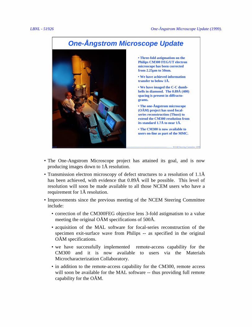
• Three-fold astigmatism on thePhilips CM300 FEG/UT electronmicroscope has been correctedfrom 2.25µm to 50nm.
• We have achieved informationtransfer to below 1Å.
• We have imaged the C-C dumb-bells in diamond. The 0.89Å (400)spacing is present in diffracto-grams.
• The one-Ångstrom microscope(OÅM) project has used focal-series reconstruction (Thust) toextend the CM300 resolution fromits standard 1.7Å to near 1Å.
• The CM300 is now available tousers on-line as part of the MMC.
maok --------------------------------------------------------------------------------------------------------------------------------------------------------------
• The One-Angstrom Microscope project has attained its goal, and is nowproducing images down to 1Å resolution.
• Transmission electron microscopy of defect structures to a resolution of 1.1Åhas been achieved, with evidence that 0.89Å will be possible. This level ofresolution will soon be made available to all those NCEM users who have arequirement for 1Å resolution.
• Improvements since the previous meeting of the NCEM Steering Committeeinclude:
• correction of the CM300FEG objective lens 3-fold astigmatism to a valuemeeting the original OÅM specifications of 500Å.
• acquisition of the MAL software for focal-series reconstruction of thespecimen exit-surface wave from Philips -- as specified in the originalOÅM specifications.
• we have successfully implemented remote-access capability for theCM300 and it is now available to users via the MaterialsMicrocharacterization Collaboratory.
• in addition to the remote-access capability for the CM300, remote accesswill soon be available for the MAL software -- thus providing full remotecapability for the OÅM.
LBNL - 51926 One-Ångstrom Microscope Update (1999).
-------------------- NCEM Steering Committee 1999maokeefe --------------------------------------------------------------------------------------------------------------------------------------------
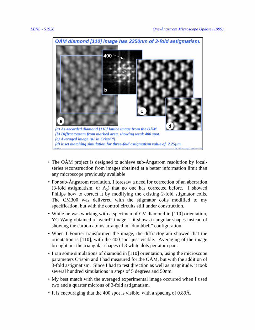
(a) As-recorded diamond [110] lattice image from the OÅM.(b) Diffractogram from marked area, showing weak 400 spot.(c) Averaged image (p1 in Crisp™).(d) inset matching simulation for three-fold astigmatism value of 2.25µm.
a
400
c
d
b
OÅM diamond [110] image has 2250nm of 3-fold astigmatism.
• The OÅM project is designed to achieve sub-Ångstrom resolution by focal-series reconstruction from images obtained at a better information limit thanany microscope previously available
• For sub-Ångstrom resolution, I foresaw a need for correction of an aberration(3-fold astigmatism, or A2) that no one has corrected before. I showedPhilips how to correct it by modifying the existing 2-fold stigmator coils.The CM300 was delivered with the stigmator coils modified to myspecification, but with the control circuits still under construction.
• While he was working with a specimen of CV diamond in [110] orientation,YC Wang obtained a “weird” image -- it shows triangular shapes instead ofshowing the carbon atoms arranged in “dumbbell” configuration.
• When I Fourier transformed the image, the diffractogram showed that theorientation is [110], with the 400 spot just visible. Averaging of the imagebrought out the triangular shapes of 3 white dots per atom pair.
• I ran some simulations of diamond in [110] orientation, using the microscopeparameters Crispin and I had measured for the OÅM, but with the addition of3-fold astigmatism. Since I had to test direction as well as magnitude, it tookseveral hundred simulations in steps of 5 degrees and 50nm.
• My best match with the averaged experimental image occurred when I usedtwo and a quarter microns of 3-fold astigmatism.
• It is encouraging that the 400 spot is visible, with a spacing of 0.89Å.

LBNL - 51926 One-Ångstrom Microscope Update (1999).
-------------------- NCEM Steering Committee 1999maokeefe --------------------------------------------------------------------------------------------------------------------------------------------
CONTRAST TRANSFER FUNCTION
Scattering Vector [Å-1]
0.10 0.30 0.50 0.70 0.90 1.10
-0.80
-0.50
-0.20
0.10
0.40
0.70
1.00V = 300.0 kV Cs = 0.6 mm Def = -2710.00 Å Del = 25.00 Å Div = 0.20 mrad
0 0 4(0.89Å)
1 1 3(1.08Å)
Defocus condition without 3-fold aberration
Image simulation without 3-fold astigmatismshows “dumbbells” from 311 and 400 spacings.
Simulation for “ideal defocus condition” produces 0.89Å “dumbbells” image.
8/26/98
OAM should show 0.89Å “dumbbells” in [110] diamond.
• The simulation shows the diamond “dumbbell” image we expected the OÅMto be able to produce -- once the corrector controls had been supplied byPhilips and A2 had been reduced to meet the OÅM specification of 500Å.
• The OÅM contrast-transfer function on the left shows how transfer should bepossible out to 0.89Å with a spread of focus as high as 25Å (the spread offocus should be closer to the specified 20Å once Philips meets the voltageripple spec by replacing our HT tank with the improved model).
• The simulated image corresponding to the OÅM parameters shows clearseparation of the carbon atoms spaced at 0.89Å.
• The many oscillations in the contrast transfer function demonstrate whyfocal-series reconstruction of the image (actually of the electron wave at thespecimen exit surface) is required for single-image resolution of complexstructures to the OÅM’s sub-Ångstrom information limit.
• Focal-series reconstruction (FSR) of the exit-surface wave (ESW) is donewith the Brite-Euram software purchased from Philips as a component of theOÅM order. The software is a combination of the PAM (parabolic method)and MAL (maximum likelihood) developed at Antwerp, and now maintainedand extended by Andreas Thust working as a consultant for Philips.
• The IBM workstation originally specified for this software was supercededin the OÅM order by the Fischione plasma cleaner, but Andreas Thustagreed to my purchase of a DEC Alpha (Durango) as a suitable replacement,and will install his software on that.

LBNL - 51926 One-Ångstrom Microscope Update (1999).
-------------------- NCEM Steering Committee 1999maokeefe --------------------------------------------------------------------------------------------------------------------------------------------
Simulation for 2.25µmof 3-fold astigmatism
Experimental image as recorded
Diffractogramfrom marked area
Averaged image ( p1 in Crisp™)
Vertical mirror symmetry ( p12 in Crisp™)Vertical & horizontal mirror symmetries ( p222 in Crisp™)
Crystallographic image processing shows “dumbbells”
Line trace
a0
a0/4 ( 0.89Å)
a0/4 is 0.89Å
Average
Mir
ror
Mirror again
8/26/98
Presented at XIVth International Congress for Electron Microscopy, Cancun, Mexico. September 2, 1998.
• The OÅM should be capable of imaging C-C “dumbbells” at 0.89Å spacing indiamond. This is supported by the presence of the 400 spot in experimentaldiffractograms as well as image simulations for OÅM parameters with A2 = 0.
• To confirm the correct spacings in the astigmatic image, I used Crisp™ to“correct” the phasings by imposing known the symmetries of the specimen.
• Imposition of a left-right mirror on the averaged image produces slightlyasymmetric black pairs of spots, which can then be symmetrized into perfect0.89Å dumbbells by the addition of a top-bottom mirror. Of course this resultis too “over processed” to be generally applicable to other structures.
• I processed the image to show that imposition of the correct diamond symmetrywould change it into an image with 0.89Å spacings present in the correctpositions for the diamond structure, and to confirm that the “weird” symmetrywas due to the 3-fold astigmatism I had predicted would limit OÅM resolution.
• I showed this slide in my presentation at the XIVth International Congress forElectron Microscopy in September 1998. At this meeting I also met withPhilips and used this result to urge them to supply the 3-fold controls I hadbeen asking for since the delivery of the OÅM (the microscope was deliveredwith the standard 2-fold stigmator coils modified as I had instructed, but the 3-fold control circuits had not yet been built and tested).
• In October (1998), the controls were delivered and installed and we were ableto correct the 3-fold astigmatism!

LBNL - 51926 One-Ångstrom Microscope Update (1999).
-------------------- NCEM Steering Committee 1999maokeefe --------------------------------------------------------------------------------------------------------------------------------------------
Pre-corrected OÅM diffractograms with 2250nm of 3-fold astigmatism.
Tilt series obtained with NCEMscript for Digital Micrograph™
shows three-fold symmetry.
• As well as using the 3-fold astigmatic diamond image, we also measured thevalue of 3-fold astigmatism by using amorphous specimens of carbon. Iwrote a Digital Microscope™ script to control the microscope and CCDcamera and produce a series of tilted diffractograms. Then I worked withCrispin Hetherington to debug it – Crispin operated the microscope to findthe beam as my initial attempts at tilting it sent it off the screen!
• The script tilts the beam, captures the image on the CCD, Fourier transformsit, then places the center area of each diffractogram in the correct positionwithin the composite.
• The slide shows a tilt series before correction of the 3-fold astigmatism. Thearrows show the 120 degree rotational symmetry.
• Using Gatan’s Digital Microscope™ plug-in, YC and I measured the 3-foldastigmatism six times for a mean value of 2.46 micron. This value is some9% higher than the value of 2.25 micron that I found to best match thediamond image.

LBNL - 51926 One-Ångstrom Microscope Update (1999).
-------------------- NCEM Steering Committee 1999maokeefe --------------------------------------------------------------------------------------------------------------------------------------------
Corrected OÅM diffractograms with < 50nm of 3-fold astigmatism*.
*tilt axis is ~53° from where I thought
60°
7°
• The 3-fold corrector box arrived one month after my Cancun meeting withPhilips, at which I showed them my processed version of YC’s diamondresults and stressed the possibilities for sub-Å resolution with the OÅM.
• I scheduled Bob Mueller to install the corrector, but there was a delay whileBob had to modify the box to add eight potentiometers to control the coilsdirectly. Then Bob and Jan Ringnalda made the adjustments while I ran theGatan software and measured the changes in the value of A2.
• Final results showed 50nm of residual 3-fold astigmatism -- exactly meetingthe specification I had placed into the original microscope specification list,and allowing spacings at 0.8Å to contribute to the image with a phasedistortion of π/4.
• After correction, I worked with YC and Ming Pan (Gatan) to obtain anddisplay a series of amorphous-carbon diffractograms using my DM™ script.
• Although the series showed no 3-fold character (120 degree symmetry), itwas not clear that the axes of the ellipses had the correct symmetry. In fact,it turned out that my DM™ script positioned the diffractograms on the“page” at about 53 degrees from the correct positions.
LBNL - 51926 One-Ångstrom Microscope Update (1999).
-------------------- NCEM Steering Committee 1999maokeefe --------------------------------------------------------------------------------------------------------------------------------------------
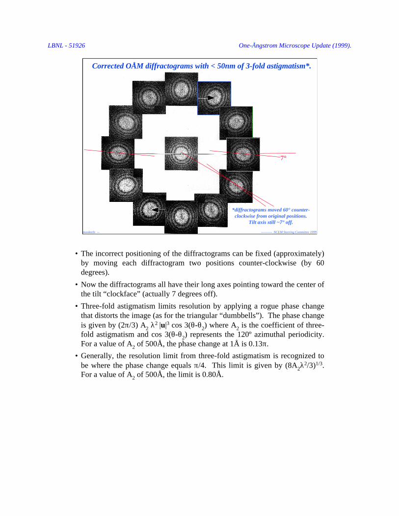
Corrected OÅM diffractograms with < 50nm of 3-fold astigmatism*.
7°
*diffractograms moved 60° counter-clockwise from original positions.
Tilt axis still ~7° off.
• The incorrect positioning of the diffractograms can be fixed (approximately)by moving each diffractogram two positions counter-clockwise (by 60degrees).
• Now the diffractograms all have their long axes pointing toward the center ofthe tilt “clockface” (actually 7 degrees off).
• Three-fold astigmatism limits resolution by applying a rogue phase changethat distorts the image (as for the triangular “dumbbells”). The phase changeis given by (2π/3) A2 λ
2 |u|3 cos 3(θ-θ2) where A2 is the coefficient of three-
fold astigmatism and cos 3(θ-θ2) represents the 120º azimuthal periodicity.For a value of A2 of 500Å, the phase change at 1Å is 0.13π.
• Generally, the resolution limit from three-fold astigmatism is recognized tobe where the phase change equals π/4. This limit is given by (8A2λ
2/3)1/3.For a value of A2 of 500Å, the limit is 0.80Å.
LBNL - 51926 One-Ångstrom Microscope Update (1999).
-------------------- NCEM Steering Committee 1999maokeefe --------------------------------------------------------------------------------------------------------------------------------------------
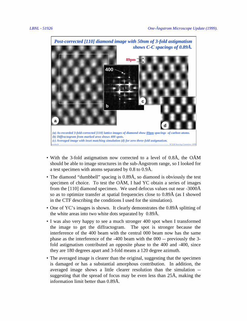
(a) As-recorded 3-fold-corrected [110] lattice images of diamond show 89pm spacings of carbon atoms.(b) Diffractogram from marked area shows 400 spots.(c) Averaged image with inset matching simulation (d) for zero three-fold astigmatism.
a
89pm
400
b c.
d
Post-corrected [110] diamond image with 50nm of 3-fold astigmatismshows C-C spacings of 0.89Å.
••
• With the 3-fold astigmatism now corrected to a level of 0.8Å, the OÅMshould be able to image structures in the sub-Ångstrom range, so I looked fora test specimen with atoms separated by 0.8 to 0.9Å.
• The diamond “dumbbell” spacing is 0.89Å, so diamond is obviously the testspecimen of choice. To test the OÅM, I had YC obtain a series of imagesfrom the [110] diamond specimen. We used defocus values out near -3000Åso as to optimize transfer at spatial frequencies close to 0.89Å (as I showedin the CTF describing the conditions I used for the simulation).
• One of YC’s images is shown. It clearly demonstrates the 0.89Å splitting ofthe white areas into two white dots separated by 0.89Å.
• I was also very happy to see a much stronger 400 spot when I transformedthe image to get the diffractogram. The spot is stronger because theinterference of the 400 beam with the central 000 beam now has the samephase as the interference of the -400 beam with the 000 -- previously the 3-fold astigmatism contributed an opposite phase to the 400 and -400, sincethey are 180 degrees apart and 3-fold means a 120 degree azimuth.
• The averaged image is clearer than the original, suggesting that the specimenis damaged or has a substantial amorphous contribution. In addition, theaveraged image shows a little clearer resolution than the simulation --suggesting that the spread of focus may be even less than 25Å, making theinformation limit better than 0.89Å.

LBNL - 51926 One-Ångstrom Microscope Update (1999).
-------------------- NCEM Steering Committee 1999maokeefe --------------------------------------------------------------------------------------------------------------------------------------------
1.13ÅCubic[110]GaN
HexagonalGaN
ARM-1000 image with 1.6Å resolutionshows the Ga-atom positions in this WPO“structure image”.
Test of Focal-series Reconstruction of a Defect Structure:Comparison of [110] GaN images of Cubic/Hexagonal Interface
CM300-FEG image has 1.7Å resolution,but also contains information to the 0.8Åinformation limit of the OÅM. Thisinformation can be reconstructed usingthe FSR software aspect of the OÅM.
ARM-1000 CM300FEG/UT
Images obtained by C. Kisielowski.
• Although a single image from a simple structure like [110] diamond candemonstrate the resolution of the OÅM, it takes a reconstruction from a focalseries to realize the full resolution in the case of a defect structure.
• The left image shows an image of the interface between cubic and hexagonalGaN taken by Christian Kisielowski using the JEOL ARM-1000. The 1.6Åresolution of the ARM is insufficient to separate the Ga and N atoms (spacedat 1.13Å), so we see them as single dots with positions close to the “center ofgravity” of the Ga-N pair (i.e. close to the Ga atom).
• The image on the right was obtained by Christian on the CM300FEG/UT. Itobviously contains higher-frequency information (details are closer together).However, this information cannot be related directly to the structure, except insome areas (marked with yellow dots) in which important reflections aretransferred with the correct phases by the highly-oscillatory CTF. Inparticular, the exact position of the interface is unclear.

LBNL - 51926 One-Ångstrom Microscope Update (1999).
-------------------- NCEM Steering Committee 1999maokeefe --------------------------------------------------------------------------------------------------------------------------------------------
Reconstruction of the phase of the electron exit wave of the 3C/2H GaN interface from a focalseries of 20 images obtained on the One-Ångstrom Microscope. Ga and N sub-lattices are clearlyresolved at a separation of 1.13Å, and are distinguished by different contrast at the (darker) Gaand (lighter) N sites. This contrast is maintained across the 3C/2H interface.
Reconstruction from [110] GaN focal series shows Ga and N atomsat a resolution of better than 1.13Å
Cubic GaN
HexagonalGaN
1.13ÅPhase image
Cubic GaN
HexagonalGaN
Image by C. Kisielowski.
• The mixed-phase CM300 result can be converted to a simple projection of thedefect structure at sub-Å resolution by using the OÅM’s software component.
• For a thin specimen (phase object), the scattered electron wave can be writtenas ψ(x,y) = exp{i σ φp(x,y)}, where σ is the cross-section for scattering andφp(x,y) is the specimen potential projected in the incident beam direction. Thephase of the scattered wave is directly proportional to the projected potential.
• The “image” shows the phase of the exit-surface wave reconstructed by theMAL software from 20 images obtained over a range of focus on the OÅM.The positions of all the atoms (both the heavy Ga and the lighter N) can nowbe seen, and the interface structure precisely determined.
• This is the first result from the OÅM using its combined resources -- theCM300 (hardware component) and the MAL reconstruction (softwarecomponent). Although the reconstruction step was slow (15mins), it willbecome much faster once the software is transferred from Kisielowki’s SiliconGraphics workstation to the much faster (3x) OÅM computer. This computer(Durango) also provides much more storage for image data than the SGI.
• Since the CM300 has been made available for remote use by NCEM users, itmakes sense to provide the full OÅM capability by making the MAL similarlyaccessible. Philips has agreed to my providing remote access by NCEM usersto the MAL once the software has been moved to Durango.
• With the MAL working, the next test of the OÅM will be to obtain andreconstruct a focal series of diamond images to demonstrate that 0.89Åresolution is possible from non-periodic and defect structures.
LBNL - 51926 One-Ångstrom Microscope Update (1999).
• The Brite-Euram software for focal-series reconstruction combines the PAM(parabolic method) and MAL (maximum likelihood). Developed at Antwerp aspart of the Brite-Euram project, it is maintained, and is being extended, byAndreas Thust in Jülich working as a consultant for Philips.
• Whilst working to get the OÅM’s 3-fold astigmatism corrected, I was alsoworking to have the software installed on the NCEM’s OÅM computer.
• Originally, the OÅM computer was to be an IBM workstation provided byPhilips to run the reconstruction software, and was so specified in the originalOÅM order. However, funds for the IBM were subsequently traded for aFischione plasma cleaner to combat contamination problems. In 1996, I found away to fund a replacement OÅM computer by using $15K that I had availablefrom a DOE prize. I consulted with Andreas, and he agreed to my purchase of aDEC Alpha (Durango) as a suitable OÅM computer to run his software.
• However, in November last year, Andreas told me he was unable to install thesoftware on Durango. I then arranged with Andreas and Philips (Sheri Kurland,Ben Bormans and Frank de Jong) to meet in January of 1999 to arrange a way toget the NCEM copy of the FSR software working on the OÅM computer.
• At the meeting, I found out why there was a problem and agreed to install Unixas the operating system on Durango instead of the current WNT so that the FSRsoftware could run using some routines that were only available under Unix. Atthis point Kisielowski made the suggestion that we temporarily install thesoftware on his personal SGI since it ran Unix. Philips and Thust agreed to ashort-term installation on the SGI with a transfer to the OÅM computer once Ihad it converted to run under Unix, just as long as we had only one copy of thesoftware at any one time.
• Unfortunately, Uli Dahmen has not yet authorized the funds for conversion ofthe operating system of the OÅM computer from WNT to Unix. At the moment,the OÅM computer remains idle, and the only access to the NCEM’s copy of theFSR software is via Christian Kisielowski’s much slower SGI. Dahmen has toldme that access to the software will remain this way for one year. Once this yearhas passed, we will be able to move the software to the OÅM computer andenjoy much faster reconstructions with fewer storage constraints for image data,as well as remote access to the OÅM’s Brite-Euram reconstruction software.
Notes on the Acquisition and Disposition of the Brite-EuramSoftware for Focal-series Reconstruction of the Exit-surface Wave
Acknowledgement
This work, and the National Center for Electron Microscopy at LBNL aresupported by the Director, Office of Science through the Office of Basic EnergySciences, Material Sciences Division, of the U.S. Department of Energy, undercontract No. DE-AC03-76SF00098.