one stop shop: 4d cta and ct perfusion...
TRANSCRIPT
7/14/2015
1
Guang-Hong Chen, PhD
Interventional Stroke Imaging Research Team at UW-Madison: PIs: Guang-Hong Chen, Charlie Strother, and Beverly Aagaard-Kienitz Members (Basic Science): Yinsheng Li, Kai Niu, Yijing Wu, John Garrett, Ke Li Members (Clinical): Pengfei Yang, David Niemann, Azam Ahmed, Howard Rowley, and Pat Turski Siemens Support: Sebastian Schafer, Kevin Royalty, Klaus Klingenbeck International consortium on interventional stroke imaging: Clinical team led by Drs. Doerfler and Struffert at the University of Erlangen-
Nuremberg Clinical team led by Dr. Guo in Taiwan
Clinical motivation of one-stop-shop imaging
Technical challenges
Enabling technology for one-stop-shop imaging:
SMART-RECON and SMART IV 3D-DSA
One-stop-shop imaging using SMART-RECON:
non-contrast CBCT images, time-resolved
CBCT angiography, and CBCT perfusion maps
Summary and discussion

7/14/2015
2
Clinical motivation of one-stop-shop imaging
Technical challenges
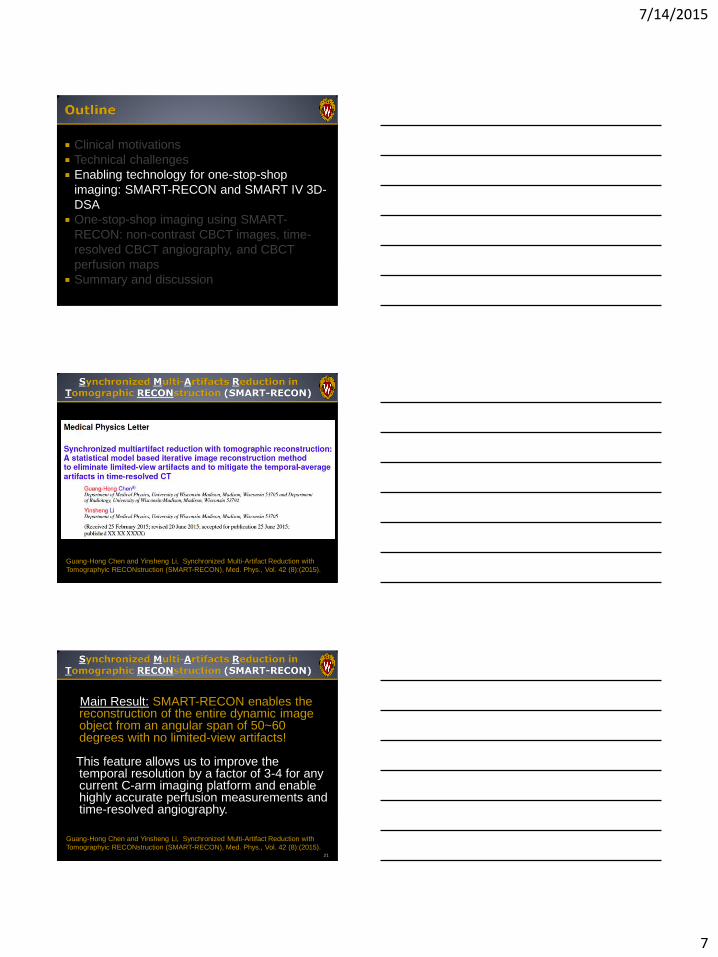
Enabling technology for one-stop-shop imaging:
SMART-RECON and SMART IV 3D-DSA
One-stop-shop imaging using SMART-RECON:
non-contrast CBCT images, time-resolved CBCT
angiography, and CBCT perfusion maps
Summary and discussion
Non-
Contrast CT
Excludes
hemorrhage
CT
Perfusion
Blood flow to
brain tissue
CTA
Head/Neck
Carotid and
vertebral
CT Head with
Contrast
Vessels in
the brain
Courtesy of Dr. Howard Rowley

7/14/2015
3
Time is brain!
JL Saver, Time is Brain---Quantified, Stroke, Vol. 37: 263-266 (2006)
In a typical acute ischemic stroke,
in every minute, the brains loses: 2 million neurons
14 billion synapses
7.5 miles of myelinated nerve fibers
Non-contrast whole brain DynaCT images to exclude hemorrhage
Time-resolved angiography to perform collateral analysis
Whole brain cone-beam CT Perfusion to detect penumbra and infarction core
Reduced motion artifacts Reduced radiation dose Reduced contrast dose ……
Plus:

7/14/2015
4
Clinical motivations
Technical challenges
Enabling technology for one-stop-shop
imaging: SMART-RECON and SMART IV 3D-
DSA
One-stop-shop imaging using SMART-
RECON: non-contrast CBCT images, time-
resolved CBCT angiography, and CBCT
perfusion maps
Summary and discussion
11 t
Inte
nsity (
a.u
.)
Why MDCT in perfusion imaging?
Superior temporal resolution (better than 0.5 seconds!)
Perfusion calculation is based on the contrast enhancement curve of each image voxel.
12 t
Inte
nsity (
a.u
.)
Low temporal resolution (5.87 seconds)-temporal average deviates from the true uptake values
Low temporal sampling density (7 or 10 data points) – a smooth curve cannot be recovered from so few sampling points
Pause Time

7/14/2015
5
13
Diagnostic MDCT C-arm
(Siemens Biplane)
Data
acquisition Continuous rotation
Back-and-forth
multiple sweeps
Temporal
resolution 0.5 s 4.3 s
Sampling
interval 0.5 s 5.87 s
Summary: A factor of 3-4 times improvement in temporal
resolution is needed to enable C-arm cone-beam CT perfusion
imaging!
14
Slow C-arm Gantry
High Temporal
resolution and high
sampling density
needed for
perfusion imaging
Odds are against us…
Why can’t we reconstruct images using
data acquired within a temporal window
shorter than 2 seconds to improve
temporal resolution and increase
temporal sampling density?

7/14/2015
6
Safety concerns limit the fastest C-arm gantry to about 3 seconds for a short-scan acquisition;
Slow detector readout speed limits the number of projections acquired in fast acquisitions (more severe view aliasing artifacts);
The negative impacts of the gantry pause (~1.5 seconds) increases for fast acquisitions (inaccuracy in perfusion measurements);
Mechanical vibrations are more severe in fast acquisitions (severe artifacts);
Limited availability of fast acquisition devices in clinical practice.
17 t
Inte
nsity (
a.u
.)
0
Devastating limited-view artifacts
render images useless!
18
The need for a factor of 3-4 temporal resolution
improvement in C-arm CT perfusion imaging requires a breakthrough in image reconstruction to enable limited-view artifact free cone-beam CT reconstructions from data acquired in an angular span of only 50-60 degrees!
At least a short-scan angular span is required to reconstruct C-arm cone-beam CT images without limited-view artifacts with the current Filtered Backprojection (FBP) method;
This alone limits the temporal resolution in current C-arm bi-plane systems to about 6 seconds
7/14/2015
7
Clinical motivations
Technical challenges
Enabling technology for one-stop-shop
imaging: SMART-RECON and SMART IV 3D-
DSA
One-stop-shop imaging using SMART-
RECON: non-contrast CBCT images, time-
resolved CBCT angiography, and CBCT
perfusion maps
Summary and discussion
Guang-Hong Chen and Yinsheng Li, Synchronized Multi-Artifact Reduction with
Tomographyic RECONstruction (SMART-RECON), Med. Phys., Vol. 42 (8):(2015).
Main Result: SMART-RECON enables the reconstruction of the entire dynamic image object from an angular span of 50~60 degrees with no limited-view artifacts!
This feature allows us to improve the
temporal resolution by a factor of 3-4 for any current C-arm imaging platform and enable highly accurate perfusion measurements and time-resolved angiography.
21
Guang-Hong Chen and Yinsheng Li, Synchronized Multi-Artifact Reduction with
Tomographyic RECONstruction (SMART-RECON), Med. Phys., Vol. 42 (8):(2015).

7/14/2015
8
22
Xp is the prior image reconstructed from all of the acquired data.
D is a diagonal matrix to incorporate photon statistics
Guang-Hong Chen and Yinsheng Li, Synchronized Multi-Artifact Reduction with
Tomographyic RECONstruction (SMART-RECON), Med. Phys., Vol. 42 (8):(2015).
23 t
Inte
nsity (
a.u
.)
0
SMART RECON
24 t
Inte
nsity (
a.u
.)
0
Contrast Injection SMART-RECON

7/14/2015
9
25 t
Inte
nsity (
a.u
.)
0
• NO inter sweep motion
• REDUCED intra sweep motion
26 t
Inte
nsity (
a.u
.)
Pause Time
0
Contrast Injection
27 t
Inte
nsity (
a.u
.)
0
• Inter sweep motion
• Intra sweep motion

7/14/2015
10
28
• Inter sweep motion
• Intra sweep motion
• NO inter sweep motion
• REDUCED intra sweep motion
Clinical motivations
Technical challenges
Enabling technology for one-stop-shop
imaging: SMART-RECON and SMART IV 3D-
DSA
One-stop-shop imaging using SMART-
RECON: non-contrast CBCT images, time-
resolved CBCT angiography, and CBCT
perfusion maps
Summary and discussion
30 t
Inte
nsity (
a.u
.)
5.87 s temporal resolution reduced to 1.5 s temporal resolution (5.9/4=1.5)
Increased sampling density (7x4=28, or 10x4=40)

7/14/2015
11
CURRENT FBP SMART-RECON
Reduced noise and artifacts
CURRENT FBP SMART-RECON
Reduced noise and artifacts

7/14/2015
12
CURRENT FBP SMART-RECON
Reduced noise and artifacts
Summary: 1. CNR improved by factor of 2.5~3.0
2. Reduced beam-hardening, noise streaks, and motion artifacts
Key elements in future hemorrhage detection using C-
arm CBCT.
σ=38 HU σ=106HU
σ=45 HU σ=113 HU
PRE-TREATMENT POST-TREATMENT

7/14/2015
13
How about C-arm cone beam CT
perfusion imaging?
Slice # = 132, 5 mm slice thickness. Comparison with CT reference
CB
F
CB
V
CT reference SMART-RECON FBP
39

7/14/2015
14
Slice # = 132, 5 mm slice thickness. Comparison with CT reference
MT
T
TT
P
CT reference SMART-RECON FBP
41
CBF Axial
Maps 5.0 mm slice thickness. Pre-treatment. C-arm cone beam
CT perfusion w/ SMART-RECON
CBF Sagittal CBF Coronal
Clinical motivations
Technical challenges
Enabling technology for one-stop-shop
imaging: SMART-RECON and SMART IV 3D-
DSA
One-stop-shop imaging using SMART-
RECON: non-contrast CBCT images, time-
resolved CBCT angiography, and CBCT
perfusion maps
Summary and discussion

7/14/2015
15
Reference CBV SMART-RECON CBV Follow-up MDCT
Clinical need: • In each hour between onset and treatment a patient loses:
• 120 Million neurons,
• 840 Billion Synapses
• 450 Miles of myelinated nerve fibers
• A quantum paradigm shift in clinical workflow is needed to
save two hours from stroke onset to start time in
endovascular therapy
• The major limiting factor is the need to use multiple imaging
modalities in different locations to determine how to best
treat each patient
Technical need: • For stroke imaging we require sub-2 second temporal
resolution, however current C-arm systems can only
achieve 6 second temporal resolution
• Therefore, quantum image reconstruction technology is
needed to achieve a quantum transition in temporal
resolution and enable time-resolved cone-beam CT
angiography and whole brain perfusion to enable new
clinical workflow.
Quantum clinical paradigm:
Quantum Reconstruction Technology: • SMART-RECON enables a quantum transition by
enabling a factor of 3-4 times improvement in temporal
resolution, achieving the needed sub-2 second temporal
resolution
• This quantum innovation provides true one-stop-shop
imaging for stroke patients.
Pre-Endovascular Therapy Post Therapy
Quantum clinical impact in five years
Thank You
e
45
7/14/2015
16
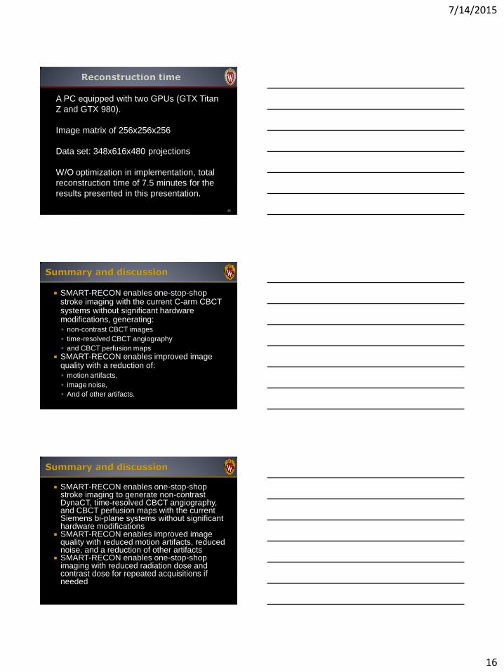
46
A PC equipped with two GPUs (GTX Titan
Z and GTX 980).
Image matrix of 256x256x256
Data set: 348x616x480 projections
W/O optimization in implementation, total
reconstruction time of 7.5 minutes for the
results presented in this presentation.
SMART-RECON enables one-stop-shop stroke imaging with the current C-arm CBCT systems without significant hardware modifications, generating:
non-contrast CBCT images
time-resolved CBCT angiography
and CBCT perfusion maps
SMART-RECON enables improved image quality with a reduction of:
motion artifacts,
image noise,
And of other artifacts.
SMART-RECON enables one-stop-shop stroke imaging to generate non-contrast DynaCT, time-resolved CBCT angiography, and CBCT perfusion maps with the current Siemens bi-plane systems without significant hardware modifications
SMART-RECON enables improved image quality with reduced motion artifacts, reduced noise, and a reduction of other artifacts
SMART-RECON enables one-stop-shop imaging with reduced radiation dose and contrast dose for repeated acquisitions if needed

7/14/2015
17
SMART IV 3D-DSA
50 t
Inte
nsity (
a.u
.)
0
Contrast Injection
51
Maps 5.0 mm slice thickness. Pre-treatment. C-arm cone beam
CT perfusion w/ SMART-RECON
CBV Axial CBV Sagittal CBV Coronal

7/14/2015
18
MTT Axial MTT Sagittal MTT Coronal
TTP Axial TTP Sagittal TTP Coronal
Improves temporal sampling density, but not to improve the accuracy of intensity value at a given temporal point
Assumes repeatable perfusion curves
Requires double injections and double scans 54
t
Inte
nsity (
a.u
.)
Ganguly, et.al., Proc. SPIE 7625, 2010; Ganguly, et.al., AJNR 32(8), 2011; Fieselmann, et.al., IEEE TMI 31(4), 2012

7/14/2015
19
Low temporal sampling density:
To recover a curve, we need adequate
sampling points.
Low temporal resolution:
If there is rapid change of contrast in the
sampling window, the reconstructed intensity
may be inaccurate.
55
For any image point inside a region of interest (ROI),
any straight line passing through the image point
should intersect the source trajectory at least once!
K. Tuy, “An inversion formula for cone-beam reconstruction," SIAM Journal on Applied Mathematics,
Vo. 43:546-552 (1983).
57
Super-short scan mode:
less than 180 degrees plus
fan angle
Short scan mode Disjoint segments:
Local ROI Imaging
Short-scan, super-short scan, and local ROI imaging

7/14/2015
20
58
Yes, the super short-scan reconstruction method does
allow us to reconstruct the crescent area. Unfortunately,
that area is completely outside the field of view and thus
useless in practice!
Wider data acquisition temporal window, stronger is the temporal-average artifacts: distortion artifacts streaking artifacts shading artifacts lower signal values for contrast enhanced
area
Narrower temporal window for data acquisition is desired!
59
Narrower data acquisition temporal
window, easier to violate the Tuy data
sufficiency condition and thus limited-view
artifacts:
Shading artifacts
Distortion artifacts
60
7/14/2015
21
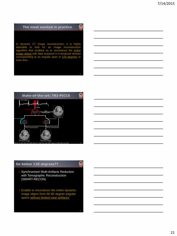
61
In dynamic CT image reconstruction, it is highly
desirable to look for an image reconstruction
algorithm that enables us to reconstruct the entire
image object with data acquired in a temporal window
corresponding to an angular span of 120 degrees or
even less.
t
TRI-PICCS Image TRI-PICCS Image
Prior Image xp
FBP
reconstruction short-scan
projection
Gating
ECG signal
half of
short-scan
projection
half of
short-scan
projection
PICCS reconstruction PICCS reconstruction
62 Chen et. al., Med. Phys. (35), 2008; Chen et. al., Med. Phys. (36), 2009; Tang et. al., Med. Phys. (37), 2010
self-consistent self-consistent
self-inconsistent
Synchronized Multi-Artifacts Reduction
with Tomographic Reconstruction
(SMART-RECON)
Enable to reconstruct the entire dynamic
image object from 50-60 degree angular
spans without limited-view artifacts!
63

7/14/2015
22
64
X(x,t)
Static statistical image reconstruction: Each static image
can be described by a vector.
SMART-RECON: Variable is a spatial-temporal matrix.
65
Xp is the prior image reconstructed from all of the acquired data.
D is a diagonal matrix to incorporate photon statistics
Time is brain! Quantum paradigm shift in clinical workflow
to save two hours from stroke onset to start time in endovascular therapy;
Quantum image reconstruction technology for a quantum transition in temporal resolution to enable time-resolved cone-beam CT angiography and whole brain perfusion to enable new clinical workflow;
Quantum clinical impact in five years.
66