oorthuva vatham
TRANSCRIPT

A STUDY ON
OORTHUVA VATHAM
Dissertation Submitted To
THE TAMIL NADU DR.M.G.R Medical University
Chennai – 32
In Partial fulfillment for The Award of Degree of
DOCTOR OF MEDICINE (SIDDHA) (Branch V - Noi Nadal)
Department of Noi Nadal
Government Siddha Medical College
Palayamkottai – 627 002
September 2007

CONTENTS
Page No
I. Acknowledgement
II. Introduction 1
Siddha physiology 3
Siddha pathology 16
III. Aim and objectives 28
IV. Reading lines between Yugi’s poem 30
V. Pathological view of reading lines in modern aspect 33
VI. Pathological view of dissertation topic in siddha aspect 39
VII. Evaluation of the dissertation topic 44
• Materials and methods 44
• Distribution of mukkuttram and udal thathukkal 54
• Picture of envagai thervugal 57
• Allied parameters 59
VIII. Modern aspects 63
• Anatomy 63
• Physiology 71
• Patho physiology 93
IX. Discussion 101
• Interpretation of clinical history 101
• Interpretation of allied parameters 103

• Interpretation of Envagai Thervugal 106
• Mukkutra nilaigal and udal kattugal 108
• Highlights of the dissertation topic 109
• Preventive measures 110
X. Conclusion 111
XI. Annexure – I 112
Annexure – II 120
XII. Bibliography 123

ACKNOWLEDGEMENT
With all the available worldly advantages, fulfilment of this work is due to
the grace of The Almighty, the first physician and to him I offer my prayers.
I thank The Vice-Chancellor, The Tamil Nadu Dr.M.G.R. Medical
University, Chennai for giving me permission to undertake this dissertation
work.
I also thank our Principal Dr.M.Dinakaran M.D.(S), Government Siddha
Medical College, Palayamkottai for permitting me to use the facilities available
in the institution for my dissertation work.
I am thankful to Dr. Mrs. I. Sornamariammal , MD(S), Joint Director,
Directorate of Indian Medicine and Homeopathy, Chennai, for encouraging me
to choose this topic.
It is my great privilege to record my deep sense of gratitude to
Dr.R.Devarajan M.D.(S)., Vice Principal, Head of the Noi Naadal Post
Graduate Department, Government Siddha Medical College, Palayamkottai for
his devoted guidance.
I express my gratitude to Dr.T.Rajasekar M.D.(S)., Post Graduate Noi
Naadal Department for his abundant support and guidance in my dissertation
work.
I wish to thank Dr.S.Sundararajan M.D.(S)., Post Graduate Noi Naadal
Department for this valuable guidance.

I owe special gratitudes to Dr.M.R.Vairamuthu Raju, M.D (GM)
Professor, Modern Medicine for his devoted guidance and timely help in the
completion of this dissertation.
I am particularly grateful to Dr. Paramasivam, M.D (Patho), M.D
(Forensic) for his valuable assistance in bringing out this book.
I thank Dr.K.Swaminathan M.D, Assistant Professor, Department of
Pathology, Tirunelveli Medical College for his guidance in the pathological view
of my dissertation.
My cordial thanks to Dr. M.S.L.Mahboobu Subuhani M.D., DM
(Cardiology), and Dr.R.Ravi Edwin M.D, D.M (Cardiology) for permitting me
to see and examine the cases. Without their constant and authentic support,
this study would not have seen the light of day.
Last but no means the least, I express my cordial thanks to Broad Band
Net Café for their co-operation in bringing out this dissertation book in a
successful manner.

1
INTRODUCTION
Medicine is not merely a science but an art as well. It deals with the
different processes of life. The ancient philosophers of siddha school
defined the mode and methods of medicine.
A system of medicine without understanding and without a true
knowledge of natural laws will remain forever a system of theories, mere
opinion and of passive observation and inactivity.
It is nature that causes disease and it is again nature that effects
their cures and therefore the physician should know the process of nature.
Siddhars who are considered to be super-human beings have
defined age and other laws of nature to which all human beings are subject
to.
In Thirumanthiram, which is the most important work of Thirumoolar,
he defined medicine as follows.
“Medicine means the prevention of bodily illness
Medicine means the prevention of mental illness
Medicine’s purpose is to avert disease
Medicine therefore is the prevention of death”.
The original stanza in Tamil reads as follows
‘kWg;gJly; Neha; kUe;njd yhFk;
kWg;gJ sNeha; kUe;njdr; rhYk;
kWg;gjpdp Neha; thuh jpUf;f
kWg;gJ rhitA kUe;njd yhNk”.
- jpU%yH

2
They seem to have realised most rationally that prevention is better
than cure and that prevention should be the main purpose of medicine.
Life is man’s most valuable possession and the next in order of value
is health. Health is the chief basis for the development of ethical,
economic, artistic and spiritual sides of man. Without health, life is deprived
not only of much if not all, of its usefulness but also of its joys and
pleasures.
Noi or disease denotes weakness and in which both mind and body
are involved (or) the disease will cause suffering to the mind and body.
It is pointed by “Tholkaapiyar” that the disease means suffering and
depression.
‘igASk; rpWikAk; Nehapd; nghUs;”.
(njhy; chp 341)
When the disease is due to the persistent action of the adverse
factors, it becomes chronic and complete cure at this stage is very difficult.
The siddhars were aware of the fact of chronicity.
‘twpjhy; ,Uisnad; ahf;if apdpatH
thpDk; Neha; kUe;jy;yu; thuhJ”.
The effect of treatment depends upon the prevention only. The
disease has no effect if we adhere to the principles of prevention in our
daily life. The Tamils of Sangam period were prompt in preventing
diseases both internal and external to lead a healthy life.

3
SIDDHA PHYSIOLOGY
Siddhars believed that five elements are the basis of the universe
and every human being. Our ancient literature Tholkaapiyam also accepts
the concepts of siddhars. The universe is a composition of five elements
viz earth, air, water, ether and fire which are known as “Panchabootham” in
siddha system. So the human body is a composition of this pancha
bootham.
‘mz;lj;jpYs;sNj gpz;lk;
gpz;lj;jpYs;sNj mz;lk;
mz;lKk; gpz;lKk; xd;Nw
mwpe;Jjhd; ghh;f;Fk; NghNj”.
- rl;lKdp Qhdk;
According to siddha physiology man is considered as the microcosm.
Universe is considered as the macrocosm. It shows that the human body is
the replica of the universe.
Vethas reveal that one of the five elements combined with the other
four elements in different proportions to form the human body. The basic
reason for the soul resting is uyirthathu or jeevathathu.
This uyirthathu divided into three thodas known as vatham, pitham,
kabam and acquires three characters (mukkunam – sathuva, rajo, thamo)
thereby it protects and develops the soul and the body.
Each and every atom consists of 96 thathuvas. These 96 thathuvas
are invisible to our naked eye until it is present in a single atom. Since it

4
mingles or joins to form a multi cellular body and it gets larger size
according to the shape and merges to act respectively.
Due to the combination of 96 thathuvas, soul originates, acquires
shape and multiplies to grow larger and finally gets a body to live and then
performs its duties, multiplies its generations, gets its old ages and dies.
Finally it reaches its initial stage where it was in primitive.
This 96 thathuvas are limited to all human beings in normal
condition. This not only consists of the physical components of the human
body but also the mental intellectual components like passions, qualities,
knowledge, the functions of the sense organs and motor organs and their
co-ordination.
The physiology of siddha system involves 96 basic factors, seven
constituent elements, 14 reflexes, aru suvaigal, four udal thee and three
udal vanmaigal.
‘cWjpahk; G+jhjp Nahiue;jhk;.......”
- Ntjhe;j jj;Jtf; fl;lis
Panchabootham 5 - Five elements
Earth - all organic living bodies and organic substances are
created
Water - It combines all the things
Air - It spreads all over the space
Fire - It gives colour and brightness to the things
Space - It gives space to all other boothams

5
Gnanenthirium or Pori 5 - Five sense organs
1. Ear
2. Skin
3. Eye
4. Tongue
5. Nose
Pulan 5 – Functions of five sense organs
1. Hearing
2. Touch sense
3. Vision
4. Taste
5. Smell
Kanmaenthiriam 5 – Five motor organs
1. Mouth - stands as space
2. Hand - stands as air
3. Leg - stands as fire
4. Anus - stands as water
5. Sex organs- stands as earth
Kanmavidayangal 5 – Functions of five motor organs
1. Speech
2. Flexion and extension of upper limbs and lower limbs
3. Walking
4. Defecation
5. Ejaculation of semen and propulsion of ova

6
Anthakaranas 4 – Four intellectual faculties
1. Manam - Mind or the reasoning faculty
2. Puththi - Knowledge
3. Siddham - Determination or firm conviction
4. Agangaram - Achievement
Arivu 1 - Intellect or wisdom
Naadies 10:
There are ten channels
1. Idakalai
2. Pinkalai
3. Suzhumunai
4. Siguvai
5. Purudan.
6. Kanthari
7. Aththi
8. Alampudai
9. Sankini – Present in external genitalia
10. Gugu – Present in anus
Vayu 10 – Ten vital airforces
1. Abanan
2. Pranan
3. Udhanan
4. Samanan

7
5. Viyanan
6. Koorman
7. Naagan
8. Kirukaran
9. Devathathan
10. Dhananjeyan
Aasayam 5 - Five visceral cavities
Amarvasayam - Stomach
Pahirvasayam - Liver, small intestine
Malavasayam - Rectum, large intestine
Salavasayam - Urinary bladder
Sukkilavasayam - Seminal vesicle and ovary
Kosam 5 - Five vestures of the soul
1. Annamaya kosam
2. Piranamaya kosam
3. Manomaya kosam
4. Vinganamaya kosam
5. Anandhamaya kosam
Aatharam 6:
1. Moolatharam
2. Swathitanam
3. Manipooragam
4. Anagatham

8
5. Vishuthi
6. Aakkinai
Malam 3 - Three mental binders
1. Aanavam
2. Maayai
3. Kanmam
Mandalam 3 – Regions
1.Gnayiru (sun) - Solar plexus
2. Thingal (moon) - Lunar plexus
3.Thee (fire)
Thodam 3 - Three humours
Three humours are the fundamental principles and essential factors
in the composition and constitution of the human body. The three humours
vatham, pitham and kabam represent wind, bile and phlegm respectively.
Relation between boothas and mukkutram
Vatham - Air
Pitham - Fire
Kabam - Water
Formation of the three humours
“te;j fiy %d;wpy; thAk ghdDld;
je;j gpuhzd; rkhdDk; - re;jKwf;
$l;LwT Nurpj;jhy; $Wk; thjk; gpj;jk;
ehl;Lq; fgNkahk; ehL”.

9
Vatham - Idakalai + Abanan
Pitham - Pinkalai + Piranan
Kabam - Suzhumunai+ Samanan
‘;thjkha;g; gilj;Jg; g;pj;j td;dpaha;f; fhj;Jr; Nrl;g
rPjkha;j; Jilj;J...............”
- Njiuah; kUj;Jg; ghujk;.
Vatham - Creation
PItham - Protection
Kabam - Destruction
‘nka;asT thjnkhd;W
Nky;gpj;j Nkhuiuahk;
Iaq;fhnyd;Nw mwp”.
- fz;Zrhkpak;
Vatham - 1 maathirai
Pitham - ½ maathirai
Kabam - ¼ maathirai
Qualities:-
Vatham Pitham Kabam
Dry Hot Cold
Cold Acid Heavy
Subtle Mobile Sweet
Rough Liquid Soft
Unstable Acute Unctuous
Light Pungent Viscid

10
Functions:-
Types:-
Vatham Pitham Kabam
Pranan Anarpitham Avalambagam
Abanan Ranjagam Kilaedhagam
Viyanan Saadhagam Bothagam
Udhanan Aalosagam Tharpagam
Samanan Pirasagam Sandhigam
Naagan
Koorman
Kirukaran
Devathathan
Dhananjayan
Vatham Pitham Kabam
Respiration Acidity Strengthens bones and
joints
Giving briskness Burning sensation Gives lustre to the body
Functions of the body Yellowish
discolouration Makes strength
Regulation of 14 Vegangal of eye, skin and
urine etc Fearlessness
Functions of 7 udal kattugal
Digestion Patience
Protects and strengthens
five sensory organs
Profuse sweating
Dizziness Immunity

11
Edanai 3 - Three physical bindings
1) Porul patru - Material bindings
2) Puthalvar patru - Off springs bindings
3) Ulaga patru - World bindings
Vinai 2 – Two deeds
1) Nalvinai - Good deeds
2) Thee vinai - Bad deeds
Gunam 3 – Three cosmic qualities
Sathuva gunam
Rasathuva gunam
Thamasa gunam
Raagam 8 – Eight passions
1. Kaamam - Desire
2. Krotham - Hatred
3. Lopam - Stinginess
4. Moham - Lust
5. Matham - Pride
6. Marcharyam - Internal conflict
7. Idumbai - Mockery
8. Agankaaram - Ego or self love.

12
Avaththai 5 – Five states of consciousness
1. Ninaivu - Wakefulness
2. Kanavu - Dream
3. Urakkam - Sleep
4. Parurakkam - Stuper
5. Uyirppadakkam - Stage of samaathy
Udal thathukkal 7 - Seven constituent elements
Seven udal thathukkal are responsible for the entire structure of the
human body.
“,urk; cjpuk; ,iwr;rp Njhy; Nkij
kUtpa tj;jp thOk; nghU kr;ir
gutpa Rf;fpyk; ghohk; cghjp
cUgk yhDly; xd;nwdyhNk”.
- jpUke;jpuk; 2080
1. Saaram - Chyle
2. Chenneer - Blood
3. Oon - Muscle
4. Kozhuppu - Fat
5. Enbu - Bone
6. Moolai - Bone marrow
7. Sukkilam / suronitham - Sperm / ovum

13
Udal thathus maintain the function of different organs, systems and
vital parts of the body. They play a very important role in the development
and nourishment of the body. Each thathu receives its nourishment from the
previous thathu. When one thathu is defective it affects the successive
thathu.
Vegangal 14 – Fourteen Urges / Reflexes
1. Vatham
2. Thummal
3. Siruneer
4. Malam
5. Kottaavi
6. Pasi
7. Neer Vetkai
8. Kasam
9. Elaippu
10. Nithirai.
11. Vaanthi
12. Kanneer
13. Sukkilam / Suronitham
14. Suvaasam.

14
Udal Vanmai 3 – Strength and vitality constitute udal vanmai.
1) Eyarkai vanmai ( Innate immunity) - Inherited vitality.
2) Kaala vanmai (Seasonal immunity) - Vitality that is generally found
in different age periods as
well as different seasons.
3) Seyarkai vanmai - Improvement of vitality
obtained by good habits and
physical exercises
Udal akkini 4 – Four body fires
Samaakkini
Mandhaakkini
Deekshanaakkini
Vishamaakkini
Suvaigal 6 – Six tastes
Suvai is a peculiar sensation caused by the contact of soluble substances
with the tongue. The sense is effected by the tongue.
Combination of two boothas constitute a suvai (taste)
‘kz;ZlNd Gdy;jPf;fhy; Kiwahfr; Nrh;e;jpl;lhy; tUNk ,dpg;G
jpz;zkpyk; Jth;g;gpurk; rjhfjpNah lhh;jPapd; jplkh Kiwg;Gk;
vz;zhpa frg;GKz;lhe; jz;zPhpy; fdypizg;ghnyOkh - Kth;g;G
cz;zhpa mWRitapd; gpwg;gpnjDk; FUrpj;jUiuj;j kiwNa”.
- kUj;Jtj; jdpg;ghly;fs;

15
Arusuvai
Inippu - Mann + Neer - Kabam
Pulippu - Mann +Thee - Pitham
Uppu - Neer +Thee - Pitham
Kaippu - Vali + Vinn - Vatham
Kaarppu - Vali + Thee - Pitham
Thuvarppu - Mann + Vali - Vatham

16
SIDDHA PATHOLOGY
Pathology is the scientific study of structure and function of the body
in a disease. It deals with causes, effects, mechanism and nature of
diseases.
Siddha pathology deals with the diseased condition of the human,
which is due to food alterations, seasonal and environmental variations,
helding the 14 reflexes and by the behaviours. The disease is reflected
through the pulse formed by the three humours.
The whole siddha system of medicine rests on the maintenance or
restoration of the equilibrium between the three thathus, which coincides
with the following kural.
‘kpfpDk; FiwapDk; Neha; nra;Ak; E}Nyhh;
tsp Kjyh vz;zpa %d;W.”
- jpUf;Fws;
Siddha system approaches and analyses the disease on the basis of
the ‘Thridosha theory’ . The doshas or humours are vatham, pitham and
kabam. Disease is due to the disturbance in the equilibrium between the
three humours. When these are in perfect balance and harmony a person
is said to be healthy. Imbalance and derangement in thridoshas causes
disease. So the diseases are studied on the basis of thridosha theory.
‘kjpj;jplw;fUik tha;e;j
khz;ghpfhuunky;yhe;
Jjpj;jpl Tzh;e;jhNdDe;;

17
Jfswg; gpzpapd;wd;ik>
gjpj;jplTzuhdhfpw;
gaDwhdhfhyhNd
tpjpj;jpL gpzpj;jpwj;ij
tpsk;GJ Kjw;fz;kd;Ndh”
- rpfpr;rhuj;d jPgk;
‘Kg;gpzp kUtp KdpT nfhs; Fwpg;igj; jg;gh
jwpAk; jd;ikAk; thj gpj;j itak; gphpitA
kitjhk; Vwpapwq;fp ,ize;J fye;J khwp
khwp tUk; nraw;ifahw; gpzp Neh;ikawpe;J
ePl;L kUe;Nj rPhpajhnkdr; nrg;Gth; rpj;jNu”.
Food variations
Diet plays a vital role in preserving the human body. The food is
formed on the basis of 6 tastes.
‘Gsp Jth; tpQ;Rq;fwp ahw;G+hpf; Fk;thjk;
xsp Ath;ifg; Ngwpy; gpj;Jr; rPWk; - fpspnkhopNa
fhh;g;gpdpg;G tpQ;rpw; fgk;tpQ;R Q;rl;bujr;
Nrug; Gzh; NehaZfhNj”.
- fz;Zrhkpak;

18
Sour and astringent cause an increase in vatham. Salt and bitter
cause an increase in pitham. While pungent and sweet cause an increase
in kabam.
Environmental changes
The environmental factors ( Thinai ) may also pave the way for the
development of diseases. Thinai has been classified into 5 types.
A. Kurinji : Kabam pertains here. Anemia, fever and abdominal
mass may develop.
B. Mullai : The dwellers of this land suffer from vatha and
pithakaba diseases.
C. Marutham : The inhabitants of this region have controlled
vatham, pitham and kabam . This is the prompt place for a
healthy inhabitation.
D. Neithal: The people of this land suffer from vatha diseases,
further it leads to increased body mass, enlargement of liver
and flatulence.
E. Palai : The inhabitants of this land suffer from vatha , pitha
and kaba diseases.
Seasonal variations
Due to seasonal variations, changes in three humours (thridosha)
occur and this lead to a disease.
1) Kar kalam (Avani and Puratasi) - Disturbances in three humours
19
is most prominent. Pitham is
increased from its normal
state. Vatham is also
increased from its normal level
and spreads continuously to
other areas of the body.
2) Koothir kalam (Iypasi & Karthigai) - Altered pitham spreads to other
areas of the body.
3) Munpani kalam (Markazhi & Thai )- Three thodas are in equilibrium.
4) Pinpani kalam (Maasi and Pankuni)- Kabam increases from its
normal state.
5) Elavenil kalam (Chithirai and Vaigasi)- Altered kabam spreads to
other parts of the body.
6) Muthuvenil kalam (Aani & Aadi) - Kabam comes to equilibrium
and vatham gets aggrevated.
Variations in 3 humours
The disease is mainly caused because of the inequilibrium in one or
more among the 3 humours that exist in human as ‘Uyir Thathukkal’.

20
Three humours Characteristic features
of increasing
Characteristic features of decreasing
Vatham Weakness and
occasionally blackening
of the body. Desire to
take hot diet, shivering,
abdominal distension ,
constipation, diminished
immunity, insomnia,
giddiness, laziness,
blabbering and
generalised weakness.
Stiffness, diminished
voice, impaired
intellectual function,
disturbance in general
activities, semi-
consciousness, fatigue,
excessive salivation,
paleness and cooling of
the body,
breathlessness, cough
and excessive sleep.
Pitham Yellowish discolouration
of eyes, faeces, urine
and skin. Polyphagia,
polydypsia, burning
sensation all over the
body and decreased
sleep.
Loss of appetite,
cooling of the body,
impaired pigmentation
of the skin, symptoms
related with decreased
kabam.
Kabam Abdominal distension,
salivation, fatigue,
paleness and cooling of
the body, heaviness of
the body,
breathlessness, cough
and excessive sleep.
Giddiness, sublaxation
of joints, prominence of
bones. Kabam present
in the lungs gets
decreased. Excessive
sweating in the hair
follicles and palpitation.

21
Udal thathukkal
When the three humours of the human body are affected by various
factors they immediately change the nature of the 7 physical constituents,
i.e. udal thathukkal.
Udal thathukkal (Physical
constituents)
Features of increasing Features of decreasing
Saaram (Chyle) Features related with
decrease in kabam, loss
of appetite
Dryness of skin,
loss of weight,
tiredness,
the functions of sense
organs are diminished
Senneer (Blood) Boils and tumours in
different parts of the
body, splenomegaly,
soolai (pain),
hypertension,
haematuria,
redness of the eyes,
leprosy,
jaundice
Desire for cold things,
dryness, discolouration
and paleness of the
skin
Oon Tumours or extra
growth around the neck,
face, abdomen, thigh
and genitalia
Lethargy of 5 sensory
organs, pain in the
joints, loss of
subcutaneous fat
Kozhuppu Identical to increasing
features of oon,
tiredness, dyspnea on
exertion
Splenomegaly, loin
pain, emaciation

22
Enbu Excessive ossification
and dentition
Weak bone pain in the
joints, splitting of hair
and nails
Moolai Sense of heaviness of
the body and eyes,
swelling of smaller joints
of hand and feet,
oliguria, non – healing
ulcers
Osteoporosis , blur
vision
Sukkilam / Suronitham Sexual activity
increases, urinary
calculi
Pain in the genitalia
and accompanied
inability to reproduce
URGES (14 VEGANGAL)
There are 14 natural reflexes involved in the physiology of normal
human beings and if willfully suppressed, the following are resulted.
1. Vatham (Flatus)
This urge should not be suppressed. If it is suppressed it leads to
chest pain, epigastric pain. Abdominal pain, body ache, constipation,
dysuria and indigestion predominates.
2. Thummal (Sneezing)
If arrested it leads to headache, facial pain, low back pain and
neuritic pain in the sense organs.

23
3. Siruneer (Urine)
If arrested it leads to urinary retention, urethral ulcer, joint pain, pain
in the penis, gas formation in abdomen.
4. Malam (Faeces)
If arrested it leads to pain in the knee joints, headache, general
weakness, flatulence and other diseases may also originate.
5. Kottavi (Yawning)
If arrested it leads to indigestion, leucorrhoea, abdominal disorders
and urinary disorders.
6. Pasi (Hunger)
If arrested it leads to the tiredness of all organs, emaciation,
syncope, apathetic face and joint pain.
7. Neer vetkai (Thirst)
If arrested it leads to the affection of all organs and pain may
supervene.
8. Kaasam (Cough)
If it is suppressed severe cough, bad breath and heart diseases will
be resulted.
9. Ilaippu (Exhaustiveness)
If suppressed it will lead to fainting, urinary disorders and rigor.

24
10. Nithirai (Sleep)
All organs will get rest only during sleep. So it should not be avoided.
If disturbed it will lead to headache, pain in the eyes, deafness and slurred
speech.
11. Vaanthi (Vomiting)
If arrested it leads to itching and symptoms of increased pitham.
12. Kanneer (Tears)
If it is suppressed it will lead to sinusitis, headache, eye diseases
and chest pain.
13. Sukkilam (Semen)
If it is suppressed there will be joint pain, difficulty in urination, fever
and chest pain.
14. Swaasam (Breathing)
If it is suppressed there will be cough, abdominal discomfort and
anorexia.
Udal Vanmai:
The disease affecting an individual is also based on the udal vanmai.
The udal vanmai is classified into 3 types.
1. Eyarkai Vanmai
This is based on sathuva, rajo and thamo gunas and it is the
strength which is present naturally.

25
2. Seyarkai Vanmai
The mukkuna based body is maintained by food habits. Seyarkai
vanmai means improving the strength by diet and medicine.
3) Kaala Vanmai:
It is based on kaalangal. The strength that is gained by seasonal
variations as well as the age of a person.
Investigations in siddha system
The methods adopted in siddha system of medicine are
poriyaalarithal, pulanaalarithal and vinaathal. Poriyaalarithal means
diagnosing through the five organs of perception namely nose, tongue,
eyes, ears and skin. Pulanaalarithal means diagnosing through the five
objects of senses namely smell, taste, vision, audio and sensation of skin.
Vinaathal is a method of interrogating the problem of the patient from his
own words or from attendars.
Envagai Thervugal
‘ehb ];ghprk; eh epwk; nkhop tpop
kyk; %j;jpukpit kUj;Jt uhAjk;”.
1. Naadi (Pulse)
2. Sparisam (Sense of touch)
3. Naa (Tongue)
4. Niram (Colour)
5. Mozhi (Speech)
6. Vizhi (Eyes)
26
7. Malam (Stool)
8. Moothiram (Urine)
‘nka;f;Fwp epwe;njhdp tpop ehtpUkyk; iff;Fwp”.
- Njiuah;
Naadi (Pulse)
This is a unique diagnostic method in siddha system of medicine. It
is responsible for existence of life. It is felt one inch below the wrist on the
radial side by palpating with the top of the index finger, middle finger and
ring finger which denotes vatham, pitham and kabam.
Suitable places to feel pulses
‘jhJ KiwNfs; jdpf; Fjpr; re;NjhL
XJW fhkpa Ke;jp neL khh;G
fhJ neL%f;Ff; fz;lk; fuk; GUtk;
NghJW Kr;R Gfo; gj;Jk; ghh;j;jpNl”.
In siddha system of medicine changes of urine is studied under two
peculiar headings. They are “Neerkuri and Neikuri”.
‘te;j ePh;f;fhp vil kzk; vQ;rnyd;
iwe;jpaYstit aiwFJ KiwNa.”
- Njuh; ePh;f;Fwp - nea;f;Fwp

27
Neikuri
1) muntd ePz;bd0Nj thjk;
2) Mop Nghw;gutpd; m0Nj gpj;jk;
3) Kj;njhj;J epw;fpd; nkhoptnjd;fgNk.
- Njuh; ePh;f;Fwp - nea;f;Fwp
This procedure is an important one in siddha system of medicine to
find out the diagnosis as well as in prognosis aspect of the disease.
So diseases in man do not originate itself. It is developed from the
alteration of three doshas or humours and are classified in to 4448 as per
siddha physician Yugi’s theory.

28
AIM AND OBJECTIVES
The Tamils of Sangam period were used to follow healthy food
habits and their life style saved them from many diseases and disease
causing factors. But now a days because of unhealthy, unwanted food
habits and modified life style of our people, many diseases gained entry
among them.
One of them is cardiovascular disease, which is the leading cause of
death in all continents. Despite of all the advances in the medical field,
which has reached its sky as a limit, the incidence of heart diseases is
rising in many developed countries.
In evaluating patients with heart failure, it is important to identify not
only the underlying cause of the heart disease but also the precipitating
cause of heart failure. Identification of such precipitating causes is of
critical importance because their prompt alleviation may be life saving.
The aim of this dissertation topic is
• To collect the evidences found in siddha literature regarding
Oorthuva Vatham.
• To review the altered thridosha (or) mukkuttram and pathology in
siddha aspect.
• To depict the unique diagnostic procedures (ie) Envagai thervugal,
mentioned in siddha literatures for Oorthuva Vatham.
• To use modern parameters in the investigation of Oorthuva Vatham.

29
• To know the pathogenesis (origin and development) of Oorthuva
Vatham.
• To find out the aggravating factors which cause the condition to
worsen and to find out the preventive methods.

30
Reading lines between Yugi’s poem
In Yugi vaithiya sinthamani under “Vatha Roga Nithanam”, “Oorthuva
Vatham” was broadly dealt in the 298th stanza. It is as follows.
CHj;Jt thjk;
‘E}yhd Rthre;jhd; NkNy Nehf;fp
EZf;fkh kpilAlNd tapW tpk;kp
thyhd tha; jdpNy EiuAKz;lha;
tha; fOj;JkhHGNk kpfT nehe;J
Myhd tq;fnky;yh kod;W fhZk;
mbj;njhilA kbf;ifA kpfNt tw;Wk;
ghyhd NkdpANk gRik fhZk;
gUT+Hj;t thjnkd;Nw gfu yhNk”.
- A+fp itj;jpa rpe;jhkzp
CHj;Jt thjk;
The author Yugi mentioned the symptoms of the above disease
under the title “Oorthuva Vatham”.
Oorthuvam means rising or tending upwards. It denotes the erect
position of the patient due to laboured breathing. The sensation of
breathlessness usually is relieved by sitting upright since this position
reduces venous return and pulmonary capillary pressure. In far advanced
heart failure orthopnea (dyspnea in the recumbent position) may become
so severe that patients cannot lie down at all and must spend the entire
night in sitting position.

31
‘E}yhd Rthre;jhd; NkNy Nehf;fp”
Dyspnea in the recumbent position which is characterised by
extreme shortness of breath and rapid, shallow breathing.
‘EZf;fkh kpilAlNd tapW tpk;kp”
Distension of the abdomen.
‘thyhd tha;jdpNy EiuA Kz;lha;”
Frothy bronchial secretions pour out of the mouth.
‘tha; fOj;JkhHGNk kpfT nehe;J”
Pain in the mouth, neck and chest due to breathlessness.
‘Myhd tq;fnky;yh kod;W fhZk;”
It denotes fatigue, poor effort tolerance, weakness and low grade
fever.
‘mbj;njhilA kbf;ifA kpfNt tw;Wk;”
It denotes serious weight loss, cardiac cachexia and skeletal muscle
atrophy.
‘ghyhd NkdpANk gRik fhZk;”
Generalised anasarca.
The summary of Oorthuva Vatham is as follows.
In congestive cardiac failure, pulmonary congestion reduces lung
compliance and can obstruct the small airways. This causes dyspnea
which means laboured breathing. Due to inability of the heart to empty
properly, congestion of tissues occurs (Hepatic congestion and ascites).
Pulmonary congestion results in escaping of bronchial secretions through

32
the mouth. Pain in the mouth, neck and chest occurs due to dyspnea. Low
cardiac output causes fatigue, listlessness, poor effort tolerance. Chronic
heart failure is associated with cardiac cachexia and skeletal muscle
atrophy. Generalised anasarca results due to congestion of tissues and
protein loss.

33
PATHOLOGICAL VIEW OF READING LINES IN MODERN ASPECT
‘E}yhd Rthre;jhd; NkNy Nehf;fp”
The clinical manifestations of left-sided heart failure result from
accumulation of fluid upstream in the lungs and from decreased left
ventricular output. Among them one of the major clinical manifestations is
pulmonary congestion and oedema which causes dyspnea and orthopnea.
A frequent cause of death in heart failure is acute pulmonary
oedema occurring in patients who have had chronic heart failure for a long
time.
Pulmonary oedema can result from either the elevation of pulmonary
hydrostatic pressure or the increased capillary permeability.
The hydrostatic pressure in the pulmonary capillaries is much lower
(average 10mm Hg). Normally the plasma oncotic pressure (25 mmHg) is
adequate to prevent the escape of fluid into the interstitial space and hence
lungs are normally free of oedema.
Elevation in pulmonary hydrostatic pressure (Haemodynamic oedema).
In heart failure, there is increase in the pressure in pulmonary veins
which is transmitted to pulmonary capillaries. This results in imbalance
between pulmonary hydrostatic pressure and the plasma oncotic pressure
so that excess fluid moved out of pulmonary capillaries into the interstitium
of the lungs. Simultaneously, the endothelium of the pulmonary capillaries

34
develops fenestrations, permitting passage of plasma proteins and fluid
into the interstitium.
The interstitial fluid so collected is cleared by the lymphatics present
around the bronchioles, small muscular arteries and veins. As the capacity
of the lymphatics to drain the fluid is exceeded (about tenfold increase in
fluid), the excess fluid starts accumulating in the interstitium (interstitial
oedema) i.e, in the loose tissues around bronchioles, arteries and in the
lobular septa.
Next follows the thickening of the alveolar walls because of the
interstitial oedema. Upto this stage, no significant impairment of gaseous
exchange occurs. However prolonged elevation of hydrostatic pressure
and due to high pressure of interstitial oedema, the alveolar lining cells
break and the alveolar air spaces are flooded with fluid (alveolar oedema)
driving the air out of alveolus, thus seriously hampering the lung function.
This increases the work of the respiratory muscles required to inflate
the lungs. The activation of receptors in the lungs results in the rapid,
shallow breathing which is the characteristic feature of cardiac dyspnea.
The oxygen cost of breathing is increased by the excessive work of the
respiratory muscles. This is coupled with the diminished delivery of oxygen
to these muscles which occurs as a consequence of the reduced cardiac
output and this may contribute to fatigue of the respiratory muscles and the
sensation of shortness of breath.
‘EZf;fkh kpilAlNd tapW tpk;kp”

35
Fullness of abdomen, anorexia, nausea associated with abdominal
pain are frequent complaints and may be related to the congested liver and
portal venous system. An enlarged, tender, pulsating liver and ascites
present.
‘thyhd tha; jdpNy EiuAKz;lha;”
This is due to pulmonary congestion with pulmonary oedema. The
patient brings out lot of sputum which is characteristically frothy as the
bronchial secretion is mixed up with air.
The fluid which enters the lungs is beaten up into a frothy by the
pulmonary ventilation.
In grave diseases both lungs are entirely filled with oedema fluid and
frothy fluid may pour out of the mouth.
‘tha; fOj;JkhHGNk kpfT nehe;J”
Pulmonary oedema reduces the compliance of the lungs and thereby
increases the work of the respiratory muscles required to inflate the lungs.
The cost of breathing is increased by the excessive work of the respiratory
muscles. This is coupled with the diminished delivery of oxygen to these
muscles, which occurs as a consequence of the reduced cardiac output
and which may contribute to fatigue of the respiratory muscles.
‘Myhd tq;fnky;yh kod;W fhZk;”
Due to inadequate blood supply to the muscle fatigue, poor effort
tolerance and weakness occur.

36
Low grade fever is because of reduction of cutaneous flow and lung
infection due to pulmonary oedema.
‘mbj;njhilA kbf;ifA kpfNt tw;Wk;”
With severe chronic heart failure there may be serious weight loss
and skeletal muscle atrophy.
The serious weight loss and cachexia are because of
1. Elevation of circulating concentrations of cachectin or tumour
necrosis factors. (TNF-∝) and interleukin–1 derived from
macrophages.
2. Elevation of the metabolic rate, which results in part from the extra
work performed by the respiratory muscles, the increased oxygen
needs of the hypertrophied heart and / or the discomfort associated
with severe heart failure.
3. Anorexia, nausea and vomiting due to congestive hepatomegaly and
abdominal fullness.
4. Impairment of intestinal absorption due to congestion of the intestinal
veins.
5. Protein losing enteropathy.
Skeletal muscle atrophy is due to prolonged diminished functional
activity i.e, wasting of muscles of limb immobilised in cast (disuse atrophy).
Reduction of the number and size of parenchymal cells of an organ or its
parts which was once normal is called atrophy. Irrespective of the
underlying cause for atrophy, the pathological changes are similar.

37
The organ is small, often shrunken. The cells become smaller in size
but are not dead cells.
Shrinkage of cell size is due to reduction in cell organells, chiefly
mitochondria, myofilaments and endoplasmic reticulum. There is often
increase in the number of autophagic vacuoles containing cell debris.
These autophagic vacuoles may persist to form residual bodies in cell
cytoplasm.
‘ghyhd NkdpANk gRik fhZk;”
Generalised oedema develops in congestive cardiac failure.
Pathogenesis of cardiac oedema is explained on the basis of the following
hypothesis.
MECHANISMS INVOLVED IN THE PATHOGENESIS OF CARDIAC OEDEMA
Chronic hypoxia
↑ Capillary permeability
↑Central venous
pressure
Capillary hydrostatic pressure
Cardiac oedema
Congestive heart failure
Extra-renal mechanism (Aldosterone)
Hypovolaemia
Intrinsic-renal mechanism
(↓GFR)
Renal retention of Na+ and water
ADH
Cardiac output

38
1. Reduced cardiac output causes hypovolaemia which stimulates
intrinsic–renal and extra–renal hormonal (renin–angiotensin– aldosterone)
mechanisms as well as ADH secretion resulting in sodium and water
retention and consequent oedema.
2. Due to heart failure, there is elevated central venous pressure
which is transmitted backward to the venous end of the capillaries, raising
the capillary hydrostatic pressure and consequent transudation, this is
known as back pressure hypothesis.
3. Chronic hypoxia may injure the capillary wall causing increased
capillary permeability and results in oedema, this is called forward pressure
hypothesis. However this theory lacks support since the oedema by this
mechanism is exudate whereas the cardiac oedema is typically transudate.
Cardiac oedema is influenced by gravity and is thus characteristically
dependent oedema i.e, in an ambulatory patient it is on the lower
extremities, while in a bed–ridden patient oedema appears on the sacral
and genital areas. The accumulation of fluid may also occur in serous
cavities.

39
PATHOLOGICAL VIEW OF DISSERTATION TOPIC IN SIDDHA ASPECT
“gpzpapDw; gj;jpiag; NgRtd; gpzpKjy;
thj gpj; jq;fg kd; ke;jphp je;jphp
tPjkh Alyuz; nka;k;Gu tuR nra;
Kiw nrAkhjyhd;............”
- Njiuah; fhg;gpak;
According to siddha aspect, vatham is said to be the initiator of all
activities of our body. So the author mentioned vatham as ‘ Arasan’ in the
above lines.
“thjgpj;jika %d;Wk;
td;gyj;JlNd jj;jk;
Ngjnkhd;wpy;yh tz;zk;
Ngrpajhde; jd;dpy;
ePjpaha; epiyj;J epw;fpy;
neLk;gpzp rpf;ftpy;iy
jhJTnkhd;Nwh nlhd;W
jhtpbw; gpzpfs;jhNd”.
- fz;Zrhkpak;
Imbalance and derangement in thridosha cause diseases. During the
acute stage of diseases any of the three thathus (3 humours) or seven udal
kattugal (7 constituent elements) may be affected. If the condition is not
treated promptly, it will lead to the affection of other thathus and the
condition will become chronic.

40
By knowing the distribution of vatham, one can understand that vatham
is capable of affecting many of the systems of the body. If we don’t treat
the derangement of vatham, the condition will lead to ‘Thontha Vinaigal’
(secondary affections) and causes suffering to the body.
ALTERED THRIDOSHA IN OORTHUVA VATHAM
I. Vatham: Ten forms of vatham are described in our siddha literatures.
1. Pranan: ‘Pranan’ is considered as heart centre. It controls heart,
circulation, inspiration and expiration.
In ‘Oorthuva Vatham’ the circulation of blood to other parts of the body
is affected i.e., pranan is affected.
2. Abanan
The functions of abanan is micturation, defecation etc.
In ‘Oorthuva Vatham’, sodium and water retention, oliguria and
steatorrhoea occur because of the derangement of abanan.
3. Viyanan
Viyanan is distributed throughout the body. It is said to be the part of
the sky. Its place in our body is heart. The functions of viyanan is walking,
movements of the body etc.
In ‘Oorthuva Vatham”, viyanan is affected and exhibits the symptoms
like dyspnea on exertion.

41
4. Samanan
The equilibrium between the ten forms of vatham is maintained by
samanan. It prevents the increase of other vayus and facilitates digestion
and absorption.
In ‘Oorthuva Vatham’, samanan is affected and results in indigestion
and poor tissue perfusion.
5. Kirukaran
Kirukaran produces appetite and its affection causes anorexia in
‘Oorthuva Vatham’.
6. Thananjayan
In ‘Oorthuva Vatham’, thananjayan is affected and it causes
generalised anasarca.
II.Pitham
‘Nghnkd;w gpj;jj;Jf;Ff;fpUg;gplNk Nfsha;>
Nguhd fz;lj;jpd; fPojhFk;.
- A+fp
From the above lines, we understand that heart is one of the dwelling
places of pitham.
There are five forms of pitha.
1. Anarpitham
Anarpitham is responsible for digestion. Due to the
derangement of anarpitham, indigestion results in ‘Oorthuva
Vatham”.

42
2. Sathaga Pitham
Ability to do our work is due to the role of sathaga
pitham.
Sathaga pitham is affected in ‘Oorthuva Vatham and exhibits in the
form of poor effort tolerance.
III. Kabam
Thorax is one of the places in which kabam dwells.
According to siddha physiology, there are five forms of kabam.
1. Avalambagam
Its living place is said to be the lungs and it is responsible for the elastic
property of pulmonary tissues (surfactant). It maintains the normal
physiological function of the heart.
In ‘Oorthuva Vatham’, avalambagam is affected and results in
impairment of the function of the heart.
IV. Udal Kattugal (7 Constituent Elements)
1 Increase of Saaram leads to loss of appetite in ‘Oorthuva Vatham’.
2 Decrease of Oon causes fatigue and atrophy of pelvic girdle muscles.
In ‘Oorthuva Vatham’ cardiac cachexia and skeletal muscle atrophy are
due to decrease of oon.
3 Decrease of Kozhuppu causes emaciation which is one of the features of
‘Oorthuva Vatham’ in chronic stage.
So, Oorthuva Vatham is a disease affecting all the three thathus and
most of the udal kattugal.

43
Eventhough heart is said to be the dwelling places of vatham, pitham
and kabam, the normal physiological function of the heart and circulation
are maintained by vatham and kabam. So primarily, there is affection of
vatham and kabam in Oorthuva Vatham. If this condition is not treated
promptly it leads to the affection of pitham and udal kattugal also.
Siddha pathology deals with the diseased condition of the human, which
is due to food alteration, seasonal and environmental variations, helding
the 14 reflexes and by the behaviour. The disease is reflected through the
pulse formed by the three humours.
The underlying cause of ‘Oorthuva Vatham’ may be due to altered food
habits and life style modification. Modern medicine also stresses the same
concept.

44
EVALUATION OF THE DISSERTATION TOPIC
MATERIALS AND METHODS
The clinical study on Oorthuva Vatham was carried out at the post
graduate department of Noi Naadal Branch in Government Siddha Medical
College, Palayamkottai.
Case selection and supervision
Cases were selected with the allied symptoms of Oorthuva Vatham
as mentioned in Yugi vaithiya sinthamani.
The detailed history of the past and present illness, personal and
family history were observed.
The author had selected 20 cases to evaluate the typical picture by
using siddha as well as modern parameters.
Evaluation of clinical parameters
A detailed history and clinical features of the patients were taken
carefully. The clinical history contains
1. History of cardinal symptoms
a) Dyspnea on exertion or breathlessness including
paroxysmal nocturnal dyspnea, orthopnea
b) Chest pain
c) Cough
d) Expectoration

45
e) Haemoptysis
f) Palpitation
g) Syncopal attacks
2. Evidences of congestion
a) Exertional breathlessness
b) Oedema of feet, puffiness of face, anasarca
c) Distension of abdomen and pain in right hypochondrium, anorexia,
nausea, vomiting
3. Detailed history of past illness
a) Hypertension
b) Diabetes
c) Coronary artery disease
d) Hyperlipidemia
e) Obesity
f) Recurrent lower respiratory tract infection
g) Tuberculosis
h) Syphilis
i) STD
j) HIV infection
4. History of hospitalization
a) Number of admissions
b) Duration of admission

46
c) Investigations done e.g. ECG, X-ray, Echo–cardiography, cardiac
catheterization
d) Diagnosis reached, if known
e) Treatment history
f) Relief obtained or not
g) Advised surgery / intervention or not
5. History of cardiac surgery, angioplasty or valvuloplasty
6. Family history
a) Hypertension
b) Diabetes
c) Coronary artery disease
d) Hyperlipidemia
e) Congenital heart disease
f) Cardiomyopathies
Study of siddha clinical diagnosis
Modes of investigating the cases are poriyaal arithal, pulanaal arithal
and vinaathal which are adopted to assess the humoral pathology. These
modes were carried out on the fundamental of udal kattugal and envagai
thervugal.
The clinical investigation
For further detailed study about the disease, modern investigatory
parameters were used and the following laboratory investigations were
done in these cases.

47
Blood
1. Total count (TC)
2. Differential count (DC)
3. Haemoglobin (Hb)
4. Erythrocyte sedimentation rate (ESR)
Bio-chemical
1. Blood sugar
2. Blood urea
3. Serum cholesterol
Urine
1. Albumin
2. Sugar
3. Deposits
Other tests
1. X- ray chest PA view
2. ECG
3. Echo-cardiogram
4. USG abdomen & pelvis

48
Results were observed with the respect of the following aspects.
1. Medical history
2. History of socio- economic status
3. Food habits
4. Family history
5. History of smoking in CAHD patients
6. History of alcoholism in Dilated Cardiomyopathy patients
7. Obstetric history (recent pregnancy)
8. Aetiological factors
9. Clinical features
10. Physical examination
11. Laboratory findings
12. X-ray chest findings
13. ECG findings
14. Echo–cardiogram findings
15. USG abdomen & pelvis findings
16. Mukkutra nilaigal
17. Udal thathukkal
18. Envagai thervugal

49
1. Medical history:
S.No Previous history
No. of. cases
1
2
Rheumatism
Angina
2
8
2. History of socio-economic status:
S.No
Socio-economic
status
No. of cases
1
2
3
Poor
Middle class
Well-to-do
5
6
9
3. Food habits:
S.No Food habits No. of cases
1
2
Vegetarian
Mixed food
habits
5
15
4. Family history in CAHD patients:
S.No Family history No. of cases
1
2
Positive family history
Negative family
history
9
5

50
5. History of smoking in CAHD patients: (Males)
S.No History of smoking
Pack years No. of cases
1
2
Present
Absent
20 x 20 = 400
30 x 10 = 300
20 x 10 = 200
-
2
1
5
3
Pack years : Duration of smoking in years X number of cigarettes
smoked / day.
6. History of alcoholism in Dilated Cardiomyopathy patients: (Males)
S.No History of
alcoholism Duration
No. of
cases
1 Present 30 yrs
20 yrs
15 yrs
1
1
1
7. Sex:
S.No Sex No. of cases
1
2
Male
Female
15
5

51
8. Aetiological factors:
S.No Aetiological factors No. of cases
1
2
3
Coronary artery heart
disease
Cardiomyopathy
Mitral stenosis
14
4
2
9. Clinical features:
S.No Clinical features No. of cases
1
2
3
4
5
6
7
8
9
Exertional dyspnea
Orthopnea
Paroxysmal nocturnal dyspnea
(PND)
Abdominal distension
Bronchial secretions pouring out of the
mouth
Fatigue
Low grade fever
Cardiac cachexia
Generalised anasarca
20
15
6
12
1
20
5
1
1
10. Physical Examination:
(i) General Examination:
S.No Build and nutrition
No. of cases
1
2
3
Obese
Moderately built
Cachectic
5
14
1

52
S.No Nails and
conjunctiva No. of cases
1
2
Pallor
Icterus
7
2
S.No Oedema No. of cases
1
2
Bilateral pitting oedema -
legs
Generalised anasarca
20
1
(ii) Cardiovascular Examination
(i) Peripheral
S.No Peripheral No. of cases
1
2
3
↑JVP
Pulse-irregularly irregular
Hypertension
(i) Grade 1 (mild)
(ii) Grade 2 (Moderate)
20
2
4
3
(ii) Central – Auscultation
S.No Central - Auscultation No. of cases
1
2
Heart sounds
S3 +
Loud P2
Murmurs
Mid diastolic murmur
12
5
2

53
RELEVANT EXAMINATION OF OTHER SYSTEMS:
(i) Abdomen:
S.No Abdomen
examination
No. of
cases
1
2
Liver
- Tenderness
- Hepatomegaly
Ascites
12
12
5
(ii) Respiratory system:
S.No Respiratory
system No. of cases
1
Basal rales 14

54
DISTRIBUTION OF MUKKUTTRAM AND UDAL KATTUGAL
Distribution of mukkuttram
a) Derangement of vatham
S.No Types of vatham
No. of cases
affected Changes
1. Pranan 20 Circulation to other parts of the body is
affected
2. Abanan 20 Sodium and water retention, oliguria
3. Viyanan 20 Dyspnea on exertion
4. Uthanan - -
5. Samanan 20 Increase of other vayus, indigestion
and poor tissue perfusion
6. Naagan - -
7. Koorman -
8. Kirukaran 20 Anorexia
9. Devathathan - -
10. Thananjayan 1 Generalised anasarca
B) Derangement of pitham
S.No Types of
pitham
No. of
cases
affected
Changes
1. Anar pitham 20 Indigestion

55
2. Ranjaga
pitham 7 Pallor
3. Sathaga
pitham 20
Poor effort tolerance, difficulty in doing
normal works
4. Alosaga
pitham - -
5. Prasaga
pitham - -
C. Derangement of kabam
S.No Types of kabam
No. of casesaffected
Changes
1. Avalambagam 20 Impairment of the function of the
heart
2. Kilethagam - -
3. Bothagam - -
4. Tharpagam - -
5. Santhigam - -
UDAL THATHUKKAL
S.No Udal
thathukkal No. of cases
affected Changes
1. Saaram 20 Loss of appetite
2. Senneer 7 Pallor

56
3. Oon 1 Fatigue, atrophy of pelvic girdle
muscles
4. Kozhuppu 1 Emaciation
5. Enbu - -
6. Moolai - -
7. Sukkilam /
suronitham - -

57
THE PICTURE OF ENVAGAI THERVUGAL
1.Naadi:
The Naadi observed in 20 cases was ‘Vathakabam’.
S.No Envagai
Thervugal No. of cases
affected
Changes in affected cases
No. of cases not affected
2 Sparisam 5 Pyrexia 15
3 Naa 7 Paleness 13
4 Niram 20
Abnormal
shining of the
skin over the
oedema
-
5 Mozhi - - 20
6 Vizhi 7 Paleness 13
7 Malam 5 Loose stools 15
8 Moothiram 4 Oliguria 16

58
Moothiram
a) Neerkuri
S.No Characters of urine Changes No. of
cases
1
2
3
4
5
Niram – Specific changes in
colour
Edai – Changes in specific
gravity
Manam – changes in smell
Nurai - abnormal froth
Enjal – quantity and
deposits
Dark yellow colour – –
–
Oliguria Increased number of puscells, epithelial cells
2
–
–
–
4
4
b) Neikuri
S.No Neikuri No. of cases Result
1
2
Lengthens like a
snake
Spreading quickly
8
12
Vatha neer
Indicates
asaathiyam

59
ALLIED PARAMETERS
1. ECG findings
The ECG may show evidence of previous myocardial infarction. The
most convincing ECG evidence of myocardial ischaemia is obtained by
demonstrating reversible ST segment depression or elevation with or
without T wave inversion at the time the patient is experiencing symptoms.
In my clinical study on Oorthuva Vatham 14 CAHD patients were
selected. The ECG changes observed in them were as follows.
S.No ECG findings No. of cases
1. T wave inversion alone 4
2. ST elevation alone 1
3. ST elevation with QS pattern 3
4. ST depression alone 2
5. ST depression with T wave inversion 2
6. Qs pattern in V1-V6 1
7. Atrial fibrillation with rapid ventricular response 1
The ECG changes observed in dilated cardiomyopathy patients are
ST depression with T wave inversion – 3 patients
In mitral stenosis patients, evidence of left atrial hypertrophy is seen
and P wave is absent (feature of atrial fibrillation).

60
2. Echo-cardiogram findings
The Echo findings observed in 14 CAHD patients were
S.No Echo findings No. of cases
1. Severe LV systolic dysfunction 5
2. Severe LV systolic dysfunction with impaired LV
relaxation
6
3. Moderate LV systolic dysfunction with impaired LV
relaxation
3
The Echo findings observed in 4 dilated cardiomyopathy patients
were
S.No Echo findings No.of cases
1. Severe LV systolic dysfunction 1
2. Moderate LV systolic dysfunction 3
3. Dilatation of all the four chambers 2
4. Global hypokinesia 2

61
The Echo findings observed in 2 mitral stenosis patients were
S.No Echo findings No.of cases
1.
LA dilatation
0.8 sq cm of M.V.O
(mitral valve orifice)
Ef 21%
1
2.
LA dilatation
1 sq cm of M.V.O
Ef 35%
1
3. Chest x- ray findings
The chest x-ray demonstrated the following findings.
S.No Chest X-ray findings No. of cases
1. Cardiomegaly
20
2. Pleural effusion (which is demonstrated by
peripheral haziness of lung fields)
5
3. Congested lung fields
5
4.
Lt. atrial enlargement
Straightening of the left heart border
2

62
4. USG abdomen & pelvis
An enlarged, tender, pulsating liver also accompanies systemic
venous hypertension and is observed in heart failure from any cause.
Among 20 patients, 12 patients demonstrated congestive
hepatomegaly in USG abdomen findings.

63
MODERN ASPECTS
ANATOMY OF THE HEART
Introduction
The heart is a conical hollow muscular organ situated in the
middle mediastinum. It is enclosed within the pericardium. It pumps
blood to various parts of the body to meet their nutritive requirements.
The Greek name for the heart is cardia from which we have the adjective
cardia. The Latin name for the heart is cor from which we have the
adjective coronary.
The heart is placed obliquely behind the body of the sternum and
adjoining parts of the costal cartilages, so that one-third of it lies to the
right and two-thirds to the left of the median plane. The direction of blood
flow, from atria to the ventricles is downwards forwards and to the left.
The heart measures about 12 x 9 cm and weighs about 300 g in males
and 250 g in females.
External Features
The human heart has four chambers. These are the right and left
atria and the right and left ventricles. The atria lie above and behind the
ventricles. On the surface of the heart they are separated from the
ventricles by an atrioventricular groove. The atria are separated from
each other by an interatrial groove.
The atria are separated from the ventricles by a circular
atrioventricular or coronary sulcus. The anterior inter-ventricular groove is

64
nearer to the left margin of the heart. The posterior interventricular groove
is situated on the diaphragmatic or inferior surface of the heart. The two
interventricular grooves meet at the inferior border near the apex.
Apex of the heart
Apex of the heart is formed entirely by the left ventricle.It is situated
in the left fifth intercostal space 9 cm lateral to the midsternal line just
medial to the midclavicular line. In the living subject, pulsations may be
seen and felt over this region.
Base of the heart
The base of the heart is also called its posterior surface. It is
formed mainly by the left atrium and by a small part of the right atrium.
Borders of the heart
The upper border is slightly oblique, and is formed by the two atria,
chiefly the left atrium. The right border is more or less vertical and is
formed by the right atrium. The inferior border is nearly horizontal and is
formed mainly by the right ventricle. The left border is oblique and curved.
It is formed mainly by the left ventricle, and partly by the left auricle.
Surfaces of the heart
The anterior or sternocostal surface is formed mainly by the right
atrium and right ventricle: and partly by the left ventricle and left auricle.
The inferior or diaphragmatic surface rests on the central tendon of the
diaphragm. It is formed in its left two-thirds by the left ventricle, and in

65
its right one-third by the right ventricle. The left surface is formed mostly
by the left ventricle, and at the upper end by the left auricle.
Musculature of the heart
Cardiac muscle fibres form long loops which are attached to the
fibrous skeleton. The atrial fibers are arranged in a superfical transverse
layer and a deep antero posterior layer. The ventricular fibres are arranged
in superficial, middle and deep layers. The middle layer of fibres of heart
are thickest.
The Right Atrium
The right atrium is the right upper chamber of the heart.
External Features
1. The chamber is elongated vertically, receiving the superior vena
cava at the upper end and the inferior vena cava at the lower end.
2. The upper end is prolonged to the left to form the right auricle.
3. Along the right border of the atrium there is a shallow vertical groove
which passes from the superior vena cava above to the inferior vena
cava below. This groove is called the sulcus terminalis. The upper
part of the sulcus contains the sinuatrial or SA node which acts as
the pacemaker of the heart.
4. The right atrioventricular groove separates the right atrium from the
right ventricle.

66
Tributaries or Inlets of the Right Atrium
(i) Superior vena cava, (ii) inferior vena cava, iii) coronary sinus,
(iv) anterior cardiac veins,(v) venae cordis minimi (Thebesian veins),
(vi) and sometimes the right marginal vein.
Right Atrioventricular Orifice
Blood passes out of the right atrium through the right atrioventricular
or tricuspid orifice and goes to the right ventricle.
The Right Ventricle
The right ventricle is a triangular chamber which receives
blood from the right atrium and pumps it to the lungs through the
pulmonary trunk and pulmonary arteries.
Features
1. Externally, the right ventricle has two surfaces anterior or
sternocostal and inferior diaphragmatic.
2. The two parts are separated by a muscular ridge called the
supraventricular crest or infundibuloventricular crest situated
between the tricuspid and pulmonary orifices.
3. The interior shows two orifices.
a. the right atrioventricular or tricuspid orifice, guarded by
the tricuspid valve, and
b. the pulmonary orifice guarded by the pulmonary valve.
4. The septomarginal trabecula or moderator band is a muscular
ridge. It contains the right branch of the AV bundle.

67
5. The wall of the right ventricle is thinner than that of the left
ventricle in a ratio of 1:3
The Left Atrium
The left atrium is a quadrangular chamber situated posteriorly.
Features
1. The posterior surface of the atrium forms the anterior wall of
the oblique of pericardium.
2. The anterior wall of the atrium is formed by the interatrial
septum.
3. Two pulmonary veins open into the atrium on each side of
the posterior wall.
Arteries supplying the heart
The heart is supplied by two coronary arteries, arising from the
ascending aorta. Both arteries run in the coronary sulcus.
Right coronary artery
Right coronary artery is smaller than the left coronary artery. It
arises from the anterior aortic sinus.
Branches
(A) Large branches: (1) Marginal, and
(2) posterior interventricular.
(B) Small branches: (1) Nodal in 60% cases, (2) right atrial,
(3) infundibular, and (4) terminal

68
Area of distribution
1. Right atrium
2. Ventricles
(i) Greater part of the right ventricle, except the area adjoining the
anterior interventricular groove.
(ii) A small part of the left ventricle adjoining the posterior
interventricular groove.
3. Posterior part of the interventricular septum.
4. Whole of the conducting system of the heart except a part of the
left branch of the AV bundle. The SA node is supplied by the left
coronary artery in about 40% of cases.
Left Coronary Artery
Left coronary artery is larger than the right coronary artery. It arises
from the left posterior aortic sinus.
Branches
A. Large branches: (1) Anterior interventricular, (2) branches to the
diaphragmatic surface of the left ventricle, including a large diagonal
branch.
B. Small branches: (1) Left atrial, (2) pulmonary, and (3) terminal.
Area of distribution
1. Left atrium
2. Ventricles

69
(i) Greater part of the left ventricle, except the area adjoining the
posterior interventricular groove.
(ii) A small part of the right ventricle adjoining the anterior
interventricular groove.
3. Anterior part of the interventricular septum
4. A part of the left branch of the AV bundle
The veins of the heart
These are the great cardiac vein, the middle cardiac vein, the right
marginal vein, the posterior vein of the left ventricle, the oblique vein of the
left atrium, the right marginal vein, the anterior cardiac veins, and the
venae cordis minimi. All veins except the last two drain into the coronary
sinus which opens into the right atrium. The anterior cardiac veins and
the venae cordis minimae open directly into the right atrium.
A. Coronary sinus
The coronary sinus is the largest vein of the heart. It is situated
in the left posterior coronary sulcus. It is about 3 cm long. It receives the
following tributaries.
1. The great cardiac vein
2. The middle cardiac vein
3. The small cardiac vein
4. The posterior vein of the left ventricle
5. The oblique vein of the left atrium
6. The right marginal vein

70
B. Anterior cardiac veins
C. Venae cordis minimi
Lymphatics of the heart
Lymphatics of the heart accompany the coronary arteries and
form two trunks. The right trunk ends in the brachiocephalic nodes, and
the left trunk ends in the tracheobronchial lymph nodes at the bifurcation
of the trachea.
Nerve supply of the heart
Parasympathetic nerves reach the heart via the vagus. These
are cardioinhibitory; on stimulation they slow down the heart rate.
Sympathetic nerves are derived from the upper two to five thoracic
segments of the spinal cord. These are cardio-acceleratory, and on
stimulation they increase the heart rate, and also dilate the coronary
arteries. Both parasympathetic and sympathetic nerves form the
superficial and deep cardiac plexuses, the branches of which run along
the coronary arteries to reach the myocardium.

71
PHYSIOLOGY OF THE HEART
The function of cardiovascular system is to supply oxygen, nutrients
and other essential substances to the tissues of the body and to remove
carbon dioxide and other metabolic end products from the tissues.
Actions of the heart
The activities of the heart are classified into four types:
1. Chronotropic action
2. Inotropic action
3. Dromotropic action
4. Bathmotropic actions.
All these actions of the heart are continuously regulated. It is
essential for the heart to cope up with the needs of the body. All the actions
are altered by the stimulation of nerves supplying the heart or some
hormones or hormonal substances secreted in the body.
1. Chronotropic action
Chronotropic action is the frequency of heartbeat or heart rate. It is of
two types:
i. Tachycardia or increase in heart rate
ii. Bradycardia or decrease in the heart rate.
2. Inotropic action
Force of contraction of heart is called inotropic action. It is of two
types:
72
i. Positive inotropic action or increase in the force of
contraction
ii. Negative inotropic action or decrease in the force of
contraction.
3. Dromotropic action
Dromotropic action is the conduction of impulse through heart. It is of
two types:
i. Positive dromotropic action or increase in the velocity of conduction
ii. Negative dromotropic action or decrease in the velocity of
conduction.
4. Bathmotropic action
Bathmotropic action is the excitability of cardiac muscle. It is also of
two types:
i. Positive bathmotropic action or increase in the excitability of
cardiac muscle
ii. Negative bathmotropic action or the decrease in the excitability of
cardiac muscle.
Divisions of circulation
The blood flows through two divisions of circulatory system:
1. Systemic circulation
2. Pulmonary circulation.

73
1. Systemic circulation
It is otherwise known as greater circulation. The blood pumped from
left ventricle passes through a series of blood vessels of arterial tree or
arterial system and reaches the tissues. The blood vessels of the arterial
system are the aorta, larger arteries, smaller arteries and arterioles. The
arterioles branch into the capillaries. The capillaries are responsible for
exchange of various substances between blood and the tissues. It is
because the wall of the capillaries is permeable to various substances.
After exchange of materials at the capillaries, the blood enters the
venous system and returns to right atrium of the heart. The blood vessels
of the venous tree or venous system are the venules, smaller veins, larger
veins and vena cava. From right atrium, blood enters the right ventricle.
Thus, through the systemic circulation, the oxygenated blood or arterial
blood is supplied from heart to the tissues and the venous blood returns to
the heart from the tissues.
2. Pulmonary circulation
It is otherwise called lesser circulation. Blood is pumped from right
ventricle to lungs through pulmonary artery. The exchange of gases occurs
between blood and alveoli of the lungs through pulmonary capillary
membrane. The oxygenated blood returns to left atrium through the
pulmonary veins.
Thus, the left side of the heart contains oxygenated or arterial blood
and the right side of the heart contains the venous blood.

74
Properties of cardiac muscle
1. Excitability
Definition
The ability of a tissue to give response to a stimulus is called
excitability. In all the tissues, the initial response to a stimulus is the
development of action potential. It is followed by the physiological action in
the form of contraction, secretion etc.
Action Potential
Action potential in a single cardiac muscle fiber occurs in 4 phases:
1. A rapid depolarization
2. Initial repolarization
3. A plateau
4. Final repolarization.
The approximate duration of the action potential in cardiac muscle is
250 to 350 m sec (0.25 to 0.35 sec).
2. Rhythmicity
Definition
Rhythmicity is the ability of a tissue to produce its own impulses
regularly. It is more appropriately named as autorhythmicity. It is also
called self-excitation. The property of rhythmicity is present in all the
tissues of the heart. However, heart has a specialized excitatory structure
from which the discharge of impulses is rapid. This specialized structure is

75
called pacemaker. From this, the impulses spread to other parts through
the specialized conductive system.
3.Conductivity
Human heart has a specialized conductive system through which the
impulses from SA node are transmitted to all other parts of the heart.
Conductive system in human heart
The conductive system in human heart comprises:
1.AV node
2.Bundle of His
3.Right and left bundle branches
4.Purkinje fibers.
SA node is situated in right atrium just below the opening of superior
vena cava. AV node is situated in the right posterior portion of intra-atrial
septum. The impulses from SA node are conducted to AV node by three
types of internodal fibers.
1.Anterior internodal fibers of Bachman
2.Middle internodal fibers of Wenckebach
3.Posterior internodal fibers of Thorel.
All these fibers from SA node converge on AV node and interdigitate
with fibers of AV node. From AV node, the bundle of His arises. It divides
into right and left branches which run on either side of the interventricular
septum. From each branch of Bundle of His, many Purkinje fibers arise and
spread all over the ventricular myocardium.

76
4. Contractility
Contractility is ability of the tissue to shorten in length (contraction)
after receiving a stimulus. Various factors affect the contractile properties
of the cardiac muscle. The different contractile properties are:
I) All or none law
When a stimulus is applied, whatever may be the strength, the whole
cardiac muscle responds to the maximum or it does not give response at
all. It is called all or none law. Below the threshold level, i.e. if the strength
of stimulus is not adequate, the muscle does not give response.
ii) Staircase phenomenon
The staircase phenomenon occurs because of quick succession of
stimuli with a time interval of only two seconds in between the stimuli.
During this period, the beneficial effect is produced which facilitates the
force of successive contraction. So there is a gradual increase in force of
contraction.
iii) Summation of subliminal stimuli
When a stimulus with a subliminal strength is applied, the quiescent
heart does not show any response. When few stimuli with same subliminal
strength are applied in succession, the heart shows response by
contraction. It is due to the summation of the stimuli.
iv) Refractory period
It is the period in which the muscle does not show any response to a
stimulus. Refractory period is of two types

77
1.Absolute refractory period
2.Relative refractory period.
Cardiac Cycle
Definition
Cardiac cycle is defined as the sequence of coordinated events which
take place during heart beat. Each heart beat consists of two major periods
called systole and diastole. During systole, there is contraction of the cardiac
muscle and pumping of blood from the heart through arteries. During
diastole, there is relaxation of cardiac muscle and filling of blood. Various
changes occur in different chambers of the heart during each heart beat.
These changes are repeated during every heart beat in a cyclic manner.
Divisions of cardiac cycle
The contraction and relaxation of atria are called atrial systole and
atrial diastole respectively. The contraction and relaxation of ventricles are
called ventricular systole and ventricular diastole respectively. However, in
clinical practice, the term 'systole' refers to ventricular systole and 'diastole'
refers to ventricular diastole. Thus events of cardiac cycle are classified into
two divisions.
1. Systole
2. Diastole.

78
Subdivisions and duration of cardiac cycle
When the heart beats at the normal rate of 72/minute, the duration of
each cardiac cycle is about 0.8 second. The duration of systole is 0.27
second and that of diastole is 0.53 second. Generally, systole is divided into
two subdivisions and diastole is divided into five subdivisions. The
subdivisions and the duration systole and diastole are:
Systole Time (second)
1. Isometric contraction = 0.05
2. Ejection period = 0.22
____
0.27
Diastole
1. Protodiastole = 0.04
2. Isometric relaxation = 0.08
3. Rapid filling = 0.11
4. Slow filling = 0.19
5. Atrial systole = 0.11
____
0.53
The total duration of cardiac cycle is 0.27 + 0.53 = 0.8 second.
Regulation of heart pumping When a person is at rest, the heart pumps only 4 to 6 liters of blood
each minute. During severe exercise, the heart may be required to pump

79
four to seven times this amount. This section discusses the means by
which the heart can adapt to such extreme increases in cardiac output.
The basic means by which the volume pumped by the heart is
regulated are
1. Intrinsic cardiac regulation of pumping in response to changes in
volume of blood flowing into the heart and
2. Control of the heart by the autonomic nervous system.
Intrinsic regulation of heart pumping – The Frank-Starling Mechanism The amount of blood pumped by the heart each minute is
determined by the rate of blood flow into the heart from the veins, which is
called venous return. That is, each peripheral tissue of the body controls its
own blood flow, and the total of all the local blood flow through all the
peripheral tissues returns by way of the veins to the right atrium. The heart
in turn automatically pumps this incoming blood into the systemic arteries,
so that it can flow around the circuit again.
This intrinsic ability of the heart to adapt to changing volumes of
inflowing blood is called the Frank - Starling mechanism of the heart, in
honor of Frank and Starling, two great physiologists of nearly a century
ago. Basically, the Frank - Starling mechanism means that the greater the
heart muscle is stretched during filling, the greater will be the force of
contraction and the greater will be the quantity of the blood pumped into
the aorta. Or another way to express this is: within physiological limits, the

80
heart pumps all the blood that comes to it without allowing excessive
damming of blood in the veins.
Explanation of the Frank-Starling Mechanism
When an extra amount of blood flows into the ventricles, the cardiac
muscle itself is stretched to a greater length. This in turn causes the
muscle to contract with increased force because the actin and myosin
filaments are then brought to a more nearly optimal degree of
interdigitation for force generation. Therefore, the ventricle, because of its
increased pumping, automatically pumps the extra blood into the arteries.
This ability of stretched muscle, up to an optimal length, to contract with
increased force is characteristic of all striated muscle, not simply of cardiac
muscle.
In addition to the important effect of stretching the heart muscle, still
another factor increases heart pumping when its volume is increased.
Stretch of the right atrial wall directly increases the heart rate by 10 to 20
percent ; this, too, helps increase the amount of blood pumped each
minute, although its contribution is much less than that of the Frank -
Starling mechanism.

81
Heart Sounds
Introduction
The mechanical activities of the heart during each cardiac cycle
produce some sounds, which are called heart sounds. Generally, heart
sounds are produced by movements of:
1. Blood through the chambers of the heart
2. Cardiac muscle
3. Valves of the heart.
The heart sounds are heard by placing the ear over the chest or by
using a stethoscope or microphone. These sounds are also recorded
graphically.
Different heart sounds
Four heart sounds are produced during each cardiac cycle. The first
and second heart sounds are more prominent and resemble the spoken
words 'LUBB' and ‘DUBB' respectively. These two heart sounds are heard by
using the stethoscope.
Third heart sound is a mild sound and it cannot be heard by using
stethoscope. It is heard by using a microphone. The fourth heart sound is an
inaudible sound. This sound is studied only by graphic registration, i.e. the
phonocardiogram.

82
Importance of heart sounds
The study of heart sounds has important diagnostic value in clinical
practice because; the alteration in the heart sounds indicates the cardiac
diseases involving the valves of the heart.
HEART SOUNDS
Features First heart sound
Second heart sound
Third heart sound
Fourth heart sound
Occurs during
Isometric contraction period and
part of ejection period.
Protodiastole and part of isometric relaxation
Rapid filling phase
Atrial systole
Cause Closure of
atrioventricular valves
Closure of semilunar
valves
Rushing of blood
into ventricle
Contraction of atrial
musculature
Characteristics
Long, soft and low pitched.
Resembles the word 'LUBB'
Short, sharp and high pitched.
Resembles the word 'DUBB'
Low pitched Inaudible sound
Duration (sec) 0.10 to 0.17 0.10 to 0.14 0.07 to 0.10 0.02 to 0.04
Frequency (cycles per sec) 25 to 45 50 1 to 6 1 to 4
Relation with ECG
Coincides with peak of
‘R' wave
Precedes or appears
0.09 second after peak of ‘T’ wave
Between ‘T’ wave and 'P' wave
Between 'P' wave and 'Q' wave
Vibrations in phonocardiogra
m 9 to 13 4 to 6 1 to 4 1 to 2

83
CONGESTIVE HEART FAILURE
Heart failure is defined as the pathophysiologic state in which
impaired cardiac function is unable to maintain an adequate circulation for
the metabolic needs of the tissues of the body.
As blood flow out of the heart slows blood returning to the heart
through the veins backs up, causing congestion in the tissues.
The term Congestive Heart Failure (CHF) is used for the chronic
form of heart failure in which the patient has evidence of congestion of
peripheral circulation and of lungs. CHF is the end–result of various forms
of serious heart diseases.
Etiology
Heart failure may be caused by one of the following factors, either
singly or in combination.
1. Intrinsic pump failure
The most common and most important cause of heart failure is
weakening of the ventricular muscle due to disease so that the heart fails
to act as an efficient pump. The various diseases which may culminate in
pump failure by this mechanisms are
i) Ischaemic heart disease
ii) Myocarditis
iii) Cardiomyopathies
iv) Metabolic disorders , e.g. beriberi.
v) Disorders of the rhythm, e.g. atrial fibrillation and flutter.

84
2. Increased workload on the heart
Increased mechanical load on the heart results in increased myocardial
demand resulting in myocardial failure. Increased load on the heart may be
in the form of pressure load or volume load.
i) Increased pressure load may occur in the following states.
a) Systemic and pulmonary arterial hypertension.
b) Valvular disease. eg: mitral stenosis, aortic stenosis,
pulmonary stenosis
c) Chronic lung diseases.
ii) Increased volume load occurs when a ventricle is required to eject more
than normal volume of the blood resulting in cardiac failure. This is found
in the following conditions.
a) Valvular insufficiency
b) Severe anemia
c) Thyrotoxicosis
d) Arteriovenous shunts
e) Hypoxia due to lung diseases.
3. Impaired filling of cardiac chambers
Decreased cardiac output and cardiac failure may result from extra
cardiac causes or defect in the filling of the heart.
a) Cardiac tamponade. eg haemopericardium, hydropericardium.
b) Constrictive pericarditis.

85
Chronic heart failure
More often, heart failure develops slowly as observed in the following
states.
i) Myocardial ischaemia from atherosclerotic coronary artery
disease.
ii) Multivalvular heart disease.
iii) Systemic arterial hypertension.
iv) Chronic lung diseases resulting in tension hypoxia and
pulmonary arterial hypertension.
v) Progression of acute into chronic failure.
Left sided and right sided heart failure
Though heart as an organ eventually fails as a whole, but functionally,
the left and right heart act as independent units.
Left-sided heart failure
Left–sided heart failure is initiated by stress to the left heart. The major
causes are:
i) Systemic hypertension
ii) Mitral or aortic valve disease (stenosis)
iii) Ischaemic heart disease.
iv) Myocardial diseases. eg. Cardiomyopathies , myocarditis.
v) Restrictive pericarditis.

86
Right–sided heart failure
Right sided heart failure occurs more often as a consequence of left
sided heart failure. However, some conditions affect the right ventricle
primarily producing right sided heart failure.
These are as follows:
i) As a consequence of left ventricular failure.
ii) Cor pulmonale in which right heart failure occurs due to intrinsic
lung diseases.
iii) Pulmonary or tricuspid valvular disease.
iv) Pulmonary hypertension secondary to pulmonary
thromboembolism.
v) Myocardial disease affecting right side.
vi) Congenital heart disease with left-to-right shunt.

87
SCHEMATIC EVOLUTION OF CONGESTIVE HEART FAILURE AND ITS EFFECTS.
Left heart failure
Cardiac output
Renal perfusion
Activation of renin - angiotensin – aldoosterone system
Na+ and water retention
Pulmonary congestion and oedema
Residual blood in left ventricle
Left atrial pressure and volume
Pulmonary arterial hypertension
Right ventricle pressure
Systemic venous congestion and peripheral oedema
Right heart failure
• Corpulmonale • Right valvular disease • Right sided myocardial
disease • Pulmonary hypertension
• Ischaemic heart disease• Myocarditis • Valvular heart disease • Restrictive pericarditis
Tissue anoxia
Pressure in pulmonary venous circulation
In evaluating patients with heart failure, it is important to identify not
only the underlying cause of the heart disease but also the precipitating
cause of heart failure. Identification of such precipitating causes is of
critical importance because their prompt alleviation may be life saving.

88
Precipitating causes
1. Infection
Patients with pulmonary vascular congestion are also more
susceptible to pulmonary infections; any infection may precipitate heart
failure. The resulting fever, tachycardia and hypoxemia and the increased
metabolic demands may place a further burden on the overloaded, but
compensated myocardium of a patient with chronic heart disease.
2. Anemia
In the presence of anemia, the oxygen needs of the metabolizing
tissues can be met only by an increase in the cardiac output. In this
manner, the combination of anemia and previously compensated heart
disease can lead to inadequate oxygen delivery to the periphery and
precipitate heart failure.
3. Thyrotoxicosis and pregnancy
As in anemia and fever in thyrotoxicosis and pregnancy, adequate
tissue perfusion requires an increased cardiac output.
Similarly heart failure not frequently occurs for the first time during
pregnancy in women with rheumatic valvular disease in whom cardiac
compensation may return following delivery.
4. Arrhythmias
In patients with compensated heart disease, arrhythmias are among
the most frequent precipitating cause of heart failure.

89
5. Rheumatic and other forms of myocarditis
Acute rheumatic fever and a variety of other inflammatory or
infectious processes affecting the myocardium may impair myocardial
function in patients with or without pre-existing heart disease.
6. Infective endocarditis
The additional valvular damage, anemia, fever and myocarditis that
often occur as a consequence of infective endocarditis may singly or in
concert, precipitate heart failure.
7. Physical, dietary, fluid, environmental and emotional excesses
The augmentation of sodium intake, the inappropriate
discontinuation of medications to treat heart failure, blood transfusions,
physical overexertion, excessive environmental heat or humidity and
emotional crises all may precipitate heart failure in patients with heart
disease who were previously compensated.
8. Systemic hypertension
Rapid elevation of arterial pressure, as may occur in some instances
of hypertension of renal origin or upon discontinuation of anti-hypertensive
medication may result in cardiac decompensation.
9. Myocardial infarction
In patients with chronic but compensated ischemic heart disease, a
fresh infarct, sometimes otherwise silent clinically, may further impair
ventricular function and precipitate heart failure.

90
10. Pulmonary embolism
Physically inactive patients with low cardiac output are at increased
risk of developing thrombi in the veins of the lower extremities of the pelvis.
Pulmonary emboli may result in further elevation of pulmonary arterial
pressure which in turn may produce or intensify ventricular failure. In the
presence of pulmonary vascular congestion, such emboli also may cause
pulmonary infarction.
Clinical Manifestations
Congestive heart failure combines the features of the both right and left
heart failure.
The clinical manifestations of left-sided heart failure result from
accumulation of fluid upstream in the lungs and from decreased left
ventricular output. Accordingly the major pathologic changes are as under:
i) Pulmonary congestion and oedema causing dyspnea and
orthopnea.
ii) Decreased left ventricular output causing hypoperfusion and
diminished oxygenation of tissues. eg
1) In kidneys causing ischemic acute tubular necrosis.
2) In brain causing hypoxic encephalopathy.
3) And in skeletal muscles causing muscular weakness and
fatigue.

91
The clinical manifestations of right-sided heart failure are upstream of
the right heart such as systemic and portal venous congestion and reduced
cardiac output. Accordingly, the pathologic changes are as under:
i) Systemic venous congestion in different tissues and organs. e.g.
subcutaneous oedema on dependent parts, passive congestion
of the liver, spleen and kidneys, ascites, hydrothorax, congestion
of leg veins and neck veins.
ii) Reduced cardiac output resulting in circulatory stagnation
causing anoxia, cyanosis and coldness of extremities.
In summary, in early stage the left heart failure manifests with features
of pulmonary congestion and decreased left ventricular output, while the
right heart failure presents with systemic venous congestion and
involvement of the liver and spleen.
Signs
1. Raised JVP – Positive hepato-jugular reflux
2. Enlarged and tender liver – systolic pulsation of liver if tricuspid
incompetence.
3. Oedema – The erect position favours collection of fluid in feet, ankles
and recumbent position favours accumulation in sacral region. If oedema is
severe it may be associated with hydrothorax (more often on right side)
and hydropericardium and occasionally ascites.

92
4. Evidence of heart disease
- Signs associated with underlying disease
- Cardiomegaly with evidence of right ventricular or combined
ventricular enlargement.
- Rt. ventricular gallop murmur of functional TR common.
5. Peripheral cyanosis – may occur due to slow peripheral circulation
6. Cardiac cachexia – loss of subcutaneous fat and muscle tissue.
Framingham criteria for diagnosis of CCF
Major criteria
• Paroxysmal nocturnal dyspnea
• Neck vein distension
• Rales
• Cardiomegaly
• Acute pulmonary oedema
• S3 gallop
• Increased venous pressure (>16cm H2O)
• Positive hepatojugular reflux
Minor criteria
• Extremity oedema
• Night cough
• Dyspnea on exertion
• Hepatomegaly
• Pleural effusion

93
• Vital capacity reduced by 1/3rd from normal
• Tachycardia (≥ 120 bpm)
Major minor
Weight loss ≥ 4.5 kg over 5 days treatment
To establish a clinical diagnosis of CCF by these criteria at least one
major and 2 minor criteria are required.
Pathophysiology
Central to any consideration of CHF is a discussion of cardiac
hypertrophy, the compensatory response of the myocardium to increased
work. Myocardial hyperfunction induces increased myocyte size (cellular
hypertrophy through addition of sarcomeres, the contractile elements) that
causes an increase in the overall mass and size of the heart. The
diameters of cardiac myocytes can increase from the normal 15 μm to
25μm or more in hypertrophy.
The structural / biochemical / molecular basis for myocardial
contractile failure is obscure in many cases. In some instances (e.g.,
myocardial infarction), there is obvious death of myocytes and loss of vital
elements of the ‘pump’; the remaining, non infarcted regions of cardiac
muscle are over worked. In contrast, in valvular heart disease, increased
pressure or volume work affects the chamber wall globally. The increased
myocyte size that occurs in cardiac hypertrophy is usually accompanied by
decreased capillary density, increased intercapillary distance and
deposition of fibrous tissue.

94
Moreover, the molecular changes in hypertrophied hearts that initially
mediate enhanced function may contribute to the development of heart
failure. With prolonged hemodynamic overload, gene expression is altered,
leading to re-expression of a pattern of protein synthesis analogous to that
seen in fetal cardiac development; other changes are analogous to events
that occur during mitosis of normally proliferating cells. Thus proteins
related to contractile elements, excitation–contraction coupling, and energy
utilization my be significantly altered through production of different
isoforms that either may be less functional than normal or may be reduced
or increased in amount.
Whatever, the underlying basis for CHF, a variety of compensatory
mechanisms come into play when the hypertrophied heart can no longer
accommodate the increased demand. The heart begins to dilate, as
discussed earlier, thereby stretching the sarcomeres and increasing the
force of contraction and secondarily the stroke volume. Increased
stretching of myocytes, however, leads to further hypertrophy.
Simultaneously there is expansion of the blood volume, further augmenting
stroke volume. Eventually, however, the compensatory mechanisms
themselves constitute an added burden. Myocardial hypertrophy may
become increasingly detrimental because of the increased metabolic
requirements of the enlarged muscle mass. Indeed, muscle mass and wall
tension are major determinants of the oxygen consumption of the heart; the
other major factors are heart rate and contractility (inotropic state, or force

95
of contraction). Increased blood volume, which supports the cardiac output
in the short term, also imposes an additional load on the failing heart.
Ultimately, the primary cardiac disease and the superimposed
compensatory burdens further encroach on the myocardial reserve until
cardiac dilatation progresses beyond the point at which adequate
myocardial tension can be generated. Then begins the downward slide of
stroke volume and cardiac output that often ends in death.

96
SCHEMATIC REPRESENTATION OF THE SEQUENCE OF EVENTS IN
HYPERTROPHY AND HEART FAILURE, EMPHASIZING CARDIAC
CELLULAR AND EXTRA CELLULAR CHANGES
HYPERTENSION VALVULAR MYOCARDIAL
DISEASE INFARCTION
↑ Cardiac work
↑ Wall stress
Cell stretch
Hypertrophy and / or Dilatation
Systolic / Diastolic dysfunction
Regional dysfunction with volume overload
Pressure overload
Pressure and / or volume overload
Characterized by
• heart size and mass • protein synthesis • induction of early intermediate
genes • induction of fetal gene program • abnormal proteins • fibrosis • inadequate vasculature
Characterized by
• Congestive heart failure • Arrhythmias • Neurohumoral stimulation

97
General measures for the management of heart failure
Education
Explanation of nature of disease, treatment and self - help strategies.
Diet
Good general nutrition and weight reduction for the obese
Avoidance of high-salt foods and added salt, especially for patients
with severe congestive heart failure.
Alcohol
Moderate or eliminate alcohol consumption
Alcohol induced cardiomyopathy requires abstinence
Smoking
Stopping
Exercise
Regular moderate aerobic exercise within limits of symptoms
Vaccination
Influenza and pneumococcal vaccination should be considered
Complications
In advanced heart failure a number of non-specific complications
may occur.
• Renal failure
• Hypokalaemia
• Hyperkalaemia
• Hyponatraemia

98
• Impaired liver function
• Atrial and ventricular arrhythmias
Diagnosis and investigations
When symptoms suggest congestive heart failure, the diagnosis
usually is confirmed by physical examination, patient history and various
tests to detect abnormal function of the left ventricle and / or heart valves.
During physical examination the physician looks for an underlying
cause and assesses heart function.
A stethoscope is used to detect abnormal heart sounds (murmurs)
that may indicate a leaky or narrowed (stenotic valve) and to detect fluid
accumulation in the lungs.
The physician also looks for enlarged (distended) veins in the neck
and for swelling in the legs (oedema particularly in the ankles and feet) and
/ or the abdomen.
A patient history may include gathering information about the
following
• Alcohol and drug use
• History of hypertension including treatment
• Prior chest pains or heart attack
• Recent viral illness
• Recent pregnancy

99
Simple tests (eg. urea, electrolytes, hemoglobin, thyroid function,
ECG, chest X-ray) may help to establish the nature and severity of the
underlying heart disease and detect any complication.
Echocardiography is a very useful investigation and should be
considered in all patients with significant heart disease in order to
• assess ejection fraction
(Normally approximately 60% of the blood in the left ventricle is
ejected each time the heart beats (contracts)
Pts with 40 A – 45 % EF – Mildly depressed ejection fraction
Pts with 35 A – 40 % EF – Moderately depressed ejection fraction
Pts with 10 A – 25 % EF – Severely depressed ejection fraction
• determine the aetiology (ie, coronary artery disease, heart attack and
valve dysfunction)
• detect hitherto unsuspected valvular heart disease (eg.occult mitral
stenosis) and other conditions that may be amenable to specific
remedies
• identify patients who will benefit from long term therapy with drugs.
Brain natriuretic peptide (BNP) is elevated in heart failure and can be
used as a screening test in breathless patients and those with oedema.
The chest X-ray in left heart failure
A rise in pulmonary venous pressure from left sided cardiac failure
first shows on the chest X-ray as an abnormal distension of the upper lobe

100
pulmonary veins (with the patient in the erect position). The vascularity of
the lung fields becomes more prominent and the right and left pulmonary
arteries dilate. Subsequently interstitial oedema cause thickened
interlobular septa and dilated lymphatics. These are evident as horizontal
lines in the costophrenic angles (septal or ‘Kerley B’ lines). More advanced
changes due to alveolar oedema cause a hazy opacification spreading
from the hilar regions and pleural effusions.
- Cardiac catheterization may be performed in patients with angina
and patients with a history of heart attack to determine if coronary heart
disease (CHD) is causing heart failure.
- In some cases, a less invasive procedure (eg stress test) is used to
assess the possibility of coronary heart disease.

101
DISCUSSION
20 patients were selected for the clinical study on “Oorthuva Vatham”
and all patients were undergone investigations by both siddha as well as
modern parameters. The results observed on various headings were
discussed for further diagnosis.
INTERPRETATION OF CLINICAL HISTORY
1) Age
In women beyond the menopausal age group, the incidence of
problems like ischaemic heart disease increases in equal propotion as that
in their male counterparts. Similarly all the 3 female patients of CAHD are
beyond the menopausal age group.
2) Sex
Males are more prone to develop conditions like CAHD, as they are
habituated to smoking and consumption of alcohol in larger numbers than
their female counterparts. In clinical study on Oorthuva Vatham 15 patients
out of 20 patients are males.
3) Address
People hailing from the urban region are prone to develop problems
related to urbanisation like exposure to constant stress and problems
developing consequent to this, eg CAHD. The study revealed that 10 out of
14 CAHD patients are hailing from urban regions.

102
4) History of previous illness
Mitral stenosis is almost always rheumatic in origin. The study
revealed that 2 mitral stenosis patients had past history of rheumatic fever
at the age of 10 years. 8 out of 14 CAHD patients had past history of
angina.
5) Family history
Presence of CAHD in the patient’s close relatives may make the
patient more prone to develop a similar problem. Similarly among 14
patients who had developed CAHD, 9 patients had positive family history.
6) Social history
i) Smoking
Since smoking is the most important single risk factor causing
CAHD, 8 out of 11 male patients had the history of smoking.
ii) Alcoholism
Alcohol is an important aetiological factor in a significant propotion
of cardiomyopathy patients. The clinical history revealed that all the 3 male
patients of dilated cardiomyopathy had the history of alcoholism.

103
INTERPRETATION OF ALLIED PARAMETERS
1.ECG
ECG of CAHD patients showed evidence of previous MI. The ECG
changes in dilated cardiomyopathy were non–specific. In mitral stenosis
there was evidence of left atrial hypertrophy, pulmonary hypertension and
features of atrial fibrillation.
2. Echo–cardiogram
Normally approximately 60% of blood in the left ventricle is ejected
each time the heart beats (contracts). Ventricular function was affected in
all patients and all had depressed Ef (Ejection fraction). There was
dilatation of all the four chambers in dilated cardiomyopathy.
In Echo findings of mitral stenosis patients, there was left atrial
dilatation and reduced M.V.O (mitral valve orifice). In rheumatic mitral
stenosis, the mitral valve orifice is slowly diminished by progressive fibrosis
and calcification of the valve leaflets. So the flow of blood from left atrium
to left ventricle is restricted and left atrial pressure rises. There is dilatation
and hypertrophy of the left atrium. Normally the M.V.O is about 5cm2. The
patients usually remain asymptomatic until the stenosis is approximately
2cm2 or less.
3. Chest X–ray
The size of the heart was enlarged in all patients. Pleural effusion
was seen because of pulmonary venous congestion. Left atrial

104
enlargement was seen because of restricted blood flow from left atrium to
left ventricle in mitral stenosis patients.
4. USG abdomen & pelvis
Due to inability of the heart to empty properly, there is increased
venous pressure and congestion of tissues in portal venous system in
congestive heart failure. The USG abdomen report revealed congested
hepatomegaly.
Routine blood tests and urine tests were done in all the patients.
5. Blood test:
In total count of WBC and differential count of WBC, there was no
significant changes.
ESR was elevated in most of the patients and this may be due to
lung infection .
Haemoglobin level was reduced in all the patients and this may be
due to indigestion and loss of appetite etc.
7 patients were diabetic and their reports revealed increased blood
sugar levels.
The level of urea was elevated in 4 patients and the level of
creatinine was elevated in 3 patients.
10 patients had elevated cholesterol levels. Among them 8 patients
had moderately increased cholesterol levels ( 200 – 250mg/dl) and 2
patients had severely increased cholesterol levels (> 300mg/dl).

105
Among 20 patients, 2 patients suffered from jaundice and they had
elevated bilirubin levels in blood.
6. Urine test
2 patients showed albuminuria and 6 patients showed presence of
sugar in the urine.
The urine of 4 patients revealed increased number of puscells and
epithelial cells.
The urine of 2 jaundice patients showed presence of bile salts and
bile pigments.

106
INTERPRETATION OF ENVAGAI THERVUGAL
1. Naadi
The naadi observed in all patients was “Vathakabam”
2. Sparisam
Five patients had mild pyrexia and this may be due to lung infection.
3. Naa
On examination of the tongue, paleness was present in 7 patients
because of reduced Hb levels.
4. Niram
In the observation of the colour of the body, all the patients had
abnormal shining of the skin over the oedema.
5. Vizhi
The conjunctiva was pale in 7 patients because of reduced Hb
levels.
6. Malam
5 patients had loose stools because of congestion of tissues in the
abdomen.
7. Moothiram
1. Niram 2 jaundice patients had dark yellow coloured urine due
to the presence of bile salts and bile pigments in the urine.
2. Enjal : The quantity was decreased in 4 patients and they had
increased pus cells and epithelial cells in the urine.

107
Neikuri
The neikuri lengthened like a snake in 8 patients and it denotes
‘Vatha Neer’.
The Neikuri spreaded quickly in 12 patients and it denotes
‘Asaathiyam’.

108
MUKKUTRA NILAIGAL AND UDAL KATTUGAL
1. Vatham
Pranan, Viyanan, Abanan, Samanan, Kirukaran and Thananjayan
are affected since of characters such as impaired circulation of the heart,
dyspnea on exertion, oliguria, steatorrhoea, generalised anasarca and
indigestion.
2. Pitham
Anarpitham, Saathagapitham and Prachaga pitham are affected
since of characters such as indigestion, poor effort tolerance and
generalised anasarca.
3. Kabam
Avalambagam is affected since of character such as impairment of
cardiac function.
UDAL KATTUGAL
Increase in Saaram causes loss of appetite in Oorthuva Vatham.
Decrease in Oon and Kozhuppu cause cardiac cachexia.

109
HIGHLIGHTS OF THE DISSERTATION
‘Oorthuva Vatham’ comes under ‘Vatha Roga Nithanam’ in Yugi
vaithiya sinthamani 800 which is characterized by orthopnea, abdominal
distension, pulmonary oedema, pain in the mouth, neck and chest, low
grade fever, fatigue, cardiac cachexia, generalised oedema (anasarca).
Since pranan is said to be the centre of the heart, it controls heart
and circulation. In Oorthuva Vatham, pranan is primarily affected and
results in impaired circulation to other parts of the body. If this acute
condition is not treated it leads to the affection of other thathus and udal
kattugal also.
In siddha text books heart diseases are classified under the
headings like Thamaraga noi, Maarbu noi and Iruthaya rogam. In Yugi
vaithiya sinthamani, Yugi discussed the symptoms of congestive heart
failure under the heading ‘Oorthuva Vatham’ which is unknown to us.
Heart is the first organ through which all the blood of the body
passes during circulation. So heart diseases have serious effects on other
systems of the body also. If properly recognized earlier, the disease can be
treated more effectively and the complications can be prevented.
110
PREVENTIVE MEASURES
Our food habits and life style may predispose to heart diseases. For
e.g., diets largely derived from fats than protein-rich diet cause
hyperlipidemia. Dietary imbalance, overnutrition, inactivity and sedentary
life style lead to obesity.
Ancient people followed healthy and restricted food habits. So they
were able to prevent many of the disease causing factors.
Suvai in the food have effects in the health of the human body. Their
proper proportion in food causes well being and their excessive or less
intake causes deleterious effects. For e.g. excessive intake of astringent
causes heart diseases, fatigue , constipation, abdominal pain etc.
Yogic physical culture tone up all the involuntary organs of the body
which are mainly concerned with such processes as circulation, respiration,
secretion, digestion, evacuation etc., Without doubt yogic exercises assure
a normal individual of his physical well being; but they are also both
curative and recuperative in action.
According to siddhars, the 14 natural reflexes (14 vegangal) involved
in the physiology of normal human beings and if willfully suppressed, it
leads to the affection of normal function of the body.

111
CONCLUSION
The lines that were said by Yugi in Yugi vaithiya sinthamani 800
under the heading “Oorthuva Vatham” well explained the clinical condition
similar to “Congestive Cardiac Failure”.
The lines of the version were well analysed on the siddha and
modern parameters and the patients were thoroughly examined with
clinical and bio-chemical analysis.
“Poriyaal arithal”, “Pulanaal arithal”, “Vinaathal”, “Envagai
Thervugal”, “Neerkuri” and “Neikuri” helped in the proper diagnosis of the
disease.
Routine biochemical examination were carried out to know the
general condition of all patients. Echo, ECG, X-ray chest and USG
abdomen & pelvis were carried out to confirm the diagnosis.
As a siddha physician we know the importance of our “Noi Illa Neri”
principles which can only prevent our people from many of these ailments.
These principles were said by learned Yogis for the sake of our people. We
should guide the people in the proper way and make them to follow these
principles to achieve the WHO target (Health for all by 2000 AD).

112
P.G.RESEARCH CENTRE
GOVT.SIDDHA MEDICAL COLLEGE, PALAYAMKOTTAI
NOI NADAL BRANH – V
Annexure – 1
Name of the medical unit :
I.P.No./OP.No : Nationality :
Name : Religion :
Age : Date of Admission :
Sex : Date of Discharge :
Occupation : Diagnosis :
Income : Results :
Address : Medical Officer :
Complaints & Duration :
H/O Present illness :
H/O Previous illness :
Personal History :
Family History :
Clinical Examination – Siddha aspect
General Examination
Yakkai :
Gunam :
Irukkai nilai :
Padukkai nilai :

113
Suvasa enn :
Kuruthi azhutham :
Special Examination
Pori / Pulan
Mei - Sensation :
Vaai - Taste :
Kan - Sight :
Mooku - Smell :
Sevi - Hearing :
Kanmendriyam / Vidayam
Vaai - Vasanam :
Kai - Dhanam :
Kaal - Kamanam :
Eruvai - Visarkam :
Karuvai - Anantham :
Paruvakalam
Karkalam :
Koothirkalam :
Munpanikalam :
Pinpanikalam :
Elavernirkalam :
Mudhuvenirkalam :
Utkayam / Athakayam
Puyam - Foreran :
Sayam - Arm :
Kaal - Leg :
Paatham - Feet :
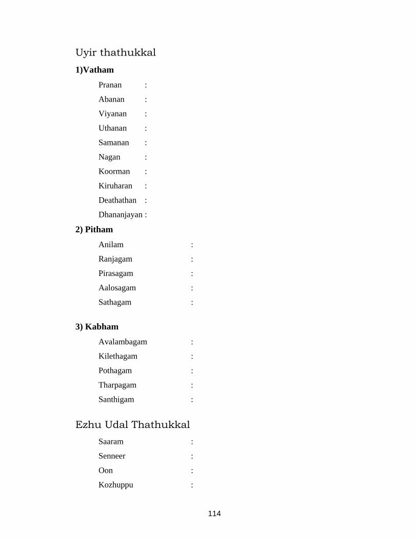
114
Uyir thathukkal
1)Vatham
Pranan :
Abanan :
Viyanan :
Uthanan :
Samanan :
Nagan :
Koorman :
Kiruharan :
Deathathan :
Dhananjayan :
2) Pitham
Anilam :
Ranjagam :
Pirasagam :
Aalosagam :
Sathagam :
3) Kabham
Avalambagam :
Kilethagam :
Pothagam :
Tharpagam :
Santhigam :
Ezhu Udal Thathukkal Saaram :
Senneer :
Oon :
Kozhuppu :

115
Enbu :
Majjai :
Sukkilam / Suronitham :
E N V A G AI T H E R V U G AL
MEI KURI (SPARISM)
Examination of the Skin
Inspection
Colour of the Skin
Eruptions
Haemorrhages
Ulcers, excoriations, fissures etc.
Boils, carbuncles, scars, trophic changes etc.
Eruption
Types of rashes
Maccular
Roseolar
Erythematous
Papular
Pustular
Lenticular
Nodular
Vesicular
Bullous
Wheals
Burrows
Blackheads

116
Plaques
Scales
Ulcers
Duration
Mode of onset
Associated pain
Size and pain
Nature of the floor
Character of the edge
Discharge
Tenderness
Surrounding skin
Lymphnodes
Pruritis
Infestation
Skin diseases
Metabolic & endocrine
Hepatic disorders
Renal diseases
Blood diseases
Examination of the hair
Falling of the hair
Patchy loss of hair
Loss of hair in temporal region
Characteristic features of the hair
Sweat
Physiological / Pathological

117
Lymphglands
Site
Shape
Size
Consistency
Mobility
Tenderness
Examination of the nails
Examination of the Head, neck, Face
Skull
Size
Shape
Face
Eyebrows
Eye lids & Eye lashes
Nose
Lips
Ears
Neck
Examination of the Chest Shape and Size
Movements
Rate of respiration
Breath Sounds : Normal / Abnormal
Heart Rate & Sounds

118
Examination of the Breast
Examination of the Abdomen Shape
Size
Examination of the Genital Organs
Examination of the Extermitis
Upper & Lower Limb : General Examinations
Special Examinations
Tests for Tone, Power & reflex
NIRAM Colour of the skin, Hair, Nail, Teeth, Tongue, Gums
Sputum – Normal / Abnormal
MOZHI Larynx
Congenital
Acquired
Traumatic
Tongue
Congential Abnormalities
Ear : Deafness
Palate : Cleft palate
VIZHI

119
Examination of Eye
Visual acuity
Visual field
Colour sense
Pupil
Size
Equality
Regularity
Reaction of light accommodation
NAA Colour
Size
Shape
IRU MALAM Malam
I. Macroscopic Examination
Amount
Colour
Odour
Consistency
Abnormal Constituents
II. Microscopic Examination
III. Chemical Examination
Siruneer
Quantity
Colour & Transparency
Specific Gravity

120
Deposit
NAADI The state of vatha, pitha and kabha naadi.
Examination of Pulse & its Indication
Rate
Rhythm
Volume
Force &
Character
Noi kanippu
MODERN ASPECTS
ANNEXURE – II
General Examination
Consciousness : General Appearance :
State : Nourishment :
Weight : Facies :
Height : Jaundice :
Skin Changes : Engorged venis :
Anaemia : Clubbing :
Cyanosis : JVP :
Pedal Odema : Koilonychia :
Abdominial distension : Brittle Nail :
Congential anomally :
Lymphadenopathy :
Pluse Rate Rhythm Volume Character
(Rt) (Lt)
Blood Presure : mm/Hg Upper limb ------- -------
Lower limb ------- -------

121
Respiratory Rate:
Systematic Examination
Cardiovascular System :
Respiratory System :
Gastro intestinal System :
Central nervous System :
Laboratory Investigations
Blood
TC : MCV :
DC: P, L, E, B, M : MCH :
Hb% : MCHC :
ESR Serum Protein :
1/2hr : Serum Cholestrol :
1hr : Blood Urea :
RBC Count : Serum Iron :
Platelet Count : Serum Ferritin :
Reticulocyte Count : Serum TIBC :
PCV : Peripheral Blood Smear :
Motion
Ova :
Cyst :
Occult blood :
Urine
Albumin :
Sugar :
Deposits :
Bile Salt :
Bile Pigment :

122
Special Investigation
Barium meal and endoscopy :
Bone marrow examination :
Skiagram :
Sputum for AFB :
Radiological investigation :
Ophthalmoscopic examination :
E.C.G.
Etc. :
Case Summary :
Fate of the Disease :
Line of treatment :

123
BIBLIOGRAPHY
1. Thotra kirama aaraichiyum siddha maruthuva varalarum – Dr. C.S.
Uthamarayan, H.P.I.M.
2. Siddha maruthuvanga surukkam
3. Noi illa neri – Dr. K. Thurairasan, H.P.I.M.
4. Siddha maruthuva noi naadal noi mudhal naadal thirattu – Dr. M.
Shanmuga Velu, H.P.I.M.
5. Siddha maruthuvam – Dr. K.N. Kuppusamy Mudaliyar, H.P.I.M.
6. Theraiyar neerkuri neikuri vilakkam
7. Udal koorugal – Dr. R. Thiyagarajan, L.I.M.
8. Udal thathuvam – Dr. D.M. Venugopal, H.P.I.M.
9. Yugimuni vaithiya sinthamani
10. T.V. Sambasivam pillai dictionary
11. Kambar Tamil Agaraathi
12. Gray’s anatomy
13. Human anatomy – B.D.Chaurasia
14. Fundamentals of human anatomy – A.S. Moni
15. Text book of medical physiology – Guyton & Hall
16. Essentials of medical physiology – K.Sembulingam, Prema
Sembulingam
17. Text book of medicine – Dr. K.V. Krishnadoss
18. Harrison’s principles of medicine
19. Davidson’s principles and practice of medicine

124
20. Oxford test book of medicine
21. Text book of medicine - P.C. Dos
22. Practical medicine – P.J. Mehta’s
23. Manual of practical medicine – A. Alagappan
24. Golwalla Medicine for students
25. Text book of pathology – Dr. Harsh Mohan
26. Robbin’s pathologic basis of disease
27. Hutchinson’s clinical methods
28. How to examine the patient


HEART - ANATOMY

CONGESTIVE HEART FAILURE

Chest x-ray pa view- normal
case no 8 cardiomegaly withcase no : 8 – cardiomegaly with pericardial effusion


no
rmal
Ecg
-n