open access research current treatments in diabetic
TRANSCRIPT

Current treatments in diabeticmacular oedema: systematic reviewand meta-analysis
John Alexander Ford,1 Noemi Lois,2 Pamela Royle,3 Christine Clar,4
Deepson Shyangdan,3 Norman Waugh3
To cite: Ford JA, Lois N,Royle P, et al. Currenttreatments in diabeticmacular oedema: systematicreview and meta-analysis.BMJ Open 2013;3:e002269.doi:10.1136/bmjopen-2012-002269
▸ Prepublication history forthis paper are availableonline. To view these filesplease visit the journal online(http://dx.doi.org/10.1136/bmjopen-2012-002269).
Received 26 October 2012Accepted 1 February 2013
This final article is availablefor use under the terms ofthe Creative CommonsAttribution Non-Commercial2.0 Licence; seehttp://bmjopen.bmj.com
1Department of PopulationHealth and Primary Care,Faculty of Medicine andHealth Sciences, NorwichMedical School, University ofEast Anglia, Norwich, UK2Centre for Vascular andVisual Sciences, QueensUniversity, Belfast, UK3Warwick Evidence, Divisionof Health Sciences, WarwickMedical School, Coventry, UK4Researcher in SystematicReviews, Berlin, Germany
Correspondence toDr John Alexander Ford;[email protected]
ABSTRACTObjectives: The aim of this systematic review is toappraise the evidence for the use of anti-VEGFdrugs and steroids in diabetic macular oedema (DMO)as assessed by change in best corrected visualacuity (BCVA), central macular thickness and adverseeventsData source: MEDLINE, EMBASE, Web of Sciencewith Conference Proceedings and the Cochrane Library(inception to July 2012). Certain conference abstractsand drug regulatory web sites were also searched.Study eligibility criteria, participants andinterventions: Randomised controlled trials wereused to assess clinical effectiveness and observationaltrials were used for safety. Trials which assessedtriamcinolone, dexamethasone, fluocinolone,bevacizumab, ranibizumab, pegaptanib or aflibercept inpatients with DMO were included.Study appraisal and synthesis methods: Risk ofbias was assessed using the Cochrane risk of bias tool.Study results are narratively described and, whereappropriate, data were pooled using random effectsmeta-analysis.Results: Anti-VEGF drugs are effective compared toboth laser and placebo and seem to be more effectivethan steroids in improving BCVA. They have beenshown to be safe in the short term but require frequentinjections. Studies assessing steroids (triamcinolone,dexamethasone and fluocinolone) have reported mixedresults when compared with laser or placebo. Steroidshave been associated with increased incidence ofcataracts and intraocular pressure rise but requirefewer injections, especially when steroid implants areused.Limitations: The quality of included studies variedconsiderably. Five of 14 meta-analyses had moderateor high statistical heterogeneity.Conclusions and implications of key findings:The anti-VEGFs ranibizumab and bevacizumab haveconsistently shown good clinical effectiveness withoutmajor unwanted side effects. Steroid results have beenmixed and are usually associated with cataractformation and intraocular pressure increase. Despitethe current wider spectrum of treatments for DMO,only a small proportion of patients recover good vision(≥20/40), and thus the search for new therapies needsto continue.
INTRODUCTIONDiabetic macular oedema (DMO) is a com-plication of diabetic retinopathy and aleading cause of blindness. The prevalenceof DMO is likely to increase with morepeople suffering from diabetes.1 IncreasingDMO has significant implications forpatients, healthcare providers and widersociety. Laser has been the mainstay of treat-ment, but recently antivascular endothelialgrowth factor (anti-VEGF) drugs and steroidshave been introduced as potential alterna-tives to laser photocoagulation.
Burden of diseaseDiabetic retinopathy is present at the time ofdiagnosis of diabetes mellitus in 0–30% ofindividuals.2 The incidence is estimated tobe 2.3/100 person-years for the overall dia-betic population and 4.5 for patients oninsulin therapy.3 There is good evidence thatprogression to DMO is associated with
ARTICLE SUMMARY
Article focus▪ To review the evidence for triamcinolone, dexa-
methasone, fluocinolone, bevacizumab, ranibizu-mab, pegaptanib and aflibercept in the treatmentof diabetic macular oedema.
Key messages▪ The anti-VEGFs ranibizumab and bevacizumab
have consistently shown good clinical effective-ness in the short term without major unwantedside effects.
▪ Steroid results have been mixed and are usuallyassociated with cataract formation and IOPincrease.
Strengths and limitations of this study▪ A robust, detailed review of the literature has
been undertaken and, when appropriate, datahave been combined in meta-analysis.
▪ The quality of studies included variedconsiderably.
Ford JA, Lois N, Royle P, et al. BMJ Open 2013;3:e002269. doi:10.1136/bmjopen-2012-002269 1
Open Access Research
on Decem
ber 1, 2021 by guest. Protected by copyright.
http://bmjopen.bm
j.com/
BM
J Open: first published as 10.1136/bm
jopen-2012-002269 on 1 March 2013. D
ownloaded from

duration of disease,4–7 poor glycaemic control8 and, intype 2 diabetes, the need for insulin,9 though the needfor insulin therapy is more a marker for duration andpoor control.The number of people with DMO is likely to increase
as diabetes becomes more common. Some reports havesuggested a decrease in progression to severe visual lossbetween 1975–1985 and 1986–2008 in a combined popu-lation of types 1 and 2.10 Regular screening for retinop-athy and better glycaemic control are thought to havereduced the progression to severe visual loss. Diabeticretinopathy is associated with a reduced quality of life.Compared with all diabetic complications, blindness wasperceived to be the third worst health state after a majorstroke and amputation.11
In the USA, the presence of DMO at diagnosis is asso-ciated with 29% additional costs within the first 3 yearscompared with individuals without retinopathy at diag-nosis.12 In 2010, the estimated healthcare costs for DMOin England were £92 million, with £65.6 million beingspent on hospital treatment and related costs.13
Visual impairment results in increased welfare costs,early retirement and costs of home help and carers.14 InEngland in 2010 (total population 52.23 million), theestimated population with diabetes was 2.34 million; theabove social costs were estimated to be £11.6 million forDMO.13
Overview of pathophysiologyDMO is caused mainly by disruption of the blood-retinalbarrier. The complex pathway that leads to this disruptionhas been previously described in this journal.15 Sustainedhyperglycaemia causes a multifactorial cascade of physio-logical processes, involving increased permeability, cytokineactivation, altered blood flow, hypoxia and inflammation.Vascular endothelial growth factor-A (VEGF-A) is a majorcontributor to the inflammatory process and, in particular,to angiogenesis and permeability.16 Hypoxia caused bymicrovascular disease stimulates the release of VEGF-A toaid perfusion. There are six major isoforms of VEGF-A:121, 145, 165, 183, 189 and 206. In addition to causingwidespread microvascular injury, there is now evidence thathyperglycaemia results in preceding neuronal dysfunction,which may contribute to visual loss.17
Overview of current treatmentsLaser photocoagulation has been the mainstay of treat-ment for DMO. The landmark Diabetic RetinopathyStudy18 and the Early Treatment Diabetic RetinopathyStudy (ETDRS)19 20 demonstrated its clinical effective-ness. However, although laser photocoagulation wasclearly effective in preserving vision, it was less successfulin restoring it, once lost. Furthermore, patients withperifoveal ischaemia are not amenable to this form oftherapy. In EDTRS, although laser was shown to reducethe risk of moderate visual loss (a loss of three ETDRSlines) by 50%, visual acuity improved in only 3% ofpatients.20 However, in some recent trials, laser has
improved the proportion of patients with more than orequal to 10 letters by 7–31%.21–24 In addition, laser isnot without side effects. Foveal burns, visual fielddefects, retinal fibrosis and laser scars have beenreported.25 Over the following decade it became appar-ent that certain patients suffered severe visual lossdespite aggressive treatment.26
Steroids and anti-VEGF drugs are newer treatments inDMO. Intravitreal corticosteroids have potent anti-inflammatory effects. Triamcinolone (Kenalog) is notlicensed for eye use but has been used to treat DMO forover 10 years. Triamcinolone (Trivaris), recently, waslicensed for eye use. The development of intravitrealimplants has allowed sustained release formulations.Fluocinolone acetonide (Iluvien, Alimera Sciences) anddexamethasone (Ozudex, Allergan) are implants thathave been introduced recently.Anti-VEGF agents have shown efficacy compared with
laser. Bevacizumab (Avastin, Genenetch/Roche) is amonoclonal antibody that targets all VEGF isoforms.Although being developed for colorectal cancer, it iswidely used off-label, as an intravitreal treatment formacular oedema of different aetiologies. Ranibizumab(Lucentis, Genentech/Roche) is a fragment of the beva-cizumab antibody (molecular weight of ranibizumab48.4 KDa compared with 149 KDa for bevacizumab). Itwas designed specifically for use in the eye. Ranibizumabis considerably more expensive than bevacizumab (theestimated cost of ranibizumab is $2000/dose comparedwith $50 for bevacizumab).27 Pegaptanib (Macugen,Eyetech Pharmaceuticals/Pfizer) is a PEGylated aptamer,with a high affinity to the VEGF isoform 165, and wasapproved for the treatment of exudative AMD in 2004.Aflibercept (Regeneron/Bayer HealthCare) is a recentaddition to the anti-VEGF class that targets all forms ofVEGF-A and placental growth factor.
Aim of the reviewThe aim of this review is to provide clinicians with anup-to-date overview of current intraocular drug treat-ments for DMO. It is hoped that the information con-tained herein will assist clinicians to present their patientswith the best evidence supporting each treatment, includ-ing possible complications. In addition, this review maybe helpful to policy makers. The review focuses on thecurrent evidence for the use of anti-VEGF drugs and ster-oids to treat DMO, as assessed by change in best cor-rected visual acuity (BCVA) (mean and proportion withmore than two lines improvement), central macularthickness (CMT), as determined by optical coherencetomography (OCT), and their adverse events.
EVIDENCE ACQUISITIONA systematic literature search was performed. The data-bases searched included MEDLINE, EMBASE, Web ofScience with Conference Proceedings and the Cochrane
2 Ford JA, Lois N, Royle P, et al. BMJ Open 2013;3:e002269. doi:10.1136/bmjopen-2012-002269
Current treatments in diabetic macular oedema
on Decem
ber 1, 2021 by guest. Protected by copyright.
http://bmjopen.bm
j.com/
BM
J Open: first published as 10.1136/bm
jopen-2012-002269 on 1 March 2013. D
ownloaded from

Library. The dates searched were from the inception ofeach database until July 2012.The search terms combined the following key words:ranibizumab or lucentis or bevacizumab or avastin or
pegaptanib or macugen or aflibercept or vegf trap-eyeor steroid* or corticosteroid* or dexamethasone or fluo-cinolone or triamcinolone or anti-VEGF* or anti-vascular endothelial growth factor*
AND
DMO or diabetic macular edema or diabetic retinopathyor diabetic maculopathy
AND
(masked or sham or placebo OR control group orrandom*) OR (systematic review or meta-analysis) OR(risk or safety or adverse or harm or pharmacovigilanceor side-effect* or precaution* or warning* or contraindi-cation* or contra-indication* or tolerability or toxic)The meeting abstracts of the Association for Research
in Vision and Ophthalmology, the American DiabetesAssociation (2002–2012) and the European Associationfor the Study of Diabetes were searched from 2002 to2012.In addition, the web sites of the European Medicines
Agency and the US Food and Drug Association weresearched for data on registration status and safety.Clinicaltrials.gov and the EU Clinical Trials Register weresearched in July 2012 for data on ongoing research.Full details of the searches are shown in appendix 1.Randomised controlled trials (RCT) were used to
evaluate clinical effectiveness. Safety was assessedthrough both RCTs and observational studies.RCTs were included provided that they (1) addressed
the use of triamcinolone, dexamethasone, fluocinolone,bevacizumab, ranibizumab, pegaptanib or aflibercept inpatients with DMO, (2) had a minimum follow-up of6 months and (3) had a minimum of 25 eyes per studyarm. Studies were excluded if they (1) evaluated laseronly, (2) assessed the effect of the aforementioned treat-ments in macular oedema due to other retinal diseases(instead of DMO), (3) used only a single dose, (4) werecombined with a surgical intervention or (5) publishedstudies in languages other than English. There were noexclusions based on drug dose. Trials were excluded ifthey evaluated combined drug treatment with surgery orsystemic treatment.Search results were screened by two independent
authors ( JF and PR/DS). Data were extracted by oneauthor (CC) and checked by a second ( JF). Dataextracted included inclusion/exclusion criteria, baselinedemographics, BCVA expressed as a change in logMAR/ETDRS letters or proportion of participants with morethan two or three lines BCVA improvement, CMT andadverse events. Risk of bias was assessed using theCochrane risk of bias tool.Studies were assessed for similarity in study popula-
tion, interventions (dose and frequency), outcomes and
time to follow-up, with a view to including similar studiesin a meta-analysis. Conference abstracts were excludedfrom the meta-analysis because their quality and detailedmethodology were not clear. A difference of 6 monthswas allowed between study follow-ups because of thepotential heterogeneity from disease progression anddifferences in the number of doses prescribed. If salientdata were not reported, such as SDs, data were sought bypersonal communication with authors. Data were ana-lysed using Review Manager software. If data from mul-tiple time-points were available, the primary end-pointdata were used. Data were entered by one author ( JF)and double-checked by a second (DS). Mean differenceswere calculated for change in BCVA and CMT and ORswere calculated for proportion of participants with morethan two lines improvement. The 95% CIs were calcu-lated for all outcomes. Statistical heterogeneity was mea-sured through I2 scores. A score of less than 30% wasconsidered as low heterogeneity, a score of more than70% was considered as high heterogeneity and scoresbetween 30% and 70% were considered as moderate.A random effects model was used throughout. Therandom effects model assumes variability betweenstudies and therefore models uncertainty into themeta-analysis. Fixed assumes no variability. Generallyspeaking, the random effects model results in wider CIs.
RESULTSThe literature search identified 430 unique articles forpossible inclusion, as shown in figure 1. In total, 328 arti-cles were excluded on the basis of title and abstract,leaving 102 full papers to be read. Fifty-one of these arti-cles were excluded; the reasons for their exclusion aresummarised in table 1. Fifty-one articles from 29 studiesmet the inclusion criteria and were included in thereview; these are described in tables 3–16. Seven studieswere suitable for meta-analysis.
Study qualityThe quality of the included studies was, in general, goodas is shown in table 2. (Note that the meeting abstractswere not quality assessed, owing to the lack of detailsreported on the methods.) Most studies adequatelydescribed sequence generation, except in three studieswhere it was unclear.28–30 However, allocation conceal-ment was poorly described throughout, with only eightreports addressing this issue appropriately.31–38
Reporting of masking also varied. A number of studiesmasked patients using sham injection or shamlaser.21 24 29 31 33 36 38 39 40 Various studies reported thatmasking of patients was impossible. Assessors, wherereported, were masked. In two studies, incomplete out-comes were not addressed.31 41 Baseline characteristicswere consistent within study treatment arms.Administration of laser followed the ETDRS protocol, ora modified version, in all studies that described laseradministration.21–24 28 30 33 34 42 43 Two studies, both
Ford JA, Lois N, Royle P, et al. BMJ Open 2013;3:e002269. doi:10.1136/bmjopen-2012-002269 3
Current treatments in diabetic macular oedema
on Decem
ber 1, 2021 by guest. Protected by copyright.
http://bmjopen.bm
j.com/
BM
J Open: first published as 10.1136/bm
jopen-2012-002269 on 1 March 2013. D
ownloaded from

available only as meeting abstracts, did not report thelaser administration details.44 45
Intravitreal anti-VEGFsThe characteristics of all published studies includingdesign, inclusion/exclusion criteria, intervention, out-comes and their timing are shown in tables 3–8. Safetydata for each drug are shown in tables 9–16.
RanibizumabNine RCTs have evaluated ranibizumab as a potentialnew treatment for patients with DMO (tables 3 and 8);seven were sponsored by industry, and two were led byindependent investigators)(table 7).21 46 READ-2 was thefirst large RCT (n=126).28 47 It compared ranibizumab(0.5 mg) alone, and ranibizumab in combination withlaser and laser alone. At 6 months, BCVA had improvedsignificantly in the ranibizumab alone group comparedwith laser alone or ranibizumab plus laser. Addition oflaser to ranibizumab did not provide additional BCVA
gain. REVEAL (n=396) compared ranibizumab (0.5 mg)with ranibizumab plus laser and laser alone.48 At12 months, both ranibizumab arms resulted in a statistic-ally significantly better improvement in BCVA comparedto laser alone. The addition of laser did not conferfurther benefit.Within the past 2 years, the results of RESOLVE,36
RESTORE24 and RISE and RIDE38 have been publishedin peer-reviewed journals. RESTORE (n=345) rando-mised similar groups as the READ-2 study (ranibizumab(0.5 mg) alone, laser alone and ranibizumab plus laser);outcomes were evaluated at 12 months. Ranibizumabimproved mean BCVA, with laser providing no additionalbenefit. Two-year extended follow-up suggested thatthese results continued.49 RESOLVE (n=151) comparedtwo doses of ranibizumab (0.3 and 0.5 mg) with shaminjection. The greatest improvement in BCVA at12 months was in the 0.3 mg group (11.8 letter gain)compared to the 0.5 mg group (8.8 letter gain) or shaminjection (1.4 letter loss). In this study, rescue laser was
Figure 1 PRISMA flow diagram.
4 Ford JA, Lois N, Royle P, et al. BMJ Open 2013;3:e002269. doi:10.1136/bmjopen-2012-002269
Current treatments in diabetic macular oedema
on Decem
ber 1, 2021 by guest. Protected by copyright.
http://bmjopen.bm
j.com/
BM
J Open: first published as 10.1136/bm
jopen-2012-002269 on 1 March 2013. D
ownloaded from

allowed after 3 months of treatment, if BCVA haddecreased by 10 letters or more, or if the investigator con-sidered the macula not to be flat as assessed by OCT.Only 4.9% of the ranibizumab group required rescuelaser, compared with 34.7% in the sham injection group.READ-2 and RESTORE were suitable for pooling
through meta-analysis and, when doing so, it was foundthat ranibizumab statistically significantly improved meanBCVA compared with laser (figure 2). In regard to the pro-portion of patients gaining more than or equal to 15letters, individual trials showed a statistically significant dif-ference between laser and ranibizumab but when thesetwo trials were pooled using a random effects model, theresult was no longer statistically significant. When a fixedeffects model was used, the result was statistically significant(figure not shown). Adding laser to ranibizumab did notadd any significant benefit (figure 3). In fact, the meanchange in BCVA and the proportion of patients with morethan 15 letter gain favoured, although not statistically sig-nificantly so, ranibizumab alone compared with ranibizu-mab plus laser. This was probably a chance effect.RISE (n=377) and RIDE (n=382) were identical in
design. The study arms are similar to those in theRESOLVE study, 0.3 or 0.5 mg ranibizumab comparedwith sham. In the RISE study, the proportion of patientswith 15 or more letter gain was greatest in the 0.3 mggroup at 24 months, whereas in the RIDE study this wasgreatest in the 0.5 mg group. In the DRCRN trial(n=854), Elman and colleagues compared ranibizumab(0.5 mg) plus prompt (within 3–10 days post ranibizu-mab) or deferred (≥24 weeks) laser with sham injectionplus prompt laser, or triamcinolone (4 mg, Trivaris) plusprompt laser (table 8). At 1 year, both ranibizumabgroups reported greater gains in mean BCVA changethan triamcinolone or laser alone. Interestingly, at2 years (n=628), the proportion of patients with 10 ormore letter gain was not statistically significantly differ-ent between ranibizumab plus prompt laser and laseralone groups, but was statistically significant in the rani-bizumab plus deferred laser compared with laser alonecomparison. The reason for this is not clear.READ-3 (n=152) has been published in abstract form
and compared monthly injections of intravitreal ranibi-zumab high dose (2.0 mg) and low dose (0.5 mg).50 At6 months, there was no statistically significant differencein BCVA between groups.One study (n=63), published in abstract form, was
identified which directly compared monthly injectionsof ranibizumab (0.5 mg) with bevacizumab (1.5 mg).51
At 48 weeks, the authors found no statistically significantdifference between bevacizumab and ranibizumab.RESTORE, READ-2 and DRCRN (12 month data used)
were suitable for pooling through meta-analysis to compareranibizumab plus laser and laser alone (figure 4).Ranibizumab plus laser resulted in a statistically signifi-cantly greater change in mean BCVA, proportion ofpatients with more than 15 letter gain and CMT reductionversus laser alone.
Table 1 List of excluded studies
Study Reason
Active comparator trials
Cho et al87 Single dose
DRCRN 2010
(Googe et al)88<6 months f/u
Faghihi et al89 Single dose
Figueroa et al90 Single dose
Isaac et al91 Single dose
Paccola et al92 Single dose
Prager et al93 <25 pts per arm
Ozturk et al94 Non-RCT
Marey and Ellakwa95 <6 months
Shahin and El-Lakkany96 Single dose
Pegaptanib
Loftus et al97 Quality of life data
Ranibizumab
Ferrone and Jonisch98 <25 pts per arm
Bevacizumab
Solaiman et al99 Single dose
DRCRN—Scott et al100 <25 pts per arm
Lee101 Non-RCT
Isaac et al91 Single dose
Trimacinolone
Audren et al102 Single dose (dosing study)
Audren et al103 Single dose
Avitabile104 Mixed RVO and DMO
Bandello et al105 Case report+PDR
Bonini et al106 Single dose injection technique
Cellini et al107 Single injection PSTI
Cardillo et al108 Single injection PSTI
Chung et al109 Single injection PSTI
Dehghan et al110 Single dose
DRCRN—Chew et al 111 <25 pts per arm
Gil et al112 <25 pts per arm
Entezari et al113 <6 months
Hauser et al114 Single dose
Jonas et al115 Single dose
Joussen et al116 Study protocol
Avci and Kaderli117 Anaesthetic technique
Kang et al118 Single dose
Kim et al119 Single injection and CME
Lam et al120 Single injection
Lee121 Single injection
Maia et al122 Single dose
Massin et al123 Single dose
Mohamed et al124 Post hoc analysis
Nakamura et al125 Single dose
Spandau et al126 Single dose
Tunc127 <6 months
Verma et al128 Single dose
Wickremasinghe et al129 Single dose
Yalcinbayir et al130 Single dose
Dexamethasone
Haller et al131 <6 months
Haller et al132 <25 pts per arm
Kuppermann et al 133 Mixture of macular oedema
causes
Boyer et al134 Non-randomised
Fluocinolone
Campochiaro et al135 <25 pts per arm
Diclofenac
Elbendary71 <35 pts per arm
CME, cystoid macular edema; DMO, diabetic macular oedemaPDR, proliferative diabetic retinopathy; PSTI, posterior subtenoninjection; RVO, retinal vein occlusion.
Ford JA, Lois N, Royle P, et al. BMJ Open 2013;3:e002269. doi:10.1136/bmjopen-2012-002269 5
Current treatments in diabetic macular oedema
on Decem
ber 1, 2021 by guest. Protected by copyright.
http://bmjopen.bm
j.com/
BM
J Open: first published as 10.1136/bm
jopen-2012-002269 on 1 March 2013. D
ownloaded from

Table 2 Study quality
Study (author
and year)
Adequate
sequence
generation
Allocation
concealment Masking
Incomplete
outcome data
addressed
Free of
selective
reporting
Free of other bias
(eg, similarity at
baseline, power
assessment) Funder
Anti-VEGFs
Ranibizumab
READ-2 Study28 47 Unclear Unclear Unclear Yes (91.3%
completion)
Yes Comparison groups
similar at baseline;
power analysis not
mentioned
Juvenile Diabetes
Research Foundation,
Genentech Inc
RESOLVE Study
(Massin et al)36Yes Yes Yes (patients and
outcome assessors)
Yes (82%
completion in
sham arm,
90.2% with
ranibizumab)
Yes Comparison groups
similar at baseline;
power analysis unclear
Novartis Pharma,
Switzerland
RESTORE Study
(Mitchell et al)24Yes Unclear Yes (patients,
outcome assessors)
Yes
(87.3–88.3%
completion)
Yes Comparison groups
similar at baseline;
power analysis carried
out (power adequate
for VA changes)
Novartis Pharma,
Switzerland
RISE and RIDE
(Nguyen et al)38Yes Yes Yes (patients, treating
physician masked to
assigned dose of
ranibizumab)
Yes (2 year
study completed
by 83.3% of
patients in RISE
and by 84.6% in
RIDE)
Yes Comparison groups
similar at baseline; ITT
analysis; power
analysis carried out
(power adequate for
primary endpoint)
Genentech Inc
Bevacizumab
BOLT Study
(Michaelides
et al)23 52
Yes Unclear Partial (outcome
assessors, not
patients)
Yes (97.5%
completion)
Yes Comparison groups
similar at baseline
(except laser group
had longer duration of
clinically significant
DMO); power analysis
carried out (power
adequate for VA
changes)
Moorfields Special
Trustees, National Institute
for Health Research
Faghihi et al53 Yes Unclear Yes (patient Yes (100%
completion)
Yes Comparable groups at
baseline
Not specified
Lam et al35 Yes Yes Yes (patients and
technicians assessing
BCVA, OCT and IOP)
Yes (92.3%
follow-up at
6 months)
Yes Comparison groups
similar at baseline;
power analysis carried
out (power adequate
for CMT changes)
Supported in part by the
Action for Vision Eye
Foundation Hong Kong
(charity)
Continued
6Ford
JA,LoisN,Royle
P,etal.BMJOpen
2013;3:e002269.doi:10.1136/bmjopen-2012-002269
Curre
nttre
atm
ents
indiabetic
macularoedema
on December 1, 2021 by guest. Protected by copyright. http://bmjopen.bmj.com/ BMJ Open: first published as 10.1136/bmjopen-2012-002269 on 1 March 2013. Downloaded from

Table 2 Continued
Study (author
and year)
Adequate
sequence
generation
Allocation
concealment Masking
Incomplete
outcome data
addressed
Free of
selective
reporting
Free of other bias
(eg, similarity at
baseline, power
assessment) Funder
Pegaptanib
Cunningham et al/
Adamis et al39 57Yes Unclear Yes (patients and
outcome assessors)
Yes (95%
completion)
Yes Comparison groups
similar at baseline;
acknowledge lack of
power to detect
differences between
doses of pegaptanib
Eyetech Pharmaceuticals
Inc, New York, and Pfizer
Inc, New York
Sultan et al40 Yes Unclear Yes (patients and
outcome assessors)
Yes
(69.9–73.8%
completion)
Yes Comparison groups
similar at baseline;
power analysis carried
out (power adequate
for VA changes)
Pfizer Inc, New York
Aflibercept
Da Vinci et al30 58 Unclear
(predetermined
randomisation
scheme)
Unclear Yes (patients) Yes (85%
completion)
Yes Comparison groups
similar at baseline,
power calculation
completed
Regeneron
Pharmaceuticals, Inc,
New York
Steroids
Dexamethasone
Haller et al59 Yes Unclear Yes (patients to
dexamethasone dose,
outcome assessors)
Yes (92%
completion)
Yes Comparison groups
similar at baseline;
power analysis carried
out, but study not
powered to detect
differences in
subgroups
Oculex Pharmaceuticals
Inc
Fluocinolone
FAME Study
(Campochiaro et al)29 60
Unclear Unclear Partial (patients,
masking of outcome
assessment not
mentioned)
Yes
(drop-out rate
19.0–22.7%)
Yes Comparison groups
similar at baseline;
power analysis not
mentioned
Alimera Sciences Inc,
Atlanta, Georgia; Psivida
Inc, Watertown,
Massachusetts
Pearson et al43 Yes Unclear Third party masked
design (patient and
investigator not
masked)
No losses to
follow-up
Yes Demographic
characteristics were
similar between implant
and SOC groups;
power calculation done,
study adequately
powered
Bausch & Lomb Inc,
Rochester, New York
Continued
FordJA,Lois
N,RoyleP,etal.BM
JOpen
2013;3:e002269.doi:10.1136/bmjopen-2012-002269
7
Curre
nttre
atm
ents
indiabetic
macularoedema
on December 1, 2021 by guest. Protected by copyright. http://bmjopen.bmj.com/ BMJ Open: first published as 10.1136/bmjopen-2012-002269 on 1 March 2013. Downloaded from

Table 2 Continued
Study (author
and year)
Adequate
sequence
generation
Allocation
concealment Masking
Incomplete
outcome data
addressed
Free of
selective
reporting
Free of other bias
(eg, similarity at
baseline, power
assessment) Funder
Triamcinolone
DRCR Network
2008 22 61 63 64Yes Unclear Partial (patients to
triamcinolone dose,
outcome assessors
not formally masked
but generally not
aware of participant’s
study group)
Yes (81–86%
completion)
Yes Comparison groups
similar at baseline;
power analysis carried
out (power adequate
for VA changes)
Cooperative agreement
from the National Eye
Institute, National Institute
of Diabetes and Digestive
and Kidney Diseases,
National Institutes of
Health, Department of
Health and Human
Services
Gillies et alSutter
et al32 136–138Yes Yes Yes (patients,
outcome assessors)
Yes (91%
completion
intervention,
83% control)
Yes Comparison groups
similar at baseline (but
limited demographic
data); power analysis
carried out (power
adequate for VA
changes)
Sydney Eye Hospital
Foundation and Juvenile
Diabetes Research
Foundation, New York
Gillies et al33 Yes Yes Yes (patients,
outcome assessors)
Yes (84.5%
completion)
Yes Power analysis carried
out (power adequate
for VA changes)
National Health and
Medical Research
Council, Canberra,
Australia, and the Sydney
Eye Hospital Foundation
Sydney, Australia
Lam et al34 Yes Yes Partial (outcome
assessors)
No losses to
follow-up
Yes Comparison groups
similar at baseline;
power analysis carried
out (power adequate
for CMT changes)
Action for Vision
Foundation, Hong Kong
Ockrim et al/
Sivaprasad et al42 62
Yes Unclear Unclear Yes (94%
completion)
Yes Comparison groups
similar at baseline;
power analysis carried
out (power adequate
for VA changes)
Special Trustees of
Moorfields Eye Hospital
Continued
8Ford
JA,LoisN,Royle
P,etal.BMJOpen
2013;3:e002269.doi:10.1136/bmjopen-2012-002269
Curre
nttre
atm
ents
indiabetic
macularoedema
on December 1, 2021 by guest. Protected by copyright. http://bmjopen.bmj.com/ BMJ Open: first published as 10.1136/bmjopen-2012-002269 on 1 March 2013. Downloaded from

Table 2 Continued
Study (author
and year)
Adequate
sequence
generation
Allocation
concealment Masking
Incomplete
outcome data
addressed
Free of
selective
reporting
Free of other bias
(eg, similarity at
baseline, power
assessment) Funder
Active comparator trials
Ahmadieh et al31 Yes Yes Yes (patients and
outcome assessors)
Unclear Yes CMT lower in control
group at baseline
(p<0.05), other
baseline values similar;
power analysis carried
out (power adequate
for CMT changes)
Not reported
DRCR Network 21 46 Yes Unclear Yes (patients, except
deferred laser group;
outcome assessors);
masking discontinued
after the first year
Yes (1 year
completion for
91–95% of
eyes)
Yes Comparison groups
similar at baseline;
power analysis carried
out (power adequate
for VA changes)
Cooperative agreement
from the National Eye
Institute, National Institute
of Diabetes and Digestive
and Kidney Diseases,
National Institutes of
Health and Human
Services; Ranibizumab
provided by Genentech,
triamcinolone provided by
Allergan Inc; companies
also provided funds to
defray the study’s clinical
site costs
Lim et al55 Yes Unclear Yes (investigators
only)
Yes (7.5% drop
out after
enrolment)
Yes Groups similar at
baseline. The
bevacizumab group
received more
injections
Not reported
Soheilian et al37 41 Yes Yes Yes (patients and
outcome assessors)
Unclear
(36 week
completion for
76–88%)
Yes CMT significantly lower
and VA significantly
better in MPC group at
baseline, other
baseline values similar;
power analysis carried
out (power adequate
for VA changes)
Ophthalmic Research
Centre, Labbafinejad
Medical Center, Tehran
MPC, macular photocoagulation.
FordJA,Lois
N,RoyleP,etal.BM
JOpen
2013;3:e002269.doi:10.1136/bmjopen-2012-002269
9
Curre
nttre
atm
ents
indiabetic
macularoedema
on December 1, 2021 by guest. Protected by copyright. http://bmjopen.bmj.com/ BMJ Open: first published as 10.1136/bmjopen-2012-002269 on 1 March 2013. Downloaded from

Table 3 Ranibizumab trials
Study Participants and baseline values Intervention
Outcome (change
from baseline at
study end)
READ-2 Study (Nguyen
et al)28 47 USA
Multicenter
Design: 3-arm RCT
Follow-up: 6 months, 2-year
extension (no relevant
outcomes as IVR received by
all groups by that time, no
safety outcomes for 2-year
data)
N: 126 eyes of 126 patients
Inclusion criteria: ≥18 years, type 1
or 2 DM, DMO, BCVA 20/40-20/
320, CMT ≥250 µm, HbA1c ≥6%within 12 months before
randomisation; expectation that
scatter laser photocoagulation not
required for 6 months
Exclusion criteria: contributing
causes to reduced BCVA other
than DMO, focal/grid laser within
3 months, intraocular steroid within
3 months, intraocular VEGF
antagonist within 2 months
Age: 62 years
Sex: 52–69% female
Diabetes type: not reported
HbA1c: 7.39–7.77%
Baseline VA: ETDRS letter score
24.85–28.35
Baseline CMT: excess foveal
thickness 198.75–262.52 µm
Comorbidities: not reported
Group 1 (IVR, n=42 eyes): IV
injections of 0.5 mg ranibizumab at
baseline, 1, 3 and 5 months
Group 2 (L, n=42 eyes): focal/grid
laser at baseline and 3 months if
CMT ≥250 µm
Group 3 (IVRL, n=42 eyes): IV
injections of 0.5 mg ranibizumab at
baseline and 3 months, followed by
focal/grid laser treatment 1 week
later
Regimen for all groups: after
6 months, patients could receive IV
injections of ranibizumab no more
than every 2 months or focal/grid
laser no more than every 3 months
if CMT ≥250 µm
Laser Modified ETDRS protocol
was used
At 6 months
BCVA (ETDRS):
BCVA (letters) p Value
IVR +7.24 0.0003 vs L
L −0.43IVRL +3.80 NS vs IVR or
L
Plus ≥3 lines
IVR 22% <0.05 vs L
L 0
IVRL 8%
CMT (OCT):
CMT (µm) p Value
IVR −106.3 All <0.01 vs
baseline, NS for
elimination of ≥50%excess foveal
thickness between
groups
L −82.8IVRL −117.2
READ-3 Study (Do et al) USA50
Design: phase 2, 2-arm RCT
Follow-up: 6 months
N: 152 eyes
Inclusion criteria: NR
Exclusion criteria: NR
Age: NR
Sex: NR
Diabetes type: NR
HbA1c: NR
Baseline VA: Mean BCVA Snellen
equivalent 20/63 in the 2.0 mg
group and 20/80 in the 0.5 mg
group
Baseline CST (central subfield
thickness): 432 µm in the 2.0 mg
group and 441 µm in the 0.5 mg
group
Comorbidities: NR
Group 1 (IVR2.0, n=NR): monthly
injections
Group 2 (IVR0.5, n=NR): monthly
injections
After month 6, eyes evaluated and
additional ranibizumab injections
given on an as needed basis if
DMO still present on OCT.
At 6 months:
BCVA
Mean BCVA
letters gain
p Value
IVR2.0 +7.46 NR
IVR0.5 +8.69 NR
CST CST reduction
IVR2.0 −163.86 µm NR
IVR0.5 −169.27 µm NR
Continued
10Ford
JA,LoisN,Royle
P,etal.BMJOpen
2013;3:e002269.doi:10.1136/bmjopen-2012-002269
Curre
nttre
atm
ents
indiabetic
macularoedema
on December 1, 2021 by guest. Protected by copyright. http://bmjopen.bmj.com/ BMJ Open: first published as 10.1136/bmjopen-2012-002269 on 1 March 2013. Downloaded from

Table 3 Continued
Study Participants and baseline values Intervention
Outcome (change
from baseline at
study end)
RESOLVE Study (Massin
et al)36
Multicenter international
Design: 3-arm
placebo-controlled RCT
Follow-up: 12 months
N: 151 eyes of 151 patients
Inclusion criteria: >18 years, type 1
or 2 DM, clinically significant DMO,
BCVA 20/40–20/160, HbA1c <12%,
decreased vision attributed to
foveal thickening from DMO, laser
photocoagulation could be safely
withheld in the study eye for at
least 3 months after randomisation
Exclusion criteria: unstable medical
status, panretinal laser
photocoagulation performed within
6 months before study entry,
previous grid/laser
photocoagulation except patients
with only mild laser burns at least
1000 µm from the centre of the
fovea performed >6 months
previously
Age: 63–65 (range 32–85) years
Sex: 43.1–49% female
Diabetes type: 96.1–98% type 2
DM
HbA1c: 7.3–7.6 (range 5.3–11.1) %
Baseline VA: ETDRS letter score
59.2–61.2 SD9.0–10.2
Baseline CMT: 448.9–459.5
SD102.8–120.1 µm
Comorbidities: not reported
Group 1 (IVR0.3, n=51 eyes):
0.3 mg (0.05 ml) IV ranibizumab,
3 monthly injections (dose up to
0.6 mg, see below)
Group 2 (IVR0.5, n=51 eyes):
0.5 mg IV (0.05 ml) ranibizumab,
3 monthly injections (dose up to
1.0 mg, see below)
Group 3 (C, n=49 eyes): sham
treatment, 3 monthly injections
Regimen for all groups: after month
1, the injection dose could be
doubled if CMT remained >300 µm
or was >225 µm and reduction in
retinal oedema from previous
assessment was <50 µm; once
injection volume was 0.1 ml it
remained that for subsequent
injections; if treatment had been
withheld for >45 days, subsequent
injections restarted at 0.05 ml;
68.6% of dose doubling with
ranibizumab, 91.8% with sham;
34.7% of rescue laser
photocoagulation in sham group,
4.9% in ranibizumab group
At 12 months
BCVA (ETDRS):
BCVA (letters) p Value
IVR0.3 +11.8 SD6.6 <0.0001 vs C
IVR0.5 +8.8 SD11.0 <0.0001 vs C
C −1.4 SD14.2
Change ≥10 letters
IVR0.3 Gain 72.5%
loss 0
<0.0001 vs C
IVR0.5 Gain 49%
loss 9.8%
0.001 vs C
C Gain 18.4%
loss 24.5%
CMT (OCT):
CMT (µm) p Value
IVR0.3 −200.7 SD122.2 <0.0001 vs C
IVR0.5 −187.6 SD147.8 <0.0001 vs C
C −48.4 SD153.4
RESTORE Study (Mitchell
et al)24 49N: 345 eyes of 345 patients
Inclusion criteria: ≥18 years, type 1
Group 1 (IVR, n=116 eyes): 0.5 mg
IV ranibizumab plus sham laser
At 12 months
BCVA (ETDRS):
Continued
FordJA,Lois
N,RoyleP,etal.BM
JOpen
2013;3:e002269.doi:10.1136/bmjopen-2012-002269
11
Curre
nttre
atm
ents
indiabetic
macularoedema
on December 1, 2021 by guest. Protected by copyright. http://bmjopen.bmj.com/ BMJ Open: first published as 10.1136/bmjopen-2012-002269 on 1 March 2013. Downloaded from

Table 3 Continued
Study Participants and baseline values Intervention
Outcome (change
from baseline at
study end)
Multicenter international
Design: 3-arm RCT
Follow-up: 12 months
or 2 DM, HbA1c ≤10%, visual
impairment due to DMO (eligible for
laser treatment), stable medication
for management of diabetes, BCVA
ETDRS letter score 39–78
Exclusion criteria: concomitant eye
conditions that could affect VA,
active intraocular inflammation or
infection, uncontrolled glaucoma in
either eye, panretinal laser
photocoagulation within 6 months
or focal/grid laser photocoagulation
within 3 months prior to study entry,
history of stroke, hypertension
Age: 62.9–64.0 SD8.15–9.29 years
Sex: 37.1–47.7% female
Diabetes type: 86.4–88.8% type 2
DM
HbA1c: not reported
Baseline VA: ETDRS letter score
62.4–64.8 SD9.99–11.11
Baseline CMT: 412.4–426.6
SD118.01–123.95
Comorbidities: not reported
(median injections 7 (range 1–12),
median sham laser treatments 2
(range 1–5))
Group 2 (IVRL, n=118 eyes):
0.5 mg IV ranibizumab plus active
laser (median injections 7 (range
2–12), median laser treatments 1
(range 1–5))
Group 3 (L, n=111 eyes): laser
treatment plus sham injections
(median sham injections 7 (range
1–12), median laser treatments 2
(range 1–4))
Regimen for all groups: 3 initial
monthly injections, followed by
retreatment schedule; 1 injection
per month if stable VA not reached;
Laser retreatments in accordance
with ETDRS guidelines at intervals
no shorter than 3 months from
previous treatment
BCVA (letters) p Value
IVR +6.1 SD6.43 <0.0001 vs L
IVRL +5.9 SD7.92 <0.0001 vs L
L +0.8 SD8.56
BCVA change categories
IVR Plus ≥10: 37.4%Loss ≥10: 3.5%
<0.0001 vs L
IVRL Plus ≥10: 43.2%Loss ≥10: 4.2%
<0.0001 vs L
L Plus ≥10: 15.5%Loss ≥10: 12.7%
CMT (OCT):
CMT (µm) p Value
IVR −118.7SD115.07
0.0002 vs L
IVRL −128.3SD114.34
<0.0001 vs L
L −61.3 SD132.29
REVEAL Study (Ohji and
Ishibashi )48
Japan Multicenter
Design: phase III
double-masked RCT
Follow-up: 12 months
N: 396 patients
Inclusion criteria: NR
Exclusion criteria: NR
Age: 61.1 years
Sex: NR
Diabetes type: 98.7% with type 2
diabetes
HbA1c: 7.5%
Baseline VA: 58.6 letters
Baseline CMT: 421.9 µm
Comorbidities: NR
Group 1 (IVR 0.5 + sham laser,
n=133): day 1, month 1, 2 and
pro-renata thereafter based on
BCVA
Group 2 (IVR 0.5+ active laser,
n=132): day 1, month 1, 2 and
pro-renata thereafter based on
BCVA
Group 3 (sham injection + active
laser, n=131): day 1, month 1, 2
and pro-renata thereafter based on
BCVA
Active/sham laser photocoagulation
performed according to ETDRS
guidelines at ≥3 month intervals
At 12 months
BCVA:
Mean average
change from
baseline to
months 1–12
p Value
IVR+sham laser +5.9 vs laser <0.0001
IVR+laser +5.7 vs laser <0.0001
Laser+sham +1.4
Mean change
from baseline to
month12 in
BCVA and CRT
IVR+sham laser +6.6; −148.0 µm vs C <0.0001
IVR+laser +6.4; −163.8 µm vs C <0.0001
Laser+sham +1.8; −57.1 µm
Continued
12Ford
JA,LoisN,Royle
P,etal.BMJOpen
2013;3:e002269.doi:10.1136/bmjopen-2012-002269
Curre
nttre
atm
ents
indiabetic
macularoedema
on December 1, 2021 by guest. Protected by copyright. http://bmjopen.bmj.com/ BMJ Open: first published as 10.1136/bmjopen-2012-002269 on 1 March 2013. Downloaded from

Table 3 Continued
Study Participants and baseline values Intervention
Outcome (change
from baseline at
study end)
RISE Study (Brown et al/
Nguyen et al)38 139
USA
Multicenter
Design: 3-arm double-blind
sham-controlled RCT
Follow-up: 24 months
N: 377 eyes of 377 patients
Inclusion criteria: ≥18 years, type 1
or 2 diabetes, BCVA 20/40–20/320,
DMO CMT ≥275 µm
Exclusion criteria: prior vitreoretinal
surgery, recent history (within
3 months of screening) of
panretinal or macular laser in the
study eye, intraocular
corticosteroids or antiangiogenic
drugs, those with uncontrolled
hypertension, uncontrolled diabetes
(HbA1c >12%), recent (within
3 months) cerebrovascular accident
or myocardial infarction
Age: 61.7–62.8 SD8.9–10.0 (range
21–87) years
Sex: 41.6–48% female
Diabetes type: type 1 or 2
HbA1c: 7.7% SD 1.4–1.5; ≤8%(65–68.3%); >8% (31.7%–35%)
Baseline VA: Mean ETDRS letter
score 54.7–57.2; ≤20/200(7.9–13.6%); >20/200 but
<20/40 (72.4–72.8%); ≥20/40(13.6–19.7%)
Baseline CMT: 463.8–474.5 µm
Comorbidities: History of smoking
46.4–51.2%
Group 1 (IVR0.3, n=125 eyes):
0.3 mg IV ranibizumab
Group 2 (IVR0.5, n=125 eyes):
0.5 mg IV ranibizumab
Group 3 (C, n=127 eyes): sham
injection
Regimen for all groups: monthly
injections; need for macular rescue
laser assessed monthly starting at
month 3
At 24 months
BCVA:
Plus ≥15 letters p Value
IVR0.3 44.8% <0.0001 vs C
IVR0.5 39.2% =0.0002 vs C
C 18.1%
Loss of <15
letters
IVR0.3 97.6% =0.0086 vs C
IVR0.5 97.6% =0.0126 vs C
C 89.8%
Snellen
equivalent of
20/40 or better
IVR0.3 60% <0.0001 vs C
IVR0.5 63.2% <0.0001 vs C
C 37.8%
Mean BCVA
gain (letters)
IVR0.3 +12.5 SD14.1 <0.0001 vs C
IVR0.5 +11.9 SD12.1 <0.0001 vs C
C +2.6 SD13.9
CFT:
Mean change
from baseline
p Value
IVR0.3 −250.6 SD212.2 <0.0001 vs C
IVR0.5 −253.1 SD183.7 <0.0001 vs C
C −133.4 SD209.0
RIDE study (Boyer et al/
Nguyen et al)38 140
USA
Multicentre
Design: 3-arm double-blind
sham-controlled RCT
Follow-up: 24 months
N: 382 eyes
Inclusion criteria: ≥18 years, type 1
or 2 diabetes, BCVA 20/40–20/320
and DMO CMT ≥275 µm
Exclusion criteria: prior vitreoretinal
surgery, recent history (within
3 months of screening) of
Group 1 (IVR0.3, n=125 eyes):
0.3 mg IV ranibizumab
Group 2 (IVR0.5, n=127 eyes):
0.5 mg IV ranibizumab
Group 3 (C, n=130 eyes): sham
injection
Regimen for all groups: Patients
At 24 months
BCVA:
More than 15
letters
p Value
IVR0.3 33.6% <0.0001 vs C
IVR0.5 45.7% <0.0001 vs C
C 12.3%
Continued
FordJA,Lois
N,RoyleP,etal.BM
JOpen
2013;3:e002269.doi:10.1136/bmjopen-2012-002269
13
Curre
nttre
atm
ents
indiabetic
macularoedema
on December 1, 2021 by guest. Protected by copyright. http://bmjopen.bmj.com/ BMJ Open: first published as 10.1136/bmjopen-2012-002269 on 1 March 2013. Downloaded from

Table 3 Continued
Study Participants and baseline values Intervention
Outcome (change
from baseline at
study end)
panretinal or macular laser in the
study eye, intraocular
corticosteroids or antiangiogenic
drugs, those with uncontrolled
hypertension, uncontrolled diabetes
(HbA1c >12%), recent (within
3 months) cerebrovascular accident
or myocardial infarction
Age: 61.8–63.5 (range 22–91)
years
Sex: 37–49.1% female
Diabetes type: type 1 or 2
HbA1c: 7.6 SD1.3–1.5; ≤8%(65.8–67.5%); >8% (32.5–34.2%)
Baseline VA: Mean ETDRS letter
score 56.9–57.5
Baseline CMT: 447.4–482.6 µm
Comorbidities: history of smoking
33.6–51.6%
were eligible for rescue macular
laser starting at month 3
Less than 15
letters
IVR0.3 1.6% >0.05 vs C
IVR0.5 3.9% <0.05 vs C
C 8.5%
Snellen
equivalent of
20/40 or better
IVR0.3 54.4% =0.0002 vs C
IVR0.5 62.2% <0.0001 vs C
C 34.6%
Mean BCVA gain (letters)
IVR0.3 +10.9 SD10.4 <0.0001vs C
IVR0.5 +12.0 SD14.9 <0.0001 vs C
C +2.3 SD14.2
CMT:
Mean change
from baseline
p Value
IVR0.3 −259.8 SD169.3 <0.0001 vs C
IVR0.5 −270.7 SD201.6 <0.0001 vs C
C −125.8 SD198.3
Injections are intravitreal unless otherwise noted. BCVA, best corrected visual acuity; C, control; CMT, central macular thickness; CSME, clinically significant macular oedema; DDS,dexamethasone; DIL, dexamethasone followed by laser; DM, diabetes mellitus; DMO, diabetic macular oedema; DP, diastolic pressure; DR, diabetic retinopathy; HR QoL, health-related qualityof life; IOP, intraocular pressure; IV, intravitreal; IVB, intravitreal bevacizumab; IVP, intravitreal pegaptanib; IVR, intravitreal ranibizumab; IVT, intravitreal triamcinolone; IVTL, intravitrealtriamcinolone plus laser; IVVTE, intravitreal VEGF Trap Eye; L, laser; MLT/MPC, macular laser therapy/macular photocoagulation; NEI VFQ-25, National Eye Institute Visual FunctionQuestionnaire-25; NPDR, non-proliferative diabetic retinopathy; NR, not reported; OCT, optical coherence tomography; PDR, proliferative diabetic retinopathy; PRP, panretinal photocoagulation;RCT, randomised controlled trial; SOC, standard of care; SP, systolic pressure; SRFA, fluocinolone; VA, visual acuity; VEGF, vascular endothelia growth factor.
14Ford
JA,LoisN,Royle
P,etal.BMJOpen
2013;3:e002269.doi:10.1136/bmjopen-2012-002269
Curre
nttre
atm
ents
indiabetic
macularoedema
on December 1, 2021 by guest. Protected by copyright. http://bmjopen.bmj.com/ BMJ Open: first published as 10.1136/bmjopen-2012-002269 on 1 March 2013. Downloaded from

Table 4 Bevacizumab studies
Study Participants and baseline values Intervention
Outcome (change from
baseline at study end)
BOLT Study
(Michaelides
et al/Rajendram
et al))23 52 85
UK
Design: 2-arm
RCT
Follow-up:
12 months
N: 80 eyes of 80 patients
Inclusion criteria: ≥18 years, type 1 or 2 DM, BCVA in
the study eye 35–69 ETDRS letters at 4 m (≥6/60 or
≤6/12), center-involving clinically significant DMO with
CMT ≥270 µm; media clarity, papillary dilation and
cooperation sufficient for adequate fundus imaging; a
least 1 prior macular laser therapy; IOP <30 mm Hg;
fellow eye BCVA ≥3/60; fellow eye received no
anti-VEGF in past 3 months and no expectation of
such therapy
Exclusion criteria: (ocular for study eye) macular
ischemia, macular oedema due to causes other than
DMO, coexistent ocular disease affecting VA or DMO,
any treatment for DMO in prior 3 months, PRP within
3 months prior to randomisation or anticipated, PDR,
HbA1c >11%, medical history of chronic renal failure;
any thromboembolic event within 6 months prior to
randomisation, unstable angina, evidence of active
ischemia on ECG; major surgery within 28 days of
randomisation or planned; participation in an
investigational drug trial; systemic anti-VEGF or
pro-VEGF treatment within 3 months of enrolment;
pregnancy, lactation; intraocular surgery within
3 months of randomisation; aphakia; uncontrolled
glaucoma; significant external ocular disease
Age: 64.2 SD8.8 years
Sex: 31% female
Diabetes type: 90% type 2 DM, 10% type 1 DM
HbA1c: 7.5–7.6 SD1.2–1.4%
Baseline VA: ETDRS letter score 54.6–55.7
SD8.6–9.7
Baseline CMT: 481–507 SD121–145 µm
Comorbidities: 19% mild NPDR (level 35), 46%
moderate NPDR (level 43), 19% moderately severe
NPDR (level 47), 13% severe NPDR (level 53), 3%
moderate PDR (level 65), 79–88% phakic
Group 1 (MLT, n=38 eyes):
modified ETDRS macular laser
therapy; reviewed every
4 months up to 52 weeks;
retreatment performed if clinically
indicated by ETDRS guidelines
(median 4 laser treatments)
Group 2 (IVB, n=42 eyes):
1.25 mg (0.05 ml) IV
bevacizumab at baseline, 6 and
12 weeks; subsequent IVB
injections (up to 52 weeks)
guided by an OCT-based
retreatment protocol (median 13
injections)
Laser modified ETDRS protocol,
retreatment by ETDRS
guidelines
At 24 months
BCVA (ETDRS):
BCVA.
mean (SD)
p Value
MLT −0.5 (10.6)
IVB +8.6 (9.1) 0.005 vs
MLT
BCVA gain categories
(letters)
MLT gaining
≥10: 7%losing >15:
4%
IVB gaining
≥10: 49%losing >15:
32%
0.001 vs
MLT
0.004 vs
MLT
CMT (µm,
quartiles)
p Value
MLT −118SD171
IVB −146SD122
0.62 vs
MLT
Lam et al35
Hong Kong
Design: 2-arm
RCT
N: 52 eyes of 52 patients
Inclusion criteria: ≥18 years, type 1 or 2 DM, clinically
significant DMO (slit-lamp biomicroscopy, ETDRS
criteria; leakage confirmed by fluorescein
Group 1 (IVB1.25, n=26 eyes):
1.25 mg bevacizumab (0.05 ml)
Group 2 (IVB2.5, n=26 eyes):
2.5 mg bevacizumab (0.1 ml)
At 6 months
BCVA (ETDRS chart):
Continued
FordJA,Lois
N,RoyleP,etal.BM
JOpen
2013;3:e002269.doi:10.1136/bmjopen-2012-002269
15
Curre
nttre
atm
ents
indiabetic
macularoedema
on December 1, 2021 by guest. Protected by copyright. http://bmjopen.bmj.com/ BMJ Open: first published as 10.1136/bmjopen-2012-002269 on 1 March 2013. Downloaded from

Table 4 Continued
Study Participants and baseline values Intervention
Outcome (change from
baseline at study end)
Follow-up:
6 months
angiography, CMT ≥250 µm on OCT), BCVA ≥1.3ETDRS logMAR units; only patients with diffuse DMO
recruited
Exclusion criteria: macular oedema due to reasons
other than diabetes, significant media opacities,
macular ischemia of ≥1 disk area, vitreomacular
traction, PDR, aphakia, glaucoma or ocular
hypertension, previous anti-VEGF treatment,
intraocular surgery except uncomplicated cataract
extraction (but > 6 months prior), focal DMO, any laser
procedure within previous 4 months, subtenon or
intravitreal triamcinolone injection within 6 months,
pregnancy
Age: 65.3 SD8.9 years
Sex: 46.2% female
Diabetes type: not reported
HbA1c: 7.5 SD1%
Baseline VA: 0.61 SD0.29 logMAR
Baseline CMT: 466 SD127 µm
Comorbidities: not reported
Regimen for all groups:
3 monthly IV injections, topical
0.5% levofloxacin 4×/day for up
to 2 weeks after each injection
BCVA (logMAR) p Value
IVB1.25 0.11
SD0.31
(+5.5
letters)
0.018 vs
baseline,
NS vs
IVB2.5
IVB2.5 0.13
SD0.26
(+6.5
letters)
0.003 vs
baseline
CMT (OCT) CMT (µm) p Value
IVB1.25 96 0.002 vs
baseline,
NS vs
IVB2.5
IVB2.5 74 0.013 vs
baseline
Subgroups:
▸ For patients with previous
DMO treatment (mainly laser):
no significant reduction in
CMT at 6 months (452 µm at
baseline to 416 µm at
6 months, p=0.22); no
significant improvement in
BCVA (0.66 logMAR at
baseline to 0.56 logMAR at
6 months (+5 letters),
p=0.074)
Continued
16Ford
JA,LoisN,Royle
P,etal.BMJOpen
2013;3:e002269.doi:10.1136/bmjopen-2012-002269
Curre
nttre
atm
ents
indiabetic
macularoedema
on December 1, 2021 by guest. Protected by copyright. http://bmjopen.bmj.com/ BMJ Open: first published as 10.1136/bmjopen-2012-002269 on 1 March 2013. Downloaded from

Table 4 Continued
Study Participants and baseline values Intervention
Outcome (change from
baseline at study end)
Faghihi et al53
Iran
Design: 2-arm
RCT
Follow-up:
6 months
N: 80 eyes of 40 patients
Inclusion criteria: Bilateral non-tractional CSME,
10/10> V.A≥1/10, Controlled blood pressure.
Exclusion criteria: Advanced or advanced active PDR,
significant cataract, glaucoma, history of recent
vascular accident (eg, MI, CVA), Previous treatment of
CSME or PDR, or pharmacotherapy for CSME,
macular ischemia and uncontrolled hypertension
Age: 57.7±8 years
Sex: 27.5% females
Diabetes type: NR
HbA1c: 8.42±1.82 g/dl
Baseline VA: 0.326–0.409 (SD 0.279–0.332)
Baseline CMT: 277 um–287 um (SD 78–98)
Comorbidities: not reported
Group 1 (IVB, n=40 eyes):
1.25 mg bevacizumab
Group 2 (IVB+MPC, n=40 eyes):
1.25 mg bevacizumab
Regimen for all groups: Eyes
examined every 2 months and if
evidence of CSME IVB was
injected. Mean of the number of
IVB injections in IVB group and
IVB+MPC group were 2.23±1.24
and 2.49±1.09, respectively
At 6 months
Mean change in BCVA (ETDRS
chart):
BCVA
(logMAR)
p Value
IVB 0.138 <0.05 vs
baseline
IVB+MPC 0.179 <0.05 vs
baseline
▸ no statistically significant
difference between the two
groups
CMT (OCT):
CMT (µm) p Value
IVB −39 <0.05 vs
baseline
IVB+MPC −39 <0.05 vs
baseline
▸ No statistically significant
difference between the two
groups
BCVA, best corrected visual acuity; C, control; CMT, central macular thickness; CSME, clinically significant macular oedema; DDS, dexamethasone; DIL, dexamethasone followed by laser; DM,diabetes mellitus; DMO, diabetic macular oedema; DP, diastolic pressure; DR, diabetic retinopathy; HR QoL, health-related quality of life; IOP, intraocular pressure; IV, intravitreal; IVB,intravitreal bevacizumab; IVP, intravitreal pegaptanib; IVR, intravitreal ranibizumab; IVT, intravitreal triamcinolone; IVTL, intravitreal triamcinolone plus laser; IVVTE, intravitreal VEGF Trap Eye; L,laser; MLT/MPC, macular laser therapy/macular photocoagulation; NEI VFQ-25, National Eye Institute Visual Function Questionnaire-25; NPDR, non-proliferative diabetic retinopathy; NR, notreported; OCT, optical coherence tomography; PDR, proliferative diabetic retinopathy; PRP, panretinal photocoagulation; RCT, randomised controlled trial; SOC, standard of care; SP, systolicpressure; SRFA, fluocinolone; VA, visual acuity; VEGF, vascular endothelia growth factor.
FordJA,Lois
N,RoyleP,etal.BM
JOpen
2013;3:e002269.doi:10.1136/bmjopen-2012-002269
17
Curre
nttre
atm
ents
indiabetic
macularoedema
on December 1, 2021 by guest. Protected by copyright. http://bmjopen.bmj.com/ BMJ Open: first published as 10.1136/bmjopen-2012-002269 on 1 March 2013. Downloaded from

Table 5 Pegaptanib and aflibercept studies
Study Participants and baseline values Intervention
Outcome (change from
baseline at study end)
Pegaptanib
Cunningham et al/
Adamis et al39 57
USA
Design: 4-arm phase
II RCT
Follow-up: 36 weeks
N: 172 eyes of 172 patients
Inclusion criteria: ≥18 years, type 1 or 2 DM, DMO
involving the center of the macula with corresponding
leakage from microaneurysms, retinal telangiectasis,
or both; clear ocular media, BCVA letter scores
between 68 and 25 in the study eye and at least 35 in
the fellow eye; IOP ≤23 mm Hg, focal
photocoagulation could be safely deferred for
16 weeks; no ECG abnormalities, no major serological
abnormalities
Exclusion criteria: history of panretinal or focal
photocoagulation; neodymium:yttrium–aluminum–
garnet laser or peripheral retinal cryoablation in
previous 6 months; any ocular abnormality interfering
with VA assessment or fundus photography;
vitreoretinal traction; vitreous incarceration; retinal vein
occlusion involving the macula; atrophy/scarring/
fibrosis or hard exudates involving the center of the
macula; history of intraocular surgery within previous
12 months, myopia of ≥8 diopters, axial length of
≥25 mm, likelihood of requiring panretinal
photocoagulation within following 9 months; cataract
surgery within 12 months; active ocular or periocular
infection; previous therapeutic radiation to the eye,
head, or neck; known serious allergies to fluorescein
dye; HbA1c ≥13%, pregnancy
Age: 61.3–64.0 SD9.3–10.1 years
Sex: 45–55% female
Diabetes type: 5–10% IDDM
HbA1c: 7.1–7.7 SD1.2–1.6
Baseline VA: letter score 55.0–57.1 SD9.1–11.5
Baseline CMT: 423.2–476.0 µm
Comorbidities: not reported
Group 1 (IVP0.3, n=44
eyes): 0.3 mg IV
pegaptanib (90 µl) (median
5 injections (range 1–6))
Group 2 (IVP1, n=44 eyes):
1 mg IV pegaptanib (90 µl)
(median 6 injections
(range 3–6))
Group 3 (IVP3, n=42 eyes):
3 mg IV pegaptanib (90 µl)
(median 6 injections (range
1–6))
Group 4 (C, n=42 eyes):
sham injection (median 5
injections (range 1–6))
Regimen for all groups:
injections at baseline, week
6 and week 12; thereafter,
additional injections
administered every 6 weeks
at the discretion of the
investigators if judged
indicated (maximum of 6
injections up to week 30);
laser photocoagulation
allowed after week 13 if
judged indicated by the
study-masked
ophthalmologist (25% for
IVP0.3, 30% for IVP1, 40%
for IVP3, 48% for C)
At 36 weeks
BCVA:
BCVA (letters) p Value
IVP0.3 +4.7 0.04 vs C
IVP1 +4.7 0.05 vs C
IVP3 +1.1 NS vs C
C −0.4Plus ≥10 letters
IVP0.3 34% 0.003 vs C
IVP1 30%
IVP3 14%
C 10%
CMT (OCT):
CMT
(µm, 95% CI)
p Value
IVP0.3 −68.0 (−118.9 to
−9.88)0.02 vs C
IVP1 −22.7 (−76.9 to
+33.8)
NS vs C
IVP3 −5.3 (−63.0 to
+49.5)
NS vs C
C +3.7
▸ Subgroups: of 16
participants with retinal
neovascularisation at
baseline, 8 of 13 (62%) in
the pegaptanib groups and
0 of 3 in the sham group
had regression of
neovascularisation at
36 weeks
Continued
18Ford
JA,LoisN,Royle
P,etal.BMJOpen
2013;3:e002269.doi:10.1136/bmjopen-2012-002269
Curre
nttre
atm
ents
indiabetic
macularoedema
on December 1, 2021 by guest. Protected by copyright. http://bmjopen.bmj.com/ BMJ Open: first published as 10.1136/bmjopen-2012-002269 on 1 March 2013. Downloaded from

Table 5 Continued
Study Participants and baseline values Intervention
Outcome (change from
baseline at study end)
Sultan et al40
Multicenter
international
Design: 2-arm
placebo-controlled
RCT
Follow-up: 2 years
(primary efficacy
endpoint at 1 year)
N: 260 eyes of 260 patients
Inclusion criteria: ≥18 years, type 1 or 2 DM, DMO
involving the center of the macula not associated with
ischemia, CMT ≥250 µm, BCVA letter score 65–35,
IOP ≤21 mm Hg, clear ocular media
Exclusion criteria: any abnormality other than DMO
affecting VA assessment, vitreomacular traction;
yttrium–aluminium–garnet laser, peripheral retinal
cryoablation, laser retinopexy for retinal tears, focal or
grid photocoagulation within prior 16 weeks; panretinal
photocoagulation <6 months before baseline or likely
to be needed within 9 months; significant media
opacities; intraocular surgery in prior 6 months;
pathological high myopia; prior radiation in region of
study eye; history of severe cardiac or peripheral
vascular disease, stroke in prior 12 months, major
surgery in prior 1 month, treatment in prior 90 days
with any investigational agent or with bevacizumab for
any nonocular condition, HbA1c ≥10% or signs of
uncontrolled diabetes, hypertension, known relevant
allergies; pregnant or lactating
Age: 62.3–62.5 SD9.3–10.2 years
Sex: 39–46% female
Diabetes type: 6.3–7.5% type 1 DM, 92.5–93.7% type
2 DM
HbA1c: 42.5–45.9% <7.6%, 54.1–57.5% >7.6%
Baseline VA: letter score 57.0–57.5 SD8.1–8.9
Baseline CMT: 441.6–464.6 SD135.5–148.5 µm
Comorbidities: not reported
Group 1 (IVP, n=133 eyes):
0.3 mg IV pegaptanib
sodium (mean number of
injections 12.7 SD4.6)
Group 2 (C, n=127 eyes):
sham injection (mean
number of injections 12.9
SD4.4)
Regimen for all groups:
injections every 6 weeks up
to week 48 (9 injections); at
investigator determination
(ETDRS criteria), laser
photocoagulation could be
performed at week 18, with
possible repeat treatment at
a minimum of 17 weeks
later (maximum 3
treatments per year) (laser
treatments in 25.2% of IVP
group and 45% of C
group); in year 2, injections
as judged necessary
At 1 year
BCVA (ETDRS): BCVA (letters) p Value
IVP +5.2 <0.05 vs C
C +1.2
Plus ≥10 letters
IVP 36.8% 0.0047 vs C
C 19.7%
Retinopathy:
Increase in degree by ≥2 steps
IVP 4.1% 0.047 vs C
C 12.4%
Decrease in degree by ≥2 steps
IVP 10.2% NS vs C
C 3.1%
CMT (OCT): Decrease in CMT
IVP ≥25%: 31.7%
≥50%: 14.6%
NS vs C
C ≥25%: 23.7%
≥50%: 11.9%
At 2 years
BCVA (ETDRS):
BCVA (letters) p Value
IVP +6.1 <0.01 vs C
C +1.3
Plus ≥10 letters
IVP 38.3% NS vs C
C 30%
Retinopathy:
Increase in degree by ≥2 steps
IVP 6.3% NS vs C
C 13.8%
Decrease in degree by ≥2 steps
IVP 16.3% 0.03 vs C
C 3.8%
CMT (OCT):
Decrease in CMT
IVP ≥25%: 40.4%
≥50%: 19.2%
NS vs C
C ≥25%: 44.6%
≥50%: 26.1%
Continued
FordJA,Lois
N,RoyleP,etal.BM
JOpen
2013;3:e002269.doi:10.1136/bmjopen-2012-002269
19
Curre
nttre
atm
ents
indiabetic
macularoedema
on December 1, 2021 by guest. Protected by copyright. http://bmjopen.bmj.com/ BMJ Open: first published as 10.1136/bmjopen-2012-002269 on 1 March 2013. Downloaded from

Table 5 Continued
Study Participants and baseline values Intervention
Outcome (change from
baseline at study end)
QoL:
▸ NEI VFQ-25: between
group differences not
significant at 54 weeks; at
102 weeks, significantly
greater improvement in
composite score and
subscales distance vision
activities, social
functioning and mental
health with pegaptanib
▸ EQ-5D: no significant
differences between
groups in EQ-5D scores at
weeks 54 or 102
Aflibercept
DA VINCI 2010 (Do
et al)30 58
Multicenter
Design: 5-arm phase
II RCT
Follow-up: 24 weeks
N: 221 eyes of 221 patients
Inclusion criteria: aged >18 years and diagnosed with
type 1 or 2 diabetes mellitus, with DMO involving the
central macula defined as CRT (>250 um in the
central subfield. Participants were required to have
BCVA letter score at 4 m of 73–24. Women of
childbearing potential were included only if they were
willing to not become pregnant and to use a reliable
form of birth control during the study period
Exclusion criteria: history of vitreoretinal surgery;
panretinal or macular laser photocoagulation or use of
intraocular or periocular corticosteroids or
antiangiogenic drugs within 3 months of screening;
vision decrease due to causes other than DMO;
proliferative diabetic retinopathy (unless regressed and
currently inactive); ocular inflammation; cataract or
other intraocular surgery within 3 months of screening,
laser capsulotomy within 2 months of screening;
aphakia; spherical equivalent of >8 diopters; or any
concurrent disease that would compromise visual
acuity or require medical or surgical intervention
during the study period: active iris neovascularisation,
vitreous hemorrhage, traction retinal detachment, or
preretinal fibrosis involving the macula; visually
significant vitreomacular traction or epiretinal
Trial of VEGF Trap-Eye
(VTE), randomised on a
1 : 1:1 : 1:1 basis
Group 1 (IVVTE1, n=44
eyes): IVVTE, 0.5 mg every
4 weeks
Group 2 (IVVTE2, n=44
eyes): IVVTE, 2 mg every
4 weeks
Group 3 (IVVTE3, n=42
eyes): IVVTE, 2 mg for 3
initial months then every
8 weeks
Group 4 (IVVTE4, n=45
eyes): IVVTE, 2 mg for 3
initial months then as
needed
Group 5 (L, n=44 eyes):
laser photocoagulation
Laser modified ETDRS
protocol
At 6 months
BCVA (letters) p Value
IVVTE1 +8.6 0.005 vs L
IVVTE2 +11.4 <0.0001 vs L
IVVTE3 +8.5 0.008 vs L
IVVTE3 +10.3 0.0004 vs L
L +2.5
plus ≥10 letters
IVVTE1 50% NR
IVVTE2 64% NR
IVVTE3 43% NR
IVVTE3 58% NR
L 32% NR
CMT(um)
IVVTE1 −144.6 0.0002 vs L
IVVTE2 −194.5 <0.0001 vs L
IVVTE3 −127.3 0.007 vs L
IVVTE3 −153.3 <0.0001 vs L
L −67.9At 12 months
BCVA (letters) p Value
IVVTE1 +11.0 ≤0.0001 vs L
IVVTE2 +13.1 ≤0.0001 vs L
IVVTE3 +9.7 ≤0.0001 vs L
IVVTE3 +12.0 ≤0.0001 vs L
Continued
20Ford
JA,LoisN,Royle
P,etal.BMJOpen
2013;3:e002269.doi:10.1136/bmjopen-2012-002269
Curre
nttre
atm
ents
indiabetic
macularoedema
on December 1, 2021 by guest. Protected by copyright. http://bmjopen.bmj.com/ BMJ Open: first published as 10.1136/bmjopen-2012-002269 on 1 March 2013. Downloaded from

Table 5 Continued
Study Participants and baseline values Intervention
Outcome (change from
baseline at study end)
membrane evident biomicroscopically or on OCT;
history of idiopathicor autoimmune uveitis; structural
damage to the center of the macula that is likely to
preclude improvement in visual acuity after the
resolution of macular oedema; uncontrolled glaucoma
or previous filtration surgery; infectious blepharitis,
keratitis, scleritis, or conjunctivitis; or current treatment
for serious systemic infection: uncontrolled diabetes
mellitus; uncontrolled hypertension; history of cerebral
vascular accident or myocardial infarction within
6 months; renal failure requiring dialysis or renal
transplant; pregnancy or lactation; history of allergy to
fluorescein or povidone iodine; only 1 functional eye
(even if the eye met all other entry criteria); or an
ocular condition in the fellow eye with a poorer
prognosis than the study eye
Age: 60.7–64.0 years (SD 8.1–11.5)
Sex: % female 35.6–47.6%
Diabetes type: percentage of type 2, 88.6–97.7%
HbA1c: 7.85–8.10 (SD 1.71–1.94)
Baseline VA: 57.6–59.9 (SD 10.1–12.5)
Baseline CMT: 426.1–456.6 µm (SD 111.8–152.4)
Comorbidities: history of any cardiac disease was
twice as common in the VEGF Trap-Eye groups
compared with the laser group
L −1.3Plus ≥15 letters
IVVTE1 40.9% 0.0031 vs L
IVVTE2 45.5% 0.0007 vs L
IVVTE3 23.8% 0.1608 vs L
IVVTE3 42.2% 0.0016 vs L
L 11.4%
Plus ≥10 letters
IVVTE1 57% 0.0031 vs L
IVVTE2 71% 0.0007 vs L
IVVTE3 45% 0.1608 vs L
IVVTE3 62% 0.0016 vs L
L
CMT(µm)
IVVTE1 −165.4 <0.0001 vs L
IVVTE2 −227.4 <0.0001 vs L
IVVTE3 −187.8 <0.0001 vs L
IVVTE3 −180.3 <0.0001 vs L
L −58.4
BCVA, best corrected visual acuity; C, control; CMT, central macular thickness; CSME, clinically significant macular oedema; DDS, dexamethasone; DIL, dexamethasone followed by laser; DM,diabetes mellitus; DMO, diabetic macular oedema; DP, diastolic pressure; DR, diabetic retinopathy; HR QoL, health-related quality of life; IOP, intraocular pressure; IV, intravitreal; IVB,intravitreal bevacizumab; IVP, intravitreal pegaptanib; IVR, intravitreal ranibizumab; IVT, intravitreal triamcinolone; IVTL, intravitreal triamcinolone plus laser; IVVTE, intravitreal VEGF Trap Eye; L,laser; MLT/MPC, macular laser therapy/macular photocoagulation; NEI VFQ-25, National Eye Institute Visual Function Questionnaire-25; NPDR, non-proliferative diabetic retinopathy; NR, notreported; OCT, optical coherence tomography; PDR, proliferative diabetic retinopathy; PRP, panretinal photocoagulation; RCT, randomised controlled trial; SOC, standard of care; SP, systolicpressure; SRFA, fluocinolone; VA, visual acuity; VEGF, vascular endothelia growth factor.
FordJA,Lois
N,RoyleP,etal.BM
JOpen
2013;3:e002269.doi:10.1136/bmjopen-2012-002269
21
Curre
nttre
atm
ents
indiabetic
macularoedema
on December 1, 2021 by guest. Protected by copyright. http://bmjopen.bmj.com/ BMJ Open: first published as 10.1136/bmjopen-2012-002269 on 1 March 2013. Downloaded from

Table 6 Dexamethasone and fluocinolone studies
Study Participants and baseline values Intervention
Outcome (change from
baseline at study end)
Dexamethasone
Callanan et alUSA44
Design: 2-arm RCT
Follow-up: 12 months
N: 253 eyes of 253 patients
Inclusion criteria: diffuse DMO, CMT ≥275 µm,
BCVA ≥34 and ≤70 letters
Exclusion criteria: not reported
Age: not reported
Sex: not reported
Diabetes type: not reported
HbA1c: not reported
Baseline VA: not reported
Baseline CMT: not reported
Comorbidities: not reported
Group 1 (DIL, n=126 eyes):
dexamethasone IV implant
followed by laser
photocoagulation after 1 month
(mean 1.6 implants; 78.6%
completion)
Group 2 (L, n=127 eyes): laser
alone (79.5% completion)
Regimen for all groups: if
needed, patients were retreated
with the dexamethasone implant
at months 6 or 9, and with laser
at months 4, 7 and 10; mean 2.2
laser treatments per patient
Laser protocol not reported
At 12 months
BCVA:
Plus ≥10letters (%)
p Value
DIL 28 NS vs L
L 24
▸ Patients in DIL group had
significantly greater
increases in BCVA from
baseline than patients in
the laser group (p<0.05) at
months 1–9 only
CMT (OCT):
▸ Patients in DIL group had
significantly greater mean
reductions from baseline in
CMT at months 1 and 6
only (p<0.001)
Haller et al59
USA
Multicenter
Design: 3-arm RCT
Follow-up: 6 months
(180 days), primary
outcome 3 months
(90 days)
N: 171 eyes of 171 patients
Inclusion criteria: ≥12 years, DMO persisting for
≥90 days after laser treatment or medical
therapy, BCVA by ETDRS between 20/40 (67
letters) and 20/200 (35 letters) due to clinically
detectable DMO; analysis includes only eyes
with DMO associated with DR
Exclusion criteria: history of vitrectomy in the
study eye; use of systemic, periocular, or
intraocular steroids within 30 days of enrolment;
moderate or severe glaucoma in the study eye;
poorly controlled hypertension (SP >160 mm Hg
or DP >90 mm Hg); poorly controlled diabetes
(HbA1c >13%)
Age: 62.9–63.8 years SD10.2–12.0
Sex: 45.6–49.1% female
Diabetes type: not reported
HbA1c: 7.3–7.6%
Baseline VA: letter score 54.4–54.7
SD9.96–11.88
Baseline CMT: 417.5–446.5 µm SD123.7–155.9
Comorbidities: 19–21% prior cataract extraction
Group 1 (DDS350, n=57 eyes):
350 µg dexamethasone IV drug
delivery system, implanted into
the vitreous cavity
Group 2 (DDS700, n=57 eyes):
700 µg dexamethasone IV drug
delivery system, implanted into
the vitreous cavity
Group 3 (C, n=57 eyes): no
treatment
Regimen for all groups: eyes
demonstrating a VA loss of ≥5letters could be treated with any
other therapy (including laser
photocoagulation and IV
triamcinolone) (n=4 with
photocoagulation or IV
triamcinolone in the C group,
n=2 in the DDS350 group, none
in the DDS700 group)
At 90 days
BCVA (ETDRS):
Plus ≥10letters
p Value
DDS350 21% (graph) NS vs C
DDS700 33% 0.007 vs C
C 12%
CMT (OCT):
CMT (µm) p Value
DDS350 −42.57SD95.96
NS
(p=0.07) vs
C
DDS700 −132.27SD160.86
<0.001 vs
C
C +30.21
SD82.12
At 180 days
BCVA (ETDRS):
Plus ≥10letters
p Value
DDS350 20% (graph) NS vs C
DDS700 33% (graph) NS vs C
C 23% (graph)
Continued
22Ford
JA,LoisN,Royle
P,etal.BMJOpen
2013;3:e002269.doi:10.1136/bmjopen-2012-002269
Curre
nttre
atm
ents
indiabetic
macularoedema
on December 1, 2021 by guest. Protected by copyright. http://bmjopen.bmj.com/ BMJ Open: first published as 10.1136/bmjopen-2012-002269 on 1 March 2013. Downloaded from

Table 6 Continued
Study Participants and baseline values Intervention
Outcome (change from
baseline at study end)
Fluocinolone
FAME Study
(Campochiaro et al/
Campochiaro et al)29 60
Multicenter
international
Design: 3-arm
placebo-controlled RCT
Follow-up: 24 months;
abstract with 36 month
outcomes
N: 956 eyes of 956 patients
Inclusion criteria: DMO, CMT ≥250 µm despite
at least 1 prior focal/grid macular laser
photocoagulation treatment, BCVA ETDRS letter
score between 19 and 68 (20/50–20/400)
Exclusion criteria: glaucoma, ocular
hypertension, IOP >21 mm Hg, taking IOP
lowering drops; laser treatment for DMO within
12 weeks of screening, any ocular surgery in
the study eye within 12 weeks of screening;
ocular or systemic steroid therapy; active ocular
infection; pregnancy
Age: 62.5 SD9.4 years
Sex: 40.6%
Diabetes type: 6.6% type 1 DM, 92% type 2
DM, 1.4% uncertain
HbA1c: 7.8 SD1.59%
Baseline VA: ETDRS letter score 53.4 SD12.23
Baseline CMT: 469.0 SD164.78 µm
Comorbidities: 47.1% cataract at baseline,
62.7–67.4% phakic
Group 1 (0.5, n=375 eyes):
intravitreal insert releasing
0.2 µg/day fluocinolone
acetonide (FA) (2, 3, or 4
treatments received by 21.3, 1.9
and 0.3%)
Group 2 (SRFA0.5, n=393 eyes):
intravitreal insert releasing
0.5 µg/day fluocinolone
acetonide (2, 3, or 4 treatments
received by 22.6, 2.5 and 0.3%)
Group 3 (C, n=185 eyes): sham
injection (2, 3, or 4 treatments
received by 19.5, 2.7 and 1.6%)
Regimen for all groups: patients
could receive rescue focal/grid
laser therapy any time after the
first 6 weeks for persistent
oedema (35.2–36.7% in FA
groups, 58.9% control group,
p<0.001); treatments were
allowed every 3 months for
persistent or recurrent oedema;
patients eligible for another FA
insert at 1 year if ≥5 letter
reduction in BCVA or >50 µm
CMT increase from best status
At 24 months
BCVA (ETDRS):
BCVA (letters) p Value
SRFA0.2 +4.4 0.02 vs C
SRFA0.5 +5.4 0.017 vs C
C +1.7
Plus ≥15letters (%)
p Value
SRFA0.2 29 0.002
SRFA vs C
SRFA0.5 29
C 16
Subgroups:
▸ BCVA benefits only in
pseudophakic eyes
(cataract surgery before or
during the study), in phakic
eyes, BCVA letter score
was reduced by 5 (high
dose) and 9 (low dose)
from baseline at 24 months
CMT (optical coherence
tomography):
CMT (µm) p Value
SRFA0.2 −167.8 0.005 vs C
SRFA0.5 −177.1 <0.001 vs
C
C −111.3▸ effect maintained at 36
months
At 36 months
Plus ≥15letters
p Value
SRFA0.2/0.5 28.7% 0.018
SRFA vs C
C 18.9%
Continued
FordJA,Lois
N,RoyleP,etal.BM
JOpen
2013;3:e002269.doi:10.1136/bmjopen-2012-002269
23
Curre
nttre
atm
ents
indiabetic
macularoedema
on December 1, 2021 by guest. Protected by copyright. http://bmjopen.bmj.com/ BMJ Open: first published as 10.1136/bmjopen-2012-002269 on 1 March 2013. Downloaded from

Table 6 Continued
Study Participants and baseline values Intervention
Outcome (change from
baseline at study end)
Pearson et al43
USA
Multicenter
Design: 2-arm RCT
Follow-up: 36 months
N: 196 patients
Inclusion criteria: persistent or recurrent
unilateral or bilateral DMO with retinal thickening
involving fixation of ≥1 disc area in size, ETDRS
visual acuity of ≥20 letters (20/400) to ≤68letters (20/50) and ≥1 macular laser treatment in
the study eye more than 12 weeks prior to
enrolment
Exclusion criteria: Ocular surgery within
3 months prior to enrolment, uncontrolled IOP
within the past 12 months while on ≥1antiglaucoma medication, IOP of ≥22 mm Hg at
screening while on ≥1 antiglaucoma medication,
peripheral retinal detachment in the area of
implantation or media opacity precluding
diagnosis of status in the study eye
Age: 61.4–62.7 years
Sex: 41.7–42% female
Diabetes type: 62.3–70% on insulin
HbA1c: not reported
Baseline VA: not reported
Baseline CMT: not reported
Comorbidities: not reported
Group 1 (SRFA, n=127): 0.5 mg
sustained release fluocinolone
acetonide intravitreal implant
Group 2 (SOC, n=69): standard
of care—either repeat laser or
observation
Laser ETDRS protocol
At 3 years
BCVA:
Gain ≥15letters
p Value
SRFA 31% NS
SOC 20%
Loss ≥15letters
SRFA 17% NS
SOC 14%
CMT:
Mean change
in baseline
CMT
p Value
SRFA −86 NS
SOC −110
BCVA, best corrected visual acuity; C, control; CMT, central macular thickness; CSME, clinically significant macular oedema; DDS, dexamethasone; DIL, dexamethasone followed by laser;DM, diabetes mellitus; DMO, diabetic macular oedema; DP, diastolic pressure; DR, diabetic retinopathy; HR QoL, health-related quality of life; IOP, intraocular pressure; IV, intravitreal; IVB,intravitreal bevacizumab; IVP, intravitreal pegaptanib; IVR, intravitreal ranibizumab; IVT, intravitreal triamcinolone; IVTL, intravitreal triamcinolone plus laser; IVVTE, intravitreal VEGF Trap Eye;L, laser; MLT/MPC, macular laser therapy/macular photocoagulation; NEI VFQ-25, National Eye Institute Visual Function Questionnaire-25; NPDR, non-proliferative diabetic retinopathy; NR, notreported; OCT, optical coherence tomography; PDR, proliferative diabetic retinopathy; PRP, panretinal photocoagulation; RCT, randomised controlled trial; SOC, standard of care; SP, systolicpressure; SRFA, fluocinolone; VA, visual acuity; VEGF, vascular endothelia growth factor.
24Ford
JA,LoisN,Royle
P,etal.BMJOpen
2013;3:e002269.doi:10.1136/bmjopen-2012-002269
Curre
nttre
atm
ents
indiabetic
macularoedema
on December 1, 2021 by guest. Protected by copyright. http://bmjopen.bmj.com/ BMJ Open: first published as 10.1136/bmjopen-2012-002269 on 1 March 2013. Downloaded from

Table 7 Triamcinolone studies
Study
Participants and baseline
values Intervention
Outcome (change from
baseline at study end)
DRCR Network 2008 (Ip
et al/Beck et al/Bressler
et al)22 61 63 64
USA
Multicenter
Design: 3-arm RCT
Follow-up: 2 years,
additional 3 year
follow-up
N: 840 eyes of 693 patients
Inclusion criteria: >18 years, type
1 or 2 DM, study eye: (1) BCVA
(E-ETDRS) between 24 and 73
(20/320 and 20/40), (2) retinal
thickening due to DMO involving
the center of the macula main
cause for visual loss, (3) CMT
≥250 µm, (4) no expectation of
scatter photocoagulation within
4 months
Exclusion criteria: any prior
treatment with IV corticosteroids,
peribulbar steroid injection within
prior 6 months, photocoagulation
for DMO within prior 15 weeks,
panretinal scatter
photocoagulation within prior
4 months, pars plana vitrectomy,
history of open-angle glaucoma
or steroid-induced
IOP elevation requiring
IOP-lowering treatment, and IOP
≥25 mm Hg
Age: 63 SD9 years
Sex: 49% female
Diabetes type: 95% type 2 DM,
5% type 1 DM
HbA1c: 7.9 SD1.8%
Baseline VA: ETDRS letter score
59 SD11 (∼20/63)Baseline CMT: 24 SD130 µm
Comorbidities: 21%
pseudophakic, 2% ocular
hypertension, 7% mild NPDR,
13% moderate NPDR, 40%
moderately severe NPDR, 11%
severe NPDR, 23.5% mild to
moderate, 3% high risk PDR
Group 1 (IVT1, n=256 eyes):
1 mg IV triamcinolone
(3.5 treatments)
Group 2 (IVT4, n=254 eyes):
4 mg IV triamcinolone
(3.1 treatments)
Group 3 (L, n=330 eyes):
focal/grid photocoagulation
(2.9 treatments)
Regimen for all groups:
retreatment protocol: where
indicated, retreatment was
performed within 4 weeks
after the follow-up visit and no
sooner than 3.5 months from
the time of last treatment;
eyes were generally retreated
unless:
(1) little or no oedema
involving the center of the
macula present and CMT
≤225 µm, (2) VA letter score
≥79 (20/25 or better),
(3) substantial improvement in
macular oedema since last
treatment (eg, ≥50%decrease in CMT), (4)
clinically significant adverse
effect from prior treatment,
(5) additional treatment
deemed futile (<5 letter
improvement in VA letter
score or lack of CMT
reduction) and (6) for laser
group, complete focal/grid
photocoagulation already
given, with no areas identified
for which additional treatment
was indicated
Laser Modified ETDRS
At 2 years
BCVA (E-ETDRS):
BCVA (letters) p Value
IVT1 −2 SD18 0.02 vs L
NS vs IVT4
IVT4 −3 SD22 0.002 vs L
L +1 SD17
BCVA gain categories
IVT1 +10 or more: 25%
+9 to −9: 50%−10 – more: 26%
0.03 vs L, NS vs
IVT4
IVT4 +10 or more: 28%
+9 to −9: 44%−10 or more: 28%
0.01 vs L
L +10 or more: 31%
+9 to −9: 50%−10 or more: 19%
Subgroups:
▸ Similar results when
considering only pseudophakic
eyes or eyes with minimal
cataract no substantially
different results based on
baseline VA, baseline CMT,
history of focal/grid
photocoagulation for DMO
▸ 3 year results consistent with 2
year results for BCVA and
CMT
CMT (OCT):
CMT (µm) p Value
IVT1 −86 SD167 <0.001 vs L,
NS vs IVT4
IVT4 −77 SD160 <0.001 vs L
L −139 SD148
Progression of retinopathy:
2 years 3 years p Value
IVT1 29% 35%
IVT4 21% 30% <0.05 vs L
L 31% 37%
Continued
FordJA,Lois
N,RoyleP,etal.BM
JOpen
2013;3:e002269.doi:10.1136/bmjopen-2012-002269
25
Curre
nttre
atm
ents
indiabetic
macularoedema
on December 1, 2021 by guest. Protected by copyright. http://bmjopen.bmj.com/ BMJ Open: first published as 10.1136/bmjopen-2012-002269 on 1 March 2013. Downloaded from

Table 7 Continued
Study
Participants and baseline
values Intervention
Outcome (change from
baseline at study end)
protocol as used in prior
DRCR.net protocols
Gillies et al
Sutter et al 32 136–138
Australia
Design: 2-arm
placebo-controlled RCT
Follow-up: 2 years,
additional 3-year
follow-up
N: 69 eyes of 43 patients
Inclusion criteria: patients with
persistent (≥3 months after
adequate laser treatment) DMO
involving the central fovea,
BCVA in the affected eye ≤6/9Exclusion criteria: uncontrolled
glaucoma, loss of vision due to
other causes, systemic treatment
with >5 mg prednisolone (or
equivalent) daily, intercurrent
severe systemic disease, any
condition affecting follow-up or
documentation
Age: 62.4–69.6
SD9.2–12.5 years
Sex: 52% female
Diabetes type: not reported
HbA1c: 7.63–8.28 SD1.12–1.41
Baseline VA: ETDRS letter score
60.5–61.3 SD11.9–13.2
Baseline CMT: 439–444
SD101–125 µm
Comorbidities: 25%
pseudophakic
Group 1 (IVT, n=34 eyes):
4 mg (0.1 ml) IV triamcinolone
acetonide (mean 2.6
injections over 2 years)
Group 2 (C, n=35 eyes):
placebo injection
(subconjunctival saline
injection) (mean 1.8 injections
over 2 years)
Regimen for all groups:
retreatment considered at
each visit as long as
treatments were at least
6 months apart (retreatment if
VA decreased ≥5 letters from
previous peak value and
persistent CMT >250 µm), if
no improvement after
4 weeks, further laser
treatment was applied (n=1
laser treatment in intervention
group, n=16 in placebo group,
p=0.0001)
Laser ETDRS protocol
At 2 years
BCVA (ETDRS):
BCVA (letters) p Value
IVT +3.1 0.01 vs C
C −2.9CVA gain categories
IVT +10 or more: 21%
+9 to −9: 70%−10 or more: 9%
0.013 vs C
C +10 or more: 12%
+9 to −9: 62%−10 or more: 25%
CMT (OCT):
CMT (µm) p Value
IVT −125 0.009 vs C,
difference
between groups
59 µm (95% CI
15 to 104)
C −75
Continued
26Ford
JA,LoisN,Royle
P,etal.BMJOpen
2013;3:e002269.doi:10.1136/bmjopen-2012-002269
Curre
nttre
atm
ents
indiabetic
macularoedema
on December 1, 2021 by guest. Protected by copyright. http://bmjopen.bmj.com/ BMJ Open: first published as 10.1136/bmjopen-2012-002269 on 1 March 2013. Downloaded from

Table 7 Continued
Study
Participants and baseline
values Intervention
Outcome (change from
baseline at study end)
Gillies et al33
Australia
Design: 2-arm RCT
Follow-up: 24 months
N: 84 eyes of 54 patients
Inclusion criteria: DMO involving
the central fovea, CMT ≥250 µm,
BCVA 17–70 letters (∼20/40–20/400), laser treatment could be
safely delayed for 6 weeks
without significant adverse
effects
Exclusion criteria: uncontrolled
glaucoma, controlled glaucoma
but with a glaucomatous visual
field defect, loss of vision
resulting from other causes,
systemic treatment with >5 mg
prednisolone (or equivalent)
daily, retinal laser treatment
within 4 months, intraocular
surgery within 6 months,
concurrent severe systemic
disease, any condition affecting
follow-up or documentation
Age: 65.4–66.9 SD8.9–9.5 years
Sex: 38.1–47.6% female
Diabetes type: not reported
HbA1c: 7.81–8.02
SD1.44–1.63%
Baseline VA: letter score
55.2–55.5 SD11.3–12.5
Baseline CMT: 482.1–477.4
SD122.7–155.5 µm
Comorbidities: not reported
Group 1 (IVTL, n=42 eyes):
4 mg (0.1 ml) IV triamcinolone
acetonide followed by laser
treatment (at least 1
retreatment in 2nd year in
69%)
Group 2 (L, n=42 eyes): sham
injection followed by laser
treatment (at least 1
retreatment in 2nd year in
45%)
Regimen for all groups:
retreatment with injection
followed by laser at discretion
of chief investigator, with at
least 6 weeks between
treatments; no retreatment if:
(1) investigator considered the
macula nearly flat and CMT
<300 µm; (2) VA was ≥79letters (20/25) or VA had
improved by ≥5 letters
compared with the best VA
after treatment or baseline
acuity; (3) laser treatment was
considered by the investigator
as inappropriate or had no
potential for improvement
At 24 months
BCVA (ETDRS):
BCVA (letters) p Value
ITL +0.76 NS vs L
L −1.49BCVA gain
categories
IVTL +10 or more: 36%
+9 to −9: 31%−10 or more: 33%
0.049 vs L
L +10 or more: 17%
+9 to −9: 59%−10 or more: 24%
Subgroups:
▸ BCVA outcome not
significantly affected by
cataract surgery
CMT (OCT):
CMT (µm) p Value
IVTL −137.1 NS vs L
L −109.6
Continued
FordJA,Lois
N,RoyleP,etal.BM
JOpen
2013;3:e002269.doi:10.1136/bmjopen-2012-002269
27
Curre
nttre
atm
ents
indiabetic
macularoedema
on December 1, 2021 by guest. Protected by copyright. http://bmjopen.bmj.com/ BMJ Open: first published as 10.1136/bmjopen-2012-002269 on 1 March 2013. Downloaded from

Table 7 Continued
Study
Participants and baseline
values Intervention
Outcome (change from
baseline at study end)
Kim et al45
Korea
Design: 2-arm RCT
Follow-up: 3 years
N: 86 eyes of 75 patients
Inclusion criteria: diffuse DMO
Exclusion criteria: not reported
Age: not reported
Sex: not reported
Diabetes type: not reported
HbA1c: not reported
Baseline VA: not reported
Baseline CMT: not reported
Comorbidities: not reported
Group 1 (IVT, n=38 eyes):
4 mg IV triamcinolone (1.88
additional treatments,
completion 68.1%)
Group 2 (IVTL, n=48 eyes):
macular laser
photocoagulation 4 weeks
after 4 mg IV triamcinolone
(0.92 additional treatments,
completion 77.1%)
Regimen for all groups:
additional treatment possible,
criteria not mentioned
Laser protocol not reported
At 3 years
BCVA: not reported
Outcomes related to DMO:
No DMO
recurrence
p Value
IVT 3.9%
IVTL 24.3% 0.028 vs IVT
Time DMO not present
IVT 10.33 months
IVTL 19.88 months 0.027 vs IVT
Lam et al34
Hong Kong
Design: 3-arm RCT
Follow-up: 6 months
(2 years planned)
N: 111 eyes of 111 patients
Inclusion criteria: >18 years, type
1 or 2 DM, clinically significant
DMO (ETDRS), CMT ≥250 µm
Exclusion criteria: macular
oedema due to causes other
than diabetic maculopathy, signs
of vitreomacular traction,
proliferative diabetic retinopathy,
aphakia, history of glaucoma or
ocular hypertension, macular
ischemia, any laser procedure
within 3 months, ocular surgery
within 6 months, significant
media opacities
Age: 64.7–67.2 SD8.2–10.3
years
Sex: 42–59% female
Diabetes type: not reported
HbA1c: not reported
Baseline VA: ETDRS logMAR
0.64–0.72 SD0.34–0.36
Baseline CMT: 385–424
SD91–108 µm
Comorbidities: 66–84% phakic
eyes
Group 1 (IVT, n=38 eyes):
4 mg IV triamcinolone (no
retreatments)
Group 2 (IVTL, n=36 eyes):
4 mg IV triamcinolone
followed by grid laser
photocoagulation (ETDRS)
(laser treatment once the
macular oedema had reduced
to <250 µm at the foveal
center or at 1 to 2 months
after injection, whichever was
earlier)
Group 3 (L, n=37 eyes): grid
laser photocoagulation (n=3
retreatments) (no
retreatments)
Regimen for all groups: in
case of recurrence or
persistence of macular
oedema, retreatment offered
according to study group, at
intervals no less than
4 months
Laser ETDRS protocol
At 6 months
BCVA (ETDRS):
BCVA
improvement
p Value
IVT −0.7 SD 10.7 log
MAR
Plus ≥15 letters:
5%
NS between
groups
IVTL −1.1 SD 10.8 log
MAR
Plus ≥15 letters:
3%
L −1.6 SD 11.5 log
MAR
Plus ≥15 letters:
5%
CMT (OCT):
CMT (µm) p Value
IVT 342 SD124 (−54) NS between
groups, <0.01 vs
baseline
IVTL 307 SD181 (−116) <0.01 vs
baseline
L 350 SD169 (−35)
Continued
28Ford
JA,LoisN,Royle
P,etal.BMJOpen
2013;3:e002269.doi:10.1136/bmjopen-2012-002269
Curre
nttre
atm
ents
indiabetic
macularoedema
on December 1, 2021 by guest. Protected by copyright. http://bmjopen.bmj.com/ BMJ Open: first published as 10.1136/bmjopen-2012-002269 on 1 March 2013. Downloaded from

Table 7 Continued
Study
Participants and baseline
values Intervention
Outcome (change from
baseline at study end)
Ockrim et al/Sivaprasad
et al42 62
UK
Design: 2-arm RCT
Follow-up: 1 year
N: 88 eyes of 88 patients
Inclusion criteria: clinically
significant DMO persisting
≥4 months, ≥1 previous laser
treatment, BCVA 6/12–3/60, VA
in fellow eye ≥3/60, durationvisual loss <24 months
Exclusion criteria: significant
macular ischemia, baseline IO
>23 mm Hg, glaucoma,
coexistent renal disease, loss of
VA due to other causes, previous
vitrectomy, intraocular surgery
within 3 months of study entry,
previous inclusion in other DR
trials, inability to return to
follow-up, inability to give
informed consent
Age: 62.3–64.8 SD7.5–10.1
years
Sex: 28.9–34.9% female
Diabetes type: 97.8–100% type
2 DM
HbA1c: 7–7.8 IQR6.5–8.7%
Baseline VA: ETDRS letter score
53.0–54.6 SD13.3–14.2
Baseline CMT: 410.4–413.4
SD127.8–134.1 µm
Comorbidities: 17.8–19.5% PDR,
13.3–18.6% pseudophakia,
15–17.8% posterior vitreous
detachment
Group 1 (IVT, n=43 eyes):
4 mg IV triamcinolone (mean
number of IVT injections 1.8
(range 1–3))
Group 2 (L, n=45 eyes):
ETDRS laser
photocoagulation (mean
number of grid laser sessions
2.1 (range 1–3))
Regimen for all groups:
patients retreated at 4 and
8 months if they had
persistent macular oedema
Laser ETDRS protocol
At 12 months
BCVA (ETDRS):
BCVA (letters) p Value
IVT −0.2 NS vs L
L +1.7
Plus ≥15 letters
IVT 4.8% NS vs L
L 12.2%
CMT (optical coherence
tomography):
CMT (µm) p Value
IVT −91.3 NS vs L
L −63.7
BCVA, best corrected visual acuity; C, control; CMT, central macular thickness; CPL, control plus laser; CSME, clinically significant macular oedema; DDS, dexamethasone; DIL,dexamethasone followed by laser; DM, diabetes mellitus; DMO, diabetic macular oedema; DP, diastolic pressure; DR, diabetic retinopathy; HR QoL, health-related quality of life; IOP, intraocularpressure; IV, intravitreal; IVB, intravitreal bevacizumab; IVP, intravitreal pegaptanib; IVR, intravitreal ranibizumab; IVT, intravitreal triamcinolone; IVTL, intravitreal triamcinolone plus laser; IVVTE,intravitreal VEGF Trap Eye; L, laser; MLT/MPC, macular laser therapy/macular photocoagulation; NEI VFQ-25, National Eye Institute Visual Function Questionnaire-25; NPDR, non-proliferativediabetic retinopathy; NR, not reported; OCT, optical coherence tomography; PDR, proliferative diabetic retinopathy; PRP, panretinal photocoagulation; RCT, randomised controlled trial; RDL,ranibizumab plus deferred laser; RPL, ranibizumab plus laser; SOC, standard of care; SP, systolic pressure; SRFA, fluocinolone; TPL, triamcinoloine plus laser; VA, visual acuity; VEGF,vascular endothelia growth factor.
FordJA,Lois
N,RoyleP,etal.BM
JOpen
2013;3:e002269.doi:10.1136/bmjopen-2012-002269
29
Curre
nttre
atm
ents
indiabetic
macularoedema
on December 1, 2021 by guest. Protected by copyright. http://bmjopen.bmj.com/ BMJ Open: first published as 10.1136/bmjopen-2012-002269 on 1 March 2013. Downloaded from

Table 8 Trials assessing more than one drug
Study Participants and baseline values Intervention
Outcome (change from
baseline at study end)
Ahmadieh et al31
Iran
Design: 3-arm
placebo-controlled RCT
Follow-up: 24 weeks
N: 115 eyes of 101 patients
Inclusion criteria: eyes with clinically
significant DMO unresponsive to previous
macular laser photocoagulation (last
session >3 months prior)
Exclusion criteria: visual acuity ≥20/40;history of cataract surgery within past
6 months; prior intraocular injection or
vitrectomy, glaucoma or ocular
hypertension; PDR with high-risk
characteristics; vitreous hemorrhage;
significant media opacity; presence of
traction on the macula; pregnancy; serum
creatinine ≥3 mg/100 ml; monocular
patients
Age: 59.7 SD8.3 years (range 39–74)
Sex: 50.5% female
Diabetes type: not reported, 27.6–33.3%
on insulin
HbA1c: 9.35–10.06%
Baeline VA: not reported
Baseline CMT: not reported
Comorbidities: (percentage of eyes)
13.9% history of cataract surgery, 81.7%
NPDR, 4.3% early PDR, 13.9% regressed
PDR; no iris neovascularisation
Group 1 (IVB, n=41 eyes):
bevacizumab 1.25 mg
(0.05 ml)
Group 2 (IVB/IVT, n=37
eyes): combined
bevacizumab (1.25 mg
(0.05 ml)) and
triamcinolone (2 mg
(0.05 ml)), followed by two
injections of bevacizumab
alone
Group 3 (C, n=37 eyes):
sham injection
Regimen for all groups: 3
consecutive IV injections at
6-week intervals
At 24 weeks
BCVA (Snellen chart):
BCVA (logMAR),
95% CI
p Value
IVB −0.18 (–0.29,
−0.08) (+9 letters
(4, 14.5))
0.01 vs C, NS vs
IVB/IVT
IVB/IVT −0.21 (−0.30,−0.12) (+10.5letters (6, 15))
0.006 vs C
C −0.03 (−0.08, 0.14)(+1.5 letters
(−7, 4))CMT (OCT):
CMT (µm), 95% CI p Value
IVB −95.7(−172.2, −19.3)
0.012 vs C, NS vs
IVB/IVT
IVB/IVT −92.1(−154.4, −29.7)
0.022 vs C
C 34.9 (7.9, 61.9)
ATEMD Oliveira Neto
et al56
Multicenter
Design: 3-arm RCT
Follow-up: 6 months
Note: only 48.3%
completion
N: 120 eyes of 120 patients
Inclusion criteria: DMO, BCVA 20/40–20/
400, CMT ≥275 µm
Exclusion criteria: PDR, laser
photocoagulation in previous 3 months, no
IV corticosteroid or anti-VEGF in previous
3 months
Age: not reported
Sex: not reported
Diabetes type: not reported
HbA1c: not reported
Baseline VA: not reported
Baseline CMT: not reported
Comorbidities: not reported
Group 1 (IVB, n=NR eyes):
1.25 mg (0.05 ml) of IV
bevacizumab
Group 2 (IVT, n=NR eyes):
4 mg (0.1 ml) of IV
triamcinolone acetonide
Group 3 (IVB/IVT, n=NR
eyes): 1.25 mg (0.05 ml) of
IV bevacizumab plus 4 mg
(0.1 ml) of IV triamcinolone
acetonide
Regimen for all groups:
monthly injections
At 6 months
BCVA:
▸ no significant difference between
groups (between 1.7 and 2.3 lines
gained in the different groups in
2010 report (n=18))
CMT (OCT):
▸ CMT reduced in all 3 groups
(between 17 and 33% reduction in the
different groups in 2010 report (n=18));
no significant difference between groups
Continued
30Ford
JA,LoisN,Royle
P,etal.BMJOpen
2013;3:e002269.doi:10.1136/bmjopen-2012-002269
Curre
nttre
atm
ents
indiabetic
macularoedema
on December 1, 2021 by guest. Protected by copyright. http://bmjopen.bmj.com/ BMJ Open: first published as 10.1136/bmjopen-2012-002269 on 1 March 2013. Downloaded from

Table 8 Continued
Study Participants and baseline values Intervention
Outcome (change from
baseline at study end)
DRCR Network 2010
(Elman et al)21 46
USA
Multicenter
Design: 4-arm
placebo-controlled RCT
Follow-up: 1–2 years;
2 years extension
(Elman)46 for
consenting patients
N: 854 eyes of 691 patients
Inclusion criteria: ≥18 years, type 1 or 2
DM; study eye: (1) BCVA letter score
78–24 (20/32–20/320), (2) definite retinal
thickening due to DMO assessed to be
main cause of visual loss, (3) retinal
thickness measured on time domain OCT
≥250 µm in central subfield (2 study eyes
per patient could be included if both were
eligible at study entry)
Exclusion criteria: (1) treatment for DMO
within the prior 3 months, (2) panretinal
photocoagulation within the prior 4 months
or anticipated need for panretinal
photocoagulation within the next
6 months, (3) major ocular surgery within
the prior 4 months, (4) history of
open-angle glaucoma or steroid-induced
IOP elevation, requiring IOP-lowering
treatment, (5) IOP ≥25 mm Hg; systolic
pressure >180 mm Hg, diastolic pressure
>110 mm Hg; myocardial infarction, other
cardiac event requiring hospitalisation,
cerebrovascular accident, transient
ischemic attack, treatment for acute
congestive heart failure within 4 months
before randomisation
Age: median 62–64 years (25th, 75th
centile 55–58, 69–70)
Sex: 41–46% female
Diabetes type: 6–9% type 1 DM, 89–92%
type 2 DM, 2–3% uncertain
HbA1c: median 7.3–7.5% (25th, 75th
centile 6.5–6.7, 8.3–8.6)
Baseline VA: letter score 63 SD12
(∼20/63 SD2.4 lines)
Baseline CMT: 405 SD134 µm
Comorbidities: 60–67% prior treatment for
Group 1 (CPL, n=293
eyes): sham injection plus
prompt (within 3–10 days
after injection) focal/grid
photocoagulation
Group 2 (RPL, n=187
eyes): 0.5 mg IV
ranibizumab plus prompt
focal/grid photocoagulation
Group 3 (RDL, n=188
eyes): 0.5 mg IV
ranibizumab plus deferred
(≥24 weeks) focal/grid
photocoagulation
Group 4 (TPL, n=186
eyes): 4 mg IV
triamcinolone plus prompt
focal/grid photocoagulation
Regimen for all groups:
Baseline treatment 0.5 mg
IV ranibizumab and 4 mg
preservative free
triamcinolone; study
treatment every 4 weeks
up to 12 weeks, then
retreatment algorithm: 16
to 20 weeks, monthly
retreatment unless
‘success’ criteria were met
(visual acuity letter score
≥84 (20/20) or OCT
central subfield thickness
<250 µm); 24–48 weeks,
patients subdivided
(according to predefined
criteria) into ‘success’,
‘improvement’, ‘no
improvement’ or ‘failure’;
At 1 year
BCVA (E-ETDRS Visual
Acuity Test): BCVA (letters) p Value
CPL +3 SD13
RPL +9 SD11 <0.001 vs CPL
RDL +9 SD12 <0.001 vs CPL
TPL +4 SD13 NS vs CPL
BCVA gain categories (letters)
CPL +10 or more: 28%
+9 to −9: 59%−10 or more: 13%
RPL +10 or more: 50%
+9 to −9: 45%−10 or more: 4%
<0.001 vs CPL
RDL +10 or more: 47%
+9 to −9: 51%−10 or more: 3%
<0.001 vs CPL
TPL +10 or more: 33%
+9 to −9: 52%−10 or more: 14%
NS vs CPL
Subgroups:
▸ BCVA results in TPL group
substantially better for
pseudophakic eyes than for
phakic eyes (comparable to
results for RPL and RDL
groups) (p not reported)
▸ No difference in results
according to prior treatment
for DMO, baseline VA,
baseline CMT, baseline
level of retinopathy, focal or
diffuse oedema
CMT (OCT):
CMT (µm) p Value
CPL −102 SD151
RPL −131 SD129 <0.001 vs CPL
RDL −137 SD136 <0.001 vs CPL
Continued
FordJA,Lois
N,RoyleP,etal.BM
JOpen
2013;3:e002269.doi:10.1136/bmjopen-2012-002269
31
Curre
nttre
atm
ents
indiabetic
macularoedema
on December 1, 2021 by guest. Protected by copyright. http://bmjopen.bmj.com/ BMJ Open: first published as 10.1136/bmjopen-2012-002269 on 1 March 2013. Downloaded from

Table 8 Continued
Study Participants and baseline values Intervention
Outcome (change from
baseline at study end)
DMO; 61–68% with NPDR, 26–36% with
PDR or PDR scars
‘improvement’ group
continued treatment, other
groups treated at
investigator discretion;
alternative treatment
permitted if eye met criteria
for ‘failure’ or ‘futility’. In
the case of retreatment,
ranibizumab could be
given as often as every
4 weeks, and
triamcinolone every
16 weeks (with sham
injections as often as
every 4 weeks).
Retreatment for focal/grid
laser (after ≥13 weeks
from previous treatment) if
there was oedema
involving or threatening the
center of the macula and if
complete laser had not
been given; retreatment
algorithms facilitated by
web-based real-time data
entry system. Median
number of drug injections
before 1 year visit was 8–9
for ranibizumab, 3 for
triamcinolone, and 5 sham
injections. Retreatment
between 1 and 2 years
(Elman 2011): median
injections 2 in RPL group,
3 in RDL group; in TPL
group 68% of eyes
received at least 1
injection; at least one focal/
grid laser sessions
between 1 and 2 years:
51% CPL, 40% RPL, 29%
RDL, 52% TPL
TPL −127 SD140 <0.001 vs CPL
Subgroups:
▸ pattern of CMT decrease
similar for groups with CMT
<400 and ≥400 µm at
baseline
▸ Significantly more patients
with severe NPDR or worse
improved by 2 levels or
more in the ranibizumab
groups (28%, no significant
change in the other groups)
At 2 years (expanded results,
Elman 2011)
BCVA (E-ETDRS Visual
Acuity Test):
BCVA (letters) p Value
CPL (n=211) +3 SD15
RPL (n=136) +7 SD13 0.03 vs CPL
RDL (n=139) +9 SD14 <0.001 vs CPL
TPL (n=142) +2 SD19 NS vs CPL
BCVA gain categories (letters)
CPL +10 or more: 36%
+9 to −9: 52%−10 or more: 13%
RPL +10 or more: 44%
+9 to −9: 49%−10 or more: 7%
NS vs CPL
RDL +10 or more: 49%
+9 to −9: 48%−10 or more: 3%
0.01 vs CPL
TPL +10 or more: 41%
+9 to −9: 40%−10 or more: 19%
NS vs CPL
CMT (OCT):
CMT (µm) p Value
CPL −138 SD149
RPL −141 SD155 0.003 vs CPL
RDL −150 SD143 0.01 vs CPL
TPL −107 SD145 NS vs CPL
Continued
32Ford
JA,LoisN,Royle
P,etal.BMJOpen
2013;3:e002269.doi:10.1136/bmjopen-2012-002269
Curre
nttre
atm
ents
indiabetic
macularoedema
on December 1, 2021 by guest. Protected by copyright. http://bmjopen.bmj.com/ BMJ Open: first published as 10.1136/bmjopen-2012-002269 on 1 March 2013. Downloaded from
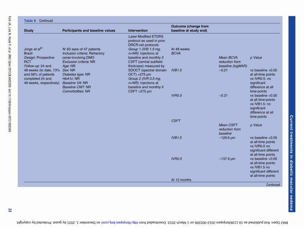
Table 8 Continued
Study Participants and baseline values Intervention
Outcome (change from
baseline at study end)
Laser Modified ETDRS
protocol as used in prior
DRCR.net protocols
Jorge et al51
Brazil
Design: Prospective
RCT
Follow-up: 24 and
48 weeks (to date, 73%
and 56% of patients
completed 24 and
48 weeks, respectively)
N: 63 eyes of 47 patients
Inclusion criteria: Refractory
cener-involving DMO
Exclusion criteria: NR
Age: NR
Sex: NR
Diabetes type: NR
HbA1c: NR
Baseline VA: NR
Baseline CMT: NR
Comorbidities: NR
Group 1 (IVB 1.5 mg,
n=NR): injections at
baseline and monthly if
CSFT (central subfield
thickness) measured by
SDOCT (spectral domain
OCT) >275 µm
Group 2 (IVR 0.5 mg,
n=NR): injections at
baseline and monthly if
CSFT >275 µm
At 48 weeks
BCVA
Mean BCVA
reduction from
baseline (logMAR)
p Value
IVB1.5 −0.21 vs baseline <0.05
at all-time points
vs IVR0.5: no
significant
difference at all
time-points
IVR0.5 −0.21 vs baseline <0.05
at all time-points
vs IVB1.5: no
significant
difference at all
time-points
CSFT
Mean CSFT
reduction from
baseline
p Value
IVB1.5 −129.6 µm vs baseline <0.05
at all-time points
vs IVR0.5 no
significant different
at all-time points
IVR0.5 −137.9 µm vs baseline <0.05
at all-time points
vs IVB1.5 no
significant different
at all-time points
At 12 months
Continued
FordJA,Lois
N,RoyleP,etal.BM
JOpen
2013;3:e002269.doi:10.1136/bmjopen-2012-002269
33
Curre
nttre
atm
ents
indiabetic
macularoedema
on December 1, 2021 by guest. Protected by copyright. http://bmjopen.bmj.com/ BMJ Open: first published as 10.1136/bmjopen-2012-002269 on 1 March 2013. Downloaded from

Table 8 Continued
Study Participants and baseline values Intervention
Outcome (change from
baseline at study end)
Lim et al55
Korea
Design: 3-arm RCT
Follow-up: 12 months
N: 111 eyes of 105 patients
Inclusion criteria: eyes with clinically
significant
DMO based on ETDRS and DMO with
central macular thickness of at least
300 µm by optical coherence tomography
(OCT)
Exclusion criteria: unstable medical
status, including glycemic control and
blood pressure; any previous treatment for
DMO, including intravitreal, sub-Tenon
injection or macular photocoagulation,
history of vitreoretinal surgery,
uncontrolled glaucoma; proliferative
diabetic retinopathy with active
neovascularisation, previous panretinal
photocoagulation, presence of
vitreomacular traction, history of systemic
corticosteroids within 6 months,
contraindications for bevacizumab or
triamcinolone acetonide
Age: 60.4 SD 7.4 (range 48–70) years
Sex: 52% female
Diabetes type: NR
HbA1c: 7.2 SD 1.2–7.4 SD1.2
Baseline VA: 0.62 SD 0.23–0.65 SD 0.28
logMAR
Baseline CMT: 447 SD 110–458 SD
92 µm
Comorbidities: NR
Group 1 (IVB/IVT, n=36):
IV injection of 1.25 mg
(0.05 ml) IVB at 0 and
6 weeks and IV injection of
2 mg (0.05 ml) IVT at
0 weeks. Mean number of
addition injection 1.28
Group 2 (IVB, n=38): IV
injection of 1.25 mg
(0.05 ml) IVB at 0 and
6 weeks. Mean number of
injections 2.54.
Group 3 (IVT, n=37): IV
injection of 2 mg (0.05 ml)
IVT at 0 weeks. Mean
number of injections 1.04
Unclear if rescue laser was
available
IVB injections were
repeated if CMT appeared
>300 µm on OCT in at
least 6 weeks in all three
groups
BCVA (logMAR) p Value
IVB/IVT −0.15 0.088 (between
groups)IVB −0.16IVT −0.16
CMT (µm) p Value
IVB/IVT −199 0.132 (between
groups)IVB −17s9IVT −200
Continued
34Ford
JA,LoisN,Royle
P,etal.BMJOpen
2013;3:e002269.doi:10.1136/bmjopen-2012-002269
Curre
nttre
atm
ents
indiabetic
macularoedema
on December 1, 2021 by guest. Protected by copyright. http://bmjopen.bmj.com/ BMJ Open: first published as 10.1136/bmjopen-2012-002269 on 1 March 2013. Downloaded from

Table 8 Continued
Study Participants and baseline values Intervention
Outcome (change from
baseline at study end)
Soheilian
et al37 41 54 141
Iran
Design: 3-arm RCT
Follow-up: 36 weeks
(Soheilian 2007 reports
12 week results of the
same trial, these were
not considered here)
N: 150 eyes of 129 patients
Inclusion criteria: eyes with clinically
significant DMO (ETDRS criteria)
Exclusion criteria: previous panretinal of
focal laser photocoagulation, prior ocular
surgery or injection, history of glaucoma
or ocular hypertension, VA ≥20/40 or <20/
300, iris neovascularisation, high risk
PDR, significant media opacity,
monocularity, pregnancy, serum creatinine
≥3 mg/dL, uncontrolled DM
Age: 61.2 SD6.1 years
Sex: 47.3% female
Diabetes type: not reported
HbA1c: not reported
Baseline VA: 0.55–0.73 SD0.26–0.28
logMAR
Baseline CMT: 300–359 SD118–149 µm
Comorbidities: 94% NPDR, 6% early PDR
Group 1 (IVB, n=50 eyes):
IV injection of
bevacizumab 1.25 mg
(0.05 ml) (retreatment IVB
14 eyes)
Group 2 (IVB/IVT, n=50
eyes): IV injection of
combined bevacizumab
(1.25 mg (0.05 ml)) and
triamcinolone (2 mg
(0.05 ml)), followed by two
injections of bevacizumab
alone (retreatment IVB/IVT
10 eyes)
Group 3 (MPC, n=50
eyes): focal or modified
grid laser (retreatment
MPC 3 eyes)
Regimen for all groups:
Retreatments performed at
12 week intervals as
required
At 36 weeks
BCVA (Snellen chart):
BCVA (logMAR),
SD
p Value
IVB −0.28 SD0.25 (+14
SD12.5 letters)
0.053 vs IVB/IVT or
MPC
IVB/IVT −0.04 SD0.33 (+2
SD16.5 letters)
NS vs MPC
MPC
+0.01 SD0.27
(−0.5 SD13.5
letters)
Snellen line
changes
IVB +2 lines or more:
37%
stable within 2
lines: 59.3%
−2 lines or more:
3.7%
NS between
groups
IVB/IVT +2 lines or more:
25%
stable within 2
lines: 54.2%
−2 lines or more:
20.8%
MPC +2 lines or more:
14.8%
stable within 2
lines: 66.7%
−2 lines or more:
18.5%
CMT (OCT):
CMT (µm), SD p Value
IVB −56 SD140 0.044 vs baseline,
NS between
groups
IVB/IVT −5 SD113
MPC −8 SD67
Continued
FordJA,Lois
N,RoyleP,etal.BM
JOpen
2013;3:e002269.doi:10.1136/bmjopen-2012-002269
35
Curre
nttre
atm
ents
indiabetic
macularoedema
on December 1, 2021 by guest. Protected by copyright. http://bmjopen.bmj.com/ BMJ Open: first published as 10.1136/bmjopen-2012-002269 on 1 March 2013. Downloaded from

Adverse events are shown in tables 9 and 16.Conjunctival haemorrhages were higher in the ranibizu-mab arms compared with laser (RESTORE) or no treat-ment (RESOLVE). In the RESOLVE, RISE and RIDEstudies, a considerably higher incidence of intraocularpressure (IOP) increase was reported in the ranibizu-mab arm compared to control. This increase in IOP wasnot demonstrated in the RESTORE study. There wereno consistent differences in systemic adverse eventsbetween ranibizumab and laser or placebo.
BevacizumabEight RCTs investigating the use of bevacizumab inDMO were identified (tables 4 and 8). One RCT, theBOLT study (n=80), randomised patients to lasertherapy or 1.25 mg intravitreal bevacizumab.23 52 At24 months, the mean changes in BCVA and the propor-tion of patients who gained 10 ETDRS letters or morewas statistically significantly higher in the bevacizumabarm than in the laser arm. Faghihi et al53 (n=80) com-pared 1.25 mg bevacizumab (average 2.23 injections perpatient) with 1.25 mg bevacizumab plus a single lasertreatment (average 2.49 injections per patient). After6 months, the authors found both treatments to beeffective at improving BCVA, but neither treatment wasfound to result in a greater benefit.Lam et al35 (n=52) compared two doses of bevacizu-
mab (1.25 and 2.5 mg) in patients with diffuse DMO.Patients with focal DMO associated with localised retinalthickening were excluded. At 6 months, following 3initial monthly injections (no treatment in the remain-ing 3 months), both groups showed a statistically signifi-cantly increased mean BCVA compared with baselinevision, but there was no difference between doses.Four trials have investigated the combination of beva-
cizumab and triamcinolone. Ahmadieh et al31 (n=115)compared combined bevacizumab (three 1.25 mg injec-tions at 6 week intervals) plus triamcinolone (2 mg base-line injection only, Triamhexal) with bevacizumab alone(three 1.25 mg at 6 week intervals) and sham injectionin patients who had DMO unresponsive (definition notreported) to previous laser (last session more than3 months previously). The combination arm and bevaci-zumab alone arm improved mean BCVA more than thesham injection. For BCVA, the combination of bevacizu-mab plus triamcinolone was non-statistically significantlybetter than bevacizumab alone.Soheilian et al37 41 (n=150) compared combined beva-
cizumab (1.25 mg) plus triamcinolone (2 mg) with beva-cizumab alone and laser alone in patients who werelaser naïve. At 36 weeks, bevacizumab alone improvedBCVA more than either combination therapy or laser,although the difference was not statistically significant.Extended follow-up at 24 months showed that there wasno statistically significant difference between groups forBCVA; however, the direction of effect favours the bevaci-zumab and combination arms more than the laser.54
Table
8Co
ntinued
Study
Participants
andbaselinevalues
Intervention
Outcome(changefrom
baselineatstudyend)
Subgroups:
▸largerCMTreductionin
subgroupwith≥400µm
at
baseline(36weeks:IVB
−27.2
SD34.8%,IVB/IVT–
8.8
SD35.9%,MPC
−15.1
SD14.6%,p<0.001vs
baselinein
IVBandMPC
groupsonly)
BCVA,bestcorrectedvisualacuity;C,control;CMT,centralmacularthickness;CSME,clinically
significantmacularoedema;DDS,dexamethasone;DIL,dexamethasonefollowedbylaser;
DM,diabetesmellitus;DMO,diabeticmacularoedema;DP,diastolic
pressure;DR,diabeticretinopathy;HR
QoL,health-relatedqualityoflife;IO
P,intraocularpressure;IV,intravitreal;IVB,
intravitrealbevacizumab;IVP,intravitrealpegaptanib;IVR,intravitrealranibizumab;IVT,intravitrealtriamcinolone;IVTL,intravitrealtriamcinolonepluslaser;IVVTE,intravitrealVEGFTrap
Eye;L,laser;MLT/M
PC,macularlasertherapy/m
acularphotocoagulation;NEIVFQ-25,NationalEyeInstitute
VisualFunctionQuestionnaire-25;NPDR,non-proliferativediabeticretinopathy;
NR,notreported;OCT,opticalcoherencetomography;PDR,proliferativediabeticretinopathy;PRP,panretinalphotocoagulation;RCT,randomisedcontrolledtrial;SOC,standard
ofcare;
SP,systolic
pressure;SRFA,fluocinolone;VA,visualacuity;VEGF,vascularendothelia
growth
factor.
36 Ford JA, Lois N, Royle P, et al. BMJ Open 2013;3:e002269. doi:10.1136/bmjopen-2012-002269
Current treatments in diabetic macular oedema
on Decem
ber 1, 2021 by guest. Protected by copyright.
http://bmjopen.bm
j.com/
BM
J Open: first published as 10.1136/bm
jopen-2012-002269 on 1 March 2013. D
ownloaded from

Table 9 Ranibizumab safety data
READ-2 study28 47 RESOLVE study36 RESTORE study24 RISE study38 RIDE study38
Number of patients IVR: n=42; L: n=42;
IVRL: n=42
IVR0.3: n=51; IVR0.5:
n=51; C: n=49
IVR: n=116; IVRL:
n=118; L: n=111
IVR0.3: 125; IVR0.5: 126;
C: 123
IVR0.3: 125; IVR0.5: 124; C: 127
Ocular adverse events
Eye pain NR IVR0.3: n=9 (18%);
IVR0.5: n=9 (18%); C:
n=10 (20%)
IVR: n=13 (11%);
IVRL: n=10 (8%); L:
n=12 (11%)
IVR0.3: 26%; IVR0.5: 21%;
C: 19%
IVR0.3: 8%; IVR0.5: 12.9%; C: 7.1%
Conjunctival
hyperaemia
NR NR IVR: n=9 (8%); IVRL:
n=6 (5%); L: n=6 (5%)
NR NR
Conjunctival
haemorrhage
NR IVR0.3: n=10 (20%);
IVR0.5: n=13 (25%); C:
n=7 (14%)
IVR: n=8 (7%); IVRL:
n=10 (8%); L: n=0
IVR0.3: 54%; IVR0.5: 52%;
C: 32%
IVR0.3: 40.8%; IVR0.5: 50%; C: 31.5%
IOP increase NR IVR0.3: n=6 (12%);
IVR0.5: n=15 (29%); C:
n=1 (2%)
IVR: n=1 (<1%);
IVRL: n=1 (<1%);
IVR0.3: 20%; IVR0.5: 14%;
C: 2%
IVR0.3 : 15.2%;IVR0.5: 18.5%; C: 11%
Vitreous
haemorrhage
IVR: n=1 (2%); L: n=4
(10%); IVRL: n=3 (7%)
IVR0.3: n=1 (2%);
IVR0.5: n=0; C: n=0
NR IVR0.3: 3.2%; IVR0.5:
3.2%; C: 13%
IVR0.3: 0.8%; IVR0.5: 2.4%; C: 15%
Substantial
worsening of DMO
L: n=1 (2%) NR NR NR
Retinal ischaemia NR IVR0.3: n=0; IVR0.5:
n=1 (2%); C: n=0
NR NR NR
Retinal artery
occlusion
NR IVR0.3: n=0; IVR0.5:
n=1 (2%); C: n=0
NR NR NR
Endophthalmitis NR IVR0.3: n=1 (2%);
IVR0.5: n=1 (2%); C:
n=0
NR IVR0.3: 0.8%; IVR0.5: 0;
C: 0
IVR0.3+IVR0.5: 1.2%; C: 0%
Retinal detachment NR IVR0.3: n=0; IVR0.5:
n=0; C: n=1 (2%)
NR IVR0.3: 0.8%; IVR0.5: 0;
C: 0.8%
IVR0.3+IVR0.5: 0.4%; C: 0%
Neovascularisation NR NR NR IVR0.3: 0; IVR0.5: 0;
C: 0.8%
IVR0.3: 0.8%; IVR0.5: 0.8%; C: 5.5%
Traumatic cataract NR NR NR IVR0.3: 0.8%; IVR0.5:
0.8%; C: 0
IVR0.3+IVR0.5: 0.4%; C: 0%
Uveitis NR NR NR NR IVR0.3+IVR0.5: 0.4%; C: 0%
Macular oedema NR NR NR IVR0.3: 16.8%; IVR0.5:
20.6%; C: 21.1%
IVR0.3: 19.2%; IVR0.5: 13.7%; C:
20.5%
Retinal exudates NR NR NR IVR0.3: 19.2%; IVR0.5:
17.5%; C: 20.3%
IVR0.3: 16%; IVR0.5: 15.3%; C: 11%
Continued
FordJA,Lois
N,RoyleP,etal.BM
JOpen
2013;3:e002269.doi:10.1136/bmjopen-2012-002269
37
Curre
nttre
atm
ents
indiabetic
macularoedema
on December 1, 2021 by guest. Protected by copyright. http://bmjopen.bmj.com/ BMJ Open: first published as 10.1136/bmjopen-2012-002269 on 1 March 2013. Downloaded from

Table 9 Continued
READ-2 study28 47 RESOLVE study36 RESTORE study24 RISE study38 RIDE study38
Retinal
haemorrhage
NR NR NR IVR0.3: 12.8%; IVR0.5:
12.7%; C: 20.3%
IVR0.3: 15.2%; IVR0.5: 22.6%;
C: 18.9%
Cataract NR NR NR IVR0.3: 16.8%; IVR0.5:
11.9%; C: 14.6%
IVR0.3: 20%; IVR0.5: 23.4%; C: 23.6%
Vitreous detachment NR NR NR IVR0.3: 13.6%; IVR0.5:
11.1%; C: 15.4%
IVR0.3: 8.8%; IVR0.5: 12.9%; C: 15%
Ocular hyperemia NR NR NR IVR0.3: 15.2%; IVR0.5:
11.1%; C: 10.6%
IVR0.3: 3.2%; IVR0.5: 3.2%; C: 7.9%
Vitreous floaters NR NR NR IVR0.3: 12.8%; IVR0.5:
14.3%; C: 5.7%
IVR0.3: 7.2%; IVR0.5: 8.1%; C: 3.1%
Eye irritation NR NR NR IVR0.3: 10.4%; IVR0.5:
9.5%; C: 6.5%
IVR0.3: 5.6%; IVR0.5: 5.6%; C: 3.1%
Foreign body
sensation in eyes
NR NR NR IVR0.3: 12.8%; IVR0.5:
7.1%; C: 4.1%
IVR0.3: 8%; IVR0.5: 2.4%; C: 5.5%
Systematic adverse events
Arterial
thromboembolic
events
Stroke in 1 pt (2%) in
IVRL group- not related
to study drug
IVR0.3: n=0; IVR0.5:
n=3 (6%); C: n=2 (4%)
IVR: n=6 (5%); IVRL:
n=1 (<1%); L: n=1
(<1%)
IVR0.3: 3.2% (n=1 stroke);
IVR0.5: 7.9% (n=5 strokes);
C: 7.3% (n=2 strokes)
IVR0.3: 1.6% (stroke), 5.6% (heart
attack); IVR0.5: 2.4% (stroke), 2.4%
(heart attack); C: 1.6% (stroke), 5.6%
(heart attack)
Hypertension NR IVR0.3: n=4 (8%);
IVR0.5: n=5 ((10%); C:
n=5 (10%)
IVR: n=9 (8%); IVRL:
n=6 (5%); L: n=9 (8%)
Serious
IVR0.3: 0.8%; IVR0.5:
3.2%; C: 0.8%
Serious
IVR0.3: 1.6%; IVR0.5: 1.6%; C: 0%
Non-ocular
haemorrhage
NR IVR0.3: n=1 (2%);
IVR0.5: n=1 (2%); C:
n=0
IVR: n=1 (<1%);
IVRL: n=0; L: n=1
(<1%)
NR NR
Proteinuria NR NR IVR: n=1 (<1%);
IVRL: n=1 (<1%); L:
n=0
NR NR
Deaths 1 (2%) due to CVA in
IVRL group
NR IVR: n=2 (2%); IVRL:
n=2 (2%); L: n=2 (2%)
IVR0.3: 2.4%; IVR0.5: 4%;
C: 0.8%
IVR0.3: 3.2%; IVR0.5: 4.8%; C: 1.6%
C, control; DMO, diabetic macular oedema; IOP, intraocular pressure; IVR, intravitreal ranibizumab; IVRL, intravitreal ranibizumab plus laser; L, laser; NR, not reported.
38Ford
JA,LoisN,Royle
P,etal.BMJOpen
2013;3:e002269.doi:10.1136/bmjopen-2012-002269
Curre
nttre
atm
ents
indiabetic
macularoedema
on December 1, 2021 by guest. Protected by copyright. http://bmjopen.bmj.com/ BMJ Open: first published as 10.1136/bmjopen-2012-002269 on 1 March 2013. Downloaded from

Table 10 Bevacizumab safety
BOLT study23 52 Lam et al35 Faghihi et al53
Number of patients MLT: n=38; IVB: n=42 IVB1.25, n=26; IVB2.5, n=26 IVB1.25 n=40 IVB 1.25
plus MLT n=40
Ocular adverse events Not reported
Loss of _15 or _30 ETDRS letters MLT: n=1 transient, 3 at 24 month analysis;
IVB: n=4 transient
No significant ocular events (IOP increase, retinal
tear, retinal detachment, endophthalmitis); no
significant difference in change in cataract scores
between groups
Vitreous haemorrhage MLT: n=1; IVB: n=0
Eye pain/irritation/watering during or
after injection
MLT:n=0; IVB: n=8
Red eye after injection MLT: n=0; IVB: n=8
Endophthalmitis NR
Transient IOP increase ≥30 mm Hg—MLT: 0; IVB:
n=4≥45 mm Hg—MLT: n=1; IVB: n=1
Floaters after injection MLT: n=0; IVB: n=2
Corneal epithelial defect MLT:n=0; IVB:n=1
Vitreomacular traction with macular
oedema
MLT: n=1; IVB: n=0
Systematic adverse events
Anaemia MLT: n=1; IVB: n=0 No systematic adverse effects (1 patient in 1.25 mg
group with foot gangrene requiring amputation due to
worsening diabetic neuropathy, considered unrelated
to treatment)
Vomiting after FFA MLT: n=1; IVB: n=0
Uncontrolled hypertension MLT:n=0; IVB: n=1
Polymyalgia rheumatica MLT:n=0; IVB: n=1
Intermittent claudication MLT:n=0; IVB: n=1
Gastroenteritis MLT:n=0; IVB: n=1
Fall MLT:n=2; IVB: n=0
Urinary tract infection MLT:n=0; IVB: n=1
Chest infection MLT:n=0; IVB: n=1
Headaches, dizziness, tiredness MLT:n=1; IVB: n=0
Bell palsy MLT:n=1; IVB: n=0
Admission for diabetic foot ulcer MLT:n=1; IVB: n=1
Admission for cholecystectomy MLT:n=0; IVB: n=1
Admission for fall/loss of
consciousness
MLT:n=1; IVB: n=0
Angina—hospital admission MLT:n=1; IVB: n=0
Cerebrovascular accident MLT:n=1; IVB: n=0
Myocardial infarction MLT:n=0; IVB: n=2
Coronary artery bypass graft MLT:n=0; IVB: n=1
Dyspnea, chest pain–admitted for
hospital observation
MLT:n=0; IVB: n=1
Death NR
ETDRS, Early Treatment Diabetic Retinopathy Study; FFA, fundus fluorescein; IOP, intraocular pressure; IVB, intravitreal bevacizumab; MLT, macular laser therapy; NR, not reported.
FordJA,Lois
N,RoyleP,etal.BM
JOpen
2013;3:e002269.doi:10.1136/bmjopen-2012-002269
39
Curre
nttre
atm
ents
indiabetic
macularoedema
on December 1, 2021 by guest. Protected by copyright. http://bmjopen.bmj.com/ BMJ Open: first published as 10.1136/bmjopen-2012-002269 on 1 March 2013. Downloaded from

Lim et al55 (n=111) also evaluated the combination ofbevacizumab plus triamcinolone when compared withbevacizumab alone or triamacinolone alone. At12 months, the authors found no statistically significantdifference between groups for BCVA or CMT.The Efficacy Study of Triamcinolone and Bevacizumab
Intravitreal for Treatment of Diabetic Macular Oedema(ATEMD) study, currently only published in abstractform, compared combined therapy with bevacizumab(1.25 mg) and triamcinolone (4 mg) with each of thesealone.56 At 6 months, they found no statistically signifi-cant difference between groups. One study comparingbevacizumab with ranibizumab is discussed above.51 Nobevacizumab trials were suitable for meta-analysisbecause treatment arms were not comparable amongincluded studies.Adverse events are shown in tables 10 and 16.There
was a low frequency of adverse events reported in theincluded trials. A higher incidence of mild anteriorchamber reaction was reported in bevacizumab groupscompared with controls. The incidence of IOP increasewas comparable between bevacizumab and laser.Soheilian et al37 41 were the only authors to report theincidence of lens opacity. No patients in the bevacizu-mab alone group were found to have lens opacities butin four patients (8%) in the bevacizumab plus triamci-nolone group, this finding was observed over the36-week follow-up period.
PegaptanibTwo studies have evaluated pegaptanib in DMO andboth compared it with sham injection (table 5).Cunningham et al39 57 compare three doses of pegapta-nib (0.3, 1 and 3 mg) and sham injection in laser-naivepatients (n=172). At 6 months, patients in the 0.3 and1 mg groups performed statistically significantly betterthan those in either the 3 mg or sham groups. Six injec-tions (median) were administered in the 0.3 and 1 mggroups, whereas only five (median) injections wereadministered in the 3 mg group.The second trial (n=260), reported by Sultan and collea-
gues in 2011, compared pegaptanib (0.3 mg) and shaminjection. At 2 years, the pegaptanib group showed a statis-tically significantly greater improvement in mean BCVAcompared with sham.40 However, there was no statisticallysignificant difference in the proportion of patients with animprovement of 10 letters or more. Patients were allowedrescue laser at the assessors’ discretion (25.2% of patientsin the pegaptanib group and 45% of patients in the shamgroup received rescue treatment). In regard tometa-analysis, data were only available to combine thesetrials for the proportion of patients with more than 15letter gain. Although neither trial individually demon-strated a statistically significant difference favouring pegap-tanib over sham (figure 5), when pooled together inmeta-analysis, a statistically significant difference was foundin favour of pegaptanib (OR 1.94, 95% CI 1.01 to 3.71).
Table 11 Pegaptanib safety
Cunningham et al / Adamis et al39 57 Sultan et al40
Number of patients IVP0.3, n=44 eyes; IVP1, n=44 eyes; IVP3, n=42 eyes IVP, n=133 eyes; C, n=127 eyes
Ocular adverse events
Eye pain Pegaptanib: 31%; C: 17% IVP: 11.1%; C: 7%
Vitreous haemorrhage Pegaptanib: 22%; C: 7% IVP: 6.3%; C: 7.7%
Punctuate keratitis Pegaptanib: 18%; C: 17% IVP: 11.8%; C: 6.3%
Cataract Pegaptanib: 13%; C: 10% IVP: 8.3%; C: 9.2%
Eye discharge Pegaptanib: 11%; C: 10% NR
Conjunctival haemorrhage Pegaptanib: 10%; C: 0% IVP: 22.2%; C: 14.1%
Vitreous opacities Pegaptanib: 9%; C: 5% NR
Blurred vision Pegaptanib: 7%; C: 5% NR
Other vitreous disorder Pegaptanib: 7%; C: 0% NR
Other visual disturbance Pegaptanib: 7%; C: 0% NR
Culture-negative endophthalmitis Pegaptanib: n=1 NR
IOP increase NR IVP: 17.4%; C: 6.3%
Retinal haemorrhage NR IVP: 6.3%; C: 10.6%
Retinal exudates NR IVP: 6.3%; C: 5.6%
Conjunctivitis NR IVP: 5.6%; C: 4.2%
Lacrimation increased NR IVP: 5.6%; C: 2.8%
Diabetic retinal oedema NR IVP: 11.1%; C: 17.6%
Macular oedema NR IVP: 9.7%; C: 11.6%
Systemic adverse events
Non-ocular hypertension NR IVP: 13.9%; C: 9.9%
Cardiac disorders NR IVP: 6.9%; C: 5.6%
Deaths NR IVP: n=4
IOP, intraocular pressure; IVP, intravitreal pegaptanib; NR, not reported.
40 Ford JA, Lois N, Royle P, et al. BMJ Open 2013;3:e002269. doi:10.1136/bmjopen-2012-002269
Current treatments in diabetic macular oedema
on Decem
ber 1, 2021 by guest. Protected by copyright.
http://bmjopen.bm
j.com/
BM
J Open: first published as 10.1136/bm
jopen-2012-002269 on 1 March 2013. D
ownloaded from

Adverse events for pegaptanib are shown in table 11.There was a higher incidence of eye pain compared tocontrol (31% vs 17%).39 57 Cataract formation wassimilar between the pegaptanib and control groups.There was a higher incidence of IOP increase in thepegaptanib arm compared to control (17.4% vs 6.3%).40
Other anti-VEGFAflibercept has been evaluated in the Da Vinci study(n=219)30 58 (table 5). Four regimens of aflibercept(0.5 mg 4 weekly, 2 mg 4 weekly, 2 mg monthly for3 months, then every 8 weeks, and 2 mg monthly for3 months followed by treatment as required) were com-pared with laser. At 6 months, all aflibercept arms had astatistically better BCVA and CMT change than the laserarm. The regimen that resulted in the greatest BCVAgain and CMT reduction was 2 mg every 4 weeks;however, statistical significance between aflibercept armswas not reported. One year extended follow-up showed
that all aflibercept arms were found to have a statisticallysignificantly better BCVA compared to laser.58
Adverse events are shown in table 12. There was ahigher incidence of IOP increase and eye pain in theaflibercept group compared with laser. Other adverseevents were too infrequent to draw meaningful conclu-sions. The incidence of cataracts was not reported.
SteroidsDexamethasoneTwo included trials assessed the use of dexamethasoneto treat DMO (table 6): Haller 2010 (full text avail-able)59 and Callanan (available to date only in anabstract form).44 Haller 2010 (n=171) compared twodoses of dexamethasone, administered as an intravitrealimplant (350 and 700 µm) through a 20-gauge trans-scleral incision, with no treatment. At 90 days only, the700 µm group showed a statistically significantly higherproportion of patients with 10 or more letter gain
Table 12 Aflibercept safety
DA VINCI 201030 58
Number of patients IVVTE (all doses) n=175, laser n=44
Ocular adverse events
Conjunctival hemorrhage At 6 months: Laser 18.2%, IVVTE 18.9%
At 12 months: Laser 18.2%, IVVTE 26.9%
IOP increase At 6 months: Laser 2.3%, IVVTE 9.7%
At 12 months: Laser 2.3%, IVVTE 9.7%
Eye pain At 6 months: Laser 4.5%, IVVTE 8.6%
At 12 months: Laser 4.5%, IVVTE 13.7%
Ocular hyperaemia At 6 months: Laser 4.5%, IVVTE 6.3%
At 12 months: Laser 4.5%, IVVTE 7.4%
Vitreous floaters At 6 months: Laser 4.5%, IVVTE 5.1%
At 12 months: Laser 4.5%, IVVTE 6.9%
Endophthalmitis At 6 months: Laser 0%, IVVTE 1.1%
At 12 months: Laser 0%, IVVTE 1.1%
Uveitis At 6 months: Laser 0%, IVVTE 0.6%
At 12 months: Laser 0%, IVVTE 0.6%
Diabetic retinal oedema At 6 months: Laser 2.3%, IVVTE 0%
At 12 months: Laser 2.3%, IVVTE 4.6%
Visual acuity reduced At 6 months: Laser 2.3%, IVVTE 0%
At 12 months: Laser 2.3%, IVVTE 0%
Vitreous hemorrhage At 6 months: Laser 2.3%, IVVTE 0%
At 12 months: Laser 6.8%, IVVTE 0%
Corneal abrasion At 6 months: Laser 0%, IVVTE 0.6%
At 12 months: Laser 0%, IVVTE 4.6%
Retinal tear At 6 months: Laser 0%, IVVTE 0.6%
At 12 months: NR
Systematic events
Hypertension At 6 months: Laser 6.8%, IVVTE 9.7%
At 12 months: Laser 0%, IVVTE 1.7%
Myocardial infarction At 6 months: Laser 0%, IVVTE 1.1%
At 12 months: Laser 0%, IVVTE 1.7%
Cerebrovascular event At 6 months: Laser 0%, IVVTE1.1%
At 12 months: Laser 2.3%, IVVTE 1.7%
Death At 6 months: Laser 0%, IVVTE 1.7%
At 12 months: Laser 2.3%, IVVTE 4%
IOP, intraocular pressure; IVVTE, intravitreal vascular endothelial growth factor Trap Eye.
Ford JA, Lois N, Royle P, et al. BMJ Open 2013;3:e002269. doi:10.1136/bmjopen-2012-002269 41
Current treatments in diabetic macular oedema
on Decem
ber 1, 2021 by guest. Protected by copyright.
http://bmjopen.bm
j.com/
BM
J Open: first published as 10.1136/bm
jopen-2012-002269 on 1 March 2013. D
ownloaded from

compared to no treatment (33% compared with 12%,p=0.007). The 350 µm group showed a non-statisticallysignificant improvement compared with laser alone(21% compared with 12%). At 180 days, there was nostatistically significant difference between either thedexamethasone group or no treatment group. The treat-ment effect appeared to peak at 3 months.The second trial, by Callanan and colleagues (n=253),
compared dexamethasone (dose not reported) pluslaser with laser alone. Although a greater improvementin mean BCVA was seen at 1–9 months in the dexa-methasone plus laser group compared with laser alone,there was no statistically significant difference at12 months. A mean of 1.6 implants were used over the12 month period.These trials were not suitable for meta-analysis since
one study is only available in abstract form.Adverse events are shown in table 13. In the 350 and
700 µm groups compared with no treatment, there wasa higher incidence of anterior chamber cells (29.1/26.4%compared with 1.8%), anterior chamber flare(27.3/20.8% compared with 8.8%), vitreous haemorrhage(20/22.6% compared with 5.3%) and increased IOP(14.5/9.4% compared with 0%). However, there was nostatistically significant difference in cataract formationbetween groups at 12 months.59 Callanan et al44 reportedan increase in IOP in the dexamethasone plus laser groupcompared with laser alone (20% compared with 1.6%).
FluocinoloneTwo trials assessed fluocinolone implant for DMO(table 6). The FAME study (n=956) compared two dosesof fluocinolone (0.2 and 0.5 µg/day) with sham injec-tion in patients with at least one prior laser treatment.29
Approximately 25% of patients in each group had morethan one prior laser treatment. At 24 months, bothdoses of fluocinolone showed a statistically significantimprovement in mean BCVA compared to sham. Therewas a modest difference between fluocinolone groups.Rescue laser was given after the first 6 weeks for persist-ent oedema and was allowed every 3 months. A range of35–37% of patients in the fluocinolone group and 59%in the sham injection group required rescue laser.Extended follow-up at 36 months showed that both thefluocinolone arms continued to result in a statisticallysignificant benefit compared with sham.60
Pearson et al43 (n=196) compared fluocinolone(0.59 mg) with standard of care, either laser or no treat-ment. At 3 years, there was no statistically significant dif-ference in the proportion of patients with 15 letter gainor more (31% fluocinolone compared with 20% stand-ard of care) between groups and the proportion ofpatients losing 15 letters or more in the fluocinolonegroup (17% compared with 14%). Increased incidenceof cataracts may have contributed to this difference.These trials were not suitable for meta-analysis.Adverse events are shown in table 14. Pearson and col-
leagues reported a higher incidence of cataracts at3 years in the fluocinolone group compared with stand-ard of care (55.9% compared with 21.7%). In theextended report of the FAME study, there was a consid-erably higher incidence of cataract surgery in phakiceyes in the 0.2 and 0.5 µg/day fluocinolone groups(80% and 87.2% compared with 27.3%) and increasedIOP at any point (37% and 46% compared with 12%).Following the demonstration in the FAME trial that a
lower dose was about as good as higher ones, the higherdoses are unlikely to be used.
Table 13 Dexamethasone safety
Callanan et al44 Haller et al59
Number of patients
Ocular adverse events
IOP elevation DIL: 20% (p<0.001); 1%
≥10 mm HgL: 1.6% ; 0% ≥10 mm Hg
Cataract NR NR
Anterior chamber
cells
NR DDS350: 29.1%; DDS700: 26.4%; C: 1.8%
Anterior chamber
flare
NR DDS350: 27.3%; DDS700: 20.8%; C: 8.8%
Vitreous
haemorrhage
NR DDS350: 20%; DDS700: 22.6%; C: 5.3%
Eye pain NR DDS350: 18.2%; DDS700: 9.4%; C: 3.5%
Vitreous disorder NR DDS350: 20%; DDS700: 15.1%; C: 3.5%
Increased IOP NR DDS350: 14.5%; DDS700: 9.4%; C: 0%
Conjunctival
haemorrhage
NR DDS350: 14.5%; DDS700: 7.5%; C: 0%
Vitreous floaters NR DDS350: 7.3%; DDS700: 17%; C: 0%
No significant differences in: reduced VA, eye irritation,
abnormal sensation in eye, macular oedema, eye pruritus,
retinal hemorrhage, DR, nonocular events
Dil, dexamethasone followed by laser; DDS, dexamethasone; IOP, intraocular pressure; NR, not reported.
42 Ford JA, Lois N, Royle P, et al. BMJ Open 2013;3:e002269. doi:10.1136/bmjopen-2012-002269
Current treatments in diabetic macular oedema
on Decem
ber 1, 2021 by guest. Protected by copyright.
http://bmjopen.bm
j.com/
BM
J Open: first published as 10.1136/bm
jopen-2012-002269 on 1 March 2013. D
ownloaded from

TriamcinoloneTen trials evaluating triamcinolone were identified(tables 7 and 8). All trials evaluated intravitreal adminis-tration of triamcinolone, but there were no trials evaluat-ing posterior or anterior subtenon injections. Two trialsused Trivaris,21 61 two trials used Kenacort,32 33 one trialused Kenalog,62 one trial used Trimahexal31 and fourtrials did not report the type of triamcinoloneused.34 3745 56 Three doses were assessed in the includedstudies (1, 4 and 8 mg) and triamcinolone has beencombined with laser or bevacizumab.
Ip and colleagues (n=840) were the only authors toevaluate triamcinolone 1 mg (Trivaris).22 61 63 64 Theyfound a statistically significant improvement in meanBCVA at 2 years in the laser group compared with thetriamcinolone group and no significant differencebetween 1 compared with 4 mg.Several trials compared 4 mg intravitreal triamcino-
lone. Ip and colleagues (n=840) found that lasertherapy resulted in a greater improvement in meanBCVA at 2 years compared to 4 mg triamcinolone(Trivaris).22 61 63 64 Lam et al34 (n=111) found no
Table 14 Fluocinolone safety
FAME study (Campochiaro et al)29 60 Pearson et al43
Number of patients
Ocular adverse events
IOP at 12 months NR NR
Progression of cataract NR NR
Cataract NR SRFA: 55.9%;
SOC: 21.7%
Transient vitreous floaters NR NR
Transient subconjunctival
haemorrhage
NR NR
Cataract surgery SRFA0.2: 41.1% (74.9% of those without cataract surgery at baseline,
80% at 36 months); SRFA0.5: 50.9% (84.5% of those without cataract
surgery at baseline, 87.2% at 36 months); C: 7% (23.1% of those
without cataract surgery at baseline, 27.3% at 36 months)
NR
Glaucoma SRFA0.2: 1.6%; SRFA0.5: 2.3%; C: 0.5% NR
Increased IOP SRFA0.2: 3.2%; SRFA0.5: 3.3%; C: 0% SRFA: 69.3%;
SOC: 11.6%
IOP >30 mm Hg at any point
during 36 months
SRFA0.2: 18.4%; SRFA0.5: 22.9%; C: 4.3% NR
Trabeculectomy SRFA0.2: 2.1%; SRFA0.5: 4.8%; C: 0% NR
Other glaucoma surgery SRFA0.2: 1.3%; SRFA0.5: 1.3%; C: 0.5% NR
Trabeculoplasty SRFA0.2: 0.8%; SRFA0.5: 2.3%; C: 0% NR
Vitreous haemorrhage NR SRFA: 40.2%;
SOC: 18.8%
Abnormal sensation in eye NR SRFA: 37%;
SOC: 11.6%
Macular oedema NR SRFA: 34.6%
Eye pain NR SRFA: 26.8%;
SOC: 15.9%
Eye irritation NR SRFA: 22%;
SOC: 10.1%
Increased lacrimation NR SRFA: 22%;
SOC: 8.7%
Photophobia NR SRFA: 21.3%;
SOC: 21.7%
Blurred vision NR SRFA: 21.3%;
SOC: 15.9%
Vitreous floaters NR SRFA: 21.3%;
SOC: 8.7%
Systemic adverse events
Serious cardiovascular events SRFA0.2: 12%; SRFA0.5: 13.2%; C: 10.3%
Pruritus NR SRFA: 38.6%;
SOC: 21.7%
Deaths NR NR
IOP, intraocular pressure; NR, not reported; SOC, standard of care; SRFA, fluocinolone.
Ford JA, Lois N, Royle P, et al. BMJ Open 2013;3:e002269. doi:10.1136/bmjopen-2012-002269 43
Current treatments in diabetic macular oedema
on Decem
ber 1, 2021 by guest. Protected by copyright.
http://bmjopen.bm
j.com/
BM
J Open: first published as 10.1136/bm
jopen-2012-002269 on 1 March 2013. D
ownloaded from

Table 15 Triamcinolone safety
DRCR Network 2008
(Ip et al/Beck et al/Bressler et al) 22 61 63 64
Gillies et al/Sutteret al32 136–138 Gillies et al33
Kim
et al45 Lam et al34Ockrim et al/Sivaprasad et al42 62
Number of patients
Ocular adverse events
At 2 years (or 3 years when
indicated)
At 2 years – Not
reported
– At 12 months
IOP ≥30 mm Hg IVT1: n=22; IVT4: n=53; L: n=3 NR NR NR IVT: IOP significantly
higher than in L group
(18.2 mm Hg, range
12–26 mm Hg); no cases
of glaucoma
IOP >22 mm Hg NR NR NR IVT: 37%
(p=0.002 vs L);
IVTL: 36%
(p=0.002 vs L); L:
5%
NR
IOP ≥10 mm Hg from
baseline
IVT1: n=41; IVT4: n=85; L:
n=12
NR NR NR NR
IOP ≥5 mm Hg NR IVT: 68% (p=0.007
vs C); C: 10%
NR NR NR
IOP lowering
medication used
IVT1: n=31; IVT4: n=76; L:
n=25
IVT: 44% (p=0.0002
vs C); C: 3%
IVTL: 64% (p<0.001); L:
24%
NR NR
Cataract surgery IVT1: 23% (of those phakic at
baseline, 46% by 3 years
(p<0.001 between all groups);
IVT4: 51% (of those phakic at
baseline, 83% by 3 years); L:
13% (of those phakic at
baseline, 31% by 3 years)
IVT: 56% (of phakic
eyes over 3 years,
p<0.001 vs C); C: 8%
(of phakic eyes over
3 years)
NR NR
Ptosis NR NR NR NR NR
Retinal detachment IVT1: n=2; IVT4: n=4; L: n=2 NR NR None NR
Retinal vein occlusion IVT1: n=1; IVT4: n=2; L: n=3 NR NR NR NR
Retinal artery occlusion IVT1: n=0; IVT4: n=0; L: n=1 NR NR NR NR
Anterior ischemic optic
neuropathy
IVT1: n=1; IVT4: n=0; L: n=0 NR NR NR NR
Vitrectomy IVT1: n=26; IVT4: n=19; L:
n=31
NR NR NR NR
Open angle glaucoma IVT1: n=2; IVT4: n=7; L: n=2 NR NR NR NR
Glaucoma filtering
surgery
IVT1: n=0; IVT4: n=2; L: n=0 NR NR NR NR
Laser trabeculoplasty IVT1: n=0; IVT4: n=1; L: n=0 IVT: n=2; C: n=0 IVTL: n=1 NR NR
Ciliary body destruction IVT1: n=0; IVT4: n=1; L: n=0 NR NR NR NR
Continued
44Ford
JA,LoisN,Royle
P,etal.BMJOpen
2013;3:e002269.doi:10.1136/bmjopen-2012-002269
Curre
nttre
atm
ents
indiabetic
macularoedema
on December 1, 2021 by guest. Protected by copyright. http://bmjopen.bmj.com/ BMJ Open: first published as 10.1136/bmjopen-2012-002269 on 1 March 2013. Downloaded from

Table 15 Continued
DRCR Network 2008
(Ip et al/Beck et al/Bressler et al) 22 61 63 64
Gillies et al/Sutteret al32 136–138 Gillies et al33
Kim
et al45 Lam et al34Ockrim et al/Sivaprasad et al42 62
Endophthalmitis IVT1: n=0; IVT4: n=;0 L: n=0 (Infectious) IVT: n=1;
C: NR
(Culture-negative) IVTL:
n=1
None (sterile) IVT: n=1
Pseudoendophthalmitis IVT1: n=0; IVT4: n=;0 L: n=0 NR NR NR NR
Chemosis NR NR NR NR NR
Percentage of increase
in cataract scores
NR NR NR IVT:+1.0 SD1.1
(p=NS vs L);
IVTL:+1.3 SD1.9
(p=NS vs L); L:
+0.5 SD0.9
NR
Ocular hypertension
(>21 mm Hg)
NR NR NR NR NR
Cataract progression NR NR Phakic eyes, progression
by ≥2 AREDS grade,
IVTL: 64% (p<0.001); L:
11% (p<0.001)
NR NR
Corneal
decompensation
NR IVT: NR; C: n=1 NR NR NR
Cataract surgery NR NR IVTL: 61% (p<0.001); L:
0%
NR IVT: n=2; L: n=1
Vitreous haemorrhage NR NR NR IVTL: n=1
Lens opacity NR NR NR NR Significantly greater
change in lens opacity in
IVT group than in L group
(1.9)
Deaths N=33, unrelated to study
treatment
IVT: n=1; C: n=2 IVTL: n=2; L: n=1 NR NR
CPL, control plus laser; IOP, intraocular pressure; NR, not reported; IVT, intravitreal triamcinolone; IVTL, intravitreal triamcinolone plus laser; RDL, ranibizumab plus deferred laser;RPL, ranibizumab plus laser; TPL, triamcinoloine plus laser.
FordJA,Lois
N,RoyleP,etal.BM
JOpen
2013;3:e002269.doi:10.1136/bmjopen-2012-002269
45
Curre
nttre
atm
ents
indiabetic
macularoedema
on December 1, 2021 by guest. Protected by copyright. http://bmjopen.bmj.com/ BMJ Open: first published as 10.1136/bmjopen-2012-002269 on 1 March 2013. Downloaded from

Table 16 Safety data in trials assessing more than one drug
Ahmadieh31
ATEMD 2011
(Oliveira
Neto et al)56DRCR Network 2010
(Elman et al, )21 46Lim
et al55 Soheilian et al37 41
Number of patients
Ocular adverse events
Mild anterior chamber
reaction
IVB: 19.5% (n=8 eyes), resolved
after 1 week of no treatment; IVB/
IVT: 18.9% (n=7 eyes), resolved
after 1 week of no treatment
NR NR NR IVB: 20% (n=10 eyes), resolved
after 1 week; IVB/IVT: 18%
(n=9 eyes), resolved after
1 week
Marked anterior chamber
reaction
IVB: n=1 (topical corticosteroid and
cycloplegic drops)
NR NR NR IVB: n=1 (topical corticosteroids
and cycloplegic drops);
Progression of fibrous
proliferation
IVB: n=1 with no sign of retinal
traction
NR NR NR IVB: n=1 with no sign of retinal
traction;
Vitreous haemorrhage IVB/IVT: n=1 after third injection
(excluded from study)
NR NR NR NR
IOP rise IVB: 23, 22 and 28 mm Hg at 6, 12
and 18 weeks (anti-glaucoma
drops)
NR IOP elevation more frequent with
triamcinolone + PL
IVB/
IVT:
8.3%
IVT:
10.8%
NR
IOP ≥10 mm Hg from
baseline
NR NR CPL: n=16; RPL: n=10; RDL: n=5;
TPL: n=70
NR NR
IOP ≥30 mm Hg from
baseline
NR NR CPL: n=3; RPL: n=2; RDL: n=4; TPL:
n=46
NR NR
Initiation of IOP lowering
treatment at any visit
NR NR CPL: n=9; RPL: n=5; RDL: n=4; TPL:
n=41
NR NR
Iris neovascularisation None NR NR NR NR
Lens opactiy None NR NR NR Severe lens opacity IVB/IVT:
n=4 eyes; MPC: n=1 eye
Endophthalmitis NR NR CPL: n=1; RPL: n=1; RDL: n=1; TPL:
n=0
NR None
Pseudoendophthalmitis NR NR CPL: n=1; RPL: n=0; RDL: n=0; TPL:
n=1
NR NR
Ocular vascular event NR NR CPL: n=1; RPL: n=1; RDL: n=0; TPL:
n=2
NR NR
Retinal detachment NR NR CPL: n=0; RPL: n=0; RDL: n=1; TPL:
n=0
NR None
Vitrectomy NR NR CPL: n=7; RPL: n=0; RDL: n=3; TPL:
n=0
NR NR
Vitreous haemorrhage NR NR CPL: n=15; RPL: n=3; RDL: n=4; TPL:
n=2
NR None
Continued
46Ford
JA,LoisN,Royle
P,etal.BMJOpen
2013;3:e002269.doi:10.1136/bmjopen-2012-002269
Curre
nttre
atm
ents
indiabetic
macularoedema
on December 1, 2021 by guest. Protected by copyright. http://bmjopen.bmj.com/ BMJ Open: first published as 10.1136/bmjopen-2012-002269 on 1 March 2013. Downloaded from

Table 16 Continued
Ahmadieh31
ATEMD 2011
(Oliveira
Neto et al)56DRCR Network 2010
(Elman et al, )21 46Lim
et al55 Soheilian et al37 41
Cataract surgery NR NR CPL: n=11 (of those phakic at
baseline); RPL: n=6 (of those phakic at
baseline); RDL: n=8 (of those phakic at
baseline); TPL: n=19 (of those phakic
at baseline)
NR NR
Glaucoma surgery NR NR NR NR NR
Retinal
neovascularisation
NR NR NR NR IVB: n=4 (all resolved); MPC:
n=3 eyes (2 resolved)
Development of early
PDR
NR NR NR NR IVB: n=1; IVB/IVT: n=4; MPC:
n=3
Progression to high-risk
PDR
NR NR NR NR IVB: n=4; IVB/IVT: n=3; MP:
n=3
Ocular hypertension
(≥23 mm HG)
NR NR NR NR IVB/IVT: 16% (n=8 of eyes),
controlled medically in all
except 1 that progressed to
neovascular glaucoma
Systemic adverse events
Acute myocardial
infarction
N=1, considered
not to be related to
the study drug
No specific systemic adverse events
that could be attributed to chance
No significant blood pressure
increase, no thromboembolic
events
Deaths C: n=1 N=1, considered
not to be related to
the study drug
CPL: n=8; RPL: n=5; RDL: n=3; TPL:
n=2
IVB/IVT: n=2; MPC: n=2
C, control; CPL, control plus laser; DMO, diabetic macular oedema; IOP, intraocular pressure; IVB, intravitreal bevacizumab; IVR, intravitreal ranibizumab; IVRL, intravitreal ranibizumab pluslaser; IVT, intravitreal triamcinolone; L, laser; NR, not reported; PDR, proliferative diabetic retinopathy; RDL, ranibizumab plus deferred laser; RPL, ranibizumab plus laser; TPL, triamcinoloineplus laser.
FordJA,Lois
N,RoyleP,etal.BM
JOpen
2013;3:e002269.doi:10.1136/bmjopen-2012-002269
47
Curre
nttre
atm
ents
indiabetic
macularoedema
on December 1, 2021 by guest. Protected by copyright. http://bmjopen.bmj.com/ BMJ Open: first published as 10.1136/bmjopen-2012-002269 on 1 March 2013. Downloaded from

statistically significant difference between laser andtriamcinolone at 6 months (triamcinolone type notreported). When these two trials were pooled throughmeta-analysis, the treatment effect favoured laser but thedifferences were not statistically significant (figure 6).Ockrim et al62 (n=88) compared 4 mg intravitreal triam-cinolone (Kenalog) with laser alone. At 12 months, they
found no statistically significant BCVA improvementbetween the triamcinolone and laser groups. Gillieset al32 (n=69) compared 4 mg of triamcinolone(Kenacort) with sham injection. Mean BCVA improvedstatistically significantly with triamcinolone at 24 monthscompared with sham injection (3.1 letter gain comparedwith 2.9 letter loss, p=0.01).
Figure 2 Ranibizumab 0.5 mg alone versus laser alone. (A) Mean change in best corrected visual acuity. (B) Proportion with
>15 letter gain. (C) central macular thickness.
Figure 3 Ranibizumab 0.5 mg plus laser versus ranibizumab 0.5 mg alone. (A) Mean change in best corrected visual acuity.
(B) Proportion with >15 letter gain. (C) central macular thickness.
48 Ford JA, Lois N, Royle P, et al. BMJ Open 2013;3:e002269. doi:10.1136/bmjopen-2012-002269
Current treatments in diabetic macular oedema
on Decem
ber 1, 2021 by guest. Protected by copyright.
http://bmjopen.bm
j.com/
BM
J Open: first published as 10.1136/bm
jopen-2012-002269 on 1 March 2013. D
ownloaded from

Lam et al34 (n=111) compared triamcinolone 4 mgalone with 4 mg of triamcinolone plus laser or laseralone. At 6 months, the authors found no difference inBCVA between any of the groups. Elman et al21 (n=854)compared 4 mg of triamcinolone (Trivaris) plus laserwith ranibizumab plus prompt (within 3–10 days) ordeferred (more than 24 week) laser and laser alone. At2 years, they found a statistically significant difference inmean BCVA between ranibizumab plus prompt/deferred laser compared with laser alone (7 letter gain/9 letter gain compared with 3 letter gain), but nodifference with triamcinolone plus laser compared withlaser alone (2 letter gain compared with 3 letter gain).Neto et al56 (n=120) compared 4 mg triamcinolonealone (triamcinolone type not reported) with 4 plus1.25 mg bevacizumab. At 6 months, they found no statis-tically significant difference between groups.The Elman and Lam studies were suitable for
meta-analysis, which showed non-statistically significantimprovements in mean BCVA and the proportions ofpatients with more or equal than 15 letter gain in thetriamcinolone plus laser group compared with laseralone (figure 7).
Adverse events are shown in tables 15 and 16.Triamcinolone was associated with consistently higherincidences of IOP increase and cataracts. Gilles and col-leagues reported a cataract rate of over 50% by 3 yearsin patients treated with triamcinolone.
Other pertinent studiesOnly one study in abstract form directly compared beva-cizumab with ranibizumab.51 Bevacizumab and ranibizu-mab have been compared through an indirectcomparison of five trials.65 There was no evidence of adifference between the drugs; however, wide credibleintervals meant that the superiority of either drug couldnot be excluded.Two-year results of the CATT (Comparison of AMD
Treatment Trials) and 1 year results of the IVAN (InhibitVEGF in Age-related choroidal Neovascularisation),recently published, have demonstrated a good safetyprofile of anti-VEGF therapies when used to treatpatients with age-related macular degeneration.66 67 TheCATT study randomised 1208 patients with AMD tomonthly or as required injection of either ranibizumabor bevacizumab. At 1 year, the mean BCVA was similar in
Figure 4 Ranibizumab 0.5 mg plus laser versus laser alone. (A) Mean change in best corrected visual acuity. (B) Proportion
with >15 letter gain. (C) Central macular thickness.
Figure 5 Pegaptanib 0.3 mg versus sham injection. (A) Proportion with >15 letter gain.
Ford JA, Lois N, Royle P, et al. BMJ Open 2013;3:e002269. doi:10.1136/bmjopen-2012-002269 49
Current treatments in diabetic macular oedema
on Decem
ber 1, 2021 by guest. Protected by copyright.
http://bmjopen.bm
j.com/
BM
J Open: first published as 10.1136/bm
jopen-2012-002269 on 1 March 2013. D
ownloaded from

both groups (8 letter gain in bevacizumab and 8.5 inranibizumab). Over 2 years, the rates of deaths, myocar-dial infarction and stroke did not differ between theranibizumab and bevacizumab treatment groups.However, there was a higher rate of serious adverseevents in the bevacizumab group compared withthe ranibizumab group. This increased event rate wasdriven mainly by hospitalisations (RR 1.29, 95% CI1.01 to 1.66). However, the hospitalisations were notcaused by known adverse events of bevacizumab.Arteriothrombotic events and heart failure occurred inless than 2% of participants in the IVAN, and they weremore often observed in the ranibizumab group than inthe bevacizumab group (p=0.03). Further data fromother ongoing clinical trials may provide more insighton the safety or anti-VEGF treatment and possible differ-ences on this respect among available drugs.Campbell et al68 conducted a population-based nested
case–control study of 91 378 older adults with a historyof physician-diagnosed retinal disease. The authorsfound that neither ranibizumab nor bevacizumab wasassociated with significant risks of ischaemic stroke,acute myocardial infarction, congestive heart failure orvenous thromboembolism.A recent systematic review specifically assessing adverse
events in anti-VEGF drugs found a low incidence of
serious (below 1 in 100) and non-serious ocular events(below 1 in 500) from ranibizumab, bevacizumab andpegaptanib.69
Fung et al70 used an internet-based survey of cliniciansto assess the safety of bevacizumab. The survey coveredover 5000 patients and found that bevacizumab was asso-ciated with an infrequent incidence of adverse events(all less than 0.21%).One study, which assessed diclofenac, did not meet
the inclusion criteria (follow-up for only 12 weeks).71
The authors randomised 32 patients to either intravitrealdiclofenac or triamcinolone and found that both diclo-fenac and triamcinolone reduced CMT, but a statisticallysignificant visual improvement was observed only in thetriamcinolone group.Sfikakis et al72 undertook a 30-week randomised cross-
over trial comparing infliximab and placebo. The studyfailed to meet our inclusion criteria (only 11 patientsincluded). The authors found that infliximab resulted ina 28.6% improvement in vision compared with 4.3%with placebo. The improvement seen with placebo couldbe due to a ‘carry over effect’, seen in cross-over trials.The Fenofibrate Intervention and Event Lowering in
Diabetes (FIELD) trial was primarily a study to see if thelipid-lowering agent fenofibrate could reduce macrovas-cular and microvascular events in type 2 diabetes.73
Figure 7 Triamcinolone 4 mg plus laser versus laser alone. (A) Mean change in best corrected visual acuity. (B) Proportion with
>15 letter gain.
Figure 6 Triamcinolone 4 mg versus laser alone. (A) Mean change in best corrected visual acuity. (B) Proportion with >15 letter
gain.
50 Ford JA, Lois N, Royle P, et al. BMJ Open 2013;3:e002269. doi:10.1136/bmjopen-2012-002269
Current treatments in diabetic macular oedema
on Decem
ber 1, 2021 by guest. Protected by copyright.
http://bmjopen.bm
j.com/
BM
J Open: first published as 10.1136/bm
jopen-2012-002269 on 1 March 2013. D
ownloaded from

However, a substudy within FIELD recruited 1012patients to a retinopathy study. The primary outcome inthe main study was need for laser therapy (3.4% onfenofibrate vs 4.9% on placebo), but the substudy usedretinal photography to assess progression of retinopathyor development of macular oedema. The HR at 6 yearsfor DMO was 0.69 (95% CI 0.54 to 0.87) in the fenofi-brate group compared to placebo.Ruboxistaurin is another oral agent which has been
assessed for the treatment of DMO. Aiello and collea-gues randomised 686 patients to receive placebo or oneof three doses of ruboxistaurin.74 75 There was no statis-tically significant difference in delay to sight-threateningDMO in any ruboxistaurin group compared to placebo.The authors suggest that differences in laser treatmentbetween groups may have contributed to the non-significant finding.
Assessment of heterogeneity within meta-analysisHeterogeneity was assessed methodologically and statistic-ally. Methodological heterogeneity was assessed by compar-ing the study population, interventions, outcome measuresand follow-up. Studies that were not methodologically com-parable were excluded from the meta-analysis. Forexample, bevacizumab trials were not pooled becauseSoheilian et al37 included patients who were laser naïve andAhmadieh et al31 included patients who were unresponsiveto laser. Some analyses were also excluded because suffi-cient details were not reported in the studies. For example,several studies failed to report SDs.35 39
Statistical heterogeneity was assessed through I2 scores.High statistical heterogeneity was found in two analyses(2.3 and 4.3). Therefore, these results should be inter-preted with due caution. Moderate heterogeneity wasfound in three analyses (2.2, 3.1 and 3.2). Low heterogen-eity was found in the remaining eight analyses.
Ongoing trialsThere are numerous ongoing studies listed in appendix 2.The most salient studies include a study to compare rani-bizumab and bevacizumab (Schmidt-Erfurth), a studyinvestigating rescue ranibizumab treatment for patientswho have failed on bevacizumab (Chaudhry), a studyevaluating two algorithms for ranibizumab, ‘treat andextend’ and ‘as required’ (RETAIN), further studies ofTrap-eye (VIVID and VISTA) and trials which are examin-ing the use of NSAIDs, such as diclofenac and nepafenac(NEVANAC and Soheilian).
DISCUSSIONIt appears that anti-VEGF treatment is effective in DMO,especially ranibizumab and bevacizumab. Meta-analysisof available short-term data (up to 2 years) suggests thatranibizumab is superior to laser and that adding laserto ranibizumab treatment does not confer additionalbenefit. Steroid treatment has demonstrated mixedsuccess and, almost uniformly, increased the incidence
of cataracts and IOP. The licence for fluocinolone takesnote of this and it is positioned as a treatment whenothers have failed.
Strengths and limitations of the reviewThere are a number of strengths of this review. A robustsystematic review methodology was used. Reliability wasimproved by excluding trials with small sample sizes orshort follow-up. Since a number of trials included similarintervention arms, consistent treatment effects furtherimprove reliability. Validity was improved by assessing thequality of trials using the Cochrane risk of bias tables.Including abstracts from ARVO provided up-to-dateresults. Pooling results through meta-analysis providedfurther evidence. The random effects model was usedthroughout to allow for heterogeneity among studies.This review, however, has limitations. Although the inclu-
sion of abstracts provides more up-to-date results, thestudies contained in these abstracts could not be assessedfor risk of bias and should therefore be interpreted withcaution. In addition, reporting of quality assessment cri-teria was variable. Allocation concealment was especiallypoorly reported. There was only one study which compareddifferent anti-VEGFs51 and none that compared steroids(fluocinolone vs dexamethasone vs triamcinolone).Therefore, it is difficult to assess the effectiveness withindrug classes. As with any meta-analysis, questions of hetero-geneity arise. Follow-up periods varied among studies.A difference of 6 months was allowed for studies to bepooled for meta-analysis, but this could have still resultedin heterogeneity. High statistical heterogeneity was foundin a quarter of the analyses. Furthermore, because of thelow number of trials included, publication bias could notbe assessed by funnel plot analysis. The manufacturersfunded most of the trials for ranibizumab, pegaptanib,dexamethasone and fluocinolone, whereas trials forbevacizumab and triamcinolone were generally funded bynon-pharmaceutical organisations. Generally, the non-commercial studies had smaller numbers, perhaps becauseof the funding restraints.It is important to note that there may be differences
in laser treatment protocol between studies. This appliesto trials which combine drug treatments with laser orinclude laser as a comparator. All studies referred to theETDRS protocol19 20 or a modified version of it. In theETDRS, once a diagnosis of clinically significant macularoedema was made, an angiogram was obtained to identi-fied ‘treatable lesions’. ‘Treatable lesions’ included dis-crete points of retinal hyperfluorescence or leakage(most of these are often microaneurysms), areas ofdiffuse leakage within the retina related to microaneur-ysms, intraretinal microvascular abnormalities, diffuselyleaking retinal capillary bed and retinal avascular zones.In the ETDRS protocol, treatment of lesions closer than500 microns from the centre of the macula was notrequired initially; however, if vision was less than 20/40and the oedema and leakage persisted, treatment up to300 microns from the centre of the macula was
Ford JA, Lois N, Royle P, et al. BMJ Open 2013;3:e002269. doi:10.1136/bmjopen-2012-002269 51
Current treatments in diabetic macular oedema
on Decem
ber 1, 2021 by guest. Protected by copyright.
http://bmjopen.bm
j.com/
BM
J Open: first published as 10.1136/bm
jopen-2012-002269 on 1 March 2013. D
ownloaded from

recommended unless there was capillary dropout; in thelatter case, treatment was not recommended as it maylead to further loss of perifoveal capillaries.However, in routine clinical practice, clinicians gener-
ally use lighter and less intense treatment than specifiedin the ETDRS protocol.76 In addition, some centres donot use fluorescein angiography (unlike the ETDRSstudy19) to guide treatment. The exact adherence to theETDRS protocol within studies is unclear. For example,in the BOLT study, a modified ETDRS protocol wasused. One of the aims of the protocol was ‘not darken-ing/whitening of microaneurysms’, which is not consist-ent with the ETDRS protocol.
Interpretation of the resultsThe anti-VEGF drugs appear to be clinically effectivein treating DMO in short-term studies (up to 2 years).Ranibizumab has the most robust evidence base and hasshown superiority compared to laser and sham injectionin all trials and meta-analyses, except for the proportionof patients with 10 or more letter gain in the DRCR.netstudy published by Elman et al46 at 2 years follow-up.Adding laser to ranibizumab conferred no benefit.Bevacizumab has also been shown to be superior tolaser. Three doses have been used (1.25, 1.5 and 2.5).The higher dose does not appear to add further benefit,and most studies in the literature use 1.25 mg. The add-ition of triamcinolone to bevacizumab did not providefurther benefits. Pegaptanib has only been comparedto sham injection. Mean change in BCVA favouredpegaptanib, but only through meta-analysis did theproportion of patients with more than 15 letter gainfavour pegaptanib. Further published data are requiredbefore drawing conclusions on aflibercept. However,although the anti-VEGF drugs are a significant advance,they fail to improve BCVA by 10 or more letters in halfor more patients, and so they do not provide a completeanswer to DMO.Steroid treatments have inconsistent results and are
undoubtedly associated with increased IOP and cataract.The effects of dexamethasone appear to peak at3 months. At 6 months, there was no significant differ-ence compared with laser. This might imply that earlierretreatment is needed if the beneficial effect is to bemaintained, but increasing the number of treatmentswould very likely increase the associated complications,especially with the relatively large needle size. The add-ition of laser did not appear to add further benefit.There was no significant difference in cataract formationat 6 months with dexamethasone compared to observa-tion, but it is likely that a higher incidence of cataractswould be seen with longer follow-up. Significantly morepatients suffered increased IOP in the dexamethasonegroup compared with observation. Fluocinolone hasbeen shown to be effective compared with sham injec-tion (FAME);29 60 however, when compared to standardof care (laser or observation at clinician’s discretion),there was no significant difference in the proportion of
patients with a 15 letter or more gain. Both studiesreported higher incidence of cataract formation in thefluocinolone group, with over 80% at 3 years at thehigher dose. Results for triamcinolone are inconsistent.Ip et al61 found that laser was more effective, whileothers have found no statistically significant difference.Triamcinolone combined with laser, however, seemed tohave similar efficacy as ranibizumab combined with laserin pseudophakic eyes.21 46 Triamcinolone is more effect-ive than sham injection. Triamcinolone has consistentlybeen associated with increased incidence of cataract andraised IOP.Steroids and laser therapy may affect CMT in a differ-
ent manner from anti-VEGF drugs. For example, whenranibizumab alone is compared with ranibizumab pluslaser, it appears to be more effective in terms of meanchange in BCVA and proportion of patients with morethan 15 letter gain. However, ranibizumab plus laser ismore effective at reducing CMT. Furthermore, whentriamcinolone plus laser is compared with ranibizumabplus laser, the latter appears to be more effective interms of change in BCVA and proportion of patientswith more than 15 letter gain, but triamcinolone pluslaser is more effective at reducing CMT. The reasons forthis are unclear. There is a weak correlation betweenCMT and BCVA. However, the long-term benefits ofreducing CMT are currently unknown.No large observational studies were identified that
compared anti-VEGF drugs. Using an internet-basedsurvey, Fung et al70 found the incidence of adverse eventsin bevacizumab to be low. One small outbreak of sterileendophthalmitis was reported with a single batch of beva-cizumab in Canada, emphasising the need for sterilitywhen preparing aliquots.77 Curtis et al78 carried out a verylarge retrospective cohort study in 146 942 patients aged65 and over with age-related macular degeneration(AMD). Their aim was to examine cardiovascular out-comes in patients treated with the four options: photo-dynamic therapy (PDT), pegaptanib, bevacizumab andranibizumab. The authors reported that one of theircomparisons showed an increase in overall mortality andstroke risk with bevacizumab compared to ranibizumab,with HRs of 0.86 (95% CI 0.75 to 0.98) and 0.78 (0.64 to0.96), respectively. However, owing to the very large costdifferences between bevacizumab and ranibizumab, theauthors noted that selection bias might be operating,with poorer people (with poorer health) more likely tobe treated with bevacizumab. They therefore carried outanother analysis using only ophthalmological clinicswhich used only one drug, to avoid selection bias. Thisanalysis showed no significant difference: overall mortal-ity HR for ranibizumab 1.10 (95% CI 0.85 to 1.141); MI0.87 (0.53 to 1.14); stroke 0.87 (0.61 to 1.24).Gower et al79 analysed 77 886 anti-VEGF injections
from Medicare data (46% ranibizumab and 54% bevaci-zumab). Results have only been published in abstractform. The authors found an increased risk of overallmortality and cerebrovascular events in the bevacizumab
52 Ford JA, Lois N, Royle P, et al. BMJ Open 2013;3:e002269. doi:10.1136/bmjopen-2012-002269
Current treatments in diabetic macular oedema
on Decem
ber 1, 2021 by guest. Protected by copyright.
http://bmjopen.bm
j.com/
BM
J Open: first published as 10.1136/bm
jopen-2012-002269 on 1 March 2013. D
ownloaded from

group (HR 1.11 99% CI 1.01 to 1.23 and 1.57, 1.04 to2.37, respectively). There was no statistically significantlyincreased risk in the ranibizumab group. The authorsacknowledge that a limitation of the study is a failure toadjust for important confounding factors (such assmoking, hypertension and hyperlipidaemia).Considering the cost difference, it is likely that patientstreated with bevacizumab would have been in a lowersocioeconomic class and therefore at high risk of mortal-ity and vascular disease.
Implications for cliniciansThe anti-VEGF drugs appear to be a significant advancein the treatment of DMO and are regarded now as thetreatment of choice for patients affected by this condi-tion. Studies assessing the effectiveness of steroids havereported mixed results. The high rates of cataract andincreased IOP are a drawback. Triamcinolone combinedwith laser may be a good option for pseudophakicpatients and may be more cost-effective than treatmentwith ranibizumab. However, the need for fewer adminis-trations, potentially one every 3 years with fluocinolone,is advantageous. From an administration perspective,some patients might prefer infrequent steroid injectionswith a sizeable risk of cataract, and a small, but existent,risk of glaucoma, to frequent anti-VEGF injections, evenif the potential gain may not be fully comparable.Steroids may also be considered for patients who do notadequately respond to anti-VEGFs. Currently, the role oflaser in the treatment of DMO is debatable. Short-termdata from available trials have demonstrated the super-iority of anti-VEGF with regard to laser treatment buthave failed to demonstrate a benefit of combining bothtreatment approaches. It is possible that some ophthal-mologists may still opt to offer laser treatment to patientswith very focal areas of leakage.Currently, there is more evidence for the effectiveness
of ranibizumab and bevacizumab than for pegaptaniband VEGF-trap eye. The results of direct head to headtrials of ranibizumab and bevacizumab are awaited.Bevacizumab is not licensed for intraocular use but costsconsiderably less than other forms of therapy.Ranibizumab is licensed and more expensive, but its useis supported by large manufacturer-funded trials demon-strating its clinical effectiveness. In the UK, the GeneralMedical Council recommends that unlicensed medica-tions should only be prescribed if ‘an alternative,licensed medicine would not meet the patient’s needs’and there is ‘a sufficient evidence base and/or experi-ence of using the medication to demonstrate its safetyand efficacy’.80 The FDA says that when using a drug‘off-label’, clinicians ‘have the responsibility to be wellinformed about the product, to base its use on firm sci-entific rationale and on sounded medical evidence, andto maintain records of the product’s use and effects’.81
Patients should be fully aware of the use of anyunlicensed medication and consent to any safety or effi-cacy uncertainties.
The place of intravitreal steroids needs considerationnow that we have the anti-VEGFs drugs, as does the roleof laser. The anti-VEGFs drugs may now be the first-linetreatment in place of laser, with laser being used select-ively for focal lesions, and in sequence after anti-VEGFtherapy once the retinal thickness has been reduced.However, it should be noted that about half of thepatients do not get good results with anti-VEGFs. InRESTORE, only 50% of patients had gains in VA of 10or more letters. So the anti-VEGFs are ‘game-changers’,but their impact should not be overestimated.In those who do not respond to anti-VEGFs or laser,
there remains a place for steroids, despite their highadverse effect rates. The European licence for fluocino-lone recognises this, by stating that it should be usedwhen other therapies have not had sufficient effect.82
The commonest adverse effect is cataract, but that isvery common in people with diabetes, and many arealready pseudophakic when treatment of DMO isrequired.Vitreoretinal surgery for the treatment of DMO was
not included in our review. Laidlaw reviewed the litera-ture and only found evidence for vitrectomy when therewere signs of clinical or OCT traction.83 However, evenin these cases, the evidence was not strong.
Implications for policy makersIn the UK, the National Institute of Health and ClinicalExcellence (NICE) has recently made the decision notto recommend ranibizumab for the treatment ofDMO.84 NICE concluded that ranibizumab, althoughclinically effective, was not cost-effective compared tolaser therapy. Bevacizumab is less than a tenth of thecost of ranibizumab but is unlikely to be licensed. Thisbeckons the question as to whether policy makersshould recommend cheaper unlicensed medicationsover a more expensive licensed alternative when theirefficacy and side effects appear to be similar.
Unanswered questionsSeveral unanswered questions remain. Studies evaluatingthe effectiveness of ranibizumab compared with bevaci-zumab are needed. Although the anti-VEGFs are clinic-ally effective and a major step forward in themanagement of DMO, it has to be noted that they havelittle effect in a large number of patients. Generallyspeaking, the proportion of patients who have demon-strated 10 or more letter gain using anti-VEGFs isbetween 30% to 50% in the trials that demonstrate thegreatest effectiveness. Most of these patients would notachieve the 20/40 visual acuity required for driving.More effective treatments, or combinations of treat-ments, are required.There is a lack of specific evidence for the use of
anti-VEGF drugs or steroids in patients with macularischaemia secondary to DMO. A number of trialsexcluded patients with macular ischaemia.23 34 35 40 53 62
The RESTORE trial included patients with macular
Ford JA, Lois N, Royle P, et al. BMJ Open 2013;3:e002269. doi:10.1136/bmjopen-2012-002269 53
Current treatments in diabetic macular oedema
on Decem
ber 1, 2021 by guest. Protected by copyright.
http://bmjopen.bm
j.com/
BM
J Open: first published as 10.1136/bm
jopen-2012-002269 on 1 March 2013. D
ownloaded from

ischaemia and undertook a subgroup analysis.24 Theauthors compared patients with (n=34) and without(n=35) macular ischaemia at baseline. They found thatthose without macular ischaemia responded better toranibizumab (mean average change in BCVA at12 months 7.2 letters gain compared with 6.3 letters).Larger trials are needed to assess the use of anti-VEGFdrugs and steroids in patients with macular ischaemia.The duration of treatment is as yet uncertain. Most of
the included studies use a retreatment protocol based onclinical need or OCT results. For example, in the BOLTstudy, patients received a median of nine injections ofbevacizumab over 24 months.23 85 However, it is not yetknown for how frequent long-term maintenance injec-tions will be needed and whether laser treatment insequence could potentially reduce the number ofanti-VEGF injections required. Other treatment strategiesto apply laser, such as using laser power at subthresholdlevels, may prove more effective.86 Future trials shoulduse active comparators which are used in routine clinicalpractice and avoid placebo-controlled trials.
CONCLUSIONThis review evaluated current treatments for DMO.Undoubtedly, the use of anti-VEGFs heralds a new era forpatients who suffer from DMO. Currently, the anti-VEGFsranibizumab and bevacizumab have consistently showngood clinical effectiveness without major unwanted sideeffects. Steroid results have been mixed and are usuallyassociated with cataract formation and IOP increase. Basedon the short-term data available, adding laser therapy toanti-VEGFs does not appear to confer additional benefit.Despite the current wider spectrum of treatments for
DMO, only a small proportion of patients recover goodvision (≥20/40), and thus the search for new therapiesto prevent and manage DMO needs to be continued.
Contributors JAF screened titles, checked data extraction, performed themeta-analysis and drafted the manuscript. NL conceived the idea, interpretedthe results and provided clinical expertise throughout. PR performed theliterature search, updated the searches, screened the titles and managed thereferences. CC extracted data from the studies. DS screened the titles andchecked the meta-analysis. NW designed the review and supervised therunning of the study. All authors contributed to the final draft.
Competing interests None.
Provenance and peer review Not commissioned; externally peer reviewed.
Disclosure The authors report no proprietary or commercial interest in anyproduct mentioned or concept discussed in this article.
Data sharing statement No additional data are available.
Protocol This review was built upon several technology appraisals for NICE,and therefore no protocol exists.
REFERENCES1. Holman N, Forouhi NG, Goyder E, et al. The Association of Public
Health Observatories (APHO) Diabetes Prevalence Model:estimates of total diabetes prevalence for England, 2010–2030.Diabet Med 2011;28:575–82.
2. Williams R, Airey M, Baxter H, et al. Epidemiology of diabeticretinopathy and macular oedema: a systematic review. Eye(London) 2004;18:963–83.
3. Henricsson M, Sellman A, Tyrberg M, et al. Progression toproliferative retinopathy and macular oedema requiring treatment.Assessment of the alternative classification of the Wisconsin Study.Acta Ophthalmol Scand 1999;77:218–23.
4. Chen E, Looman M, Laouri M, et al. Burden of illness of diabeticmacular edema: literature review. Curr Med Res Opin2010;26:1587–97.
5. Hirai FE, Knudtson MD, Klein BE, et al. Clinically significantmacular edema and survival in type 1 and type 2 diabetes. Am JOphthalmol 2008;145:700–6.
6. Klein R, Klein BE, Moss SE, et al. The Wisconsin EpidemiologicStudy of Diabetic Retinopathy: XVII. The 14-year incidence andprogression of diabetic retinopathy and associated risk factors intype 1 diabetes. Ophthalmology 1998;105:1801–15.
7. Klein R, Knudtson MD, Lee KE, et al. The Wisconsin EpidemiologicStudy of Diabetic Retinopathy XXIII: the twenty-five-year incidenceof macular edema in persons with type 1 diabetes. Ophthalmology2009;116:497–503.
8. Knudsen LL, Lervang HH, Lundbye-Christensen S, et al. The NorthJutland County Diabetic Retinopathy Study (NCDRS)2. Non-ophthalmic parameters and clinically significant macularoedema. Br J Ophthalmol 2007;91:1593–5.
9. Thomas RL, Dunstan F, Luzio SD, et al. Incidence of diabeticretinopathy in people with type 2 diabetes mellitus attending theDiabetic Retinopathy Screening Service for Wales: retrospectiveanalysis. BMJ 2012;344:e874.
10. Wong TY, Mwamburi M, Klein R, et al. Rates of progression indiabetic retinopathy during different time periods: a systematicreview and meta-analysis. Diabetes Care 2009;32:2307–13.
11. Huang ES, Brown SE, Ewigman BG, et al. Patient perceptions ofquality of life with diabetes-related complications and treatments.Diabetes Care 2007;30:2478–83.
12. Shea AM, Curtis LH, Hammill BG, et al. Resource use and costsassociated with diabetic macular edema in elderly persons. ArchOphthalmol 2008;126:1748–54.
13. Minassian DC, Owens DR, Reidy A. Prevalence of diabeticmacular oedema and related health and social care resource use inEngland. Br J Ophthalmol 2012;96:345–9.
14. Happich M, Reitberger U, Breitscheidel L, et al. The economicburden of diabetic retinopathy in Germany in 2002. Graefes ArchClin Exp Ophthalmol 2008;246:151–9.
15. Bhagat N, Grigorian RA, Tutela A, et al. Diabetic macular edema:pathogenesis and treatment. Surv Ophthalmol 2009;54:1–32.
16. Murata T, Ishibashi T, Khalil A, et al. Vascular endothelial growthfactor plays a role in hyperpermeability of diabetic retinal vessels.Ophthalmic Res 1995;27:48–52.
17. Barile GR, Pachydaki SI, Tari SR, et al. The RAGE axis in earlydiabetic retinopathy. Invest Ophthalmol Vis Sci 2005;46:2916–24.
18. The Diabetic Retinopathy Study Research Group. Preliminaryreport on effects of photocoagulation therapy. Am J Ophthalmol1976;81:383–96.
19. Early Treatment Diabetic Retinopathy Study research group.Photocoagulation for diabetic macular edema. Early TreatmentDiabetic Retinopathy Study report number 1. Arch Ophthalmol1985;103:1796–806.
20. Early Treatment Diabetic Retinopathy Study research group.Treatment techniques and clinical guidelines for photocoagulationof diabetic macular edema. Early Treatment Diabetic RetinopathyStudy report number 2. Ophthalmology 1987;94:761–74.
21. Elman MJ, Aiello LP, Beck RW, et al. Randomized trial evaluatingranibizumab plus prompt or deferred laser or triamcinolone plusprompt laser for diabetic macular edema. Ophthalmology2010;117:1064–77.
22. Ip MS, Bressler SB, Antoszyk AN, et al. A randomized trialcomparing intravitreal triamcinolone and focal/grid photocoagulationfor diabetic macular edema: baseline features. Retina2008;28:919–30.
23. Michaelides M, Kaines A, Hamilton RD, et al. A prospectiverandomized trial of intravitreal bevacizumab or laser therapy in themanagement of diabetic macular edema (BOLT study) 12-monthdata: report 2. Ophthalmology 2010;117:1078–86.
24. Mitchell P, Bandello F, Schmidt-Erfurth U, et al. The RESTOREstudy: ranibizumab monotherapy or combined with laser versuslaser monotherapy for diabetic macular edema. Ophthalmology2011;118:615–25.
25. Lovestam-Adrian M, Agardh E. Photocoagulation of diabeticmacular oedema—complications and visual outcome. ActaOphthalmol Scand 2000;78:667–71.
54 Ford JA, Lois N, Royle P, et al. BMJ Open 2013;3:e002269. doi:10.1136/bmjopen-2012-002269
Current treatments in diabetic macular oedema
on Decem
ber 1, 2021 by guest. Protected by copyright.
http://bmjopen.bm
j.com/
BM
J Open: first published as 10.1136/bm
jopen-2012-002269 on 1 March 2013. D
ownloaded from

26. Lee CM, Olk RJ. Modified grid laser photocoagulation for diffusediabetic macular edema. Long-term visual results. Ophthalmology1991;98:1594–602.
27. Nwanze CC, Akinwale A, Adelman RA. Bevacizumab vs.Ranibizumab in preserving or improving vision in patients with wet,age-related macular degeneration: a cost-effectiveness review. ClinMed Insights: Ther 2012;4:29–38.
28. Nguyen QD, Shah SM, Khwaja AA, et al. Two-year outcomes ofthe ranibizumab for edema of the mAcula in diabetes (READ-2)study. Ophthalmology 2010;117:2146–51.
29. Campochiaro PA, Brown DM, Pearson A, et al. Long-term benefitof sustained-delivery fluocinolone acetonide vitreous inserts fordiabetic macular edema. Ophthalmology 2011;118:626–35.
30. Do DV, Schmidt-Erfurth U, Gonzalez VH, et al. The DAVINCIStudy: phase 2 primary results of VEGF Trap-Eye in patients withdiabetic macular edema. Ophthalmology 2011;118:1819–26.
31. Ahmadieh H, Ramezani A, Shoeibi N, et al. Intravitrealbevacizumab with or without triamcinolone for refractory diabeticmacular edema; a placebo-controlled, randomized clinical trial.Graefes Arch Clin Exp Ophthalmol 2008;246:483–9.
32. Gillies MC, Sutter FK, Simpson JM, et al. Intravitreal triamcinolonefor refractory diabetic macular edema: two-year results of adouble-masked, placebo-controlled, randomized clinical trial.Ophthalmology 2006;113:1533–8.
33. Gillies MC, McAllister IL, Zhu M, et al. Intravitreal triamcinoloneprior to laser treatment of diabetic macular edema: 24-monthresults of a randomized controlled trial. Ophthalmology2011;118:866–72.
34. Lam DS, Chan CK, Mohamed S, et al. Intravitreal triamcinoloneplus sequential grid laser versus triamcinolone or laser alone fortreating diabetic macular edema: six-month outcomes.Ophthalmology 2007;114:2162–7.
35. Lam DS, Lai TY, Lee VY, et al. Efficacy of 1.25 MG versus 2.5 MGintravitreal bevacizumab for diabetic macular edema: six-monthresults of a randomized controlled trial. Retina 2009;29:292–9.
36. Massin P, Bandello F, Garweg JG, et al. Safety and efficacy ofranibizumab in diabetic macular edema (RESOLVE Study): a12-month, randomized, controlled, double-masked, multicenterphase II study. Diabetes Care 2010;33:2399–405.
37. Soheilian M, Ramezani A, Bijanzadeh B, et al. Intravitrealbevacizumab (avastin) injection alone or combined withtriamcinolone versus macular photocoagulation as primarytreatment of diabetic macular edema. Retina 2007;27:1187–95.
38. Nguyen QD, Brown DM, Marcus DM, et al. Ranibizumab fordiabetic macular edema: results from 2 phase III randomized trials:RISE and RIDE. Ophthalmology 2012;119:789–801.
39. Cunningham ET Jr, Adamis AP, Altaweel M, et al. A phase IIrandomized double-masked trial of pegaptanib, an anti-vascularendothelial growth factor aptamer, for diabetic macular edema.Ophthalmology 2005;112:1747–57.
40. Sultan MB, Zhou D, Loftus J, et al. A phase 2/3, multicenter,randomized, double-masked, 2-year trial of pegaptanib sodium forthe treatment of diabetic macular edema. Ophthalmology2011;118:1107–18.
41. Soheilian M, Ramezani A, Obudi A, et al. Randomized trial ofintravitreal bevacizumab alone or combined with triamcinoloneversus macular photocoagulation in diabetic macular edema.Ophthalmology 2009;116:1142–50.
42. Sivaprasad S, Ockrim Z, Massaoutis P, et al. Posterior hyaloidchanges following intravitreal triamcinolone and macular laser fordiffuse diabetic macular edema. Retina 2008;28:1435–42.
43. Pearson PA, Comstock TL, Ip M, et al. Fluocinolone acetonideintravitreal implant for diabetic macular edema: a 3-yearmulticenter, randomized, controlled clinical trial. Ophthalmology2011;118:1580–7.
44. Callanan D, Gupta S, Ciulla TA, et al. Efficacy and safety ofcombination therapy with dexamethasone intravitreal implant (DEXImplant) plus laser photocoagulation versus monotherapy with laserfor treatment of diffuse diabetic macular edema (DDME) [abstract].Invest Ophthalmol Vis Sci 2011;52:E-Abstract 3968.
45. Kim Y, Kang S, Yi CH. Three-year follow-up of intravitrealtriamcinolone acetonide injection and macular laserphotocoagulation for diffuse diabetic macular edema [abstract].Invest Ophthalmol Vis Sci 2010;51:E-Abstract 4260.
46. Elman MJ, Bressler NM, Qin H, et al. Expanded 2-year follow-up ofranibizumab plus prompt or deferred laser or triamcinolone plusprompt laser for diabetic macular edema. Ophthalmology2011;118:609–14.
47. Nguyen QD, Shah SM, Heier JS, et al. Primary end point (sixmonths) results of the ranibizumab for edema of the mAcula indiabetes (READ-2) study. Ophthalmology 2009;116:2175–81.
48. Ohji M, Ishibashi T Sr, REVEAL study group. Efficacy and safety ofranibizumab 0.5 mg as monotherapy or adjunctive to laser versuslaser monotherapy in Asian patients with visual impairment due todiabetic macular edema: 12-month results of the REVEAL Study[abstract]. Invest Ophthalmol Vis Sci 2012;53:E-Abstract 4664.
49. Mitchell P, RESTORE extension study group. 2-Year safety andefficacy outcome of ranibizumab 0.5 mg in patients with visualimpairment due to diabetic macular edema (DME): an interimanalysis of the RESTORE extension study [abstract]. InvestOphthalmol Vis Sci 2012;53:E-Abstract 4667.
50. Do DV, Campochiaro PA, Boyer DS, et al. 6 month results of theREAD 3 Study: ranibizumab for edema of the mAcula in diabetes[abstract]. Invest Ophthalmol Vis Sci 2012;53:E-Abstract 5282.
51. Jorge R, Nepomuceno AB, Takaki E, et al. A prospectiverandomized trial of intravitreal bevacizumab versus ranibizumab forthe management of refractory diabetic macular edema [abstract].Invest Ophthalmol Vis Sci 2012;53:E-Abstract 347.
52. Michaelides M, Fraser-Bell S, Hamilton R, et al. Macular perfusiondetermined by fundus fluorescein angiography at the 4-month timepoint in a prospective randomized trial of intravitreal bevacizumabor laser therapy in the management of diabetic macular edema(Bolt Study): report 1. Retina 2010;30:781–6.
53. Faghihi H, Esfahani MR, Harandi ZA, et al. Intravitrealbevacizumab vs. combination of intravitreal bevacizumab plusmacular photocoagulation in clinically significant diabetic macularedema: 6 months results of a randomized clinical trial. Iranian JOphthalmol 2010;22:21–6.
54. Soheilian M, Garfami KH, Ramezani A, et al. Two-year results of arandomized trial of intravitreal bevacizumab alone or combined withtriamcinolone versus laser in diabetic macular edema. Retina2012;32:314–21.
55. Lim JW, Lee HK, Shin MC. Comparison of intravitreal bevacizumabalone or combined with triamcinolone versus triamcinolone indiabetic macular edema: a randomized clinical trial.Ophthalmologica 2012;227:100–6.
56. Oliveira Neto HL, Andrade RE, Casella M, et al. A randomized clinicaltrial to compare the efficacy and safety of isolated or combinedintravitreal injection of triamcinolone acetonide and bevacizumab fordiabetic macular edema ATEMD protocol—a Brazilian clinical trial[abstract]. Invest Ophthalmol Vis Sci 2011;52:E-Abstract 5331.
57. Adamis AP, Altaweel M, Bressler NM, et al. Changes in retinalneovascularization after pegaptanib (Macugen) therapy in diabeticindividuals. Ophthalmology 2006;113:23–8.
58. Do DV, Nguyen QD, Boyer D, et al. One-year outcomes of the DAVINCI Study of VEGF Trap-Eye in eyes with diabetic macularedema. Ophthalmology 2012;119:1658–65.
59. Haller JA, Kuppermann BD, Blumenkranz MS, et al. Randomizedcontrolled trial of an intravitreous dexamethasone drug deliverysystem in patients with diabetic macular edema. Arch Ophthalmol2010;128:289–96.
60. Campochiaro PA, Brown DM, Pearson A, et al. Sustained deliveryfluocinolone acetonide vitreous inserts provide benefit for at least 3Years in patients with diabetic macular edema. Ophthalmology2012;119:2125–32.
61. Beck RW, Edwards AR, Aiello LP, et al. Diabetic RetinopathyClinical Research Network (DRCR.net). Three-year follow-up of arandomized trial comparing focal/grid photocoagulation andintravitreal triamcinolone for diabetic macular edema. ArchOphthalmol 2009;127:245–51.
62. Ockrim ZK, Sivaprasad S, Falk S, et al. Intravitreal triamcinoloneversus laser photocoagulation for persistent diabetic macularoedema. Br J Ophthalmol 2008;92:795–9.
63. Bressler NM, Edwards AR, Beck RW, et al. Exploratory analysisof diabetic retinopathy progression through 3 years in arandomized clinical trial that compares intravitreal triamcinoloneacetonide with focal/grid photocoagulation. Arch Ophthalmol2009;127:1566–71.
64. Diabetic Retinopathy Clinical Research Network. A randomized trialcomparing intravitreal triamcinolone acetonide and focal/gridphotocoagulation for diabetic macular edema. Ophthalmology2008;115:1447–9.
65. Ford JA, Elders A, Shyangdan D, et al. The relative clinicaleffectiveness of ranibizumab and bevacizumab in diabetic macularoedema: an indirect comparison. BMJ 2012;345:e5182.
66. Chakravarthy U, Harding SP, Rogers CA, et al. Ranibizumabversus bevacizumab to treat neovascular age-related maculardegeneration: one-year findings from the IVAN randomized trial.Ophthalmology 2012;119:1399–411.
67. Martin DF, Maguire MG, Fine SL, et al. Ranibizumab andbevacizumab for treatment of neovascular age-related maculardegeneration: two-year results. Ophthalmology 2012;119:1388–98.
Ford JA, Lois N, Royle P, et al. BMJ Open 2013;3:e002269. doi:10.1136/bmjopen-2012-002269 55
Current treatments in diabetic macular oedema
on Decem
ber 1, 2021 by guest. Protected by copyright.
http://bmjopen.bm
j.com/
BM
J Open: first published as 10.1136/bm
jopen-2012-002269 on 1 March 2013. D
ownloaded from

68. Campbell RJ, Gill SS, Bronskill SE, et al. Adverse events withintravitreal injection of vascular endothelial growth factor inhibitors:nested case-control study. BMJ 2012;345:e4203.
69. Van der Reis MI, La Heij EC, De Jong-Hesse Y, et al. A systematicreview of the adverse events of intravitreal anti-vascular endothelialgrowth factor injections. Retina 2011;31:1449–69.
70. Fung AE, Rosenfeld PJ, Reichel E. The International IntravitrealBevacizumab Safety Survey: using the internet to assess drugsafety worldwide. Br J Ophthalmol 2006;90:1344–9.
71. Elbendary AM, Shahin MM. Intravitreal diclofenac versus intravitrealtriamcinolone acetonide in the treatment of diabetic macularedema. Retina 2011;31:2058–64.
72. Sfikakis PP, Grigoropoulos V, Emfietzoglou I, et al. Infliximab fordiabetic macular edema refractory to laser photocoagulation: arandomized, double-blind, placebo-controlled, crossover, 32-weekstudy. Diabetes Care 2010;33:1523–8.
73. Keech AC, Mitchell P, Summanen PA, et al. Effect of fenofibrate onthe need for laser treatment for diabetic retinopathy (FIELD study):a randomised controlled trial. Lancet 2007;370:1687–97.
74. PKC-DMES Study Group. Effect of ruboxistaurin in patients withdiabetic macular edema: thirty-month results of the randomizedPKC-DMES clinical trial. Arch Ophthalmol 2007;125:318–24.
75. PKC-DMES Study Group. Effect of ruboxistaurin on visual loss inpatients with diabetic retinopathy. Ophthalmology2006;113:2221–30.
76. Fong DS, Strauber SF, Aiello LP, et al. Comparison of the modifiedEarly Treatment Diabetic Retinopathy Study and mild macular gridlaser photocoagulation strategies for diabetic macular edema. ArchOphthalmol 2007;125:469–80.
77. Health Canada. Reports of eye inflammation, endophthalmitis, andToxic Anterior Segment Syndrome (TASS) following off-labelintravitreal use of Avastin (bevacizumab). 2008 [cited 24 Oct 2012];http://www.hc-sc.gc.ca/dhp-mps/medeff/advisories-avis/prof/_2008/avastin_4_hpc-cps-eng.php
78. Curtis LH, Hammill BG, Schulman KA, et al. Risks of mortality,myocardial infarction, bleeding, and stroke associated withtherapies for age-related macular degeneration. Arch Ophthalmol2010;128:1273–9.
79. Gower EW, Cassard S, Chu L, et al. Adverse event rates followingintravitreal injection of Avastin or Lucentis for treating age-relatedmacular degeneration [abstract]. Invest Ophthalmol Vis Sci2011;52:E-Abstract 6644.
80. General Medical Council. Prescribing medicines for use outside theterms of their licence (off-label). 2012 [cited 24 Oct 2012];http://www.gmc-uk.org/guidance/ethical_guidance/prescriptions_faqs.asp#10
81. U.S.Food and Drug Administration. Off-Label" and investigationaluse of marketed drugs, biologics, and medical devices—Information Sheet. 2011 [cited 24 Oct 2012];http://www.fda.gov/RegulatoryInformation/Guidances/ucm126486.htm
82. Alimera Sciences. Alimera Sciences’ ILUVIEN Receives MarketingAuthorization in France for the Treatment of Chronic DiabeticMacular Edema. 2012 [cited 24 Oct 2012];http://investor.alimerasciences.com/releasedetail.cfm?ReleaseID=692876
83. Laidlaw DA. Vitrectomy for diabetic macular oedema. Eye2008;22:1337–41.
84. National Institute for Health and Clinical Excellence. Ranibizumabfor the treatment of diabetic macular oedema:TA237. 2011 [cited24 Oct 2012];http://publications.nice.org.uk/ranibizumab-for-the-treatment-of-diabetic-macular-oedema-ta237
85. Rajendram R, Fraser-Bell S, Kaines A, et al.. A 2-year prospectiverandomized controlled trial of intravitreal bevacizumab or lasertherapy (BOLT) in the management of diabetic macular edema:24-month data: report 3. Arch Ophthalmol 2012;130:972–9.
86. Sivaprasad S, Dorin G. Subthreshold diode laser micropulsephotocoagulation for the treatment of diabetic macular edema.Expert Rev Medical Devices 2012;9:189–97.
87. Cho WB, Moon JW, Kim HC. Intravitreal triamcinolone andbevacizumab as adjunctive treatments to panretinalphotocoagulation in diabetic retinopathy. Br J Ophthalmol2010;94:858–63.
88. Googe J, Brucker AJ, Bressler NM, et al. Randomized trialevaluating short-term effects of intravitreal ranibizumab ortriamcinolone acetonide on macular edema after focal/grid laser fordiabetic macular edema in eyes also receiving panretinalphotocoagulation. Retina 2011;31:1009–27.
89. Faghihi H, Roohipoor R, Mohammadi SF, et al. Intravitrealbevacizumab versus combined bevacizumab-triamcinolone versusmacular laser photocoagulation in diabetic macular edema. Eur JOphthalmol 2008;18:941–8.
90. Figueroa MS, Contreras I, Noval S. Surgical and anatomicaloutcomes of pars plana vitrectomy for diffuse nontractional diabeticmacular edema. Retina 2008;28:420–6.
91. Isaac DL, Abud MB, Frantz KA, et al. Comparing intravitrealtriamcinolone acetonide and bevacizumab injections for thetreatment of diabetic macular oedema: a randomized double-blindstudy. Acta Ophthalmol 2012;90:56–60.
92. Paccola L, Costa RA, Folgosa MS, et al. Intravitreal triamcinoloneversus bevacizumab for treatment of refractory diabetic macularoedema (IBEME study). Br J Ophthalmol 2008;92:76–80.
93. Prager SG, Kriechbaum K, Mylonas G, et al. Comparison ofintravitreally applied bevacizumab and triamcinolone on diabeticmacular edema [abstract]. Invest Ophthalmol Vis Sci 2010;51:E-Abstract 4262.
94. Ozturk BT, Kerimoglu H, Bozkurt B, et al. Comparison of intravitrealbevacizumab and ranibizumab treatment for diabetic macularedema. J Ocul Pharmacol Ther 2011;27:373–7.
95. Marey HM, Ellakwa AF. Intravitreal bevacizumab alone or combinedwith triamcinolone acetonide as the primary treatment for diabeticmacular edema. Clin Ophthalmol 2011;5:1011–16.
96. Shahin MM, El-Lakkany RS. A prospective, randomized comparisonof intravitreal triamcinolone acetonide versus intravitreal bevacizumab(avastin) in diffuse diabetic macular edema.Middle East Afr JOphthalmol 2010;17:250–3.
97. Loftus JV, Sultan MB, Pleil AM, et al. Changes in vision- andhealth-related quality of life in patients with diabetic macular edematreated with pegaptanib sodium or sham. Invest Ophthalmol Vis Sci2011;52:7498–505.
98. Ferrone PJ, Jonisch J. Ranibizumab dose comparison for thetreatment of diabetic macular edema [abstract]. Invest OphthalmolVis Sci 2011;52:E-Abstract 5333.
99. Solaiman KA, Diab MM, Abo-Elenin M. Intravitreal bevacizumaband/or macular photocoagulation as a primary treatment for diffusediabetic macular edema. Retina 2010;30:1638–45.
100. Scott IU, Edwards AR, Beck RW, et al. Diabetic RetinopathyClinical Research Network. A phase II randomized clinical trial ofintravitreal bevacizumab for diabetic macular edema.Ophthalmology 2007;114:1860–7.
101. Lee SJ, Kim ET, Moon YS. Intravitreal bevacizumab alone versuscombined with macular photocoagulation in diabetic macularedema. Korean J Ophthalmol 2011;25:299–304.
102. Audren F, Lecleire-Collet A, Erginay A, et al. Intravitrealtriamcinolone acetonide for diffuse diabetic macular edema: phase2 trial comparing 4 mg vs 2 mg. Am J Ophthalmol 2006;142:794–9.
103. Audren F, Erginay A, Haouchine B, et al. Intravitreal triamcinoloneacetonide for diffuse diabetic macular oedema: 6-month resultsof a prospective controlled trial. Acta Ophthalmol Scand2006;84:624–30.
104. Avitabile T, Longo A, Reibaldi A. Intravitreal triamcinolone comparedwith macular laser grid photocoagulation for the treatment of cystoidmacular edema. Am J Ophthalmol 2005;140:695–702.
105. Bandello F, Pognuz DR, Pirracchio A, et al. Intravitrealtriamcinolone acetonide for florid proliferative diabetic retinopathy.Graefes Arch Clin Exp Ophthalmol 2004;242:1024–7.
106. Bonini MA, Jorge R, Barbosa JC, et al. Intravitreal injection versussub-Tenon’s infusion of triamcinolone acetonide for refractorydiabetic macular edema: a randomized clinical trial. InvestOphthalmol Vis Sci 2005;46:3845–9.
107. Cellini M, Pazzaglia A, Zamparini E, et al. Intravitreal vs. subtenontriamcinolone acetonide for the treatment of diabetic cystoidmacular edema. BMC Ophthalmol 2008;8:5TN.
108. Cardillo JA, Melo LA Jr, Costa RA, et al. Comparison of intravitrealversus posterior sub-Tenon’s capsule injection of triamcinoloneacetonide for diffuse diabetic macular edema. Ophthalmology2005;112:1557–63.
109. Chung EJ, Freeman WR, Azen SP, et al. Comparison ofcombination posterior sub-tenon triamcinolone and modified gridlaser treatment with intravitreal triamcinolone treatment in patientswith diffuse diabetic macular edema. Yonsei Medi J2008;49:955–64.
110. Dehghan MH, Ahmadieh H, Ramezani A, et al. A randomized,placebo-controlled clinical trial of intravitreal triamcinolone forrefractory diabetic macular edema. Int Ophthalmol 2008;28:7–17.
111. Chew E, Strauber S, Beck RW, et al. Diabetic Retinopathy ClinicalResearch Network. Randomized trial of peribulbar triamcinoloneacetonide with and without focal photocoagulation for mild diabeticmacular edema: a pilot study. Ophthalmology 2007;114:1190–6.
112. Gil AL, Azevedo MJ, Tomasetto GG, et al. Treatment of diffusediabetic maculopathy with intravitreal triamcinolone and laserphotocoagulation: randomized clinical trial with morphological and
56 Ford JA, Lois N, Royle P, et al. BMJ Open 2013;3:e002269. doi:10.1136/bmjopen-2012-002269
Current treatments in diabetic macular oedema
on Decem
ber 1, 2021 by guest. Protected by copyright.
http://bmjopen.bm
j.com/
BM
J Open: first published as 10.1136/bm
jopen-2012-002269 on 1 March 2013. D
ownloaded from

functional evaluation. Arq BrasArquivos Brasileiros de Oftalmol2011;74:343–7.
113. Entezari M, Ahmadieh H, Dehghan MH, et al. Posterior sub-tenontriamcinolone for refractory diabetic macular edema: a randomizedclinical trial. Eur J Ophthalmol 2005;15:746–50.
114. Hauser D, Bukelman A, Pokroy R, et al. Intravitreal triamcinolonefor diabetic macular edema: comparison of 1, 2, and 4 mg. Retina2008;28:825–30.
115. Jonas JB, Kamppeter BA, Harder B, et al. Intravitreal triamcinoloneacetonide for diabetic macular edema: a prospective, randomizedstudy. J Ocul Pharmacol Ther 2006;22:200–7.
116. Joussen AM, Weiss C, Bauer D, et al. Triamcinolone versusinner-limiting membrane peeling in persistent diabetic macularedema (TIME study): design issues and implications. Graefes ArchClin Exp Ophthalmol 2007;245:1781–7.
117. Kaderli B, Avci R. Comparison of topical and subconjunctivalanesthesia in intravitreal injection administrations. Eur JOphthalmol 2006;16:718–21.
118. Kang SW, Sa HS, Cho HY, et al. Macular grid photocoagulationafter intravitreal triamcinolone acetonide for diffuse diabetic macularedema. Arch Ophthalmol 2006;124:653–8.
119. Kim JE, Pollack JS, Miller DG, et al. ISIS-DME: a prospective,randomized, dose-escalation intravitreal steroid injection study forrefractory diabetic macular edema. Retina 2008;28:735–40.
120. Lam DS, Chan CK, Mohamed S, et al. A prospective randomisedtrial of different doses of intravitreal triamcinolone for diabeticmacular oedema. Br J Ophthalmol 2007;91:199–203.
121. Lee HY, Lee SY, Park JS. Comparison of photocoagulation withcombined intravitreal triamcinolone for diabetic macular edema.Korean J Ophthalmol 2009;23:153–8.
122. Maia OO Jr, Takahashi BS, Costa RA, et al. Combined laser andintravitreal triamcinolone for proliferative diabetic retinopathy andmacular edema: one-year results of a randomized clinical trial. AmJ Ophthalmol 2009;147:291–7.
123. Massin P, Audren F, Haouchine B, et al. Intravitrealtriamcinolone acetonide for diabetic diffuse macular edema:preliminary results of a prospective controlled trial. Ophthalmology2004;111:218–24.
124. Mohamed S, Leung GM, Chan CK, et al. Factors associated withvariability in response of diabetic macular oedema after intravitrealtriamcinolone. Clin Experiment Ophthalmol 2009;37:602–8.
125. Nakamura A, Shimada Y, Horio N, et al. Vitrectomy for diabeticmacular edema with posterior subtenon injection of triamcinoloneacetonide. [Japanese]. Folia Ophthalmol Japonica2004;55:958–62.
126. Spandau UH, Derse M, Schmitz-Valckenberg P, et al. Dosagedependency of intravitreal triamcinolone acetonide astreatment for diabetic macular oedema. Br J Ophthalmol2005;89:999–1003.
127. Tunc M, Onder HI, Kaya M. Posterior sub-Tenon’s capsuletriamcinolone injection combined with focal laser photocoagulationfor diabetic macular edema. Ophthalmology 2005;112:1086–91.
128. Verma LK, Vivek MB, Kumar A, et al. A prospective controlled trialto evaluate the adjunctive role of posterior subtenon triamcinolonein the treatment of diffuse diabetic macular edema. J OculPharmacol Ther 2004;20:277–84.
129. Wickremasinghe SS, Rogers SL, Gillies MC, et al. Retinal vascularcaliber changes after intravitreal triamcinolone treatment fordiabetic macular edema. Invest Ophthalmol Vis Sci2008;49:4707–11.
130. Yalcinbayir O, Gelisken O, Kaderli B, et al. Intravitreal versussub-Tenon posterior triamcinolone injection in bilateral diffusediabetic macular edema. Ophthalmologica 2011;225:222–7.
131. Haller JA, Bandello F, Belfort R Jr, et al. Randomized,sham-controlled trial of dexamethasone intravitreal implant inpatients with macular edema due to retinal vein occlusion.Ophthalmology 2010;117:1134–46.
132. Haller JA, Dugel P, Weinberg DV, et al. Evaluation of the safetyand performance of an applicator for a novel intravitrealdexamethasone drug delivery system for the treatment of macularedema. Retina 2009;29:46–51.
133. Kuppermann BD, Blumenkranz MS, Haller JA, et al. Randomizedcontrolled study of an intravitreous dexamethasone drug deliverysystem in patients with persistent macular edema. Arch Ophthalmol2007;125:309–17.
134. Boyer DS, Faber D, Gupta S, et al. Dexamethasone intravitrealimplant for treatment of diabetic macular edema in vitrectomizedpatients. Retina 2011;31:915–23.
135. Campochiaro PA, Hafiz G, Shah SM, et al. Sustained oculardelivery of fluocinolone acetonide by an intravitreal insert.Ophthalmology 2010;117:1393–9.
136. Gillies MC, Islam FM, Zhu M, et al. Efficacy and safety of multipleintravitreal triamcinolone injections for refractory diabetic macularoedema. Br J Ophthalmol 2007;91:1323–6.
137. GilliesMC, Simpson JM,GastonC, et al. Five-year results of arandomized trial with open-label extension of triamcinolone acetonide forrefractory diabeticmacular edema.Ophthalmology 2009;116:2182–7.
138. Sutter FK, Simpson JM, Gillies MC. Intravitreal triamcinolone fordiabetic macular edema that persists after laser treatment:three-month efficacy and safety results of a prospective, randomized,double-masked, placebo-controlled clinical trial.Ophthalmology2004;111:2044–9.
139. Brown DM, Nguyen QD, Rubio RG, et al. Ranibizumab for DiabeticMacular Edema (DME): 24-Month Efficacy and Safety Results ofRISE—a phase 3 randomized controlled trial [abstract]. InvestOphthalmol Vis Sci 2011;52:E-Abstract 6647.
140. Boyer D, Sy J, Rundle AC, et al. Ranibizumab (Anti-VEGF) forvision loss due to diabetic macular edema—results of two phase IIIrandomized trials [abstract]. 71st Scientific Sessions June 24–28,2011, San Diego Convention Center—San Diego, California,USA2011;Abstract No, 133-LBOR.
141. Soheilian M, Ramezani A, Yaseri M, et al. Initial macular thicknessand response to treatment in diabetic macular edema. Retina2011;31:1564–73.
APPENDIX 1: METHODS OF THE LITERATURE SEARCHSearches for clinical trialsOvid MEDLINE 1948-week 2 July 2012 and Ovid MEDLINE(R)
In-Process and Other Non-Indexed Citations 11 July 2012
1. Diabetic Retinopathy/dt (Drug Therapy)
2. Macular Edema/dt (Drug Therapy)
3. (diabet* adj2 macular adj (edema or oedema)).tw.
4. (diabet* adj2 maculopathy).tw.
5. (diabet* adj2 retinopathy).tw.
6. 1 or 2 or 3 or 4 or 5
7. (ranibizumab or lucentis or bevacizumab or avastin or pegaptanib
or macugen or aflibercept or vegf trap-eye or steroid* or cortico-
steroid* or dexamethasone or fluocinolone or triamcinolone or
anti-VEGF* or anti-vascular endothelial growth factor*).tw.
8. exp Vascular Endothelial Growth Factor A/
9. exp Fluocinolone Acetonide/
10. exp Triamcinolone/
11. 7 or 8 or 9 or 10
12. 6 and 11
13. randomised controlled trial.pt.
14. controlled clinical trial.pt.
15. (masked or sham or placebo or control group or random*).tw.
16. 13 or 14 or 15
17. 12 and 16
18. (case reports or editorial or letter or review).pt.
19. 17 not 18
20. limit 19 to humans
EMBASE 1947–2012 week 271. (ranibizumab or lucentis or bevacizumab or avastin or pegaptanib
or macugen or aflibercept or vegf trap-eye or dexamethasone or
fluocinolone or triamcinolone or anti-VEGF* or anti-vascular endo-
thelial growth factor*).m_titl.
2. (diabetic macular edema or diabetic macular oedema or diabetic
retinopathy or diabetic maculopathy).m_titl.
3. 1 and 2
4. random*.tw.
5. 3 and 4
Cochrane Central Register of Controlled Trials, Issue 7 of 12, July 2012Ranibizumab or lucentis or bevacizumab or avastin or pegaptanib
or macugen or aflibercept or vegf trap-eye or steroid* or corticosteroid*
or dexamethasone or fluocinolone or triamcinolone or anti-VEGF* or
anti-vascular endothelial growth factor* in Record Title and diabetic
macular edema or diabetic macular oedema or diabetic retinopathy or
diabetic maculopathy in Record Title
Web of Science—with Conference Proceedings (updated 12 July 2012)Title=(ranibizumab or lucentis or bevacizumab or avastin or pegap-
tanib or macugen or aflibercept or vegf trap-eye or steroid* or
Ford JA, Lois N, Royle P, et al. BMJ Open 2013;3:e002269. doi:10.1136/bmjopen-2012-002269 57
Current treatments in diabetic macular oedema
on Decem
ber 1, 2021 by guest. Protected by copyright.
http://bmjopen.bm
j.com/
BM
J Open: first published as 10.1136/bm
jopen-2012-002269 on 1 March 2013. D
ownloaded from

corticosteroid* or dexamethasone or fluocinolone or triamcinolone or
anti-VEGF* or anti-vascular endothelial growth factor*) AND Title=(dia-
betic macular edema or diabetic macular oedema or diabetic retinop-
athy or diabetic maculopathy) AND Title=(random*)
Searches for systematic reviewsOvid MEDLINE(R) Daily Update 11 July 2012, Ovid MEDLINE(R)
In-Process and Other Non-Indexed Citations 11 July 2012
1. Diabetic Retinopathy/dt (Drug Therapy)
2. Macular Edema/dt (Drug Therapy)
3. (diabet* adj2 macular adj (edema or oedema)).tw.
4. (diabet* adj2 maculopathy).tw.
5. (diabet* adj2 retinopathy).tw.
6. 1 or 2 or 3 or 4 or 5
7. (ranibizumab or lucentis or bevacizumab or avastin or pegaptanib
or macugen or aflibercept or vegf trap-eye or steroid* or cortico-
steroid* or dexamethasone or fluocinolone or triamcinolone or
anti-VEGF* or anti-vascular endothelial growth factor*).tw.
8. exp Vascular Endothelial Growth Factor A/
9. exp Fluocinolone Acetonide/
10. exp Triamcinolone/
11. 7 or 8 or 9 or 10
12. 6 and 11
13. (systematic review or meta-analysis or pubmed or medline).tw.
14. meta-analysis.pt.
15. cochrane.af.
16. 13 or 14 or 15
17. 12 and 16
Cochrane Database of Systematic Reviews and Technology AssessmentsDatabase, Cochrane Library July Issue, 2012Ranibizumab or lucentis or bevacizumab or avastin or pegaptanib
or macugen or aflibercept or vegf trap-eye or steroid* or corticosteroid*
or dexamethasone or fluocinolone or triamcinolone or anti-VEGF* or
anti-vascular endothelial growth factor* in Record Title and diabetic
macular edema or diabetic macular oedema or diabetic retinopathy or
diabetic maculopathy in Record Title
Searches for safety and adverse eventsOvid MEDLINE(R) Daily Update 11 July 2012, Ovid MEDLINE(R)
In-Process and Other Non-Indexed Citations 11 July 2012 ; EMBASE
1980–2012 week 27
1. (ranibizumab or lucentis or bevacizumab or avastin or pegaptanib
or aflibercept or vegf trap-eye or macugen or dexamethasone or
fluocinolone or triamcinolone or anti-VEGF* or anti-vascular endo-
thelial growth factor*).m_titl.
2. (diabetic macular edema or diabetic macular oedema or diabetic
retinopathy or diabetic maculopathy).m_titl.
3. 1 and 2
4. (risk or safety or adverse or harm or pharmacovigilance).tw.
5. (side-effect* or precaution* or warning* or contraindication$ or
contra-indication* or tolerability or toxic*).tw.
6. 4 or 5
7. 3 and 6
Searches of the annual meeting abstracts (for trials,reviews and safety studies)▸ ARVO (Association for Research in Vision and Ophthalmology)
(2002–2012)
▸ ADA (American Diabetes Association) (2002–2012)
▸ EASD (European Association for the Study of Diabetes) (2002–
2012)
Other searchesWeb sites of the following
▸ Drugs@FDA: FDA Approved Drug Products
▸ European Medicines Association
▸ ClinicalTrials.gov
▸ EU Clinical Trials Register
National Institute for Health and Clinical Excellence
APPENDIX 2: ONGOING TRIALS IN CLINICALTRIALS.GOV▸ Schmidt-Erfurth and colleagues are comparing ranibizumab and
bevacizumab in DME (NCT00545870)
▸ TRIASTIN study is comparing ranibizumab, triamcinolone and
sham injection (NCT00682539)
▸ Maturi and colleagues are comparing bevacizumab plus dexa-
methasone with bevacizumab alone (NCT01309451)
▸ IBeTA study ( Jorge and colleagues) is comparing bevacizumab
(1.5 mg) plus laser, triamcinolone (4 mg) plus laser with laser
alone (NCT00997191)
▸ Chaudhry and colleagues are evaluating ranibizumab in patients
who have failed with 3–6 injections of bevacizumab
(NCT01253694)
▸ MIDME study (Pfizer) is comparing pegaptanib 0.3 mg with sham
injection (NCT01175070)
▸ Figueira and colleagues are comparing pegaptanib plus laser with
laser alone (NCT01281098)
▸ RESPOND (Novartis) is comparing ranibizumab (0.5 mg) alone
with ranibizumab plus laser or laser alone (NCT01135914)
▸ RETAIN (Novartis) study is comparing two different ranibizumab
algorithms; ‘treat and extend’ versus as needed (NCT01171976)
▸ RED-ES (Novartis) is comparing ranibizumab with laser in patients
with visual impairment due to DME (NCT00901186)
▸ READ 3 study (Do and colleagues) are comparing two doses of
ranibizumab 0.5 and 2 mg (NCT01077401)
▸ VIVID-DME and VISTA DME studies (Bayer) are comparing afliber-
cept with laser. (NCT01331681 and NCT01363440)
▸ Gillies and colleagues are comparing bevacizumab with dexa-
methasone (NCT01298076)
▸ Soheilian and colleagues are performing a phase I study looking at
the use of diclofenac compared with bevacizumab in DME
(NCT00999791)
▸ López-Miranda and colleagues are comparing the use of bevacizu-
mab before and after laser therapy (NCT00804206)
▸ NEVANAC study is comparing triamcinolone alone with triamcino-
lone plus nepafenac (NSAID) (NCT00780780)
▸ Elman and colleagues are comparing laser alone, laser combined
with an intravitreal injection of triamcinolone, laser combined with
an intravitreal injection of ranibizumab, or intravitreal injection of
ranibizumab alone (NCT00444600)
▸ BRDME (Schlingemann and collagues) study is comparing the use
of bevacizumab and ranibizumab in the treatment of patients with
DME (OCT central area thickness > 275 μm) (NCT01635790)
▸ Wiley and colleagues are comparing bevacizumab and ranibizu-
mab in patients with DME in at least one eye (NCT01610557)
▸ Protocol T study (Wells and colleagues) is comparing effectiveness
of a aflibercept, bevacizumab and ranibizumab for DME
(NCT01627249)
▸ Allergan-funded study comparing safety and efficacy of 700 µg
dexamethasone implant against 0.5 mg ranibizumab in patients
with DME (NCT01492400)
▸ Pfizer-funded study comparing effectiveness of 0.3 mg pegaptanib
against sham injection (NCT01100307)
▸ Allergan-funded study comparing safety and efficacy of an intravi-
treal dexamethasone implant (700 and 350 µg) against sham in
patients with DME (NCT00168389)
▸ Allergan-funded study comparing safety and efficacy of an intravi-
treal dexamethasone implant (700 and 350 µg) against sham in
patients with DME (NCT00168337)
58 Ford JA, Lois N, Royle P, et al. BMJ Open 2013;3:e002269. doi:10.1136/bmjopen-2012-002269
Current treatments in diabetic macular oedema
on Decem
ber 1, 2021 by guest. Protected by copyright.
http://bmjopen.bm
j.com/
BM
J Open: first published as 10.1136/bm
jopen-2012-002269 on 1 March 2013. D
ownloaded from