open access research impairment of brainstem implicit
TRANSCRIPT

Impairment of brainstem implicitlearning paradigms differentiatesmultiple system atrophy (MSA) fromidiopathic Parkinson syndrome
Friederike von Lewinski,1 Michaela Schwan,2 Walter Paulus,1
Claudia Trenkwalder,3 Martin Sommer1
To cite: von Lewinski F,Schwan M, Paulus W, et al.Impairment of brainstemimplicit learning paradigmsdifferentiates multiple systematrophy (MSA) fromidiopathic Parkinsonsyndrome. BMJ Open2013;3:e003098.doi:10.1136/bmjopen-2013-003098
▸ Prepublication history forthis paper is available online.To view these files pleasevisit the journal online(http://dx.doi.org/10.1136/bmjopen-2013-003098).
FvL and MS contributedequally.
Received 22 April 2013Revised 13 July 2013Accepted 31 July 2013
1Department of ClinicalNeurophysiology, UniversityMedical Center Göttingen,Göttingen, Germany2Praxis Dr. Karlbauer,Munich, Germany3Paracelsus-Elena-Klinik,Kassel, Germany
Correspondence toDr Martin Sommer;[email protected]
ABSTRACTObjectives: Learning as measured by eyeblinkclassical conditioning is preserved in patients withidiopathic Parkinson’s disease, but severely affected inpatients with progressive supranuclear palsy. We heresought to clarify whether procedural learning isimpaired in multiple system atrophy (MSA), andwhether it may be helpful for the differentiation ofparkinsonian syndromes.Design: We investigated learning using (1) eyeblinkclassical conditioning with a delay (interstimulusinterval 0 ms) and a trace (600 ms) paradigm and (2) aserial reaction time task.Setting: Participants were recruited from academicresearch centres.Participants: 11 patients with MSA and 11 healthycontrols.Results: Implicit learning in eyeblink classicalconditioning (acquisition of conditioned responses) aswell as the serial reaction time task measures ofimplicit learning (reaction time change) are impaired inpatients with MSA as compared with controls, whereasexplicit learning as measured by the sequence recall ofthe serial reaction time task is relatively preserved.Analysis: We hypothesise that the learning deficits ofpatients with MSA are due to lesions of cerebellar andconnected brainstem areas.Conclusions: A retrospective synopsis of these noveldata on patients with MSA and groups of patients withidiopathic Parkinson’s disease and progressivesupranuclear palsy studied earlier suggest that eyeblinkclassical conditioning may contribute to the earlydifferentiation of atypical Parkinson syndromes fromidiopathic Parkinson’s disease. This hypothesis shouldbe tested in a prospective trial.
INTRODUCTIONMultiple system atrophy (MSA) is a progressiveneurodegenerative disorder characterised byan absent or a poor levodopa responsive par-kinsonism, cerebellar dysfunction and auto-nomic failure.1 A predominantly parkinsonian
(MSA-P) and a cerebellar subtype (MSA-C)are recognised. Despite the development ofthe consensus criteria,2 the differential diagno-sis between MSA and other hypokinetic rigidsyndromes, such as idiopathic Parkinson’sdisease (IPD) or progressive supranuclearpalsy (Steele-Richardson-Olszewski syndrome,PSP), remains a clinical challenge.3 4
Recent years have seen an increasing focuson non-motor symptoms such as cognitivedeficits in parkinsonian disorders. Differentpatterns of cognitive impairment includingdeficits in executive function and learningabilities have been described.5–9
A well-established task to study associative,procedural learning10 is eyeblink classicalconditioning (EBCC), which some regard asa model of implicit learning.11 Previousstudies have shown that learning as measuredby EBCC is normal in patients with IPD, but,severely affected in patients with PSP.12–14 Incontrast to tracking or pointing tasks,15 16
EBCC has the advantage not to depend onmanual motor skills. Learning assessed bythe serial reaction time task (SRTT) showedthe implicit motor skill close to normal inpatients with IPD, whereas patients with PSPwere markedly impaired; in contrast, theSRTT sequence recall component asmeasure of explicit learning was largely pre-served in both groups.12 14 We sought toinvestigate whether implicit learning deficitsare specific for PSP or present in MSA as
ARTICLE SUMMARY
Strength and limitation of this study▪ The study differentiates feasible and non-feasible
assessments of procedural learning in MSA.▪ The comparison to other patient groups is
clearly retrospective and needs to be validated bya prospective trial.
von Lewinski F, Schwan M, Paulus W, et al. BMJ Open 2013;3:e003098. doi:10.1136/bmjopen-2013-003098 1
Open Access Research
on January 31, 2022 by guest. Protected by copyright.
http://bmjopen.bm
j.com/
BM
J Open: first published as 10.1136/bm
jopen-2013-003098 on 13 Septem
ber 2013. Dow
nloaded from

well, thereby comparing the clinical feasibility of SRTTand of EBCC in this patient group.
METHODSSubjectsEleven patients with MSA were recruited from the out-patient clinics in Munich and Göttingen between 1999and 2008 (table 1). The clinical diagnosis of ‘probableMSA’ was established following consensus criteria.2
Seven patients with MSA were taking L-Dopa, two dopa-mine agonists (ropinirole, pramipexole), one amanta-dine, one budipine, one biperiden, another metixenand two were taking antidepressants. L-Dopa equivalentdoses were calculated according to Tomlinson et al17
except for budipine, biperiden and metixen, where noconversion factor was given.To rule out an immediate impact of medication on the
patients’ memory performance, the antiparkinsonianmedication was discontinued on the morning of the dayof the study. Patients with MSA were compared with 11healthy control participants, matched for age (t test), andchosen for the absence of neurodegenerative or anyother neurological disease, and for the absence of intakeof central nervous system-active medication (mean age59.5±10 years, 6 men, 5 women). A subgroup was alreadyinvolved in our earlier published study (numbers 2, 3, 5,6, 8, 9, 11, 12, 14 according to table 2 in Ref. 12). All par-ticipants gave written informed consent; the researchprotocol was approved by the local ethics committee.Neither the patients nor the control participants had anysign of cranial nerve impairment or auditory deficits inroutine neurological examination.
Clinical testing proceduresThe Hamilton18 rating scale for depression and theMini-Mental State Examination19 were used to quantifythe affective and general cognitive status, respectively,with pragmatic and established tests. Motor features inpatients were evaluated with the Unified Parkinson’sDisease Rating Scale (UPDRS, part III).20 Further clin-ical assessments are listed in table 1.To investigate eyeblink function we used the blink reflex
(BR) and its R2 recovery cycle detailed elsewhere.12 21 22
In brief, a single electrical stimulation of the supraorbitalnerve (duration: 0.2 ms) elicits an early unilateral (R1)and a late bilateral (R2) blink response. Paired-pulsesupraorbital stimulation probes brainstem interneuronalexcitability and yields an inhibition of R2 of the secondstimulus. We used interstimulus intervals (ISIs) of 100, 300and 600 ms on both sides. For EBCC we stimulated theside in which amplitudes and latencies were within orclose to the normal range or arbitrarily the right supra-orbital nerve if no side-differences were found.
EBCC-implicit learningThe procedures were virtually identical to and detailedin the earlier studies from our group.12 14 In brief, an
Table
1Characteristicsofthepatients
withMSA
Patient
numbers
MSA
type
Age
(year)
Sex
Duration
(year)
L-D
opa
response
LED
(mg)
UPDRS
maxim
um=108
Cerebellar
maxim
um=4
Autonomic
maxim
um=5(f),
6(m
)
Pyramidal
maxim
um=2
Hamilton
maxim
um=69
MMS
maxim
um=30
1P
66
F9
Poor
0+
50
01
011
27
2P
69
M4.5
Poor
125
20
02
020
30
3P
73
M8
Absent
255
16
03
015
28
4P
59
F1.5
Poor
125#
30
02
116
26
5P
71
M4
Absent
150
35
04
06
29
6P
75
M5
Modest
524
38
03
06
29
7P
75
F3
Poor
375
40
03
010
28
8P
58
M3
Poor
105#
18
01
02
30
9C
64
M2
Poor
900
69
22
022
27
10
C56
M2.5
*0
53
10
16
28
11
C60
F8
*0
30
33
114
26
Mean
66.0
4.6
31.9
0.7
2.3
0.2
12.5
28.0
S.D.
7.1
2.6
17.7
1.3
1.0
0.4
6.2
1.4
Type:parkinsonian(M
SA-P)orcerebellar(M
SA-C
)predominance;LED,L-D
opaequivalentdose,+indicatesadditionalbudipinemedication,#additionalanticholinergic
medication;UPDRS,
UnifiedParkinson’s
diseaseratingscale,motorexaminationonly
(highnumberofpoints
indicateshighdisability);MMS,Mini-MentalState
(30points
are
norm
al,≤26is
usually
consideredas
cognitiveim
pairment).Cerebellarim
pairmentwasevaluatedforataxia
(arm
,leg),saccadesandintentiontremor(finger–nosetest),autonomic
impairmentforposturalfaintness,syncopes,
urinary
incontinence,urinary
retention,faecalincontinenceandim
potence(m
alesonly).Pyramidaltractim
pairmentwasscoredforhyper-reflexia
andBabinskisign.Eachitem
wasscoredif
presentwith1,otherw
ise0.In
theHamiltonscale
alow
score
indicatesfew
depressivesymptoms.*N
otinvestigated.
MSA,multiple
system
atrophy.
2 von Lewinski F, Schwan M, Paulus W, et al. BMJ Open 2013;3:e003098. doi:10.1136/bmjopen-2013-003098
Open Access
on January 31, 2022 by guest. Protected by copyright.
http://bmjopen.bm
j.com/
BM
J Open: first published as 10.1136/bm
jopen-2013-003098 on 13 Septem
ber 2013. Dow
nloaded from
unconditioned stimulus (UCS), that is, an electric pulseover the supraorbital nerve, invariably induces an eyeblinkreflex. It is paired with a preceding conditioned stimulus(CS), that is, a tone, which by itself may elicit early startleresponses (so called α blinks, within 200 ms after thetone). With repeated presentation of the tone and theelectric pulse, a conditioned eyeblink response is expectedto occur before the onset of the electrical stimulus. In thecurrent study, the tone (CS, 400 ms duration) was gener-ated by a custom built sound generator (Department ofElectronic Engineering, University of Goettingen) andpresented through earphones (Cherry Inc, Japan) at anintensity perceived as very loud by each participant, usually80 dB. A brief electrical stimulus to the supraorbital nerveeliciting BR served as UCS. We tested the EBCC with twodifferent ISIs between the end of the tone and the begin-ning of the electric pulse, either ISI 0 ms (delay paradigm)or 600 ms (trace paradigm), in randomised order. Foreach paradigm we administered six learning blocks, eachwith CS and UCS in trials 1–9, UCS only in trial 10 (tocontrol for random blinks) and CS only in trial 11 (to testfor a persistent learning effect). To control for extinctionof learned responses a seventh block consisted of 11 trialswith CS only.23 The intertrial interval was randomisedbetween 10 and 30 s.Electromyographic (EMG) activity was recorded using
silver-silverchloride cup electrodes fixed with adhesivetape over the lower eyelid (active electrode) and overthe ipsilateral temple (reference electrode); with a sam-pling rate of 10 kHz.12 14 EMG signals were fed into arecording device and filtered at 100 Hz and 5 kHz(SynAmps, Neuroscan Inc, Virginia, USA). To detect anyongoing muscular activity we recorded 400 ms beforeand 1600 ms after CS onset.
Serial reaction time taskThe SRTT is established as a test of implicit learning.12 24
Participants were sitting in front of a computer screen,and were told that single asterisks would appear in oneof four positions on a computer screen. They wereinstructed to press a marked key on a computer key-board that was underneath the position of the asteriskon the screen. The asterisks were presented in threerandom blocks (blocks 1, 2 and 7) and four identicalsequence blocks (blocks 3–6), in each of which asequence of 10 elements (CBDABCDCBA) was pre-sented 10 times. After each block, participants wereasked to repeat the last 10 asterisk positions manually onthe computer keyboard, which may have accentuatedexplicit aspects of the task.25 26 We analysed reactiontime, errors and number of correctly repeated parts ofthe sequence. This test was difficult for many patients:Only six patients completed the test as required, onepatient discontinued after block 1 and was excludedfrom the analysis. Two others discontinued after block 4,and one after block 3. To enable some kind of statisticalanalysis, the result that these patients reached in theirlast sequence block was carried forward to the following
sequence blocks, and the result of the second randomblock was assumed for block 7. One patient apparentlyresponded with random typing to the letters presentedand was therefore excluded from the analysis.
Comparison of patients with MSA with PSP and IPDstudied earlierWhile clearly retrospective, we have taken the liberty tocompare the patients with MSA results obtained herewith the group of patients with PSP that we studied in200114 and a subgroup of patients with IPD studied in1999 with identical electrophysiological methods(numbers 1–4 and 6–11 according to table 1,12 selectedto match as good as possible the current MSA groupwith regard to the disease severity (according to UPDRSpart III)), even though retrospective matching based inpart on different scales used in different laboratories iscertainly not perfect. Demographical data are cited intable 2. In figures 1, 2 and 4A, data from these patientswith IPD and PSP are given in dashed lines.
Data analysisUPDRS scores in the patient groups, and age in all fourgroups, were compared using factorial analysis of variances(ANOVAs) with group (three or four levels) as between-participant factor. R2 latencies were measured off-line. Foranalysis of the BR recovery cycle, we entered the R2 ampli-tudes of the second pulse normalised to the R2 amplitudesof the first pulse into a repeated measure ANOVA with‘interstimulus interval’ (three levels: 100; 300, 600 ms) aswithin-subject factor and ‘group’ (two levels: control andMSA or four levels: control, MSA, IPD and PSP) asbetween-subject factor.12 21 22 In the EBCC, EMG burstswere regarded as present if their peak-to-peak amplitudeexceeded baseline noise by at least 1.5-fold and reached atleast 50 µV. They were counted as α blinks, that is, startleresponses or conditioned responses (CRs) if they occurredwithin the appropriate time window (α blinks: within200 ms after onset of tone (CS); CRs: within 200 ms beforeelectrical stimulus (UCS)). For the tone-alone-trials weextended the time window until 300 ms after the end ofthe UCS to detect delayed CRs.27 Random blinks werecounted as EMG bursts occurring in the CR time windowin the absence of a CS, that is, in the UCS only trials. Theiroccurrence rate was reported numerically. We analysed thepercentage of conditioned eyeblink responses repeatedmeasures ANOVAs with ‘block’ (six levels: blocks 1–6) aswithin-subject factor and ‘group’ (two levels: control andMSA; or four levels: control, MSA, IPD and PSP) and‘paradigm’ (two levels: delay vs trace) as between-subjectfactors. In addition, we repeated the ANOVAs for condi-tioned eyeblink responses with the individual average αblink rate across blocks 1–6 as covariate. We calculated sep-arate repeated measures ANOVAs for the tone-alone trials(trial 11, blocks 1–6), with ‘block’ (six levels: blocks 1–6)as within-subject factor and ‘group’ (two levels: controland MSA; or four levels: control, MSA, IPD and PSP) and‘paradigm’ (two levels: delay vs trace) as between-subject
von Lewinski F, Schwan M, Paulus W, et al. BMJ Open 2013;3:e003098. doi:10.1136/bmjopen-2013-003098 3
Open Access
on January 31, 2022 by guest. Protected by copyright.
http://bmjopen.bm
j.com/
BM
J Open: first published as 10.1136/bm
jopen-2013-003098 on 13 Septem
ber 2013. Dow
nloaded from

factors. For α blink rate, we calculated a repeated-measuresANOVA with ‘block’ (seven levels: blocks 1–6 and CS onlyblock) as within-subject factor and ‘group’ (two levels:control and MSA; or four levels: control, MSA, IPD andPSP) and ‘paradigm’ (two levels: delay vs trace) asbetween-subject factors.For SRTT, we analysed reaction time, accuracy errors
and retrieval using separate repeated measures ANOVAswith ‘block’ (seven levels: blocks 1–7) as within-subjectfactor and ‘group’ (two levels: control and MSA; or fourlevels: control, MSA, IPD and PSP) as between-subjectfactor. Post hoc, we compared the effect change fromthe last sequence block 6 to random block 7, which isconsidered a measure of implicit learning, within-groupand with uncorrected, two-tailed t tests.
In all analyses, Mauchly’s sphericity test was performedand Greenhouse-Geisser correction was applied whennecessary. The level of significance was set at p<0.05.Post hoc t tests were calculated for the four-group com-parisons and Bonferroni-corrected. A correlationbetween two parameters was determined by calculatingPearson’s correlation coefficient and was reported if itwas higher than 0.75 or lower than −0.75. The resultsare given as mean values±one SD.
RESULTSRating scalesDetails of age, sex, disease duration, L-Dopa responseand rating scales of patients with MSA are displayed in
Table 2 Characteristics of controls, patients with IPD and PSP in part taken from earlier publications12 14
Number Group
Age
(year) Sex
Duration
(year)
UPDRS
maximum=108
BDI
maximum=63
MMS
maximum=30
MDRS
maximum=144
1 C 57 M – – 2 144
2 C 60 F – – 9 142
3 C 50 M – – 0 141
4 C 64 F – – 0 142
5 C 58 M – – 1 138
6 C 73 M – – 6 134
7 C 49 F – – 0 143
8 C 45 M – – 1 144
9 C 53 M – – 1 142
10 C 73 F – – 11 30
11 C 72 F – – 0 30
Mean 59.5 2.6
S.D. 10.0 3.6
1 IPD 69 F 2 45 11 138
2 IPD 64 F 6 39 15 143
3 IPD 62 M 5 21 9 132
4 IPD 45 M 6 28 5 141
5 IPD 47 M 7 31 6 139
6 IPD 49 M 7 25 6 140
7 IPD 64 M 9 47 11 135
8 IPD 63 M 8 16 11 141
9 IPD 61 M 5 33 8 143
10 IPD 50 M 3 44 11 143
Mean 57.4 5.8 32.9 9.3 139.5
S.D. 8.7 2.1 10.7 3.1 3.7
1 PSP 54 M 2 22 6 30 127
2 PSP 69 M 9 34 0 28 110
3 PSP 65 F 2 44 13 28 107
4 PSP 57 F 3 30 35 30 135
5 PSP 66 M 2 30 13 28 135
6 PSP 59 M 6 50 4 22 100
7 PSP 68 F 4 43 50 18 116
8 PSP 63 M 5 54 * 23 112
Mean 62.6 4.1 38.4 17.3 25.9 117.8
S.D. 5.4 2.5 11.1 18.4 4.4 13.1
Dementia had been ruled out using the Mattis Dementia Rating Scale (MDRS),47 48 where higher scores out of a maximum of 144 indicatebetter performance, with a cut-off ≤123 considered as cognitive impairment.49 Depression had been assessed using the Beck DepressionInventory (BDI), where higher scores out of a maximum of 63 points indicate a more severe depressed state, and a score of 15 is regarded ascut off for a self-report of mild depression.12 50 *Not investigated. The MDRS was not available at the German study sites.IPD, idiopathic Parkinson’s disease; MMS, Mini-Mental State; PSP, progressive supranuclear palsy; UPDRS, Unified Parkinson’s diseaserating scale.
4 von Lewinski F, Schwan M, Paulus W, et al. BMJ Open 2013;3:e003098. doi:10.1136/bmjopen-2013-003098
Open Access
on January 31, 2022 by guest. Protected by copyright.
http://bmjopen.bm
j.com/
BM
J Open: first published as 10.1136/bm
jopen-2013-003098 on 13 Septem
ber 2013. Dow
nloaded from

table 1. UPDRS scores for motor impairment placed thepatients in an intermediately impaired range. TheHamilton depression score revealed mild depressivesymptoms (mean 12.5±6.2 of 69 points) and theMini-Mental State scored between 26 and 30 points(mean 28±1.4) indicating mild cognitive impairment inmore than half of the patients. These results are com-parable to the IPD and PSP groups reported earlier.12 14
The UPDRS score did not differ between the threepatient groups (factorial ANOVA; no effect of group, nopost hoc difference on Bonferroni-corrected t tests); inaddition, all four groups did not differ with regard toage.
BR pathwaysBR responses in patients with MSA showed normal laten-cies (ipsilateral R1: 11.6±1 ms, ipsilateral R2: 31.5±4.9 ms
contralateral R2: 34.4±4.1 ms). An R2 recovery cyclecould be obtained in all patients (figure 1) with no sig-nificant side difference between the ipsilateral and con-tralatral R2 recovery. Patients with MSA showedsignificantly less R2 inhibition compared with thecontrol group (repeated-measures ANOVAMSA-controls, effect of group, F(1, 20)=15.0 p=0.001).
Conditioned eyeblink responsesAll patients with MSA showed few random blinks asassessed by the UCS only trials (3±6.7% across bothparadigms). The CRs were first analysed for blocks 1–6excluding the tone alone trials (see below). In eitherparadigm patients with MSA showed significantly fewerCRs than the control group (figure 2; repeated-measuresANOVA MSA-control, effect of group, F(1, 39)=37.1,p<0.0001; effect of block, F(3.4, 39)=7, p<0.0001; inter-action of group by block, F(3.4, 266)=3.325, p=0.017, nomain effect of paradigm). Adding the rate of α blinks ascovariate to the ANOVA did not abolish the effect ofgroup (F(1, 38)=31.5, p<0.0001).These results were supported by a separate analysis of
the tone alone trials (trial 11, blocks 1–6), in which theMSA group yielded an average number of CRs of 14±17% in the delay and 12±17% in the trace paradigm,which was significantly less than the control group with73±23% and 55±27% of CRs, respectively (ANOVAMSA-control, effect of group, F(1,37)=59.1, p<0.0001).There was again no main effect of paradigm, that is,trace and delay paradigm did not differ from eachother.
α BlinksIn either paradigm patients with MSA tended to showless α blinks than controls (repeated measures ANOVA,effect of group F(1,38)=4, p=0.054; figure 3). The meanpercentage of α blinks across all blocks in patients withMSA was 17.6±4.6% in the delay and 14.4±4.1% in the
Figure 2 Conditioned eyeblink responses in two different paradigms: left, delay (interstimulus interval 0 ms), right, trace
paradigm (interstimulus interval 600 ms). In both paradigms, the number of conditioned responses was significantly lower in
patients with multiple system atrophy (MSA) and progressive supranuclear palsy (PSP) than in the control and idiopathic
Parkinson’s disease (IPD) groups indicating impaired implicit learning. CS, conditioned stimulus (tone), UCS, unconditioned
stimulus (electrical stimulation to the supraorbital nerve). Data of patients with MSA and controls are indicated as average value
and single SD. Data for patients with IPD and PSP (dashed lines) were taken from our earlier studies using identical
methods.12 14
Figure 1 Blink reflex recovery cycle with interstimulus
intervals of 100, 300 and 600 ms. In patients with multiple
system atrophy (MSA), the inhibition of the ipsilateral R2
response to the second stimulus is weaker than in the control
group. Data of patients with MSA and controls are indicated
as average value and single SD. Data for patients with
idiopathic Parkinson’s disease and progressive supranuclear
palsy (dashed lines) were taken from our earlier studies using
identical methods.12 14
von Lewinski F, Schwan M, Paulus W, et al. BMJ Open 2013;3:e003098. doi:10.1136/bmjopen-2013-003098 5
Open Access
on January 31, 2022 by guest. Protected by copyright.
http://bmjopen.bm
j.com/
BM
J Open: first published as 10.1136/bm
jopen-2013-003098 on 13 Septem
ber 2013. Dow
nloaded from

trace paradigm, for control participants 31.5±11.1% and35.2±11.3%, respectively. There were significantly moreα blinks in earlier blocks than in late blocks for bothparadigms (effect of block, F(4.3, 163.7)=8.5, p<0.0001).
Serial reaction time taskReaction timePatients with MSA showed longer reaction times com-pared with controls (repeated measures ANOVAMSA-control, effect of group, F(1,18)=20.2, p<0.0001;and a trend for an interaction of group by block, F(1.52,27.34)=2.77, p=0.10, figure 4A). In both groups reactiontimes decreased from block 1 to the sequence blocks 3–6 (effect of block, F(1.52, 27.34)=5.5, p=0.016). Thereaction time increase from sequence block 6 torandom block 7, which is considered a measure of impli-cit learning, was significant in the control group only (ttest p<0.01; MSA p=0.1).
Accuracy errorsThe average error rate of patients with MSA acrossblocks was 19.7±4.2%, which is significantly higher com-pared with controls with a rate of 2.6±0.8% (repeatedmeasures ANOVA MSA-control, effect of group, F(1,18)=10.1, p=0.005). In both groups error rates decreasedfrom the first random to the sequence blocks (effect ofblock, F(3.37, 42.66)=3.9, p=0.022) and tended toincrease between the last sequence block and therandom block 7 without being significant.
Figure 4 (A) Reaction time in a
serial reaction time task (SRTT).
An implicit learning effect is
indicated by the reaction time
increase between the last
sequence block (6) and the
following random block (7). (B)
Explicit learning in the SRTT was
tested after each block by manual
retrieval of the sequence
(repetition of the last 10 key
presses) and revealed no
significant difference between
groups. Data are indicated as
average value and single SD.
Data for patients with idiopathic
Parkinson’s disease (IPD) and
progressive supranuclear palsy
(PSP) were taken from our earlier
studies using identical
methods.12 14 Asterisks indicate a
significant difference for the
comparison of blocks 6 and 7
(p<0.05, post hoc t test).
Figure 3 Occurrence of ‘α blinks’. These bursts are a startle
reaction to the tone (conditioned stimulus) and are less
frequent in patients with multiple system atrophy and
progressive supranuclear palsy (PSP) than in the control and
idiopathic Parkinson’s disease (IPD) groups. Data for patients
with IPD and PSP were taken from our earlier studies using
identical methods.12 14 Data are indicated as average value
and single SD and were pooled for both paradigms.
6 von Lewinski F, Schwan M, Paulus W, et al. BMJ Open 2013;3:e003098. doi:10.1136/bmjopen-2013-003098
Open Access
on January 31, 2022 by guest. Protected by copyright.
http://bmjopen.bm
j.com/
BM
J Open: first published as 10.1136/bm
jopen-2013-003098 on 13 Septem
ber 2013. Dow
nloaded from
Retrieval of sequenceThere was no significant difference between patientswith MSA and controls in the measures of sequencedetection (manual sequence retrieval, ANOVAMSA-control, effect of group, F(1,18)=0.7, p=0.42). Bothgroups remembered more items of the sequence in post-block reproduction of the last 10 items during thecourse of the experiment (ANOVA, effect of block, F(3.58, 64.48)=31, p<0.001). A small percentage of repeti-tion was seen even before the sequence was presented,which indicates the baseline guessing rate (figure 4B).
Correlation analyses for patients with MSAWe did not find a significant correlation between theaverage number of CRs across blocks 3–6 (steady state)and the duration of disease, the Hamilton scale score,the Mini Mental Status Examination (MMST), themotor examination of the UPDRS, the cerebellar orautonomic impairment as assessed by the scores (seetable 1) for either paradigm. The percentage of manualretrieval in the SRTT did not correlate with any of theseparameters either.
Retrospective comparison of all patient groups (MSA, PSPand IPD) and the control groupIn the BR recovery cycle, the R2 inhibition was not differ-ent between the patient groups (figure 1). For the EBCCparadigms, the lack of eyeblink conditioning in patientswith MSA was similar to the PSP group and differed sig-nificantly from the increasing occurrence of CRs inpatients with IPD and controls (ANOVA, effect of group,F(3,63)=23.2, p<0.0001; interaction of group by block, F(11.1, 233)=3.6, p<0.0001; figure 2). Post hoc t test withBonferroni correction confirmed a difference betweenpatients with MSA and PD, and between patients withMSA and control participants. Adding the rate of α blinksas covariate did not abolish the effect of the group (F(1,32)=16.7, p<0.0001). Also in the tone alone trials, patientswith MSA and PSP showed fewer CRs than the IPD andcontrol groups (ANOVA, effect of group, F(3,64)=19,p<0.0001; interaction of group by block, F(15 320)=1.8,p=0.04). MSA and PSP groups both showed fewer αblinks than patients with IPD and controls (ANOVA,effect of group, F(3,61)=3.5, p=0.02; interaction of groupby block (F(12.73, 259)=2, p=0.025; see figure 3).However, with post hoc, Bonferroni-corrected t tests thesedifferences were not significant.For the differentiation of MSA and PSP from IPD we
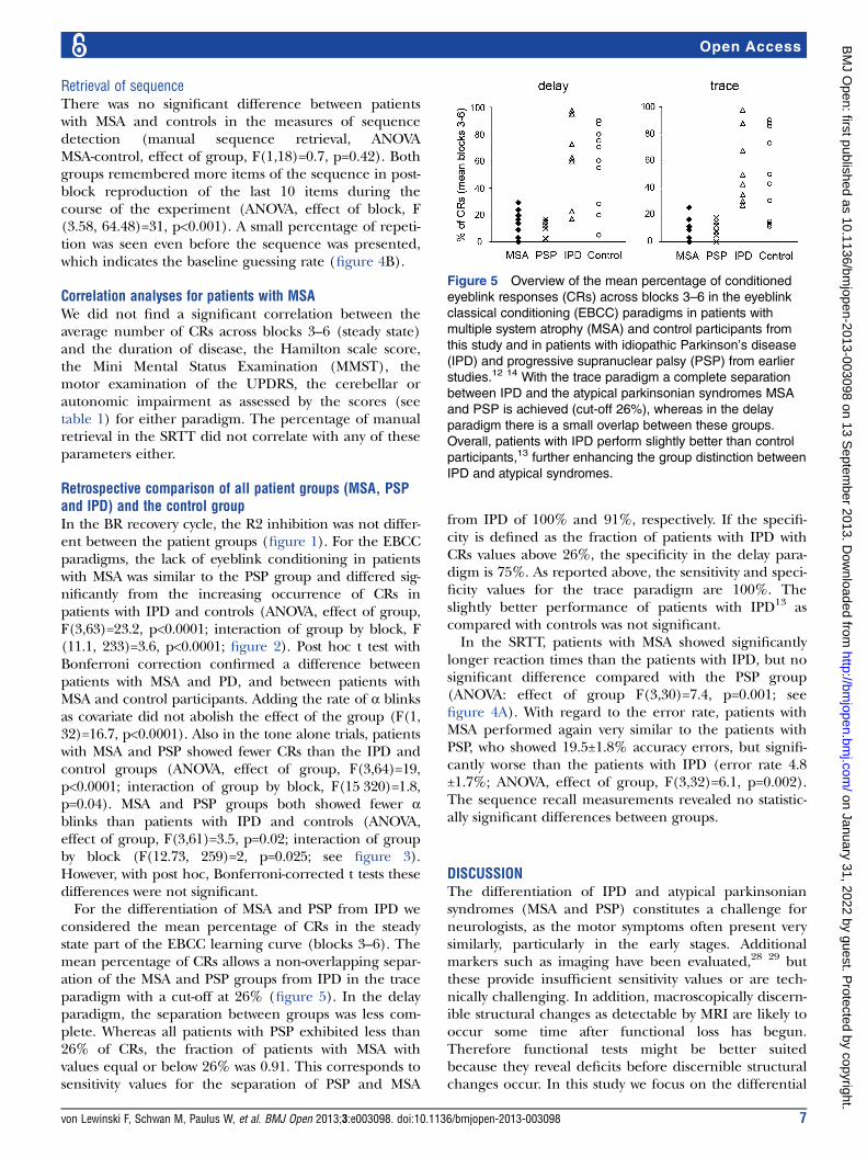
considered the mean percentage of CRs in the steadystate part of the EBCC learning curve (blocks 3–6). Themean percentage of CRs allows a non-overlapping separ-ation of the MSA and PSP groups from IPD in the traceparadigm with a cut-off at 26% (figure 5). In the delayparadigm, the separation between groups was less com-plete. Whereas all patients with PSP exhibited less than26% of CRs, the fraction of patients with MSA withvalues equal or below 26% was 0.91. This corresponds tosensitivity values for the separation of PSP and MSA
from IPD of 100% and 91%, respectively. If the specifi-city is defined as the fraction of patients with IPD withCRs values above 26%, the specificity in the delay para-digm is 75%. As reported above, the sensitivity and speci-ficity values for the trace paradigm are 100%. Theslightly better performance of patients with IPD13 ascompared with controls was not significant.In the SRTT, patients with MSA showed significantly
longer reaction times than the patients with IPD, but nosignificant difference compared with the PSP group(ANOVA: effect of group F(3,30)=7.4, p=0.001; seefigure 4A). With regard to the error rate, patients withMSA performed again very similar to the patients withPSP, who showed 19.5±1.8% accuracy errors, but signifi-cantly worse than the patients with IPD (error rate 4.8±1.7%; ANOVA, effect of group, F(3,32)=6.1, p=0.002).The sequence recall measurements revealed no statistic-ally significant differences between groups.
DISCUSSIONThe differentiation of IPD and atypical parkinsoniansyndromes (MSA and PSP) constitutes a challenge forneurologists, as the motor symptoms often present verysimilarly, particularly in the early stages. Additionalmarkers such as imaging have been evaluated,28 29 butthese provide insufficient sensitivity values or are tech-nically challenging. In addition, macroscopically discern-ible structural changes as detectable by MRI are likely tooccur some time after functional loss has begun.Therefore functional tests might be better suitedbecause they reveal deficits before discernible structuralchanges occur. In this study we focus on the differential
Figure 5 Overview of the mean percentage of conditioned
eyeblink responses (CRs) across blocks 3–6 in the eyeblink
classical conditioning (EBCC) paradigms in patients with
multiple system atrophy (MSA) and control participants from
this study and in patients with idiopathic Parkinson’s disease
(IPD) and progressive supranuclear palsy (PSP) from earlier
studies.12 14 With the trace paradigm a complete separation
between IPD and the atypical parkinsonian syndromes MSA
and PSP is achieved (cut-off 26%), whereas in the delay
paradigm there is a small overlap between these groups.
Overall, patients with IPD perform slightly better than control
participants,13 further enhancing the group distinction between
IPD and atypical syndromes.
von Lewinski F, Schwan M, Paulus W, et al. BMJ Open 2013;3:e003098. doi:10.1136/bmjopen-2013-003098 7
Open Access
on January 31, 2022 by guest. Protected by copyright.
http://bmjopen.bm
j.com/
BM
J Open: first published as 10.1136/bm
jopen-2013-003098 on 13 Septem
ber 2013. Dow
nloaded from

learning abilities tested by eyeblink conditioning(EBCC) and an SRTT. First, the results of the patientswith MSA will be discussed, followed by a comparisonwith PSP and the putative impact for differentiationfrom IPD.The patients with MSA showed severely impaired impli-
cit learning in the trace as well as in the delay eyeblinkconditioning paradigm, with SDs in the range of otherstudies,10 30 whereas non-learning associated BR latenciesas indicators of oculomotor pathways were normal.Histopathological alterations in both subtypes of MSA
include the substantia nigra, putamen, descending andascending fibre tracts of the motor system, olivary andpontine nuclei, brainstem-cerebellar circuits as well ascerebellar structures (hemispheres and vermis).31–33 Thishas been confirmed in vivo by diffusion tensor imagingof white matter microstructure.34 We suggest that damageof cerebellar structures and associated brainstem-cerebellar circuits are responsible for the failure of EBCClearning in patients with MSA. This assumption is sup-ported by the findings of impaired EBCC in patients withcerebellar damage,27 35–37 positron-emission tomographymeasurements in healthy humans showing changes inglucose metabolism in the cerebellum and pons duringEBCC23 38 as well as in experiments studying the influ-ence of selective pharmacological blockade of cerebellarinput on EBCC in rabbits.39 Most patients in our studywere clinically characterised by MSA-P and not MSA-C.However, the failure in BR acquisition in both groupspoints to a subclinical cerebellar involvement in MSA-P,which is in accordance with the histopathologicalstudies.32 33 EBCC therefore seems to detect cerebellarinvolvement at a subclinical stage.In addition to the cerebellum, several studies indicate
that acquisition of CR in the trace paradigm depends onforebrain areas such as the hippocampus or prefrontalcortex, in particular for longer ISIs.40 41 In our study,the failure of CR acquisition in patients with MSA wasslightly more pronounced in the trace compared withthe delay paradigm. Therefore, lesions of the frontallobe, which have been suggested by neuropsychologicaltesting6 42 and confirmed histopathologically in a varietyof MSA cases,43 44 may have contributed to impairedEBCC acquisition in the trace paradigm.An alternative explanation that was brought up by an
anonymous reviewer is that the tone may be a less salientCS to the patients with MSA than to the control group.The reduced number of α blinks would support thisassumption. Following that very elegant line of thought,the EBCC group difference between patients with MSAand control participants would have to do less with impli-cit learning and more with responsiveness and associativeprocesses related to external stimuli. While this may havesome relevance, adding the number of α blinks to theANOVAs on conditioned eyeblink responses did notabolish the between-group differences.In the SRTT the patients with MSA showed severe
impairment in terms of prolonged reaction time, high
error rate and, in some patients, early discontinuationdue to fatigue and exhaustion. In contrast to the controlgroup they showed no significant reaction time increasebetween blocks 6 (random) and 7 (sequence) and thisis indicative of implicit learning deficits. On the otherhand they showed good performance on the parametersof sequence recall (explicit learning). This preservationof SRTT explicit learning parts may be explained by therelative preservation of posterior association (temporaland parietal) cortex and hippocampus in MSA. It has tobe interpreted with some caution, though, given limita-tions of spatial working memory in MSA.42 However, thevalidity of the SRTT learning results is limited by the dis-continuation of patients and our ‘last observationcarried forward approach’ (see Methods section). Inaddition, the patients’ wide range of motor impairment,which may interfere with the motor part of the task, andthe fact that sequence learning and movement prepar-ation seem to share similar attentional and workingmemory resources45 have to be considered. Thereforethe SRTT seems to be inappropriate to assess learningabilities in patients with MSA. This is in contrast to theEBCC, which is independent of the motor performanceof patients. Furthermore, EBCC circuits are located ana-tomically closer to the affected brainstem regions.With all the limitations of such a retrospective com-
parison of data acquired in different patient groups bythe same authors, the implicit learning impairment inpatients with MSA as clearly revealed by the EBCC andwith some limitation by the SRTT parallels the previ-ously reported findings in patients with PSP.14
Interestingly, MSA and PSP are characterised by differ-ent histopathological alterations, α-synuclein-positiveinclusions versus τ-positive aggregations, which lead tothe presumption of different pathophysiologicalmechanisms. However, the common involvement ofcerebellar structures in both diseases31 46 seems to beresponsible for the clinical phenomenology independ-ent of the cellular mechanism.In contrast to patients with MSA and PSP, patients with
IPD show normal12 or even enhanced13 acquisition ofCRs in EBCC. EBCC may therefore contribute to distin-guish IPD from these atypical parkinsonian syndromes.As the development of cerebellar and related neuropath-ology in MSA or PSP often occurs prior to or evenwithout clinical manifestation,33 46 we propose impairedEBCC particularly in the trace paradigm as an early indi-cator of neurodegeneration of areas beyond those typic-ally affected in IPD. However, as the retrospectivesynopsis is clearly limited, the pivotal questions ofwhether EBCC can serve as predictor for the develop-ment of typical or atypical disease and whether EBCC isa useful addition to imaging techniques in establishingan early differential diagnosis are unanswered yet andrequire further prospective investigation.
Acknowledgements The authors would like to thank Professor Mark Hallettfor commenting on an earlier draft of the manuscript.
8 von Lewinski F, Schwan M, Paulus W, et al. BMJ Open 2013;3:e003098. doi:10.1136/bmjopen-2013-003098
Open Access
on January 31, 2022 by guest. Protected by copyright.
http://bmjopen.bm
j.com/
BM
J Open: first published as 10.1136/bm
jopen-2013-003098 on 13 Septem
ber 2013. Dow
nloaded from
Contributors All authors were involved in substantial contributions toconception and design, acquisition of the data or analysis and interpretationof the data; drafting the article or revising it critically for important intellectualcontent; and final approval of the version to be published. MSo, CT and WPconceived the study. MSc, FvL and MSo were responsible for obtaining thedata. FvL and MSo conducted the data analysis. All authors contributed todecisions on the interpretation of results. FvL and MSo contributed to thedrafting of the manuscript. CT and WP were responsible for editing andproviding guidance on the paper. All authors were responsible for criticallyrevising the paper; and approved the final version of the manuscript prior tosubmission.
Funding This work was supported by the Deutsche Forschungsgemeinschaft(DFG, grant SO 429/2-2 (MSo)), the Bernstein Center for ComputationalNeuroscience (grant # 01GQ0432 (WP)) and the University of Göttingen(Heidenreich von Siebold-Programm (FvL)).
Competing interests None.
Ethics approval Ethics committee of the Medical Faculty of the University ofGoettingen.
Provenance and peer review Not commissioned; externally peer reviewed.
Data sharing statement No additional data are available.
Open Access This is an Open Access article distributed in accordance withthe Creative Commons Attribution Non Commercial (CC BY-NC 3.0) license,which permits others to distribute, remix, adapt, build upon this work non-commercially, and license their derivative works on different terms, providedthe original work is properly cited and the use is non-commercial. See: http://creativecommons.org/licenses/by-nc/3.0/
REFERENCES1. Kollensperger M, Geser F, Ndayisaba JP, et al. Presentation,
diagnosis, and management of multiple system atrophy in Europe:final analysis of the European multiple system atrophy registry. MovDisord 2010;25:2604–12.
2. Gilman S, Wenning GK, Low PA, et al. Second consensusstatement on the diagnosis of multiple system atrophy. Neurology2008;71:670–6.
3. Poewe W, Wenning G. The differential diagnosis of Parkinson’sdisease. Eur J Neurol 2002;9(Suppl 3):23–30.
4. Hughes AJ, Daniel SE, Ben-Shlomo Y, et al. The accuracy ofdiagnosis of parkinsonian syndromes in a specialist movementdisorder service. Brain 2002;125(Pt 4):861–70.
5. Lange KW, Tucha O, Alders GL, et al. Differentiation of parkinsoniansyndromes according to differences in executive functions. J NeuralTransm 2003;110:983–95.
6. Burk K, Daum I, Rub U. Cognitive function in multiple systematrophy of the cerebellar type. Mov Disord 2006;21:772–6.
7. Robbins TW, James M, Owen AM, et al. Cognitive deficits inprogressive supranuclear palsy, Parkinson’s disease, and multiplesystem atrophy in tests sensitive to frontal lobe dysfunction. J NeurolNeurosurg Psychiatry 1994;57:79–88.
8. Kehagia AA, Barker RA, Robbins TW. Neuropsychological andclinical heterogeneity of cognitive impairment and dementia inpatients with Parkinson’s disease. Lancet Neurol 2010;9:1200–13.
9. Balas M, Balash Y, Giladi N, et al. Cognition in multiple systematrophy: neuropsychological profile and interaction with mood.J Neural Transm 2010;117:369–75.
10. Hoffland BS, Kassavetis P, Bologna M, et al. Cerebellum-dependentassociative learning deficits in primary dystonia are normalized byrTMS and practice. Eur J Neurosci 2013;2013:1–6.
11. Christian KM, Thompson RF. Neural substrates ofeyeblink conditioning: acquisition and retention. Learn Mem2003;10:427–55.
12. Sommer M, Grafman J, Clark K, et al. Learning in Parkinson’sdisease: eyeblink conditioning, declarative learning, and procedurallearning. J Neurol Neurosurg Psychiatry 1999;67:27–34.
13. Daum I, Schugens MM, Breitenstein C, et al. Classicaleyeblink conditioning in Parkinson’s disease. Mov Disord1996;11:639–46.
14. Sommer M, Grafman J, Litvan I, et al. Impairment of eyeblinkclassical conditioning in progressive supranuclear palsy. Mov Disord2001;16:240–51.
15. Soliveri P, Brown RG, Jahanshahi M, et al. Learning manual pursuittracking skills in patients with Parkinson’s disease. Brain 1997;120(Pt 8):1325–37.
16. Hwang EJ, Smith MA, Shadmehr R. Dissociable effects of theimplicit and explicit memory systems on learning control of reaching.Exp Brain Res 2006;173:425–37.
17. Tomlinson CL, Stowe R, Patel S, et al. Systematic review oflevodopa dose equivalency reporting in Parkinson’s disease. MovDisord 2010;25:2649–53.
18. Hamilton M. A rating scale for depression. J Neurol NeurosurgPsychiatry 1960;23:56–62.
19. Folstein MF, Folstein SE, McHugh PR. ‘Mini-mental state’.A practical method for grading the cognitive state of patients for theclinician. J Psychiatr Res 1975;12:189–98.
20. Fahn S, Elton R; Committee amotUD. Recent developments inParkinson’s disease. Florham Park, NJ: Macmillan HealthcareInformation, 1987.
21. Sommer M, Wobker G, Ferbert A. Voluntary eyelid contractionmodifies the blink reflex recovery cycle. Acta Neurol Scand1998;98:29–35.
22. Kimura J. The blink reflex. In: Kimura J, ed. Electrodiagnosis indiseases of nerve and muscle: principles and practice. Philadelphia:Davis, F. A., 1989:307–31.
23. Molchan SE, Sunderland T, McIntosh AR, et al. A functionalanatomical study of associative learning in humans. Proc Natl AcadSci U S A 1994;91:8122–6.
24. Pascual-Leone A, Grafman J, Clark K, et al. Procedural learning inParkinson’s disease and cerebellar degeneration. Ann Neurol1993;34:594–602.
25. Moisello C, Crupi D, Tunik E, et al. The serial reaction time taskrevisited: a study on motor sequence learning with an arm-reachingtask. Exp Brain Res 2009;194:143–55.
26. Bo J, Jennett S, Seidler RD. Working memory capacity correlateswith implicit serial reaction time task performance. Exp Brain Res2011;214:73–81.
27. Topka H, Valls-Sole J, Massaquoi SG, et al. Deficit in classicalconditioning in patients with cerebellar degeneration. Brain 1993;116(Pt 4):961–9.
28. Mahlknecht P, Hotter A, Hussl A, et al. Significance of MRI indiagnosis and differential diagnosis of Parkinson’s disease.Neurodegener Dis 2010;7:300–18.
29. von Lewinski F, Werner C, Jorn T, et al. T2*-weighted MRI indiagnosis of multiple system atrophy. A practical approach forclinicians. J Neurol 2007;254:1184–8.
30. Holloway JL, Trivedi P, Myers CE, et al. Enhanced conditionedeyeblink response acquisition and proactive interference in anxietyvulnerable individuals. Front Behav Neurosci 2012;6:1–8.
31. Braak H, Rub U, Del Tredici K. Involvement of precerebellar nuclei inmultiple system atrophy. Neuropathol Appl Neurobiol 2003;29:60–76.
32. Wenning GK, Tison F, Elliott L, et al. Olivopontocerebellar pathologyin multiple system atrophy. Mov Disord 1996;11:157–62.
33. Ozawa T, Paviour D, Quinn NP, et al. The spectrum of pathologicalinvolvement of the striatonigral and olivopontocerebellar systems inmultiple system atrophy: clinicopathological correlations. Brain2004;127(Pt 12):2657–71.
34. Nair SR, Tan LK, Mohd Ramli N, et al. A decision tree fordifferentiating multiple system atrophy from Parkinson’s diseaseusing 3-T MR imaging. Eur Radiol 2013;10:1459–66.
35. Gerwig M, Dimitrova A, Kolb FP, et al. Comparison of eyeblinkconditioning in patients with superior and posterior inferior cerebellarlesions. Brain 2003;126(Pt 1):71–94.
36. Gerwig M, Haerter K, Hajjar K, et al. Trace eyeblink conditioning inhuman subjects with cerebellar lesions. Exp Brain Res 2006;170:7–21.
37. Daum I, Schugens MM, Ackermann H, et al. Classicalconditioning after cerebellar lesions in humans. Behav Neurosci1993;107:748–56.
38. Logan CG, Grafton ST. Functional anatomy of human eyeblinkconditioning determined with regional cerebral glucose metabolismand positron-emission tomography. Proc Natl Acad Sci USA1995;92:7500–4.
39. Attwell PJ, Ivarsson M, Millar L, et al. Cerebellar mechanisms ineyeblink conditioning. Ann N Y Acad Sci 2002;978:79–92.
40. Clark RE, Squire LR. Classical conditioning and brain systems: therole of awareness. Science 1998;280:77–81.
41. Takehara-Nishiuchi K, Kawahara S, Kirino Y. NMDAreceptor-dependent processes in the medial prefrontal cortex areimportant for acquisition and the early stage of consolidation duringtrace, but not delay eyeblink conditioning. Learn Mem2005;12:606–14.
42. Robbins TW, James M, Lange KW, et al. Cognitive performance inmultiple system atrophy. Brain 1992;115(Pt 1):271–91.
von Lewinski F, Schwan M, Paulus W, et al. BMJ Open 2013;3:e003098. doi:10.1136/bmjopen-2013-003098 9
Open Access
on January 31, 2022 by guest. Protected by copyright.
http://bmjopen.bm
j.com/
BM
J Open: first published as 10.1136/bm
jopen-2013-003098 on 13 Septem
ber 2013. Dow
nloaded from

43. Konagaya M, Konagaya Y, Sakai M, et al. Progressive cerebralatrophy in multiple system atrophy. J Neurol Sci 2002;195:123–7.
44. Armstrong RA, Lantos PL, Cairns NJ. Multiple system atrophy: laminardistribution of the pathological changes in frontal and temporal neocortex—a study in ten patients. Clin Neuropathol 2005;24:230–5.
45. Marinelli L, Perfetti B, Moisello C, et al. Increased reaction timepredicts visual learning deficits in Parkinson’s disease. Mov Disord2010;25:1498–501.
46. Jellinger KA, Bancher C. Neuropathology. In: Litvan I, Agid Y, eds.Progressive supranuclear palsy. New York: University Press,1992:45–88.
47. Coblentz JM, Mattis S, Zingesser L. Presenile dementia. Clinicalaspects and evaluation of cerebrospinal fluid dynamics. Arch Neurol1973;29:299–308.
48. Schmidt R, Freidl W, Fazekas F, et al. The Mattis Dementia RatingScale: normative data from 1,001 healthy volunteers. Neurology1994;44:964–6.
49. Llebaria G, Pagonabarraga J, Kulisevsky J, et al. Cut-off score ofthe Mattis Dementia Rating Scale for screening dementia inParkinson’s disease. Mov Disord 2008;23:1546–50.
50. Beck AT, Steer RA, Garbin MG. Inventory for measuring depression.Arch Gen Psychiatry 1961;4:561–71.
10 von Lewinski F, Schwan M, Paulus W, et al. BMJ Open 2013;3:e003098. doi:10.1136/bmjopen-2013-003098
Open Access
on January 31, 2022 by guest. Protected by copyright.
http://bmjopen.bm
j.com/
BM
J Open: first published as 10.1136/bm
jopen-2013-003098 on 13 Septem
ber 2013. Dow
nloaded from