open access surgical management of cervical ... · surgical management of cervical...
TRANSCRIPT
The Open Orthopaedics Journal, 2010, 4, 39-43 39
1874-3250/10 2010 Bentham Open
Open Access
Surgical Management of Cervical Spondyloarthropathy in Hemodialysis Patients
Panayiotis Spinos1, Charalambos Matzaroglou
*,2, Meni Partheni
1, Angeliki Deli
2,
Menelaos Karanikolas3 and Dimitrios Konstantinou
4
1Department of Neurosurgery, Patras University Hospital, Rion, Greece
2Department of Orthopaedic Surgery, Patras University Hospital, Rion, Greece
3Department of Anaesthesiology and Critical Care Medicine, University of Patras School of Medicine, Rion, Greece
4Department of Neurosurgery, University of Patras School of Medicine, Rion, Greece
Abstract: Dialysis-related spondyloarthropathy is a rare cause of spinal deformity and cervical myelopathy. Optimal
management of cervical spine spondyloarthropathy often requires circumferential reconstructive surgery, because affected
patients typically have both the anterior column and the facet joints compromised. The occasional presence of non-
contiguous or "skip lesions" adds an additional level of complexity to surgical management, because decompression and
fusion in an isolated segment of neural compression can worsen spine deformity by applying increased stress to adjacent
cervical spine segments. We report two cases of hemodialysis patients who presented with cervical myelopathy and
initially had anterior cervical discectomy or corpectomy. Because symptoms recurred due to hardware failure, both
patients required posterior spine fusion as well. In retrospect, because of the hardware failure, both of these patients might
have benefited from a circumferential (combined anterior and posterior) cervical spine reconstruction as their initial
treatment.
Keywords: Hemodialysis, spondyloarthropathy, surgical management.
INTRODUCTION
Since first described by Kuntz in 1984, dialysis-related spondyloarthropathy has been recognized as a rare cause of cervical myelopathy and deformity [1]. Deposition of 2 microglobulin amyloid, which is seen in nearly all hemodialysis patients at post-mortem [2], seems to be a key factor in the development of hemodialysis-related complications, because amyloid deposition in cartilage surfaces, tendons, and ligaments leads to destruction of joints and surrounding bone. Frequently encountered clinical manifestations of amyloid deposition in dialysis patients include carpal tunnel syndrome and generalized chronic arthropathy. In the spine, thickening of the posterior longitudinal ligament may cause spinal cord compression and destructive spondyloarthropathy (DSA), especially in the cervical spine [3-5], but renal osteodystrophy is also a possible cause of DSA.
Evaluation of cervical spine radiographs in hemodialysis patients demonstrates that DSA is common, but frequently asymptomatic, presumably due to the absence of deformity or neural compression [6]. Length of dialysis treatment appears to be a predictive factor for development of the disease [6]. Destruction of adjacent endplates can make differentiation from discitis and vertebral osteomyelitis difficult on imaging grounds alone [7].
*Address correspondence to this author at the Department of Orthopaedics,
University of Patras, Rion, Greece; Tel: +30 2610 999556; Fax: +30 2610
994579; E-mail: [email protected]
We present two hemodialysis patients with cervical myelopathy due to DSA. Both patients initially had anterior column surgery (discectomy or corpectomy, instrumentation and fusion) but later required revision combined with posterior instrumentation due to hardware failure and myelopathy recurrence.
PATIENT 1
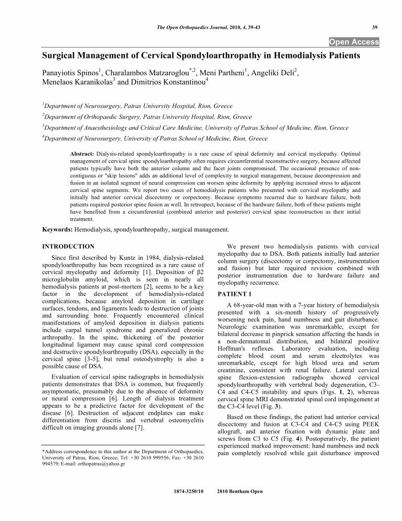
A 68-year-old man with a 7-year history of hemodialysis presented with a six-month history of progressively worsening neck pain, hand numbness and gait disturbance. Neurologic examination was unremarkable, except for bilateral decrease in pinprick sensation affecting the hands in a non-dermatomal distribution, and bilateral positive Hoffman's reflexes. Laboratory evaluation, including complete blood count and serum electrolytes was unremarkable, except for high blood urea and serum creatinine, consistent with renal failure. Lateral cervical spine flexion-extension radiographs showed cervical spondyloarthropathy with vertebral body degeneration, C3-C4 and C4-C5 instability and spurs (Figs. 1, 2), whereas cervical spine MRI demonstrated spinal cord impingement at the C3-C4 level (Fig. 3).
Based on these findings, the patient had anterior cervical discectomy and fusion at C3-C4 and C4-C5 using PEEK allograft, and anterior fixation with dynamic plate and screws from C3 to C5 (Fig. 4). Postoperatively, the patient experienced marked improvement: hand numbness and neck pain completely resolved while gait disturbance improved
40 The Open Orthopaedics Journal, 2010, Volume 4 Spinos et al.
Fig. (1). Lateral cervical spine extension X-Ray showing
spondyloarthropathy with vertebral body degeneration, C3-C4 and
C4-C5 instability and spurs.
Fig. (2). Lateral cervical spine flexion X-Ray showing
spondyloarthropathy with vertebral body degeneration, C3-C4 and
C4-C5 instability and spurs.
Fig. (3). Cervical spine MRI demonstrating spinal cord
impingement at the C3-C4 level. Red arrows show the point of cord
impingement.
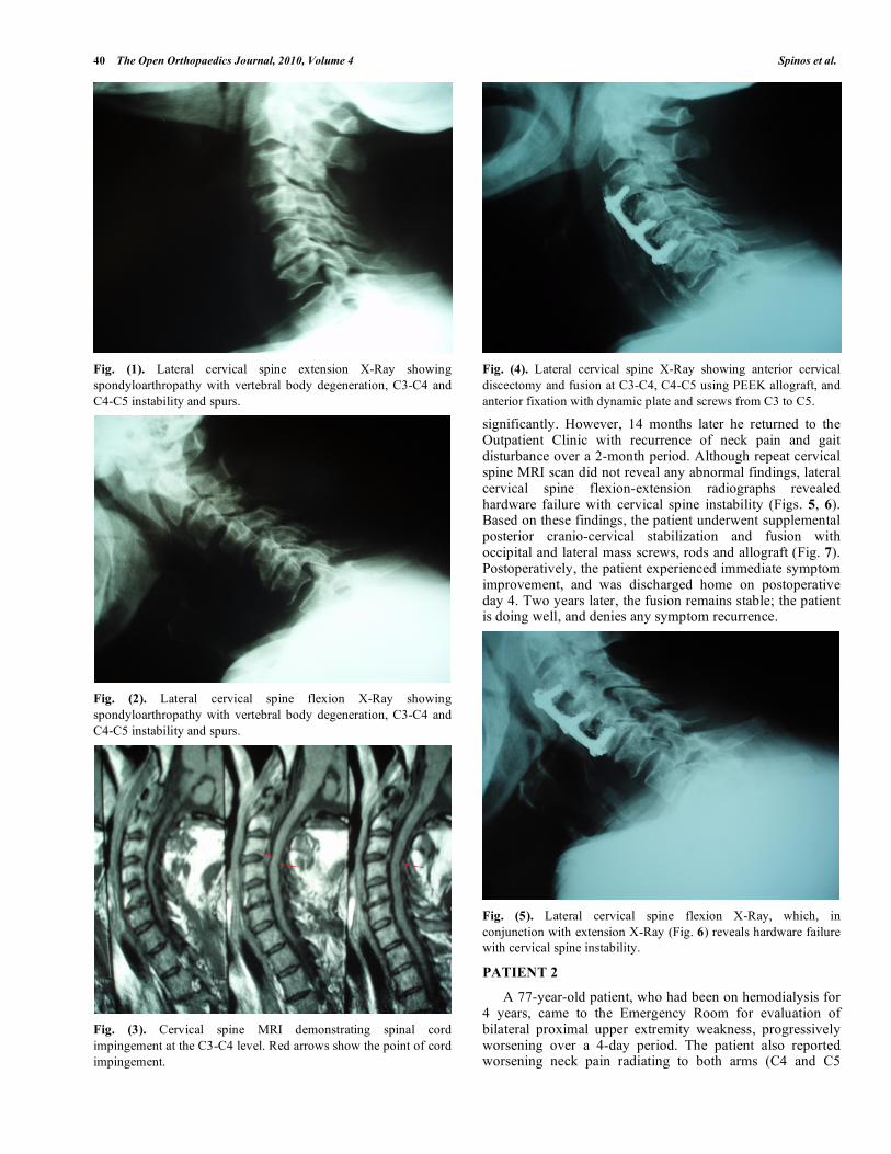
Fig. (4). Lateral cervical spine X-Ray showing anterior cervical
discectomy and fusion at C3-C4, C4-C5 using PEEK allograft, and
anterior fixation with dynamic plate and screws from C3 to C5.
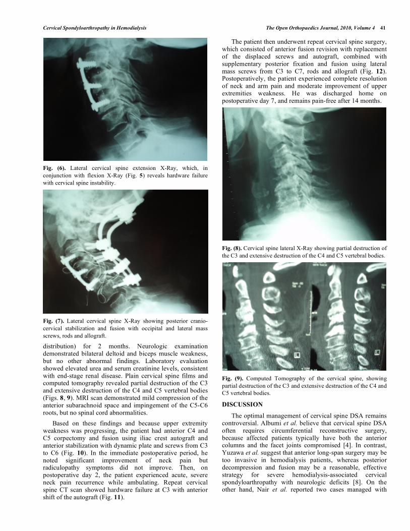
significantly. However, 14 months later he returned to the Outpatient Clinic with recurrence of neck pain and gait disturbance over a 2-month period. Although repeat cervical spine MRI scan did not reveal any abnormal findings, lateral cervical spine flexion-extension radiographs revealed hardware failure with cervical spine instability (Figs. 5, 6). Based on these findings, the patient underwent supplemental posterior cranio-cervical stabilization and fusion with occipital and lateral mass screws, rods and allograft (Fig. 7). Postoperatively, the patient experienced immediate symptom improvement, and was discharged home on postoperative day 4. Two years later, the fusion remains stable; the patient is doing well, and denies any symptom recurrence.
Fig. (5). Lateral cervical spine flexion X-Ray, which, in
conjunction with extension X-Ray (Fig. 6) reveals hardware failure
with cervical spine instability.
PATIENT 2
A 77-year-old patient, who had been on hemodialysis for 4 years, came to the Emergency Room for evaluation of bilateral proximal upper extremity weakness, progressively worsening over a 4-day period. The patient also reported worsening neck pain radiating to both arms (C4 and C5
Cervical Spondyloarthropathy in Hemodialysis The Open Orthopaedics Journal, 2010, Volume 4 41
Fig. (6). Lateral cervical spine extension X-Ray, which, in
conjunction with flexion X-Ray (Fig. 5) reveals hardware failure
with cervical spine instability.
Fig. (7). Lateral cervical spine X-Ray showing posterior cranio-
cervical stabilization and fusion with occipital and lateral mass
screws, rods and allograft.
distribution) for 2 months. Neurologic examination demonstrated bilateral deltoid and biceps muscle weakness, but no other abnormal findings. Laboratory evaluation showed elevated urea and serum creatinine levels, consistent with end-stage renal disease. Plain cervical spine films and computed tomography revealed partial destruction of the C3 and extensive destruction of the C4 and C5 vertebral bodies (Figs. 8, 9). MRI scan demonstrated mild compression of the anterior subarachnoid space and impingement of the C5-C6 roots, but no spinal cord abnormalities.
Based on these findings and because upper extremity weakness was progressing, the patient had anterior C4 and C5 corpectomy and fusion using iliac crest autograft and anterior stabilization with dynamic plate and screws from C3 to C6 (Fig. 10). In the immediate postoperative period, he noted significant improvement of neck pain but radiculopathy symptoms did not improve. Then, on postoperative day 2, the patient experienced acute, severe neck pain recurrence while ambulating. Repeat cervical spine CT scan showed hardware failure at C3 with anterior shift of the autograft (Fig. 11).
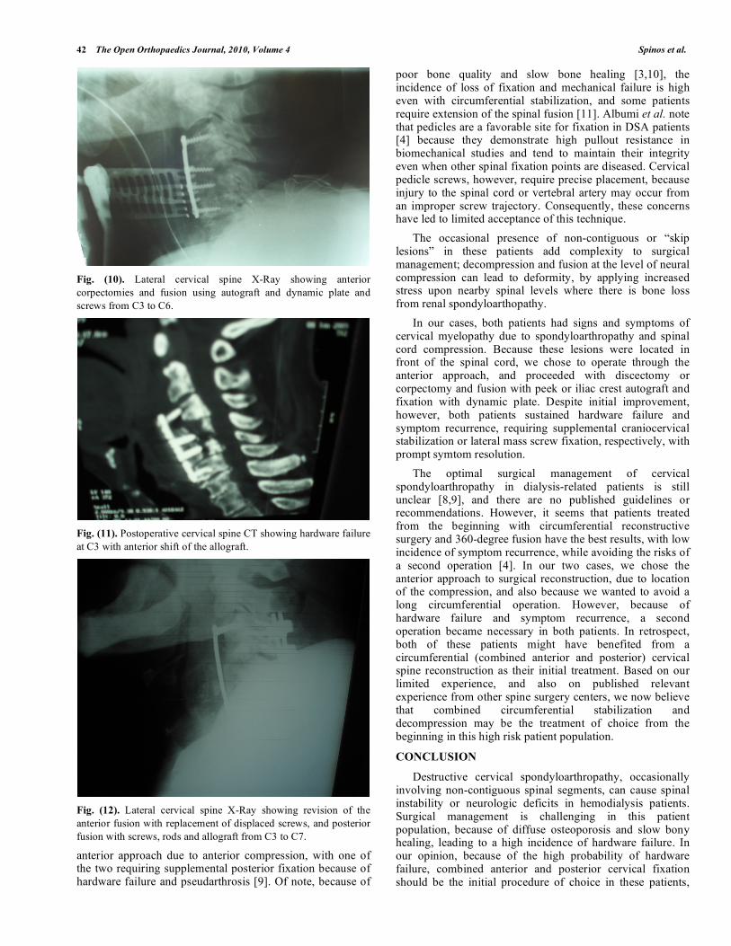
The patient then underwent repeat cervical spine surgery, which consisted of anterior fusion revision with replacement of the displaced screws and autograft, combined with supplementary posterior fixation and fusion using lateral mass screws from C3 to C7, rods and allograft (Fig. 12). Postoperatively, the patient experienced complete resolution of neck and arm pain and moderate improvement of upper extremities weakness. He was discharged home on postoperative day 7, and remains pain-free after 14 months.
Fig. (8). Cervical spine lateral X-Ray showing partial destruction of
the C3 and extensive destruction of the C4 and C5 vertebral bodies.
Fig. (9). Computed Tomography of the cervical spine, showing
partial destruction of the C3 and extensive destruction of the C4 and
C5 vertebral bodies.
DISCUSSION
The optimal management of cervical spine DSA remains controversial. Albumi et al. believe that cervical spine DSA often requires circumferential reconstructive surgery, because affected patients typically have both the anterior columns and the facet joints compromised [4]. In contrast, Yuzawa et al. suggest that anterior long-span surgery may be too invasive in hemodialysis patients, whereas posterior decompression and fusion may be a reasonable, effective strategy for severe hemodialysis-associated cervical spondyloarthropathy with neurologic deficits [8]. On the other hand, Nair et al. reported two cases managed with
42 The Open Orthopaedics Journal, 2010, Volume 4 Spinos et al.
Fig. (10). Lateral cervical spine X-Ray showing anterior
corpectomies and fusion using autograft and dynamic plate and
screws from C3 to C6.
Fig. (11). Postoperative cervical spine CT showing hardware failure
at C3 with anterior shift of the allograft.
Fig. (12). Lateral cervical spine X-Ray showing revision of the
anterior fusion with replacement of displaced screws, and posterior
fusion with screws, rods and allograft from C3 to C7.
anterior approach due to anterior compression, with one of the two requiring supplemental posterior fixation because of hardware failure and pseudarthrosis [9]. Of note, because of
poor bone quality and slow bone healing [3,10], the incidence of loss of fixation and mechanical failure is high even with circumferential stabilization, and some patients require extension of the spinal fusion [11]. Albumi et al. note that pedicles are a favorable site for fixation in DSA patients [4] because they demonstrate high pullout resistance in biomechanical studies and tend to maintain their integrity even when other spinal fixation points are diseased. Cervical pedicle screws, however, require precise placement, because injury to the spinal cord or vertebral artery may occur from an improper screw trajectory. Consequently, these concerns have led to limited acceptance of this technique.
The occasional presence of non-contiguous or “skip lesions” in these patients add complexity to surgical management; decompression and fusion at the level of neural compression can lead to deformity, by applying increased stress upon nearby spinal levels where there is bone loss from renal spondyloarthopathy.
In our cases, both patients had signs and symptoms of cervical myelopathy due to spondyloarthropathy and spinal cord compression. Because these lesions were located in front of the spinal cord, we chose to operate through the anterior approach, and proceeded with discectomy or corpectomy and fusion with peek or iliac crest autograft and fixation with dynamic plate. Despite initial improvement, however, both patients sustained hardware failure and symptom recurrence, requiring supplemental craniocervical stabilization or lateral mass screw fixation, respectively, with prompt symtom resolution.
The optimal surgical management of cervical spondyloarthropathy in dialysis-related patients is still unclear [8,9], and there are no published guidelines or recommendations. However, it seems that patients treated from the beginning with circumferential reconstructive surgery and 360-degree fusion have the best results, with low incidence of symptom recurrence, while avoiding the risks of a second operation [4]. In our two cases, we chose the anterior approach to surgical reconstruction, due to location of the compression, and also because we wanted to avoid a long circumferential operation. However, because of hardware failure and symptom recurrence, a second operation became necessary in both patients. In retrospect, both of these patients might have benefited from a circumferential (combined anterior and posterior) cervical spine reconstruction as their initial treatment. Based on our limited experience, and also on published relevant experience from other spine surgery centers, we now believe that combined circumferential stabilization and decompression may be the treatment of choice from the beginning in this high risk patient population.
CONCLUSION
Destructive cervical spondyloarthropathy, occasionally involving non-contiguous spinal segments, can cause spinal instability or neurologic deficits in hemodialysis patients. Surgical management is challenging in this patient population, because of diffuse osteoporosis and slow bony healing, leading to a high incidence of hardware failure. In our opinion, because of the high probability of hardware failure, combined anterior and posterior cervical fixation should be the initial procedure of choice in these patients,

Cervical Spondyloarthropathy in Hemodialysis The Open Orthopaedics Journal, 2010, Volume 4 43
thereby avoiding the risk and expense of a second operation in this high risk cohort. However, as not all experts agree on this, and there is clearly some controversy in the literature, more data, preferably from large, rigorous prospective studies are required before the controversy settles and guidelines or consensus statements can be formulated.
CONFLICT OF INTEREST STATEMENT
This work was supported solely by Department funds. All authors declare they have no conflict of interest to report.
REFERENCES
[1] Kuntz D, Naveau B, Bardin T, Drueke T, Treves R, Dryll A. Destructive spondylarthropathy in hemodialyzed patients. A new
syndrome. Arthritis Rheum 1984; 27(4): 369-75. [2] Danesh F, Ho LT. Dialysis-related amyloidosis: history and clinical
manifestations. Semin Dial 2001; 14(2): 80-5. [3] Cuffe MJ, Hadley MN, Herrera GA, Morawetz RB. Dialysis-
associated spondylarthropathy. Report of 10 cases. J Neurosurg 1994; 80(4): 694-700.
[4] Abumi K, Ito M, Kaneda K. Surgical treatment of cervical destructive spondyloarthropathy (DSA). Spine 2000; 25(22): 2899-
905.
[5] Shiota E, Naito M, Tsuchiya K. Surgical therapy for dialysis-
related spondyloarthropathy: Review of 30 cases. J Spinal Disord 2001; 14(2): 165-71.
[6] Leone A, Sundaram M, Cerase A, Magnavita N, Tazza L, Marano P. Destructive spondyloarthropathy of the cervical spine in long-
term hemodialyzed patients: A five-year clinical radiological prospective study. Skeletal Radiol 2001; 30(8): 431-41.
[7] Flipo RM, Cotten A, Chastanet P, et al. Evaluation of destructive spondyloarthropathies in hemodialysis by computerized
tomographic scan and magnetic resonance imaging. J Rheumatol 1996; 23(5): 869-73.
[8] Yuzawa Y, Kamimura M, Nakagawa H, et al. Surgical treatment with instrumentation for severely destructive spondyloarthropathy
of cervical spine. J Spinal Disord Tech 2005; 18(1): 23-8. [9] Nair S, Vender J, McCormack TM, Black P. Renal osteodystrophy
of the cervical spine: Neurosurgical implications. Neurosurgery 1993; 33(3): 349-54; Discussion 354-5.
[10] Kumar A, Leventhal MR, Freedman EL, Coburn J, Delamarter R. Destructive spondyloarthropathy of the cervical spine in patients
with chronic renal failure. Spine 1997; 22(5): 573-7; Discussion 78. [11] Sudo H, Ito M, Abumi K, et al. Long-term follow up of surgical
outcomes in patients with cervical disorders undergoing hemodialysis. J Neurosurg Spine 2006; 5(4): 313-9.
Received: December 2, 2009 Revised: December 21, 2009 Accepted: December 22, 2009
© Spinos et al.; Licensee Bentham Open.
This is an open access article licensed under the terms of the Creative Commons Attribution Non-Commercial License (http://creativecommons.org/licenses/by-nc/3.0/) which permits unrestricted, non-commercial use, distribution and reproduction in any medium, provided the work is properly cited.