openeds: open eye dataset - arxiv.org
TRANSCRIPT

OpenEDS: Open Eye Dataset
Stephan J. Garbin∗1, Yiru Shen2, Immo Schuetz2, Robert Cavin2, Gregory Hughes†3, and Sachin S.Talathi‡2
1University College London2Facebook Reality Labs
3Google via Adecco
Abstract
We present a large scale data set, OpenEDS: Open EyeDataset, of eye-images captured using a virtual-reality (VR)head mounted display mounted with two synchronized eye-facing cameras at a frame rate of 200 Hz under controlledillumination. This dataset is compiled from video cap-ture of the eye-region collected from 152 individual partic-ipants and is divided into four subsets: (i) 12,759 imageswith pixel-level annotations for key eye-regions: iris, pupiland sclera (ii) 252,690 unlabelled eye-images, (iii) 91,200frames from randomly selected video sequence of 1.5 sec-onds in duration and (iv) 143 pairs of left and right pointcloud data compiled from corneal topography of eye re-gions collected from a subset, 143 out of 152, participantsin the study. A baseline experiment has been evaluated onOpenEDS for the task of semantic segmentation of pupil,iris, sclera and background, with the mean intersection-over-union (mIoU) of 98.3 %. We anticipate that OpenEDSwill create opportunities to researchers in the eye trackingcommunity and the broader machine learning and computervision community to advance the state of eye-tracking forVR applications. The dataset is available for downloadupon request at https://research.fb.com/programs/openeds-challenge
1. IntroductionUnderstanding the motion and appearance of the human
eye is of great importance to many scientific fields [14]. Forexample, gaze direction can offer information about the fo-cus of a person’s attention [4], as well as certain aspects oftheir physical and psychological well-being [12], which in
∗This work was done during internship at Facebook Reality Labs.†This work was done during employment at Facebook Reality Labs.‡Corresponding author
turn can facilitates the research on eye tracking to aid in thestudy and design of how humans interact with their environ-ment [39].
In the context of virtual reality (VR), accurate and pre-cise eye tracking can enable game-changing technologicaladvances, for example, foveated rendering, a technique thatexploits the sensitivity profile of the human eye to renderonly those parts of a virtual scene at full resolution that theuser is focused on, can significantly alleviate the computa-tional burden of VR [30]. Head-mounted displays (HMDs)that include gaze-contingent variable focus [21] and gaze-driven rendering of perceptually accurate focal blur [47]promise to alleviate vergence-accommodation-conflict andincrease visual realism. Finally, gaze-driven interactionschemes could enable novel methods of navigating and in-teracting with virtual environments [40].
The success of data driven machine learning modelslearned directly from images ([22], [13]) is accompanied bythe demand for datasets of sufficient size and variety to cap-ture the distribution of natural images sufficiently for thetask at hand [37]. While being able to source vast collec-tions of images from freely available online data has led tothe successful creation of datasets such as ImageNet [22]and COCO [24], many other research areas require specialequipment and expert knowledge for data capture. For ex-ample, the creation of the KITTI dataset required synchro-nization of cameras alongside a laser scanner and localiza-tion equipment ([11, 10]. We seek to address this challengefor eye-tracking with HMDs.
Commercially available eye tracking systems often donot expose the raw camera image to the user, complicatingthe generation of large-scale eye image datasets. We optto capture eye-images using a custom-built VR HMD withtwo synchronized cameras operating at 200Hz under con-trolled illumination. As outlined below, we also use special-ist medical equipment to capture further information aboutthe shape and optical properties of each participant’s eyes
1
arX
iv:1
905.
0370
2v2
[cs
.CV
] 1
7 M
ay 2
019

Figure 1: Examples of the acquired HMD images.
contained in the dataset. It is the unique combination ofadvanced data capture, usually only performed in a clinicalsetting, with high resolution eye images and correspondingannotation masks for key eye-regions that sets OpenEDSapart from comparable datasets. We hope that OpenEDSbridges the gap between the vision and eye tracking com-munities and provides novel opportunities for research inthe domain of eye-tracking for HMDs.
Our contributions are summarized as follows:
• A large scale dataset captured using an HMD withtwo synchronized cameras under controlled illumina-tion and high frame rates;
• A large scale dataset of annotation masks for key eye-regions: the iris, the sclera and the pupil;
• point cloud data from corneal topography captures ofeye regions.
2. Related WorkEye Tracking Datasets:
Due to the difficulty of capturing binocular eye data es-pecially in the VR context, there exists only a limited num-ber of large-scale high resolution human image datasets inthis domain. A comparison of our dataset to some existingdatasets can be found in Table 1.
Figure 2: Corneal topography of left eye.
Figure 3: Examples of corneal topography, represented aspoint clouds. Row from top to bottom: left eye, right eye.Column from left to right: rotations along Y-axis. Colorvariation is along Z-axis. Better viewed in color.
The most similar dataset in terms of domain and im-age specifications is the recently published NVGaze dataset[19], consisting of 2.5 million infrared images recordedfrom 30 participants using an HMD (640x480 at 30 Hz).NVGaze includes annotation masks for key eye-regions foran additional dataset of 2 million synthetic eye images butdoes not provide segmentation annotations for the humaneye image set at this point. The LPW dataset [42] includesa number of images recorded from 22 participants wearinga head mounted camera. Images are from indoor and out-door recordings with varying lighting conditions and thusvery different from the controlled lighting conditions in aVR HMD.
Some eye focused image sets are aimed at gaze predic-tion and released with gaze direction information, but donot include annotation masks, such as the Point of Gaze(PoG) dataset [28]. Other large-scale eye image datasetswere captured in the context of appearance-based gazeestimation and record the entire face using RGB camerasas opposed to the eye region [9, 17, 48]. For example,Gaze Capture [20] consists of over 2.5 million imagesat various resolutions, recorded through crowd-sourcingon mobile devices, and its images are not specificallyfocused on the eye but contain a large portion of thesurrounding face. In all these datasets, the focus is notsolely on captures of eye-images, making them lesssuitable for the specific computer vision and machinelearning challenges in the VR context. Finally, a differentcategory of dataset, such as the UBIRIS [32] and UBIRISv2 [33], were conceived with iris recognition in mind and
2

Dataset #Images #Participants Resolution FrameRate ControlledLight
Sync.Left/Right
OptometricData
STARE [15] - - - - No No YesPoG [28] - 20 - 30 Yes No NoMASD [7] 2,624 82 - - No No NoUbiris v2 [32] 11,102 261 400×300 - No - NoLPW [42] 130,856 22 480×640 95Hz No No NoNVGaze [19] 2,500,000 30 480×640 30Hz Yes No NoGaze Capture [20] 2,500,000 1,450 Various - - No NoOurs 356,649 152 400×640 200Hz Yes Yes Yes
Table 1: Publicly available datasets in the field of eye tracking. Note that studies in [20] and [32] offer a large number ofparticipants through crowd-sourcing, where images usually include a large portion of the face and hence lack eye details.
therefore contain only limited annotation mask information.
Eye Segmentation:Segmentation of ocular biometric traits in eye region, suchas the pupil, iris, sclera, can provide the information tostudy fine grained details of eye movement such as saccade,fixation and gaze estimation [43]. A large amount of stud-ies have been investigated on segmenting a single trait (e.g.,only the iris, sclera or eye region) [36, 34, 41, 6, 26]. A de-tailed survey of iris and sclera segmentation is presented in[1, 5]. We note that, although there are several advantagesto having segmentation information on all key eye-regionssimultaneously, a very limited amount of studies have beendone on multi-class eye segmentation [35, 27]. However,study in [35] trained a convolutional encoder-decoder neu-ral network on a small data set of 120 images from 30 partic-ipants. Study in [27] trained a convolutional neural networkcoupled with conditional random field for post-processing,on a data set of 3,161 low resolution images to segment onlytwo classes: iris and sclera. In this paper, we try to addressthe gap for multi-class eye segmentation including pupil,iris, sclera and background, in a large data set of images inhigh resolution of 400×640.Eye Rendering:Generating eye appearances under various environmentalconditions including facial expressions, color of the skinand the illumination settings, play an important role in gazeestimation and tracking [19]. Two approaches have beenstudied in eye rendering: graphics-based approaches to gen-erate eye images using a 3D eye model usually with a ren-dering framework to provide geometric variations such asgaze direction or head orientation [46, 45]. Another is ma-chine learning based approach. Study in [38] used a gener-ative adversarial network to train models with synthetic eyeimages while testing on realistic images. Study in [44] useda conditional bidirectional generative adversarial network tosynthesize eye images consistent with the given eye gaze.However, these studies are focused on rendering synthetic
eye images. We are interested in rendering realistic eye im-ages that conform to the captured distribution of an individ-ual and anticipate OpenEDS will encourage researchers touse the large corpus of eye-images and the annotation masksto develop solutions that can render realistic eye images.
3. Data CollectionTo ensure eye-safety during our data-collection process,
which is important because OpenEDS exposes users’ eyesto infrared light, the exposure levels were controlled to bewell below the maximum permissible exposure as laid outin IEC 62471:2006, as well as the American National Stan-dard for the Safe use of Lasers (ANSI Z136.1-2000, (IEC)60825).
OpenEDS was collected from voluntary participants ofages between 19 and 65. These participants have providedwritten informed consent for releasing their eye images be-fore taking part in the study. There was no selection bias inour selection of participants for OpenEDS study, except thatwe required subjects to have corrected visual acuity abovelegal blindness, have a working knowledge of the Englishlanguage, and not be pregnant. Participants were paid fortheir participation per session, and were given the choice towithdraw from the study at any point.
In addition to the image data captured from the HMD,OpenEDS also comes with an anonymized set of metadatadata per participant:
• Age (19-65), sex (male/female), usage of glasses(yes/no);
• Corneal topography.
Other than a pre-capture questionnaire for every participant,the rest of the data is captured in an approximately hour longsession which is further split into sub-sessions as follows:All optometric examination are conducted in the first 10minutes, followed by a 5 minute break. The break is takento prevent any effect of optometric examinations on data
3
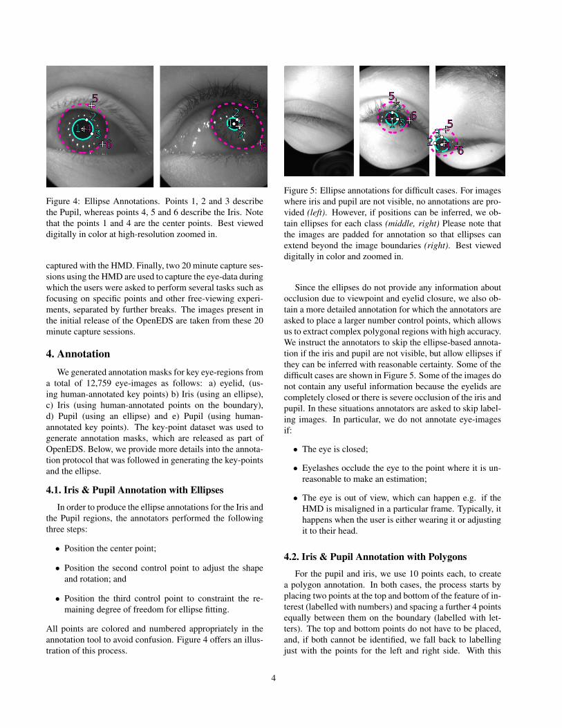
Figure 4: Ellipse Annotations. Points 1, 2 and 3 describethe Pupil, whereas points 4, 5 and 6 describe the Iris. Notethat the points 1 and 4 are the center points. Best vieweddigitally in color at high-resolution zoomed in.
captured with the HMD. Finally, two 20 minute capture ses-sions using the HMD are used to capture the eye-data duringwhich the users were asked to perform several tasks such asfocusing on specific points and other free-viewing experi-ments, separated by further breaks. The images present inthe initial release of the OpenEDS are taken from these 20minute capture sessions.
4. AnnotationWe generated annotation masks for key eye-regions from
a total of 12,759 eye-images as follows: a) eyelid, (us-ing human-annotated key points) b) Iris (using an ellipse),c) Iris (using human-annotated points on the boundary),d) Pupil (using an ellipse) and e) Pupil (using human-annotated key points). The key-point dataset was used togenerate annotation masks, which are released as part ofOpenEDS. Below, we provide more details into the annota-tion protocol that was followed in generating the key-pointsand the ellipse.
4.1. Iris & Pupil Annotation with Ellipses
In order to produce the ellipse annotations for the Iris andthe Pupil regions, the annotators performed the followingthree steps:
• Position the center point;
• Position the second control point to adjust the shapeand rotation; and
• Position the third control point to constraint the re-maining degree of freedom for ellipse fitting.
All points are colored and numbered appropriately in theannotation tool to avoid confusion. Figure 4 offers an illus-tration of this process.
Figure 5: Ellipse annotations for difficult cases. For imageswhere iris and pupil are not visible, no annotations are pro-vided (left). However, if positions can be inferred, we ob-tain ellipses for each class (middle, right) Please note thatthe images are padded for annotation so that ellipses canextend beyond the image boundaries (right). Best vieweddigitally in color and zoomed in.
Since the ellipses do not provide any information aboutocclusion due to viewpoint and eyelid closure, we also ob-tain a more detailed annotation for which the annotators areasked to place a larger number control points, which allowsus to extract complex polygonal regions with high accuracy.We instruct the annotators to skip the ellipse-based annota-tion if the iris and pupil are not visible, but allow ellipses ifthey can be inferred with reasonable certainty. Some of thedifficult cases are shown in Figure 5. Some of the images donot contain any useful information because the eyelids arecompletely closed or there is severe occlusion of the iris andpupil. In these situations annotators are asked to skip label-ing images. In particular, we do not annotate eye-imagesif:
• The eye is closed;
• Eyelashes occlude the eye to the point where it is un-reasonable to make an estimation;
• The eye is out of view, which can happen e.g. if theHMD is misaligned in a particular frame. Typically, ithappens when the user is either wearing it or adjustingit to their head.
4.2. Iris & Pupil Annotation with Polygons
For the pupil and iris, we use 10 points each, to createa polygon annotation. In both cases, the process starts byplacing two points at the top and bottom of the feature of in-terest (labelled with numbers) and spacing a further 4 pointsequally between them on the boundary (labelled with let-ters). The top and bottom points do not have to be placed,and, if both cannot be identified, we fall back to labellingjust with the points for the left and right side. With this
4

Figure 6: Iris and pupil annotations with dots (up to 10points per feature). Top / bottom points are labelled withnumbers, and further boundary points are denoted with let-ters. We note that even if one or the other of the top andbottom points is not visible, we still obtain annotations.
Figure 7: Eyelid annotation process. Note how the anno-tators proceed by splitting the annotation boundary recur-sively (left to right).
process, the annotators are instructed to proceed with theleft side first, and then the right (see Figure 6). Again, theannotators are asked to skip difficult-to-label images, as ex-plained in Section 4.1.
4.3. Eyelid Annotation
18 points are used for annotating the upper and lowereyelid. Similar to the iris and pupil case, the annotatorsare instructed to place these points equally spaced along theeyelid boundaries. Since the eyelid is much bigger than theother two cases (Iris and Pupil), in order to help with equalspacing we give the instruction to split the line recursivelywhile adding points. Examples of this process are givenin Figure 7. Figure 8 shows completed annotations for avariety of cases.
5. General StatisticsOpenEDS has a total of 12,759 images with annotation
masks, 252,690 further images, 91,200 frames from con-
Figure 8: Eyelid annotation examples. Note how in case ofthe eye fully closed, we require the eyelid annotation pointsto overlap.
tiguous 1.5 second video snippets, and 286 point clouddatasets for corneal topography. The latter image and imagesequence sets are not accompanied by semantic segmenta-tion annotations. Table 2 shows the statistics w.r.t. demo-graphics (sex, age group, wearing glasses or not), and theamount of images and point clouds provided.
5.1. Corneal Topography
We provide corneal topography point cloud data thatcaptures the surface curvature of the cornea for both leftand right eyes. Corneal topography of each participant wasmeasured via Scheimpflug imaging using an OCULUS R©
Pentacam R© HR corneal imaging system, where corneal el-evation maps were exported and converted to a point cloud.Figure 3 shows an example. We note that there is at mostone point cloud estimate per participant.
6. Statistical AnalysisAs outlined above, we choose to split OpenEDS by iden-
tity of the study participants as we found this to be both in-tuitive, and an easy setting to assess and avoid bias. Whenselecting the validation and test sets, we resample (or alter-natively reweigh for evaluation) the data to account for fac-tors such as age and sex. Resampling or weighting of thiskind is motivated by the fact that under-sampled modes ofthe true data distribution are the hardest to accurately cap-ture by data-driven approaches. To avoid bias arising fromthis, we ensure that the reweighing and selection of the testset penalizes approaches that do not take this into consider-ation.
An example of this is the age distribution. Figure 10shows histograms characterizing the age distribution of thetraining vs test and validation images (with a bin-width of5 years). Note how our choice of dataset splits already re-moves a significant amount of bias.
Apart from the information we can directly gain fromthe collected metadata, we further investigate the dataset
5
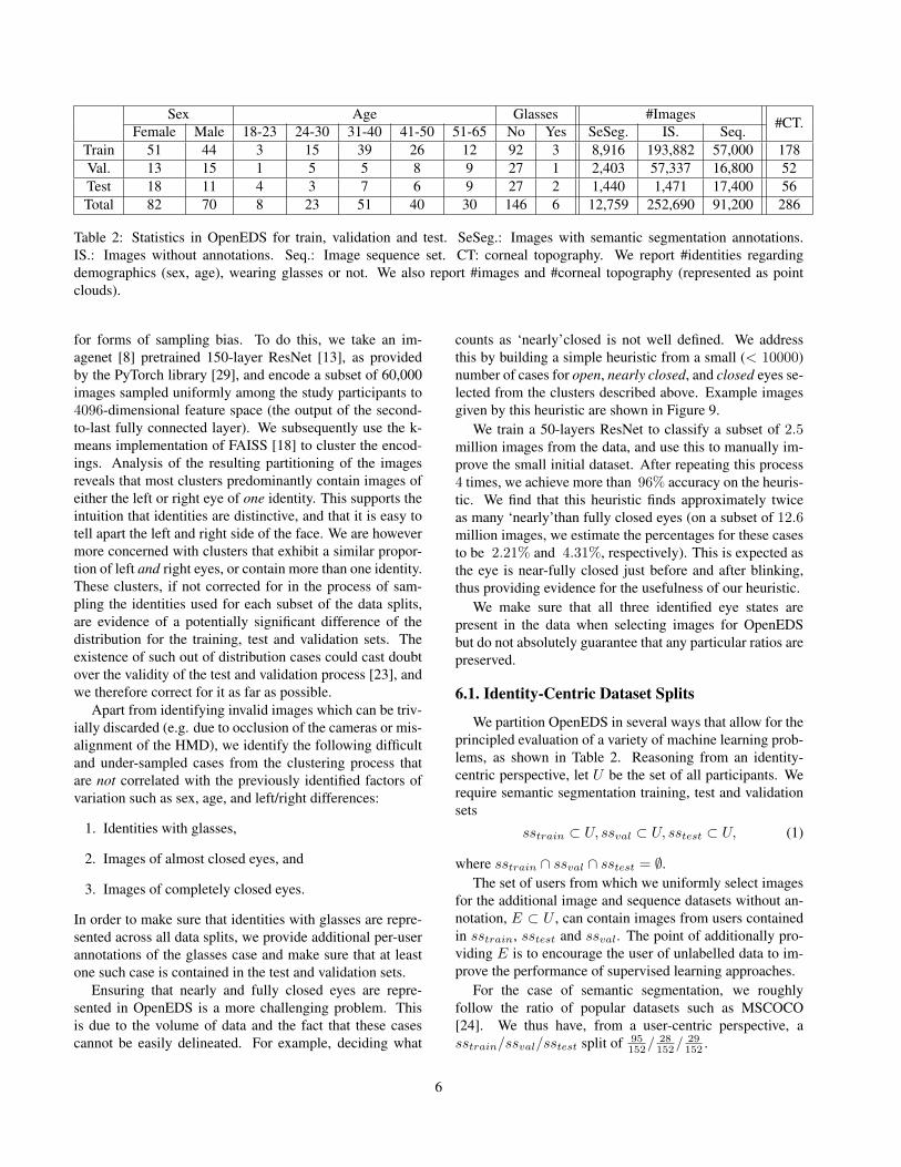
Sex Age Glasses #Images #CT.Female Male 18-23 24-30 31-40 41-50 51-65 No Yes SeSeg. IS. Seq.Train 51 44 3 15 39 26 12 92 3 8,916 193,882 57,000 178Val. 13 15 1 5 5 8 9 27 1 2,403 57,337 16,800 52Test 18 11 4 3 7 6 9 27 2 1,440 1,471 17,400 56Total 82 70 8 23 51 40 30 146 6 12,759 252,690 91,200 286
Table 2: Statistics in OpenEDS for train, validation and test. SeSeg.: Images with semantic segmentation annotations.IS.: Images without annotations. Seq.: Image sequence set. CT: corneal topography. We report #identities regardingdemographics (sex, age), wearing glasses or not. We also report #images and #corneal topography (represented as pointclouds).
for forms of sampling bias. To do this, we take an im-agenet [8] pretrained 150-layer ResNet [13], as providedby the PyTorch library [29], and encode a subset of 60,000images sampled uniformly among the study participants to4096-dimensional feature space (the output of the second-to-last fully connected layer). We subsequently use the k-means implementation of FAISS [18] to cluster the encod-ings. Analysis of the resulting partitioning of the imagesreveals that most clusters predominantly contain images ofeither the left or right eye of one identity. This supports theintuition that identities are distinctive, and that it is easy totell apart the left and right side of the face. We are howevermore concerned with clusters that exhibit a similar propor-tion of left and right eyes, or contain more than one identity.These clusters, if not corrected for in the process of sam-pling the identities used for each subset of the data splits,are evidence of a potentially significant difference of thedistribution for the training, test and validation sets. Theexistence of such out of distribution cases could cast doubtover the validity of the test and validation process [23], andwe therefore correct for it as far as possible.
Apart from identifying invalid images which can be triv-ially discarded (e.g. due to occlusion of the cameras or mis-alignment of the HMD), we identify the following difficultand under-sampled cases from the clustering process thatare not correlated with the previously identified factors ofvariation such as sex, age, and left/right differences:
1. Identities with glasses,
2. Images of almost closed eyes, and
3. Images of completely closed eyes.
In order to make sure that identities with glasses are repre-sented across all data splits, we provide additional per-userannotations of the glasses case and make sure that at leastone such case is contained in the test and validation sets.
Ensuring that nearly and fully closed eyes are repre-sented in OpenEDS is a more challenging problem. Thisis due to the volume of data and the fact that these casescannot be easily delineated. For example, deciding what
counts as ‘nearly’closed is not well defined. We addressthis by building a simple heuristic from a small (< 10000)number of cases for open, nearly closed, and closed eyes se-lected from the clusters described above. Example imagesgiven by this heuristic are shown in Figure 9.
We train a 50-layers ResNet to classify a subset of 2.5million images from the data, and use this to manually im-prove the small initial dataset. After repeating this process4 times, we achieve more than 96% accuracy on the heuris-tic. We find that this heuristic finds approximately twiceas many ‘nearly’than fully closed eyes (on a subset of 12.6million images, we estimate the percentages for these casesto be 2.21% and 4.31%, respectively). This is expected asthe eye is near-fully closed just before and after blinking,thus providing evidence for the usefulness of our heuristic.
We make sure that all three identified eye states arepresent in the data when selecting images for OpenEDSbut do not absolutely guarantee that any particular ratios arepreserved.
6.1. Identity-Centric Dataset Splits
We partition OpenEDS in several ways that allow for theprincipled evaluation of a variety of machine learning prob-lems, as shown in Table 2. Reasoning from an identity-centric perspective, let U be the set of all participants. Werequire semantic segmentation training, test and validationsets
sstrain ⊂ U, ssval ⊂ U, sstest ⊂ U, (1)
where sstrain ∩ ssval ∩ sstest = ∅.The set of users from which we uniformly select images
for the additional image and sequence datasets without an-notation, E ⊂ U , can contain images from users containedin sstrain, sstest and ssval. The point of additionally pro-viding E is to encourage the user of unlabelled data to im-prove the performance of supervised learning approaches.
For the case of semantic segmentation, we roughlyfollow the ratio of popular datasets such as MSCOCO[24]. We thus have, from a user-centric perspective, asstrain/ssval/sstest split of 95
152/28152/
29152 .
6
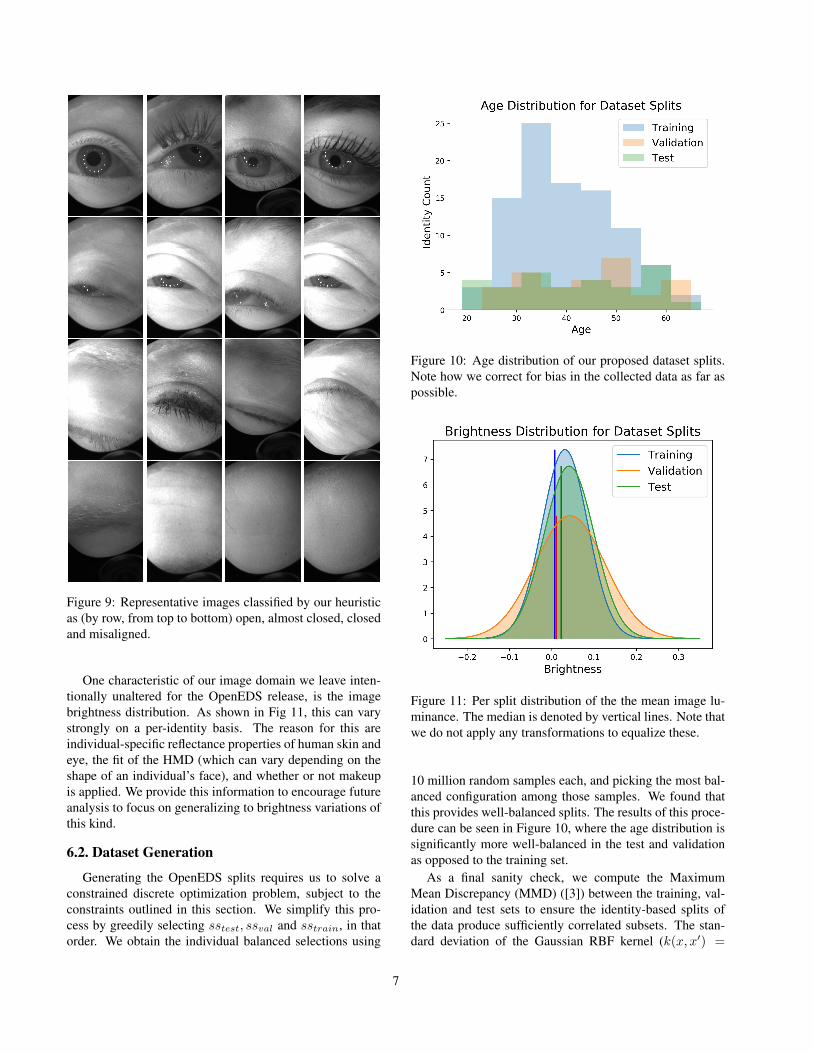
Figure 9: Representative images classified by our heuristicas (by row, from top to bottom) open, almost closed, closedand misaligned.
One characteristic of our image domain we leave inten-tionally unaltered for the OpenEDS release, is the imagebrightness distribution. As shown in Fig 11, this can varystrongly on a per-identity basis. The reason for this areindividual-specific reflectance properties of human skin andeye, the fit of the HMD (which can vary depending on theshape of an individual’s face), and whether or not makeupis applied. We provide this information to encourage futureanalysis to focus on generalizing to brightness variations ofthis kind.
6.2. Dataset Generation
Generating the OpenEDS splits requires us to solve aconstrained discrete optimization problem, subject to theconstraints outlined in this section. We simplify this pro-cess by greedily selecting sstest, ssval and sstrain, in thatorder. We obtain the individual balanced selections using
Figure 10: Age distribution of our proposed dataset splits.Note how we correct for bias in the collected data as far aspossible.
Figure 11: Per split distribution of the the mean image lu-minance. The median is denoted by vertical lines. Note thatwe do not apply any transformations to equalize these.
10 million random samples each, and picking the most bal-anced configuration among those samples. We found thatthis provides well-balanced splits. The results of this proce-dure can be seen in Figure 10, where the age distribution issignificantly more well-balanced in the test and validationas opposed to the training set.
As a final sanity check, we compute the MaximumMean Discrepancy (MMD) ([3]) between the training, val-idation and test sets to ensure the identity-based splits ofthe data produce sufficiently correlated subsets. The stan-dard deviation of the Gaussian RBF kernel (k(x, x′) =
7

Train Validation TestTrain 0.0 0.003932 0.00487Validation 0.003932 0.0 0.00489Test 0.00487 0.00489 0.0
Table 3: Maximum Mean Disrepancy between the training,test and validation sets (Evaluated on a random set of 1024images per split, averaged over 10 runs)
exp(−‖x−x′‖22
2σ2 )) used in computing the MMD is set basedon the median Euclidian norm between all image pairsin the dataset [23]. In our case, this amounts to setting1
2σ2 = 0.0087719. Evaluating the metric for random sets of1024 images across the data splits gives the results shownin Table 3, which strongly indicates that the three derivedimage sets are drawn from the same underlying data distri-bution.
7. Investigation into neural network model topredict key eye-region annotation masks
A number of convolutional encoder-decoder neural net-work architectures, derived from the SegNet architecture,[2], (keeping top 4 layers from SegNet in the encoderand the decoder network) are investigated for predictingkey eye-segments: boundary refinement (BR) modeling theboundary alignment as a residual structure to improve lo-calization performance near object boundaries [31]; sepa-rable convolution (SC) factorizing a standard convolutioninto a depth wise convolution and a 1×1 convolution to re-duce computational cost [16]. In addition, we introduced amultiplicative skip connection between the last layer of theencoder and the decoder network. We refer to this modifiedSegnet neural network as the mSegnet model
We trained each network for 200 epochs on a NVIDIARTX 2080 GPU using PyTorch [29] with ADAM optimizer,initial learning rate 0.0001, batch size 8, and weight decay1e−5. No data augmentation was performed.
Table 4 shows the results of semantic segmentation. Wefollow the same evaluation metric as in [25]. In addition,#parameters are considered as well. In terms of accuracy,SegNet with BR achieved the best performance with a meanIoU of 91.4%. In terms of model complexity, as measuredin terms of the model size, SegNet with SC requires thesmallest model size of 1.6 MB and the number of trainableparameters of 0.4 M. We also observed that in general allthese models fail to generate high fidelity eye-region masksfor eye-images with eyeglasses and heavy mascara and non-conforming pupil orientation. Figure 12 shows examplesfrom these failure cases.
Model Pixel Mean Mean Mean Model #Param.acc. acc. F1 IoU Size
(MB)(M)
mSegnet 98.0 96.8 97.9 90.7 13.3 3.5mSegnetw/ BR
98.3 97.5 98.3 91.4 13.3 3.5
mSegnetw/ SC
97.6 96.6 97.4 89.5 1.6 0.4
Table 4: Results on semantic segmentation. Model size:size of the PyTorch models on disk. #Params: the number oflearnable parameters, where M stands for million. mSegnet:4-layer segnet with multiplicative skip connection betweenthe last layer of the encoder and the decoder network
Figure 12: Examples of challenging samples on test dataset of semantic segmentation. Rows from top to bottom:images, ground truth, predictions from SegNet w/ BR.Columns from left to right: eyeglasses, heavy mascara, dimlight, varying pupil size. Better viewed in color.
8. Conclusion
We have presented OpenEDS, a new dataset of imagesand optometric data of the human eye. Initial algorithmicanalysis of the provided labels demonstrates the usefulnessof the data for semantic segmentation and we hope that fu-ture work can improve on these results. Our statistical anal-
8

ysis of the data and the resulting dataset splits should be ofuse to density estimation tasks for the eye-tracking domain.
AcknowledgementThe authors gratefully acknowledge the helpful com-
ments and feedback from Abhishek Sharma, Alexander Fix,and Nick Sharp.
References[1] B. Adegoke, E. Omidiora, S. Falohun, and J. Ojo. Iris seg-
mentation: a survey. International Journal of Modern Engi-neering Research (IJMER), 3(4):1885–1889, 2013.
[2] V. Badrinarayanan, A. Kendall, and R. Cipolla. Segnet: Adeep convolutional encoder-decoder architecture for imagesegmentation. IEEE transactions on pattern analysis andmachine intelligence, 39(12):2481–2495, 2017.
[3] K. Borgwardt, A. Gretton, M. Rasch, H. P. Kriegel,B. Scholkopf, and A. Smola. Integrating structured biolog-ical data by kernel maximum mean discrepency. Bioinfor-matics, 22:e49–457, 2010.
[4] A. Borji and L. Itti. State-of-the-art in visual attention mod-eling. IEEE transactions on pattern analysis and machineintelligence, 35(1):185–207, 2013.
[5] A. Das, U. Pal, M. Blumenstein, and M. A. F. Ballester.Sclera recognition-a survey. In 2013 2nd IAPR Asian Con-ference on Pattern Recognition, pages 917–921. IEEE, 2013.
[6] A. Das, U. Pal, M. A. Ferrer, M. Blumenstein, D. Stepec,P. Rot, Z. Emersic, P. Peer, V. Struc, S. A. Kumar, et al.Sserbc 2017: Sclera segmentation and eye recognitionbenchmarking competition. In 2017 IEEE InternationalJoint Conference on Biometrics (IJCB), pages 742–747.IEEE, 2017.
[7] A. Das, U. Pal, M. A. Ferrer, M. Blumenstein, D. Stepec,P. Rot, Z. Emersic, P. Peer, V. Struc, S. V. A. Kumar, andB. S. Harish. Sserbc 2017: Sclera segmentation and eyerecognition benchmarking competition. In 2017 IEEE Inter-national Joint Conference on Biometrics (IJCB), pages 742–747, Oct 2017.
[8] J. Deng, W. Dong, R. Socher, L. Li, K. Li, and L. Fei-Fei. Im-agenet: A large-scale hierarchical image database. In 2009IEEE Conference on Computer Vision and Pattern Recogni-tion, pages 248–255, June 2009.
[9] K. A. Funes Mora, F. Monay, and J.-M. Odobez. Eyediap: Adatabase for the development and evaluation of gaze estima-tion algorithms from rgb and rgb-d cameras. In Proceedingsof the Symposium on Eye Tracking Research and Applica-tions, pages 255–258. ACM, 2014.
[10] A. Geiger, P. Lenz, C. Stiller, and R. Urtasun. Vision meetsrobotics: The kitti dataset. The International Journal ofRobotics Research, 32(11):1231–1237, 2013.
[11] A. Geiger, P. Lenz, and R. Urtasun. Are we ready for au-tonomous driving? the kitti vision benchmark suite. In 2012IEEE Conference on Computer Vision and Pattern Recogni-tion, pages 3354–3361. IEEE, 2012.
[12] K. Harezlak and P. Kasprowski. Application of eye track-ing in medicine: a survey, research issues and challenges.Computerized Medical Imaging and Graphics, 65:176–190,2018.
[13] K. He, X. Zhang, S. Ren, and J. Sun. Deep residual learningfor image recognition. CoRR, abs/1512.03385, 2015.
[14] K. Holmqvist, M. Nystrom, R. Andersson, R. Dewhurst,H. Jarodzka, and J. van de Weijer. Eye tracking: A com-prehensive guide to methods and measures. 01 2011.
[15] A. D. Hoover, V. Kouznetsova, and M. Goldbaum. Locat-ing blood vessels in retinal images by piecewise threshold
9

probing of a matched filter response. IEEE Transactions onMedical Imaging, 19(3):203–210, March 2000.
[16] A. G. Howard, M. Zhu, B. Chen, D. Kalenichenko, W. Wang,T. Weyand, M. Andreetto, and H. Adam. Mobilenets: Effi-cient convolutional neural networks for mobile vision appli-cations. arXiv preprint arXiv:1704.04861, 2017.
[17] Q. Huang, A. Veeraraghavan, and A. Sabharwal. Tabletgaze:A dataset and baseline algorithms for unconstrainedappearance-based gaze estimation in mobile tablets. CoRR,abs/1508.01244, 2015.
[18] J. Johnson, M. Douze, and H. Jegou. Billion-scale similaritysearch with gpus. CoRR, abs/1702.08734, 2017.
[19] J. Kim, M. Stengel, A. Majercik, S. De Mello, S. Laine,M. McGuire, and D. Luebke. Nvgaze: An anatomically-informed dataset for low-latency, near-eye gaze estimation.In Proceedings of the SIGCHI Conference on Human Fac-tors in Computing Systems, CHI ’19, New York, NY, USA,2019. ACM.
[20] K. Krafka, A. Khosla, P. Kellnhofer, H. Kannan, S. Bhan-darkar, W. Matusik, and A. Torralba. Eye tracking for every-one. In IEEE Conference on Computer Vision and PatternRecognition (CVPR), 2016.
[21] G. Kramida. Resolving the vergence-accommodation con-flict in head-mounted displays. IEEE transactions on visual-ization and computer graphics, 22(7):1912–1931, 2016.
[22] A. Krizhevsky, I. Sutskever, and G. E. Hinton. Imagenetclassification with deep convolutional neural networks. InAdvances in neural information processing systems, pages1097–1105, 2012.
[23] S. Liang, Y. Li, and R. Srikant. Principled detection ofout-of-distribution examples in neural networks. CoRR,abs/1706.02690, 2017.
[24] T. Lin, M. Maire, S. J. Belongie, L. D. Bourdev, R. B.Girshick, J. Hays, P. Perona, D. Ramanan, P. Dollar, andC. L. Zitnick. Microsoft COCO: common objects in context.CoRR, abs/1405.0312, 2014.
[25] J. Long, E. Shelhamer, and T. Darrell. Fully convolutionalnetworks for semantic segmentation. In Proceedings of theIEEE conference on computer vision and pattern recogni-tion, pages 3431–3440, 2015.
[26] D. R. Lucio, R. Laroca, E. Severo, A. S. Britto Jr, andD. Menotti. Fully convolutional networks and generative ad-versarial networks applied to sclera segmentation. CoRR,vol. abs/1806.08722, 2018.
[27] B. Luo, J. Shen, Y. Wang, and M. Pantic. The iBUG EyeSegmentation Dataset. In E. Pirovano and E. Graversen, ed-itors, 2018 Imperial College Computing Student Workshop(ICCSW 2018), volume 66 of OpenAccess Series in Infor-matics (OASIcs), pages 7:1–7:9, Dagstuhl, Germany, 2019.Schloss Dagstuhl–Leibniz-Zentrum fuer Informatik.
[28] C. D. McMurrough, V. Metsis, J. Rich, and F. Makedon. Aneye tracking dataset for point of gaze detection. In Proceed-ings of the Symposium on Eye Tracking Research and Appli-cations, pages 305–308. ACM, 2012.
[29] A. Paszke, S. Gross, S. Chintala, G. Chanan, E. Yang, Z. De-Vito, Z. Lin, A. Desmaison, L. Antiga, and A. Lerer. Auto-matic differentiation in pytorch. 2017.
[30] A. Patney, J. Kim, M. Salvi, A. Kaplanyan, C. Wyman,N. Benty, A. Lefohn, and D. Luebke. Perceptually-basedfoveated virtual reality. In ACM SIGGRAPH 2016 EmergingTechnologies, SIGGRAPH ’16, pages 17:1–17:2, New York,NY, USA, 2016. ACM.
[31] C. Peng, X. Zhang, G. Yu, G. Luo, and J. Sun. Large kernelmatters–improve semantic segmentation by global convolu-tional network. In Proceedings of the IEEE conference oncomputer vision and pattern recognition, pages 4353–4361,2017.
[32] H. Proenca, S. Filipe, R. Santos, J. Oliveira, and L. A.Alexandre. The ubiris. v2: A database of visible wavelengthiris images captured on-the-move and at-a-distance. IEEETransactions on Pattern Analysis and Machine Intelligence,32(8):1529–1535, 2010.
[33] H. Proenca, S. Filipe, R. Santos, J. Oliveira, and L. A.Alexandre. The ubiris.v2: A database of visible wavelengthiris images captured on-the-move and at-a-distance. IEEETransactions on Pattern Analysis and Machine Intelligence,32(8):1529–1535, Aug 2010.
[34] P. Radu, J. Ferryman, and P. Wild. A robust sclera segmenta-tion algorithm. In 2015 IEEE 7th International Conferenceon Biometrics Theory, Applications and Systems (BTAS),pages 1–6. IEEE, 2015.
[35] P. Rot, Z. Emersic, V. Struc, and P. Peer. Deep multi-class eye segmentation for ocular biometrics. In 2018 IEEEInternational Work Conference on Bioinspired Intelligence(IWOBI), pages 1–8. IEEE, 2018.
[36] W. Sankowski, K. Grabowski, M. Napieralska, M. Zubert,and A. Napieralski. Reliable algorithm for iris segmentationin eye image. Image and vision computing, 28(2):231–237,2010.
[37] A. Shafaei, M. Schmidt, and J. J. Little. Does your modelknow the digit 6 is not a cat? A less biased evaluation of”outlier” detectors. CoRR, abs/1809.04729, 2018.
[38] A. Shrivastava, T. Pfister, O. Tuzel, J. Susskind, W. Wang,and R. Webb. Learning from simulated and unsupervisedimages through adversarial training. In Proceedings of theIEEE Conference on Computer Vision and Pattern Recogni-tion, pages 2107–2116, 2017.
[39] B. A. Smith, Q. Yin, S. K. Feiner, and S. K. Nayar. Gazelocking: passive eye contact detection for human-object in-teraction. In UIST, 2013.
[40] V. Tanriverdi and R. J. Jacob. Interacting with eye move-ments in virtual environments. In Proceedings of the SIGCHIconference on Human Factors in Computing Systems, pages265–272. ACM, 2000.
[41] M. Thoma. A survey of semantic segmentation. arXivpreprint arXiv:1602.06541, 2016.
[42] M. Tonsen, X. Zhang, Y. Sugano, and A. Bulling. Labelledpupils in the wild: a dataset for studying pupil detection inunconstrained environments. In Proceedings of the Ninth Bi-ennial ACM Symposium on Eye Tracking Research & Appli-cations, pages 139–142. ACM, 2016.
[43] R. Venkateswarlu et al. Eye gaze estimation from a sin-gle image of one eye. In Proceedings Ninth IEEE Inter-national Conference on Computer Vision, pages 136–143.IEEE, 2003.
10

[44] K. Wang, R. Zhao, and Q. Ji. A hierarchical generative modelfor eye image synthesis and eye gaze estimation. In Proceed-ings of the IEEE Conference on Computer Vision and PatternRecognition, pages 440–448, 2018.
[45] E. Wood, T. Baltrusaitis, L.-P. Morency, P. Robinson, andA. Bulling. Learning an appearance-based gaze estimatorfrom one million synthesised images. In Proceedings of theNinth Biennial ACM Symposium on Eye Tracking Research& Applications, pages 131–138. ACM, 2016.
[46] E. Wood, T. Baltrusaitis, X. Zhang, Y. Sugano, P. Robinson,and A. Bulling. Rendering of eyes for eye-shape registra-tion and gaze estimation. In Proceedings of the IEEE Inter-national Conference on Computer Vision, pages 3756–3764,2015.
[47] L. Xiao, A. Kaplanyan, A. Fix, M. Chapman, and D. Lan-man. Deepfocus: Learned image synthesis for computa-tional displays. ACM Trans. Graph., 37(6):200:1–200:13,Dec. 2018.
[48] X. Zhang, Y. Sugano, M. Fritz, and A. Bulling. Appearance-based gaze estimation in the wild. In 2015 IEEE Conferenceon Computer Vision and Pattern Recognition (CVPR), pages4511–4520, June 2015.
[49] D. Deshraj, R. Jain, H. Agrawal, P. Chattopadhyay, T. Singh,A. Jain, B. Singh, S. Lee and D. Batra. EvalAI: Towards Bet-ter Evaluation Systems for AI Agents In arXiv, 1902.03570,2019.
11