ophthalmic anaesthesia 1
TRANSCRIPT

8/6/2019 Ophthalmic Anaesthesia 1
http://slidepdf.com/reader/full/ophthalmic-anaesthesia-1 1/66
OPHTHALMIC ANAESTHESIA
The Journal of the British Ophthalmic Anaesthesia Society
2009
web: www.boas.org
BOAS COUNCIL
PRESIDENTProf Chandra Kumar
SECRETARYDr K-L Kong
TREASURERMr Tom Eke
EDITORDr Steve Mather
OTHER COUNCIL MEMBERSDr Keith AllmanProf Ezzat AzizDr Peter JamesDr Jonathan LordDr Hamish McLureProf Peter ShahDr Roger SlaterDr Sean TigheDr Guri ThindDr Shashi Vohra

8/6/2019 Ophthalmic Anaesthesia 1
http://slidepdf.com/reader/full/ophthalmic-anaesthesia-1 2/66
2
EDITORIALThere has been a metamorphosis in thelast year at BOAS. The Council hassome new members, recently elected.Dr Keith Allman from Exeter, ProfessorEzzat Aziz, professor in Cairo and aconsultant in anaesthesia atChesterfield, UK, Professor Peter Shahfrom Birmingham and Dr Peter Jamesfrom Basingstoke have joined us and welook forward to receiving their ideas andwisdom.The second metamorphosis is that ourprevious Society publication,Ophthalmic Anaesthesia News hasbeen renamed Ophthalmic Anaesthesia to more reflect its role as a Society
journal rather than a newsletter. We aimto publish the views of members, andindeed, non members, on any topicrelated to ophthalmic anaesthesia, andalso to give a forum to trainees andothers to publish articles and casereports, which are peer reviewed, but inan ―easier‖ environment than themainstream anaesthesia journals.The contribution from our President thistime is centred on the role of theanaesthetist in the care of ophthalmicpatients, and in the widest sense. It is
not just about performing blocks!The question of how to manage patientson anticoagulants and antiplatelet drugshas been an ongoing discussion inBOAS circles for over 2 years now, andin 2008 the Council commissioned aworking group (myself, Dr K-L Kong andDr Shashi Vohra) to produce a positionpaper on this subject. I presented ourconclusions at the BOAS annualScientific Meeting in Manchester thissummer and invited comments from
delegates. So far I have received nonefrom any members outside the Council.Accordingly, a summary of that positionpaper is published in this edition, to Ihope a wider audience. We need tohear your views and practice, as this iswhat is happening up and down the UKand abroad, and reflects the current
management of patients. Getting a widerange of views will enable us to build apicture of current practice and facilitaterecommendations for best practice. Inthis summary document we do drawsome conclusions and make somerecommendations, based on theavailable evidence and what weconsider to be best practice currently.So please let me have your opinionsand tell us what you are currently doing.We hope our position paper will informthe Royal Colleges when they reviewtheir advice.We have a selection of interestingarticles and case reports for you in thisedition, some of which are based onpresentations given at the ScientificMeeting this year.I hope you find the journal bothinformative and interesting, and that youwill be prompted to make a contributionyourself.We welcome letters giving opinion,articles both research and review, andcase reports – in fact anything which willbe of interest to your colleagues workingwith ophthalmic patients.Please send your contributions to the
editor by email at the address below.It will soon be time to think about nextyear‘s study leave. BOAS 2010 will behosted by Dr Jonathan Lord and hiscolleagues at Moorfield‘s Eye Hospital inLondon on 3 and 4 June. More details,including the programme, will follow onthe website.
Steve Mather
Please send contributions to theeditor at:

8/6/2019 Ophthalmic Anaesthesia 1
http://slidepdf.com/reader/full/ophthalmic-anaesthesia-1 3/66
3
REPORT OF BOAS ANNUAL SCIENTIFIC MEETING 2009MANCHESTER 18 – 19 JUNE 2009
Manchester was the location for the 10th annual scientific meeting of the society. Themeeting was held in the Manchester Conference Centre, just a few minutes walk from
the city centre.
The faculty included specialists from the UK, Europe and the USA. Those attendingcomprised consultants and trainees from the UK, as well as guests from France,Switzerland, Russia, Brazil and Chile.
In the first session, chaired by Anthony Rubin, Hamish Mclure outlined the appliedanatomy of orbital blocks and examined the alternative scoring systems for formalassessment of ophthalmic regional anaesthesia. Keith Allman then gave details ofclinical work he had conducted into the use of the relatively new local anaesthetic agentarticaine for ophthalmic regional anaesthesia and demonstrated his novel technique ofincisionless sub-Tenon‘s block. The session ended with Malachy Column‘s review of
local anaesthesia therapy and toxicity and the conclusion that despite extensiveresearch and developments there has been no really significant advance in localanaesthetic agents since the development of bupivicaine.
The next session, chaired by Steve Gayer, began with a comprehensive and clearreview by Nigel Harper of the problems of anaphylaxis encountered by anaesthetists andsurgeons. He included an update of the revised AAGBI guidelines in suspectedanaphylaxis and the new National Anaesthesia Anaphylaxis Database. Brian Pollardthen provided an exposition of the problems around the risks of awareness and recallafter general anaesthesia, including Hollywood‘s recent contribution to the issue in thefilm ―Aware‖. He reviewed methods of assessing depth of anaesthesia including recentwork carried out in Manchester. Finally Ralph MacKinnon gave an energetic and
detailed presentation on the use of simulation in paediatric anaesthetic emergencytraining. He emphasised the importance of simulators in medical education anddiscussed regional and national plans for the development of training with scenarios inthe management of specific emergencies. Over the two days Ralph ran informalworkshops for delegates using full paediatric simulation equipment.
8/6/2019 Ophthalmic Anaesthesia 1
http://slidepdf.com/reader/full/ophthalmic-anaesthesia-1 4/66
4
Paediatric simulation
The first afternoon session, chaired by Roger Slater, began with Brian Leatherbarrowproviding an overview of orbital and oculoplastic surgery, including slides and videos ofpathology encountered in his practice, and the implications for the anaesthetist. DanConway then looked at the choices of anaesthesia in oculoplastic surgery and outlined
his experience and the techniques he uses in his everyday practice.
Chandra Kumar chaired the second afternoon session which encompassedpresentations from the USA, France and Bristol. Firstly Steve Gayer from the Universityof Miami demonstrated the potential use of ultrasound of the globe in providing saferperibulbar blocks. Jacques Ripart then gave a fascinating presentation relating thescientific background and his experience of the use of xenon as a general anaestheticagent. Finally Steve Mather presented the preliminary findings of the BOAS WorkingGroup following a systematic review of the subject of anticoagulant and antiplatelettherapy and ophthalmic surgery. The society was then asked to consider therecommendations produced. The day concluded with the annual dinner which tookplace in the Conference Centre.
The first session of the second day, chaired by Hamish McLure, began with K- L Kong‘scomprehensive review of anaesthesia for vitreo-retinal surgery. The second lecture wasgiven by Simon Howell and examined the subject of pre-operative assessment inrelation to cardiovascular issues such as hypertension, aspirin therapy and coronarystents. After the break Chandra Kumar returned to the chair and Jacques Ripartreturned to the podium to give the French view of the role of the anaesthetist inophthalmic surgery. Chris Dodds then gave the view from the Royal College ofAnaesthetists of the impending change to medical regulation in the Revalidation andRecertification process.
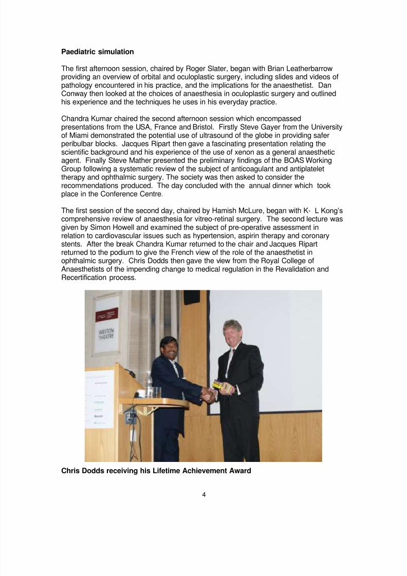
Chris Dodds receiving his Lifetime Achievement Award

8/6/2019 Ophthalmic Anaesthesia 1
http://slidepdf.com/reader/full/ophthalmic-anaesthesia-1 5/66
5
During the session the BOAS annual general meeting was held and included thegranting of the BOAS Lifetime Achievement Award to Chris Dodds. Chandra Kumargave an eloquent and touching tribute to his friend and colleague.
The free paper presentations took the final session of the morning. This was chaired jointly by Shashi Vohra and Jonathan Lord.
Council member Shashi Vohra
Dr Peres Bota from Lille gave her presentation on the use of low-dose ketamine
infusions following enucleation. Vip Gill from Moorfields then outlined a technique ofhigh-volume sub-tenon‘s anaesthesia in V-R surgery. Laura Tulloch from Birminghampresented an audit of bedside INR monitoring in ophthalmic patients. The final traineepresentation was by Richard Lee from Norwich who presented work on the ―face to face‖position for cataract surgery.
Three international speakers followed; Haroldo Carneiro from Brazil compared CT scansof the orbit in intraconal and extraconal blocks; Dagobert Lerch from Switzerlanddescribed a novel method to reduce subconjunctival haemorrhage after subtenon‘sblocks; then Pavlov Rylov from Russia gave a DVD presentation from his ophthalmicsurgery unit in Yekaterinburg and the interaction between surgeon and anaesthetist.Tom Eke showed a video of a patient undergoing cataract surgery using the ―face to
face‖ position. Finally there were two case presentations of patients with challengingmedical conditions. Sofia Khan from Stockport presented ―Severe pulmonaryhypertension in a patient requiring enucleation‖ and Kailash Bhatia from Manchester presented ―Tracheal stenosis in Wegener‘s Granulomatosis; managing orbital surgery‖.
Before lunch Jonathan Lord took the opportunity to invite the delegates to the 2010BOAS meeting in London.

8/6/2019 Ophthalmic Anaesthesia 1
http://slidepdf.com/reader/full/ophthalmic-anaesthesia-1 6/66

8/6/2019 Ophthalmic Anaesthesia 1
http://slidepdf.com/reader/full/ophthalmic-anaesthesia-1 7/66
7
FROM THE PRESIDENT
The Anaesthetist’s role in 21st century ophthalmology
SummaryAnaesthetists play a pivotal role in 21st century ophthalmology by providing anaesthesia for various ophthalmic surgical procedures. They not only administer general anaesthesia but also perform local blocks and provide sedation, orbital pain relief for both acute as well as chronic conditions, monitor patients and manage life threatening complications should the need arise.
Keywords: anaesthesia, ophthalmology, anaesthetist‘s role
Ophthalmic surgery has been carried out in one form or another for centuries but thereis little detail of any anaesthetic that might have been used. According to the publishedliterature, it is apparent that advances in ophthalmic surgery occur in tandem withadvances in anaesthesia. In the late 19th century ophthalmic surgery was limited tocataract treatment and iridectomy performed sometimes with topical cocaine. Later,needle techniques were introduced using at first cocaine and then procaine. However,general anaesthesia provided the opportunity for more complex surgery without fear ofpain and this led to advances in ophthalmic surgery over the last century. There hasbeen a steady and progressive development in ophthalmic surgical technique thatcontinues to evolve.
With recent various global healthcare reforms and consequent financial constraint,various models of reorganisation, restructure and centralisation of services have beenintroduced with increasing super specialization. It is important to understand how thesehave been adopted into everyday practice and how anaesthetic practice has evolvedalongside to maximise the benefit to our patients. Models such as ‗One Stop Clinics‘ 1,‗High Volume Cataract Surgery‘ 2 and ‗Fast Track Surgery‘ 3 have been introduced tostreamline the patient‘s journey through diagnosis and treatment impacting on waitingtimes and cost. Although these initiatives have benefits, they are seen to have adverseeffects on training 4. ‗One Stop Clinics‘ and ‗Fast Track Surgery‘ concepts are possible if there are strict procedures and protocols in place but success also depends on patientsbeing adequately assessed and prepared before surgery itself.
At present ophthalmic surgery is performed in single large tertiary referral specialistcentres, district general hospitals or stand alone dedicated cataract centres. Tertiarycentres perform routine to complex surgical procedures and anaesthesia is provided bydedicated trained ophthalmic anaesthetists. District General Hospitals performextraocular and intraocular as well as oculoplastic procedures with anaesthesia often
provided by general anaesthetists with one or two weekly sessions in the ophthalmictheatre. With closure and amalgamation of various eye units in recent years, manyophthalmologists have become interested in a particular sub-speciality of ophthalmologyeven in large district hospitals. In cataract surgery-dedicated stand alone day units,anaesthesia is usually provided by ophthalmologists without an anaesthetist.

8/6/2019 Ophthalmic Anaesthesia 1
http://slidepdf.com/reader/full/ophthalmic-anaesthesia-1 8/66
8
The provision of anaesthesia for ophthalmic surgical procedures varies around theworld with an increasing tendency towards orbital regional and local anaesthesia 5, 6, theexception being where general anaesthesia is desirable or essential.
Advances in general anaesthesia over the last 50 years include new intravenousinduction agents ( eg propofol), muscle relaxants (vecuronium, rocuronium and
cisatracurium), analgesic agents (fentanyl,alfentanil,remifentanil), volatile agents(sevoflurane and desflurane), supraglottic airway devices (laryngeal mask airway) andnewer technology including computerised anaesthetic machines, monitoring andinfusion devices. Intravenous induction and balanced anaesthesia with a securedairway aided with muscle relaxant, analgesic and volatile agent or equivalent remain thegold standard technique. However, the introduction of supraglottic devices has allowedmany ophthalmic surgical procedures to be performed whilst patients breathespontaneously. The role of anaesthetists in general anaesthesia is very well definedand no further reference to general anaesthesia will be made in the rest of this article.Cataract surgery with lens implant is the commonest ophthalmic surgical procedure butit is not uncommon for other surgical procedures such as glaucoma operations,viteroretinal surgery, oculoplastic surgery and other less common procedures such as
enucleation and evisceration to be performed under orbital regional anaesthesia.
Orbital regional and local anaesthesia for ophthalmic surgery has traditionally been andcontinues to be performed by ophthalmologists. Orbital regional anaesthesia is aconduction block. Safe and successful use of a conduction block depends on manyfactors but knowledge of anatomy, pharmacology and resuscitation skills are necessaryprerequisites. Anaesthetists by training are individuals who possess these prerequisitesand no wonder many anaesthetists undertook the task of performing orbital regionalanaesthesia in the1970s and many more have followed since then.
On a wider level, anaesthetists have defined and established roles both in clinical aswell as non-clinical areas. They play a pivotal role in the provision of anaesthesia and
perioperative care for various sub-specialities including ophthalmology as well as painmanagement. They play an important role in non technical fields which includemanaging the theatre, equipment, enforcing minimum monitoring standards, training,education, research and audit.
Patients undergoing routine ophthalmic surgery are usually elderly with morbiditiesrequiring multiple drugs 7. Many suffer from hypertension, diabetes, cardiorespiratory &other systemic diseases. Their well being depends on proper preoperative assessment,evaluation and preparation before surgery. Written guidelines, protocols and structuredpreassessment help in initiating appropriate investigations and selection of appropriateanaesthesia resulting in minimum disruption to the patient‘s routine life, lesscancellation and the safe conduct of surgery. Anaesthetists are important in providing
these services by organising and running successful preoperative assessment andoptimization clinics while guiding other healthcare professionals.
Provision of orbital regional or local anaesthesia is fundamental to the concept of fasttrack cataract surgery. The use of topical anaesthesia is seen as crucial to make thesystem work. However, topical anaesthesia is not and cannot be suitable for allpatients8. If the ophthalmologist has to perform his regional block the option is limited tosub-Tenon‘s block as the presence of an anaesthetist is considered essential if a needleblock is performed 9. Many ophthalmologists do not agree with the concept of fast track

8/6/2019 Ophthalmic Anaesthesia 1
http://slidepdf.com/reader/full/ophthalmic-anaesthesia-1 9/66
9
surgery and insist on a good anaesthesia and akinesia service being provided by ananaesthetist.
Ophthalmic anaesthetists can provide suitable orbital regional anaesthesia according tothe requirements of surgery, surgeons‘ and patients‘ wishes 10. Orbital regionalanaesthesia can be performed with a needle (intraconal or extraconal blocks) or a blunt
cannula (sub-Tenon‘s block) to match the type of surgery 7.
The popularity of needle block has declined in recent years due to its complications 11,
12. Although these complications are rare, they can be life threatening. Responsibilityfor treating complications such as cardiorespiratory arrest cannot entirely rest with theoperating ophthalmologist. Many ophthalmic surgeons are of the opinion that their jobis to perform surgery alone and the provision of anaesthesia should be left to the expert.The Joint Colleges report 9 recommends that an anaesthetist must be present when aneedle block is performed.
Sub-Tenon‘s block was introduced as a simple, safe and effective technique.13.Although the presence of an anaesthetist is not considered essential 9
, many
serious sight and life threatening complications including central spread of localanaesthetic 14 and death have occurred 15. Other serious adverse events unrelated tosub-Tenon‘s block have also been reported 16 and if they are undetected or untreatedthe patient‘s life may be at risk. Arguably, the presence of an anaesthetist is important18 but often ignored on financial grounds.
Anaesthetists are trained specialists who provide regional anaesthesia not only forophthalmic surgery but also for non-ophthalmic surgery and they have a wide and in-depth knowledge and skills base. Over the last three decades anaesthetists havebecome proficient in performing orbital regional anaesthesia and led the development inanaesthetic techniques with resulting reduced morbidity and mortality. Althoughscientific data is not available to prove this, the clinical view supports this. Provision of
anaesthesia by an anaesthetist can only result in good quality anaesthesia withoutinterruption and unnecessary delay with safety and improved quality for the patient‘s
journey through the surgical pathway.
Dacryocystorhinostomy (DCR) surgery is usually performed under general anaesthesia,the preferred technique. DCR is known to be performed under local anaesthesia butusually multiple injections are required and sedation may be necessary requiring thepresence of an anaesthetist. DCR can be performed under a single injection techniqueand with or without sedation 18 but the presence of an anaesthetist will be a necessity ifsedation is used 19.
In hospitals where topical anaesthesia is common, it is not unusual for the patient to feel
discomfort and anxiety during surgery. The use of sedation and topical anaesthesia forcataract and non-cataract surgery in finance and quality driven centres (private sector)is not uncommon 5. The routine use of sedation in ophthalmic surgery is discouraged 9
but sedation may be used in anxious patients and in patients undergoing longer surgerysuch as vitreoretinal procedures, dacryocystorhinostomy and others 19. Sedation shouldonly be administered by an anaesthetist, he/she is not there only to monitor the patientbut also to deal with any adverse events which do occur in these high risk patients 19.

8/6/2019 Ophthalmic Anaesthesia 1
http://slidepdf.com/reader/full/ophthalmic-anaesthesia-1 10/66
10
Anaesthetists can help in alleviating both postoperative and chronic orbital pain.Anaesthetists are trained to treat postoperative pain and have variety of techniques intheir armamentarium (including PCA, retrobulbar and sub-Tenon‘s catheters).
Treatment of chronic orbital pain is an area which has not attracted the attention ofophthalmic anaesthetists. Patients suffering from chronic orbital pain are mainly treated
by ophthalmologists pharmacologically and rarely by neurolytic injections.Ophthalmologists refer these patients to the chronic pain specialist if drug therapy fails.Chronic pain specialists may not be conversant with ophthalmological neurolyticinjections. Neurolytic injections are known to be helpful in the treatment of severeorbital pain in seeing and non-seeing eyes. Anaesthetists who are trained to administerretrobulbar block and have knowledge of neurolytic agents can help in the managementof chronic orbital pain 20.
Anaesthetists have many non clinical roles which include theatre management andscheduling, utilization of appropriate resources and facilitating teaching and training.
Conclusion
Anaesthetists play a pivotal role in ophthalmic surgery. They can administeranaesthesia both general and orbital blocks, sedation, monitor patients, manage lifethreatening complications should the need arise and provide orbital pain relief as well ascontribute in essential non-clinical areas.
Chandra M KumarPresident of BOAS
[email protected]: +44 164 2854246

8/6/2019 Ophthalmic Anaesthesia 1
http://slidepdf.com/reader/full/ophthalmic-anaesthesia-1 11/66
11
References
1 Hughes E, Diamond J. One stop cataract surgery: the Bristol Eye Hospitalexperience 1997-1999. Eye 2001; 15: 306-308.
2 Burton RL. Norwich experience-high volume & low cost cataract surgery (personalcommunication).
3 Kerr C, Kavanagh S. Fast-track surgery. I can see clearly now the wait has gone.Health Serv J 2002; 112: 28-9.
4 Au L, Saha K, Fernando B, Ataullah S, Spencer F. 'Fast-track' cataract services anddiagnostic and treatment centre: impact on surgical training. Eye 2008; 22: 55-9.
5 Leaming DV. Practice styles and preferences of ASCRS members - 2003 survey. J Cataract Refract Surg 2004; 30: 892-900.
6 Eke T, Thompson JR. Serious complications of local anaesthesia for cataractsurgery: a 1 year national survey in the United Kingdom. Br J Ophthalmol 2007; 91:470-5.
7 Kumar CM, Dowd TC. Ophthalmic regional anaesthesia. Current Opin Anaesthesiol 2008;oct 21(5):632-7
8 Rodrigues PA, Vale PJ, Cruz LM, Carvalho RP, Ribeiro IM, Martins JL. Topical
anaesthesia versus sub-Tenon block for cataract surgery: Surgical conditions andpatient satisfaction. Eur J Ophthalmol 2008; 18: 356-60.
9 Local Anaesthesia for Intraocular Surgery: The Royal College of Anaesthetists andThe Royal College of Ophthalmologists 2001http://www.rcoa.ac.uk/docs/RCARCOGuidelines.pdf.
10 Friedman DS, Reeves SW, Bass EB, Lubomski LH, Fleisher LA, Schein OD. Patientpreferences for anaesthesia management during cataract surgery. Br J Ophthalmol 2004; 88: 333-5.
11 Kumar CM, Dowd TC. Complications of ophthalmic regional blocks: Their treatmentand prevention. Ophthalmologica 2006; 220: 73-82.
12 Kumar CM. Orbital regional anaesthesia: complications and their prevention. Indian J Ophthalmol 2006; 54: 77-84.
13 Kumar CM, Williamson S, Manickam B. A review of sub-Tenon‘s block: Currentpractice and recent development. Eur J Anaesth . 2005; 22:.567-77.
14 Kumar CM, Dodds C. Sub-Tenon's Anaesthesia. Ophthalmol Clin North Am 2006;19: 209-19.
15 Quantock CL, Goswami T. Death potentially secondary to sub-Tenon's block.Anaesthesia 2007; 62: 175-7.
16 Guise P. Sub-Tenon‘s anaesthesia: A prospective study of 6000 blocks.Anesthesiology 2003; 98: 964-8.
17 Guise P. Aeroplanes rarely crash nowadays, therefore they don't need pilots:anaesthesia, anaesthetics and cataract surgery. Clin Experiment Ophthalmol 2005;33: 451-2.
18 Kumar CM, Dodds C, AhKine D . Single medial peribulbar block for
dacryocystorhinostomtomy. 15th
World Congress of Anaesthesiologists 2 - 7th
March2008, Cape Town, South Africa (abstract).
19 Greenhalgh DL, Kumar CM. Sedation during ophthalmic surgery. Eur J Anaesthesiol 2008; 25:701-7.
20 Kumar CM, Dowd TC, Hawthorne M. Retrobulbar alcohol injection for orbital painrelief under difficult circumstances: a case report. Ann Acad Med Singapore 2006;35: 260-5.

8/6/2019 Ophthalmic Anaesthesia 1
http://slidepdf.com/reader/full/ophthalmic-anaesthesia-1 12/66
12
Loco- regional anaesthesia for ocular surgery:Patients on anticoagulant and antiplatelet drugs
BOAS Working Party on anticoagulant and antiplatelet
drugs
This article is a summary of the Working Party’s position document which was submitted to BOAS Council in June 2009 and presented at the Annual Scientific Meeting in Manchester. The subject of anticoagulants and antiplatelet drugs in relation to ophthalmic regional anaesthesia is one of current debate and the Working Party would like to receive comments from members of BOAS.
The Working Par ty’s full report and references will be available in due course on theBOAS website.
BackgroundCataract operation is by far the most common ophthalmic surgical procedure and thevast majority of the patients are elderly with a higher incidence of associated systemicdisease such as coronary artery disease. There has been a steady increase in the useof local anaesthetic (LA) techniques in recent years but many of these patients are onantiplatelet or anticoagulant drugs.
Why are patients on anticoagulant and antiplatelet drugs? It is well established that long-term use of these drugs reduces the risk ofthromboembolic events in patients with atrial fibrillation (AF) and a history ofatheroslerotic disease, such as cerebrovascular accident (CVA), myocardial infarction(MI) or peripheral vascular disease. A dilemma arises when these patients present forocular surgery under LA due to the risk of ocular haemorrhagic complications.
Randomized controlled trial There has been no randomized controlled trial (RCT) comparing the thromboembolicevents rate and the haemorrhagic anaesthetic and surgical complications rate in cataractor other ocular surgical patients who stopped their anticoagulant and antiplatelet drugsand those who continued them during the perioperative period. Such a trial wouldrequire a prohibitively large sample size.
Review of the literature The use of local anaesthetic techniques grew from 46% in 1990 to 96% in 2003 and isnow even higher in some institutions.1,2 There has been a move away from sharp
needle blocks recently towards sub-Tenon‘s (ST) anaesthesia and to a lesser extenttopical-intracameral or topical anaesthesia alone. This varies considerably by centrewith a wide variation in technique. There are, however, still reports of sight-threateningand life- threatening complications which are almost certainly under-reported. Is therecent trend toward sub-Tenon‘s block because it is perceived to be safer, and is thisborne out by any evidence?
After over 6000 uncomplicated ST blocks in Auckland, New Zealand, ST blocks replacedsharp needle blocks3 in Auckland – but are needle blocks that much less safe? The

8/6/2019 Ophthalmic Anaesthesia 1
http://slidepdf.com/reader/full/ophthalmic-anaesthesia-1 13/66
13
complication rate for both ST and peribulbar block seems to be low . Some ―peribulbar blocks‖ can actually be retrobulbar although still extraconal (periconal) since a 25mm (1inch) needle is longer than an average eye (22 - 23mm), and the cornea protrudesforward of the lower orbital rim. This ―pericone‖ type of block presents less risk to theoptic nerve than an intraconal retrobulbar block but the risk to blood vessels andmuscles remains. The question is, do antiplatelet drugs and anticoagulants significantly
increase the risk and is there evidence to show the risk is less with sub-Tenon‘s blocks? One study looked at 1383 patients having medial peribulbar and inferolateral retrobulbarblocks.4 35% of patients were on aspirin, 5.5% on warfarin and 19% on non-steroidalanti-inflammatory drugs (NSAIDs). 4% developed lid haemorrhages but there were noserious orbital or intraocular haemorrhagic complications. The authors concluded that the preoperative use of aspirin, non-steroidal anti-inflammatory drugs or warfarin,whether or not they had been discontinued, did not predispose to haemorrhage associated with retrobulbar/peribulbar block.
In a study of haemorrhagic complications with ST block,5 75 patients were taking aspirin,65 were on warfarin and 40 on clopidogrel. The control group of 75 patients was not onany of these drugs. Subconjunctival haemorrhage occurred in 19% of the control group,
40% of the clopidogrel group, 35% of the warfarin group and 21% of the aspirin group.However, no sight-threatening haemorrhagic complications were noted and no surgerywas postponed due to an anaesthetic complication. The authors concluded that the results of their study support the continued use of anticoagulant agents during cataract surgery using a sub- Tenon’s block.
There have been no large randomised controlled trials to compare LA techniques andhaemorrhagic complications. Most reports are based on historical data recordingcomplications. The largest is the National Cataract Dataset Electronic Multicentre Audit .6 This attempts to record data about the operation, including major and minorcomplications and the anaesthetic technique and complications, using an electronicproforma which is then stored in a database. However, an incorrect record for
anaesthesia may be entered if it is not done or checked by the anesthetist doing theblock since assumptions may be made about technique. The maximum data whichcould be input is often not completed, merely the name of the technique and the drugname. Such incorrect entries ―skew‖ the data. This may mean a recorded complicationis attributed to the wrong technique. We do not know the scale of this problem. Thusthere is difficulty for reviewers in obtaining meaningful data.
A further limitation is that the data are derived from only 12 participating UK NHS Trustsand may therefore not be representative of the whole country. To obtain truly meaningfulcomparative data, an extremely large number of subjects would be required. Bearingthese limitations in mind, the following observations were made:
In 55,567 cataract operations, LA was used in 95.5% of cases. 46.9% were ST blocks,19.5% peribulbar, 0.5% retrobulbar (presumed intraconal) and the remainder receivedtopical or topical-intracameral anaesthesia. Of the total of 38,058 patients who receivedST, peribulbar or retrobulbar blocks, there were no complications in 95.6% of patients.4.3% suffered a ―minor‖ complication (not sight or life-threatening) and 0.066% [N=25]suffered a ―serious‖ complication (sight or life-threatening). Peribulbar or retrobulbarhaemorrhage occurred in 12 patients, suprachoroidal haemorrhage in 2. Of the 25―serious‖ complications, 13 occurred with needle block and 12 with sub -Tenon‘s

8/6/2019 Ophthalmic Anaesthesia 1
http://slidepdf.com/reader/full/ophthalmic-anaesthesia-1 14/66
14
anaesthesia. Of these, 8 and 4 respectively were periocular haemorrhages. ―Minor‖complications were significantly more common after ST block.The data showed anaesthesia was delivered by a consultant in 62.1% of cases. Datarecorded on the professional group administering anaesthesia showed 56.7% ofanesthetics given by surgeons and 42.1% by anaesthetists, including 4.5% generalanaesthesia (GA) with or without LA. However, there are concerns that this data may
not be robust; For example, the consultant might be supervising a more junior personwho actually performed the block. Nevertheless, complication rates were similar for thevarious professional groups and grades of doctors delivering LA.
Anticoagulants and antiplatelet drugs Aspirin use is widespread in the UK and fewer patients take warfarin, clopidogrel ordipyridamole. Dipyridamole is more often used in combination with aspirin. In a UKsurvey of 48,862 cataract operation,7 28.1% of patients were using aspirin, 5.1%warfarin, 1.9% clopidogrel and 1% dipyridamole. Clopidogrel or warfarin use was foundto be associated with a significant increase in minor complications of sharp needle andST block but was not associated with a significant increase in potentially sight-threatening local anaesthetic or operative haemorrhagic complications such as
choroidal/suprachoroidal haemorrhage and hyphaemia. An unexpected finding in theclopidogrel group was an increased incidence of posterior capsular rupture (3.23% vs1.77% for non-users). More importantly, there was no increased risk of serioushaemorrhagic complications in patients using any antiplatelet or anticoagulantmedication.
This series was large in terms of the number of patients (48,862) with a complete drughistory. However, the actual numbers taking antiplatelet or anticoagulant drugs wassmall; 94 patients were taking warfarin plus aspirin, 190 aspirin plus clopidogrel and 317aspirin plus dipyridamole.
No information was recorded about the doses of drugs used, which is common in
reported studies. The dose may have an important impact on the results of thesestudies as for example, one could postulate a real difference in antiplatelet effectbetween 75mg aspirin and 300mg aspirin.
Risks and benefitsIn a study of 19,283 cataract operations8 in patients over 50 years of age, patients wereobserved intra-operatively and for the first 7 days post-operatively for retrobulbarhaemorrhage, vitreous or choroidal haemorrhage, hyphaema, transient ischaemic attack(TIA), CVA, deep venous thrombosis (DVT) and myocardial infarction (MI). 24.2% ofpatients used aspirin regularly and 4.0% took warfarin regularly. 22.5% in the aspiringroup and 28.3% in the warfarin group discontinued the drugs pre-operatively.
Ocular haemorrhagic events and rates of retrobulbar haemorrhage were similar inpatients who were not routine aspirin users and those routine users who continued itsuse within 2 weeks of surgery. There were no ocular haemorrhages among any warfarinusers whether the use was routine or not and whether they continued or discontinueduse within 4 days of surgery. Rates of CVA, TIA or DVT were 1.5 per 1000 among non-users and 1 in 1000 in those who stopped aspirin pre-operatively but there were noepisodes in those who stopped warfarin preoperatively. The incidence was 3.8 per 1000among those who continued the drugs until surgery. Rates of MI or ischaemia were 5.1per 1000 in the aspirin group and 7.6 per 1000 in the warfarin group of those who were

8/6/2019 Ophthalmic Anaesthesia 1
http://slidepdf.com/reader/full/ophthalmic-anaesthesia-1 15/66
15
routine users and who continued medication. These users probably represent a highercardiac risk group than non-users. There was no statistical difference between thosewho continued and those who discontinued medication.
The authors concluded that the risk of medical or ophthalmic events associated with cataract surgery is so low that absolute differences in risk associated with changes in
aspirin or warfarin use are minimal.
In a study of patients with prosthetic heart valves undergoing non-cardiac surgery,patients were converted from warfarin to heparin (―seamless anticoagulation‖) or hadwarfarin stopped and restarted post-operatively, or continued with warfarinanticoagulation throughout.9 235 patients (mean age 63 years) were included in thestudy (none ophthalmic). Thromboembolic and haemorrhagic events included 5 CVA,11 peripheral emboli, 10 wound haematoma and 8 increased bleeding. Mostthromboembolic complications were seen in patients with mitral valve disease and atrialfibrillation (AF). Most complications occurred after surgery within 10 days of restartingoral anticoagulants. This paper stresses that thromboembolism may occur up to 1month following surgery despite a “therapeutic” international normalized ratio (INR ) and
that minor surgical procedures can be performed safely without discontinuinganticoagulation.
It is more difficult to support absolutely continuous antiplatelet therapy in those with ahistory of TIA or CVA but the relative risk of continuing the medication is small andconfined to the eye.
The risk of operative haemorrhage affecting sight appears to be small. Evidence fromthe large studies suggests that there is no significant difference between sharp needletechniques in common use, and sub-Tenon‘s block (but this may exclude retrobulbar,intraconal block). However, even these large studies have insufficient power to detectrare adverse events (0.5% or less).
The range of ophthalmic surgery involves operations other than cataract. The risk ofcomplications in these other ophthalmic procedures may be very different and theavailable studies too small to quantify this. We should not extrapolate data from cataractsurgery to other types of operation. The joint Royal Colleges‘ guidelines ―Local Anaesthesia for Intraocular surgery ‖ are largely based on experience from cataract surgery . There is some evidence that consideration must be given to other risk factorsin more complex surgery such as glaucoma operations.
DiscussionMost studies are too small to detect real differences between groups taking anti-plateletdrugs or anticoagulants (such as warfarin) and those who are not. Complicationsreported, even in large studies, were usually minor. Severe sight - threateninghaemorrhagic events are rare, of the order of 2 or 3 per 10,000 operations. It isimportant to distinguish between the nature of haemorrhagic complications as theoutcome is very different. Retrobulbar haemorrhage, even if severe enough to cause
There is no doubt that patients with prosthetic heart valves or recently stented coronary arteries are at high risk of possibly fatal thrombosis if their medication is stopped.10, 11

8/6/2019 Ophthalmic Anaesthesia 1
http://slidepdf.com/reader/full/ophthalmic-anaesthesia-1 16/66
16
proptosis, is usually associated with a good visual outcome12 whereas suprachoroidalhaemorrhage is associated with a high rate of permanent visual deficit. Fortunately, theincidence of suprachoroidal haemorrhage appears to be much lower than that ofretrobulbar haemorrhage.6
Evidence suggests that stopping antiplatelet or anticoagulant medication, particularly in
patients with atrial fibrillation, prosthetic heart valves or recent coronary stent carries ahigh risk of thromboembolic sequelae. This risk greatly outweighs the risk of intraocular or extraocular haemorrhage.
Warfarin has a biologic half-life of 36 to 42 hours. Following commencement of warfarintherapy, it takes approximately 3 to 4 days for the (international normalised ratio) INR torise above 2.0 and on cessation of therapy it requires several days for the INR to fallbelow 2.0. In addition, there is also concern regarding life-threatening reboundhypercoagulability following the abrupt cessation of anticoagulation.13 The jointguidelines from the Royal College of Anaesthetists and the Royal College ofOphthalmologists recommends that in patients on warfarin scheduled for sharp needleor sub-Tenon‘s block, the INR should be known and that the level should be within the
recommended therapeutic ratio for the condition for which the patient is beinganticoagulated. It is recognised that several medications and foods interact with warfaringenerally potentiating its effects. These include antimicrobials (macrolides, quinolones,and "azoles"), lipid-lowering agents, NSAIDs, selective serotonin reuptake inhibitors,cimetidine, amiodarone, omeprazole, fluorouracil, chloral hydrate, anabolic steroids andherbal supplements. Holbrook et al (2005)14 recommend frequent INR testing during the2 weeks of the onset or discontinuation of treatment with other medications. Shouldevery patient who is on warfarin have their INR checked on the day of surgery? From apractical point of view, preoperative INR should be tested as close to the time of surgeryas possible and if there have been any recent changes in the patient‘s diet or routinemedication or its compliance, then it would seem sensible to re-check the INR on theday of surgery.
The type of anaesthetic block and surgery also need consideration. There is nostatistically significant difference demonstrated between peribulbar and sub-Tenon‘sblock with respect to sight-threatening haemorrhage, but with sharp needle techniquesoverall (peribulbar plus retrobulbar) there is a higher risk of haemorrhagic complications(El Hindy 2009). Both peribulbar and sub-Tenon‘s anaesthesia are widely practised inthe UK and are associated with a very low rate of complications. There may be anadvantage to be gained by using ‗short‘ (13-16mm) needles for peribulbar block, thusavoiding major blood vessels, but there is as yet no large published evidence base tosupport a recommendation for this. However such ‗short needle‘ techniques may begaining popularity amongst some ophthalmic anaesthetists (personal communications).
RECOMMENDATIONS
Grades of recommendations[A] Based on at least one randomized controlled trial as part of a body of literature of
overall good quality and consistency addressing the specific recommendation.[B] Based on the availability of well conducted clinical studies but no randomized
controlled trials on the topic of recommendation.

8/6/2019 Ophthalmic Anaesthesia 1
http://slidepdf.com/reader/full/ophthalmic-anaesthesia-1 17/66
17
[C] Based on evidence from expert committee reports or opinions and/or clinicalexperiences of respected authorities. Indicates an absence of directly applicableclinical studies of good quality.
Good practice points[] Recommended best practice based on the clinical experience of the guideline
development group.
1 In general patients with prosthetic heart valves and coronary stents should nothave anticoagulant or antiplatelet agents discontinued for cataract surgery [A].
2 We recommend continuing warfarin for routine cataract surgery. Theinternational normalized ratio (INR) must be checked and the INR should bewithin the range that is determined by the condition for which the patient is beinganticoagulated [B].
3 Patients who self medicate or receive prescribed low dose aspirin may have aslightly increased risk of haemorrhage but the benefit to be derived from stoppingaspirin is, at best, questionable. It is therefore recommended that low-doseaspirin should not be stopped prior to cataract surgery under LA [B].
4 Patients on clopidogrel, dipyridamole or combinations of these with aspirin areusually on these drugs for sound medical reasons. Withdrawal of the drugs inthese circumstances may lead to dangerous thromboembolic events. It istherefore recommended that these drugs should not be stopped [B].
5 Evidence is lacking to allow a firm recommendation to be made with regard totechnique. In particular, a recommendation for sub-Tenon‘s block over needleblock cannot be supported by weight of evidence at this time [B].
6 The use of short (less than 25mm) needles may be inherently safer but there isas yet no published evidence to support this.If appropriate , topical-intracameral local anaesthetic or topical alone is a saferalternative than needle or subtenon‘s block by cannula with regards tohaemorrhagic complications related to anaesthetic technique.
For operations on patients unsuitable for topical or topical-intracameralanaesthesia, the risk/benefit of a needle or cannula technique vs. generalanaesthetic must be considered individually for each patient. []
7 If indicated, a fresh INR result should be obtained on the day of surgery, prior toanaesthetic/surgical intervention [].
8 In general, whenever there are any specific concerns (e.g. complicated surgery,only eye surgery) there should be discussion between anaesthetist, surgeon andpatient (and where appropriate, the patient‘s cardiologist) regarding the risks andbenefits of continuing anticoagulants and antiplatelet drugs to agree anacceptable approach [].
References
1 Courtney P. The National Cataract Surgery Survey. Eye 1992;6:487-492. 2 Eke T, Thompson JR. Serious complications of local anaesthesia for cataract
surgery: a one year national survey in the United Kingdom. Ophthalmology2007;91:470-475.
3 Guise PA. Sub-Tenon anaesthesia : a prospective study of 6,000 blocks.Anesthesiology 2003 ;98:964-968.

8/6/2019 Ophthalmic Anaesthesia 1
http://slidepdf.com/reader/full/ophthalmic-anaesthesia-1 18/66
18
4 Kallio H, Paloheimo M, Maunuksela EL. Haemorrhage and risk factors associatedwith retrobulbar/peribulbar block : a prospective study in 1383 patients. BritishJournal of Anaesthesia 2000 ;85 :708-711.
5 Kumar N, Jivan S, Thomas P, McLure H. Sub-Tenon‘s anaesthesia with aspirin,warfarin and clopidogrel. J Cataract Refract Surg 2006;32:1022-1025.
6 El-Hindy N, Johnston RL, Jaycock P, Eke T, Braga AJ, Tole DM, Galloway P,
Sparrow JM, et al. The Cataract National Dataset Electronic Multi-centre Audit of 55567 operations: anaesthetic techniques and complications. Eye 2009:23:50-55.
7 Benzimra JD, Johnston RL, Jaycock P, Galloway PH, Lambert G, Chung AKK, EkeT, Sparrow JM, et al. The Cataract National Dataset Electronic Multi-centre Audit of55 567 operations: antiplatelet and anticoagulant medications. Eye 2009;23:10-16.
8 Katz J, Feldman MA, Bass EB, Lubomski LH, Tielsch JM, Petty BG, Fleisher LA,Schein OD. Risks and benefits of anticoagulant and antiplatelet medication usebefore cataract surgery. Ophthalmology 2003;110:1784-1788.
9 Carrel TP, Klingenmann W, Mohacsi PJ, Berdat P, Althaus U. Perioperativebleeding and thromboembolic risk during non-cardiac surgery in patients withmechanical prosthetic valves: An institutional review. J Heart Valve Dis 1999;8:392-398.
10 Douketis JD, Berger PB, Dunn AS, Jaffer AK, Spyroulos AC, Becker RC, Ansell J.Chest 2008 ;133 :299-339.
11 Aoki J, Lansky AJ, Mehran R, Moses J, Bertrand ME, McLaurin BT, Cox DA, LincoffAM, Ohman EM, White HD, Parise H, Leon MB, Stone GW. Circulation2009;119 :687-698.
12 Krausher MF, Seelenfreund MH, Freilich DB. Central retinal artery closure duringorbital haemorrhage from retrobulbar injection. Trans Am Acad OphthalmolOtolaryngol 1974;78:65-70
13 Rockson SG, Albers GW. Comparing the guidelines: anticoagulation therapy tooptimize stroke prevention in patients with atrial fibrillation. J AM Coll Cardiol2004;43:929-935.
14 Holbrook AM, Pereira JA, Labiris R, McDonald H, Douketis JD, Crowther M, Wells
PS. Systematic overview of warfarin and its drug and food interactions. Arch InternMed 2005;165:1095-1106.
Appendix
Anticoagulation and vitreoretinal, glaucoma and oculoplastic surgery In recent years there has been an expansion of rather complex and invasive vitreoretinalglaucoma and oculoplastic surgery being done under local anaesthesia. Unfortunately,there is a paucity of firm evidence in the literature looking at the issues of anticoagulationand local anaesthesia for these types of procedures. There is however a risk that theguidelines for routine ‗ambulatory cataract surgery‘ may be applied to what is much moreintricate work. What follows is a review of the literature for overall care of patients on
anticoagulant and antiplatelet therapy (rather than just local anaesthesia) for theseprocedures.
A Vitreoretinal surgeryAlthough anticoagulant therapy may safely be continued for patients scheduled forvitreoretinal surgery, the literature does report complications. Subretinal haemorrhage isone such complication associated with external drainage during scleral bucklingprocedures. This may occur due to trauma to choroidal vessels, or acute hypotony.

8/6/2019 Ophthalmic Anaesthesia 1
http://slidepdf.com/reader/full/ophthalmic-anaesthesia-1 19/66
19
Raj et al1 have reported a spontaneous massive subretinal bleed in a patient withbackground diabetic retinopathy and on treatment with warfarin.
In a study by Fu AD et al2 25 patients on systemic anticoagulation with warfarin(international normalized ratio 1.5 to 3.1: median 2.0) had vitreoretinal surgery. Nointraoperative complications were observed except in one patient. This patient had an
intraoperative subretinal haemorrhage associated with scleral buckling and the drainageprocedure.
In another study3 60 patients (mean age 73 yrs) had vitreoretinal surgery under sub-Tenon anaesthesia. Twenty-two (36.7%) were on vitamin K antagonists and 38 (67.3%)on antiplatelet agents (clopridogel or aspirin). One patient who underwent a majorprocedure for a complicated retinal detachment had an intraoperative subretinalhaemorrhage requiring retinectomy. No other complications occurred.
Narendran and Williamson4 studied seven patients undergoing vitreoretinal surgery whileon anticoagulation with aspirin and warfarin. Two of the seven suffered hemorrhagiccomplications, including one postoperative hemorrhagic choroidal detachment and one
recurrent vitreous haemorrhage. The authors concluded that warfarin anticoagulationwas associated with an increased risk of haemorrhagic complications.
Degree of anticoagulationThis may have a bearing on the overall outcome of the procedure. A retrospectivestudy5 of 1737 patients undergoing pars plana vitrectomy identified 54 patients onwarfarin who underwent 57 vitreoretinal surgical procedures. These patients weregrouped into categories depending on the INR. Group S (subtherapeutic) 1.2 to 1.49.Group B (borderline therapeutic) 1.5 to 1.99. Group T (therapeutic) 2.0 to 2.49. GroupHT (highly therapeutic) had INRs of 2.5 or greater.
There were no anaesthesia-related or intraoperative haemorrhagic complications. Four
patients (7.0%) however suffered postoperative haemorrhage. Two of 26 eyes (7.7%)were in group S and two of 12 eyes (16.7%) in group HT (one patient had an INR of2.68, another 2.69).
Combination therapiesDrug combinations may pose additional concern. Antiplatelet agents are increasinglyprescribed in combination or taken with non-steroidal anti-inflammatory drugs (NSAIDs),which potentiate their action. Herbert6 et al have reported four cases of intraocularhaemorrhage associated with these combinations.
References
1 Raj A, Sekhri R, Salam A, Priya P. Massive subretinal bleed in a patient withbackground diabetic retinopathy and on treatment with warfarin. Eye July2003;7/5(649):0950-222X.
2 Fu AD, McDonald HR, Williams DF, Cantrill HL, Ryan EH Jr, Johnson RN, Ai E,Jumper JM. Anticoagulation with warfarin in vitreoretinal surgery. Retina March2007;27/3(290-5):0275-004X.
3 Chauvaud D. Anticoagulation and vitreoretinal surgery. [French] Chirurgievitreoretinienne et anticoagulants. Bulletin de l Academie Nationale de Medecine.April 2007;191/4-5(879-84):0001-4079.

8/6/2019 Ophthalmic Anaesthesia 1
http://slidepdf.com/reader/full/ophthalmic-anaesthesia-1 20/66
20
4 Narendran N, Williamson TH. The effects of aspirin and warfarin therapy onhaemorrhage in vitreoretinal surgery. Acta Ophthalmol Scand 2003;81:38-40.
5 Dayani PN, Grand MG. Maintenance of warfarin anticoagulation for patientsundergoing vireoretinal surgery. Trans Am Ophthalmol Soc 2006December;104:149-160.
6 Herbert EN, Mokete B, Williamson TH, Laidlaw DAH. Haemorrhagic vitreoretinal
complications associated with combined antiplatelet agents. British Journal ofOphthalmology 2006;90(9):1209-10.
B Glaucoma surgeryChronic anticoagulant and antiplatelet therapy are associated with a statisticallysignificant increase in the rate of hemorrhagic complications in patients undergoingglaucoma surgery. Perioperative anticoagulation and a high preoperative intraocularpressure are potential risk factors for hemorrhagic complications in these patients.
In a study by Law et al1 three hundred and forty-seven patients (eyes) who were onanticoagulant therapy (ACT) or antiplatelet therapy (APT) prior to glaucoma surgerywere studied. The haemorrhagic complications were higher in this group than 347
control patients (10.1% vs 3.7%, respectively, P = .0002). Patients on ACT had a higherrate of hemorrhagic complications than patients on APT (22.9% vs 8.0% respectively, P= .003). Patients who continued ACT during glaucoma surgery had the highest rate ofhemorrhagic complications (31.8%) when compared to patients who discontinued ACTprior to surgery or patients who used APT alone (P = .001).
Currently there is no definitive evidence or guideline available for management ofpatients on anticoagulant or antiplatelet therapy undergoing glaucoma surgery. In aquestionnaire survey of glaucoma surgeons in England2, diversity was observed withregard to continuation of anticoagulation therapy. The majority of surgeons do not stopwarfarin or aspirin prior to glaucoma surgery.
References
1 Law SK, Song BJ, Yu F, Kurbanyan K, Yang TA, Caprioli J. HemorrhagicComplications from Glaucoma Surgery in Patients on Anticoagulation Therapy orAntiplatelet Therapy. American Journal of Ophthalmology, April;145(4):736-746.Epub 2008 Feb 6.
2 Alwitry A, King AJ, Vernon SA. Anticoagulation therapy in glaucoma surgery.Graefes Arch Clin Exp Ophthalmol. 2008 Jun;246(6):891-6. Epub 2008 Apr 8.
C Oculoplastic SurgerySerious haemorrhagic complications have been reported with oculoplastic procedures.Fortunately the incidence remains low. In a prospective study in patients undergoing
oculoplastic surgery1
intraoperative bleeding prolonging surgery was reported in 9.2% ofcases. Severe bleeding affecting surgical outcome occurred in 0.4% of patients. Ahistory of previous stroke was linked with increased risk of postoperative bleeding. Age>60 years, history of hypertension and recent discontinuation of aspirin therapy wereassociated with increased risk of postoperative bruising. There was no statisticaldifference between the incidence of haemorrhagic complications among patients onACT/APT therapy and those who were not. Cessation of continuation of these therapiesmade no statistically significant difference.

8/6/2019 Ophthalmic Anaesthesia 1
http://slidepdf.com/reader/full/ophthalmic-anaesthesia-1 21/66
21
The authors suggested individualisation of patients with respect to discontinuation ofantiplatelet or anticoagulant medications before surgery and concluded that selectedprocedures may be safely performed without stopping these medications.
References
1 Custer PL, Trinkaus KM. Hemorrhagic complications of oculoplastic surgery.Ophthal Plast Reconstr Surg. 2002 Nov;18(6):409-15.
ConclusionAs the surgical outcome following vitreoretinal, glaucoma and oculoplastic proceduresmay be directly influenced by the haematological status of the patient, it is important thatseparate attention be given to these procedures. There is a need for individualisation ofpatients with respect to anticoagulation, anaesthetic and surgical management.
Steve Mather, K-L Kong and Shashi Vohra
Correspondence to [email protected]

8/6/2019 Ophthalmic Anaesthesia 1
http://slidepdf.com/reader/full/ophthalmic-anaesthesia-1 22/66
22
ARTICLESContinuous intra and postoperative ketamineadministration reduces the analgesic consumption ineye amputation
D Peres Bota, D Cantineua, A Galet, B Vallet, G LebuffeDepartment of Anaesthesia and Intensive Care, Claude Huriez Hospital, Faculty ofMedicine, Rue Michel Polonovski 59037 Lille CEDEX France
Correspondence to [email protected]
Abstract
BackgroundEye amputation (evisceration and/or enucleation) requires high doses of peri andpostoperative analgesics compared to other ophthalmic procedures. Ketamine was
shown to reduce anaesthetic needs in different types of surgery. We thought that addinga low dose continuous intravenous ketamine infusion would reduce perioperative opioidand non opioid requirement in patients with eye amputation.
Method A retrospective study was performed in a 40 bed ophthalmic surgery department of atertiary care hospital for a period of 2 years (January 2007- December 2008). Theketamine protocol was introduced in January 2008 and it consists of a bolus of 0.5mg.kg-1 at induction, followed by intraoperative infusion of 0.1 mg . kg-1
.h-1for the first 24 hpostoperatively. Demographic and clinical data, duration of surgery, intra andpostoperative (24h) analgesic consumption were recorded. Paracetamol and ketoprofenare prescribed systematically. A visual analogue score (VAS) >7 provided nefopam and,
if persistently high, nalbuphine administration.
Results Forty –seven adult patients were included in the study, 22 in the non-ketamine (NK) and25 in the ketamine (K) group, respectively. There was a statistically significantdifference between intraoperative sufentanil (0.53 vs. 0.31µg .kg-1, p=0.02) andpostoperative nefopam (0.9 vs. 0.2 mg
.kg-1.24h-1, p=0.01) and nalbuphine (280 vs. 90 µg.kg-1.24h-1, p=0.01) consumption between the two groups.
ConclusionThe administration of a low dose continuous ketamine infusion reduces the total peri andpostoperative analgesic administration in patients undergoing eye amputation.
Key words: eye amputation, ketamine infusion, analgesic consumption.
Introduction Among ophthalmic surgical procedures, eye amputation (enucleation and /orevisceration) is one of the most painful, requiring high doses of peri and postoperativeanalgesics1. Although some techniques of local anaesthesia were described for this typeof surgery2, general anaesthesia (GA) remains the most used in these patients. Thelatter provides more comfort for relatively young and distressed patients undergoing eyeamputation. Multimodal analgesia is used for these patients in the postoperative period.

8/6/2019 Ophthalmic Anaesthesia 1
http://slidepdf.com/reader/full/ophthalmic-anaesthesia-1 23/66
23
A low – dose infusion of ketamine has been used as an adjuvant to opioids in differenttype of surgery, such as: orthopaedics, traumatology, cardiac and abdominal surgery3,4.A decrease in the use of opioids and postoperative administration of nonopioid analgesiawas associated with low-dose ketamine. Moreover, low-dose ketamine is reported to beneutral for changes in intraocular pressure (IOP), in contrast to anaesthetic doses which
are recognised for their effect in increasing the IOP.
We thought, therefore, that adding a continuous low-dose infusion of ketamine, wouldreduce the perioperative use of opioids along with the postoperative requirements fornonopioid analgesics in patients undergoing eye amputation.
MethodA retrospective study was performed in a 40 bed ophthalmic surgery unit of a tertiarycare hospital, between January 2007 and December 2008. The low-dose ketamineprotocol was introduced in January 2008 and consists of the administration of a bolus of0.5mg. kg-1 at induction, followed by intraoperative 0.1 mg.kg-1h-1. and for the first 24 hpostoperatively. Propofol (2mg/kg) and sufentanil (0.25 µg /kg) were used for GA
induction and desflurane for maintainance. A 15% increase in mean arterial pressurerequired sufentanil readministration (0.1µg.kg-1). Before the implementation of thisprotocol, postoperative analgesia relied on the use of a combination of paracetamol andketoprofen prescribed systematically. When the visual analogue scale (VAS) was higherthan 7, nefopam was administered, and if VAS remained > 7, nalbuphine wasprescribed.
Demographic and clinical data, duration of surgery, intra and postoperative (24h)analgesic consumption and the presence of postoperative nausea and vomiting (PONV)were recorded for each patient.
Statistics
A student T – test was performed to compare the two groups, with a p value <0.5considered as statistically significant.
ResultsForty –seven adult patients were included in the study, 22 in the non-ketamine (NK) and25 in the ketamine (K) group, respectively. There was no difference in age, gender, ASAscore and duration of surgery, but the sufentanil consumption was lower in the Kcompared to the NK group (Table 1.). There was also a statistically significant differencebetween nefopam and nalbuphine administration in the postoperative period (Table1.).
Six patients in the K group and 9 in the NK group received the PONV preventionprotocol5. Only 2 patients in the K group and 5 patients in the NK group had PONV,
which made the comparison between the two groups not feasible.
DiscussionOur study shows that the administration of a continuous low dose- ketamine infusiondecreases the requirements for intraoperative opioids and postoperative nonopioids inpatients undergoing eye amputation.
Used as an efficient anaesthetic in high-doses and as a potent analgesic at low doses6,ketamine was recognised in the last decades as an opioid adjuvant in various types of
8/6/2019 Ophthalmic Anaesthesia 1
http://slidepdf.com/reader/full/ophthalmic-anaesthesia-1 24/66
24
surgery. Indeed, Chapman et al described a sparing effect of low dose ketamine onopioid administration up to 50%, as well as the superior pain relief of the combinationcompared with opioid use alone7. This effect was described in abdominal, orthopaedicand cardiac surgery, as well as in chronic pain treatment3. Ketamine was also added asan adjuvant to epidural and patient controlled analgesia (PCA) opioid administration3.Nevertheless, there are several studies showing that associating ketamine with opioids
did not decrease the intraoperative opioid consumption, nor improve postoperative pain3.
In our study, we were able to show that bolus administration followed by continuous lowdose ketamine infusion, had a sparing effect on intraoperative opioid consumption andon postoperative nonopioid analgesic use. To our knowledge, this is the only studyfocused on the use of ketamine for analgesia in ophthalmic surgery. Although weapplied a technique mainly used for ―heavy‖ and very painful types of surgery, acomparison between the results obtained in our study and those performed onabdominal, orthopaedic or cardiac surgery is not reasonable. Nevertheless, inophthalmic surgery this association had beneficial effects for postoperative analgesia,and it was not associated with notable side effects, except in one case in which we hadto stop the continuous infusion after 4h due to patient agitation.
Although the effect of ketamine on postoperative analgesic consumption was obvious, acritique that might be addressed to our study is that for the first 24h after surgery thepatient remains ‖ bed arrested‖ due to the continuous infusion. The question of theefficacy of short time ketamine administration on postoperative nonopioid analgesia willbe addressed in a future study.
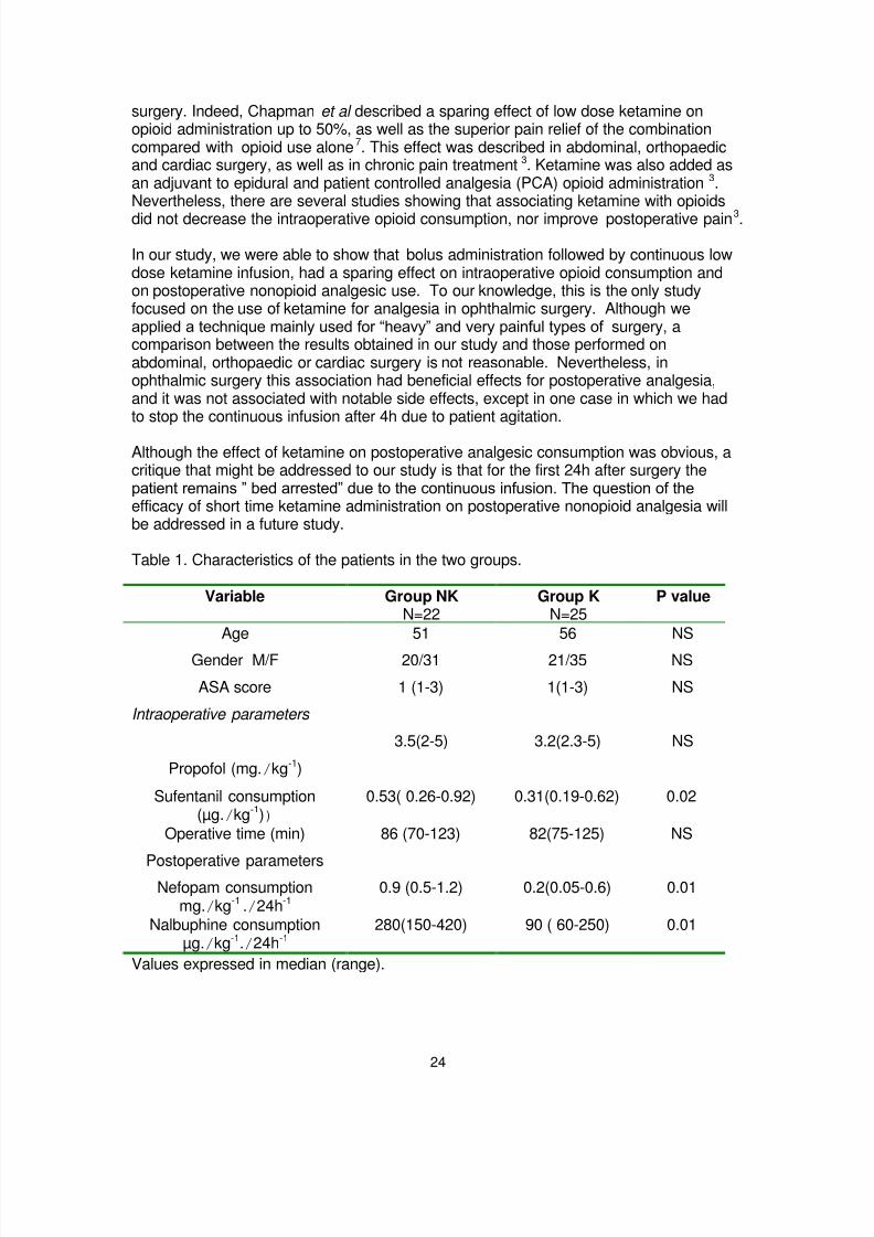
Table 1. Characteristics of the patients in the two groups.
Variable Group NKN=22
Group KN=25
P value
Age 51 56 NS
Gender M/F 20/31 21/35 NS
ASA score 1 (1-3) 1(1-3) NS
Intraoperative parameters
Propofol (mg./kg-1)
3.5(2-5) 3.2(2.3-5) NS
Sufentanil consumption(µg./kg-1))
0.53( 0.26-0.92) 0.31(0.19-0.62) 0.02
Operative time (min) 86 (70-123) 82(75-125) NS
Postoperative parametersNefopam consumption
mg./kg-1 ./24h-10.9 (0.5-1.2) 0.2(0.05-0.6) 0.01
Nalbuphine consumptionµg./kg-1./24h-1
280(150-420) 90 ( 60-250) 0.01
Values expressed in median (range).

8/6/2019 Ophthalmic Anaesthesia 1
http://slidepdf.com/reader/full/ophthalmic-anaesthesia-1 25/66
25
References
1 Coppens M, Versichelen L, Mortier E. Treatment of postoperative pain afterophthalmic surgery. Bull Soc Belge Ophtalmol. 2002; (285): 27-32.
2 Burroughs JR, Soparkar CN, Patrinely JR, Kersten RC, Kulwin DR, Lowe CL.Monitored anaesthesia care for enucleations and eviscerations. Ophthalmology.
2003; 110 (2): 311-3.3 Subramaniam K, Subramaniam B, Steinbrook RA. Ketamine as adjuvant analgesic
to opioids: a quantitative and qualitative systematic review. Anesth Analg. 2004;99(2): 482-95.
4 Aveline C, Gautier JF, Vautier P, Cognet F, Hetet HL, Attali JY, Leconte V, LeborgneP, Bonnet F. Postoperative analgesia and early rehabilitation after total kneereplacement: a comparison of continuous low-dose intravenous ketamine versusnefopam. Eur J Pain. 2009; 13 (6): 613-9.
5 Apfel CC, Korttila K, Abdalla M, Kerger H, Turan A, Vedder I, Zernak C, Danner K,Jokela R, Pocock SJ, Trenkler S, Kredel M, Biedler A, Sessler DI, RoewerN;IMPACT Investigators. A factorial trial of six interventions for the prevention ofpostoperative nausea and vomiting. N Engl J Med. 2004; 350(24): 2441-51.
6 Sigtermans M, Dahan A, Mooren R, Bauer M, Kest B, Sarton E, Olofsen E. S(+)-ketamine Effect on Experimental Pain and Cardiac Output: A PopulationPharmacokinetic-Pharmacodynamic Modelling Study in Healthy Volunteers.Anesthesiology. 2009 Sep 7. EPUB ahead of print.
7 Chapman V, Dickenson AH. The combination of NMDA antagonism and morphineproduces profound antinociception in the rat dorsal horn. Brain Res 1992; 573; 321-323.

8/6/2019 Ophthalmic Anaesthesia 1
http://slidepdf.com/reader/full/ophthalmic-anaesthesia-1 26/66
26
A novel method of reducing subconjunctivalhaemorrhage after sub-Tenon's block
Dagobert Lerch
Consultant Anaesthetist, Voregg 2, CH – 5085 Sulz, Switzerland
Correspondence to [email protected]
IntroductionThe incidence of subconjunctival haemorrhage following Sub-Tenon's block is known tovary from 30-100% 1, 2, 3, 4, 5, 6, 7. Various methods including handheld cautery and topicalepinephrine have been tried but at present, no full proof technique is known to beeffective.
AimReduce the incidence of subconjunctival haemorrhage after sub-Tenon's block.
Method In a prospective observational study which included more than 2500 subjects age range18 -75 but mostly between 25 and 60 undergoing clear lens exchange by phaco andphakic intra ocular Lens (IOL) implant, patients received sub-Tenon's block in the inferonasal quadrant after incision and dissection with Wescott scissors and Colibri forceps.Sub-Tenon's block was performed by an anaesthetist who has extensive experienceinvolving over 5000 such blocks.
A volume ranging from 2-3 ml of 2 % lidocaine with 15 IU/ml and without adrenalinewas injected into the sub-Tenon's space with the help of a plastic 22 G cannula made ofPolyurethane (PUR). The plastic part of a common 22 G "IV indwelling cannula(B/Braun: Introcan Certo/PUR 22G x 1" 0.9x25mm Ref 4251318)" was used.
8/6/2019 Ophthalmic Anaesthesia 1
http://slidepdf.com/reader/full/ophthalmic-anaesthesia-1 27/66
27
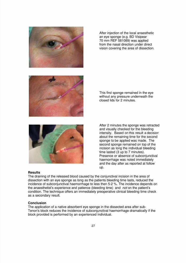
ResultsThe draining of the released blood caused by the conjunctival incision in the area ofdissection with an eye sponge as long as the patients bleeding time lasts, reduced the
incidence of subconjunctival haemorrhage to less then 5-2 %. The incidence depends onthe anaesthetist‘s experience and patience (bleeding time) and not on the patient‘scondition. The technique offers an immediately preoperative clinical bleeding time checkas a secondary result.
ConclusionThe application of a native absorbent eye sponge in the dissected area after sub-Tenon's block reduces the incidence of subconjunctival haemorrhage dramatically if theblock provided is performed by an experienced individual.
After injection of the local anaesthetican eye sponge (e.g. BD Visipear70 mm REF 581089) was applied
from the nasal direction under directvision covering the area of dissection.
This first sponge remained in the eyewithout any pressure underneath theclosed lids for 2 minutes.
After 2 minutes the sponge was retractedand visually checked for the bleedingintensity. Based on this result a decision
about the remaining time for the secondsponge to be applied was made. Thesecond sponge remained on top of theincision as long the individual bleedingtime lasted (3 up to 7 minutes).Presence or absence of subconjunctivalhaemorrhage was noted immediatelyand the day after as reported at followup.

8/6/2019 Ophthalmic Anaesthesia 1
http://slidepdf.com/reader/full/ophthalmic-anaesthesia-1 28/66
28
International Normalised Ratio (INR) Monitoring inPatients Undergoing Ocular Surgery: A Survey
L Tulloch1 and SB Vohra2 1 ST3 Anaesthesia, Russell’s Hall Hospital, Dudley, West Midlands DY1 2HQ, UK. 2 Consultant Anaesthetist. Birmingham and Midland Eye Centre, City Hospital, Sandwell and West Birmingham Hospitals NHS Trust, Birmingham, B18 7QH, UK.
Correspondence to [email protected]
IntroductionAnticoagulated patients are at increased risk of haemorrhagic complications during sub-Tenon‘s and shar p needle regional anaesthetic techniques. The Joint Colleges WorkingParty guidelines recommend that warfarinised patients have their INR checked prior toocular surgery and that their INR is kept within therapeutic range 1. It is generallyaccepted that the INR be 2.5 or less prior to performing peribulbar blocks. A recentnational UK survey of consultant ophthalmic anaesthetists showed that most would
accept an INR 3.5 or less prior to performing sub-Tenon‘s blocks2.
Current national guidelines do not stipulate how soon before surgery the INR should bechecked. Since warfarin and hence INR can be affected by many drug interactions(prescription, over-the-counter and herbal supplements), intercurrent illness as well asdietary changes it is not unreasonable to suggest that the gold standard should bechecking INR on the day of surgery.
At Birmingham and Midland Eye Centre (BMEC), INRs are checked on the day ofsurgery either by; 1. A venous blood sample, or 2. A bedside anticoagulation testingdevice (Roche CoaguChek).
The ward nurses are responsible for taking the venous blood sample when the patientarrives on the ward. The sample is then taken by the porters over to the main hospitalsite to be processed by the pathology laboratory. The ward nurses telephone thelaboratory to obtain the INR result.
The bedside testing service is run by the hospital pathology department. It is performedby specialist anticoagulation nurses when a bedside INR is requested by the wardnurses. Unfortunately this service is only available during office hours and is subject tonursing availability.
It has been observed that with the current systems, preoperative INR results were oftenunavailable causing frustration, changes in theatre list order, delays, cancellations,
complaints, not to mention the financial ramifications of inefficient use of theatre time.The aims of this study were to identify inefficiencies in the current system for checkingINRs at BMEC, to identify the time taken to obtain an INR result on the day of surgeryand to suggest ways to improve current system,
MethodA prospective survey of INR monitoring for patients scheduled for ocular surgery wasperformed over a three month period. We recorded the method of INR testing, the lengthof time it took to receive the result, the anaesthetic technique used for the surgical

8/6/2019 Ophthalmic Anaesthesia 1
http://slidepdf.com/reader/full/ophthalmic-anaesthesia-1 29/66
29
procedure, as well as the reasons and consequences that resulted from any delayedINR results.
Results26 warfarinised patients scheduled for surgery were identified within the three monthperiod. As expected the majority of patients had regional anaesthesia for their surgery.
58% (15) of INRs were checked by the bedside testing device and 42% (11) by venoussampling. (Figure 1)
65% (17) had sub-Tenon‘s block, 19% (5) had peribulbar, 12% (3) had generalanaesthesia, and 4% (1) had topical anaesthesia (Figure 2).
The range of time taken from requesting INR to getting the result was with the traditionalvenous sampling was 43 – 810 minutes (avg. 200 min) and that with the point of carebedside service run by the haematology department was 3-270 minutes, mean 40 min(table 1)
Fig 1 Method of INR testing
Table 1 Time taken to obtain INR result
Time taken Range (min) Mean Time (min) Venous sample 43-810 200 (3hrs 20min)
Bedside test 3-270 40

8/6/2019 Ophthalmic Anaesthesia 1
http://slidepdf.com/reader/full/ophthalmic-anaesthesia-1 30/66
30
Fig 2 Anaesthetic technique
Reasons for delays
The main reasons for delays with venous sampling included nursing staff being too busyto take the blood sample, lost samples and problems with the portering service. Otherreasons were that the pathology laboratory had not processed the sample as a priority orhad not informed the ward of problems with the samples.The reasons for delays with the bedside testing service were mainly due to the lack ofavailability of the anticoagulation nurses since they only work from 9am to 5pm and hadother clinical commitments and were unable to attend the ward when requested.
Effect of delayed INR results on theatre list management:Waiting to obtain INR results led to surgical delays and required alterations in list orderin 30% (8) cases. Of these delays 75% (6) were due to problems with venous sampling.There were no cancellations as a result of delayed INR results.
DiscussionThe results of this study showed that the current INR monitoring system at BMEC couldbe significantly improved. Inefficiencies in the system were causing considerabledelays, discomfort to patients and disruption to theatre lists.The traditional venous sampling took longer, was more labour intensive and causednoticeably more delays than bedside testing. On an average it took nearly three hours toobtain the INR value. The delay with the point of care bedside testing although small,still took nearly 40 minutes on average.
On cost benefit analysis, it appeared that the average cost of one bedside INR test(£2.60) versus the cost of venous sampling (£0.50) was acceptable since the hidden
costs of consumables, nursing, porters and laboratory technician time, patient discomfortand the cost of cancellations and lost operating time were not included in the cost of avenous sample.
The bedside INR tests are as accurate as venous sampling,3 quicker and easy to use,convenient for staff, more comfortable for patients and avoid processing and transportdelays.

8/6/2019 Ophthalmic Anaesthesia 1
http://slidepdf.com/reader/full/ophthalmic-anaesthesia-1 31/66
31
In order to make the INR testing system at BMEC more efficient it was clear that bedsidetesting should be used in preference to venous sampling. Unfortunately since the servicewas dependent on the haematology nurses (who were only available within office hoursand had other commitments) the delays were still creeping in. The only way of avoidingthis delay was to gain independence. We therefore decided to obtain an independentbedside anticoagulation device for the BMEC. A business plan to buy the device has
recently been approved. Plans are afoot to teach and train the ward nurses in its useand maintenance. The aim and hope of the authors is to reduce the time taken toreceive an INR result to less than ten minutes, and eliminate the delays andcancellations resulting from traditional ways of INR monitoring.
References 1 Local anaesthesia for intraocular surgery. Joint report by the Royal College of
Anaesthetists and Royal College of Ophthalmologists London: 2001.http://www.rcophth.ac.uk/docs/publications/published-guidelines/LocalAnaesthesia.pdf
2 Vohra SB, Murray PI. Sub-Tenon's Blocks: A National United Kingdom Survey.Ophthalmic Surgery Lasers and Imaging 2008; 39: 379-385.
3 Cromheecke M et al. Oral anticoagulation self-management and management by aspecialist anticoagulation clinic: a randomised cross-over comparison. Lancet 2000;356: 97 –102.

8/6/2019 Ophthalmic Anaesthesia 1
http://slidepdf.com/reader/full/ophthalmic-anaesthesia-1 32/66
32
Intra-vitreal injection of local anaesthestic agentimproves the quality of peribulbar block in patientsundergoing evisceration surgery
Ezzat Samy Aziz1 and Essam El Toukhy2
1Department of Anaesthesia, Faculty of Medicine, Cairo University 35 A Abou Elfeda street, Zamalek, Cairo, Egypt, 2 Department of Ophthalmology, Cairo University, Cairo,Egypt.
Correspondence to [email protected]
IntroductionEvisceration surgery of the globe entails removal of intraocular contents leaving thesclera shell intact. This surgery is usually performed under general anaesthesia but hasalso been successfully performed under peribulbar block1,2 Using the latter technique, itis not unusual for the patients to experience intraoperative discomfort during surgery.Monitored anaesthesia care with sedation has been proposed as a method for managingthis pain3.
We have performed evisceration surgery in our clinical practice under peribulbar blockand many patients reported intraoperative pain. Perforation of or damage to the globeduring performance of the peribulbar block and evisceration surgery is of little concern.we postulated that deliberate injection of 1 ml of local anaesthetic into the vitreous gelcan relieve intraoperative pain. This hypothesis was supported by a subsequent pilotstudy whose aim was to compare the use of peribulbar block with or without deliberateintravitreal injection of local anaesthetic agent in patients undergoing evisceration of theglobe.
Patients and methodsAfter obtaining the approval of the institutional ethics committee and written informedconsent, 100 patients scheduled for elective evisceration surgery were enrolled in thisstudy. We estimated our sample size using the method described by Lerman4. Asample size of 45 was estimated to be sufficient to detect a 20% difference betweengroups; this will give an alpha value of 0.05 with a power of 80%. With a fall-out rate ofapproximately 19%, we increased the number of patients in each group to 50.
All patients were fasted. They were randomly allocated to one of two groups using theclosed envelope method. Group I (n=50) received peribulbar block alone whereas group11 (n=50) received peribulbar block and intravitreal injection of local anaesthetic. Beforethe administration of anaesthesia, a peripheral vein was cannulated ECG, pulse
oximetry and non-invasive arterial blood pressure were monitored.
Peribulbar block was performed with a 25 gauge, 25 mm sharp disposable needle usingthe three injections technique (extreme inferotemporal, medial and superonasal) with theglobe in the neutral gaze position to ensure complete anaesthesia. Each injection wasperformed with a 3 ml of local anaesthetic solution consisting of equal volumes of 0.5%isobaric bupivacaine and 2% lidocaine with 30 iu /ml of hyaluronidase.

8/6/2019 Ophthalmic Anaesthesia 1
http://slidepdf.com/reader/full/ophthalmic-anaesthesia-1 33/66
33
Group II also received an intravitreal injection of 1 ml of the above local anaestheticmixture. The ophthalmic surgeon injected the intravitreal solution at the inferotemporalquadrant 4 mm behind the limbus. Akinesia and anaesthesia were assessed every 5minutes. Complete or near akinesia was required before proceeding to surgery.
Adequacy of anaesthesia was checked by grasping the conjunctiva at the limbus area in
various quadrants using Castroviejo 0.12 tissue forceps.
Surgical procedureA 360 degree conjunctival incision around the limbus (periotomy) was performedfollowed by a stab incision at the limbus. A spatula was then introduced to separate theuveal tissues from the sclera. The intraocular contents (iris, ciliary body, choroid,vitreous, retina and lens) were removed using a scoop and aided by suction. The insideof the scleral shell was carefully scraped to remove any adherent pigments and thescleral shell washed with hydrogen peroxide solution to dislodge any adherent ocularpigments as well. Any bleeding coming from the vortex veins was cauterized. Specialattention was also given to the vessels at the optic disc and application of directpressure for 2-3 minutes was sufficient. Direct cauterization of the disc vessels was
carried out if necessary.
A simple pain scoring system was used: no pain = 0, discomfort = 1, pain = 2.Pain was subjectively reported by the patient. The scoring was done at various stages ofsurgery: stab incision at limbus, separation of uveal tissues from sclera, removal onintraocular contents and pressure on the disc vessels. If the pain score was 0 or 1, nofurther management was required. However, if the pain score was 2 at any stage, 20 ugof fentanyl and 1 mg of midazolam were given.
Parametric data were analyzed using Student‘s t-test and non parametric data werecompared using the Chi-square x2 test. A p value of <0.05 was considered statisticallysignificant.
Results There was no significant difference between the patient characteristics of the two groups(table1).
There was no significant difference in pain scores during stab incision between the twogroups. 11 patients in group 1 reported pain (pain score =2) during separation of uvealtissues compared to 1 patient in group 2 (p<0.0001). 13 patients reported pain (painscore=2) in group 1 during removal of ocular tissues compared to 2 patients in group 2(p<0.0001). 16 patients in group 1 reported no pain (pain score=0) during pressure andcauterization of disc vessels compared to 36 in group II (p<0.0001). Pain scores ateach stage of surgery are shown in table 2.
Discussion The globe is made up of three layers. The outer layer consists of cornea and sclera andis very sensitive to local anaesthetic injection. The inner layer is relatively insensitiveand consists of the retina. The middle layer consists of iris, ciliary body and choroid. It isvery vascular, contains sensitive neurons and is very sensitive to pain. Most of thepatients feel discomfort or even pain when the middle layer is touched even within a fullyfunctional peribulbar block. For this reason many surgeons prefer general anaesthesia.

8/6/2019 Ophthalmic Anaesthesia 1
http://slidepdf.com/reader/full/ophthalmic-anaesthesia-1 34/66
34
We observed that the deliberate injection of 1 ml of local anaesthetic solution alleviatedthis pain and improved the quality of anaesthesia and analgesia in most patients.
Injection of local anaesthetic into the vitreous can be associated with ocular explosion.Ocular explosion has been reported during cataract surgery if approximately 3ml of localanaesthetic is injected accidentally into the vitreous during needle block because the
intra-orbital pressure increases to a dangerous level5. Globe explosion has beenreported after intravitreal injection of triamicinolone acetonide6. Intravitreal injectionshave become popular for the treatment of retinal disorders, however an increase in theintraocular pressure has been reported6. Globe rupture has been reported after simpledigital pressure following peribulbar block in a 75-year- old woman7. This reinforces theview that any rise in intraocular pressure is of concern when an ocular block isperformed.
We are not aware of any adverse event when 1 ml of local anaesthetic is injected intothe vitreous. We believe that the rise in intraocular pressure will not be high enough tocause any damage to the globe. Our study shows that this small amount of deliberateintravitreal injection improves the quality of anaesthesia and analgesia in patients
undergoing evisceration surgery with a peribulbar block and only a very insignificantnumber of patients felt any pain as shown in table 2.
ConclusionWe conclude that a deliberate intravitreal injection of 1 ml of local anaesthetic agentimproves the quality of periblubar block thus making the intraoperative procedure muchmore acceptable to patients, avoiding general anaesthesia.
Table 1 Patient characteristics weight: mean (SD) age: mean (range)
Group I Group II
Peribulbar block Peribulbar block + intraocular anaesthesian=50 n=50
Men/Women
30/20 35/15
Mean age(range)(yr)
25(18-37) 24(19-38)
Mean weight (SD)(Kg)
75(18-37) 68(10.8)
ASA 1 (n)50 50

8/6/2019 Ophthalmic Anaesthesia 1
http://slidepdf.com/reader/full/ophthalmic-anaesthesia-1 35/66
35
Table 2 Numbers of patients with a specific pain score at different stages of theoperation. Data were analyzed using the Chi-square test. *P <0.0001 between groups.--------------------------------------------------------------------------------------------------------------------
Group I Group II
Peribulbar block Peribulbar block + intraocular anaesthesia
n=50 n=50
______________________________________________________________________
No pain discomfort pain No pain Discomfort pain
______________________________________________________________________
Stab incision
47 3 0 49 1 0
Separation of uveal tissues
17 22 11 * 31 18 1
Removal of ocular tissues
14 23 13* 30 18 2
Pressure & cauterization of disc vessels
16 27 7 36* 11 3 ______________________________________________________________________
References
1 Calenda E, Assadi C, Retout A, Cardon A. Is eye enucleation or eviscerationpossible under peribulbar anaesthesia? Eur J Anaesthesiol . 1997;14(5):551-2.
2 Calenda E, Retout A, Muraine M. Peribulbar anaesthesia for peroperative andpostoperative pain control in eye enucleation or evisceration: 31 cases J Fr Ophtalmol. 1999;22(4):426-30. French.
3 Burroughs JR, Soparkar CNS, Patrinely JR, Kersten RC, Kulwin DR, Lowe CL.Monitored anaesthesia care for enucleations and eviscerations. Ophthalmology 2003;110:311-13.
4 Lerman J. Study design in clinical research: sample size estimation and poweranalysis statistics. Can J Anaesth 1996;43:184-91 Bullock JD, Warwar RE andGreen WR.
5 Ocular explosion during cataract surgery: a clinical, histopathological, experimentaland biophysical study. Trans Am Ophthalmol Soc , 1998;96:243-281.
6 Viestenz A, Kuchle M and Behrens-Baumann W. Globe explosion caused byintravitreal injection of triamcinolone acetonide, a case report. Klin Monatsbl Augenheilkd 2008;12:1087-90.
7 Rathi V, Basti S, Gupta S. Globe rupture during digital massage after peribulbaranaesthesia. J Cataract Refract Surg 1997;23(2):297-9.

8/6/2019 Ophthalmic Anaesthesia 1
http://slidepdf.com/reader/full/ophthalmic-anaesthesia-1 36/66
36
Two-quadrant high volume sub-Tenon‘s anaesthesia for vitreoretinal surgery: a randomized controlled trial
VS Gill1, AH Presland1, JA Lord1, and DG Charteris2
1Department of Anaesthesia ,2 Vitreoretinal Surgeon,Moorfields Eye Hospital NHS Foundation Trust, City Rd, London, EC1V 2PD, UK
Correspondence to [email protected]
AbstractThe total volume of a standard single inferonasal injection of local anaesthetic for a sub-Tenon‘s block is limited by an increase in intraocular pressure and commonly requiresthe operating surgeon to top up the block intraoperatively. This study compares theefficacy and safety of a two-quadrant technique which allows the use of a higher volumeof local anaesthetic.
Methods Fifty-four patients undergoing vitreoretinal surgery were randomized into two groups.The control group (n=27) received a standard 5ml single infero-nasal sub-Tenoninjection of a 50:50 mixture of 2% lidocaine and 0.5% bupivacaine with 150 IUhyaluronidase. The study group (n=27) received a 5ml inferonasal injection and a 5mlsuperotemporal injection of the same mixture (10ml total). The primary outcomemeasure was the number of intraoperative top ups required. Secondary outcomemeasures were: intraoperative pain score, postoperative pain scores, intraocularpressure, block onset time, ocular akinesia, eyelid akinesia and chemosis.
ResultsTwo-quadrant sub-Tenon‘s anaesthesia using 10 ml of a 50:50 mixture of 2% lidocaine
and 0.5% bupivacaine with 150 IU hyaluronidase significantly reduced the need for anintraoperative top up during vitreoretinal surgery (P<0.001) without a significant increasein intraocular pressure. Block onset time was shorter and eyelid akinesia was improvedin the two-quadrant high volume group. Mean pain scores were significantly lower in thestudy group compared to the control group immediately postoperatively and at 0-2hours, 4-6 hours, 10-14 hours and 20-24 hours postoperatively.
ConclusionsTwo-quadrant sub-Tenon‘s anaesthesia using 10 ml of a 50:50 mixture of 2% lidocaineand 0.5% bupivacaine with 150 IU hyaluronidase seems to be more effective than astandard inferonasal approach in preventing perioperative and postoperative pain duringvitreoretinal surgery under local anaesthesia.

8/6/2019 Ophthalmic Anaesthesia 1
http://slidepdf.com/reader/full/ophthalmic-anaesthesia-1 37/66
IntroductionSub-Tenon‘s block has been described as a safe and effective method to achieve localanaesthesia for vitreoretinal surgery.1-9 A perceived advantage is the avoidance of sharpneedles and a reduction in their associated complications such as retrobulbar haemorrhage,globe perforation and optic nerve damage.6 7 10-13 However, the variable quality of ocularakinesia and short block duration with subsequent need for an additional sub-Tenon‘s blockto be performed by the operating surgeon intraoperatively is not ideal and may be a limitingfactor in its popularity for vitreoretinal surgery. Total volume and local anaesthetic mixtureused varies and has an effect on block onset time, quality and duration. 14 15 Localanaesthetic concentration is limited by the risk of myotoxicity to the extra-ocular muscleswhilst total volume of local anaesthetic used is limited by an increase in intraocular pressureand chemosis. 16 17 A common technique is a single 5ml injection of a 50:50 mixture oflidocaine 2% and bupivacaine 0.5% via the inferonasal quadrant which allows for a shortonset time and reliable ocular akinesia but frequently still requires the need for furtherintraoperative top ups.7 9 14 The aim of this study was to compare the efficacy and safety of ahigh volume two-quadrant sub-Tenon‘s technique compared to the usual low volume singlequadrant approach for vitreoretinal surgery.
MethodAfter local ethics committee approval and written informed consent, 54 patients (aged 30-81yr and ASA grade I-III) undergoing vitreoretinal surgery (vitrectomy +/- laser +/- cryotherapy+/- gas exchange) for retinal detachment were recruited into this prospective, randomizedstudy. Patients allergic to local anaesthetics or hyaluronidase, with communicationdifficulties or with a scleral buckle present were excluded.
Using a random number generator, a batch of numbered sealed envelopes containing eachrandomization was produced and kept in a designated area. At recruitment, each patientwas allocated a sequential envelope to be opened in the anaesthetic room only by theanaesthetist performing the block. All blocks were performed by a single anaesthetist
experienced in both types of blocks. Patients were randomized to one of two groups. Thecontrol group received a 5ml single inferonasal sub-Tenon injection of a 50:50 mixture of 2%lidocaine and 0.5% bupivacaine with 150 IU hyaluronidase19-21 The study group received a5ml inferonasal injection and a 5ml superotemporal injection of the same mixture. Theprincipal investigator was masked to which block had been administered.
In the anaesthetic room monitoring included ECG, pulse oximetry and non-invasive arterialpressure. No sedation was given. The conjunctiva and cornea was initially anaesthetisedwith topical drops of proxymetacaine 0.5% followed by tetracaine 1% and sterilised withdrops of 5% aqueous iodine solution. The surrounding skin was sterilised with 10% aqueousiodine solution. Using non-toothed Moorfield forceps to grip the conjunctiva and Tenon‘scapsule, with Westcott scissors, a small incision was made to expose the white sclera in the
inferonasal quadrant only (control group) or inferonasal and superotemporal quadrants(study group). After injection using a blunt 19G, 25mm long curved Visitec sub-Tenon‘scannula the principal investigator then assessed the block. Ocular akinesia was assessed at1,3,6,9,12,15 and 18 min after injection using a scoring system as described by Brahma,22 inwhich globe movement is scored either 0, no movement; 1, a flicker of movement; 2, partialmovement; or 3, full movement; in the secondary directions of gaze (abduction, adduction,

8/6/2019 Ophthalmic Anaesthesia 1
http://slidepdf.com/reader/full/ophthalmic-anaesthesia-1 38/66
elevation and depression). An immobile eye scored 0 and a fully mobile eye scored 12.
Table 1 Patient characteristics, type of operation, duration of operation and type of previoussurgery. Results are n or mean (SD).
Patients were deemed ready for surgery when the score was 4 or less. Chemosis wasassessed on the number of quadrants affected. No quadrants affected scored 0 and all fourquadrants affected scored 4. Intraocular pressure was measured at 1,3,6,9,12,15,18 minusing a Tonopen.23 24 At the end of the operation, type of surgery, duration of surgery andthe timing of any intraoperative top ups (3ml of bupivacaine 0.5%) were recorded. Theoperating surgeon was also asked to score perioperative eyelid movement from 0 to 2. 0, nomovement; 1, partial function and 2, full function.25 Pain scores were taken post injection andpostoperatively using an 11-point (0-10) numerical visual analogue scale (VAS).26 Patientsrecorded pain scores and analgesia taken at four intervals within the first 24 hours aftersurgery; at 0-2 hours, 4-6 hours, 10-14 hours and 20-24 hours. Age, gender, ethnic origin,previous operations and postoperative complications were also recorded.
Statistical analysisThe primary outcome measure was the requirement for an intraoperative top up. On thebasis of previous studies it was assumed that the proportion of patients who require a top upafter a 5 ml injection is 60 % and that the proportion of patients who require a top up after a10 ml injection is 10 %.7-9 From this it was calculated that 25 patients would be required pertreatment group to detect a difference with 95% power (two-tailed alpha of 0.05). Thenumbers were then increased by 5% in each group to allow for drop out, resulting in 2 extrapatients per treatment group so that the final number in each group was 27.
ResultsThe groups were similar in respect of patient characteristics, previous surgery, type and
duration of surgery (Table 1). Mean operating time for the control group was 74.2 +/- 20.7minutes and 74.4 +/- 17.6 minutes for the study group. Within the control group twenty-three
Single-Quadrant(n =27)
Two-Quadrant(n =27)
Age (yr)
SexMF
VitrectomyCryotherapyGasLaser
Op Time (min)Prev Surgery
ant chamberpost chamber
62.8 (11.2)
11
16
2719199
74.2 (20.7)
97
63.6 (10.4)
15
12
2723256
74.4 (17.6)
95

8/6/2019 Ophthalmic Anaesthesia 1
http://slidepdf.com/reader/full/ophthalmic-anaesthesia-1 39/66
patients (85.19%) required a single top up at a mean time of 50.8 +/- 11.8 minutes and onepatient (3.7%) required two top ups. No patients required a top up in the study group(P <0.001). Block onset time was significantly lower for the study group. Median time to anakinesia score of 4 or less was 9 minutes for the control group compared to 3 minutes for thestudy group. Median eyelid akinesia score was 2 for the control group and 0 for the studygroup. There was no significant difference in the changes in mean IOP with time betweenthe two groups.
0
5
10
15
20
25
Pre-Block 1 3 6 9 12 15 18Time (min
Single-Quadrant
Two-Quadrant
Fig 1 Mean intraocular pressure (IOP) measured in mmHg at 1,3,6,9,12,15 and 18 min afterblock performed.
Fig 2 Mean intraoperative and postoperative pain scores.
0
1
2
3
4
5
Post-
Block
Intra-
Op
0 to 2 4 to 6 10 to
14
20 to
24Time (hrs)
Single-QuadrantTwo-Quadrant

8/6/2019 Ophthalmic Anaesthesia 1
http://slidepdf.com/reader/full/ophthalmic-anaesthesia-1 40/66
Single-Quadrant Two-Quadrant(n =27) (n =27)
None 2 (7.4%) 12 (44.4%)
Paracetamol 3 (11.1%) 14 (51.9%)
Codeine Phosphate 22 (81.5%) 1 (3.7%)
Maximum IOP measured was 32 mmHg in the control group and 28 mmHg lower in thecontrol group at 33.3% compared to 52% in the study group. Mean pain scores were lowerfor the study group compared to control group at injection (0.8 vs. 2.2), immediatelypostoperatively (0.1 vs. 3.4) and at 0-2 hours (0.4 vs.2.5), 4-6 hours (0.8 vs. 4.4), 10-14hours (0.9 vs. 4.6) and 20-24 hours (0.7 vs. 3.6) postoperatively (Fig. 2). The need forrescue analgesia within the first 24 hours postoperatively was less in the study group(Table 2). In the control group 2 patients (7.4%) required no analgesia and 22 patients(81.5%) required codeine phosphate 30-60mg. In the study group 12 patients (44.4%)required no analgesia and only 1 patient (3.7%) required codeine phoshate 30-60mg. There
were no complications in either group.
DiscussionThis study shows that two-quadrant sub-Tenon‘s anaesthesia using 10ml of a 50:50 mixtureof lidocaine 2% and bupivacaine 0.5% with 150 IU hyaluronidase via the inferonasal andsuperotemporal quadrants significantly increases block duration and reduces the need for anintraoperative top up during vitreoretinal surgery without any significant increase inintraocular pressure. In addition, intraoperative pain, postoperative pain and the need forrescue analgesia postoperatively are all significantly reduced when compared to a single 5mlinjection of the same mixture in the inferonasal quadrant. Quality of block as assessed bythe onset of ocular akinesia and perioperative eyelid movements are also significantlyimproved. Our findings for the low volume (control) group are similar to previous reported
studies. Kwok and colleagues7
used a 4ml single inferonasal injection of a 50:50 mixture oflidocaine 1% and bupivacaine 0.5% with 1,200 IU of hyaluronidase. 77% of patients in theirstudy needed one or two top ups at a mean time of 50 +/- 46 minutes compared to 89% inour study at a similar mean time of 50.8 +/- 11.8 minutes. Lai and colleagues9 reported 37%of patients receiving a 5ml single inferonasal sub-Tenon‘s injection of a 50:50 mixture of lidocaine 4% and bupivacaine 0.75% requiring a top up (times of top up not mentioned).Their use of a stronger anaesthetic mixture may account for the lower top up rate. Themean intraoperative pain score in our control group was 3.4 compared to 1.17 and 2.1 in theabove studies. However all the patients in these studies were sedated with varyingcombinations of midazolam, alfentanil or fentanyl and propofol. None of the patients in ourstudy were sedated. This may explain the higher top up rate and mean intraoperative painscores in the control group compared to previous studies. Nevertheless anaesthesia was
significantly more effective in the high volume (study) group with no one requiring a top upcompared to 89% requiring a top up in the low volume (control) group. Anaesthesia
Table 2 Postoperative analgesic requirements within the first 24 hours

8/6/2019 Ophthalmic Anaesthesia 1
http://slidepdf.com/reader/full/ophthalmic-anaesthesia-1 41/66
continued for up to 24 hours postoperatively in the high volume group, providing adequateanalgesia without the need for rescue analgesia in 44.4% of patients compared to 7.4% notrequiring any rescue analgesia in the low volume group.
Cryotherapy was the most stimulating procedure performed within our study followed bylaser therapy and then gas exchange. The type of surgery performed in both groups wasnot significantly different. 23 patients underwent cryotherapy in the high volume groupcompared to 19 patients in the low volume group. Patients who have had previous ocularsurgery have tissue adhesions which may limit diffusion of the local anaesthetic agent andthus efficacy. Both groups were similar with no significant difference between the groupswith respect to previous ocular surgery. Previous studies have demonstrated sub-Tenon‘sanaesthesia is a safe and effective technique to achieve local anaesthesia for vitreoretinalsurgery.1-9 The risk of globe compression and consequent increase in intraocular pressure(IOP) associated with an injection of a volume of local anaesthetic agent into the fixed orbitalspace has limited the maximum volume used for sub-Tenon‘s anaesthesia. IOP is known torise with increasing volume of local anaesthetic agent used for peribulbar and retrobulbaranaesthesia techniques.27 There are few studies investigating the relationship between
volume and IOP after sub-Tenon‘s anaesthesia.
17 18
IOP may rise slightly after injection of upto 5ml of local anaesthetic into the sub-Tenon space.17 No comparative data observing theeffect on IOP of injecting more than 5ml of local anaesthetic into the sub-Tenon spaceexists in the current literature. An important new finding in this study is that injection of 10mlof local anaesthetic agent into the sub-Tenon space did not cause any significant rise in IOP.In fact we observed a small gradual reduction in IOP up to 18 min after injection. Oneprevious study has identified this phenomenon.17 The reduction in IOP may be a directpharmacological effect of the local anaesthetic agent on ocular blood flow, or secondary to areduction in aqueous production due to an effect on the ciliary ganglion, or possibly becauseof a reduction in extraocular muscle tone.
Block quality as assessed by onset time and eyelid akinesia was also improved in the two-
quadrant high volume group. A median reduction in block onset of 6 min may not beclinically important but continual intraoperative eyelid movements are distracting for thesurgeon, interfere with surgery and may interfere with clinical outcome.
The incidence of chemosis increases as the volume of local anaesthetic injected into thesub-Tenon space increases.4 13 14 Our findings are consistent with this and the incidence of52% in the high volume group is within the range of 25 to 60% reported by previous studiesusing a lower volume of local anaesthetic agent.6 However this is not clinically significant.Chemosis interferes less with vitreoretinal surgery compared to anterior segment surgeryand usually resolves after the application of digital pressure.
In conclusion, our study has shown that 10ml of local anaesthetic agent may be injected via
the inferonasal and superotemporal quadrants into the sub-Tenon space without significantlyincreasing intraocular pressure. A 50:50 mixture of lidocaine 2% and bupivacaine 0.5% with150 IU hyaluronidase provides significantly better block quality, perioperative anaesthesiaand postoperative analgesia for vitreoretinal surgery when compared to a standard single-quadrant low volume (5ml) technique using the same mixture. Further studies are requiredto resolve whether this is due to an increased dose of local anaesthetic agent, increaseddiffusion secondary to an increase in volume of local anaesthetic or due to increaseddiffusion associated with the anatomy of a two-quadrant approach.
8/6/2019 Ophthalmic Anaesthesia 1
http://slidepdf.com/reader/full/ophthalmic-anaesthesia-1 42/66
References
1 Mein CE, Woodcock MG. Local anaesthesia for vitreoretinal surgery. Retina 1990; 10:47-49.
2 Friedberg MA, Spellman FA, Pilkerton AR, et al . An alternative technique of localanaesthesia for vitreoretinal surgery. Arch Ophthalmol 1991; 109: 1615-16. 3 Stevens JD, Franks WA, Orr G, Leaver PK, Cooling RJ. Four-quadrant local
anaesthesia technique for vitreoretinal surgery. Eye 1992; 6: 583-86.4 Greenbaum S. Parabulbar Anaesthesia. Am J Ophthalmol 1992; 114: 776.5 Sharma T, Gopal L, Parikh S, et al. Parabulbar anaesthesia for primary vitreoretinal
surgery. Ophthalmology 1997; 104: 425-28.6 Roman SJ, Chong Sit DA, Boureau CM, Auclin FX, Ullern MM. Sub-Tenon‘s
anaesthesia: an efficient and safe technique. Br J Ophthalmol 1997; 81:673-76.7 Kwok AK, Van Newkirk MR, Lam DS, Fand DS. Sub-Tenon‘s anaesthesia in
vitreoretinal surgery: a needleless technique. Retina 1999; 19: 291-96.8 Li HK, Abouleish A, Grady J, Groeschel W, Gil KS. Sub-Tenon‘s injection for local
anaesthesia in posterior segment surgery. Ophthalmology 2000; 107: 41-46.9 Lai MM, Lai JC, Lee W-H, et al. Comparison of retrobulbar and sub-Tenon‘s capsule
injection of local anaesthetic in vitreoretinal surgery. Ophthalmology 2005; 112: 574-79.10 Eke T, Thompson JR. The national survey of local anaesthesia for ocular surgery. II.
Safety profiles of local anaesthesia techniques. Eye 1999; 13: 196-204.11 Canavan KS, Dark A, Garrioch MA. Sub-Tenon‘s administration of local anaesthetic: a
review of the technique. Br J Anaesth 2003; 90: 787-93.12 Guise PA. Sub-Tenon‘s anaesthesia: a prospective study of 6000 blocks. Anesthesiol
2003; 98: 964-68.13 Ruschen H, Bremner FD, Carr C. Complications after sub-Tenon‘s eye block. Anaesth
Anal 2003; 96: 273-77.14 Kumar CM, Williamson S, Manickam B. A review of sub-Tenon‘s block: current practice
and recent development. Eur J Anaesthesiol 2005; 22: 567-77.15 Tokuda Y, Oshika T, Amano S, Yoshitomi F, Inouye J. Anaesthetic dose and analgesic
effects of sub-Tenon‘s anaesthesia. J Cataract Refract Surg 1999; 25: 1250-53.16 McLure HA, Kumar CM, Ahmed S, Patel A. A comparison of lidocaine 2% with
levobupivacaine 0.75% for sub-Tenon‘s block. Eur J Anaesthesiol 2005; 22: 500-03.17 Alwitry A, Koshy Z, Browning AC, Keil W, Holden R. The effect of sub-Tenon‘s
anaesthesia on intraocular pressure. Eye 2001; 15: 733-35.18 Patton N, Malik T, Aslam T, Vallance J. Effect of volume used in sub-Tenon‘s
anaesthesia on efficacy and intraocular pressure: a randomized clinical trial of 3 mlversus 5 ml. Clin Exp Ophthalmol 2004; 32: 488-91.
19 Guise P, Laurent S. Sub-Tenon‘s block: the effect of hyaluronidase on speed of onsetand block quality. Anaesth Intensive Care 1999; 27: 179-81.
20 Rowley SA, Hale JE, Finlay RD. Sub-Tenon‘s local anaesthesia: the effect of hyaluronidase. Br J Opthalmol 2000; 84: 435-36.
21 Schulenburg HE, Sri-Chandana C, Lyons G, Columb MO, McLure HA. Hyaluronidasereduces local anaesthetic volumes for sub-Tenon‘s anaesthesia. Br J Anaesth 2007; 99:717-20.
22 Brahma AK, Pemberton CJ, Ayeko M, Morgan LH. Single medial injection peribulbaranaesthesia using prilocaine. Anaesthesia 1994; 49: 1003-05.
23 Kao SF, Lichter PR, Bergstrom TJ, Rowe S, Musch DC. Clinical comparison of theOculabe Tono-Pen to the Goldmann applanation tonometer. Ophthalmology 1987; 94:1541-44.
24 Frenkel RE, Hong JY, Shin DH. Comparison of the Tono-Pen to the Goldmann
applanation tonometer. Arch Ophthalmol 1998; 106: 750-53.

8/6/2019 Ophthalmic Anaesthesia 1
http://slidepdf.com/reader/full/ophthalmic-anaesthesia-1 43/66
25 Rauz S, Subramaniam S. Sub-Tenon‘s anaesthesia and orbicularis oculi function.Ophthalmic Surg Lasers 1997; 28: 727-30.
26 Ohnhaus EE, Adler R. Methodological problems in the measurement of pain: acomparison between the verbal rating scale and visual analogue scale. Pain 1975; 1:375-84.
27 Watkins R, Beigi B, Yates M, Chang B, Linardos E. Intraocular pressure and pulsatileocular blood flow after retrobulbar and peribulbar anaesthesia. Br J Ophthalmol 2001;85: 796-98.

8/6/2019 Ophthalmic Anaesthesia 1
http://slidepdf.com/reader/full/ophthalmic-anaesthesia-1 44/66
44
Prospective evaluation of deep topical fornix nerveblock versus topical anaesthesia in patients undergoingphacoemulsification and intraocular lens implantation
Ezzat Samy Aziz1 and Noha Khater2
1Department of Anaesthesia, Faculty of Medicine, Cairo University 35 A abou elfeda street, Zamalek, Cairo, Egypt. and 2 Department of Ophthalmology, Cairo University,Cairo, Egypt.
Correspondence to [email protected]
IntroductionGeneral anaesthesia may be hazardous in many patients undergoing ophthalmicsurgery, as most of them are elderly with systemic disease receiving multiplemedications. This is especially true for diabetic patients in whom local anaesthesia ispreferable as it reduces to a great extent the endocrine response to surgery1
.
Traditionally, retrobulbar injections were performed deep into the orbit, but it is nowaccepted that peribulbar injections using shorter needles are safer. In the last few years,continuing concern over the rare but serious complications of sharp needle blocks hasled to increasing interest in the use of sub-Tenon`s blocks utilizing a blunt cannula 2,3,4,5.Even with the use of blunt canulae sub-Tenon`s block serious problems can still occur.With advances in cataract extraction using the technology of phacoemulsification,topical anaesthesia has been used successfully6.
Topical anaesthesia has several advantages over regional infiltrative techniques, theforemost of which is the abolition of any risk of inadvertent injury of the globe or orbitalcontents 7,8. It has a high rate of patient satisfaction, but still there some patients thatexperience intraoperative discomfort.
Deep topical fornix nerve block anaesthesia (DTFNB) is done by placing two smallsponges soaked with 0.5% bupivicaine in the conjunctival fornices. It has theadvantage of a needle free technique with patient satisfaction9. In this study wecompared topical anaesthesia with DTFNBA in patients undergoing phacoemulsificationcataract surgery.
Patients and methodsOne hundred patients scheduled for elective phacoemulsification cataract surgery wereenrolled in this study after obtaining approval from the institutional ethical committee andwritten patient` consent. We estimated our sample size based on a method described
by Lerman10. A sample size of 45 will detect a 20% difference between each group; thiswill give an alpha value of 0.05 with a power of 80%. With a fall-out rate ofapproximately 19%, we decided to increase the number of patients in each group to 50.All patients were assessed and only those who were deemed suitable for topical andDTFNBA were included in the study.
Selection criteria of patients are essential for the above technique which includes fullcooperation of the patient. The level of patient anxiety is another important indicator of

8/6/2019 Ophthalmic Anaesthesia 1
http://slidepdf.com/reader/full/ophthalmic-anaesthesia-1 45/66
45
success of non-akinetic anaesthesia. Very anxious patients were omitted from thestudy. All the operations were done by one experienced surgeon.
Patients were randomly allocated to one of two groups (closed envelope method).Group I (n=50) received topical anaesthetic drops and Group 2 (n=50) receivedDTFNBA. Before giving the anaesthetics, a peripheral vein was cannulated and heart
rate, oxygen saturation and non-invasive arterial blood pressure were monitored.
Topical anaesthesia was done with 2% tetracaine local anaesthetic drops and DTFNBAwas performed using two sponges (2x3mm) soaked with 0.5% bupivicaine, applied deepin the conjunctival fornices after anaesthetising the conjunctiva with tetracaine localanaesthetic drops9. The sponges were removed after 15 minutes. The anaestheticeffect was tested by grasping the limbus with Castroviejo 0.12 tissue forceps.
Pain was estimated by the patient using a simple pain score: no pain =0; that does notinterfere with the surgical technique, discomfort=1; the surgical technique is performedwith difficulty, pain=2; the surgeon is unable to continue the surgical technique. Thescoring was done during different stages of surgery: lid retraction while inserting a
speculum, tolerance to the microscope light, corneal incision, phacoemulsification,irrigation and aspiration, and intraocular lens insertion.
The surgical technique was performed through a clear corneal 3mm tunnel incision,followed by capsulorrhexis under sodium hyalurounate. The nucleus was removed bythe stop and chop technique followed by bimanual irrigation aspiration of the cortex.Finally, implantation of a foldable acrylic intraocular lens in the capsular bag with wash ofthe viscoelastic and hydration of the side ports. The total operative time was recorded forevery case.
If the pain score was 0 or 1, no further management was required but if the pain scorewas 2 at any stage, 1% preservative free lidocaine was injected intracamerally.
Parametric data were analyzed using Students t-test; non parametric data werecompared using the Chi-square test. A P value of <0.05 was considered statisticallysignificant.
Results There was no significant difference between the patient characteristics of the two groups(Table1).
About 50% of patients receiving topical anaesthesia only reported pain - especially withspeculum application, phacoemulsification. Furthermore, they reported severe pressuresensation which required the use of intracameral lidocaine. The other 50% reported
varying degrees of discomfort that was tolerated without the need for intracamerallidocaine. Most patients in group 2 (DTFNBA), tolerated the operation well, Slightdiscomfort was noted as a sensation of heaviness reported by some patients during theintroduction of the phacoemulsification probe inside the eye and duringirrigation/aspiration. Another moment of discomfort occurred with the IOL implantation.However none of the patients had pain strong enough to require intracameral injection oflidocaine.

8/6/2019 Ophthalmic Anaesthesia 1
http://slidepdf.com/reader/full/ophthalmic-anaesthesia-1 46/66
46
The number of patients in each group with different pain scores at each stage of surgeryare shown in Table 2.
Table I Patient characteristics. Age is mean (range) and weight is mean (SD)
Group 1 Topical Group II DTFNBA
Men/Women 30/20 35/15
Mean age(range)(yr) 25(18-37) 24(19-38)
Mean weight (SD)(Kg) 75(18-37) 68(10.8)
ASA 1 9 10
ASA 2 31 30
ASA 3 20 20

8/6/2019 Ophthalmic Anaesthesia 1
http://slidepdf.com/reader/full/ophthalmic-anaesthesia-1 47/66
47
Table 2 Number of patients in relation to pain score at different stages of theoperation. Data were analyzed using the Chi-square test. *P < 0.0001 between groups
Group I Topical n=50 Group II DTFNBA n=50
No pain Discomfort Pain No pain discomfort Pain
Lid retraction 0 50* 0 28* 22 0
Tolerance to light 0 50* 0 35 15 0
Keratotomy (corneal incision) 30 20* 0 50 0 0
Phacoemulsification 11 29 10 41* 9 0
Intraocular lens insertion 0 30 20* 10 40 0
Irrigation and aspiration 14 27 9 39* 11 0
DiscussionTopical anaesthesia has gained popularity over the last few years as it is safer than thetraditional peribulbar and sub-Tenon`s block especially with the advancedphacoemulsification machines. It has a high rate of patient satisfaction with immediatevisual rehabilitation in most cases 7. Topical anaesthesia has the advantage of having asofter globe during surgery with minimal positive vitreous pressure compared toperibulbar blocks.
However, some patients squeeze orbicularis oculi muscle (squeezers), which is apotential problem with either no or inadequate lid block. Administration of fascial blocksis a potential solution and results in loss of time, additional expense, complications and
pain. Patients can be intolerant of the microscope light which makes them squeeze theireyes more, obscuring the surgical field for the surgeon. Moreover there is always adegree of discomfort (ranging from slight to severe) at the time of phacoemulsification,irrigation and aspiration and finally intraocular lens insertion. Topical anaesthesia wascompared to needle blocks with contradictory results proving that topical anaesthesiaalone is still not the method of choice for elective phacoemulsification cataract surgery11,12.
With DTFNBA patients did not squeeze their eyes and their tolerance to the microscopelight was better. Patients experienced no pain or slight discomfort duringphacoemulsification, irrigation/aspiration and during intraocular lens insertion. Nopatients felt discomfort or pain on the pain scale which makes DTFNBA superior to
topical anesthesia in phacoemulsification.
In DTFNBA, placing the anaesthetic in the fornix is more neuro-anatomically andpharmacologically sound as the fornix is contiguous with the peribulbar space.Placement of the sponges in the fornices allows absorption by the nerve trunks subserving the conjunctiva. At the same time, by being absorbed posteriorly into theperibulbar space, the posterior cilliary nerves, which supply the anterior sclera, anteriorconjunctiva and limbus as well as the iris and ciliary body are anaesthetised at theirnerve roots 13,14,15. The fornix is also a logical space for direct absorption across the

8/6/2019 Ophthalmic Anaesthesia 1
http://slidepdf.com/reader/full/ophthalmic-anaesthesia-1 48/66
48
Tenon`s capsule. There is in addition likely absorption into the lid, producing somedegree of lid akinesia.
The presence of lid hypokinesia, the ability to manipulate the iris freely without pain andthe few case of incidental superior rectus pariesis suggest an anaesthetic effect thatgoes beyond its topical anaesthesia. In group 2 where DTFNBA was used, tolerance to
microscopic light was much better. It has been suggested that nerve endingsresponsible for temperature sensation in the cornea lies deeper in the stroma andtherefore more difficult to block than pain fibres (16), suggest that DTFNBA provide aprofound degree of anaesthesia than topical anaesthesia.
However, proper selection of patients as well as experienced surgeons is the mainsecrets of success of using non-akinetic anaesthesia for phacoemulsification (17).
References
1 Barker JP, Robinson PN, Vafidis GC, Hart GR, Spased-Byme S, Hall GM. Localanalgesia prevents the cortisol and glycaemic responses to cataract surgery. Br J
anaesth 1990;64:442-5.2 Stevens JD: A new local anaesthesia technique for cataract extraction by one
quadrant subtenons infiltration. Br J Ophthalmol 1992;76:670-74.3 Bergman L, Berglin L, Algreve PV: Limbal subtenons administration of retrobulbar
anesthesia using a blunt irrigating cannula. Ophthalmic Surg Lasers 1996;27:106-12.
4 Kapran Z, Uyar M, Eltutar K: One quadrant sub-tenons capsule anesthesia inanterior segment surgery. Eur J Ophthalmol 1996;6:131-36.
5 Khoo B-K, Lim T-H, Yong V: Subtenon versus retrobulbar anaesthesia for cataractsurgery. Ophthalmic Surg Lasers 1996;27:773-77.
6 Fichman RA. Topical anaesthesia. Ophthalmic Clinics of North America 1998;11:57-63.
7 Dinsmore SC: Drop, then decide approach to topical anaesthesia. J Cataract Refract Surg 1995;21:666-71.
8 Duguid IGM, Claoue CMP, Thamby-Rajah Y, et al. Topical anaesthesia forphacoemulsification surgery. Eye 1995,9:456-59.
9 Rosenthal DB, Mandel MR. Deep topical fornix nerve block anaesthesia. J Catarct surg 1995;21:499-503.
10 Lerman J. Study design in clinical research: sample size estimation and poweranalysis statistics. Can J Anaesth 1996;43:184-91.
11 Gombos K, Jakubovits E, Kolos A, Salacz G, Németh J. Cataract surgeryanaesthesia: is topical anaesthesia really better than retrobulbar?. Acta Ophthalmologica Scandinavica : Volume 2007, 85:309-16.
12 Sosis MB. Topical anaesthesia is the technique of choice for routine cataract
surgery. Anesthesiology 2004,100:197.13 Hustead RF, Kornneef, Zonneveld FW. Anatomy. In: Gills JP, Hustead RF, Sanders
DR. Ophthalmic Anaesthesia. Thorofare, NJ, Slack, 1993;63.14 Srinivasan BD, Jakobeic FA, Iwamoto T. Conjunctival In: Duane TD, Jaeger EA, Eds.
Biomedical Foundations of Ophthalmology. Philadelphia, Harper and Row, 1985;vol1,chap 29:23.
15 Reech MJ. The globe. In: Jones LT, Reech MJ, Wirtschafter JD. OphthalmicAnatomy: A Manual with some clinical applications. Rochester, MN, AmericanAcademy of ophthalmology and otolaryngology, 1970;105.

8/6/2019 Ophthalmic Anaesthesia 1
http://slidepdf.com/reader/full/ophthalmic-anaesthesia-1 49/66
49
16 Miller Sc, Himmelstein KJ, Patton TF. A physiological based pharmacokinetic modelfor the intraocular distribution of pilocarpine in rabbits. J Pharmacokinetic Biopharm 1981;9:653-67.
17 Aziz ES. Non-akinetic anaesthesia for squint surgery Current Anaesth Crit Care 2006,17:171-74.

8/6/2019 Ophthalmic Anaesthesia 1
http://slidepdf.com/reader/full/ophthalmic-anaesthesia-1 50/66
50
Notification of Council Vacancy
Three members of Council of the British Ophthalmic Anaesthesia Society will completetheir elected terms of Office in June 2010. Two of these colleagues are eligible for re-election. This is to notify all members of BOAS of the three vacant seats (one surgicaland two anaesthetic) on Council in June next year and to invite nominations for BOASmembers to stand for election to Council.
Nominations must be made in writing.
Nominations must have the prior consent of the nominee.
Nominees must provide a brief personal statement about their background, reasons forstanding for election and how they intend to contribute to the work of BOAS (maximum
200 words).
Nominations should be addressed to the Honorary Secretary and sent by post or emailand must be received no later than 30 th April 2010.
Dr K-L KongHonorary Secretary, BOASConsultant AnaesthetistCity HospitalDudley RoadBirmingham B15 2QT [email protected]

8/6/2019 Ophthalmic Anaesthesia 1
http://slidepdf.com/reader/full/ophthalmic-anaesthesia-1 51/66
51
Post market surveillance study of BIS use in orbital andvitreo-retinal surgery
SJA Gold1, W Khalaf1, D Conway2
Specialist Registrars, North West Deanery, UK 1Consultant in Anaesthesia and Intensive Care, CMFT NHS Trust, UK 2
Correspondence to [email protected]
BackgroundModern anaesthesia is a balance between drug induced unconsciousness and surgicalstimulation. Imbalance of either of these factors can lead to serious complications ofsurgery and the risk of patient movement or even awareness during the procedure.
During ophthalmic surgery there can be huge variety in the level of surgical stimulationmaking titrating the level of anaesthesia very difficult. In an effort to avoid this there can
be a tendency to use overly deep anaesthesia. This increased use of anaesthetic hasconsequences for recovery from anaesthesia and time to discharge from the recoveryroom.1,2 The other major concern is that a large surgical stimulus could lead toanaesthetic imbalance and then onto patient movement or awareness. Particular toophthalmic surgery, even a small movement can have catastrophic effects on outcome.(There have been 2 cases of patient movement recently in our unit).
There have been no studies to date evaluating the use of depth of anaesthesia monitorsin ophthalmic surgery. There are concerns that ophthalmic surgery may interfere withthe function of these monitors. We were interested to see if these monitors have clinicalutility in this field, furthermore, if their use can decrease the risk of patient movement.
Depth of anaesthesia monitors have been available since 1996, using signals from theelectrical activity of the brain, to further inform the anaesthetist to the patient‘s plane of
anaesthesia. One such commercial monitor is BIS which has been extensivelyevaluated and its benefits are well documented.3,4,5 These include decreased risk ofawareness, decreased use of anaesthetic drugs, faster awakening, and faster recoverytime.
Methods From 1/11/07 to 1/11/08, patients undergoing vitreo-retinal (VR) surgery, orbitaldecompression, enucleation and evisceration were invited to participate. All patientsreceived a standardised anaesthetic, (Table 1) and a surgically placed local anaestheticblock. Standard monitoring as per Royal College of Anaesthetists guidelines was used.
Prior to induction the BIS
electrodes were placed on the forehead according tomanufacturer‘s instructions. This provided a baseline record of the awake BIS number.All patients were then pre-oxygenated.
Detailed notes were taken throughout the procedure and recorded on a separate datacollection form along with a photocopy of the anaesthetic record. The informationcollected included: HR, NIBP, SaO2, concentration of sevoflurane, remifentanil dose,BIS levels throughout the procedure, and timing of surgical insults (e.g. knife to skin,cryotherapy, gas insufflation).

8/6/2019 Ophthalmic Anaesthesia 1
http://slidepdf.com/reader/full/ophthalmic-anaesthesia-1 52/66
52
Table 1: Standardised Anaesthetic
Induction Remifentanil 0.3mcg/kg/min, Propofol 2-4mg/kg,Rocuronium 0.5mg/kg
Maintenance Remifentanil 0.1-0.15mcg/kg/min, Sevoflurane. Age adjusted MAC0.8Analgesia Paracetamol 1g, Paracoxib 40mg (Unless contraindicated)Anti-emetics Ondansetron 4mgReversal Neostigmine 2.5g / Glycopyrrolate 500mcg
ResultsData was collected for 21 patients. The mean age was 48 years (range 22-81). Therewere 13 females and 8 males. The median ASA was 2(range 1-3). The mean BIS
awake was 98. (range: 97-99) The mean post induction BIS was 44. The mean BISreading intra-operatively was 48. The standard deviation of the BIS was 5.5. Asexpected, the BIS values varied during surgery and more so during surgical stimulation.
There were minimal artefacts / interference during surgery. The variation in BIS valuescorrelated positively to some surgical insults.
A Typical BIS trace during anaesthesia for VR surgery
Discussion
We have demonstrated that the BIS monitor can be used successfully in ophthalmicsurgery. We believe that its routine use in ophthalmic surgery should be encouraged inan aim to reduce the risk of awareness and decrease the risk of movement during
8/6/2019 Ophthalmic Anaesthesia 1
http://slidepdf.com/reader/full/ophthalmic-anaesthesia-1 53/66
53
surgery. It allows for an improved titration of hypnotic agent requirement and may leadto a reduced agent use and improved recovery. It has been recommended to use longacting neuromuscular blocking agents to decrease the risk of movement 6, however theiruse exposes the patient to an almost doubled increased risk of awareness (0.18% whenmuscle relaxants used, 0.10% when not used) 7.
The FutureThere has been recent interest in the correlation of analgesic state and variability of BIS during anaesthesia 8. This has implications for both the perioperative and critical care
settings. Perioperatively, being able to accurately titrate analgesia to BIS variabilitywould have many advantages (less side effects, better pain relief, more costeffectiveness, faster recovery etc). In the critical care setting, BIS monitoring may be
useful for guidance of sedation and BIS variability may provide a window into assessingan unconscious patients‘ analgesic state allowing better titration of analgesics. Finally,as a follow on from this study, we are now completing a randomised controlled trial
investigating variability using bilateral BIS in vitreo-retinal surgery.
References1 Gan TJ, Glass PS, Windsor A et al. Bispectral index monitoring allows faster
emergence and improved recovery from propofol, alfentanil, and nitrous oxideanaesthesia. BIS Utility Study Group. Anesthesiology 1997 Oct;87(4):808-15.
2 Song G, Joshi GP, White PF. Titration of volatile anaesthetics using bispectral indexfacilitates recovery after ambulatory anaesthesia. Anesthesiology 1997; 87: 842-848.
3 Myles PS, Leslie K, McNeil J, Forbes A, Chan MT. Bispectral index monitoring toprevent awareness during anaesthesia: the B-Aware randomised controlled trial.Lancet. 2004 May 29;363(9423):1757-63.
4 Mak S, Crowley J. The utility of the Bispectral Index vs. standard practiceanaesthetic care: a meta analysis of randomized trials comparing drug reduction andrecovery time. SAMBA 2002.
5 Mayfield JB, Quigley JD. BIS monitoring reduces phase I PACU admissions in an
ambulatory surgical unit (ASU). Anesthesiology 1999; 91 (3A): A28.6 Rossiter JD, Lockwood A, Wood M, Lewis K. Operating conditions for ocular surgery
under general anaesthesia: an eccentric problem. Anaesthesia, 59 (2):207-208,February 2004.
7 Sandin R, Englund G et al. Awareness during Anaesthesia : A prospective Casestudy. The Lancet 2000 Feb 26;355(9205):707-11.
8 Bloom M, Jurmann A, Cuff G, Bekker A. BIS Variability Reflects Analgesia. JNeurosurg Anesthesiol 2005; 17 (4):254-5.

8/6/2019 Ophthalmic Anaesthesia 1
http://slidepdf.com/reader/full/ophthalmic-anaesthesia-1 54/66
54
Don’t forget BOAS 2010…..
3-4 June 2010 London, at the Royal College
of Anaesthetists

8/6/2019 Ophthalmic Anaesthesia 1
http://slidepdf.com/reader/full/ophthalmic-anaesthesia-1 55/66
55
Personal view The uncooperative patient: the surgeon’s nightmare.How does the anaesthetist manage?
V V Jaichandran1, Bhanulakshmi InderMohan2
1 Consultant Anaesthetist, 2 Director, Department of Anaesthesia Sankara Nethralaya 41/18, College Road, Nungambakkam,Chennai 600 006, India.
Correspondence to [email protected]
Our eye hospital is primarily a referral centre. We come across patients with manysystemic complications such as diabetics with end stage renal disease, ischaemic heartdisease or heart failure, chronic bronchitis etc. We perform approximately 25,000cataract operations under peribulbar anaesthesia in a year.
Once surgery has been advised, patients undergo a complete physical examination byour house physicians, with routine lab investigations (complete haemogram, bloodsugar, ECG for patients 40 years and above and urine examination). Usually, theanaesthetist examines the patient on the day of surgery. If the patient is classified ashigh risk (ASA III) for surgery under local anaesthesia, then he is admitted as aninpatient so that the anaesthetist examines the patient a day before surgery is planned.The above protocol is followed routinely at our institution for all patients. Despite thesemeasures, occasionally we come across complications for example pulmonary oedemaresulting from acute left ventricular failure, or acute asthma.
However the most challenging and not infrequently encountered problem during surgeryis sudden lack of co-operation by the patient – every surgeon‘s nightmare for severalreasons, for example the high risk of expulsive haemorrhage or suprachoroidalhaemorrhage. In this article we would like to share our experiences regarding thecauses which might lead to a patient becoming uncooperative half way through theprocedure and how we manage these problems on the operating table.
Anxiety: If a patient is found to be anxious during physical examination, our physicians usuallyadvise them to have oral anti-anxiety tablets the night before surgery. Preoperatively ifthe anaesthetist finds the patient to be very apprehensive about the local anaestheticprocedure, midazolam 0.5-1mg is given intravenously. Most patients respond to this lineof management. However there are occasions when despite these measures, an acuteanxiety attack can occur in some patients causing tense moments for the surgeon.
Holding the patient‘s hand and gently reassuring him helps to ease anxiety.
B reathing difficulty: Some patients can experience difficulty in breathing due to plastic drapes sticking toboth the nostrils. To avoid this iatrogenic cause of breathing difficulty we have devisedindigenous frames for normal (Figure 1) and obese (Figure 2) patients, who are operatedupon under local anaesthesia. These frames help to lift the drapes away from the faceso that the patient can breathe easily.

8/6/2019 Ophthalmic Anaesthesia 1
http://slidepdf.com/reader/full/ophthalmic-anaesthesia-1 56/66
56
C laustrophobia: For a patient with a history of claustrophobia we conduct a trial of draping over thepatient‘s face preoperatively and advise the patient‘s attendant to do the same at homeso that the patient can be mentally prepared for this necessary intervention at the time ofsurgery. In the past thick cloth drapes were used for draping. These have been replacedsince by thin transparent plastic drapes which are more ―patient compatible‖..These
drapes are permeable to both air and light, thus making the patient more comfortableduring surgery. We have, in addition, indigenously designed a frame (Figure 3) which wefound to be useful in patients with claustrophobia.
It is not entirely uncommon for a patient to become claustrophobic in the middle ofsurgery. With reassurance, a little bit of sedation with midazolam and lifting the drapesfurther away from the face, we were able to successfully complete the procedure.Sometimes we have even tied the drape ends to the intravenous stand, (Figure 4).Rarely we were forced to cancel surgery for a patient after advising the surgeon to dothe procedure under general anaesthesia at another time.
D ifficulty in voiding urine:
Normally we advise the patient to empty their bladder before transfer to the operatingtheatre. Some patients, especially the elderly, those patients who are on diuretics andthose with a history of prostatic hypertrophy do complain of bladder fullness in themiddle of surgery and this in turn makes them uncomfortable and unable to lie still on theoperating table. Rarely some patients find it difficult to void because of their supineposture. We once had a patient who suddenly became uncooperative in the middle ofsurgery, because of bladder fullness and refused to use the urine can. Surgery had tobe stopped, the eye patched with sterile gauze and the patient was allowed to void in hisown natural way, following which surgery was completed uneventfully!.
E motional upset: Patients may be mentally or emotionally disturbed due to personal conflicts and are
often not forthcoming with their problems unless presented with an opportunity to ‗pour their hearts out‘. In such a functionally disordered frame of mind if a patient presents for surgery, especially, as in our experience, for eye surgery, he may be more prone tobeing uncooperative. Only a detailed and thorough history, both with the patient andtheir attendant can reveal problems.
F ailure of the heart: Acute cardiac failure with pulmonary oedema can cause dyspnoea and restlessness.The patient with heart failure finds it difficult to lie down flat. While treating the patientsymptomatically, we advise the surgeon to complete the surgical procedure as soon ashe is able.
G lucose level decreased: Hypoglycemic attacks can produce excessive sweating, restlessness and discomfort tothe patient. If these are the symptoms present, we check the random blood sugar level.If it is low we administer 25% glucose intravenously.
H ard of hearing: It is not uncommon for elderly patients to be hard of hearing. If they do not use ahearing aid this could lead to communication problems, especially if verbal commands

8/6/2019 Ophthalmic Anaesthesia 1
http://slidepdf.com/reader/full/ophthalmic-anaesthesia-1 57/66
57
are given by the surgeon or anaesthetist. Hence we allow the patient to use a hearingaid during surgery.
I nadvertent injection of local anaesthetic systemically or into the CNS: Accidental inadvertent vascular or dural sheath injection of local anaesthetic canproduce cardiovascular or central nervous system toxicity. Toxic manifestations range
from twitching, restlessness, bradycardia and hypotension to respiratory arrest. Thesecan occur either immediately after the block or even after draping the patient. Monitoringvital signs at regular intervals and maintaining verbal contact with the patient during theblock can help to detect signs of toxicity and so prevent further complications as a result.We generally treat the toxic signs and symptoms symptomatically. Rarely, we haveintubated the patient with apnoea.
Language barrier: The preoperative visit helps us to find out if there is likely to be a communicationproblem with the patient due to differences in language. If both the surgeon as well asthe anaesthetist is not well versed with the language spoken by the patient, thennaturally it will be difficult for him to respond to our verbal commands. In such a situation
we normally allow an attendant / translator to be with the patient in the theatre at thetime of surgery.
M entally challenged: Patients who are mentally challenged (Down‘s syndrome, dementia) also present for eyesurgery. Managing these patients under local anaesthesia is possible and challenging.If there is a degree of understanding between patient, anaesthetist and surgeon, theprocedure of local anaesthesia can be explained to the patient including the need todrape during surgery. A trial of draping can be performed before surgery. Thepresence of the patient‘s attendant in the operating theatre at the time of surgery alsohelps. We have successfully operated on patients with Down‘s and the elderly patientwith dementia using this strategy.
P ain: Inadequate block resulting in pain during surgery can cause anxiety, stress, sweatingand restlessness. Always encourage the patient to communicate with the surgeon ifthere is any pain during surgery. When possible a parabulbar injection of localanaesthetic could provide adequate pain relief to the patient.
W hispering: Remember that the patient for cataract surgery is usually under local anaesthesia Theeye is blocked and not the ears! Whispering words like high blood pressure, bleedinginside the eye etc can cause a patient to become anxious, afraid and make themrestless too. Hence if any event or occurrence has to be communicated among the
team in the operating theatre, try to maintain communication with your surgicalcolleagues using hand symbols and not verbally.
Conclusion From the above causes cited (―ABCD EFGH ILMPW”), for which a patient presenting forcataract surgery can become unco-operative, we feel that in most instances they areiatrogenically induced. Hence for surgery to be completed uneventfully, maintain a goodrapport with the patient, ascertain the native language of the patient beforehand, spendmore time with them in the preoperative area and explain clearly about the procedure

8/6/2019 Ophthalmic Anaesthesia 1
http://slidepdf.com/reader/full/ophthalmic-anaesthesia-1 58/66
58
you are going to perform (block). Advise the patient to void before transfer to thetheatre, encourage them to use their hearing aid, maintain verbal communication withthe patient during the block and after draping. If necessary sedation can be given butreassurance is essential. Monitor vital sings at regular intervals.
It is not enough to listen to the murmur of the heart; listen to the murmur of the patient
too, to avoid any murmur from your surgical colleagues.
BOAS Annual Scientific Meeting 2010
Remember to book your leave now to be sure you can attend
BOAS 2010 in London on 3 and 4 June, hosted by the
Departments at Moorfield’s Eye Hospital, one of the world’s
most renowned eye centres. Details on the website:
www.boas.org

8/6/2019 Ophthalmic Anaesthesia 1
http://slidepdf.com/reader/full/ophthalmic-anaesthesia-1 59/66
59
Tracheal Stenosis in Wegener’s Granulomatosis:Managing Orbital surgery
K Bhatia, R M Slater
Central Manchester University Hospitals NHS Foundation Trust, Oxford Road,Manchester M13 9WL UK
Correspondence to [email protected]
AbstractWegener‘s granulomatosis (WG) is a rare autoimmune vasculitis characterized by ageneralised disease, associated with a necrotizing glomerulonephritis and high mortality,and by a more limited form, associated with respiratory tract, nasal, oral, or orbitalinflammation1-2. The incidence of this immunopathological disease is approximately onein 20000. Tracheal and subglottic stenosis (SGS) occurs in 12-23% of these cases3-4.Eye involvement occurs in 52% of patients and varies from mild conjunctivitis,episcleritis, nasolacrimal duct obstruction to proptosis, retinal vasculitis and opticneuropathy5. We report two patients with WG who presented to our ophthalmic unit forsurgery under general anaesthesia.
Case 1A 48 year old lady presented with proptosis, severe pain and poor vision in the right eye.She had a 9 year history of WG with severe subglottic stenosis which had beenmanaged by CO2 laser therapy. CT scan of the right orbit revealed an extensive rightpseudotumour of the orbit. She developed antibiotic-resistant orbital cellulitis andsurgery for an orbital enucleation or exenteration was planned as a joint case with ENTsurgery. An arterial line was inserted and an intravenous propofol, fentanyl, atracuriumtechnique was used for induction of anaesthesia. She was intubated with a size 4
microlaryngeal tube (grade 1 laryngoscopy) and remifentanil with sevoflurane was usedfor maintenance. Dexamethasone steroid cover and multimodal analgesia were used.Surgery lasted for about three hours, was uneventful and she was extubated andtransferred to the high dependency unit (HDU) with patient controlled analgesia. Shewas discharged from HDU the subsequent day.
Case 2A 45 year old lady presented with recurrent nasolacrimal duct obstruction. She had a 5year history of a limited form of WG (renal-sparing) but had limited exercise tolerancepredominantly due to severe subglottic stenosis. She had undergone trachealdilatations 5 times in 12 months. She was assessed and referred for a repeat dilatation1 week prior to Dacrocystorhinostomy. Again after a thorough preoperative assessment
she was anaesthetised with ENT back up in theatre. Total intravenous anaesthesiausing remifentanil and propofol was used for induction and maintenance of anaesthesia.After checking mask ventilation she was paralysed with atracurium and the airway wassecured using a size 3 reinforced laryngeal mask (LMA). Surgery lasted just over threehours and the intraoperative course was uneventful. Post operatively the LMA wasremoved when she was awake and she was sent back to the ward.

8/6/2019 Ophthalmic Anaesthesia 1
http://slidepdf.com/reader/full/ophthalmic-anaesthesia-1 60/66
60
DiscussionAnaesthetic management of patients with WG presents a challenge to the anaesthetistdue to multisystem involvement resulting in potential abnormalities of the airway,respiratory, circulatory, renal, ophthalmic and central/peripheral nervous systems. Amultidisciplinary approach should be used in the anaesthetic management of thesepatients especially those presenting with signs and symptoms of upper airwayobstruction. The preoperative anaesthetic assessment should focus on the upper airwayevaluation, chest X-ray, an ENT review and pulmonary function tests if required whichcould also include a CT thorax. Other systems would need to be evaluated dependingon their involvement and additional considerations relate to the use ofImmunosuppressants and corticosteroids in the management of this condition.Both our patients had tracheal stenosis resulting from granulomata with repeatedepisodes of stridor and both had an airway intervention from the ENT surgeons beforethey actually presented for the ophthalmic surgery. The airway of choice in this group ofpatients would vary depending on the condition of the patient, severity of upper airwayobstruction and an elective or emergency scenario at the time of presentation. The aimin these patients is to avoid hypoxia and hypercarbia and protect the airway but at thesame time avoid any damage to vocal cords, trachea or precipitating any bleeding fromgranulomata within the tracheo-bronchial tree.
The airway options here vary from local anaesthesia, sedation or anaesthesia with anasopharyngeal airway and oxygen insufflation6, general anaesthesia with anendotracheal tube or a supraglottic airway device, jet ventilation or an electivetracheostomy. In view of the extensive nature of the surgery and patient choice, generalanaesthesia was chosen for both the techniques. Jet ventilation was avoided as wewere concerned about the risk of pneumothorax. Tracheostomy with an ENT surgeonstanding by for this procedure was kept as a last option in case we had a problem intheatre.
There is paucity of literature regarding which is the best airway device to manage theairway of these patients. Our first patient was intubated as she was going to have anextensive orbital exenteration. The intubation or the extubation did not pose anyproblems but as there was a risk of precipitating airway oedema she was sent to HDUwith steroid cover. The laryngeal mask airway has been used as an alternative to anendotracheal tube (ETT) and there is some evidence that it does result in less vocal corddamage when compared to an ETT7 as well as protecting against airway contaminationduring nasal / paranasal surgery8. It also has the advantage that we can pass otherairway adjuncts and even an ETT via it. Also there would theoretically be less chance ofprecipitating bleeding from the tracheal granulomata as it avoids infraglottic intervention.The second patient had a reinforced LMA inserted without any problems and wasdischarged to the ward after an uneventful surgical procedure thus avoiding an overnightHDU admission.
Overall we can conclude that a good preoperative evaluation, a multidisciplinaryapproach with an ENT review, and both an infra or a supraglottic airway device can besafely used in managing this group of complicated patients when they present forophthalmic surgery.

8/6/2019 Ophthalmic Anaesthesia 1
http://slidepdf.com/reader/full/ophthalmic-anaesthesia-1 61/66
61
References
1 Harman LE, Margo CE. Wegener‘s granulomatosis. Surv Ophthalmol 1998; 42(5):458 –480.
2 Woo TL, Francis IC, Wilcsek GA, Coroneo MT, McNab AA, Sullivan TJ. Australasianorbital and adnexal Wegener‘s granulomatosis. Ophthalmology 2001; 108(9): 1535 –
1543.3 Mc Donald TJ, DeRemee RA. Wegener's granulomatosis. Laryngoscope 1983 ;
93:220 –31.4 Langford CA, Sneller MC, Hallahan CW, et al. Clinical features and therapeutic
management of subglottic stenosis in patients with Wegener's granulomatosis.Arthritis Rheum 1996 ; 39:1754 –60.
5 Rookard P, Hechtman J, Baluch AR, Kaye AD, Manmohansingh V . Wegener'sgranulomatosis. Middle East J Anesthesiol 2009 Feb; 20(1):21-29.
6 A. Scholz K. Srinivas, M. R. W. Stacey and P. Clyburn Subglottic stenosis inpregnancy. British Journal Of Anaesthesia 2008; 100(3) : 385-388.
7 Lee SK, Hong KH, Choe H and Song HS. Comparison of the effects of thelaryngeal mask airway and endotracheal intubation on vocal Function. British
Journal Of Anaesthesia 1993; 71: 648-650.8 M. Zubair Ahmed.Akbar Vohra. The reinforced laryngeal mask airway (RLMA)
protects the airway in patients undergoing nasal surgery — an observational study of200 patients. Canadian Journal Of Anaesthesia 2002; 49 (8): 863 -866.

8/6/2019 Ophthalmic Anaesthesia 1
http://slidepdf.com/reader/full/ophthalmic-anaesthesia-1 62/66
62
Case report Severe Pulmonary Hypertension and Anaesthesia
S Khan, R Slater
Central Manchester University Hospitals NHS Foundation Trust, Oxford Road,Manchester M13 9WL
Correspondence to [email protected]
IntroductionMr C a 57-year-old gentleman with severe pulmonary hypertension (PHTN) presentedrequiring surgery for a blind and increasingly painful left eye. Mr C suffered a fall whilston sildenafil, a pulmonary vasodilator for his PHTN. He also had portal pulmonaryhypertension secondary to liver cirrhosis from alcohol consumption. Cardiaccatheterisation 2 years prior (at diagnosis) revealed mean pulmonary artery pressure(MPAP) of 53 mmHg, right atrial pressure of 13 mmHg, CI of 2.2 L/min/m2 andpulmonary vascular resistance of 985 dyne.s/cm5. On 6 minute walk test he couldmanage 330m but desaturated from 98% to 87%. This had improved from 200m oncespecific PHTN medication was started. He was classified as NYHA II-III. Additionally,his past medical history included ischaemic heart disease (and stent insertion), chronicatrial fibrillation, hypertension, obesity (BMI 33) and he continued to smoke.
Pulmonary hypertensionPulmonary hypertension (PH) is defined as a MPAP >25mmHg at rest or >30mmHg onexercise[1,2,3,4,5]. It is difficult to diagnose, the initial symptoms being non-specific.Symptoms of dyspnoea and fatigue progress to palpitations, angina, syncope, legswelling, weakness and abdominal fullness. There can be a delay of up to 3 years fromthe onset of symptoms to diagnosis. The 5-year survival has improved from 27% to 54%in patients with severe disease[1]. Therefore a rising number of patients with PHTN willbe presenting for surgery.
The WHO classification states there are 5 main groups of PHTN;1 Pulmonary arterial hypertension2 Pulmonary hypertension with left heart disease3 Pulmonary hypertension associated with lung diseases and/or hypoxaemia4 Pulmonary hypertension due to thrombotic and/or embolic disease5 Miscellaneous group
The importance of this classification is regarding treatment. Patients in groups 2 and 3do not require specialist treatment and their treatment is best aimed at the underlyingdisease[1]. Group 4 require lifelong anticoagulation and can potentially be cured bysurgery whereas group 1 can be treated with selective pulmonary arterial vasodilators[1].In general, as the pulmonary vascular resistance (PVR) increases the right heart is putunder greater strain. The right ventricular afterload is normally only ¼ to 1/5th that of theleft ventricle and the left and right ventricles are interdependent. Therefore as the rightventricle is exposed to higher than normal pressures it becomes increasingly like the leftventricle. As the right ventricle hypertrophies it can affect the function of the leftventricle. The right coronary blood supply is normally throughout systole and diastole

8/6/2019 Ophthalmic Anaesthesia 1
http://slidepdf.com/reader/full/ophthalmic-anaesthesia-1 63/66
63
but as the right ventricle enlarges the coronary artery blood supply mimics that of theleft. The blood supply only flows during diastole and is dependent on the coronary arteryperfusion pressure and therefore the systemic vascular resistance (SVR).
Eventually with increasing pulmonary vascular pressures the right heart will fail. This
leads to the symptoms of right heart failure but also to a decrease in cardiac output (CO)and finally bi-ventricular failure.
A number of investigations should be carried out. Simple non-invasive tests include:ECG (tachycardia, right ventricular hypertrophy and strain), CXR (prominent pulmonaryarteries, peripheral pruning of vessels and cardiomegaly), Lung Function test (reportedbreathlessness is disproportionate to the results) and Echo. The gold standardinvestigation is cardiac catheterisation with direct measurement of cardiac pressures andoutput and calculation of pulmonary vascular resistance. The 6 minute walk test is veryimportant to assess function but also any improvement with treatment.
Treatment as mentioned earlier is directed to the underlying disease in the majority of
groups according to WHO classification. Pulmonary arterial hypertension is controlledwith vasodilators. Chronic treatment includes subcutaneous, intravenous or inhaledprostacyclin and its analogues, calcium channel blockers, phosphodiesterase inhibitorsand endothelin receptor antagonists. The right ventricle has a huge potential to remodelif the PVR is reduced1. Treatment for these patients should be instituted in a specialistcentre
CaseIn regard to Mr C, after correspondence with his PHTN specialist, who stated he wouldbe a ―significant risk with general anaesthesia‖ and if possible a local anaesthetictechnique would be preferable, the latter option was taken. In the anaesthetic roomoxygen was administered and he was sedated with propofol (0.8-1.6 mcg/ml) andremifentanil (0.03-0.05 mcg/kg/min) and a peribulbar block (12ml 50:50 2% lignocaine +0.5% bupivacaine) was performed. During the operation there was a period ofdesaturation to 80% associated with a drop in systolic blood pressure. This recoveredwith his blood pressure. He made an uneventful recovery following his surgery with agood result.
Anaesthetic ManagementPatients with PHTN are difficult to manage. They are a significant perioperative risk formajor complications and a good understanding of normal physiology and thepathophysiology of the disease are necessary.
As mentioned earlier it is important to prevent increases in PVR and to maintain SVR.Factors that increase PVR are hypoxia, hypercapnia, acidosis (respiratory or metabolic),noxious stimuli e.g. pain or airway instrumentation and over or under distension of thelungs. Increases in PVR can lead to right heart failure. Maintenance of SVR isimportant as it maintains the coronary perfusion pressure.
Mortality and morbidity rates are increased in this group of patients. Mortality rates (innon-cardiac surgery) of 7%2 to 18%3 have been published and up to 42% experiencingone or more short-term morbid events3.

8/6/2019 Ophthalmic Anaesthesia 1
http://slidepdf.com/reader/full/ophthalmic-anaesthesia-1 64/66
64
Current PTHN treatment should be continued as sudden withdrawal can bedetrimental[4]. If PHTN is discovered in the preoperative period then it may be worthdelaying elective surgery to start treatment. Maintenance of sinus rhythm is just asimportant as with left heart disease[5] and recognition that these patients may have afixed cardiac output.
Local anaesthetic blocks (as in this case) including peripheral nerve blocks are veryuseful if the surgery permits[4]. Epidurals have been used, mostly in the obstetricpopulation, with success but care must be taken with the induction to prevent rapidhaemodynamic changes and treatment of a pulmonary hypertensive crisis must beavailable. Spinals have been used but are not the ideal anaesthetic because of therapid haemodynamic changes.
Sedation needs to be administered cautiously. These patients can be very sensitive tosedation and with an unprotected airway can lead to hypoxia and hypercapnia.
For a general anaesthetic no single anaesthetic technique has been shown to be
superior
[4,5]
. Induction has been carried out with all agents but thiopentone is notadvocated. It is important to carry out a balanced anaesthetic technique, using opiatesand benzodiazepines to reduce the amount induction agent required and to maintainhaemodynamic stability. Volatile agents (mostly sevoflurane and isoflurane) and totalintravenous anaesthesia (TIVA) have been used with success. Opiates or lignocaineare recommended to cover the stimulation of airway instrumentation [4]. Muscle relaxantsthat avoid histamine release are preferable. Additionally, invasive monitoring is at thediscretion of the anaesthetist depending on the patient, surgery and anaesthetic.Cardiac output monitoring is advised for major surgery.
Pulmonary Hypertensive Crisis is a life-threatening situation and can occur in theatre orduring the days following surgery. A rapid rise in PVR leads to right heart failure, thisdramatically reduces the cardiac output and therefore decreases coronary perfusion to ahypertrophied and failing ventricle. Ultimately it produces biventricular ischaemia andfailure and potentially death. Treatment involves decreasing the PVR, increasing theSVR and increasing the cardiac output. Initially treating any of the known causes ofincreased PVR by hyperventilating with 100% oxygen, correcting a metabolic acidosis(pH >7.4), recruitment manoeuvres (avoid V/Q mismatch), avoiding over distension ofalveoli, avoiding catecholamine release (noxious stimuli) and avoiding shivering [5].Cautious fluid administration can be given if the patient is under filled or diuretics if overfilled.
Drug treatment of a crisis includes inhaled nitric oxide (iNO) or inhaled prostacyclin asspecific pulmonary vasodilators. Alternatively intravenous vasodilators (dobutamine,prostacyclin, phosphodiesterase inhibitors) can be utilised but with the unfortunate sideeffect of peripheral vasodilation. To maintain SVR a vasoconstrictor may be needed(noradrenaline or phenylephrine).
References1 Elliot MB, Kiely DG. Pulmonary Hypertension. Continuing Education in Anaethesia,
Critical Care & Pain 2006 vol 6, no 1: 17-22.

8/6/2019 Ophthalmic Anaesthesia 1
http://slidepdf.com/reader/full/ophthalmic-anaesthesia-1 65/66
65
2 Ramakrishna G, Sprung J, Ravi BS, Chandrasekaran K, McGoon MD. Impact ofPulmonary Hypertension on the Outcomes of Noncardiac Surgery. J Am Coll Cardiol 2005 45:1691-1699.
3 Minai OA, Venkateshiah SB, Arroliga AC. Surgical Intervention in Patients withModerate to Severe Pulmonary Arterial Hypertension. Connecticut Medicine 2006
70:239-243.4 Blaise G, Langleben D, Hubert B. Pulmonary Arterial Hypertension. Anesthesiology 2003 99:1415-32.
5 Fischer LA, Van Aken H, Burkle H. Management of Pulmonary Hypertension:Physiological and Pharmacological Considerations for Anesthesiologists. Anesth Analg 2003 96:1603-16.
8/6/2019 Ophthalmic Anaesthesia 1
http://slidepdf.com/reader/full/ophthalmic-anaesthesia-1 66/66
Copyright The British Ophthalmic Anaesthesia Society 2009