optical aberrations
TRANSCRIPT

Jagdish Dukre

Optical aberration is an imperfection in the
image formation of an optical system.
Aberrations fall into two classes:
monochromatic and
chromatic.

Monochromatic aberrations are caused by the geometry of the lens and occur both when light is reflected and when it is refracted. They appear even when using monochromatic light, hence the name.
Chromatic aberrations are caused by dispersion, the variation of a lens's refractive index with wavelength. They do not appear when monochromatic light is used.
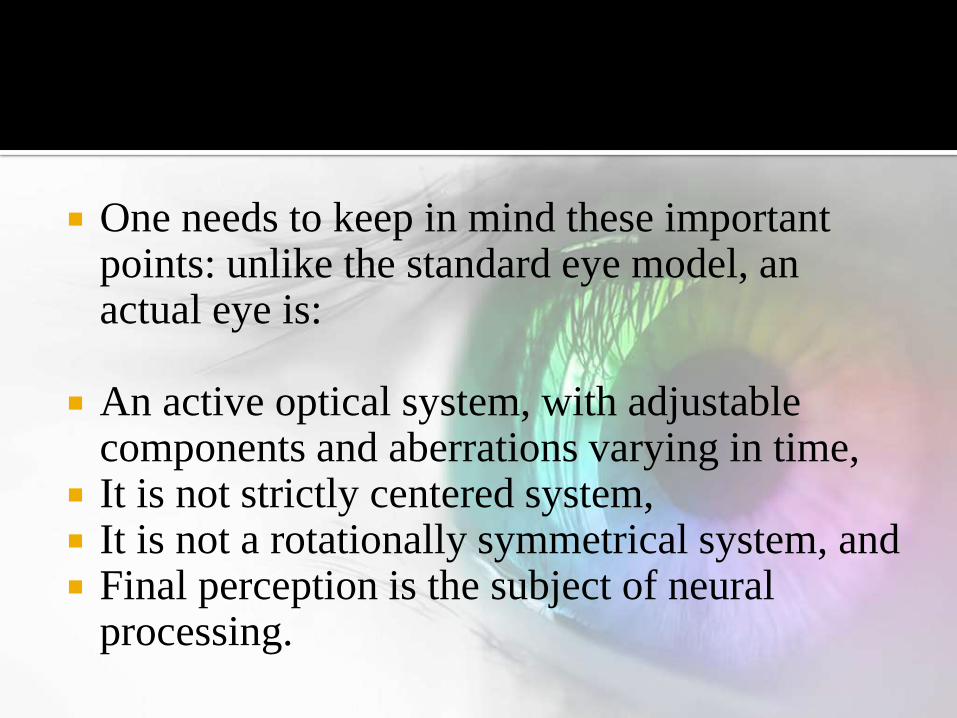
One needs to keep in mind these important points: unlike the standard eye model, an actual eye is:
An active optical system, with adjustable components and aberrations varying in time,
It is not strictly centered system, It is not a rotationally symmetrical system, and Final perception is the subject of neural
processing.
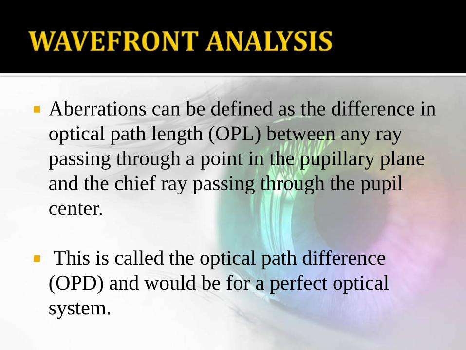
Aberrations can be defined as the difference in
optical path length (OPL) between any ray
passing through a point in the pupillary plane
and the chief ray passing through the pupil
center.
This is called the optical path difference
(OPD) and would be for a perfect optical
system.

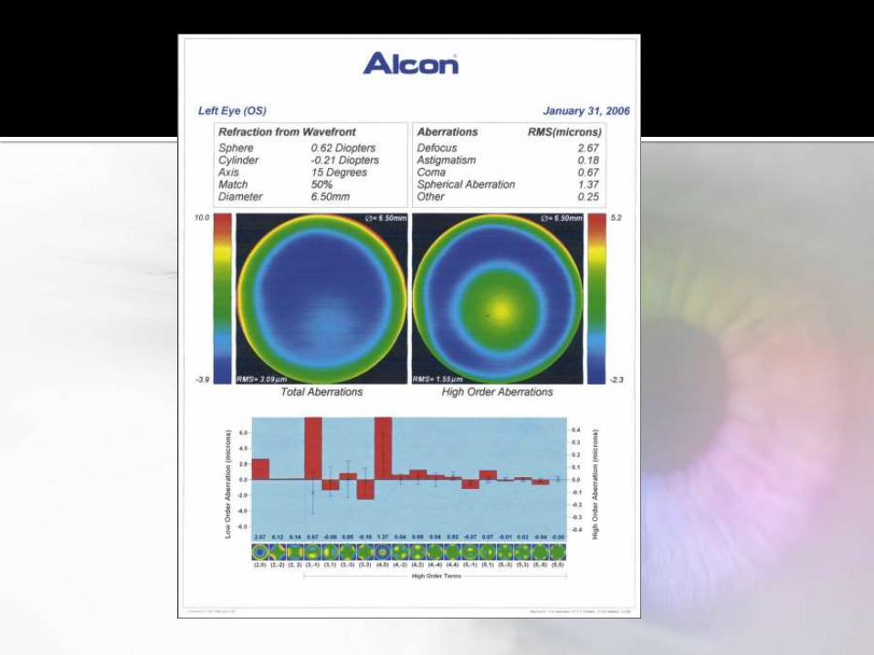
Wavefront aberrometer shines a perfectly
shaped wave of light into the eye and captures
reflections distorted based on the eye’s surface
contours.
Thus, it generates a map of the optical system
of the eye, which can be used to prescribe a
solution, correcting the patient’s specific
vision problem.

Another way of characterizing the wavefront is to measure the actual slope of light rays exiting the pupil plane at different points in the plane and compare these to the ideal; the direction of propagation of light rays will be perpendicular to the wavefront.
This is the basic principle behind the Hartman-Shack devices commonly used to measure the wavefront.
Wavefronts exiting the pupil plane are allowed to interact with a microlenslet array.

If the wavefront is a perfect flat sheet, it will form a perfect lattice of point images corresponding to the optical axis of each lenslet.
If the wavefront is aberrated, the local slope of the wavefront will be different for each lenslet and result in a displaced spot on the grid as compared to the ideal.
The displacement in location from the actual spot versus the ideal represents a measure of the shape of the wavefront.

Wavefront maps are commonly displayed as
2-dimensional maps.
The color green indicates minimal wavefront
distortion from the ideal.
While blue is characteristic of myopic
wavefronts and red is characteristic of
hyperopic wavefront errors.

Once the wavefront image is captured, it can be analyzed.
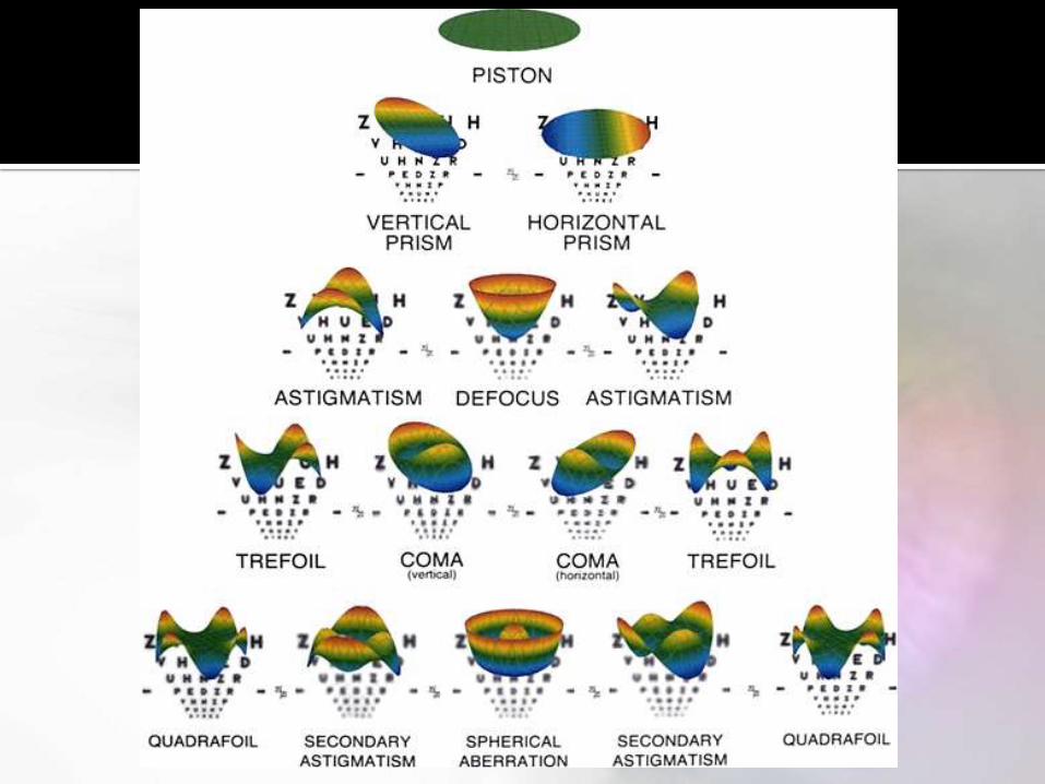
One method of wavefront analysis and classification is to consider each wavefront map to be the weighted sum of fundamental shapes.
Zernike and Fourier transforms are polynomial equations that have been adapted for this purpose.
Zernike polynomials have proven especially useful since they contain radial components and the shape of the wavefront follows that of the pupil, which is circular.

Following the above division of the Zernike expansion adopted in ophthalmology, monochromatic eye aberrations are addressed as:
(1) lower-order aberrations, with the Zernike radial order n<3, and
(2) higher-order aberrations, with n≥3.
The important optical aberrations that affect vision are:
2nd Order optical aberrations – currently measured in all eye exams providing sphere, cylinder and axis corrections
3rd and 4th Order optical aberrations – high order aberrations currently not measured in today’s eye exams but can account for up to 20% of the eye’s refractive error.

5th and 6th Order optical aberrations –also high
order aberrations not currently measured in
today’s eye exam.
These aberrations are of less significance
clinically, however they manifest in reduced
vision for a small percentage of eyes.

The lower-order aberrations are
Piston
Tilt
Defocus
Astigmatism
The 2nd order aberrations, defocus and primary astigmatism - are the most significant contributors to the overall magnitude of eye aberrations
Lower-order aberrations
Remaining lower-order forms, piston and tilt,
or distortion, are usually ignored.
The former being not an aberration with a
single imaging pupil, and
The latter being not a point-image quality
aberration).


Higher order aberrations are measured with
wavefront aberrometers and expressed in
terms that describe the shape and severity of
the deviated light rays as they pass through the
eye's optical system and strike the retina.
Coma, spherical aberration, and trefoil are the
most common higher order aberrations .

Coma causes light to be smeared like the tail of a
comet in the night sky.
Double vision is a common symptom of coma.
Trefoil causes a point of light to smear in three
directions, like a Mercedes-Benz symbol.
Spherical aberration is characterized by halos,
starbursts, ghost images, and loss of contrast
sensitivity (inability to see fine detail) in low light.
Starbursts – Patterns of Small Lights Around Light Sources
Haloes – Circles of Light Around Light Sources Ghosting – A Faint Duplicate of Each Object
Similar to Double Vision Glare – Intensification of Light Sources.
It's quite common for a patient to have an increase in all of these aberrations, resulting in distorted night vision when the pupil opens and allows light to enter through a larger area of the irregular corneal surface.
A comet-like tail or directional flare appearing in the retinal image, when a point source is viewed.
Because the eye is a somewhat nonaxial imaging device, and because the cornea and lens are not perfectly centered with respect to the pupil, coma generally is present in all human eyes.
A large amount of coma (0.3 μm of coma alone) may point to known corneal diseases, such as keratoconus.

Fortunately, spherical aberration is relatively easy to understand.
For a normal photopic eye, spherical aberration may vary from approximately 0.25 D to almost 2 D.
Light rays entering the central area of a lens are bent less and come to a sharp focus at the focal point of a lens system.
However, peripheral light rays tend to be bent more by the edge of a given lens system so that in a plus lens, the light rays are focused in front of the normal focal point of the lens and secondary images are created.

This is why many lens systems incorporate an aspheric grind, so that the periphery of the lens system gradually tapers and refracts or bends light to a lesser degree than if this optical adaptation was not included.
The variation in index of refraction of the crystalline lens (higher index in the nucleus, lower index in the cortex) is responsible for neutralization of a good part of the spherical aberration caused by the human cornea.

Because the index of refraction of the ocular
components of the eye varies with
wavelength, colored objects located at the
same distance from the eye are imaged at
different distances with respect to the retina.
This phenomenon is called axial chromatic
aberration. In the human eye the magnitude of
chromatic aberration is approximately 3 D.
However, significant colored fringes around
objects generally are not seen because of the
preferential spectral sensitivity of human
photoreceptors.
Studies have shown that humans are many
times more sensitive to yellow–green light
with a central wavelength at 560 nm than to
red or blue light.