optimization of biopsy incisions in cutaneous surgery … tilleman.pdf · optimization of incisions...
TRANSCRIPT

Optimization of Incisions in Cutaneous Surgery
including Mohs’ Micrographic Surgery. The validity of paradigms in skin surgery
Het optimaliseren van de incisievorm bij cutane
chirurgie waaronder de Mohs’ Micrografische
Chirurgie. Een validatieonderzoek van paradigma van cutane chirurgie
Proefschrift ter verkrijging van de graad van doctor
aan de Erasmus Universiteit Rotterdam
op gezag van de Rector Magnificus
Prof.dr. S.W.J. Lamberts
en volgens besluit van het College voor Promoties
De openbare verdediging zal plaatsvinden op
donderdag 1 juli 2004 om 16.00 uur
door
Tamara Raveh Tilleman
geboren te Tel Aviv, Israël
3

Promotiecommissie
Promotor: Prof.dr. H.A.M. Neumann
Overige leden: Prof.dr. S.E.R. Hovius
Prof.dr. J. Jeekel
Dr. M.J.P. Gerritsen
4

“Je le pansay et Dieu le guarist"
(I dressed the wound and God healed it)
Ambroise Paré‘s motto, as inscribed above his chair
in the Collège de St-Cosme (1510-1590).
This work is dedicated to my late mother Tova.
5

6

Contents
Chapter 1 – Introduction … 09-48
Chapter 2 – Skin waste, vertex angle and scar length in excisional biopsies. Comparing five
excision patterns: fusiform ellipse, fusiform circle, rhomboid, mosque and S-
shape … 49-60
Chapter 3 – Evidence Based Surgery: The paradigm of surgical ellipse length-to-width ratio
and vertex angle … 61-78
Chapter 4 – The analyses of skin waste during excision of benign skin lesions. Is the surgical
ellipse cut an unnecessary cut? … 79-92
Chapter 5 – Skin waste in elliptical excision biopsies of non-melanoma skin cancers. 93-104
Chapter 6 – The elastic properties of cancerous skin: Poisson’s ratio and Young’s modulus
…105-116
Chapter 7 – Minimal beveling angle in Mohs’ micrographic surgery cut …117-126
Chapter 8 – Optimizing the cut in Mohs’ micrographic surgery with regard to skin sparing
and microscopic view: Is a circular incision necessary? … 127-136
Chapter 9 – A new surgical technique for direct closure of circular skin defects without dog
ear excision. … 137-152
Chapter 10– Summary and conclusions … 153-158
Samenvatting … 159-164
Dankwoord and acknowledgements … 165-168
Curriculum Vitae …169-170
Index … 171-174
7

8

Chapter 1
General Introduction
9

10

Introduction to rational surgery
Tradition has been a restraining barrier in the process of converting surgery from
empirical craft into a scientific discipline. For centuries many surgeons were committed to
seeing and believing what they were taught rather than believing what they saw (1).
Innovators, always in the minority, were people who abandoned theories when their
observations conflicted with what they had been taught.
The French barber-surgeon Ambroise Paré is considered the "father of rational
surgery". Paré's pioneering work was chiefly in the department of military surgery, when in
1536 he introduced a scientific and humane treatment for battlefield wounds (2). Prior to his
work surgeons stopped bleeding wounds by pouring boiling oil on them. Ambroise Paré
discovered that treatment of gunshot wounds by boiling oil was worse than leaving them
alone or covering them with other materials. His findings were accidental because he ran out
of oil, and as a stopgap he tried a cold mixture of egg yolks, oil of roses, and turpentine (3).
This coincidence also created two test groups, and as the soldiers healed, the ones who were
not burnt with boiling oil recovered far more quickly than those who were. Paré, who was a
gifted observer, created a new concept in medicine and surgery: questioning current
treatments, testing alternatives, creating two groups, observing, comparing and then
concluding what he saw. In 1545 Paré authored a short treatise on the treatment of gunshot
wounds based upon his observations. This short practical volume was the first in a lifetime
of voluminous medical manuals (4). In addition, Paré invented several surgical instruments
and surgical techniques. Among them: truss for hernia, artificial limbs, reimplantation of
teeth and ligating arteries instead of cauterizing vessels during amputation. Although
suturing and ligating were already used in the Aztec-Mexican medicine (5), their wide use
began only after Paré’s work. Paré's pioneering work set a frame for future generations in
surgery and was a milestone in incorporating scientific studies into medicine.
During the next centuries, general surgery, plastic surgery and dermatosurgery were
tremendously developed focusing on establishing surgical techniques (6). Later the focus
turned to spreading this knowledge through written textbooks, tutoring, training new
generations and establishing professional surgical associations (7, 8)
Until recently, the clinical training of residents was based on an apprenticeship
model in which the residents learned medicine and surgical techniques from experienced
11

attending doctors. The apprenticeship model is gradually being transformed from pure
experience-based medicine and surgery to an evidence-based medicine. The development of
a surgeon’s judgment based on the physician’s practice and experience is important (9).
However, even the most experienced physician may be influenced by recent occurrences in
selected patients or anecdotal experiences. Currently, the importance of using a more
objective and systematic approach for making decisions and treatments is being established
(10). A new model for medical practice emerged a decade ago, which diminishes intuition,
unsystematic clinical experience and rational explanations, as sufficient grounds for clinical
decisions (11). This new model is called Evidence-Based Medicine (EBM), which
integrates individual clinical expertise with the best available external clinical and research
evidence from systematic data. It is defined by Sackett as “the conscientious, explicit and
judicious use of current best evidence in making decisions about the care of individual
patients” (12). Evidence Based medicine became the standard of treatment from the patient
perspective on the one hand, and from the health care system and the physician perspective
on the other hand. Providing evidence based care improves outcomes for patients (12).
Choosing an optimal intervention procedure is also becoming a must in the current financial
restrictions that tend to reduce health care expenditure (13). For these two reasons, as well as
the superior systematic approach, physicians increasingly tend to treat patients based on
satisfactory evidence (14,15).
In practicing EBM the need for information is converted into a question answered by
the current best evidence resources such as textbooks and electronic databases. This
information is finally integrated with the clinical expertise (16). The best evidence
resources are prospectively designed, double-blind, placebo-controlled randomized clinical
trials. They represent the “golden standard”, yet not the only source, of evidence regarding
therapeutic decisions. Valuable evidence may stem from prospective cohort studies and
analytical surveys. Evidence is strengthened immensely after it has been confirmed in
multiple investigations. The sources may be compared with one another and presented in a
meta analysis or systematic overview (17). The skill of rapid access to the best available
evidence is not sufficient for optimum treatment. The ability of a physician to apply sound
evidence in a particular situation in a patient is becoming the state-of-the-art in medicine.
12

In this study the excisional biopsy incisions are explored. The importance of such
study lies in preserving healthy tissue and shortening of scars. If sparingly used, the saved
skin may serve as a future resource for reconstruction, a flap, for instance. In order to study
these incisions a brief overview of excisional biopsy techniques, Mohs’ micrographic
surgery (MMS), surgical ellipse closures and the phenomenon of dog-ear creation are
presented. An insight into the principles of these physiological dynamics is essential to solve
the skin-related problems which arise during and after performing these procedures.
Excisional biopsy technique
In cutaneous biopsy a piece of skin is removed from a patient in order to investigate
whether the probed area is benign or malignant and to confirm the diagnosis in suspected
cases. The removed specimen is pathologically examined and processed. An excisional
biopsy is the removal of the entire lesion with some additional normal tissue as margins. For
the complete removal of the lesion these margins are small if the lesion is deemed benign
(18), or large if the lesion is clinically suspected to be malignant (19-22). If there is a
residual tumor, simple re-excision or MMS are undertaken. The latter is the best local
intervention to remove the residual tumor (23-29).
Many skin lesions are circular, yet the final excision pattern is often different.
Surgeons do not simply cut a circle around the lesion to remove it, even though a circular
cut removes less skin and leaves a shorter scar than any other skin excision. The reason is
that a circular excision does not stitch very well when directly closed. It leaves an elevated
skin bunched up at the ends. This excess skin at the wound apices is called “dog-ear”,
because its shape resembles a pointed ear on a dog's head (Figure 1).
Cuts of an unequal width and length on the skin are easier to close and produce less
tissue protrusion and dog-ear formation compared with a circular pattern (30). The cut is
chosen after determining the necessary extent of excision that includes the lesion and the
margins. The specimen is designed such that it provides an adequate amount of tissue for
pathological examination. After marking the lesion to be removed and its margins, a cutting
pattern surrounding the lesion is drawn. The short axis of the cutting pattern, or width, is the
diameter of the lesion whereas the long axis exceeds the width by a certain factor (Figure 2).
13

Several cutting patterns have been described in the literature. Among them are the
ellipse, rhomboid, s-shape, mosque and circular incision (Table 1). The objective in
cutaneous plastic surgery is to remove the lesion and yet to obtain an esthetical and
functional scar. Skin scars need be designed for minimal scar length, minimal healthy tissue
waste, a well oriented scar confined to a single cosmetic unit and without any damage to
tissue function (31). A hidden incision yields the best scar, as seen in closed rhinoplasty or
in a scar hidden by hair. If the scar is visible, it should be restricted to the subunit and to the
favorable skin tension lines, for instance the cosmetic result is optimum when the final scar
is in a wrinkle. All these factors should be taken into consideration when a certain skin cut is
planed for an excision.
Mohs’ Micrographic Surgery
The incidence of skin cancer rates have continued to rise during the last few decades
(32). Skin cancers make up a half of all cancers diagnosed (33-4) with a total estimated
number of 54,000 new cases of malignant melanoma (35) and over 1,200,000 new cases
annually of non melanoma skin cancer (36,37) in the USA. These numbers are most likely
to be underestimates of the true tumor incidence, because not all of the skin cancers are
registered and because many patients with such tumors are treated in private medical
practices. Complete removal of the tumor is mandatory, whereas incomplete excision or
neglected lesion may result in an invasion of cancer cells into deeper layers, destroying local
vital structures, and eventually causing metastasis and possibly death. Excisional surgery is
effective for skin cancer and is the mainstay of therapy (38,39).
Mohs’ micrographic surgery was developed by Dr. Frederick Mohs at University of
Wisconsin in the 1930s (40). The technique and its modifications focus on complete surgical
excision of the tumor with immediate microscopic examination of horizontal frozen sections
(41). The operation begins by cutting the cancerous lesion with some margins. The first
layer is removed by a saucer shaped incision. This saucer incision was recommended by Dr.
Mohs’ to obtain adequate number of frozen sections. The specimen’s margins are color-
coded in the laboratory, and any residual tumor at the margin is mapped. Additional
excisions are performed according to this Mohs’ map (Figure 3). The operation is completed
when all the residual tumor has been removed (42).
14

In the past twenty years, MMS has been recognized as a superior treatment for some
non melanoma skin cancers (43 – 46) and may also present advantages in treatment of
melanoma tumors (47,48). It should be mentioned that large, prospective, long term follow
up studies for deferent treatment modalities are lacking in the literature. There are, however,
systematic review articles concerning treatment options in skin cancer. Three of such articles
describe that MMS has the lowest recurrent rate for both primary and recurrent basal cell
carcinoma (43,44,45). The five years recurrent rates for non Mohs modalities are reported as
8.7%, on the average, while the recurrent rates for the same tumor treated by Mohs
technique is 1%. Similar results were found in previously treated basal cell carcinoma. The
five years recurrence rate was 17.4% for surgical excision, 9.8% for radiotherapy, and 40%
for curettage and electrodessication. In comparison, Mohs’ microscopic surgery recurrence
rate for the same cancer was only 5.6%. Consequently, the MMS technique offers an
extremely high cure rate (44) while minimizing skin waste, which often allows for the
preservation of functions, and an optimal esthetic outcome (49). Considering the
effectiveness of MMS in minimizing local recurrence and bearing in mind that it is
performed under local anesthesia in an office setting, the procedure is cost-effective. In the
past decades MMS has been modified. The fixed tissue technique (chemosurgery) has been
replaced with a fresh tissue technique that makes the procedure less painful, shorter and
permits immediate reconstruction (23).
The surgical ellipse
The surgical ellipse, also known as a fusiform ellipse (50, 51), is the classical
approach for removing cutaneous lesions. A geometrical ellipse on the one hand possesses a
rounded apex, which is difficult to both incise and close. A surgical ellipse on the other
hand, is the overlapped zone of two ellipses or two circles (Figure 4), thus producing two
vertices. An excision having this shape is preferred because of the subsequent ease of
planning, cutting and closing. The fusiform ellipse is incised perpendicular to the skin,
yielding a straight-line specimen and wound. The specimen’s borders are clear when
observed microscopically and the straight borders of the wound are easy to close, resulting
in a thin, flat scar.
15
As a general rule, the traditional recommendation in the literature is to form ellipse
dimensions that have a length-to-width ratio between 3:1 to 4:1 (52, 53), and an apical
(vertex) angle of 30° or less. This ratio results in longer scars compared with the original
round lesion diameter, but it permits primary closure of the wound without creating dog-ears
(54-60).
The open wound created after incising the lesion can be closed by either a primary
closure or by skin graft and flap. Of these, the primary closure is superior yielding optimum
esthetic results. Therefore, the focus of the present study was on direct and primary closures.
Tissue dynamics in basic local flaps
Skin is a viscoelastic material whose complex mechanical properties include the
elastic properties of solid materials and the viscous properties of fluids (61). The elastic
characteristics of the skin relate to the immediate changes that occur when force is applied to
the skin. They govern the ability of the skin to deform, i.e. to stretch, contract and compress.
These characteristics are defined by two physical constants namely the Young’s modulus,
which relates the proportionality of the longitudinal deformation to the applied force
(Hooke’s law), and Poisson’s ratio, which relates the dimensional deformations to one
another (62). The viscous characteristics of the skin relate to the delayed changes occurring
after time: the decrease in stress over time when a constant strain is applied (the stress
relaxation effect) and the increase in length over time when a constant strain is applied (the
creeping effect) (63). Surgeons are familiar with these effects and count on the stress
relaxation effect to release the tension in scar with time. They use the creeping effect to
absorb some irregular scar features and dog-ears and to cause tissue elongation after
inflating expanders.
Advancing movement and rotating movement are two main movements that can be
applied to the skin. The two are vastly used in reconstructive surgery (64, 65). The
transpositions of skin tissues may be manipulated by either of these basic movements or any
combination thereof such as Millard’s rotation-advancement cheiloplasty (66). The two
basic movements are described below.
16

a. Advancement movement
In an advancement movement the tissue is transferred from the donor to the recipient
site by a linear shift in the same plane. The dynamic change of the advancement movement
stretches the tissue and exerts tension at the center of the movement or flap (Figure 5).
Excess tissue is formed at the flap base or movement. To overcome the discrepancy in tissue
elongation during the advancement across the flap, two small triangles at the base, referred
to as Bürow’s triangles, should be excised (67) (Figure 6). The advancement movement is
usually used to close a square defect.
b. Rotational movement
In the rotational movement the tissue is transferred from the donor to the recipient
site by an angular movement (Figure 7 ). This movement is usually used to open or close a
circular or triangular defect. All rotation flaps produce tissue protrusion on the outer
perimeter (Figure 8). This can be corrected by removing the excess tissue.
The result of a rotational movement is an angular manipulation that produces a
conical skin deformation around the pivot (68). Formed are two kinds of cones: vertical and
horizontal. A vertical cone is produced on rotating a flap to close an incised sector (Figure
9). Its axis is at a right angle to the skin surface. In this case there is a shortage of tissue in
the lesion (horizontal) plane and an excess of tissue above or beneath this plane (Figure 10).
When there is enough supporting tissue beneath, the standing cone produced is upright and
everted, when there is no support beneath, the cone is pushed under the surface creating an
inverted, or a sunken dog-ear. In dog-ear formation, the smaller the angle to be closed, the
smaller is the angle of rotation, and the smaller is the cone protrusion (Figure 11). For an
apical angle smaller than 30°, the protruding tissue is considered negligible and a dog-ear is
hardly observed (68). Figure 10 is as Figure 11, however viewed from a different angle,
emphasizing the point that the base of the flap is narrower for a larger apical angle creating a
larger protrusion.
A horizontal cone is produced on rotating a flap to open an angle and insert a tissue
sector (Figure 12). It is also formed when the wound to be closed possesses unequal arc
lengths (69, 70). The central axis of a horizontal (laying) cone is at an acute angle to the skin
surface. In comparison with a vertical cone, there is excess of tissue both in the horizontal
17
and the vertical planes, either above or beneath the lesion plane. The horizontal cone
occasionally appears as multiple wrinkles or rippling of the skin (Figure 13) radiating from
the opened angle. The laying cone is often referred to as pseudo dog-ear.
Tissue dynamics in closure of surgical ellipse
The surgical ellipse is an excision comprising two arcs that together constitute a
perimeter. On closure of an elliptical defect each of the arcs is pulled towards an imaginary
meridian such that the scar follows the meridian. In fact one performs two closures,
between each of the arcs designated as A, and the meridian designated as M (Figure 14).
Dog-ears are formed at the vertices because these two closure lines possess uneven lengths
(71) (Figure 15).
The moving zones along the ellipse can be divided into two areas: the center and the
apex. During closure different movements such as advancement, rotation, and a hybrid
rotation-advancement are observed in each zone. Pure advancement movement is observed
at the center of the ellipse whereas pure rotation movement is observed at the two ellipse
vertices (Figure 16). Upon closing, the central zone of the ellipse moves in an advancing
manner and traverses the greatest distance, thus creating tension in the central zone of the
scar (Hooke’s law). The apical zones move rotationally thus forming tissue puckering or
dog-ears (Figure 16). All the other ellipse points that are not central or apical move in a
hybrid rotation-advancement.
The tension created in the scar center is proportional to the traversed distance, or the
ellipse width. The dog-ear protrusion at the vertices is a function of the apical angle. In fact
the two latter effects depend on one another such that the greater the discrepancy between
the ellipse length and the arcs, the greater is the vertex angle and skin protrusion. A circular
excision is the extreme case of the discrepancy between the ellipse length and the arcs with
the largest vertex angle (180°) (Figure 17). Therefore, a circular excision by definition
creates the highest tension in the center and consequently, the largest dog-ear protrusion at
the vertices (Figure 18). Accordingly, any other cutting pattern possessing a long and a
short axis is superior to the circular excision. Classical elliptical techniques recommend an
excision with an ellipse length that is 3 to 4 times the ellipse width (72). This general rule is
used to obtain an acute angle of 30º or less at the vertex. Such an ellipse can always be
18

closed primarily without excessive tension and with a lower tendency to form a dog-ear
(73).
The ability of the skin to overcome the shortage of tissue at the center and the excess
of tissue at the apex depends on its aforementioned elastic and viscoelastic properties (61).
Explicitly, the skin may compensate for tensions and wrinkles and even by absorb
protrusions without breaking or tearing.
Correction of dog-ear
The term dog-ear is deeply embedded in surgical parlance (74) and has become an
intimidating term to surgeons. Dog-ear is the result of rotated skin created by: 1) closure of
any cutting pattern with equal arcs possessing an apical angle exceeding 30°, 2) closure of
any cutting pattern with unequal arcs, 3) wide angle of rotation at the vertices, 4) closure of
a wound located in inelastic skin, or when tissues are bonded to the dermis or subdermis,
and 5) closure of a wound located in a convex plane.
Dog-ear or puckering can be repaired by the following approaches:
1. Increasing the length of the ellipse whereby the length-to-width ratio is increased,
creating a smaller angle at the vertex and a smaller rotation movement that can be
closed without forming a protrusion (55). There are two techniques to achieve this:
a) preventing the formation of dog-ear by incising a longer ellipse than the designed
ellipse in advance, and b) repairing dog-ear by trimming off the excess tissue after
closing the original ellipse (Figure 19). The trimming may be accomplished by either
elongating the scar along the main axis (straight one-line dog-ear repair), or by a
continued incision at an angle to the ellipse length. This trimming may be a curved
one-line dog-ear repair, angled one-line dog-ear repair (hockey stick line, L-shaped
line) (Figure 20) or a two-line dog-ear repair (T shape line). A dog-ear repair by
trimming is better than directly enlarging the a priori ellipse excision because it
spares more tissue (56).
2. Correcting an uneven length of a wound closure. Equalizing the two sides of the
excess tissue (80) (Figure 21) may be accomplished by three possible techniques: a)
shortening the long arc with a small triangle known as: Bürow’s triangle, when at the
vertex, and Szymanowski’s triangle, when at the center (51, 76, 77). This correction
19

is a two-line dog-ear repair or a V-shaped line, b) lengthening the shorter arc by
adding a right-angled incision at the vertex (78), c) redistributing the excess length
by the “rule of halves”, a surgical method for dividing uneven lengths (Figure22). In
this technique, the initial suture is placed at the center of both arcs. The remaining
defects are continually halved by placing suture (79) (Figure 23).
3. A wide angle of rotation may be avoided by dividing the vertex angle and drawing
an M-plasty. The shortening of the scar and a less protruding dog-ear are thus
achieved (70, 80). This correction is a two-line dog-ear repair.
4. Some of the skin inelasticity or stiffness may be overcome by a wide undermining. A
pseudo dog-ear is formed when the dermis and subcutaneous tissues are released
unevenly or improperly. Undermining and removing the excess subcutaneous tissue
will straighten the pseudo dog-ear (81).
5. Closure of ellipses located on a convex surface may enhance the appearance of the
dog-ear. The vertex tissue is pushed farther out relative to the plane, causing the dog-
ear to become more visible. Drawing S-plasty instead of the ellipse prevents this
phenomenon (82).
6. Doing nothing is also an option. Some spontaneous flattening of dog-ears occurs in
time. The reason for this partial absorption is a longitudinal and vertical contraction
of the linear scar (51). Therefore, not doing anything to the dog-ear and waiting for a
future result is an acceptable option that does not cause any extra scar and requires
no excision of healthy tissue. Perhaps in many cases this should be the prime option,
following the rule “Primum non nocere”.
Circular defect closure
Most of open wounds after excising lesions are circular or oval (83). This section
describes the methods to treat circular defects. The open wound may be closed or
reconstructed depending on a number factors, including the size and location of the defect,
the surrounding tissue, the age of the patient, the patient’s preference and the surgeon
experience (84). Small size defects in the head and neck area should be considered for direct
closure and medium size defects should be considered for closure by local flap. Large
defects may be closed by local flaps or skin grafts. Next, location is crucial in areas where
20

small tension or scar may create secondary deformity. Small size defects in the scalp or neck
are easy to close whereas the same size defect located on the eyelid may present a problem
for reconstruction and may end with an ectropion. The surrounding tissue must be examined
to determine if a wound may be closed primarily, with a local flap or graft.
In general there are three options to close a defect: primary, secondary or tertiary
closure (85). Many skin defects especially surgical wound may be closed by first intention
or primary closure where the wound edges are directly approximated or with some minor
variation or modifications such as M-plasty or a purstring. Purstring suture is aimed at
directly closing a circular defect or at reducing the size of the wound (86).
Second intention or secondary closure allows the wound to contract and epithelize
without formal repair. Wound contraction is an active essential part of wound repair in
which the organism closes a gap in the soft tissues (52, 87,88). The second intention
approach yields nice small contracted scars when the wound is small or an excessive
contraction with esthetic deformity, such as ectropion or contracture deformity (89), when
the wound is large. This method achieves good esthetic results especially for wounds placed
on a concave surface, for instance: nose, eye and ear. (90)
The next option is third intention or tertiary closure, a surgical closure by bringing
tissue from elsewhere in the form of flap or graft.
Skin grafts can be classified as either split thickness consisting of the epidermis and
part of the dermis or full thickness grafts including the entire thickness of the skin (91-93).
The best donor sites for head and neck grafting are considered to be the preauricular, post
auricular and supraclavicular areas. The advantages of using a skin graft are its being a
simple technical procedure and the ability to recruit large amounts of skin for closing large
defects. Yet, skin grafts in the head and neck region do not always fully survive, appearing
pale or more pigmented than the surrounding skin (94) and leave additional scars at the
donor site. Skin grafts are used to close circular defects in locations that are difficult to close
primarily or to be reconstructed by a flap. An example includes small size defects in the
medial canthus or lower eyelid (Figures 24,25) and large size defects located in the temporal
area. When a wound defect has a tumor at high risk for recurrence, grafting or second
intention healing may be the treatment of choice (95).
21

Flaps present another option for tertiary closure. Using local flaps bring skin with
similar color and texture to the defect, and there is frequently little or no scar contracture
with the use of flaps (96). The major problems with the use of local flaps are that they
require planning and experience. In addition, all flaps have the disadvantage of creating
excess scars in the donor site. Most local flaps are random in their vascular supply therefore
they are not classified by vascular pattern but rather according to the flap movements. There
are only two basic movements available to the skin: advancement and rotation (83) and a
combination thereof.
Advancement flap slide, stretch and push tissue into the defect. A round defect can
be closed by a single advancement flap or by a double advancement flap that results in an H
shaped scar. A single advancement flap can be used when a round defect is on the lateral
eyebrow, then the flap movement will add length to the lateral brow (83). A single
advancement flap (Pang flap) can be also used for reconstructing a round defect in the nasal
tip (97). This flap conforms perfectly to the three dimensional shape of the nasal tip while
most of the scars are limited to the subunits of the nose being well hidden in the sidewalls
(98). A double advancement flap can be used in the forehead or neck where most of the flap
scars can be hidden in wrinkles. A bipedicle flap is an advancement flap suitable for closing
defects on the forehead, nose, ear, eyelid and chin. Its greatest advantages are that it is a well
vascularized flap and its scars can be easily camouflaged (99).
Rotation flap is a semicircular flap that may be used as a single flap or as a double
flap (referred to as an O-to-Z flap). These flaps are useful in locations where the arc of
rotation may be less noticeable as in the lateral side of the face: preauricular, temporal, and
mandibular areas or in the nasolabial area (Figures 26, 27). Among the limitation of rotation
flaps one fined some tissue resistance to rotation and the noticeable large dog ear that is
created at the rotation pivot. The cheek and the scalp tend to rotate well while the tip of the
nose, the ala nasi region and the auricle, rotate poorly. In addition, in order to stretch the flap
and achieve lengthening, any back-cut at the base of the flap compromises the blood supply
to the flap.
Transposition flap rotates about a pivot into an adjacent defect. The transposition
flap has a wide range of uses. The bilobe flap is used on a convex surface as the nose and the
Limberg or rhomboid flap and the Dufourmental flap are examples of flaps (100) used to
22

close a diamond shape defects. The nasolabial flap is a transposition flap that interpolates
the path between two separated tissues .The related pedicle passed over intervening tissue.
The advantages of this flap are a superior donor site and the disadvantages are additional
incisions at the donor site and the need for a second intervention, especially to obliterate the
nose-cheek junction or to match the flap thickness.
In this section the options to close circular defects were overviewed and were
referred to the head and neck region. The same principles can be applied elsewhere in the
body.
Objectives of the present study
There were several different objectives of the studies described in this thesis. The
two common excisional biopsy techniques namely the surgical ellipse used in general and
cutaneous surgery and the saucer excision used in MMS were analyzed. The medical
validity of these two axiomatic cuts was questioned and examined. In the initial study, five
different excision patterns namely the fusiform ellipse (82, 101-1034), the fusiform circle
(104), the rhomboid shape (105,106), the mosque shape and the S-shape (107-109) were
theoretically analyzed and examined in details. These were also compared with a circular
excision (110).
In the second study, the present paradigm of surgical ellipse dimensions was
questioned and a meticulous examination of the accurate proportions of the most common
skin pattern was undertaken. The length-to-width ratio and the vertex angle were examined
by reviewing the date on ellipses in plastic, general and dermatosurgery (30, 31, 85,110-
148).
The issue of what the surgeons use for surgical ellipse dimensions (149) and the
waste of healthy skin in these operations was investigated in the next two studies. It was also
examined whether there was a relationship between the waste of healthy skin and a specific
site or size of the lesion and whether the surgical ellipse was a necessary cutting pattern
(150).
The elastic properties of frozen and cancerous skin (151) were examined by
determining the elastic constants of Poisson’s ratio and Young’s Modulus (152,153). In the
subsequent study the elastic constants that were calculated were used to challenge the saucer
23

incision, a paradigm used in MMS to answer a persisting question on the minimum beveling
angle effective for strata projection in a MMS cut (154).
The paradigm of saucer cut and an alternative to the circular incision were further
investigated in the next study.
In the final study, the paradigm that a direct closure of circular defects is not feasible
without additional surgical manipulation was re-examined (55,155,156) with the aim to find
a new technique which would permit a direct closure of circular defects without the need for
additional excision.
In summary, defining the most common cutting patterns, investigating them, finding
their advantages and disadvantages, understanding the physiology of dog-ear creation, were
milestones in exploring paradigms in cutaneous surgery and developing two new surgical
techniques: one for general cutaneous surgery and another for Mohs’ micrographic surgery.
24

References
1. Wangensteen OH, Wangensteen SD. Overview. In: Wangensteen OH, Wangensteen
SD, eds.The rise of surgery. University of Minnesota press, Minneapolis 1978;3-15.
2. Catholic encyclopedia: Ambroise Paré. http://www.newadvent.org/
cathen/11478a.htm
3. Malgaigne, J. F. Surgery and Ambroise Paré. Norman, OK: University of Oklahoma
Press, 1965.
4. Rutkow IM. The renaissance. In: Rutkow IM, ed. Surgery and illustrative history.
Mosby 1993; 119-185.
5. An Overview of Medical History. http://www.glaciermedicaled.com/history/
hxmed01b.html
6. Converse JM. Introduction to plastic surgery. In: Converse JM, ed. Reconstructive
plastic surgery. WB Saunders company, Philadelphia. 1964;3-20.
7. Brieger GH. The development of surgery. In: Sabiston DC, ed. Textbook of surgery.
The biological basis of modern surgical practice. WB Saunders company 1997;1-15.
8. Rogers BO. History of aesthetic plastic surgery of the face. In: Lewis JR, ed. The art
of aesthetic plastic surgery. Little Brown and Company 1989;9-19.
9. Jurkiewicz MJ. Plastic surgery: a conceptual definition. In: Jurkiewicz MJ, Krizek TJ,
Mathes SJ and Ariyan S, eds. Plastic surgery principles and practice. CV Mosby
company, St Louis.1990; 3-6.
10. Braunwald E, Fauci AS, Kasper DL, Hauser SL, Longo DL, Jameson JL Introduction
to clinical medicine. In: Braunwald E, Fauci AS, Kasper DL, Hauser SL, Longo DL,
Jameson JL, eds. Harrison’s principles of internal medicine, 15 th edition. McGraw-
Hill. New York 2001;1-5.
11. Guyatt G, Evidence based medicine working group. Evidence-based Medicine .A
new approach to teaching the practice of medicine. JAMA 1992;7:2420-5.
12. Sackett DL, Straus SE, Richardson WS, Rosenberg W, Haynes RB.Evidence-based
Medicine: how to practice and teach EBM.1st Ed. New York: Churchill Livingstone,
1997.
13. Howes N, Chagla L, Thorpe M, McCulloch P. Surgical practice is evidence based. Br
J Surg 1997;84:1220-3.
25
14. Ellise J, Mulligan I, Rowe J, Sackett DL. Inpatient general medicine is evidence
based. Lancet 1995;346:407-10.
15. Sackett DL, Rosenberg WM, Gray JA, Haynes RB, Richardson WS. Evidence-based
Medicine: what it is and what it isn’t. Br Med J 1996;312:71-2.
16. Berg D, Asgari M, Evidence based medicine for dermatologic surgeons: Concepts in
critical appraisal of information. Dermatol Surg 2001;27:511-4.
17. Offer GJ, Perks GB. In search of evidence based plastic surgery: the problem faced
by the specialty. Br J Plast Surg 200;53:427-33.
18. Hudson-Peacock MJ, Matthews JN, Lawrence CM. Relation between size of skin
excision, wound, and specimen. J Am Acad Dermatol. 1995 ;32(6):1010-5.
19. Thomas DJ, King AR, Peat BG. Excision margins for nonmelanotic skin cancer.
Plast Reconstr Surg. 2003 Jul; 112(1): 57-63
20. Breuninger H, Schippert W, Black B, Rassner G. The margin of safety and depth of
excision in surgical treatment of basalioma. Use of 3-dimensional histologic study of
2,016 tumors. Hautarzt. 1989;40 (11):693-700
21. Brodland DG, Zitelli JA. Surgical margins for excision of primary cutaneous
squamous cell carcinoma. J Am Acad Dermatol. 1992; 27:241-8.
22. Thomas JM, Newton-Bishop J, A'Hern R, Coombes G, Timmons M, Evans J, Cook
M, Theaker J, Fallowfield M, O'Neill T, Ruka W, Bliss JM, United Kingdom
Melanoma Study Group, British Association of Plastic Surgeons, Scottish Cancer
Therapy Network. Excision margins in high-risk malignant melanoma. N Engl J Med.
2004;350:757-66
23. Lang PG Jr. The role of Mohs' micrographic surgery in the management of skin
cancer and a perspective on the management of the surgical defect. Clin Plast Surg.
2004;31:5-31.
24. Shriner DL, McCoy DK, Goldberg DJ, Wagner RF Jr. Mohs micrographic surgery. J
Am Acad Dermatol. 1998;39:79-97.
25. Malhotra R, Huilgol SC, Huynh NT, Selva D.The Australian Mohs database, part II:
periocular basal cell carcinoma outcome at 5-year follow-up. Ophthalmology.
2004;111:631-6.
26. Gloster HM Jr, Harris KR, Roenigk RK. A comparison between Mohs micrographic
26

surgery and wide surgical excision for the treatment of dermatofibrosarcoma
protuberans. J Am Acad Dermatol. 1996;35:82-7.
27. Zitelli JA, Brown CD, Hanusa BH. Surgical margins for excision of primary
cutaneous melanoma. J Am Acad Dermatol. 1997; 37: 422-9.
28. Tromovitch TA, Beirne G, Beirne C. Mohs’ technique treatment of recurrence
cutaneous carcinoma. Cancer 1966;19:867.
29. Rowe DE, Carroll RJ, Day CL. Mohs surgery is the treatment of choice for recurrent
basal cell carcinoma. J dermatol Surg Oncol 1989;15:424-431.
30. Borges AF. Dog ear repair. Plast Reconst Surgery 1982;69:707-13.
31. Leshin B. Proper planning and execution of surgical excisions. In: Wheeland RG,ed.
Cutaneous surgery. W.B.Saunders company;1994:171-177.
32. Gloster HM, Brodland DG. The epidemiology of skin cancer. Dermatol Surg
1996;22:217-26.
33. Marks R. An overview of skin cancer. Incidence and causation. Cancer 1995;75:607-
12.
34. Miller DL, Weinstock MA. Nonmelanoma skin cancer in the United States:
incidence. J Am Acad Dermatol. 1994;30:774-8.
35. http://www.infoplease.com/ipa/A0883543.html
36. Geller AC, Annas GD. Epidemiology of melanoma and nonmelanoma skin cancer.
Semin Oncol Nurs. 2003;19:2-11.
37. Higashi M.K.; Veenstra D.L.; Langley P.C. Health Economic Evaluation of Non-
Melanoma Skin Cancer and Actinic Keratosis. PharmacoEconomics 2004;22:2:83-94.
38. Preston DS, Stern RS. Nonmelanoma cancers of the skin. N Eng J Med
1992;325:23:1649-62.
39. Krown SE, Chapman PB. Defining adequate surgery for primary melanoma. N Eng J
Med 2004;350:8:823-5.
40. Mohs FE.Chemosurgical method for microscopically controlled excision of
cancer.Am J Proctol. 1957;8:273-82.
41. Robins P, Albom MJ. Mohs' surgery-fresh tissue technique.J Dermatol Surg. 1975
;1:37-41.
42. Mohs FE. Chemosurgery. Clin Plast Surg. 1980;7:349-60.
27

43. Thissen MRTM, Neumann MHA, Schouten LJ. A systemic review of treatment
modalities for primary basal cell carcinoma. Arch Dermaol 1999;135:1177-83.
44. Rowe D,Carroll R, Day C. Long term recurrent rates in previously untreated
(primary) basal cell carcinoma: Implications for patient follow-up. J Dermatol Surg
Oncol 1989;15:315-28.
45. Rowe D,Carroll R, Day CL. Mohs surgery is the treatment of choice for recurrent (
previously treated) basal cell carcinoma. J Dermatol Surg Oncol 1989;15:424-31.
46. O'Connor WJ, Roenigk RK, Brodland DG. Merkel cell carcinoma. Comparison of
Mohs micrographic surgery and wide excision in eighty-six patients. Dermatol Surg.
1997;23:929-33
47. Wacker J, Khan-Durani B, Hartschuh W. Modified Mohs micrographic surgery in the
therapy of dermatofibrosarcoma protuberans: analysis of 22 patients.Ann Surg Oncol.
2004;11:438-44.
48. Bienert TN, Trotter MJ, Arlette JP. Treatment of cutaneous melanoma of the face by
Mohs micrographic surgery.J Cutan Med Surg. 2003;7:25-30.
49. Mohs FE. Mohs micrographic surgery. A historical perspective. Dermatol Clin.
1989;7:609-11.
50. Dunlavey E and Leshin B. The simple excision. Dermatol Clinics. 1998;16(1):49-64.
51. Bennett RG .Complex closures. In: Bennett RG, ed. Fundamentals of cutaneous
surgery. C.V. Mosby Company;1988:473-491
52. Wound closure. In: Rohrich RJ and Robinson JB, eds. Wound healing. Selected
readings in plastic surgery. University of Texax southwestern medical center and
Bayelor University medical center;1999:9(3):18-20.
53. McCarthy JG. Introduction to plastic surgery. in: McCarthy JG, ed. Plastic surgery.
W.B. Saunders Company 1990;1-54.
54. Wound care, and flaps, pedicle and tubes. In: McGregor IA, ed. Fundamental
techniques of plastic surgery and their surgical applications. E & S Livingstone Ltd.
Edinburgh and London 1960;3-39, 39-141.
55. Hudson-Peacock MJ and Lawrence CM. Comparison of wound closure by means of
dog ear repair and elliptical excision. J Am Acad Dermatol 1995;32:627-30.
56. Hudson-Peacock MJ, Lawrence CM. Excision with dog ear repair is superior to
28

elliptical excision. Br J Dermatol 1993;129(42):48.
57. Dzubow LM. The dynamics of dog ear formation and correction, J Dermatol Surg
Oncol 1985;11:722-8.
58. Robertson DB. Dog ear repair. In: Wheeland RG, ed. Cutaneous surgery.
W.B.Saunders Company, 1994;295-303.
59. Dog ear repair. In: Rompel JPR,ed. Operative dermatologie. Lehrbuch und atlas.
Springer 1996;54-55.
60. Complex excision. In: Fewkes JL, Cheney ML and Pollack SV, eds. Illustrated atlas
of cutaneous surgery. J.B. Lippincott Company. Philadelphia 1992;16.1-16.8.
61. Gilbson T, Kenedi RM. Biomechanical properties of skin. Surg North Am
1967;47:279-294.
62. Lee C, Vincent JF, and Hillerton JE. Poisson’s ratio in skin. Biomed Matwe Eng.
199;1(1):19-23.
63. Wexler DB, Gilbertson LG, Goel VK, Bardach J. Biomechanics of the rotation-
advancement skin flap: experimental and theoretical studies. In: Bardach J, ed. Local
flaps and free skin grafts. Mosby year book, 1992. 53-68.
64. Advancements flaps. In: Davidson TM, Webster RC, and Gordon BR,eds. The
principles and dynamics of local flaps. American Academy of Otolaryngology -
Head and neck surgery foundation, Inc 1998;26-8.
65. Le surjets. In:Letessier S, Bachellier-Beuzelin Y, and Grognard C,eds. Manuel
partique de chirurgie dermatologique. Masson, Paris 1986;35-38.
66. Millard DR. Refinements in rotation-advancement cleft lip technique. Plast Reconstr
Surg 1964;33:26-9.
67. Jackson IT. General consideration. In: Jackson IT, ed. Local flaps in head and neck
reconstruction. The CV Mosby Company, 1985. 1-34.
68. Limberg AA. Design of local flaps. In: Gibson T,ED. Modern trends in plastic
surgery. Butterworth & co. London. 1966;38-61.
69. Vaughan TK, Samlaska CP, Mulvaney MJ. Hello tricone; good-bye "dog-ear".
Arch Dermatol. 1990 Oct;126(10):1366.
70. Weisberg NK, Nehal KS, Zide BM. Dog-ears: a review. Dermatol Surg 200;26:363-
70.
29

71. Mizunuma M, Yanai A, Tsutsumi S, Yoshida H, Seno H, Inoue M, and Nisbida M.
Can dog ear formation be decreased when an s shape resection is used instead of
spindle skin resection? A three dimensional analysis of skin surgery techniques using
finite element method. Plast Reconstr Surg. 2000;106(4):845-8.a
72. Weinzweig J, Weinzweig N. Techniques and geometry of wound repair. In:
Weinzweig J, ed. Plastic surgery secrets. Henley and Belfus, Inc, Philadelphia.1999.
6-13.
73. Robertson DB. Dog ear repair. In: Wheeland RG, ed. Cutaneous surgery.
W.B.Saunders Company, 1994;295-303.
74. Complex closure. In: Bennett RG,ed. Fundamentals of cutaneous surgery. The C.V.
Mosby Company 1988;473-91.
75. Dog-ear repair and closure of sides of unequal length. In: Stegman SJ, Tromovitch
TA, and Glogau RG,eds. Basics of dermatologic surgery. Year Book Medical
Publishers, Inc. Chicago, London 1982;69-72.
76. Goldwyn AM. Carl August Bürow. Plast Reconstr Surg 1984;73(4):687-690.
77. Bürow A. Symanowski und Uhde’s Kritik der methode der seitlichen Dreiecke. Berl
Klim Wochensch 1870;8:350.
78. Grabb WC. Basic techniques in plastic surgery. In: Grabb WC, and Smith JW, eds.
Plastic surgery. A concise guide to clinical practice. Little, Brown and Company,
Boston 1973;3-74.
79. Maloney M. Basic skin surgery techniques. In: Harper J, Oranje A, and Prose N, eds.
Textbook of pediatric dermatology. Blackwell Science 2000;1767-80.
80. Weisberg NK, Nehal KS, Zide BM.Dog-ears: a review. Dermatol Surg. 2000
Apr;26(4):363-70.
81. Leffell DJ, Brown MD: Basic excisional surgery. In: Manual of Skin Surgery: A
Practical Guide to Dermatologic Procedures. 1997:149-80.
82. Rosemberg L. A versatile procedure for the closure of small skin defects. Aesthetic
Plast Surg 1985;9:23-6.
83. Whitaker DC. Random pattern flaps. In: Cutaneous surgery. Wheeland RG,ed. WB
Saunders Company 1994;329-52.
84. Bardach J. Analysis of defects in the head and neck area and planning of
30

reconstruction using local flaps and free skin grafts. In: Local flaps and free skin
grafts. Bardach J,ed. Mosby Year Book 1992;69-86.
85. Stasko T. Advanced suturing techniques and layer closure. In: Wheeland RG,ed.
Cutaneous surgery. W.B.Saunders Company 1994;304-17.
86. Krizek TJ. The problematic wound. In: Plastic surgery secrets. Weinzweig J,ed.
Hanley and Belfus, Inc. Philadelphia 1999;29-33.
87. Rudolph R. inhibition of myofibroblasts by skin grafts. Plast Reconstr Surg
1979;63:473- 8.
88. Majno G, Gaddiani G, Hirshel BJ et all. Contraction of granulation tissue in vitro:
similarity to smooth muscle. Science 1971;173:548-50.
89. Harris DR. healing of the surgical wound. J Am Acad Dermatol 1979;1:197-207.
90. Zitelli JA. Wound healing by second intention. J Am Acad Dermatol 1983;9:407-15.
91. Vitnes LM. Grafting of skin. Surg Clin North Am 1977;57:939- 45.
92. Flowers r. Unexpected postoperative problems in skin grafting. Surg Clin North Am
1970;50:439- 43.
93. Lopez-Mas J, Ortiz-Monasterio, Degonzales MV, et al. Skin graft pigmentation: a
new approoach to prevention. Plast Reconstr Surg 1972;49:18- 21.
94. Kim JH, Kim KC. Principles of skin grafts. In: Plastic surgery secrets. Weinzweig
J,ed. Hanley and Belfus, Inc. Philadelphia 1999;40814.
95. Evans GRD. Local flaps of the head and neck. In: Plastic surgery secrets. Weinzweig
J,ed. Hanley and Belfus, Inc. Philadelphia 1999;185-193.
96. Peng VT, Sturm RL, Marsh TW. Pinch modification of the linear advancement flap. J
Dermatol Surg. Oncol 1987;13:251-3.
97. Lambert RW, Dzubow LM. A dorsal nasal advancement flap for off-midline defects.J
Am Acad Dermatol. 2004 Mar;50(3):380-3.
98. Flint ID, Siegle RJ. The bipedicle flap revisited. J Dermatol Surg Oncol 1994;20:394-
400.
99. Keser A, Sensoz O, Mengi AS. Double opposing semicircular flap: a modification of
Opposing Z-plasty for closing circular defects. Plast Reconstr Surg 1998;102:1001-7.
100. Jackson TI. General considerations. Jackson TI,ed. Local flaps in head and neck
reconstruction. CV Mosby 1985;1-34.
31
101. Usatine RP, Moy RL, Tobinick EL: Elliptical excision. In: Skin Surgery. A Practical
Guide. 1998:120-36.
102. Harrison PV.A guide to skin biopsies and excisions. Clin Exp Dermatol. 1980 Jun;
5(2): 235-43.
103. Raveh Tilleman T, Tilleman M.M, Krekels GAM and Neumann MHA. Skin waste,
vertex angle and scar length in excisional biopsies. Comparing five excision patterns:
fusiform ellipse, fusiform circle, rhomboid, mosque and S-shaped. Plast Reconst Surg
2004;113:3:857-861.
104. Ashbell TS. The rhomboid excision and limberg flap reconstruction in difficult tense-
skin areas.Plast Reconstr Surg. 1982 Apr; 69(4): 724.
105. Izaguirre H, Navarro C. Rhomboid-to-"W" technique for excision and closure of
facial skin lesions. J Maxillofac Surg. 1983 Oct; 11(5): 207-10.
106. Mizunuma M, Yanai A, Tsutsumi S, Yoshida H, Seno H, Inoue M, Nishida M. Can
dog-ear formation be decreased when an S-shaped skin resection is used instead of a
spindle skin resection? A three-dimensional analysis of skin surgery techniques using
the finite element method. Plast Reconstr Surg. 2000 Sep; 106(4): 845-8
107. Yoshida H, Tsutsumi S, Mizunuma M, Yanai A.Three-dimensional finite element
analysis of skin suture. Part 1: spindle model and S-shaped modified model. Med Eng
Phys. 2000 Sep; 22(7): 481-5.
108. Paolo B, Stefania R, Massimiliano C, Stefano A, Andrea P, Giorgio L.Modified S-
plasty: an alternative to the elliptical excision to reduce the length of suture. Dermatol
Surg. 2003 Apr; 29(4): 394-8.
109. Davis TS, Graham WP 3rd, Miller SH. The circular excision. Ann Plast Surg. 1980
Jan; 4(1): 21-4.
110. Skin and subcutaneous lesions. In: Plastic and reconstructive surgery, essential for
students. Plastic Surgery Educational Foundation, Illinois 1979;18-30.
111. Marchac D. Techniques of surgical repair. In: Marchac D,ed. Surgery of basal cell
carcinoma of the face. Spring-Verlag 1988;14.
112. Manstein CH, Manstein ME, and Manshtein G. Creating curvilinear scar. Plast
Reconst Surgery 1988;83(5):914-5.
113. Borges AF. Unfavorable results in scar revision. In: Goldwyn RM, ed. The
32
unfavorable results in plastic surgery avoiding and treatment.Little Brown and
Company, Boston,Toronto 1984;203-211.
114. Wheeland RG. Random pattern flaps. In: Roenigk RK, and Roenigk HH,eds.
Dermatologic Surgery Principles and practice. Marcel Dekker Inc, New York, Basel
1988;265-273.
115. Maloney M. Basic skin surgery techniques. In: Harper j, Oranje A, and Prose N,
eds.Textbook of pediatric dermatology. Blackwell Science Ltd 2000;28.1:1767-81.
116. Atlas of cutaneous surgery and basic excisional technique. In: Fewkes JL, Cheney
ML and Pollack SV,eds. Illustrated atlas of cutaneous surgery. J.B.Lippincott
Company. Philadelphia 1992;6.2-6.7.
117. Le surjets. In:Letessier S, Bachellier-Beuzelin Y, and Grognard C,eds. Manuel
partique de chirurgie dermatologique. Masson, Paris 1986;35-38.
118. Indikationsabwagung bei der operativen therapie benigner hautveranderungen. In:
Konz B, and Braun-Falco O,eds. Komplikationen in der operativen dermatologie.
Springer-Verlag 1984;145.
119. Wundverschlub in dermatochirurgischen bereich. In: von B. Knoz H, and Burg G,
eds.Dermatochirurgie in klinik und praxis. Springer-Verlag 1977;29-31.
120. Surgical techniques. In: Petres J, and Hundeiker M,eds. Dermatosurgery. Spring-
Verlag 1978;42-45.
121. General considerations. In: Jackson TI,ed. Local flaps in head and neck
reconstruction. CV Mosby 1985;1-34
122. Standardtechniken in der operativen dermatologie and Nahlappenplastiken. In:
Kaufmann VR, and Landes E,eds. Dermatologische Operationen. George Thieme
Verlag Stuutgart, New York 1987;22-3, 45.
123. Emergency room. In: Pletta FX,ed. Pediatric plastic surgery volume 1 Trauma. C.V.
Mosby Company, Saint Louis 1967;5-20.
124. Chang WHJ. Wound management. In: Chang WHJ,ed. Fundamentals of plastic and
reconstructive surgery. Williams and Wilkins, Baltimore London 1980;9-61.
125. Skin flaps. In: Padgett EC and Stephenson KL, eds. Plastic and reconstructive
surgery. Charles C Thomas, Springfield Illinois ;18-40. YEAR!!
126. Facial scars. In:Fomon S,ed. Cosmetic surgery. Principles and practice.
33

J.B.Lippincott Company, Philadelphia Montreal 1960;134-160.
127. Wounds. In: Berson MI,ed. Atlas of plastic surgery. Grune and Stratton, New York
1948;3-54.
128. General technique of plastic operqations. In:Bankoff G,ed. Plastic surgery. Medical
Publications Ltd, London 1943;45-62.
129. Barron JN, and Saad MN. An introduction to operative plastic and reconstructive
surgery. In:Barron JN, and Saad MN,eds. Operative plastic and reconstructive
surgery. Churchill Livingstone 1980;3-45.
130. Fundamental techniques. In:Mckinney P, and Cunningham BL,eds. Handbook of
plastic surgery. Williams and Wilkins, Baltimore london 1981;15-25.
131. Closure of wounds. In: Burian F,ed. The plastic surgery atlas. Butterworths London
Czechoslovak Medical Press Prague. 1967;19-32.
132. Linear scar revision techniques. In:Borges AF, ed. Elective incisions and scar
revision. Little, Brown and company 1973;35-60.
133. Penn J, Brown LJ, Berry TB, Schulmeister SA, Roylance J and Ormerod CL..Fat loss.
In: Penn J,ed. Brenthurst papers. Problems in eye lid and socket reconstruction.
Witwatersrand University Press Johannesberg 1944:4-16.
134. Grabb WC. Basic techniques in plastic surgery. In: Grabb WC, and Smith JW, eds.
Plastic surgery. A concise guide to clinical practice. Little, Brown and Company,
Boston 1973;3-74.
135. Free skin grafts. In: Barsky AJ, Kahn S, and Simon BE,eds. Principles and practice of
plastic surgery. McGraw-Hill Book Company 1964;34-62.
136. Scar revision. In: Georgiade GS, Riefkohl R, and Levin LS,eds. Georgiade plastic,
maxillofacial and reconstructive surgery. Williams and Wilkins 1997;117.
137. Anatomy and physiology. In: Battle RJV,ed. Clinical surgery. Plastic surgery.
Butterworths, London 1965;115.
138. Skin flaps and pedicles. In:Mclaughlin CR,ed. Plastic surgery. An introduction for
nurses. Faber and Faber Limited London :49-57.
139. Primary repair. In:Mustarde JC,ed. Repair and reconstruction in the orbital region. A
practice guide. Churchill Livingstone 1969;2-16.
140. Vistnes LM. Basic principles of cutaneous surgery. In:Epstein E, and Epstein E
34
Jr,eds. Skin Surgery. W.B.Saunders Company 1987;44-55.
141. Weisberg NK, Nehal KS, and Zide BM. Dog-ears: a review. Dermatol Surg
2000;26:363-370.
142. Zitelli JA. Tips for a better ellipse. J Am Acad Dermatol 1990;22:101-3.
143. Complex closure. In:Bennett RG,ed. Fundamentals of cutaneous surgery. The C.V.
Mosby Company 1988;473-91.
144. Dunlavey E, and Leishin B. The simple excision. Dermatol Clinics 1998;16(1):49-64.
145. Ellipse. In: Stegman SJ, Tromovitch TA, and Glogau RG,eds. Basics of
dermatologic surgery. Year Book Medical Publishers, Inc. Chicago, London 1982;60-
8.
146. Mackay GJ, Carlson GW, and Bostwick J. Plastic and maxillofacial surgery. In:
Sabiston DC,ed. Textbook of surgery seventh edition. WB Saunders company
1997;1298-33.
147. Wood RJ and Jurkiewicz. Plastic and reconstructive surgery. In: Schwartz SI, Shires
GA, Spencer FC, Daly JM, Fischer JE, and Galloway AC, eds. Principles of surgery
seventh editon. McGraw Hill 1999;2091-143.
148. Walker WF. Plastic surgery. In: Walker WF,ed. A colour atlas of minor surgery.
Wolfe Medical Publications Ltd 1986;134-43.
149. Zuber TJ. Fusiform excision.Am Fam Physician. 2003;67:1539-44.
150. Pardasani AG, Leshin B, Hallman JR, White WL. Fusiform incisional biopsy for
pigmented skin lesions.Dermatol Surg. 2000;26:622-4.
151. Miller LJ, Argenyi ZB, Whitaker DC. The preparation of frozen sections for
micrographic surgery. A review of current methodology. J Dermatol Surg Oncol.
1993;19:1023-9.
152. Ishikawa T, Ishikawa O, Miyachi Y.Measurement of skin elastic properties with a
new suction device (I): Relationship to age, sex and the degree of obesity in normal
individuals. J Dermatol. 1995;22:713-7.
153. Cua AB, Wilhelm KP, Maibach HI. Elastic properties of human skin: relation to age,
sex, and anatomical region.Arch Dermatol Res. 1990;282:283-8.
154. Weber PJ, Moody BR, Dryden RM, Foster JA. Mohs surgery and processing: novel
optimizations and enhancements. Dermatol Surg. 2000;26:909-14.
35

155. De Giorgi V, Mannone F, Quercioli E, Giannotti V, Piccolo E, Carli P. Dog-ears: a
useful artifice in the closure of extensive wounds. J Eur Acad Dermatol Venereol.
2003 Sep;17(5):572-4.
156. Pritchard GA, Zhang LJ, Hughes LE. Suture or graft? Changing trends in melanoma
wound closure. Eur J Surg Oncol. 1988 Oct;14(5):371-7.
36
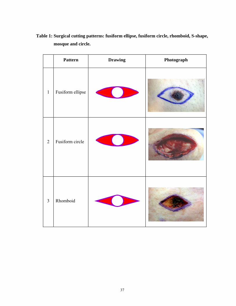
Table 1: Surgical cutting patterns: fusiform ellipse, fusiform circle, rhomboid, S-shape,
mosque and circle.
Pattern Drawing Photograph
1
Fusiform ellipse
2
Fusiform circle
3
Rhomboid
37

4
Mosque
Pattern Drawing Photograph
5
S-shaped
6
Circular shape
38

39

40

41

42

43

44

45

46

47

48

Chapter 2
Skin waste, vertex angle and scar length in excisional biopsies.
Comparing five excision patterns: fusiform ellipse, fusiform
circle, rhomboid, mosque and S-shape.
Publication:
Raveh Tilleman T ,Tilleman MM ,Krekels GAM and Neumann HAM. Skin waste and
scar length in excisional biopsies. Comparing five excision patterns: fusiform ellipse,
fusiform circle, diamond, mosque and S-shape. Plast Reconst Surg 2004:113:857-861.
Presentation:
Combined annual meeting of the American Society for Dermatologic Surgery (ASDS)
and the American College of Mohs Micrographic Surgery (ACMMSCO). New Orleans
October 2003
2nd Annual Research Meeting. Rabin Medical Center, Petach Tikva ,Israel, November
2003.
29th Annual Israel Plastic Surgery Meeting. Tel Aviv, Israel, November 2002.
49

50
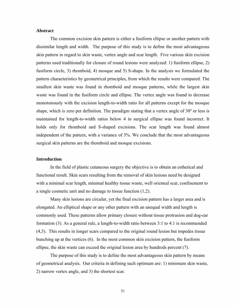
Abstract
The common excision skin pattern is either a fusiform ellipse or another pattern with
dissimilar length and width. The purpose of this study is to define the most advantageous
skin pattern in regard to skin waste, vertex angle and scar length. Five various skin excision
patterns used traditionally for closure of round lesions were analyzed: 1) fusiform ellipse, 2)
fusiform circle, 3) rhomboid, 4) mosque and 5) S-shape. In the analysis we formulated the
pattern characteristics by geometrical principles, from which the results were compared. The
smallest skin waste was found in rhomboid and mosque patterns, while the largest skin
waste was found in the fusiform circle and ellipse. The vertex angle was found to decrease
monotonously with the excision length-to-width ratio for all patterns except for the mosque
shape, which is zero per definition. The paradigm stating that a vertex angle of 30º or less is
maintained for length-to-width ratios below 4 in surgical ellipse was found incorrect. It
holds only for rhomboid and S-shaped excisions. The scar length was found almost
independent of the pattern, with a variance of 3%. We conclude that the most advantageous
surgical skin patterns are the rhomboid and mosque excisions.
Introduction
In the field of plastic cutaneous surgery the objective is to obtain an esthetical and
functional result. Skin scars resulting from the removal of skin lesions need be designed
with a minimal scar length, minimal healthy tissue waste, well oriented scar, confinement to
a single cosmetic unit and no damage to tissue function (1,2).
Many skin lesions are circular, yet the final excision pattern has a larger area and is
elongated. An elliptical shape or any other pattern with an unequal width and length is
commonly used. These patterns allow primary closure without tissue protrusion and dog-ear
formation (3). As a general rule, a length-to-width ratio between 3:1 to 4:1 is recommended
(4,5). This results in longer scars compared to the original round lesion but impedes tissue
bunching up at the vertices (6). In the most common skin excision pattern, the fusiform
ellipse, the skin waste can exceed the original lesion area by hundreds percent (7).
The purpose of this study is to define the most advantageous skin pattern by means
of geometrical analysis. Our criteria in defining such optimum are: 1) minimum skin waste,
2) narrow vertex angle, and 3) the shortest scar.
51

Materials and Methods
A geometrical analysis of five various skin excision patterns wad studied:
1. Fusiform ellipse, tangent to the round lesion,
2. Fusiform circle, tangent to the round lesion,
3. Rhomboid, contained by lines tangent to the round lesion,
4. Mosque, modeled as sinusoidal lines containing the lesion, and
5. S-shape, modeled as a combination of circle arcs and mosque lines.
Though the first two patterns in the table seem similar they are not strictly identical, as
explained in the formulae and results below.
From basic geometrical principles, the above pattern areas, vertex angles and scar
lengths are mathematically formulated. In the following the pattern areas are
expressed:
Fusiform ellipse: is defined by the area overlapped by two fused ellipses. Its area is:
( )⎪⎭
⎪⎬⎫
⎪⎩
⎪⎨⎧
−−
⎥⎥⎦
⎤
⎢⎢⎣
⎡
−−
⎟⎠⎞
⎜⎝⎛
−−−
= −
21
12
sin212
21
22
atatatat
atat
atataSA (1)
where; S is the width of the fusiform ellipse, a is the length-to-width ratio, and t is the
tangent of the half vertex-angle:
( )2tan θ=t
Fusiform circle: is a special case of a fusiform ellipse where the two forming shapes are
circles. The vertex angle is determined by the length-to-width ratio:
( )[ ]12tan2 21 −= − aaθ
By substituting this angle in equation (1) one obtains:
( ) ( ⎥⎦
⎤⎢⎣
⎡−−⎟
⎠⎞
⎜⎝⎛
++= − 12
12sin1
82
2122
2
aaa
aaSA ) (2)
Rhomboid shape: is defined by lines tangent to the round lesion stretching to two
vertices. Its area is expressed by:
⎟⎠⎞
⎜⎝⎛ +−= −
aaSA 1sin1
212
2
(3)
Mosque pattern: is modeled based on a sinusoidal line tangent to the round lesion.
52
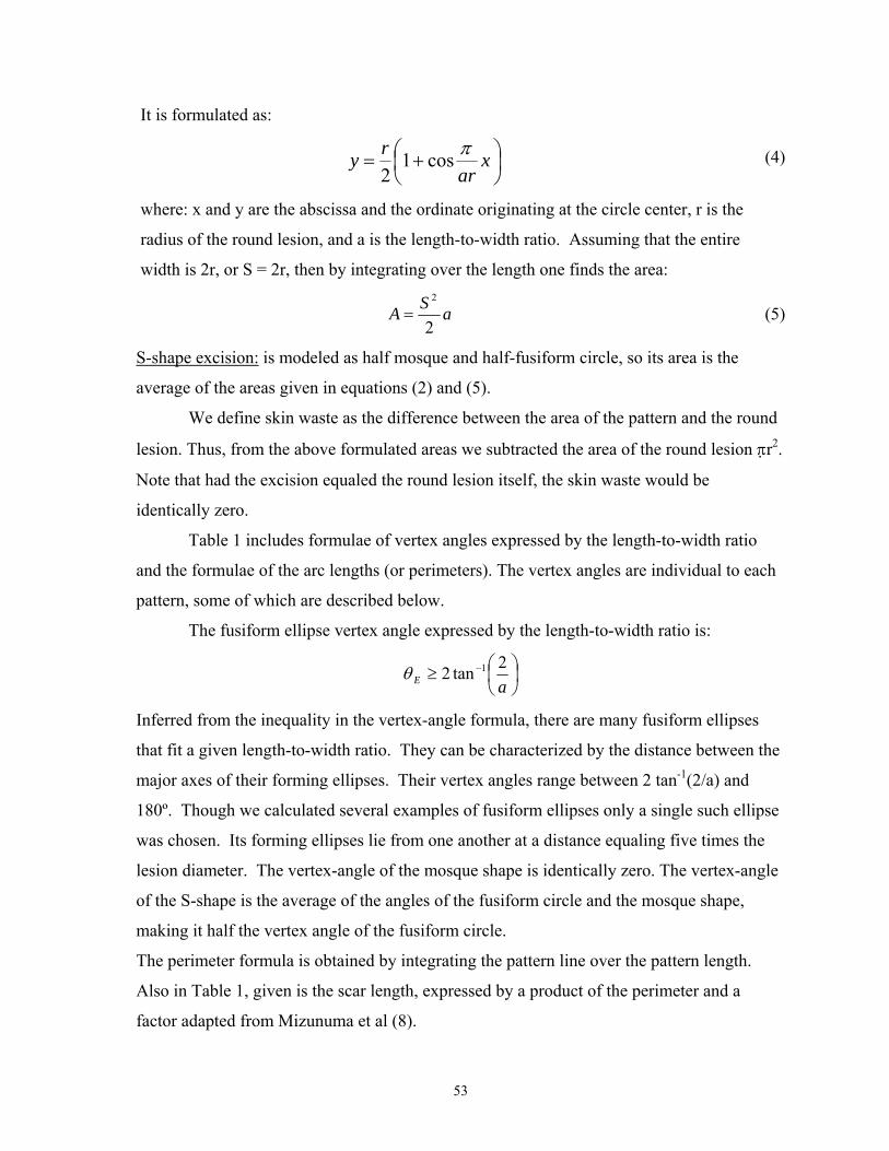
It is formulated as:
⎟⎠⎞
⎜⎝⎛ += x
arry πcos12
(4)
where: x and y are the abscissa and the ordinate originating at the circle center, r is the
radius of the round lesion, and a is the length-to-width ratio. Assuming that the entire
width is 2r, or S = 2r, then by integrating over the length one finds the area:
aSA2
2
= (5)
S-shape excision: is modeled as half mosque and half-fusiform circle, so its area is the
average of the areas given in equations (2) and (5).
We define skin waste as the difference between the area of the pattern and the round
lesion. Thus, from the above formulated areas we subtracted the area of the round lesion πr2.
Note that had the excision equaled the round lesion itself, the skin waste would be
identically zero.
Table 1 includes formulae of vertex angles expressed by the length-to-width ratio
and the formulae of the arc lengths (or perimeters). The vertex angles are individual to each
pattern, some of which are described below.
The fusiform ellipse vertex angle expressed by the length-to-width ratio is:
⎟⎠⎞
⎜⎝⎛≥ −
aE2tan2 1θ
Inferred from the inequality in the vertex-angle formula, there are many fusiform ellipses
that fit a given length-to-width ratio. They can be characterized by the distance between the
major axes of their forming ellipses. Their vertex angles range between 2 tan-1(2/a) and
180º. Though we calculated several examples of fusiform ellipses only a single such ellipse
was chosen. Its forming ellipses lie from one another at a distance equaling five times the
lesion diameter. The vertex-angle of the mosque shape is identically zero. The vertex-angle
of the S-shape is the average of the angles of the fusiform circle and the mosque shape,
making it half the vertex angle of the fusiform circle.
The perimeter formula is obtained by integrating the pattern line over the pattern length.
Also in Table 1, given is the scar length, expressed by a product of the perimeter and a
factor adapted from Mizunuma et al (8).
53

Results
Figure 1 shows the ratio of skin waste-to-lesion area for the various patterns. Skin
waste is defined as the net area of the excision calculated from each pattern formula, for
instance equations 1, 2, 3 and 5, and the round lesion area. The smallest waste is found for
rhomboid and mosque patterns, while the largest waste is found for the fusiform circle and
ellipse. The median waste is found for the S-shape, which is a mid-way pattern between the
fusiform circle and the mosque patterns. This trend is quite expected owing to our
geometrical definition of the S-shape model. For the common length-to-width ratio range of
3 – 4 the skin waste is 90% - 245%. One can also observe from the figure that the skin waste
is proportional to the length-to-width ratio for all the shapes. Note that had a circular cut
been considered, its waste area would be zero.
Figure 2 shows the vertex angle of the various patterns. The angle decreases
monotonously with the excision length-to-width ratio for all patterns except for the mosque
shape. For a surgical ellipse, the most common excision shape, we calculated vertex angles
between 44º - 74º and 33º - 56º, respective to length-to-width ratios of 3 and 4. The vertex-
angle range reflects the distance between the major axes of their forming ellipses as was
explained in the previous section. Note that the vertex angle of 30º within the range of
length-to-width ratio of 3 to 4 holds for only the rhomboid and S-shapes.
Figure 3 plots the scar length as a function of the length-to-width for all the patterns.
The scar length is proportional to the excision perimeter, such that it equals 96% of half the
perimeter for the S-shaped pattern, and 92% for all other patterns: fusiform ellipse, fusiform
circle and mosque. We assumed a similar change for a rhomboid pattern as well. These
proportions have been adapted from Mizunuma et al (8). We found that the scar length is
nearly proportional to the lesion radius and the length-to-width ratio, and almost
independent of the pattern. What stands out is that the scar is typically longer than the
lesion diameter by a factor approximately the length-to-width ratio. Therefore, one can
predict the scar length of any surgical pattern knowing only its length and width (equaling
the lesion diameter). According to this rational a circular cut would result in the shortest
possible scar.
Table 2 summarizes the results for the five excision patterns in terms of waste area,
vertex angle and scar length. The results are given for a length-to-width ratio of 4. The
54

excess skin waste is calculated as the ratio of the areas of the net excision pattern to the
circular lesion. Observe that the least skin waste is obtained for the rhomboid and mosque
excisions, for which also the vertex angles are the smallest. The scar length depends linearly
on the lesion diameter, and is approximately the same for all the patterns.
Discussion
The final excision pattern of cutaneous lesion is usually elliptical. New patterns are
being suggested to exchange that tradition of choosing the elliptical excision. This article
tries to find the most economical pattern concerning extra skin waste and scar length. First
compared are the excision skin-waste areas. Strictly from the standpoint of minimum waste
area, the superior approach is to perform a rhomboid or mosque excision. If, for instance,
one selects the length-to-width ratio of 3 – 4 then the skin waste is 90% - 160% in excess of
the original round lesion, for the mosque and rhomboid shape, and up to 245% for the
fusiform ellipse or fusiform circle.
Second comparison is of vertex angles. The angle decreases with the excision length-
to-width ratio for all patterns. Yet, the current paradigm maintaining that the vertex angle
should be 30° or less for length-to-width ratios below 4 is incorrect. Only rhomboid and S-
shapes can produce such narrow angles while other patterns, those of fusiform ellipse and
circle exhibit a vertex angle of up to 60°. A large vertex angle therefore creates stress on the
skin during suturing rendering unfavorable conical deformations resulting in dog-ears, as
postulated by Limberg in 1966 (1). Note that while performing an excision with a small
angle is desirable, the mosque shape with a zero vertex angle presents surgical and technical
difficulties.
It is noteworthy that a circular excision pattern, i.e. a direct excision of the round
lesion, creates no skin waste and results in the shortest scar length. On the other hand a
circular excision possesses the greatest vertex angle (180°) thus creating huge dog-ears
during closure and a bad scar.
The next chapter examines the theoretical results of the fusiform ellipse treated in
this chapter vis a vis clinical data.
55

References
1. Limberg AA. Design of local flaps. In: Gibson T,ed. Modern trends in plastic
surgery. Butterworth & Co. London;1966:38-61.
2. Leshin B. Proper planning and execution of surgical excisions. In: Wheeland RG,ed.
Cutaneous surgery. W.B.Saunders company;1994:171-177.
3. Borges AF. Dog ear repair. Plast Reconst Surgery;1982:69(4):707-13.
4. Dunlavey E and Leshin B. The simple excision. Dermatol Clinics. 1998;16(1):49-64.
5. Bennett RG. Complex closures. In: Bennett RG, ed. Fundamentals of cutaneous
surgery. C.V. Mosby Company;1988:473-491
6. Rohrich RJ and Robinson. Wound closure. In: Rohrich RJ and Robinson, eds.
Wound healing. Selected redings in plastic surgery. University of Texax
southwestern medical center and Bayelor University medical center;1999:9(3):18-20.
7. Raveh Tilleman T, Smeets N, Tilleman M.M, and Neumann HAM. Skin waste in
elliptical excision biopsies . Accepted to the Scandinavian Journal of Plastic and
Reconstructive Surgery and Hand Surgery.
8. Mizunuma M, Yanai A, Tsutsumi S, Yosida H, Seno H, Inoue M, and Nisbida M.
Can dog ear formation be decreased when an s shape resection is used instead of
spindle skin resection? A three dimensional analysis of skin surgery techniques using
finite element method. Plast Reconstr Surg. 2000;106(4):845-8.
56

Table 1: Formulae of vertex angles and scar length of five excision patterns.
Formula of Scar Length
Shape
Formula of Vertex Angle
Arc Length Factor
1
Fusiform
ellipse
⎟⎠⎞
⎜⎝⎛≥ −
aE2tan2 1θ
( )( )∫
− −
−=
2/
2/2
22
1
1aS
aS
dxx
xkL
α
α
0.92
2
Fusiform
circle
( )[ ]12tan2 21 −= − aaθ
( )( )∫
−
−
++=
−=
2/
2/2
12
2 1sin1
1
aS
aS aaaS
Rx
dxL
0.92
3
Rhomboid
(diamond)
( )a/1sin2 1−=θ
( )aaSL 1sin1 12 −+−=
0.92
4
Mosque
0
∫−
⎟⎠⎞
⎜⎝⎛+=
2/
2/
22
sin2
1aS
aS
dxara
L ππ
0.92
5
S-shape
Half of No.2
Average of No.2 and 4
0.96
57

Table 2: Summarized results for the five excision patterns by the three criteria. The
results are given for a length-to-width ratio of 4. The excess skin waste is the
difference between the areas of the excision pattern and the round lesion.
Shape Length-to-width ratio
Arc length (cm)
Excess skin waste
Vertex angle (degree)
Scar length (cm)
1
Fusiform
ellipse
4
4.18
245%
33º
3.85
2
Fusiform
circle
4
4.16
244%
56º
3.83
3
Rhomboid
4
4.13
162%
28º
3.80
4
Mosque
4
4.18
155%
0º
3.82
5
S-shape
4
4.16
200%
28º
3.99
58

0
1
2
3
4
5
2 3 4 5 6aspect ratio
ratio
of w
aste
d-to
-lesi
on a
rea fusiform ellipse
fusiform circleS-shaperhomboidmosque
Figure 1: Ratios of wasted areas to round lesion areas.
0
20
40
60
80
100
2 3 4 5 6aspect ratio
verte
x an
gle
[deg
]
fusiform ellipsefusiform circleS-shaperhomboid
Figure 2: Vertex angle as a function of the length-to-width ratio.
59

20
40
60
80
100
120
140
160
2 3 4 5 6length-to-width ratio
scar
leng
th (m
m) lesion diameter = 30 mm
Figure 3: Scar length as a function of the length-to-width ratio. The parameter is the
lesion diameter, depicted at 10, 20 and 30 mm. The S-shape is represented
by the broken lines, whereas all the other shapes overlap being denoted by
the solid line.
60

Chapter 3
Evidence Based Surgery: The paradigm of surgical ellipse length-
to-width ratio and vertex angle.
Publication:
Raveh Tilleman T ,Tilleman MM,and Neumann HAM. Evidence Based Surgery: The
paradigm of surgical ellipse dimensions: relation of length-to-width ratio and vertex angle.
Submitted to: Br J Plast Surg 2004.
61

62

Abstract
The paradigm concerning the dimensions of a surgical ellipse is examined. To date it
has been postulated that elliptical excisions must possess a length-to-width ratio of 3:1 to 4:1
and a vertex angle of 30º or less in order to be closed primarily without creating a “dog-ear”.
These dimensions became axiomatic in teaching cutaneous surgery. The present article
examines the validity of this paradigm.
We collected data from two sources: published ellipses in plastic, general and
dermato - surgery literature (fifty seven cases) and elliptical excisions performed at the
authors’ outpatient clinic (eighty three cases). The surgical ellipse lengths, widths and vertex
angles were analyzed, setting an empirical law. Then, the data were compared with a
calculation based on a mathematical formula of a fusiform ellipse.
The results showed that the length-to-width ratio of 3:1 – 4:1 is inconsistent with the
vertex angle of 30º. To meet this length-to-width ratio a surgical ellipse must posses a vertex
angle of 63º – 48º, respectively.
In conclusion the paradigm that the surgical ellipses have a vertex angle of 30º
within the said range of length-to-width ratios is incorrect. The vertex angles are appreciably
larger, as predicted by the evidence-based approach. These results have important
implications in cutaneous surgery both on the skin waste and on the scar length.
Introduction
The objective of all physicians is to provide their patients with the best health care. To
do so a physician is trained for years and undergoes a long residency during which he or she
learn to diagnose patients as well as to treat and try to prevent diseases. Clinical training of
residents is based on an apprenticeship model in which the residents learn medicine and
surgical techniques from experienced attending doctors. The current apprenticeship model of
clinical training perpetuates bad habits including: preserving the present hypothesis for the
diseases etiology, pathology and treatment, patient treatment based on personal experience or
nonsystematically collected results and treatment bias toward the newest methods presented
(1). The apprenticeship model is experience-based medicine, being gradually substituted by
evidence-based medicine (EBM) (2). In practicing EBM, the need for information is
converted into a question answered by the current best evidence resources such as textbooks
63

and electronic databases. This information is integrated with the clinical expertise. The
principles of EBM were developed by physicians and epidemiologists for both teaching
medical students and helping physicians in using the best and most up-to-date evidence in
order to make decisions about their patient care. (3)
Evidence Based Medicine became the golden standard in practicing medicine (4),
whereas Evidence Based Surgery is lagging behind the scientific evidence (5). In addition,
the surgical literature is poor in highly valuable evidence in comparison with the general
medical literature (6). The reasons for such discrepancy include the fact that not all surgical
procedures are suitable for trials, patients fear of practicing in surgical trails, difficulty to
standardize surgical treatment as opposed to medicine, the lack of surgeons’ enthusiasm to
participate in clinical trials, and last, the funding of surgical trials are often difficult to obtain
(7).
In this evidence-based-surgery study we explore the most common technique used for
removing skin lesions, the surgical ellipse (8-10). This shape, also known as a fusiform
ellipse, is the overlapped zone of two ellipses thus producing two vertices. The paradigm of
cutaneous-cut dimensions relating the length-to-width ratio to the vertex angle of a surgical
ellipse excision is examined. To date it has been postulated that elliptical excisions must
possess a length-to-width ratio of 3:1 to 4:1 and a vertex angle of 30º or less in order to be
closed primarily without creating a “dog-ear” (11-19). These dimensions became axiomatic
in surgery, taught in apprenticeship model for a few generations. The present article
questions the validity of this paradigm by measuring and calculating the exact dimensions of
a surgical ellipse: length-to-width ratio and vertex angle.
Method
In an attempt to find the underlying rule relating the vertex angle to the length-to-
width ratio we analyzed two data sets: 1. Fifty seven surgical ellipses published in the
literature, 2. Eighty three surgical ellipses excised at our outpatient, and a calculation based
on a mathematical formula. The data were compared with each other and with the
calculation.
We collected data from published ellipses in plastic surgery, general surgery and
cutaneous surgery literature. Books and articles from the private library of a plastic surgeon
64

(first author), a dermato-surgeon (last author), the library of the plastic surgery department,
the medical library of the hospital Academisch Ziekenhuis Maastricht and Medline research
results were reviewed. Forty one references presented drawings or photographs of surgical
ellipses (20-61). Extracted from them are fifty seven ellipses, fifteen of which are
photographed in vivo and the remainder are graphically drawn ellipses. We also collected
consecutive data of eighty three surgical ellipses performed at the authors’ outpatient clinic.
The data were compared with a calculation of a fusiform ellipse vertex angles,
expressed by the following formula (62):
⎟⎠⎞
⎜⎝⎛≥ −
aE2tan2 1θ
where a is the length-to-width ratio. By comparing the clinical ellipses to the formula an
empirical law was set for the length, width, length-to-width ratio and vertex angle.
Results
The ellipse dimensions of the compiled data from references 20 – 61 are summarized
in Table 1. Their vertex angles are plotted against the length-to-width ratio in Figure 1. The
ellipse dimensions of our clinical data are summarized in Table 2. Their vertex angles are
plotted against the length-to-width ratio in Figure 2. Included in both figures are the plots of
the theoretical vertex angles calculated by the above equation, denoted by the pink line.
In Figure 1 the reviewed length-to-width ratio varies between 1.7 and 6.2, and the
vertex angle varies between 32.5º and 110º, with a measurement error of ±2.5º. Assuming a
power regression curve the vertex angle is typified by θ DATA = 128.7a-0.71 where a is the
length-to-width ratio, with R2 = 0.48. In Figure 2 the length-to-width ratio of the clinical
ellipses varies between 1.3 and 6.3, and the vertex angle varies between 42.5º and 118º, with
a measurement error of ±1º. In both figures we used as vertex angle the average of the two
vertices. Assuming a power regression curve the vertex angle is typified by θ DATA = 125.5a-
0.71, with R2=0.82. Both power regression curves are very similar, setting an empirical law to
this relation. These curves, however, are somewhat removed from the theoretical fusiform
ellipse, for the reason that the surgical ellipse is hardly ever a simple, canonical geometrical
65

pattern. Note, that our original data scatter is significantly smaller than the data from the
literature. We explain this by the fact that a single surgeon excisions and measurements are
inherently more uniform than data from multiple sources.
Observe that the vast majority of the data have angles approaching those of a
theoretical fusiform ellipse. In contrast, it is necessary to stress out that in the literature and
in our clinical data the angle corresponding to the length-to-width ratio between 3:1 and 4:1
features best-fit angle values of θ DATA = 58º to 48º (Figure 1) and 59º to 47º (Figure 2),
respectively.
None of the data matches the paradigm of a 30º angle for a length-to-width ratio of
3:1 – 4:1. Note that the angle of 30º, axiomatically used in the literature, corresponds only to
the aspect ratio of 7.5. Table 3 summarizes the vertex angles obtained for the length-to-width
ratios of 3:1 and 4:1. In all, they span the range from 67º to 48º.
Discussion
High level available evidence in surgical literature is less abundant the in the general
medical literature (6). The gold standard in EBM is the randomized controlled trial (RCT).
Only 0.3% – 6% of the references in the surgical literature are RCTs (63). Given that RCTs
are rarely performed in surgery, case studies are the majority of the surgical literature (64).
This work is a case-control study with a systematic review.
The case used in this chapter is of a common, widely used procedure in surgery, the
elliptical excision. The relation between the vertex angle of an ellipse and its length-to-width
ratio has become axiomatic. By following the literature and by analyzing eighty three cases
of clinical ellipses, then comparing them to a calculation based upon geometrical principles,
we show the relation of the surgical-ellipse vertex-angle to the length-to-width ratio. What is
proved is that within the commonly used range of length-to-width ratio of 3:1 to 4:1, the
vertex angle is between 67º to 48º, never reaching the postulated value of 30º. Consistently,
the 30º angle can be only reached for large length-to-width ratios, in excess of 7.5. It is also
shown that the length to width ratio of 4:1 is rarely used in excisional biopsies and usually
the aspect ratio is shorter, reaching an average of 3.1 in the published literature and 2.5 in the
clinical data. The above results agree with a previous analysis covering a part of the theory
on the apical angle (65) and with previously presented empirical data of length-to-width
66

ratios in measured surgical ellipses (66) (see Chapter 5). These results have important
implications on the skin wasted in cutaneous surgery and on the scar length. The smaller the
length-to-width ratio and the greater the vertex angle, the smaller is the skin waste and the
shorter is the scar length.
By using evidence based medicine, the question of what are the accurate dimensions
of surgical ellipses was answered by the current best evidence resources. This information
was integrated with our clinical experience. As many scientific theories in general, also
medical paradigms should be questioned, tested, analyzed and verified by medical empirical
findings. By doing so correct paradigms will prevail, while erroneous theorems will be
disqualified and replaced.
67

References
1. Bigby M. Paradigm lost. Arch Dermatol 2000;136:26-7.
2. Evidence-Based-Medicine working group. Evidence-Based-Medicine: a new
approach to teaching the practice of medicine. JAMA 1992;268:2420-5.
3. Sackett DL, Straus SE, Richardson WS, Rosenberg W, Haynes RB. Evidence-based
Medicine: how to practice and teach EBM.1st Ed. New York: Churchill Livingstone,
1997.
4. Braunwald E, Fauci AS, Kasper DL, Hauser SL, Longo DL, Jameson JL Introduction
to clinical medicine. In: Braunwald E, Fauci AS, Kasper DL, Hauser SL, Longo DL,
Jameson JL, eds. Harrison’s principles of internal medicine, 15th edition. McGraw-
Hill. New York 2001;1-5.
5. Young JM, Hollands MJ, Wared J, Holman CDJ. Role for opinion leaders in
promoting Evidence Based Surgery. Arch Surg 2003;138:785-91.
6. Offer GJ, Perks AGB. In search of evidence-based-plastic surgery: the problems
faced by the specialty.
7. Beahrs OH. Clinical trails from the surgeon’s view. Cancer 1990;65:2383-4.
8. Jankauskas S, Cohen IK, and Grabb WC. Basic Techniques of plastic surgery. In:
Grabb WC, and Smith JW, eds. Plastic surgery. Little, Brown and Company 1991;3-
90.
9. Mackay GJ, Carlson GW, Wood RJ, Bostwick J IIII. Plastic and maxillofacial
surgery. In: Sabiston DC, Lyeryl HK, eds. Textbook of surgery. WB Saunders
company 1997; 1298-1305.
10. Swanson NA and Gerkin RC. Recognition and treatment of skin lesions. In:
Cummings CW, Fredrickson JM, Harker LA, Krause CJ,and Schuller DE, eds.
Otolaryngology, head and neck surgery. Mosby Year Book, Inc. 1993;315-32.
11. McCarthy JG. Introduction to plastic surgery. in: McCarthy JG, ed. Plastic surgery.
W.B. Saunders Company 1990;1-54.
12. McGregor IA. Wound care, and flaps, pedicle and tubes. In: McGregor IA, ed.
Fundamental techniques of plastic surgery and their surgical applications. E & S
Livingstone Ltd. Edinburgh and London 1960;3-39, 39-141.
13. Dermatological surgery. In: Champion RH, Burton JL, Burns DA, Breathnach SM.
68

Textbook of dermatology. Blackwell Science ltd. Oxford 1998;3602-5.
14. Hughes NC. Excision of tumors with direct closure of defect. In: Operative surgery.
Fundamental international techniques. Butterworths 1979;12-3.
15. Dzubow LM. The dynamics of dog ear formation and correction, J Dermatol Surg
Oncol 1985;11:722-8.
16. Robertson DB. Dog ear repair. In: Wheeland RG, ed. Cutaneous surgery.
W.B.Saunders Company, 1994;295-303.
17. Dog ear repair. In: Rompel JPR,ed. Operative dermatologie. Lehrbuch und atlas.
Springer 1996;54-55.
18. Complex excision. In: Fewkes JL, Cheney ML and Pollack SV, eds. Illustrated atlas
of cutaneous surgery. J.B. Lippincott Company. Philadelphia 1992;16.1-16.8.
19. Limberg AA. Design of local flaps. In: Gibson T,ED. Modern trends in plastic
surgery. Butterworth & co. London. 1966;38-61.
20. Skin and subcutaneous lesions. In: Plastic and reconstructive surgery, essential for
students. Plastic Surgery Educational Foundation, Illinois 1979;18-30.
21. Marchac D. Techniques of surgical repair. In: Marchac D,ed. Surgery of basal cell
carcinoma of the face. Spring-Verlag 1988;14.
22. Manstein CH, Manstein ME, and Manshtein G. Creating curvilinear scar. Plast
Reconst Surgery 1988;83(5):914-5.
23. Borges AF. Unfavorable results in scar revision. In: Goldwyn RM, ed. The
unfavorable results in plastic surgery avoiding and treatment.Little Brown and
Company, Boston,Toronto 1984;203-211.
24. Wheeland RG. Random pattern flaps. In: Roenigk RK, and Roenigk HH,eds.
Dermatologic Surgery Principles and practice. Marcel Dekker Inc, New York, Basel
1988;265-273.
25. Maloney M. Basic skin surgery techniques. In: Harper j, Oranje A, and Prose N,
eds.Textbook of pediatric dermatology. Blackwell Science Ltd 2000;28.1:1767-81.
26. Atlas of cutaneous surgery and basic excisional technique. In: Fewkes JL, Cheney
ML and Pollack SV,eds. Illustrated atlas of cutaneous surgery. J.B.Lippincott
Company. Philadelphia 1992;6.2-6.7.
27. Le surjets. In:Letessier S, Bachellier-Beuzelin Y, and Grognard C,eds. Manuel
69

partique de chirurgie dermatologique. Masson, Paris 1986;35-38.
28. Indikationsabwagung bei der operativen therapie benigner hautveranderungen. In:
Konz B, and Braun-Falco O,eds. Komplikationen in der operativen dermatologie.
Springer-Verlag 1984;145.
29. Wundverschlub in dermatochirurgischen bereich. In: von B. Knoz H, and Burg G,
eds.Dermatochirurgie in klinik und praxis. Springer-Verlag 1977;29-31.
30. Surgical techniques. In: Petres J, and Hundeiker M,eds. Dermatosurgery. Spring-
Verlag 1978;42-45.
31. General considerations. In: Jackson TI,ed. Local flaps in head and neck
reconstruction. CV Mosby 1985;1-34
32. Standardtechniken in der operativen dermatologie and Nahlappenplastiken. In:
Kaufmann VR, and Landes E,eds. Dermatologische Operationen. George Thieme
Verlag Stuutgart, New York 1987;22-3, 45.
33. Emergency room. In: Pletta FX,ed. Pediatric plastic surgery volume 1 Trauma. C.V.
Mosby Company, Saint Louis 1967;5-20.
34. Chang WHJ. Wound management. In: Chang WHJ,ed. Fundamentals of plastic and
reconstructive surgery. Williams and Wilkins, Baltimore London 1980;9-61.
35. Skin flaps. In: Padgett EC and Stephenson KL, eds. Plastic and reconstructive
surgery. Charles C Thomas, Springfield Illinois ;18-40. YEAR!!
36. Facial scars. In:Fomon S,ed. Cosmetic surgery. Principles and practice.
J.B.Lippincott Company, Philadelphia Montreal 1960;134-160.
37. Wounds. In: Berson MI,ed. Atlas of plastic surgery. Grune and Stratton, New York
1948;3-54.
38. General technique of plastic operqations. In:Bankoff G,ed. Plastic surgery. Medical
Publications Ltd, London 1943;45-62.
39. Barron JN, and Saad MN. An introduction to operative plastic and reconstructive
surgery. In:Barron JN, and Saad MN,eds. Operative plastic and reconstructive
surgery. Churchill Livingstone 1980;3-45.
40. Fundamental techniques. In:Mckinney P, and Cunningham BL,eds. Handbook of
plastic surgery. Williams and Wilkins, Baltimore london 1981;15-25.
41. Closure of wounds. In: Burian F,ed. The plastic surgery atlas. Butterworths London
70
Czechoslovak Medical Press Prague. 1967;19-32.
42. Linear scar revision techniques. In:Borges AF, ed. Elective incisions and scar
revision. Little, Brown and company 1973;35-60.
43. Penn J, Brown LJ, Berry TB, Schulmeister SA, Roylance J and Ormerod CL..Fat loss.
In: Penn J,ed. Brenthurst papers. Problems in eye lid and socket reconstruction.
Witwatersrand University Press Johannesberg 1944:4-16.
44. Grabb WC. Basic techniques in plastic surgery. In: Grabb WC, and Smith JW, eds.
Plastic surgery. A concise guide to clinical practice. Little, Brown and Company,
Boston 1973;3-74.
45. Free skin grafts. In: Barsky AJ, Kahn S, and Simon BE,eds. Principles and practice of
plastic surgery. McGraw-Hill Book Company 1964;34-62.
46. Scar revision. In: Georgiade GS, Riefkohl R, and Levin LS,eds. Georgiade plastic,
maxillofacial and reconstructive surgery. Williams and Wilkins 1997;117.
47. Anatomy and physiology. In: Battle RJV,ed. Clinical surgery. Plastic surgery.
Butterworths, London 1965;115.
48. Skin flaps and pedicles. In:Mclaughlin CR,ed. Plastic surgery. An introduction for
nurses. Faber and Faber Limited London :49-57.
49. Primary repair. In:Mustarde JC,ed. Repair and reconstruction in the orbital region. A
practice guide. Churchill Livingstone 1969;2-16.
50. Vistnes LM. Basic principles of cutaneous surgery. In:Epstein E, and Epstein E
Jr,eds. Skin Surgery. W.B.Saunders Company 1987;44-55.
51. Stasko T. Advanced suturing techniques and layer closure. In: Wheeland RG,ed.
Cutaneous surgery. W.B.Saunders Company 1994;304-17.
52. Borges AF. Dog ear repair. Plast Reconst Surg 1982;69(4):707-13.
53. Weisberg NK, Nehal KS, and Zide BM. Dog-ears: a review. Dermatol Surg
2000;26:363-370.
54. Zitelli JA. Tips for a better ellipse. J Am Acad Dermatol 1990;22:101-3.
55. Complex closure. In:Bennett RG,ed. Fundamentals of cutaneous surgery. The C.V.
Mosby Company 1988;473-91.
56. Leshin B. Proper planning and execution of surgical excisions.In: Wheeland RG,ed.
Cutaneous surgery. W.B.Saunders Company 1994;171-7.
71
57. Dunlavey E, and Leishin B. The simple excision. Dermatol Clinics 1998;16(1):49-64.
58. Ellipse. In: Stegman SJ, Tromovitch TA, and Glogau RG,eds. Basics of
dermatologic surgery. Year Book Medical Publishers, Inc. Chicago, London 1982;60-
8.
59. Mackay GJ, Carlson GW, and Bostwick J. Plastic and maxillofacial surgery. In:
Sabiston DC,ed. Textbook of surgery seventh edition. WB Saunders company
1997;1298-33.
60. Wood RJ and Jurkiewicz. Plastic and reconstructive surgery. In: Schwartz SI, Shires
GA, Spencer FC, Daly JM, Fischer JE, and Galloway AC, eds. Principles of surgery
seventh editon. McGraw Hill 1999;2091-143.
61. Walker WF. Plastic surgery. In: Walker WF,ed. A colour atlas of minor surgery.
Wolfe Medical Publications Ltd 1986;134-43.
62. Raveh Tilleman T, Tilleman M.M ,Krekels GAM and Neumann HAM. Skin waste
and scar length in excisional biopsies. Comparing five excision patterns: fusiform
ellipse, fusiform circle, diamond, mosque and S-shape. Plast Reconst Surg
2004:113:857-861.
63. Moss RL, Henry MCW, Dimmitt RA, Rangel S, Geraghty NG, Skarsgard ED. The
role of prospective randomized clinical trails in pediatric surgery:State of the art?. J
Pediatr Surg 2001;36:1182-6.
64. Solomon MJ, McLeod RS. Should we be performing more randomized controlled
trails evaluating surgical operations. Surgery 1995;118:495-67.
65. Moody BR, McCarthy EM, and Sengelmann RD. The apical angle: a mathematical
analysis of the ellipse. Dermatol Surg 2001;27:61-3.
66. Raveh Tilleman T, Tilleman M.M, Smeets N, and Neumann HAM. Skin waste in
elliptical excision biopsies . Accepted to the Scand J Plast Reconstr Surgery Hand
Surg.
72
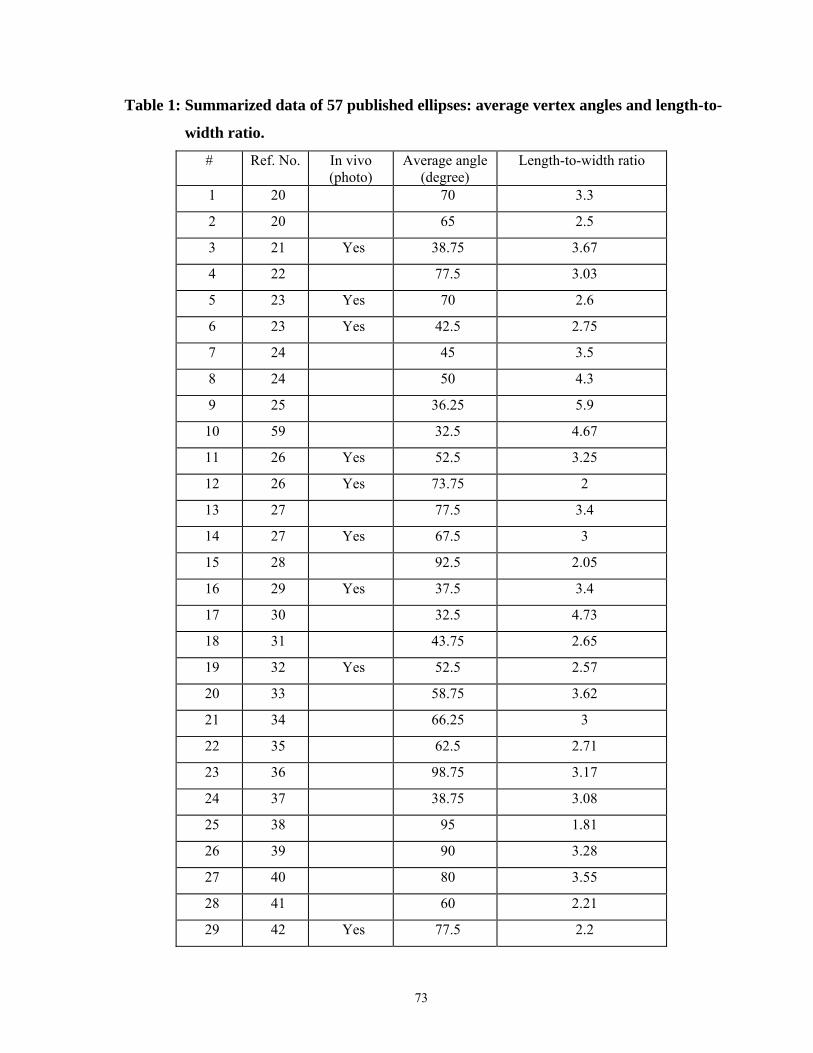
Table 1: Summarized data of 57 published ellipses: average vertex angles and length-to-
width ratio.
# Ref. No. In vivo (photo)
Average angle (degree)
Length-to-width ratio
1 20 70 3.3
2 20 65 2.5
3 21 Yes 38.75 3.67
4 22 77.5 3.03
5 23 Yes 70 2.6
6 23 Yes 42.5 2.75
7 24 45 3.5
8 24 50 4.3
9 25 36.25 5.9
10 59 32.5 4.67
11 26 Yes 52.5 3.25
12 26 Yes 73.75 2
13 27 77.5 3.4
14 27 Yes 67.5 3
15 28 92.5 2.05
16 29 Yes 37.5 3.4
17 30 32.5 4.73
18 31 43.75 2.65
19 32 Yes 52.5 2.57
20 33 58.75 3.62
21 34 66.25 3
22 35 62.5 2.71
23 36 98.75 3.17
24 37 38.75 3.08
25 38 95 1.81
26 39 90 3.28
27 40 80 3.55
28 41 60 2.21
29 42 Yes 77.5 2.2
73

# Ref. No. In vivo (photo)
Average angle (degree)
Length-to-width ratio
30 43 57.5 3.29
31 44 60 2.88
32 44 50 2.67
33 44 63.75 2.56
34 44 67.5 2.3
35 45 47.5 2.75
36 46 110 1.85
37 47 65 2.75
38 48 75 2.69
39 49 37.5 6.17
40 50 67.5 3.27
41 51 52.5 3.8
42 52 Yes 70 2.8
43 53 Yes 42.5 3.89
44 54 57.5 3
45 55 36.25 5.67
46 55 60 3.73
47 56 86.25 2.05
48 56 61.25 3.25
49 57 62.5 2.875
50 58 50 3.21
51 60 47.5 2.8
52 60 50 3
53 60 52.5 3.24
54 61 yes 88.75 1.73
55 61 yes 75 1.71
56 61 yes 60 2.38
57 61 yes 90 1.67
74

Table 2: Summarized data of 83 clinical ellipses: average vertex angles and length-to-
width ratio.
# Average angle (degree)
Length-to-width ratio
1 48.1 6.33
2 50.4 4.5
3 42.5 4.4
4 45.0 4.17
5 58.6 3.5
6 65.1 3.5
7 54.1 3.5
8 47.2 3.43
9 46.4 3.33
10 58.5 3.29
11 58.4 3.25
12 46.4 3.22
13 51.1 3.17
14 53.5 3.17
15 47.9 3.17
16 51.3 3.17
17 45.6 3.14
18 55.2 3.13
19 58.4 3.09
20 58.5 3
21 68.5 2.91
22 53.5 2.91
23 58.4 2.89
24 57.7 2.88
25 60.8 2.8
26 60.8 2.73
27 63.1 2.69
28 61.7 2.64
29 58.4 2.56
# Average angle (degree)
Length-to-width ratio
30 65.0 2.56
31 60.0 2.55
32 55.9 2.53
33 72.0 2.50
34 63.3 2.50
35 60.0 2.5
36 61.7 2.5
37 60.0 2.5
38 62.5 2.5
39 65.9 2.5
40 60.8 2.5
41 63.4 2.45
42 61.8 2.44
43 63.3 2.42
44 66.9 2.38
45 60.1 2.33
46 63.3 2.30
47 72.2 2.3
48 59.3 2.3
49 83.0 2.29
50 63.4 2.23
51 69.5 2.20
52 65.0 2.18
53 72.9 2.1
54 75.2 2.06
55 91.8 2.05
56 71.1 2.03
57 85.7 2.00
58 76.9 1.96
75

# Average angle (degree)
Length-to-width ratio
59 79.7 1.93
60 83.0 1.84
61 105.6 1.83
62 87.7 1.79
63 97.6 1.78
64 97.6 1.75
65 79.7 1.74
66 87.7 1.73
67 78.9 1.71
68 85.7 1.70
69 90.2 1.69
70 89.6 1.67
71 87.7 1.64
72 88.5 1.62
73 78.9 1.60
74 97.9 1.59
75 93.1 1.57
76 107.7 1.55
77 109.7 1.53
78 92.2 1.50
79 95.0 1.49
80 98.6 1.47
81 95.7 1.46
82 88.4 1.36
83 117.9 1.29
76

Table 3: Comparison of empirical vertex angles and theoretical calculations.
Vertex angle for Length-to-width
ratio of 3
Vertex angle for Length-to-width
ratio of 4
Length-to-width ratio for vertex
angle of 30°
Data (literature)
58°
48°
7.5
Data (our excisions)
59°
47°
7.5
Theory –
fusiform ellipse
67°
53°
7.5
77

y = 128.71x-0.71
R2 = 0.48
30
50
70
90
110
1 2 3 4 5 6 7 8length-to-width ratio
verte
x an
gle
[deg
]
Literature DataTheory (Fusiform ellipse)Bestfit (Literature Data)
Figure 1: Vertex angle of the cited surgical ellipses as a function of the length-to-width
ratio. The data are represented by the dots, the best-fit curve is the black solid
line and the theoretical curve is the pink solid line.
y = 125.51x-0.71
R2 = 0.82
30
50
70
90
110
1 2 3 4 5 6 7
Length-to-width ratio
Verte
x an
gle
[deg
]
Clinical DataTheory (Fusiform ellipse)Bestfit (Clinical Data)
Figure 2: Vertex angle of our clinical ellipses as a function of the length-to-width ratio.
The data are represented by the dots, the best-fit curve is the black solid line
and the theoretical curve is the pink solid line.
78

Chapter 4
The analyses of skin waste during excision of benign skin lesions.
Is the surgical ellipse cut an unnecessary cut?
Publication:
Raveh Tilleman T , Neumann HAM, Tilleman MM. The analyses of skin waste during
excision of non cancerous skin lesions. Is the surgical ellipse cut an unnecessary cut?
Submitted to: Plast Reconst Surg 2004
Presentation:
2nd Annual Research Meeting. General Health Services, Rabin Medical Center, Petach
Tikva ,Israel, November 2003.
79

80

Abstract
Elliptical excision is the common cut in cutaneous surgery. In this work we
investigate the skin waste of the surgical ellipse in biopsies of benign skin lesions. The skin
waste is defined as the ratio of the difference between the excised elliptical area and the
original lesion area, to the lesion area (plus margins for both).
Ninety consequential excisional biopsies performed at our outpatient clinic are
analyzed. The results show that the surgical ellipse cut is a skin wasting procedure. The
measured skin waste spans the range between 57% and 733%, with a mean of 181%. The
largest skin waste was observed for small lesions, occurring in the head and neck regions.
The ellipse cut is therefore an unnecessary surgical procedure especially for small, benign
lesions. For these lesions a shave biopsy or a circular block excision is the best procedure. It
wastes less skin and produces shorter scars. Consequently, we see no apparent justification
for the elliptical cut paradigm for benign lesions.
Introduction
All humans have a number of benign skin lesions (1-3). Some of these are removed
during the life span of a person for either cosmetic reasons or for preventive treatment (4-6).
In order to extract tissue from a skin lesion, an excisional biopsy is frequently performed.
The removed specimen undergoes further laboratory processing and microscopic analysis.
Skin biopsies are a common procedure in cutaneous surgery. Out of several possible
surgical cuts, the surgical ellipse is the most common cutting pattern used for biopsies. The
elliptical cut offers fast healing, good esthetic result, and sufficient specimen for histological
examination (7). A properly designed elliptical excision has a length-to-width ratio of 3:1
and produces an angle of 30º at both apices of the wound (8). So designed an excision can
be closed primarily thus avoiding the formation of excessive mounds of tissue at the wound
vertices or dog-ears. Otherwise, the dog-ears will require additional mending by one out of
ten or more methods of dog-ear correction (9,10).
The purpose of this study is to quantify the wasted skin in excisional biopsies of non
cancerous skin lesions. Skin waste is hence defined as the ratio of the difference between the
excised elliptical area and the original lesion area, to the lesion area.
81

Patients and Methods
Data of ninety consequential biopsies performed at our outpatient clinic for benign
skin lesions were recorded. Only those lesions that were further histologically proved as
completely excised were included. All the histological results were benign including: nevi,
warts, fibro epithelial polyps, seborrheic keratoses, dermatofibromas, and pyogenic
granuloma. Measured dimensions of the lesions and of the excised ellipses are summarized
in Table I. Forty five patients had lesions located on the head and neck and forty five
patients had lesions located on the body.
The area of the lesion is calculated by:
Alesion = π/4 b×d (1)
where: b is the short diameter and d is the long diameter of the ellipse.
We chose the following ellipse formula to model the surgical ellipse area (11):
( ) ( ⎥⎦
⎤⎢⎣
⎡−−⎟
⎠⎞
⎜⎝⎛
++= − 12
12sin1
82
2122
2
aaa
aaSAellipse ) (2)
where: S is the width of the fusiform ellipse and a is the aspect ratio (length-to-width ratio).
The skin waste is defined as the ratio of the difference of equations (1) and (2), and the
lesion area plus margins (Eq.(1)):
lesion
lesionellipse
AAA
waste−
= (3)
Results
Summarized in Table I are the lesion and excision data, and the calculated skin
waste. The lesion length varied between 2 mm and 32 mm and the lesion width varied
between 2 mm and 29.5 mm. The excision length varied between 6.5 mm and 54.5 mm and
the width varied between 3 mm and 33 mm. The mean length-to-width ratio of the excisions
was 2.43:1. The area of the lesions varied between 3.14 mm2 and 741 mm2 and the area of
the excised surgical ellipse varied between 13.54 mm2 and 1283 mm2. The skin waste was
found in the range of 57% to 733% with a mean of 181%.
Figure 1 plots the skin waste (percent) versus the lesion area. Observe that the
wasted skin decreases monotonously with the lesion area. Note that the skin waste is vastly
82

larger for small lesions, 733% for 3.14 mm2, than for large lesions, 57% for 616 mm2. Thus
small lesions cause large skin waste whereas large lesions cause small waste.
Approximating a power regression curve the wasted area is typified by: 37.015.1 −= lesionAwaste , with R2=0.69.
Figure 2 breaks up the data of Figure 1 into two groups: lesions from the head and
neck, and lesions from the rest of the body. To emphasize the small lesions, the skin waste
is plotted against the lesion area on a logarithmic scale. The largest amount of skin waste
was found when small lesions in the head and neck were excised. The average skin waste of
the head and neck lesions is 240%, compared with an average of 120% for body lesions.
This twofold waste occurs due to the following facts: 1) the head and neck excisions have a
larger length-to-width ratio than the body excisions (2.75 versus 2.09), and 2) the head and
neck lesions are smaller than the body lesions, yet the excision margins for both areas are
similar. For the above two reasons, paradoxically, larger skin waste is obtained for the small
lesions.
Another data fit shows that the excision area is proportional to the excision length, as
shown in Figure 3. What follows is that even a small addition to the excision length affects
dramatically the skin waste. For instance, by increasing the average length from 20 mm to
30 mm (growth of 50%), the excision area grows from 70 mm2 to 142 mm2 (growth of
100%).
Discussion
The surgical ellipse is the most common pattern used in excision and closure of
cutaneous lesions. This study quantifies the wasted skin in excising benign cutaneous
lesions. It varies between 57% and 733% in excess of the lesion area. This enormous wasted
tissue noticed in both small and large lesions is for the purpose of producing a linear scar
and avoiding dog-ear formation. What was noticed is that the smaller the lesion, the larger is
the skin waste. This might be due to the reason that many smaller lesions were located on
the head and neck where attention is paid to the final scar and a considerable effort is made
to conceal it, ending up with a longer length-to-width ratio and larger skin waste.
It is recommended to prefer a shave biopsy or a round excision of the lesion which
ends with a circular defect. With the use of a circular incision, adequate safe margins are
83

obtained and, at the same time, waste of sound skin is minimized. Such a circular defect can
be easily closed by one of three alternatives. The first is leaving the tissue open to heal by
second intention (11), the second is a direct closure of the defect without dog-ear excision
(12,13), and the third is a purse-string closure which uses circumferential tissue
advancement (14-16). These three techniques produce excellent results with no additional
scarring or tissue wasting and often the final scar is smaller than the original lesion. From
our experience with the figure-of-8 direct closure (13) (see chapter 9) any wound diameter
that can be closed by an ellipse is suitable for this technique as well. Other options for
closing a circular defect (17,18) include: primary closure and dog-ear correction, skin graft,
M-plasty, A-to-T closure, O-to-Z closure (19), lazy S (20), H-advancement flap (21) and
variety of local skin flaps (22,23). These solutions are less effective. They compromise
either the scar length or the amount of healthy skin wasted.
The elliptical cut is therefore an unnecessary surgical procedure especially for small
benign lesions. For these lesions a shave diagnostic biopsy or a round block cosmetic
excision is the best procedure. It yields less skin waste and shorter scars. Though the
surgical ellipse cut was found as the least advantageous skin excision pattern (24, see
Chapter 2), it is routinely used. We see no apparent justification for the elliptical cut
paradigm.
The conclusion is that for better tissue preservation and scar formation it is
recommended that the surgical ellipse should be replaced by other skin cuts.
84

References
1. Pariser RJ. Benign neoplasms of the skin. Med Clin North Am. 1998;82:1285-307.
2. Luba MC, Bangs SA, Mohler AM, Stulberg DL. Common benign skin tumors. Am Fam
Physician. 2003 15;67:729-38.
3. Rudolph P. Benign adnexal skin tumors. Pathologe. 2002 ;23(1):71-8.
4. Monteagudo C, Ferrandez A, Gonzalez-Devesa M, Llombart-Bosch A. Psammomatous
malignant melanoma arising in an intradermal naevus. Histopathology. 2001;39:493-7.
5. Betti R, Menni S, Cerri A, Vergani R, Crosti C. Seborrheic keratosis with compound
nevus, junctional nevus and basal cell carcinoma in the same lesion.
Dermatology. 2001;203:265-7.
6. Jin ZH, Kumakiri M, Ishida H, Kinebuchi S. A case of combined nevus: compound
nevus and spindle cell Spitz nevus. J Dermatol. 2000 ;27:233-7.
7. Dunlavey E, Leshin B. The simple excision. Dermatol Clin. 1998;16:49-64.
8. Zuber TJ, Dewitt DE. The fusiform excision. Am Fam Physician. 1994:1;49:371-80.
9. Weisberg NK, Nehal KS, Zide BM. Dog-ears: a review. Dermatol Surg. 2000;26:363-
70.
10. Zitelli JA. Tips for wound closure. Pearls for minimizing dog-ears and applications
of periosteal sutures. Dermatol Clin. 1989;7:123-8.
11. Lawrence CM, Comaish JS, Dahl MG. Excision of skin tumours without wound
closure. Br J Dermatol. 1986 ;115:563-71.
12. Davis TS, Graham WP 3rd, Miller SH. The circular excision. Ann Plast Surg. 1980
;4:21-4.
13. Raveh Tilleman T. Direct closure of rounded skin defects: A four-step technique
with multiple subcutaneous and cutaneous “figure-of-8” sutures alleviating dog-ears.
Accepted to the Plast Reconstr Surg.
14. Yuen JC. Versatility of the subcuticular purse-string suture in wound closure.
Plast Reconstr Surg. 1996;98:1302-5.
15. Tremolada C, Blandini D, Beretta M, Mascetti M. The "round block" purse-string suture:
a simple method to close skin defects with minimal scarring. Plast Reconstr Surg. 1997
;100:126-31.
16. Weisberg NK, Greenbaum SS. Revisiting the purse-string closure: some new methods
85

and modifications. Dermatol Surg. 2003;29:672-6.
17. Romiti R, Randle HW. Revisiting the purse-string closure: some new methods and
modifications. Dermatol Surg. 2003;29:672-6.
18. Hanke CW. Repair of surgical defects. Dermatol Clin. 1987;5:287-302.
19. Robbins TH. Double rotation skin closure: an alternative to elliptical closure. Aust N Z J
Surg. 1978;48:650-2
20. Buckingham ED, Quinn FB, Calhoun KH. Optimal design of O-to-Z flaps for closure of
facial skin defects.Arch Facial Plast Surg. 2003;5:92-5.
21. Rosenberg L A versatile procedure for the closure of small skin defects. Aesthetic Plast
Surg. 1985;9:23-6.
22. Albom MJ. Closure of excisional wounds with "H" flaps.J Dermatol Surg. 1975;1(4):26-
7.
23. Alvarado A. Reciprocal incisions for closure of circular skin defects. Plast Reconstr
Surg. 1981;67:482-91.
24. Warren SE, Snell GF. Complex wound closure, excisional biopsy, and use of simple
flaps. Prim Care. 1986;13:433-45.
25. Raveh Tilleman T ,Tilleman M.M ,Krekels GAM and Neumann HAM. Skin waste and
scar length in excisional biopsies. Comparing five excision patterns: fusiform ellipse,
fusiform circle, diamond, mosque and S-shape. Plast Reconst Surg 2004;113:857-861.
86
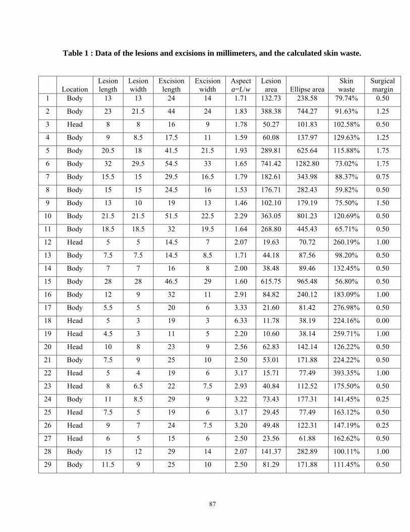
Table 1 : Data of the lesions and excisions in millimeters, and the calculated skin waste.
Location Lesion length
Lesion width
Excision length
Excision width
Aspect a=L/w
Lesion area Ellipse area
Skin waste
Surgical margin
1 Body 13 13 24 14 1.71 132.73 238.58 79.74% 0.50
2 Body 23 21.5 44 24 1.83 388.38 744.27 91.63% 1.25
3 Head 8 8 16 9 1.78 50.27 101.83 102.58% 0.50
4 Body 9 8.5 17.5 11 1.59 60.08 137.97 129.63% 1.25
5 Body 20.5 18 41.5 21.5 1.93 289.81 625.64 115.88% 1.75
6 Body 32 29.5 54.5 33 1.65 741.42 1282.80 73.02% 1.75
7 Body 15.5 15 29.5 16.5 1.79 182.61 343.98 88.37% 0.75
8 Body 15 15 24.5 16 1.53 176.71 282.43 59.82% 0.50
9 Body 13 10 19 13 1.46 102.10 179.19 75.50% 1.50
10 Body 21.5 21.5 51.5 22.5 2.29 363.05 801.23 120.69% 0.50
11 Body 18.5 18.5 32 19.5 1.64 268.80 445.43 65.71% 0.50
12 Head 5 5 14.5 7 2.07 19.63 70.72 260.19% 1.00
13 Body 7.5 7.5 14.5 8.5 1.71 44.18 87.56 98.20% 0.50
14 Body 7 7 16 8 2.00 38.48 89.46 132.45% 0.50
15 Body 28 28 46.5 29 1.60 615.75 965.48 56.80% 0.50
16 Body 12 9 32 11 2.91 84.82 240.12 183.09% 1.00
17 Body 5.5 5 20 6 3.33 21.60 81.42 276.98% 0.50
18 Head 5 3 19 3 6.33 11.78 38.19 224.16% 0.00
19 Head 4.5 3 11 5 2.20 10.60 38.14 259.71% 1.00
20 Head 10 8 23 9 2.56 62.83 142.14 126.22% 0.50
21 Body 7.5 9 25 10 2.50 53.01 171.88 224.22% 0.50
22 Head 5 4 19 6 3.17 15.71 77.49 393.35% 1.00
23 Head 8 6.5 22 7.5 2.93 40.84 112.52 175.50% 0.50
24 Body 11 8.5 29 9 3.22 73.43 177.31 141.45% 0.25
25 Head 7.5 5 19 6 3.17 29.45 77.49 163.12% 0.50
26 Head 9 7 24 7.5 3.20 49.48 122.31 147.19% 0.25
27 Head 6 5 15 6 2.50 23.56 61.88 162.62% 0.50
28 Body 15 12 29 14 2.07 141.37 282.89 100.11% 1.00
29 Body 11.5 9 25 10 2.50 81.29 171.88 111.45% 0.50
87

30 Head 6 8 17 8 2.13 37.70 94.56 150.84% 0.00
31 Head 9 5 19 8 2.38 35.34 104.84 196.64% 1.50
32 Head 4 4 18 5.5 3.27 12.57 67.22 434.89% 0.75
33 Head 3 3 13 4 3.25 7.07 35.31 399.60% 0.50
34 Head 7 5 15 6 2.50 27.49 61.88 125.10% 0.50
35 Body 6.5 7 20 9 2.22 35.74 124.73 249.03% 1.00
36 Head 3 3 17 4.5 3.78 7.07 51.71 631.51% 0.75
37 Body 16.5 9 28 11 2.55 116.63 211.54 81.37% 1.00
38 head 5.5 4.5 16 6 2.67 19.44 65.77 238.32% 0.75
39 head 3 2.5 11.5 4 2.88 5.89 31.40 433.00% 0.75
40 head 6.5 4 14 5.5 2.55 20.42 52.88 158.98% 0.75
41 head 5 5 15 7 2.14 19.63 72.96 271.58% 1.00
42 head 6.5 4 19 6 3.17 20.42 77.49 279.50% 1.00
43 head 2 2 11 3.5 3.14 3.14 26.18 733.31% 0.75
45 body 4.5 5 15 5.5 2.73 17.67 56.45 219.45% 0.25
46 head 3.5 3 10.5 4 2.63 8.25 28.80 249.19% 0.50
47 Head 3.5 3 10 4 2.50 8.25 27.50 233.49% 0.50
48 Head 2 2 6.5 3 2.17 3.14 13.54 330.93% 0.50
49 Body 9 9 17.5 9.5 1.84 63.62 117.11 84.09% 0.25
50 Body 7 6 15 7 2.14 32.99 72.96 121.18% 0.50
51 Body 4 4.5 10.5 5.5 1.91 14.14 40.54 186.74% 0.50
52 Head 11 9 20.5 10 2.05 77.75 142.97 83.87% 0.50
53 Body 7.5 7 15.5 8 1.94 41.23 86.92 110.79% 0.50
54 Head 5 5 16 7 2.29 19.63 77.45 294.46% 1.00
55 Body 10 10 22 12 1.83 78.54 186.07 136.91% 1.00
56 Head 8 6 23 7 3.29 37.70 109.30 189.92% 0.50
57 Head 2.5 3 10 4 2.50 5.89 27.50 366.88% 0.50
58 Head 5 5 15 6 2.50 19.63 61.88 215.14% 0.50
59 Head 4.5 4.5 11.5 5 2.30 15.90 39.75 149.91% 0.25
60 Head 4 3 12.5 4 3.13 9.42 34.01 260.82% 0.50
61 Head 3.5 3.5 14 4 3.50 9.62 37.94 294.30% 0.25
62 Head 2 2 9 3 3.00 3.14 18.39 485.50% 0.50
64 Body 12 10 24 11 2.18 94.25 183.19 94.37% 0.50
88

65 Head 4 4 13 4.5 2.89 12.57 39.92 217.67% 0.25
66 Head 4 4 11 4.5 2.44 12.57 34.08 171.20% 0.25
67 Body 15 14 31.5 15 2.10 164.93 328.85 99.39% 0.50
68 Head 18 12 42 14 3.00 169.65 400.58 136.13% 1.00
69 Body 22 14 46 18 2.56 241.90 568.55 135.03% 2.00
70 Body 17 16 42 18 2.33 213.63 522.06 144.38% 1.00
71 Body 9 9 17 10 1.70 63.62 120.83 89.93% 0.50
72 Body 15 15 28 17.5 1.60 176.71 350.92 98.58% 1.25
73 Head 9 6 18.5 7 2.64 42.41 88.76 109.28% 0.50
74 Head 11 9 24 9.5 2.53 77.75 156.66 101.48% 0.25
75 Head 14 4 25 6 4.17 43.98 101.14 129.96% 1.00
76 Body 13.5 14 30.5 15 2.03 148.44 319.28 115.09% 0.50
77 Body 9 9 16.5 10 1.65 63.62 117.70 85.01% 0.50
78 Body 12.5 9 23 10 2.30 88.36 158.98 79.93% 0.50
79 Body 21 13.5 43 16 2.69 222.66 471.13 111.59% 1.25
80 Body 20 16 39 19 2.05 251.33 516.71 105.59% 1.50
81 Body 12 12 26 15 1.73 113.10 276.56 144.54% 1.50
82 Body 11 10 24.5 11 2.23 86.39 186.71 116.12% 0.50
83 Head 9 8 22 9 2.44 56.55 136.32 141.06% 0.50
84 Head 8 8 18 9 2.00 50.27 113.22 125.25% 0.50
85 Body 18 17 35 19 1.84 240.33 468.46 94.92% 1.00
86 Head 8 7 16 9 1.78 43.98 101.83 131.52% 1.00
87 Head 11 7 21.5 9 2.39 60.48 133.41 120.61% 1.00
88 Head 9 11 23 13 1.77 77.75 211.54 172.06% 1.00
89 Body 12 10 30.5 11 2.77 94.25 229.38 143.38% 0.50
90 Body 10.5 8 16 10 1.60 65.97 114.59 73.69% 1.00
Average 2.43 180.96%
STD 0.71 121.06%
Max 32.00 29.50 54.50 33.00 6.33 741.42 1282.80 733% 2.00
Min 2.00 2.00 6.50 3.00 1.46 3.14 13.54 57% 0.00
89

y = 1.15x-0.37
R2 = 0.69
0%
100%
200%
300%
400%
500%
600%
700%
800%
0 2 4 6 8Lesion area (cm2)
Was
ted
skin
Figure 1: The skin waste (percent) versus the lesion area (cm2). Observe that the
wasted skin decreases monotonously with the lesion area. The solid line is the
best–fit curve of the data.
0%
100%
200%
300%
400%
500%
600%
700%
800%
1 10 100 1000Lesion area (mm2), logarithmic scale
Was
ted
skin
body head
Figure 2: The skin waste (percent) versus the lesion area (mm2) in a logarithmic scale.
Lesions on the head and neck are triangular green and lesions on the body
are round pink.
90

y = 0.0027x1.85
R2 = 0.94
0
1
2
3
4
5
6
0 10 20 30 40 50 60Excision length (mm)
Exci
sion
are
a (c
m2 )
Figure 3: Excision area versus lesion length. The solid line is the best–fit curve of the
data.
91

92

Chapter 5
Skin waste in elliptical excision biopsies of non melanoma skin
cancers.
Publication:
Tamara Raveh Tilleman, Martino H.A. Neumann, Nicole W.J. Smeets, and Michael M.
Tilleman. Accepted for publication in the Scand J Plast Reconstr Surgery Hand Surg
2004.
Presentation:
Combined annual meeting of the American Society for Dermatologic Surgery (ASDS)
and the American College of Mohs Micrographic Surgery (ACMMSCO). New Orleans
October 2003
2nd Annual Research Meeting. General Health Services, Rabin Medical Center, Petach
Tikva ,Israel, November 2003.
93

94

Abstract.
This work quantifies the skin waste during elliptical excision biopsies of non
melanoma skin cancers. The skin waste is defined as a ratio of the difference between the
excised elliptical area and the original lesion area plus oncological margins, to the lesion
area plus margins.
The analysis uses basic geometrical calculations of twenty six excisional biopsies of
patients with non-melanoma malignant tumors. The measurements were performed at the
last stage of Mohs’ Micrographic Surgery (MMS). Huge tissue waste is noticed in both
small and large excised lesions. The largest skin waste measured was 230% and the smallest
was 34% with a mean of 130%.
Mohs’ Micrographic Surgery is a skin sparing procedure, therefore the above
mentioned huge tissue waste is unacceptable. The conclusion is that for better tissue
preservation in closure of circular defects as a result of malignant tumor excision, other
patterns should be adopted instead of surgical ellipses.
Introduction
Skin excisions are widely performed for skin lesions suspected as cancerous.
Treatment of such cutaneous lesions should completely remove the tumor while preserving
maximum of the healthy surrounding skin (1). Therefore, the surgery is a compromise
between safe excision margins and obtaining a satisfactory esthetic result (2). The healthy
skin removed in addition to the lesion is a byproduct of the skin cutting pattern and the
necessary oncological therapeutic margin. In the literature there is no agreement as to the
optimal width of surgical therapeutic margins. This amount of excised skin depends on the
skin cancer type. Skin cancer such as basal cell carcinoma requires minimal margins
whereas squamous cell carcinoma or malignant melanoma requires wider margins (3-5).
Even the histological subtypes of a cancer play a role: for example, certain basal cell
carcinomas behave more aggressively and require more aggressive treatment (6). Some
authors recommend an optimum margin of 4 mm for all non melanoma skin cancer (7) while
others found that these margins are suitable only for cutaneous lesions clinically diagnosed
as basal cell (8). Others still believe that well demarcated lesions, such as a nodular basal
95

cell carcinoma, may be excised with a 3 mm margin (2). Cited are also opinions which
maintain that surgeons resect excessively wide surrounding skin carcinomas (9).
The therapeutic margins cannot be considered skin waste, because they are an
oncological necessity. On the other hand, the healthy skin removed due to the cutting pattern
enabling wound closure is considered pure skin waste. Many lesions are circular yet rarely
the final excision is identical to the original pattern, resulting in the removal of healthy skin.
The most common skin pattern used is a fusiform ellipse, which has become the standard
pattern for skin lesion removal (10 – 12). To avoid dog-ear formation during elliptical
excisions a vertex angle of 30˚ or less (13,14) and a length-to-width ratio between 3:1 to 4:1
are recommended (15 – 17).
The purpose of this study is to quantify the wasted skin in excisional biopsies of non
melanoma skin cancers. Skin waste is hence defined as the ratio of the difference between
the excised elliptical area and the original lesion area plus the therapeutic margins, to the
lesion area.
Patients and Methods
Twenty–six patients, with non–melanoma malignant tumors (twenty four with basal
cell carcinoma and two with squamous cell carcinoma tumors), on the forehead or the cheek,
were consequentially chosen for this study from the outpatient department of dermatology at
the Hospital Ziekenhuis Maastricht, Netherlands. All the patients underwent MMS and the
measurements of the skin defect and the ellipse incision were performed at the last stage of
the operation. Seventeen patients were males and the rest were females. Measured
dimensions of the lesions and of the excised ellipses were recorded and are summarized in
Table I. The excision length varied between 6 mm and 34 mm and the excision width varied
between 6 mm and 27 mm. The mean length-to-width ratio of the excision was 3.13:1.
The area of the lesion is calculated by:
A = π/4 b×d
where: b is the short diameter and d is the long diameter of the ellipse. We chose a fusiform
ellipse to model the surgical ellipse. This is the area overlapped by two identical ellipses
somewhat removed from one another. From basic geometrical principles the area of the
fusiform ellipse is determined by (see Chapter 2):
96

( ) ( )⎥⎦
⎤⎢⎣
⎡−−⎟
⎠⎞
⎜⎝⎛
++= − 12
12sin1
82
2122
2
aaa
aaSAellipse
where S is the width of the fusiform ellipse and a is the aspect ratio (length-to-width ratio).
The skin waste is determined by the difference of the elliptical excision and the original
lesion areas.
Results
Summarized in Table I are the lesion and excision data, and the calculated skin
waste. The area of the excised lesions varied between 0.3 cm2 and 7.2 cm2. The area of the
fusiform ellipse varied between 0.9 cm2 and 15 cm2. Figure 1 shows the areas of the excised
lesion with oncological margins (orange bars) and the areas of the final excised ellipse (blue
bars). The excision areas seem considerably larger than the lesion areas, implying excessive
skin cut. Whereas the greatest lesion area (with its 3 millimeter margins) is 7 cm2, its
corresponding excision area measures 15 cm2.
The lesion area is plotted against the excision area in Figure 2, showing a linear
relation between the two. Discrete points denote the data and a bestfit curve is drawn as a
line with a slope of 0.46 and a standard deviation of less than 10%. This means that the
greater the lesion the greater is the excision area. The mean wasted skin area is 130%, with a
standard deviation decreasing from 50% at 0.3 cm2 to 30% at 4 cm2. The largest skin waste
was 230% and the smallest was 34%.
Shown in Figure 3 is the percentage of the skin waste versus the lesion area. Observe
that the data scatter of the skin waste is vastly larger for small lesions, 130% for 0.3 cm2,
than for large lesions, 110% for 7.2 cm2. Hence, the average fraction of the skin waste is
nearly constant, independent of the lesion area.
The excision area is proportional to the square of the average length of the excision,
as shown in Figure 4. What is implied is that any additional lengthening of the skin cut
dramatically affects the amount of the skin waste. For instance, by increasing the average
length from 30 mm to 40 mm (a growth of 33%), the excision area grows from 4.6 cm2 to
8.2 cm2 (a growth of 78%).
97

Discussion
Surgical extirpation of skin lesions and especially malignant tumors often results in
circular or oval defects. The routine use of elliptical excision as a standard means of closure
often creates needlessly long scars and skin waste. Many surgical alternatives exist for
closing a circular defect (18 – 25). This study quantifies the wasted skin during cutaneous
surgery of non-melanoma cancerous lesion. It varies between 34% and 210% beyond the
lesion area. This skin waste seems extremely inappropriate when the operation performed is
a Mohs’ Micrographic surgery, an operation that heralds skin sparing as an aim.
The findings of this chapter are consistent with those of Chapter 4. The difference in
the magnitudes of skin waste stems from the fact that in MMS discussed herewith the
lesions are larger ab initio though they are located in the face. For the current lesion size of
0.3 cm2 and 7.2 cm2 we found a mean waste of 120%, similar to the finding of equivalent
sizes in Chapter 4 (see figures 1 and 2 in Chapter 4).
The importance of this study lies in the sparing use of healthy tissue and shortening
of scars. If sparingly used, the saved skin may serve as a future resource for reconstruction,
a flap, for instance. This is true for both regular excisional biopsies, and for Mohs’
Micrographic surgery (MMS). For the latter it is even more emphasized since there skin
sparing is an objective in itself, therefore it is highly significant to optimize the cut. It would
be imprudent for a surgeon performing MMS that spares millimeters of healthy margins
throughout its stages, to willingly sacrifice lots of healthy skin in the reconstruction stage.
The conclusion is that for better tissue preservation in closure of circular and oval defects
patterns other than a surgical ellipse should be adopted.
98

References
1. Hudson-Peacock MJ, Matthews JN, Lawrence CM. Relation between size of skin
excision, wound, and specimen. J Am Acad Dermatol. 1995 ;32:1010-5.
2. Lalloo MT, Sood S. Head and neck basal cell carcinoma: treatment using a 2-mm
clinical excision margin. Clin Otolaryngol. 2000;25:370-3.
3. Epstein E. How accurate is the visual assesment of basal call carcinoma margins?. Br
J Dermatol; 1973:89:37-43.
4. Brodland DG, Zitelli JA. Surgical margins for excision of primary cutaneous
squamous cell carcinoma. J Am Acad Dermatol. 1992;27:241-8.
5. Haigh PI, DiFronzo LA, McCready DR.Optimal excision margins for primary
cutaneous melanoma: a systematic review and meta-analysis. Can J Surg.
2003;46:419-26.
6. Orengo IF, Salasche SJ, Fewkes J, Khan J, Thornby J, Rubin F. Correlation of
histologic subtypes of primary basal cell carcinoma and number of Mohs stages
required to achieve a tumor-free plane. J Am Acad Dermatol. 1997;37:395-7.
7. Thomas DJ, King AR, Peat BG. Excision margins for nonmelanotic skin cancer.
Plast Reconstr Surg. 2003;112:57-63.
8. Wolf DJ, Zitelli JA. Surgical margins for basal cell carcinoma. Arch Dermatol
1987;123:340-4.
9. Bisson MA, Dunkin CS, Suvarna SK, Griffiths RW. Do plastic surgeons resect basal
cell carcinomas too widely? A prospective study comparing surgical and histological
margins. Br J Plast Surg. 2002;55:293-7.
10. Grabb WC. Basic techniques in plastic surgery. In: Grabb WC and Smith JW, eds.
Plastic surgery third edition. Little, Brown and Company, Boston;1979:3-8.
11. Chapter 8 ellipse. In: Stegman SJ, Tromovitch TA, and Glogau, eds. Basics of
dermatologic surgery. Year book medical publishers Inc;1982:60-72.
12. Vistnes LM. Basic principales of cutaneous surgery. In: Epstein E and Epstein E Jr,
eds. Skin surgery sixth edition. W.B.Saunders company;1987:44-55.
13. Dunlavey E and Leshin B. The simple excision. Dermatol Clinics. 1998;16:49-64.
14. Complex closures. In: Bennett RG, ed. Fundamentals of cutaneous surgery. C.V.
Mosby Company;1988:473-491
99

15. Leshin B. Proper planning and execution of surgical excisions. In: : Wheeland
RG,ed. Cutaneous surgery. W.B.Saunders company;1994:171-177.
16. Borges AF. Dog ear repair. Plast Reconst Surgery;1982:69:707-13.
17. Wound closure. In: Rohrich RJ and Robinson, eds. Wound healing. Selected redings
in plastic surgery. University of Texax southwestern medical center and Bayelor
University medical center;1999:9:18-20.
18. Healing by second intention. In: Fewkes JL, Cheney ML, and Pollack SV,eds.
Illustrated atlas of cutaneous surgery. 1992;13.1-13.13.
19. Iriondo M. Healing by second intention. In: Wheeland RG,ed. Cutaneous surgery.
W.B.Saunders company;1994: 892-900.
20. Drake LA, Dienehart SM, Goltz RW et al. Guidelines of care for Mohs
micrographic surgery. J AM Acad Dermatol 1995;33:271-8.
21. Greenbaum SS, and Radonich MA. The purse-string suture. Dermatol Surg
1996;22:1054-6.
22. Teitelbaum S. The purse-string suture. Plast Reconst Surger 1998;101:1748-9.
23. Hudson-Peacock M.J., and Lawrence C.M. Comparison of wound closure by means
of dog ear repair and elliptical excision. J Am Academ Dermato 1995;32:627-30.
24. Kirsner RS, Falanga V, and Eaglstein WH. The biology of skin grafts. Arch
Dermatol. 1993;129:481-483.
25. Advancement flaps. In: Davidson TM, Webster RC, and Gordon BR, eds. The
principles and dynamics of local skin flaps. American Academy of Otolaryngology.
1998; 26-37.
100

Table 1. Measurements of the lesion, the excised ellipse lengths and widths, and skin
waste. Location 1 denotes the forehead and location 2 denotes the cheek.
# A
ge
Loca
tion
Prev
ious
trea
tmen
t
Gen
der
Su
rgeo
n
Le
sion
dim
ensi
ons
(mm
)
Ex
cisi
on d
imen
sion
s (m
m)
Le
sion
are
a (
cm2 )
Ex
cisi
on a
rea
(cm
2 )
Skin
was
te
(%
)
1 66 1 Rec F 1 6 x 6 21 x 6 0.28 0.85 2022 69 2 Prim M 2 15 x 9 25 x 9 1.06 1.54 453 71 1 Rec F 2 13 x 12 33 x 13 1.23 2.95 1414 72 2 Rec F 2 13 x 12 35 x 12 1.23 2.86 1345 82 1 Rec F 1 20 x 8 42 x 8 1.26 2.26 806 82 1 Rec M 2 14 x 12 30 x 13 1.32 2.70 1047 60 1 Rec F 1 16 x 11 45 x 11 1.38 3.34 1428 63 1 Rec M 2 16 x 12 39 x 16 1.51 4.30 1859 85 1 Prim M 1 16 x 12 39 x 12 1.51 3.18 11110 73 1 Rec F 2 15 x 15 34 x 15 1.77 3.53 10011 86 1 Prim M 1 17 x 15 45 x 15 2.00 4.60 13012 86 2 Prim M 2 20 x 13 28 x 14 2.04 2.74 3413 60 1 Rec M 1 18 x 15 52 x 15 2.12 5.29 14914 60 2 Rec F 2 19 x 16 41 x 16 2.39 4.50 8915 70 2 Rec F 1 20 x 16 73 x 16 2.51 7.86 21316 83 1 Prim M 2 18 x 20 62 x 20 2.83 8.44 19817 83 1 Prim M 1 22 x 19 70 x 19 3.28 9.00 17418 71 1 Prim M 2 21 x 20 50 x 20 3.30 6.88 10819 86 2 Rec F 2 23 x 21 47 x 22 3.79 7.19 8920 79 1 Rec M 2 24 x 20 62 x 20 3.77 8.44 12421 77 1 Prim M 2 26 x 16 73 x 16 3.27 7.86 14122 60 1 Rec M 1 25 x 20 60 x 20 3.93 8.18 10823 86 2 Prim M 2 34 x 27 84 x 27 7.21 15.43 11424 58 1 Prim M 1 14 x 10 45 x 12 1.10 3.65 23225 58 2 Prim M 1 20 x 19 56 x 20 2.98 7.65 15626 58 2 Prim M 1 15 x 15 40 x 15 1.77 4.11 133
101

0
2
4
6
8
10
12
14
16
18
1 6 11 16 21
Area
(cm2 )
lesion
excision
Parient number
Figure 1: Areas of lesions (orange bars) and excisions made in a form of an fusiform
ellipse (blue bars).
0
2
4
6
8
10
12
14
16
18
0 2 4 6 8lesion area (cm2)
exci
sion
are
a (c
m2 )
Figure 2: Excision area versus lesion area. The data are the discrete points and the
solid line is a best--fit curve with a slope of 0.461.
102

0
50
100
150
200
250
0 2 4 6 8lesion area (cm2)
wast
ed a
rea
(%)
Figure 3: Percentage of wasted skin during excision. The average waste recedes slowly
with the lesion area from 140% to 120%. On the other hand the point
scattering decreases appreciably with the lesion area.
0
5
10
15
20
0 10 20 30 40 50 60Average length (mm)
Exci
sion
are
a (c
m2 )
fusiform ellipse
fusiform circle
elipse
measurement
Figure 4: The excision area versus the average excision length. The solid line is the
best–fit curve of the data and the broken line is the best–fit curve of the
calculated fusiform ellipse.
103

104

Chapter 6
The elastic properties of cancerous skin: Poisson’s ratio and
Young’s modulus.
Publication:
Raveh Tilleman T, MD , Tilleman MM, Neumann HAM. The elastic properties of
cancerous skin: Poisson’s ratio and Young’s modulus. Submitted to and under second
revision to:IMAJ 2004.
105

106

Abstract
Physical properties of cancerous skin tissue have rarely been measured in either fresh
or frozen skin specimens. Of interest are the elastic properties associated with the skin
ability to deform, i.e. to stretch and compress. Two constants, the Young’s modulus and
Poisson’s ratio, represent the basic elastic behavior pattern of any elastic material, skin being
one. The former relates the applied stress on a specimen to its deformation via the Hooke’s
law, while the latter is the ratio between the axial and lateral strains.
Our objective is to investigate the elastic properties of cancerous skin tissue. For this
purpose frozen sections of twenty three consecutive cancerous tissue specimens prepared
during Mohs micrographic surgery (MMS) were analyzed. We calculated from the data the
change in the radial length defined as the radial strain and the change in the tissue thickness
defined as axial strain.
Based on the above two strains we determined a Poisson’s ratio of 0.43 ± 0.12 and
an average Young’s modulus of 52 KPa. Defining the elastic properties of cancerous skin
may become the first step in turning elasticity into a clinical tool. Correlating these constants
with the histopathological features of a cancerous tissue can contribute an additional
noninvasive, in vivo and in vitro diagnostic tool. The elastic properties found in this work
were used for calculations of the saucer incisions in MMS discussed in Chapter 7.
Introduction
Many physiological processes significantly change tissue properties. Pathological
changes are generally correlated with changes in tissue elasticity. Cancerous tissue, such as
carcinoma of the breast, appears as extremely hard nodules (11). This is the rational for
palpation as a diagnostic tool in finding tumors of the prostate and breast (22,3). The
palpation is a subjective test while in medicine the aim is to convert a physiological process
into an objective test. Measuring the elasticity of cancerous tissues can yield an objective
tool to be used in diagnostic models.
The properties of skin tissue in biomechanical research are commonly cited in the
literature as applying to a material that is purely elastic, homogeneous and isotropic (44). It
should be emphasized that real skin is unisotropic, heterogeneous and elastically nonlinear
107

or viscoelastic, in either healthy or cancerous tissues. Therefore, a direct measurement of
the cancerous skin properties is necessary.
Skin complex mechanical properties include the elastic properties of solid materials
and the viscous properties of fluids (5). The elastic characteristics of the skin relate to the
immediate changes that occur when force is applied to the skin. They govern the skin ability
to deform, i.e. to stretch, contract and compress. These characteristics are defined by two
physical constants: the Young’s modulus, which relates the proportionality of the
longitudinal deformation to the applied force (Hooke’s law), and Poisson’s ratio, which
relates the dimensional deformations to one another (6). The viscous characteristics of the
skin relate to the delayed changes occurring after time: the decreased stress obtained over
time when a constant strain is applied (stress relaxation effect) and the increased length
obtained over time when a constant strain is applied (creeping effect) (7). Surgeons are
familiar with these effects, counting on stress-relaxation to release tension scar with time.
They use the creeping effect to absorb some irregular scar features and dog-ears and to cause
tissue elongation after inflating expanders.
Our interest is to explore the skin ability to deform. To gain understanding of the
elastic behavior of the skin we set to measure the Young’s modulus and Poisson’s ratio. A
common approach is to controllably compress a sample and measure the resulting
deformation. One of the immediate implementations is to perform these measurements on a
tissue before and after compression in a cryostat. This is a common process for immediate
histology evaluation, available from surgeries that produce frozen sections.
Defining the elastic properties of normal and cancerous skin may become the first
step in turning elasticity into a clinical tool. In the present work we measured the Young’s
modulus and Poisson’s ratio of cancerous skin specimens, the first such measurement to the
best of our knowledge.
Material and methods
In the analysis we used twenty three consecutive specimens excised from patients at
our outpatient clinic. The specimen dimensions: length and thickness, in millimeters, were
measured before and after compression and freezing using a caliper with an accuracy of 50
�m (0.05 mm). The cryostat chamber was set to -27°C (see Table 1), and a weight of 478
108

gram was used to compress the specimen (Figure 1). The compression in the cryostat is the
first stage in producing frozen sections from the specimen. Inspection of the processed
specimens under the microscope provides the histological diagnosis.
From the data we measured the change in the radial length and thickness of the
tissue, and determined the resulting radial and axial strains. Based on the compressing
weight and tissue dimensions we determined the stresses acting on the sample. From these,
in turn, we determined the Young’s modulus (the proportion coefficient between the strain
and stress) and Poisson’s ratio (the ratio between the radial strain and the axial strain). For
this study we used only frozen sections that were further histologically proven as basal cell
carcinoma.
Ice granules do not interfere with the measurement of the specimens because of two
main reasons: 1) the time duration for the freezing of water having the size of our smallest
sample is a few minutes, longer than the duration of the compression in the cryostat, 2)
frozen water, or ice, would dramatically decrease the deformability of the tissue.
Considering that ice Young’s modulus is about 8 GPa, under the weight of 478 gram the
deformation of the typical 10×2 mm is less than the dimension of an atom! Therefore, what
was measured was the deformation of the cancerous tissue, with minimum freezing effect.
Our statistical evaluation assumes the present sample as normal, calculating
accordingly the statistical averages, standard deviations and p-values.
Results
Twenty three consecutive cancerous tissue specimens prepared during frozen
sections were analyzed. The twenty three analyzed tissue segments showed, after
compression in the cryostat, an axial elongation varying from 12.66% to 44.33 %, with a
mean of 17.7% (Table 1) and a standard deviation (STD) of 0.06. These tissues varied in
width (radial thinning) from 7.33% to 30.86%, with a mean of 30.55% and STD of 0.09.
The Poisson’s ratio determined by the mean dimensions, with a measurement error of about
1%, resulted in 0.43 ± 0.12. With the positive stress being the ratio of the weight of 478
gram and the specimen area, we found a Young’s modulus of 52 ± 45 KPa. Note that the
Poisson’s ratio calculated is not a regular average of the measured lengths. The reason is that
commonly accepted elastic properties and the Hooke’s law pertain to small deformations of
109

the specimen. Because the present specimens underwent large deformations, one must use
the known formulae carefully. Thus, one uses the mean lengths rather than the initial lengths
as the normalizing dimensions.
The p-values were calculated based on the assumption of normal distribution. They
are 15% the Poisson’s ratio and 17% for the Young’s modulus. These large values are
consistent with the large variation of elastic properties of the skin cited in the literature
(44,8,9).
Discussion
Normal skin and pathological skin possess different characteristics. For instance,
cancerous tissue can appear as extremely hard nodules (11). The abnormal skin can be either
cancerous or infected or a scarred tissue (88,10,11). Defining the elastic properties of
cancerous skin and correlating these constants with the histopathological features of a
cancerous tissue can contribute an additional noninvasive, in vivo and in vitro diagnostic
(13). Poisson’s ratio, a skin elastic characteristic, describes the behavior of materials under
stress. For biological materials this constant ranges from 0.25 to 0.85 (14-22). Our
measured Poisson’s ratio for cancerous skin is 0.43, well within the cited range in the
literature for biological tissues. In comparison, the Poisson’s ratio for healthy human skin at
room temperature has been cited in Ref. 8 as 0.5.
An example for clinical use of the Poisson’s ratio was presented at the last annual
meeting of the American College of Mohs Micrographic Surgery and Cutaneous Oncology
(23). By using finite element analysis (ANSYS 5.6 Finite Element software package, with
tetrahedron shaped elements represented by 1000 nodes), inputting the Poisson’s ratio we
determined, the minimum angle for an unobstructed view of a Mohs micrographic surgery
cut was calculated. The angle was found 10º, smaller than the current norm in Mohs’. This
approach is an example where elastic properties of the cancerous skin have a clinical
application (2424).
The further measured Young’s modulus shows an average of 52 KPa, well within the
known values for various skins (44,13,25-28). Its deviation of 45 KPa about the mean is also
well understood due to the significant variations in skin elasticity as a function of age,
location, actinic changes and racial features. This variation explains the large p-value we
110

found, 17% for Young’s modulus and 15% for Poisson’s ratio.
Our further objective is to investigate the correlation between the skin elastic
properties and the histological findings of healthy skin and cancerous skin morphology.
Such a clinical research is presently held in our departments aiming to specify the skin
elastic properties according to the cancer features. Future practical uses of the a priori
knowledge of the elastic characteristics of cancerous tissues is, for instance, finding the
exact border between healthy and malignant tissue by a noninvasive measurement on the
skin surface. Thus cancer screening, diagnosis and treatment modalities can benefit from a
well defined, compiled and stored data body of these characteristics.
Acknowledgments
The authors wish to acknowledge Ms. Ismene Muermans and Ms. Ilse Muermans of
AZM Hospital, Maastricht, the Netherlands, and Ms. Ida Lubetsky of Rabin Medical Center,
Petach Tikva, Israel, for their assistance in processing the tissue specimens.
111

References
1. Anderson RC, Curtin HR. Ultrasonic holography. A promising medical imaging tool.
Radiology. 1973;109:417-21
2. Owen AW, Forrest AP, Anderson TJ, Samuel E, Young GB, Scott AM Breast screening
and surgical problems. Br J Surg. 1977;64:725-8
3. Hedlund PO, Das D, Lowhagen T, Esposti P. Rectal palpation and transrectal fine
needle aspiration of the prostate in the monitoring of prostate cancer: a study of 59
patients during treatment with estramustine phosphate or estrogens. Prostate.
1989;15:327-33
4. Larrabee, W.F.Jr. A finite element model of skin deformation: I Biomechanics of skin
and soft tissue: A review. Laryngoscope. 1986;96(4):399-405.
5. Gilbson T, Kenedi RM. Biomechanical properties of skin. Surg North Am 1967;47:279-
294.
6. Lee C, Vincent JF, and Hillerton JE. Poisson’s ratio in skin. Biomed Matwe Eng.
199;1(1):19-23.
7. Wexler DB, Gilbertson LG, Goel VK, Bardach J. Biomechanics of the rotation-
advancement skin flap: experimental and theoretical studies. In: Bardach J, ed. Local
flaps and free skin grafts. Mosby year book, 1992. 53-68.
8. Larrabee,W.F.Jr., and Sutton, D. A finite element model of skin deformation: II An
experimental model of skin deformation. Laryngoscope. 1986;96(4):406-12.
9. Kennedy RM, Gibson T, Daly CH. Bioengineering studies of the human skin in: Proc.
symposium on biomechanics and related bioengineering topics. Kennedy RM, ed.
Pergamon Press, Oxford, 1965; 147-58.
10. Miyauchi T, Maruoka M, Nagayama T. Malignant melanoma of the penis associated
with von Recklinghausen’s neurofibromatosis: report a case. Hinyokika Kiyo.
1988;34(4):710-3.
11. Tiu CM, Chiou HJ, Chou YH, Hsu CC, Lin KJ, Chen CM, Ko JS, Tseng LM, Lai CR,
Lui WY. Sonographic features of breast abscesses with emphasis on "hypoechoic rim"
sign.Zhonghua Yi Xue Za Zhi (Taipei). 2001;64(3):153-60.
12. Ulrich J, Voit C.Ultrasound in dermatology. Part II. Ultrasound of regional lymph node
basins and subcutaneous tumours. Eur J Dermatol. 2001;11(1):73-9
112

13. Dobrin PB, Mrkvicka R.Estimating the elastic modulus of non-atherosclerotic elastic
arteries. J Hypertens Suppl. 1992;10(6):S7-10.
14. Maaß H, Kühnapfel U. Noninvasive Measurement of Elastic Properties of Living
Tissue, Proceedings of the European Medical and Biological Engineering Conference
EMBEC,99, Medical & Biological Engineering & Computing. 1999;37(2):1460-1.
15. Mizunuma M, Yanai A, Tsutsumi S, et al. Can dog ear formation be decreased when an
s shape resection is used instead of spindle skin resection? A three dimentional analysis
of skin surgery techniques using finite element method. Plast Reconstr Surg.
2000;106(4):845-8.
16. Lee C, Vincent JF, and Hillerton JE. Poisson’s ratio in skin. Biomed Matwe Eng.
199;1(1):19-23.
17. Boissieux L, Kiss G, Magnenat Thalmann N, Kalra P. Simulation of skin aging and
wrinkles with cosmetics insight. http://66.102.11.104/search?q=cache:_traSCNhG7MJ
18. Ophir J, Cespedes I, Ponnekanti H, Yazdi Y, and Li X. Elastography: A quantitative
method for imaging the elasticity of biological tissues. Ultrasonic imaging 1991;13:
111-34.
19. Mente, P.L., Lewis, J.L., 1994. Elastic modulus of calcified cartilage is an order of
magnitude less than that of subchondral bone. Journal of Orthopaedic Research 12,
637}647.
20. Manios A, Katsantonis J, Tosca A, Skulakis CH, and Tsifisis D. The finite element
method as a research and teaching tool in the analysis of local skin flaps. Dermatol
Surg. 1996;22:1029-1033.
21. Erkamp RQ, Wiggins P, Skovoroda AR, Emelianov SY, O’Dodonnell M. Measuring
the elastic modulus of small tissue samples.Ultrason Imaging. 1998;20(1):17-28.
22. Pieper SD, Laub DR, and Rosen JM. A finite element facial model for simulating
plastic surgery. Plast Reconstr Surg. 1995;96(5):1100-5.
23. Raveh Tilleman T, Tilleman M and Neumann MHA. Minimum beveling in Mohs
surgery cut. In: The combined annual meeting of the ASDS and ACMMSCO. October
9-12 New Orleans 2003.
24. Pellettiere J. A. Materiam model for bone under impact loading. http://asb-
biomech.org/onlineabs/NACOB98/280
113

25. Mossel WP. Summary E-modulus of skin. [email protected].
http://isb.ri.ccf.org/biomch-l/archives/biomch-l-1997-12/00080.html
26. Mackay DR, Saggers GC, Kotwal N, Manders EK. Stretching skin: undermining is
more important than intraoperative expansion. Plast Reconstr Surg 1990;86:722-30.
27. Khatyr F.; Imberdis C.; Vescovo P.; Varchon D.; Lagarde J-M. Model of the
viscoelastic behaviour of skin in vivo and study of anisotropy. Skin Research Tech
2004;10:96-103.
28. Kiwaki H, Miyaoka Y, Arase S. Analysis of the absorbance spectra of skin lesions as a
helpful tool for detection of major pathophysiological changes. Skin Res Technol.
2004;10:130-5.
114

Table 1: Summary of the Measured Specimen Dimensions, Strain, Poisson’s ratio and
Young’s Modulus.
Initial diameter
(mm)
Initial height (mm)
Final diameter
(mm)
Final height (mm)
Radial strain
Axial strain
Poisson's ratio
Axial stress-to-strain
ratio
Young's modulus
(KPa) 8.85 3.15 9.85 2.4 10.70% 27.03% 0.40 0.026 42.57
9.9 3.15 11.85 2.45 17.93% 25.00% 0.72 0.021 25.83
9.75 4.85 11.55 2.7 16.90% 56.95% 0.30 0.009 17.26
8.1 4.85 10.6 2.7 26.74% 56.95% 0.47 0.012 18.83
10.2 4.65 11.2 3.65 9.35% 24.10% 0.39 0.022 36.75
8.45 4.65 9.8 3.65 14.79% 24.10% 0.61 0.030 41.25
9.85 4.55 10.75 3.4 8.74% 28.93% 0.30 0.020 36.11
9.1 4.55 10.95 3.4 18.45% 28.93% 0.64 0.021 27.92
7.5 3.95 8.05 3.45 7.07% 13.51% 0.52 0.075 109.35
8.75 3.15 10.2 2.25 15.30% 33.33% 0.46 0.020 31.63
9.65 3.15 10.8 2.2 11.25% 35.51% 0.32 0.016 29.38
5.45 3.15 5.85 2.55 7.08% 21.05% 0.34 0.091 158.99
7.35 4.05 8.95 2.45 19.63% 49.23% 0.40 0.019 30.67
4.15 3.85 5.2 2.5 22.46% 42.52% 0.53 0.065 95.73
10.45 3.95 12.5 2.55 17.86% 43.08% 0.41 0.011 17.41
4.5 3.95 5.55 2.45 20.90% 46.88% 0.45 0.051 80.98
7.15 2.4 8.9 1.55 21.81% 43.04% 0.51 0.022 32.71
6.5 2.5 8 1.5 20.69% 50.00% 0.41 0.023 37.60
9.9 2.45 11.35 1.85 13.65% 27.91% 0.49 0.019 29.24
6.35 2.45 7.3 1.95 13.92% 22.73% 0.61 0.057 78.28
8.85 3.15 10.75 1.9 19.39% 49.50% 0.39 0.013 21.24
3.8 2.75 4.25 1.85 11.18% 39.13% 0.29 0.096 177.98
10.05 3.7 12.15 2.1 18.92% 55.17% 0.34 0.009 15.61
Average 15.86% 36.72% 0.43 51.88
Std. dev. 5.33% 12.83% 0.12 44.85
115

116

Chapter 7
Minimal beveling angle in Mohs’ micrographic surgery cut
Publication:
Raveh Tilleman T ,Tilleman M.M , Neumann HAM. Minimum beveling in Mohs
surgery cut Submitted to: Clinic Exp Dermatol 2004
Presentation:
XXIII congress of the International Society for Dermatologic Surgery. Porto, September
2002 Portugal.
Combined annual meeting of the American Society for Dermatologic Surgery (ASDS)
and the American College of Mohs Micrographic Surgery (ACMMSCO). New Orleans
October 2003
2nd Annual Research Meeting. General Health Services, Rabin Medical Center, Petach
Tikva ,Israel, November 2003.
117

118

Abstract
Saucer incision with a taper angle of 45°is the common cut in Mohs’ micrographic
surgery (MMS). The beveling is necessary for microscopic viewing of the cutting strata,
however, its minimal angle has not been studied thus far. In this work we examine the 45°
aspect of the saucer incision and answer the question what is the minimal beveling angle that
permits viewing all the layers after the cryostat compression. The calculation method used is
a finite elements analysis (ANSYS). Microscopic views of Mohs specimens are inspected to
backup the analysis. It was found that 10° is the critical cone angle that permits complete
layer viewing without obstruction. The conclusion, therefore, is that a minimal beveling of
10° instead of 45° can be carried out during MMS. It is more skin sparing, reducing the risk
of invading the cancer and producing a better scar, while still providing adequate
microscopic information.
Introduction
In the teaching of cutaneous surgery a perpendicular 90° cut is the norm (1,2). However,
the skin cut in Mohs’ micrographic surgery (MMS) differs from that in any other skin
surgery. It aims to produce horizontal sections for histopathological viewing. Dr. Frederic
Mohs recommended that tissue removed by the first excision should possess a saucer shape
(3,4), i.e. a 45° beveled cut combined with a circular marginal incision. The beveled cut is
needed to produce a good microscopic strata projection (5) by eliminating any tissue bulging
during condensation in the cryostat (6).
This behavior is illustrated in Figure.1, where all the cone layers can be viewed
before and after the compression. In contrast, the compression of a cylinder (an object with
no taper) creates a barrel shape with a protruding middle section, therefore, masking layers
from a microscopic view. The projection of the cone layers diminishes as the cone angle
decreases. So far the minimal beveling of the Mohs specimen edge has not been shown or
calculated, thus the taper angle of 45° in MMS remained the norm (7,8). In this work we
examined the tapered aspect of the saucer incision and answered the question of what is the
minimal beveling angle that permits viewing all the layers after the cryostat compression.
119

Materials and Methods
The method uses a three-dimensional finite element analysis and microscopic views
of Mohs specimens. The finite element method (ANSYS 5.6 Finite Element software
package, with tetrahedron shaped elements represented by 1000 nodes) is a basic
engineering tool used for structure analysis, also used lately for modeling of body tissues (9-
19). This software has the capacity to compute and display anatomic predictions of surgical
manipulations (20). The finite analysis can model geometrically complex bioforms, such as
human tissues, by discretizing the anatomy into many smaller components termed
“elements”, the fundamental unit of the finite elements model. A thoughtfully constructed
finite element model of the skin can simulate the stretch and elasticity of a tissue (21).
Current models of wound closure and skin flap movement are based upon geometrical or
paper models (see the figures in Chapter 1). These models are not realistic because they
ignore the elastic properties of skin and its subcutaneous attachments (9). The skin
deformation based upon the finite elements model is a more effective and realistic model to
describe skin movements.
In the calculation we used a Poisson ratio of 0.43, described in the previous chapter.
Recall that we measured this Poisson’s ratio in a cancerous tissue, rendering it valid for all
the specimens used in this chapter. This value is within the cited range in the literature (4-
17,22). The finite elements served to assess quantitatively the minimal angle cut that permits
a complete microscopic layer projection.
Finally, specimens with various cut angles were photographed during MMS. The
microscopic views are used to backup the above theoretical calculations by showing their
layer projections.
Results
Using the finite elements method we analyzed a number of cones with a taper
ranging from 0° to 45°. This simulates specimen compression in a cryostat during the
preparation of frozen sections. Specifically, a dynamic analysis consisting of the original
specimen sagging due to applied vertical force is conducted. Figure 2 presents a model
having a taper of 45°. In the figure shown is the specimen before (left hand side) and after
(right hand side) the compression. Observe that a complete layer is unobstructedly viewed
120

after the compression, as schematically shown at the top of Figure 2.
Figure 3 presents a calculated model having a taper of 10° before (left hand side) and
after (right hand side) the compression. As before, a complete layer viewing is attained after
the compression though with a smaller projection. In analyzing cones with a taper ranging
from 0° to 45° it was found that 10° is the critical cone angle which permits complete layer
viewing without obstruction after cryostat compression. A smaller angle obscures some
layers and renders the viewing imperfect.
This finding was supported by numerous MMS operations. A microscopic view of
Mohs specimens is demonstrated in Figure 4. What is shown is that both a specimen with
beveling angles of both 45° (a), and of an acute angle cut of 10° (b) present good layer
projections.
Discussion
Mohs’ micrographic surgery is based on the concept of microscopically scanning
layers taken from the entire undersurface of the excised specimen (23). As a rule, the greater
the angle the better is the viewing of the layers after compression. On the other hand, the
greater the angle, the greater is the tissue waste and greater is the chance that the cancerous
tissue will be revealed at the bottom of the specimen, increasing the probability of an
additional MMS stage. Also, a 45° angle produces an epidermis and dermis acute tissue step
that upon closure results in a nonlinear scar and a dog-ear creation. A smaller angle reduces
the probability of the above drawbacks.
The commonly used taper angle of 45°, the legacy of Dr Mohs, has never been tested
before. By using the finite elements method we found that a taper of 10° is the minimal
beveling that permits projection and viewing of all the skin layers after the cryostat
compression. This finding was supported by MMS specimens. Note that the calculation
became possible once elasticity data of frozen, cancerous skin have been found (see Chapter
6). The conclusion is that a beveling of less than 45° but no less than 10° is recommended. It
is more skin sparing, reduces the probability of intercepting the cancerous tissue, and
produces a better scar.
121

References
1. Walkwer NPJ, Lawrence CM, Dawber RPR Simple excision. In: Textbook of
dermatology, eds: Champion RH, Burton JL, Burns DA, Breathnach SM. Blackwell
Science Ltd. Oxford 1998;3603-5.
2. Hak DJ, Goulet JA. Operative planning and perioperative management. In:Chapman’s
orthopedic surgery, ed: Chapman MW. Lippincott Williams and Wilkins 2001;75-120.
3. Brodland DG, Amonette R, Hanke CW and Robins P. The history and evolution of
Mohs micrographic surgery. Dermatol Surg 2000;26:303-7
4. Drake LA, Dinehart SM, Goltz RW, Graham GF, Hordinsky MK, Lewis CW, Pariser
DM, Salasche SJ, Skouge JW, Chanco Turner ML, Webster SB, Whitaker DC, Bulter B
and Lowery BJ. Guidelines of care for Mohs micrographic surgery. J Am Acad
Dermatol 1995;33:271-8.
5. Steinman HK. Mohs surgical techniques. In: Gross KG, Steinman HK, and Rapini
RP,eds. Mohs Surgery: fundamentals and techniques. Mosby, Inc;1999:49-66.
6. Mohs’ micrographic surgery. Chapter 29. In:. Fewkes JL, Cheney ML, and Pollack
SV,eds. Illustrated atlas of cutaneous surgery. JB Lippincott Company;1992:29.2-29.12
7. Walkwer NPJ, Lawrence CM, Dawber RPR.Dermatological surgery: micrographic
(Mohs) surgery. In:Textbook of dermatology, eds: Champion RH, Burton JL, Burns DA,
Breathnach SM. Blackwell Science Ltd. Oxford 1998;3593-3623.
8. Greenway HT, Leslie DF. Mohs micrographic surgery for skin cancer. Australas J
Dermatol 1991;32:159-64.
9. Larrabee, W.F.Jr. A finite element model of skin deformation: I Biomechanics of skin
and soft tissue: A review. Laryngoscope. 1986;96:399-405.
10. Larrabee,W.F.Jr., and Sutton, D. A finite element model of skin deformation: II An
experimental model of skin deformation. Laryngoscope. 1986;96:406-12.
11. Larrabee,W.F.Jr., and Galt, J.A. A finite element model of skin deformation: III The
finite element model of skin deformation. Laryngoscope. 1986;96:413-9.
12. Manios A, Katsantonis J, Tosca A, Skulakis CH, and Tsifisis D. The finite element
method as a research and teaching tool in the analysis of local skin flaps. Dermatol Surg.
1996;22:1029-1033.
13. Mizunuma M, Yanai A, Tsutsumi S, et al. Can dog ear formation be decreased when an
122

s shape resection is used instead of spindle skin resection? A three dimentional analysis
of skin surgery techniques using finite element method. Plast Reconstr Surg.
2000;106:845-8.
14. Remmler D. Can dog ear formation be decreased when an s shape resection is used
instead of spindle skin resection? A three dimentional analysis of skin surgery
techniques using finite element method. Discussion. Plast Reconstr Surg. 2000;106:849-
851.
15. Erkamp RQ, Wiggins P, Skovoroda AR, Emelianov SY, O’Dodonnell M. Measuring the
elastic modulus of small tissue samples.Ultrason Imaging. 1998;20:17-28.
16. Costa, K.D., Hunter, P.J., Wayne, J.S., Waldman, L.K., Guccione, J.M., and McCulloch,
A.D. A three dimentional finite element method for large elastic deformations of
ventricular myocardium: II – prolate sphenoidal coordinates. J Biomech Eng.
1996;118:464-72.
17. Keyak JH, Fourkas MG, Meagher JM, and Skinner HB. Validation of an automated
method of three-dimentional finite element modelling of bone. J Biomed Eng.
1993;15:505-9.
18. McCulloch A, Waldman L, Rogers J, and Guccione J. Large scale finite element analysis
of the beating heart. Crit Rev Biomed Eng. 1992;20:427-49.
19. Pieper SD, Laub DR, and Rosen JM. A finite element facial model for simulating
plastic surgery. Plast Reconstr Surg. 1995;96:1100-5.
20. Manios A, Katsantonis J, Tosca A, Skulakis CH, Tsifisis D, Finite element method as a
research and teaching tool in the analysis of local skin flaps. Dermatol Surg
1996;22:1029-33.
21. Kirby S, Wang B, To C, Lampe HB. Non linear three dimensional finite element model
of skin biomechanics. J otolaryngol 1998;27:153-60.
22. H. Maaß, U. Kühnapfel. Noninvasive Measurement of Elastic Properties of Living
Tissue, Proceedings of the European Medical and Biological Engineering Conference
EMBEC,99, Medical & Biological Engineering & Computing. 1999;37:1460-1.
23. Mohs FE. Mohs Micrographic Surgery a historical perspective. Dermatol Clinics
1989;7:609-626.
123

Cylinder Cone
(a)
(b)
(c)
Figure 1: Comparison of tapered and cylindrical shapes before (a) and after
compression (b and c). The view shown in (c) simulates the frozen cut
sections analyzed under microscope during Mohs’ micrographic surgery.
124

Figure 2: Finite Elements simulation of specimen compression: a calculated model
having a taper of 45° before (left hand side) and after the compression (right
hand side). Observe a complete layer viewing after the compression as
presented both by the finite element model and by the small geometrical
illustration (on top).
Figure 3: Finite Elements simulation of specimen compression: a calculated model
having a taper of 10° before (left hand side) and after the compression (right
hand side). Observe a complete layer viewing after the compression as
presented both by the finite element model and by the small geometrical
illustration (on top).
125
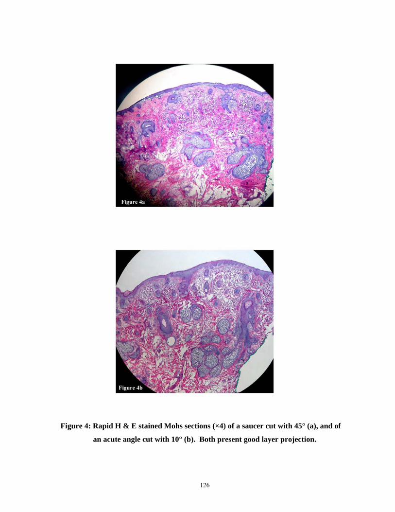
Figure 4: Rapid H & E stained Mohs sections (×4) of a saucer cut with 45° (a), and of
an acute angle cut with 10° (b). Both present good layer projection.
126

Chapter 8
Optimizing the cut in Mohs micrographic surgery in regard to
skin sparing and microscopic view: Is a circular incision
necessary?
Publication:
Raveh Tilleman T ,Tilleman M.M , Neumann HAM. Optimizing the cut in Mohs
micrographic surgery in regard to skin sparing and microscopic view: Is a round incision cut
necessary? Accepted to: Clin Exp Dermatol 2004
Presentation:
XXIII congress of the International Society for Dermatologic Surgery. Porto, September
2002 Portugal.
Combined annual meeting of the American Society for Dermatologic Surgery (ASDS)
and the American College of Mohs Micrographic Surgery (ACMMSCO). New Orleans
October 2003
2nd Annual Research Meeting. General Health Services, Rabin Medical Center, Petach
Tikva ,Israel, November 2003.
127

128
Abstract
Saucer incision is the common cut in Mohs’ micrographic surgery. To date no proof
to the superiority of this cut to other patterns has been presented. In this work we examine
the round pattern aspect of the saucer incision and answer two questions: does the circular
cut provide the best skin-sparing pattern and does it provide the best microscopic view. A
two dimensional geometrical analysis is used to determine whether a circular incision is
optimal from the standpoint of skin sparing and microscopic view. Mohs’ micrographic
surgery views are used to backup the geometrical hypothesis.
The result is that the circular incision pattern is skin-wasteful compared with an
incision that follows the contour of the cancerous lesion. In the present lesion the two cuts
have a ratio of 1.5 between the two excised skin areas, indicating a 50% waste of healthy
skin. It is also shown that specimens with a pointed edge provide better layer projection than
the circular contour.
The conclusion is that a tailored cut following the lesion pattern is the optimal Mohs
incision. Therefore in the first stage of Mohs’ micrographic surgery the skin cut should
replicate the lesion pattern instead of a circular saucer cut. Although many Mohs surgeons
already implement this philosophy, in the literature the saucer incision recommended by Dr.
Frederic Mohs remains the norm.
Introduction
Saucer shape excision is the common first layer cut in Mohs’ micrographic surgery
(MMS) (1). The persistence of Dr. Frederic Mohs saucer pattern excision as a standard is
found in various textbooks including general dermatology (2,3), dermatosurgery (4,5) and
also in surgery textbooks (6).
The circular cut in the first stage was suggested by Dr. Frederic Mohs (7,8,9). The
literature predominantly quotes the saucer incision even when the cancerous tissue drawn or
photographed is not circular (2,5,10,11,12). In some publications the drawn cut has rather an
oval shape (13,14). However, none of the published patterns exhibits pointed edges such as
used in a surgical ellipse.
Considering the microscopic view of a saucer cut, the advantages of a specimen
having beveled edge are detailed in previous studies (15,16). On the other hand, the
129

superiority of a circular cut for microscopic view to a pointed edge cut has not been
described or proven. In this work we examine the circular pattern aspect of the saucer
incision and answer two questions: does the circular cut provide the best skin-sparing pattern
and does it provide the best microscopic view.
Materials and Methods
We inspected cutting patterns illustrated by two dimensional geometrical drawings in
regard to skin waste and microscopic view. The skin waste of a circular, saucer cut was
compared with that of a skin cut following the lesion contour. A rounded excision
circumventing the lesion such that the closest points between them are separated by 3 mm
was compared with an excision extended only by minimal oncological margins around the
lesion (17,18).
In the literature various surrounding margins of normal skin are suggested in order
to ensure a complete excision. Some authors suggest 2 mm of excisional margins of non
melanoma skin cancer cases presenting clearance of up to 95% (19). Others suggest a 3 mm
margin which presents clearance of up to 96% (20, 21). Still other authors found that only 4
mm margins can clear the cancerous tissue up to 96% (22- 25). We chose a 3 mm margin to
use in our measurement and in the estimation of skin waste.
A geometrical analysis of the microscopic view for specimens having a pattern with
circular and pointed edges was performed. The analysis demonstrates which of the two cuts
provides a better layer view. To back up the hypothesis, related microscopic views of frozen
sections having a fusiform ellipse pattern, and excised during MMS, are presented.
Results
From the aspect of skin sparing a pattern that follows the skin lesion rather than a
circular pattern is preferable, as demonstrated in Figure 1. Shown is an example of Basal
Cell Carcinoma with a complex form located on a forehead measuring 14×10 mm. An
excision that follows the lesion providing a minimum healthy skin excised is drawn in
Figure 1a, compared with a circular cut circumventing the lesion drawn in Figure 1b. The
ratio between the two areas is about 1.5, indicating a 50% waste of healthy skin comparing
the circular cut to the tailored excision.
130

As for the aspect of a microscopic view, both specimens with round and pointed
edges can achieve good layer projection. The pointed edge, as evidenced at the vertex of a
fusiform ellipse, mosque pattern, rhomboid pattern and S-shape incision (26), does not
compromise the microscopic view. On the contrary, any pointed edge in the incision offers
more information. The radial projection thickness (t) in the outermost layer is shorter than
the horizontal thickness (E) at the pointed edge, such that:
⎪⎭
⎪⎬⎫
⎪⎩
⎪⎨⎧
⎥⎦
⎤⎢⎣
⎡⎟⎠⎞
⎜⎝⎛ −+−−=
wt
awtLE 2111211
2 2
where L and w are respectively the excision length and width, and a is their ratio (= L / w) .
Consider for example a fusiform ellipse with a length-to-width ratio of 3:1, L = 30 mm, w =
10 mm and radial projection thickness of 2.5 mm, E becomes 4.7 mm, exceeding by 88%
the radial projection thickness t = 2.5 mm.
Frozen sections produced during MMS having a shape of a fusiform ellipse (Figure
2) and its related microscopic views are presented. In Figure 3, a microscopic view of this
specimen with a round edge (green margin) and a pointed edge (red margin) is shown. Both
edges present a complete layer view. Therefore, a pointed edge presents no difficulty to a
microscopic view.
Discussion
In 1936 Dr. Mohs began the clinical use of chemosurgery, originally intended as an
in situ fixative on the patient himself (14). At the end of such an operation, the tissue was
left untouched until separation took place. No immediate closure or reconstruction was part
of the original MMS (27). Therefore, less attention was paid to the original incision shape.
In the 1970s a new era of fresh tissue technique began, popularized by Dr. Theodore
Tromovitch (28). Subsequently the immediate closure of defects became feasible. This
allows for more accurately excised margins, primary repair of the defect, and a more rapid
completion of the operation (29). Nowadays, this is the dominant procedure in MMS, thus
special attention must be paid to the shape of the excised specimen (30).
In this work we examined the circular pattern aspect of the Mohs saucer incision. It
131

was demonstrated that a tailored cut has two advantages in comparison with a round cut: 1)
it is more skin sparing, and 2) it provides a better microscopic view. Therefore, a tailored cut
that follows the lesion contour is the optimal Mohs incision.
In conclusion, in the first stage of MMS a skin cut following the lesion pattern
should be applied instead of a circular saucer cut. Though many Mohs surgeons already
implement this philosophy, in the literature the saucer incision recommended by Dr.
Frederic Mohs remains the norm (31).
132

References
1. Mikhail GR. Techniques in Mohs surgery. Saucerized excision of a disc of tissue. In:
Mikhail GR, ed. Mohs micrographic surgery. WB Saundres company 1991;15-6.
2. NPJ Walker, CM Lawrence, RPR Dawber. Dermatological surgery. In: RH Champion,
JL Burton, DA Burns SA Brearhnach, eds. Textbook of dermatology. Blackwell Science
Ltd, Oxford 1998;3593-623.
3. GJ Hruza, JL Fewkes. Surgery in dermatology. In: TB Fitzpatrick, AZ Eisen, K Wolff,
IM Freedberg, KF Austen, eds. Dermatology in general medicine. McGraw-Hill, Inc.
1993;2917-20.
4. Dufresne RG. Mohs surgery: fresh tissue techniques. In: Wheeland RG, ed. Cutaneous
surgery. WB Saundres company 1994;880-91.
5. Fewkes JL, Cheney ML, Pollack SV. Mohs’ micrographic surgery. In: Fewkes JL,
Cheney ML, Pollack SV,eds. Illustrated atlas of Cutaneous surgery. JB Lippincott
company New York 1992;29.2-29.12.
6. NA Swanson, TM Johnson. Management of basal cell carcinoma ans squamous cell
carcinoma. In: CW Cummings, JM Fredrickson, CJ Krause, LA Harker, DE Schuller,
MA Richardson, eds. Otolaryngology head and neck surgery. Mosby 1998;486-501.
7. Steinman HK. Mohs surgical techniques. In: Gross KG, Steinman HK and Rapini RP,
eds. Mohs surgery. Fundamentals and techniques. Mosby 1999:49-66.
8. Mohs FE. Chemosurgery for skin cancer: fixed tissue and fresh tissue techniques. Arch
dermatol 1976;112:211-5.
9. Tromovitch TA, Beirne G, and Beirne C. Mohs technique (cancer chemosurgery),
treatment of recurrent cutaneous carcinomas. Cancer 1966;19:867-8.
10. Shriner D, McKoy DC, Goldberg DJ, and Wagner RF Jr. Mohs micrographic surgery. J
Am Acad Dermatol. 1998;39:79-97.
11. Mora RG. Mapping out skin cancer: Mohs micrographic surgery. Virginia medical
quarterly, Autumn 1990:361-6.
12. Pearson GL Jr. Mohs micrographic surgery. Fresh tissue technique. Dermatol Clinics
1989;7:613-26.
13. Zitelli JA. Mohs surgery concept and misconcept. .Intl J Dermatol 1985;24:541-8.
14. Picoto AM and Picoto A. Technical procedures for Mohs fresh tissue sugery. J
133

Dermatol Surg Oncol 1986;12:134-8.
15. Nelson BR, Railan D, and Cohen S: Mohs’ micrographic surgery for nonmelanoma skin
cancer. Clin Plast Surg. 1997;24:705-18.
16. Raveh Tilleman T, Tilleman MM, Krekels GAM and Neumann MHA. Minimum
beveling in Mohs micrographic surgery cut. In: Combined annual meeting ASDS-
ACMMSCO. New Orleans 2003.
17. Burg G, Hirsch RD, Konz B, Braun-Falco O. Histographic surgery: accuracy of visual
assessment of the margins of basal cell epithelioma. J Dermatol Surg 1975;1:21-4.
18. Ratner D, and Bagiella E. The efficacy of curettage in delineating margins of Basal Cell
Carcinoma before Mohs Micrographic Surgery. Dermatol Surg 2003;29:899-903.
19. Lalloo MT, Sood S. Head and neck basal cell carcinoma: treatment using a 2-mm
clinical excision margin. Clin Otolaryngol. 2000;25: 370-3.
20. Bisson MA, Dunkin CS, Suvarna SK, Griffiths RW. Do plastic surgeons resect basal cell
carcinomas too widely? A prospective study comparing surgical and histological
margins. Br J Plast Surg. 2002;55:293-7.
21. De Visscher JG, Gooris PJ, Vermey A, Roodenburg JL Surgical margins for resection of
squamous cell carcinoma of the lower lip. Int J Oral Maxillofac Surg.2002;31:154-7.
22. Wolf DJ, Zitelli JA. Surgical margins for basal cell carcinoma. Arch Dermatol.1987;123:
340-4.
23. Thomas DJ, King AR, Peat BG. Excision margins for nonmelanotic skin cancer. Plast
Reconstr Surg. 2003;112:57-63.
24. Brodland DG, Zitelli JA. Surgical margins for excision of primary cutaneous squamous
cell carcinoma. J Am Acad Dermatol.1992;27:241-8.
25. Breuninger H, Schippert W, Black B, Rassner G. The margin of safety and depth of
excision in surgical treatment of basalioma. Use of 3-dimensional histologic study of
2,016 tumors. Hautarzt. 1989;40:693-700.
26. Raveh Tilleman T, Tilleman MM, Krekels GAM and Neumann MHA.,Skin waste,
vertex angle and scar length in excisional biopsies. Comparing five excision
patterns: fusiform ellipse, fusiform circle, rhomboid, mosque and S-shape. Plast
Reconst Surg 2004:113:857-861.
27. Mohs FE. Chemosurgery: a microscopically controlled method of cancer excision. Arch
134

Surg 1941;42:279-295.
28. Tromovitch TA, Stegma SJ.Microscopically controlled excision of skin tumors:
chemosurgery (Mohs): fresh tissue technique. Arch Dermatol 1974;110:231-2.
29. Hart V. Mohs’ chemosurgery. In: Emmett AJJ and O’rourke MGE,eds. Malignant skin
tumors.Longman Group Limited;1982:175.
30. Brodland, D.G., Amonette, R., Hanke, C.W., and Robins, P. The history and evolution
of Mohs micrographic surgery. Dermatol Surg. 2000;26:303-7.
31. Robins P. Chemosurgery: my 15 years of experience. J Dermatol Surg Oncol
1981;7:779-89.
135

136

Chapter 9
A new surgical technique for direct closure of circular skin
defects without dog-ear excision.
Publication:
Raveh Tilleman T. Direct closure of rounded skin defects. A four-step technique with
multiple subcutaneous and cutaneous “figure-of-8” sutures alleviating dog-ears. Accepted
for publication by: Plast Reconstr Surg 2004.
137

138

Abstract
Excisional biopsies of circular lesions are performed daily by surgeons. A
circular excision yields the smallest skin waste and scar length, but presents a surgical
challenge in closure due to dog-ear formation. This chapter establishes a surgical
technique for direct closure of circular or elliptical defects without producing long scars,
skin wasting and dog-ears. A four-step technique based on multiple cutaneous and
subcutaneous “figure-of-8” sutures is presented. When correctly placed, those sutures
can equally divide excess tissue and alleviate dog-ears.
It is shown that skin can be redistributed, alleviating dog-ears, yielding short
scars and saving healthy skin. A significant reduction of the length-to-width ratio and
the arc-to-scar length ratio are obtained. In Conclusion a direct closure of a circular or
elliptical defect without stipulating a 3:1 length-to-width ratio is feasible. In this
technique no excessive healthy tissue is removed and the final scar length is small. The
long-term outcome is a thin scar that is linear, flat and concealed in the body structure.
Introduction
Excisional biopsies are daily performed by surgeons for indications varying from
esthetic excisions to oncological skin malignancy removal. The purpose of an experienced
surgeon is to completely remove the skin lesion while leaving an optimum scar, performing
a short operation and accomplish fast healing without complications. Many cutaneous
lesions possess a circular or mildly elliptical shapes, yet from a surgical point of view it is a
challenge to close a circular defect without creating permanent dog-ears.
The most popular cutting pattern used for excisional biopsies is the surgical ellipse
followed by less popular patterns such as the rhomboid, mosque, and S-shape (1).
Traditionally the long axis of a surgical ellipse has been at least three times longer than its
short axis (2 – 6). This ratio permits avoiding dog-ear formation at the cost of wasting
healthy skin tissue and the creation of a long scar (7).
The present surgical technique is a new concept that eliminates the need for any
artificial excision pattern. It permits a direct closing a round skin wound while alleviating
dog-ears. In four steps this technique enables the closure of any circular or elliptical defect
139

without the need for a 3:1 length-to-width ratio, therefore mitigates undue skin waste. The
principle of the present method is that excess tissue can be redistributed provided guiding
sutures are placed correctly on the created scar. After several weeks a fine, blunt and
concealed linear scar is obtained. The advantage of the present technique is a considerable
saving of healthy skin without compromising the quality of the scar.
Surgical technique
The four-step technique for closing circular defects is based on multiple cutaneous
and subcutaneous “figure-of-8” sutures that equally divide excess tissue. The first step is
excision of the lesion with the recommended oncological margins shown in Figure 1, and
performing wide undermining shown in Figure 2. The end result of the first step is an open
round wound with skin margins that can be brought together with minimal tension.
The second step is selecting the best line of closure, preferably coinciding with skin
lines, such as relaxed skin tension lines (8). The end result of the second step is determining
the scar longitudinal axis. Drawn in Figure 3 are the scar axis and some smaller auxiliary,
perpendicular lines.
The third step is the closure itself beginning with a row of absorbable, “figure-of-8”,
buried sutures (Figure 4). The sutures fill two functions: obliterating the dead space of deep
margins and starting to equally divide the tissue excess by the mechanism explained below.
Photographed in Figure 5 is the end result of the third step: an open wound whose margins
are brought together. Observed around the wound is the beginning of wrinkling,
pronounced at the scar edges.
The fourth step is the final closure of the epidermis with multiple cutaneous, “figure-
of-8” sutures (Figure 6). Note that the wrinkles induced by the suture technique are a
byproduct of dog-ear alleviation. The two key sutures are located at the vertices. They
intercept at the dog-ear peak, as shown in Figure 13. The peak points can be found by using
a skin hook. The pleated scar starts fading within 3 – 4 weeks, and vanishes completely
within 3 months. During this period, the scar straightens out, the dog-ears are redistributed
and eventually disappear (see Figures 7-12).
Figure 7 shows a circular lesion located on the neck with a lesion size of 15×15 mm.
The scar length produced by this closure technique was 24 mm (Figure 8). The same scar
140
after five months shown in Figure 9. Note that the scar is located on a convex surface,
indicating that this closure technique is suitable for curved surfaces.
Figure 10 shows an open wound in a paranasal region. The lesion was 7×9 mm and
the produced scar length was 11 mm. After two months the resulting scar is hardly noticed
(Figure 11). With a similar bearing on the suitability for curved surface, this scar is located
on a concave surface. In Figure 12 a scar located on the forearm after six months post
operative period is shown. The round lesion was 17×43 mm and the produced scar length
was 54 mm.
Observations and Results
Shown in Table 1 are sixty five excisional biopsies taken at the outpatient clinic
using the above surgical technique. In the data the aspect ratio is 1:1.45 on the average and
the scar length is 53±7% of the arc length. The present aspect ratio is less than a half of the
recommended aspect ratio for surgical ellipses (1:3 – 1:4). Mizunuma et al found that a scar
length of a surgical ellipse is 92% of the pattern arc (9). In the present work the scar length
is only 53% of the arc length, shortening the scar by 40% relative to any other cutting
pattern. Consequently, the data indicate a double saving: first, by excising precisely the
circular lesion instead of an ellipse, second, by creating a short scar in comparison with the
arc length. In sum, this technique offers a shorter scar by a factor of approximately four
with respect to a surgical ellipse and less tissue waste by a factor greater than two (1).
Concerning tissue redistribution along the scar, the present technique reduces excess
skin at the apex and compensates for skin shortages at the center. On closing an open
wound, the apex moves in a rotation manner producing excess skin that forms a vertical
cone or dog-ear. At the same time, the central part of the scar advances to cover the longest
distance, thus creating tension at the scar center. These trends are even more pronounced if
the wound is circular. This takes place because the pseudo-apical angle is 180° and the
pattern width is maximal. However, by the present method the stresses around the scar are
redistributed. The “figure-of-8” stitch when tightly closed, squeezes tissue between the two
loops of the suture (see Figure 13). At the apex the “figure-of-8” suture applies a vertical
stress on the dog-ear. Thus the excess skin is distributed forming several smaller,
considerably less protruding cones. Around the wound center the “figure-of-8” stitch pulls
141

skin from the horizontal surroundings. Thus while some ripples are formed perpendicular to
the longitudinal axis, the scar is contracted. Over time, this lateral stress produces a creeping
effect on the scar, which also flattens the dog-ears. Note that the process is limited, because
excessive tension of the suture may cause scar ischemia.
An interim effect is a wrinkled scar in the immediate postoperative period. This
effect disappears within several weeks yet the patient should be given an educated
explanation assuring that the immediate ripple is only temporary.
Over the recent five years of clinical experience, the present technique has proved
very safe, reliable and producing good results. Among the main advantages are skin saving
and minimal scar length. Follow-ups revealed good cutaneous scars, independent of
whether the tissue surface is flat, convex or concave, as presented in Figures 7-12.
Discussion
Many skin lesions are circular, yet the applied excision pattern is often different
given skin wasteful shapes, such as the surgical ellipse. Surgeons do not simply cut a circle
around the lesion to remove it, even though a circular cut removes less skin and leaves a
shorter scar than any other skin excision, as shown in Chapters 2, 3 and 4. The reason is that
a circular excision does not stitch very well when directly closed. It leaves an elevated skin
bunched up at the ends, namely a “dog-ear”.
The objective of this study was to develop a surgical technique for closing circular
defects without removing healthy tissue while solving the dog-ear problem. This method
could be realized owing to numerous clinical observations about the skin ability to
redistribute. For example: skin, which has been strained by tension, relaxes over time (2). A
small circular excision cut by a punch biopsy allows primary closure without future
noticeable dog-ears (10). Some dog-ears flatten spontaneously due to contraction of the
linear scar both longitudinally and vertically (11). Closing a wound of unequal sides can be
accomplished by the “rule of halves” where the excised skin of the longer side is equally
divided without forming a dog-ear (12). In the Z-plasty technique the surgeon induces a
better scar by borrowing skin from a zone with excess skin to a zone with a shortage (13).
This elongates one axis and shortens the other. Over time the stretched skin in one area and
142

the shrunk skin in the perpendicular area relax.
All of the above are examples of the skin ability to redistribute, where one of the
most prominent observations of this ability is in the vertical reduction mammoplasty. In
order to avoid horizontal inframammary scar, a wrinkled, shrunk vertical suture is
performed (14). Subcuticular running suture can reduce the vertical subareolar length by
50% or more (15) resulting in fine wrinkling around the vertical scar that vanish over time.
These examples were the basis for the present suture technique. By forcing the skin to
redistribute, a short scar is formed, with some temporary wrinkles that disappear within
several weeks. The excess skin that would otherwise form dog-ears is equally divided along
the scar. A pivotal advantage of this technique is in saving healthy skin without
compromising the scar quality or the healing process.
This technique has been established for any surface topography. It has good results
for concave, convex or flat surface as shown in the figures. It is also a solution for
alleviating a dog-ear created during flap movements. Though at the pivot of a rotation flap a
dog-ear is created (16) and at the base of an advancement flap excess skin folds (Bürow’s
triangles) are formed (17), by applying the present suture technique the excess tissue can be
immediately corrected without any healthy skin removal and without creating additional
scars and backcuts.
This technique is suitable for many surgeons who previously ignored the circular cut
fearing the creation of unpleasant wrinkled scars and dog-ears. It is also an alternative to the
axiomatic use of surgical ellipse for skin biopsies.
To conclude, a direct closure of a circular or elliptical defect without stipulating a 3:1
length-to-width ratio is feasible. The immediate outcome is saving of healthy skin tissue. A
byproduct of skin sparing is a significant reduction of the scar length. The described suture
technique prevents the formation of dog-ears during excisional biopsies. It is also a method
relaxing the excess of tissue created at the pivot of a rotation flap, or the excessive triangles
during advancement flap. This technique is used daily at our practice yielding good results.
The long-term outcome is a thin scar that is linear, flat and concealed in the body structure.
143

References
1. Raveh Tilleman T, Tilleman M.M, Krekels GAM and Neumann HAM. Skin waste,
vertex angle and scar length in excisional biopsies. Comparing five excision patterns:
fusiform ellipse, fusiform circle, rhomboid, mosque and S-shape. Plast Reconst Surg
2004;
2. Grabb WC. Basic techniques in plastic surgery. In: Grabb WC, and Smith JW, eds.
Plastic surgery. A concise guide to clinical practice. Little, Brown and Company, Boston
1973;3-74.
3. Wood RJ and Jurkiewicz. Plastic and reconstructive surgery. In: Schwartz SI, Shires
GA, Spencer FC, Daly JM, Fischer JE, and Galloway AC, eds. Principles of surgery
seventh editon. McGraw Hill 1999;2091-143.
4. Length of scar and the “dog-ear”. In: McGregor IA, ed. Fundamental techniques of
plastic surgery and their surgical applications. E & S Livingstone Ltd. Edinburgh and
London 1960;31.
5. Ellipse. In: Stegman SJ, Tromovitch TA, and Glogau RG,eds. Basics of dermatologic
surgery. Year Book Medical Publishers, Inc. Chicago, London 1982;60-68.
6. Rohrich RJ and Robinson JB. Wound healing. Selected Reading 1999;9:1-39.
7. Raveh Tilleman T, Tilleman M.M, Smeets N, and Neumann HAM. Skin waste in
elliptical excision biopsies . Accepted to the Scand J Plast Reconstr Surgery Hand Surg
2004.
8. Borges AF. Relaxed skin tension lines (RSTL) versus other skin lines. Plast Reconstr
Surg 1984;73:144-50.
9. Mizunuma M, Yanai A, Tsutsumi S, Yosida H, Seno H, Inoue M, and Nisbida M. Can
dog ear formation be decreased when an s shape resection is used instead of spindle skin
resection? A three dimensional analysis of skin surgery techniques using finite element
method. Plast Reconstr Surg. 2000;106:845-8.
10. Schultz BC. Skin biopsy. In: Roenigk RK, and Roenigk HH,eds. Dermatologic Surgery
Principles and practice. Marcel Dekker Inc, New York, Basel 1989;139-154.
11. Complex closure. In:Bennett RG,ed. Fundamentals of cutaneous surgery. The C.V.
Mosby Company 1988;473-91.
144

12. Dog-ear repair and closure of sides of unequal lenth. In: Stegman SJ, Tromovitch TA,
and Glogau RG,eds. Basics of dermatologic surgery. Year Book Medical Publishers, Inc.
Chicago, London 1982;69-72.
13. Furnas DW and Fischer GW. The Z- plasty: biomechanics and mathematics. Br J Plast
Surg 1971:144-160
14. Lassus C. A 30-year experience with vertical. Plast Reconstr Surg 1996;97:373-80
15. LeJour M. Vertical mammaplasty: update and appraisal of late results. Plast Reconstr
Surg 1999;104:771-81
16. General considerations. In: Jackson TI,ed. Local flaps in head and neck reconstruction.
CV Mosby 1985;1-34
17. Bardach J, Levine NS, Buchanan RT, Cohen M and Marschall MA. Surgical techniques
using local flaps in head and neck reconstruction. In: Bardach J,ed. Local flaps and free
skin grafts. CV Mosby 1992;87-126
145
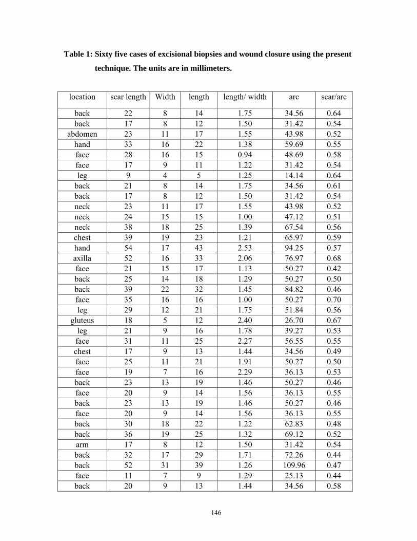
Table 1: Sixty five cases of excisional biopsies and wound closure using the present
technique. The units are in millimeters.
location scar length Width length length/ width arc scar/arc
back 22 8 14 1.75 34.56 0.64 back 17 8 12 1.50 31.42 0.54
abdomen 23 11 17 1.55 43.98 0.52 hand 33 16 22 1.38 59.69 0.55 face 28 16 15 0.94 48.69 0.58 face 17 9 11 1.22 31.42 0.54 leg 9 4 5 1.25 14.14 0.64
back 21 8 14 1.75 34.56 0.61 back 17 8 12 1.50 31.42 0.54 neck 23 11 17 1.55 43.98 0.52 neck 24 15 15 1.00 47.12 0.51 neck 38 18 25 1.39 67.54 0.56 chest 39 19 23 1.21 65.97 0.59 hand 54 17 43 2.53 94.25 0.57 axilla 52 16 33 2.06 76.97 0.68 face 21 15 17 1.13 50.27 0.42 back 25 14 18 1.29 50.27 0.50 back 39 22 32 1.45 84.82 0.46 face 35 16 16 1.00 50.27 0.70 leg 29 12 21 1.75 51.84 0.56
gluteus 18 5 12 2.40 26.70 0.67 leg 21 9 16 1.78 39.27 0.53 face 31 11 25 2.27 56.55 0.55 chest 17 9 13 1.44 34.56 0.49 face 25 11 21 1.91 50.27 0.50 face 19 7 16 2.29 36.13 0.53 back 23 13 19 1.46 50.27 0.46 face 20 9 14 1.56 36.13 0.55 back 23 13 19 1.46 50.27 0.46 face 20 9 14 1.56 36.13 0.55 back 30 18 22 1.22 62.83 0.48 back 36 19 25 1.32 69.12 0.52 arm 17 8 12 1.50 31.42 0.54 back 32 17 29 1.71 72.26 0.44 back 52 31 39 1.26 109.96 0.47 face 11 7 9 1.29 25.13 0.44 back 20 9 13 1.44 34.56 0.58
146

location scar length Width length length/ width arc scar/arc
back 17 8 13 1.63 32.99 0.52 face 22 13 15 1.15 43.98 0.50 face 21 11 15 1.36 40.84 0.51 neck 20 10 11 1.10 32.99 0.61 face 20 9 14 1.56 36.13 0.55 face 9 4 6 1.50 15.71 0.57 face 33 19 29 1.53 75.40 0.44 face 14 7 10 1.43 26.70 0.52 leg 19 11 15 1.36 40.84 0.47
finger 29 10 16 1.60 40.84 0.71 face 18 9 12 1.33 32.99 0.55 face 42 22 29 1.32 80.11 0.52 neck 30 15 21 1.40 56.55 0.53 face 17 12 15 1.25 42.41 0.40 face 32 20 28 1.40 75.40 0.42 face 15 7 10 1.43 26.70 0.56 leg 20 9 10 1.11 29.85 0.67 face 13 7 10 1.43 26.70 0.49 leg 13 7 10 1.43 26.70 0.49
abdomen 16 8 11 1.38 29.85 0.54 chest 18 10 12 1.20 34.56 0.52 chest 15 11 13 1.18 37.70 0.40 back 21 10 13 1.30 36.13 0.58 back 18 11 11 1.00 34.56 0.52 face 12 10 11 1.10 32.99 0.36 leg 25 15 19 1.27 53.41 0.47
back 14 10 11 1.10 32.99 0.42
147
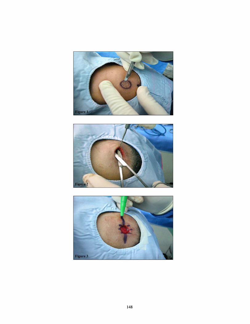
148
148

149
149

150
150

151
151

152

Chapter 10
Summary and Conclusions
153

154

In chapter 1, a general introduction into the rationale of surgery and various
techniques for cutaneous excisional biopsies and repairing the defects were described. In the
studies described in chapter 2, the most economical patterns with regard to extra skin waste,
scar length and vertex angle were investigated. A comparison of minimum waste area
showed that the best patterns were the rhomboid and the mosque excision. The second
comparison of the scar length showed that the length was almost independent of the pattern.
The third comparison showed that the rhomboid and the S-shape patterns possessed the
smallest vertex angles and thus minimized the formation of dog ear. Interestingly, the
common surgical ellipse was observed not to have any advantage in any of the cut pattern
categories.
The currently held paradigm that the vertex angle of a surgical ellipse should be 30°
or less for length-to-width ratios of lower than 4 was found to be mathematically incorrect.
The circular excision pattern, i.e. a direct excision of a round lesion, was found superior to
other patterns that were examined in that there was no skin waste and that the resulting scar
length was the shortest. However, the circular excision had the highest vertex angle (180°),
which resulted in large dog ear formation during closure.
The present paradigm of surgical ellipse dimensions was also observed to be
incorrect from the clinical point of view which was noted in studies described in chapter 3.
The relation of length-to-width ratio and vertex angle of ellipses cited in the literature and in
our own outpatient clinical cases proved that within the commonly used range of length-to-
width ratio of 3 to 4, the vertex angle was between 67º to 48º, but never reached the assumed
value of 30º. It was also shown that the length-to-width ratios in those data were shorter than
the recommended 4, with an average of 3.1 in the published studies and 2.5 in our clinical
studies.
The amount of healthy skin that was wasted in surgical ellipse biopsies was
quantified in studies described in chapters 4 and 5. A large amount of tissue wastage was
observed to occur both in malignant and benign lesions and in small and large lesions for the
purpose of producing a linear scar and avoiding dog-ear formation. The measured skin
wastage varied between 57% and 733% relative to the lesion area. Therefore, the surgical
ellipse was found to be an unnecessary cutting pattern. It is recommended that the ellipse
pattern should be replaced by a circular excision for suspected malignant lesions and a shave
155

biopsy for benign lesions in order to minimize skin wastage and scar formation.
The elastic properties of frozen and cancerous skin specimens were investigated in
the studies described in Chapter 6. The elastic constants were measured resulting in a
Poisson’s ratio of 0.43 ± 0.12 and a Young’s modulus of 52 ± 45 KPa. Defining the elastic
properties of the cancerous skin allowed us to use a finite elements analysis (ANSYS) in
subsequent investigation into the clinical simulation of tissue compression in a cryostate
during the preparation of frozen sections.
The current paradigmal cut used in Mohs’ micrographic surgery, the saucer incision,
was noted to have disadvantages in the next two studies. In studies described in Chapter 7,
using the finite elements method, it was observed that a taper of 10° was the minimum
beveling which still permitted projection and viewing of all the skin layers after the cryostat
compression. Therefore, it may be applied instead of the commonly used 45° taper.
The studies described in Chapter 8 demonstrated that a cutting pattern that followed
the lesion contour rather than the circular cut had two advantages: it was more skin sparing
and provided a better microscopic view. Therefore, an alternative to the saucer incision that
has been used for 80 years, since the first day the Mohs’ micrographic surgery was
undertaken, is presented. It is a tailored cut following the shape of a lesion with a taper of
10°.
The findings of the studies described in the chapters 2 to 8 showed that the surgical
ellipse cut was inferior to the circular excision pattern which was superior in terms of skin
sparing and scar length. In the studies described in Chapter 9, an attempt to overcome the
only disadvantage of the circular excision, namely the dog-ear formation during closure, was
pursued. The surgical paradigm that a direct closure of circular defects is not feasible
without additional surgical manipulation or excisions was observed to be inaccurate. The
study provided a new surgical technique permitting a direct closure of circular defects with
no loss of healthy tissue and with a solution for the problem of dog-ear. The sparing of
healthy skin significantly reduced the length of the scar without compromising the quality of
the scar and the healing. This technique may be applied after a circular excision of
cutaneous lesions or in the last round of Mohs’ micrographic surgery.
The studies exploring paradigms in cutaneous surgery described in this thesis
showed that three paradigms were incorrect and provided two new surgical techniques: one
156

for general cutaneous surgery and another for Mohs’ micrographic surgery, as alternatives to
obtain optimum therapy and esthetic outcome for the patient.
There are several innovations in the present work. First, it introduces new methods to
the surgical research. They include rigorous mathematical and engineering tools, unbiased
by doctrine or surgeon’s experience, enabling accurate quantitative criteria to assess the
effectiveness of surgical cuts. For instance, by using these methods the parameters of skin
wastage, scar length, beveling angle and vertex angle were explored. Second, optimization
of cutting patterns was accomplished by defining a set of parameters. Optimized patterns
were thus found in regard to skin wastage, scar length, good microscopic view and esthetic
results. Third, factors which were not of prime importance heretofore received additional
attention, and were perhaps brought to the forefront of considerations in cutaneous surgery.
An example of such parameter is the preservation of healthy skin. Fourth, a new surgical
method was established for the closure of circular cuts without dog-ear formation. Fifth,
based on our measured finding of the elastic properties of cancerous skin, a novel Mohs’ cut
was developed.
The present systematic pursuit of accurate, optimal cutting patterns and new surgical
techniques offer a contribution to the doctrine of surgery. It adds a new aspect also to EBM
in the context of cutaneous surgery, whereby contemporary research tools may become one
of the criteria in the designing and performing of operations.
157

158

Samenvatting en Conclusies
159

160
Hoofdstuk 1 is een algemene introductie over de grondgedachten van chirurgie en de
verschillende voor handen zijnde technieken voor het verrichten van excisies/biopten en het
herstellen van huiddefecten. In hoofdstuk 2 beschreven studies worden de meest
economische technieken onderzocht in verband met het potentieel en nodeloze verlies van
gezonde huid, in relatie tot littekenlengte als in de relatie tot de longitudinale as hoek
(toppunt). Bij een vergelijking van het minimaal verloren huidgebied blijkt dat de beste
excisie patronen de romboidaal en de “moskee” excisies zijn. Een tweede onderzoek betreft
de vergelijking van de lengte van het litteken. Hierin wordt aangetoond dat deze lengte bijna
onafhankelijk is van het patroon van de excisie. Een derde vergelijking laat zien dat de
romboidaal en de S-vormige patronen de kleinste hoeken aan de longitudinale as hebben
waardoor het ontstaan van een zogenaamd “hondenoor” geminimaliseerd wordt. Interessant
is het feit dat de algemene chirurgische ellips, welke zoveel wordt toegepast, geen enkel
voordeel heeft ten opzichte van de vele andere mogelijkheden van een excisie vorm. Het
huidige paradigma dat de longitudinale as hoek van een chirurgische ellips 30° of minder
moet bedragen bij een lengte/breedte ratio van minder dan 4, is wiskundig onjuist gebleken.
De cirkelvormige excisie, dat wil zeggen een excisie van een ronde laesie, welke deze vorm
direct volgt, blijkt superieur te zijn in vergelijking met de andere onderzochte vormen omdat
het nodeloze huidverlies hierbij minimaal is en omdat de resulterende littekenlengte korter is
dan bij alle andere vormen. De cirkelvormige excisie had echter de hoogste longitudinale
hoek (180°), waardoor een groot hondenoor ontstaat bij het primair sluiten van deze wond.
Het huidige geldende paradigma over de afmetingen van de chirurgische ellips wordt
beschreven en onderzocht in Hoofdstuk 3. De conclusie is dat dit paradigma klinisch onjuist
blijkt te zijn. De relatie van lengte tot breedte en longitudinale as hoek van de ellips, zoals
vaak in de literatuur geciteerd wordt en eveneens veel toegepast wordt bij onze patiënten,
bewijst dat binnen de algemeen toegepaste verhouding van lengte tot breedte van 3 tot 4
waarbij de longitudinale hoek tussen 67º tot 48º bedraagt. Deze hoek bereikt echter nooit de
algemeen aanvaardbare waarde van 30º. Tevens wordt gezien dat de lengte / breedte ratios
minder zijn dan de aanbevolen 4, met een berekend gemiddelde van 3,1 van uit de
gepubliceerde studies en zelfs slechts 2,5 bedraagt in onze eigen klinische studies.
Het ongewenste verlies van gezonde huid bij de chirurgische ellips wordt besproken
in de Hoofdstukken 4 and 5. Om zowel een lineair litteken te verkrijgen en tevens een
161

hondenoor te vermijden gaat een grote hoeveelheid gezond weefsel verloren. Dit ongeacht
of het een maligne dan wel een benigne tumor betreft. Dit is bovendien onafhankelijk van de
grote van de tumor. Het gemeten huidverlies varieert tussen de 57 en 733% afhankelijk van
de omvang van de laesie. Daarom wordt de chirurgische ellips als een weefsel verspillende
en dus onnodig snijpatroon aangemerkt. Aanbevolen wordt de ellips te vervangen door een
cirkelvormige excisie bij (verdachte) maligne tumoren en een shavebiopsie voor
goedaardige tumoren om zodoende onnodig huidverlies en de vorming van het litteken te
minimaliseren.
De elastische eigenschappen van bevroren maligne huidtumoren werden onderzocht
en de resultaten van dit onderzoek wordt vermeld in Hoofdstuk 6. De elasticiteits
karakteristieken werden gemeten en blijken een Poisson’s ratio van 0,43 ± 0,12 en een
Young’s modulus van 52 ± 45 Kpa te bezitten. Het definiëren van de elastische
eigenschappen van deze monsters maakte het mogelijk om een beperkte element analysis
(ANSYS) toe te passen in een vervolgonderzoek naar de klinische simulatie van weefsel
compressie in bevroren staat tijdens het maken van vriescoupes.
Het huidige paradigma, de 45º snede, ook wel genoemd de schotelincisie welk
gebruikt wordt in Mohs’ micrografische chirurgie, bleek in de volgende twee studies
nadelen te hebben. In het onderzoek vermeldt in Hoofdstuk 7, wordt gebruik gemaakt van de
finite elements method, waarbij wordt waargenomen dat een hoek van 10° het minimum is
voor een goede projectie om alle huidlagen na compressie in de cryostat in één coupe te
verkrijgen. Daarom kan deze hoek beter gebruikt worden bij het verrichten van een Mohs
snede en is de algemeen gebruikte hoek van 45°, die bovendien lastiger bij de patiënt uit te
voeren is, niet nodig.
De studies beschreven in Hoofdstuk 8 tonen aan dat het snijpatroon welke de
natuurlijke vorm van een huidlaesie volgt, in plaats van een zuivere cirkelvormig snede twee
voordelen heeft: 1.) het bespaart meer gezonde huid en 2.) het geeft een beter microscopisch
beeld. Daarom wordt een alternatief voor de schotelincisie, welke al 80 jaar gebruikt wordt,
namelijk sinds the eerste dag dat Frederik Mohs deze operatie uitvoerde, gepresenteerd. Het
is een aangepaste snede (incisie) welke de vorm van de laesie direct volgt met een snijhoek
van 10°.
162

De resultaten van de onderzoeken beschreven in de Hoofdstukken 2 tot 8 laten zien
dat de chirurgische ellipsexcisie inferieur is aan het cirkelvormige excisie patroon omdat de
cirkelvormige excisie meer gezonde huid spaart en een korter litteken oplevert. In het
onderzoek in Hoofdstuk 9, wordt een poging gedaan om het enige nadeel van de
cirkelvormige excisie, namelijk het ontstaan van een hondenoor tijdens de primaire sluiting,
te voorkomen. Het chirurgische paradigma dat een primiare sluiting van ronde defecten niet
haalbaar zou zijn zonder aanvullende chirurgische manipulatie blijkt onjuist te zijn. Dit
onderzoek leidde tot een nieuwe chirurgische techniek, welke een primaire sluiting van
ronde defecten mogelijk maakt zonder nodeloos verlies van gezond weefsel en tevens een
oplossing geeft voor het probleem van het ontstaan van een het hondenoor. Het sparen van
gezonde huid verkleint bovendien in belangrijke mate de lengte van het litteken zonder het
optreden van verlies van kwaliteit van het litteken en de genezing ervan. Deze sluiting kan
gebruikt worden na een cirkelvormige excisie van een cutane laesies maar ook bij de laatste
ronde van operatie volgens de Mohs’ micrografische chirurgische techniek.
De oriënterende onderzoeken naar paradigma’s in cutane chirurgie zoals beschreven
in dit proefschrift laten zien dat drie paradigma’s onjuist blijken te zijn. Twee nieuwe
chirurgische technieken (één voor algemene cutane chirurgie en de andere voor Mohs’
micrografische chirurgie) zijn naar aanleiding van de resultaten van dit onderzoek
ontwikkeld als alternatieven met het doel optimale therapie met eveneens een optimaal
esthetisch resultaat voor de patiënt.
In dit proefschrift worden verschillende innovaties gepresenteerd. Ten eerste de
introductie van nieuwe technieken voor onderzoek op het vakgebied chirurgie. Dit betreft
ondermeer uitgebreide mathematische constructieve hulpmiddelen, zonder beinvloeding van
doctrines of ervaringsfeiten van de chirurg. Hiermee kunnen nauwkeurige kwantitatieve
kriteria gegeven worden voor de effectiviteit van bepaalde chirurgische sneden. Bij
voorbeeld kon met deze methoden de parameters van huidverlies, litteken lengte, snijhoek
en longitudinale as hoek worden onderzocht. Ten tweede, het optimaliseren van snijpatronen
wordt tot stand gebracht door een groep parameters te definiëren. Optimale patronen konden
zodoende vastgesteld worden met betrekking tot huidverlies, littekenlengte, optimaal
microscopisch beeld en het cosmetisch resultaat. Ten derde, factoren welke niet van
doorslaggevend belang zijn voor het primaire doel kregen additionele aandacht en worden
163

naar het voorste front gehaald in de overwegingen voor cutane chirurgie. Een voorbeeld van
een degelijke parameter is het sparen van gezonde huid. Ten vierde werd een nieuwe
chirurgische methode ontwikkeld voor het sluiten van een rond deffect zonder de nadelen
van optredende hondeoren. Ten vijfde kon door de elastische karakteristieken van
huidtumoren te bepalen een nieuwe Mohs’ snede worden ontwikkeld. De huidige
systematische opeenvolging van een optimaal snijpatroon en nieuwe heelkundige
technieken, leveren een bijdrage aan de chirurgische leer.
Het voegt nieuwe aspecten toe aan het abc van de chirurgie in de context van cutane
chirurgie, waarbij hedendaagse onderzoekstechnieken mogelijk één van de criteria zal
worden voor het ontwikkelen en uitvoeren van operaties.
164

Dankwoord and acknowledgements
165

166
This section is dedicated to the many people who inspired and stood by me
throughout my research and work on this thesis and the years that prepared me for it. I want
to thank these people and my good fortune to have met them and work with them.
First I am indebted to Professor Yehuda Skornik who taught me the essence of
general surgery along with human dignity. I learned from him that a great doctor is both
technically skillful and empathetic to his patients. He has mentored myself and many of my
colleagues showing extreme interest and care for all of us during the years of our training
and long thereafter.
I am grateful to Professor Fernando Ortiz Monasterio, a renowned innovator of many
techniques in plastic surgery. He showed me that reconstructive plastic surgery is both
magical and enjoyable. He set an example for pioneering scientific work, contribution to the
community, and hard work for the benefit of humankind.
My thanks go to two Israeli fellow plastic surgeon, senior physicians in Israel,
Doctor Michael Scheflan and Professor Haggai Tsur, who shared with me their experience
and professional secrets of esthetic plastic surgery.
To the kind Doctor Gertrrud Krekels, who went out of her way to help me
professionally and personally during my fellowship period in Maastricht, I am obliged.
Special thanks to Professor Martino Neumann, who taught me the principles of the
Mohs Micrographic Surgery and conceived the idea of this thesis. Professor Neumann
encouraged me to speak up my mind in matters of the medical science, being directed by my
conscience and professionalism. Without his support this work would not have come to
fruition.
Acknowledged is my medical stuff: Svetlana Popik, Medi Nir, Simi Elishakashvili,
Ida Lubetsky, Lolita Boterashvili, Varda Atad, Carmit Shai and Tami Ben-Noun. Their
understanding and support in the paramedical domain have been very important. Good deals
of my accomplishments are due to their hard work.
Special thanks go to a special person: my husband Doctor Michael Tilleman. He is a
brilliant gentleman, an intellectual, a talented scientist and a warm person. Life and research
by joint scientific work with him become straightforward, enriching, amazingly challenging
and fun. His contribution to this work, with mathematical and engineering tools, is
countless. In addition I thank him for the endurance and sleepless nights.
167

Last but not least, I want to dedicate this work to my many patients during fourteen
years of surgery, from whom I learn every day and am empowered by.
168

Curriculum Vitae Tamara Raveh Tilleman was born on August 18th 1959 in Tel Aviv, Israel. During
her school years she played basketball in the youth national league teams and
simultaneously was active in the youth council of Tel Aviv. There she was elected
chairperson for two years, deputy of the youth city of Tel Aviv and Mayor of the youth city
of Tel Aviv (1976 – 77).
Tamara earned four academic degrees: 1) Doctor of Medicine from Tel Aviv
University, 2) Bachelor of Arts (cum laude) in Economics from Hebrew University,
Jerusalem, 3) Bachelor of Science from Hebrew University, Jerusalem, and 4) Master’s
diploma in Plastic Surgery from Tel Aviv University.
Her professional experience includes: 1) Surgery resident at Tel Aviv Medical
Center, 2) Plastic Surgery resident at Hadassah Medical Center, Jerusalem, 3) Research
fellowship at V.A. Hospital in NY University, New York, 4) Esthetic and Reconstructive
Surgery fellowship at Hospital Angeles, Mexico City, Mexico, and 5) Mohs’ Micrographic
Surgery fellow at AZM Hospital Maastricht, The Netherlands. In addition, Dr. Raveh
Tilleman is a trained Advanced Trauma Life Support (ATLS) Instructor of the American
College of Surgeons.
Tamara Raveh Tilleman is a board certified Plastic Surgeon, a Mohs’ surgeon and is
the head of the Mohs’ Micrographic Surgery Unit at Rabin Medical Center, Israel.
She has published fourteen papers in peer reviewed medical journals and presented
seventeen papers at international conferences. Tamara is a member in the following
Societies: the American Society of Plastic Surgery, the Israel Society of Plastic Surgery, the
Israel Society of Dermato-Surgery and the European Society of Mohs’ Surgery.
Tamara is deeply involved in voluntary activities in Israel including having
established a breast cancer clinic at Neve-Tirza female prison and running it during the
years 1993-2003, volunteer in the Israeli Defense Forces as an ATLS instructor. She
currently operates an internet consultation column on skin cancer treatment at:
http://www.ynet.co.il/home/0,7340,L-2227-14825,00.html
Tamara is married to Dr. Michael M Tilleman, PhD, a scientist, engineer and
inventor.
169

170

Index A
advancement flap 22,84,143
advancement movement 17,18
Ambroise Paré 5,11
apex, apices, apical 13,15,16,17,18,19,66,72,81,141
apprenticeship 11,12,63,64
B
basal cell carcinoma 15,95,96,130
benign 17,79,81,82,83,84,155,156
beveling 24,117,119,121,129,156,157
Bürow triangle 17,l19,143
C
cancer 14,15,23,79,81,93,95,96,98,105,107,108,109,110,111,119,120,121,129,130,156,157
circle 13,15,23,37,49,51,52,53,54,55,57,58,72,86,142
circular 13,14,17,18,20,21,22,23,24,38,51,54,55,81,83,84,85,95,96,98,119,127,129,130,131,
132, 137,139,140,141,142,143,155,156,157
cone 17,18,119,120,121,124,141
creeping 16,108,142
cutaneous 3,13,14,15,20,23,24,26,51,55,56,63,64,67,81,83,95,98,110,119,120,137,139,140,
142,155,156,157
D
direct closure 20,24,84,137,139,143,156
dog-ear 13,16,17,18,19,20,24,51,55,63,81,83,84,96,108,121,137,139,140,141,142,143,155,
156,157
E
elastic properties 16,19,23,105,107,108,109,110,111,120,156,157
ellipse 13,15,16,18,19,20,23,51,52,53,55,61,63,64,65,66,79,81,82,83,84,87,96,97,98,101,
102,130,131,139,141,143,155,156
Evidence-Based Medicine (EBM) 12,63,64,66,157
Evidence-Based Surgery 61,64
171

excisional biopsy 13,23,49,66,81,95,96,98,139,141,143,146,155
F
flap 13,16,17,20,21,22,23,84,98,120,143
frozen section 14,107,108,109,120,130,131,156
fusiform 15,23,37,49,51,52,53,54,55,57,58,63,64,65,66,77,82,86,96,97,102,103,130,131
G
graft 16,20,21,84
H
Hooke’s law 16,18,107,108,109
horizontal 14,17,18,119,131,142,143
I
incision 3,13,14,19,20,23,24,83,96,107,119,127,129,130,131,132,156
L
length-to-width ratio 16,19,23,51,52,53,54,55,58,59,60,61,63,64,65,66,67,73,74,75,76,77,
78,81,82,83,96,97,131,139,140,143,146,147,155
length of scar 14,49,51,52,54,55,57,58,60, 63, 67,84,86,134,139,140,141,142,143,146,147,
155,156,157
M
malignant 13,14,95,96,98,155
malignant melanoma 14,15,95
margin 13,14,81,82,83,87,95,96,97,98,119,130,131,140
microscopic view 14,15,81,119,120,121,127,129,130,131,132,156,157
Mohs (MMS) 3,13,14,15, 23,24,49,93,95,96,98,107,110,117,119,120,121,124,126,127,129,
130,131,132,156, 157
mosque 14,23,37,49,51,52,53,55,57,58,72,131,139,155
N
non melanoma 14,15,93,95,96,130
O
optimal, optimize,optimum 3,12,14,15,16,35,51,95,98,127,129,132,139,157
P
paradigm 3,23,24,51,55,61,63,64,66,67,81,84,155
172

pathology 13,63,107,119
pattern 13,14,18,19,23,24,37,38,44,51,52,53,54,55,57,58,59,66,69,81,83,84,95,96,98,107,
129,130,131,132,139,141,142,155,156,157
Poisson’s ratio 16,23,105,107,108,109,110,11,120,156
primary closure 16,21,51,84,142
projection 24,119,120,121,126,129,131,156
R
reconstruction 13,15,16,20,21,22,98,131
rhomboid 14,22,23,37,49,51,52,54,55,57,58,131,139,155
rotation, rotational 16,17,18,19,20,141,143
S
saucer 14,23,24,107,119,126,129,130,131,132,156
scar length 14,49,51,52,54,55,57,58,60, 63, 67,84,86,134,139,140,141,142,143,146,147,
155,156,157
second intention 21,84
skin sparing 25,98,127,129,130,143,156
skin waste 15,49,51,53,54,55,58,63,67,79,81,82,83,84,87,90,93,95,96,97,101,130,139,
140,142,155
S-shape 14,23,37,38,49,51,52,53,54,55,57,58,131,139,155
stress relaxation effect 16,108
surgical technique 11,24,63,137,139,140,141,156,157
suture 20,21,137,139,140,141,142,143
T
tumor 13,14,15,21,95,96,98,99,107
V
vertex angle 18,20,23,49,51,52,53,54,55,57,58,59,61,63,64,65,66,67,73,75,77,78,96,134,
155,157
viscoelastic 16,19,108
Y
Young’s modulus 16,23,105,107,108,109,110,111,115,156
173

Z
zone 15,18,64,142
174