optimizing cardiomyocyte transfection with lipofectamine...
TRANSCRIPT

APPLICATION NOTE Lipofectamine MessengerMAX Transfection Reagent
Optimizing cardiomyocyte transfection with Lipofectamine MessengerMAX reagent
IntroductionCardiovascular disease is the leading cause of mortality in the United States, and understanding the pathophysiology of the disease is a critical research area. In order to study the changes in signaling pathways that give rise to this class of diseases, in vitro experimental models are frequently employed. Transfection of cardiac muscle cells, or cardiomyocytes, has posed a particularly challenging problem, and achieving uniform protein expression at a high efficiency has been one of the biggest hurdles for reagent-based systems. Methodologies currently being employed include electroporation and viral overexpression, which come with a range of limitations including cost and time. Reagent-based transfection of DNA in nondividing primary cells is very inefficient due to ineffective nuclear entry and thus is not a viable tool—emphasizing the need for a reagent that can address this limitation. To simplify the process of overexpressing protein in cardiomyocytes, we developed the Invitrogen™ Lipofectamine™ MessengerMAX™ Transfection Reagent. Here we provide a simple protocol and examples of how Lipofectamine MessengerMAX reagent can be used for high-efficiency transfection of cardiomyocytes. The efficacy of this reagent system provides researchers with a superior overexpression system compared to the commonly used methods that include adenovirus and electroporation (Figure 1).
Figure 1. Recommended transfection methods for cardiomyocytes. As can be seen in the bright-field and GFP images above, the high transfection efficiency achieved using mRNA transfection makes Lipofectamine MessengerMAX reagent the recommended choice for use in cardiomyocytes. For researchers interested in other delivery methods, we’ve listed some additional potential solutions by cell type. The trade-offs of the alternative solutions are provided in Table 1.
Cell type
Alternative
transfection
methods
Primary cardiomyocytes
Recommended
solution
Lipofectamine MessengerMAX reagent
Cardiomyocyte stem cells
1. Adenovirus2. Electroporation3. Lipofectamine 3000
reagent
1. Adenovirus2. Electroporation
Posttransfection, bright-field
Posttransfection, GFP: 57% transfection efficiency

Protocol overview The Lipofectamine MessengerMAX reagent has been optimized for the delivery of mRNA, making it an ideal choice for delivery in isolated primary cardiomyocytes. Utilizing mRNA as a payload bypasses the need for nuclear entry, which greatly improves transfection effi ciency and subsequent protein expression, specifi cally in nondividing primary cells (Figures 2 and 3). A detailed protocol is outlined in Figure 4.
mRNA transfection
DNA transfection
Step 2.Endocytosis
into cell
Step 1.Attachment
to cell
Step 3.Escape
from endosome
Step 4.Nuclear
entry
Proteinexpression
Reagent–mRNA complex
Endosome
DNA
Protein
mRNA
Reagent–DNA complex
Nucleus
Figure 2. Faster protein expression with no risk of genomic integration. Transfection of mRNA with Lipofectamine MessengerMAX reagent typically results in faster protein expression with greater homogeneity of expression among the transfected cells. Additionally, delivery of mRNA does not require nuclear entry (step 4, DNA transfection), which eliminates the risk of genomic integration and makes transfection effi ency cell cycle–independent.
Expressionvector DNA
21 3
T7 ORF
Cloning option
PCR option
ARCA-capped mRNAwith poly(A) tail
Prepare DNA templatePrepare your DNA template with a T7 promoter. You may choose to clone your gene with a Gateway pcDNA-DEST40 vector or amplify it with PCR with T7-containing primers.
Generate mRNATranscribe your template DNA with the mMESSAGE mMACHINE T7 ULTRA Transcription Kit to generate mRNA for transfection.
Transfect mRNATransfect your mRNA with our simple Lipofectamine MessengerMAX transfection protocol.
Timeline Steps
Day
0
1
Seed cells to be 70–90% confluent at transfection
Day
1
2
Diluted MessengerMAX Reagent
Vortex 2–3 sec
3
Diluted mRNA
Prepare diluted mRNA master mix by adding mRNA to
Opti-MEM medium—mix well
4
5
Incubate
6
Add mRNA–lipid complex to cells
Day
2–4
7
Visualize/analyzetransfected cells
Gateway pcDNA-DEST40 Vector
SV40 ori
f1 ori
BGH pAP CMV
Am
pR
pUC oriSV40 pA
N
eoR
T7 attR1 attR2 6xHisccdBCmR V5 epitope
Dilute Lipofectamine MessengerMAX reagent in
Opti-MEM medium (2 tubes)—mix well
Add diluted mRNA to each tube of diluted
Lipofectamine MessengerMAXreagent (1:1 ratio)
Figure 3. Workfl ow for mRNA transfection using Lipofectamine MessengerMAX reagent.
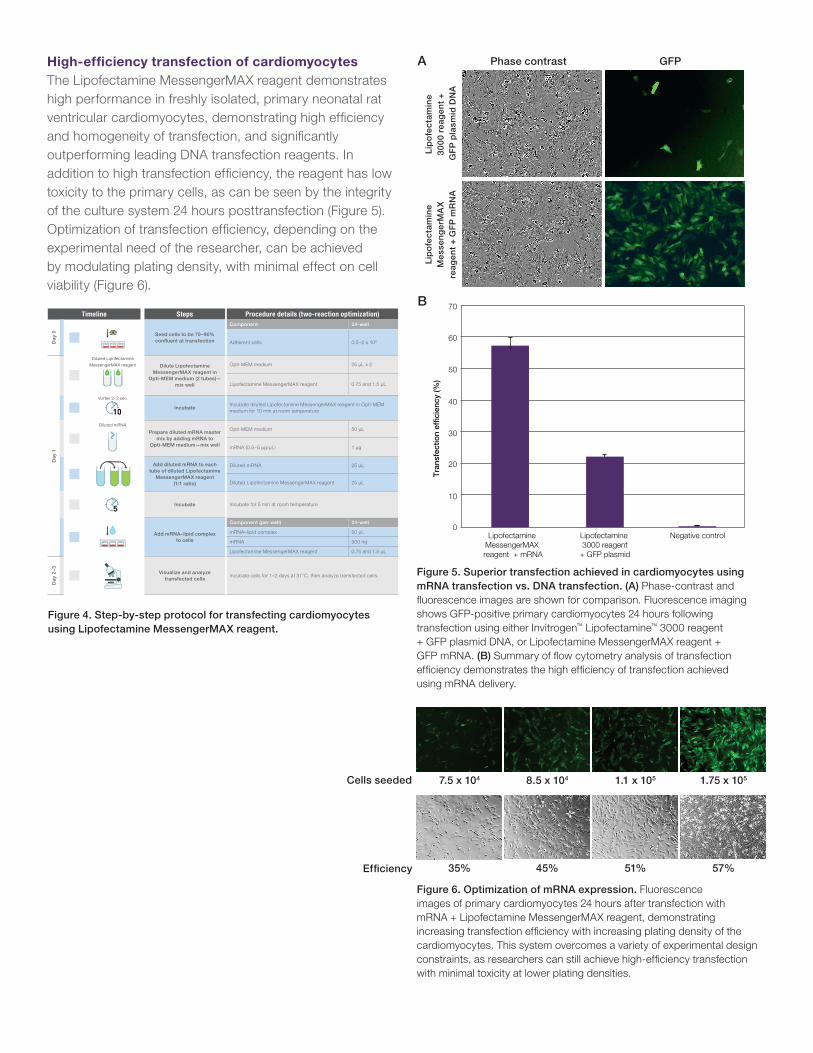
High-effi ciency transfection of cardiomyocytesThe Lipofectamine MessengerMAX reagent demonstrates high performance in freshly isolated, primary neonatal rat ventricular cardiomyocytes, demonstrating high effi ciency and homogeneity of transfection, and signifi cantly outperforming leading DNA transfection reagents. In addition to high transfection effi ciency, the reagent has low toxicity to the primary cells, as can be seen by the integrity of the culture system 24 hours posttransfection (Figure 5). Optimization of transfection effi ciency, depending on the experimental need of the researcher, can be achieved by modulating plating density, with minimal effect on cell viability (Figure 6).
Figure 5. Superior transfection achieved in cardiomyocytes using mRNA transfection vs. DNA transfection. (A) Phase-contrast and fl uorescence images are shown for comparison. Fluorescence imaging shows GFP-positive primary cardiomyocytes 24 hours following transfection using either Invitrogen™ Lipofectamine™ 3000 reagent + GFP plasmid DNA, or Lipofectamine MessengerMAX reagent + GFP mRNA. (B) Summary of fl ow cytometry analysis of transfection effi ciency demonstrates the high effi ciency of transfection achieved using mRNA delivery.
Figure 4. Step-by-step protocol for transfecting cardiomyocytes using Lipofectamine MessengerMAX reagent.
Figure 6. Optimization of mRNA expression. Fluorescence images of primary cardiomyocytes 24 hours after transfection with mRNA + Lipofectamine MessengerMAX reagent, demonstrating increasing transfection effi ciency with increasing plating density of the cardiomyocytes. This system overcomes a variety of experimental design constraints, as researchers can still achieve high-effi ciency transfection with minimal toxicity at lower plating densities.
Phase contrast GFP
Lip
ofe
ctam
ine
30
00
reag
ent
+ G
FP
pla
smid
DN
A
Lip
ofe
ctam
ine
Mes
sen
ger
MA
X
reag
ent
+ G
FP
mR
NA
Timeline Steps
Day
0
1
Seed cells to be 70–90% confl uent at transfection
Day
1
2
Diluted Lipofectamine
MessengerMAX reagent
Dilute Lipofectamine MessengerMAX reagent in
Opti-MEM medium (2 tubes)—mix well
3 10Incubate
4
Diluted mRNA
Prepare diluted mRNA master mix by adding mRNA to
Opti-MEM medium—mix well
5
Add diluted mRNA to each tube of diluted Lipofectamine
MessengerMAX reagent (1:1 ratio)
6
Incubate
7
Add mRNA–lipid complex to cells
Day
2–3
8
Visualize and analyze transfected cells
Procedure details (two-reaction optimization)Component 24-well
Adherent cells 0.5–2 x 10⁵
Opti-MEM medium 25 μL x 2
Lipofectamine MessengerMAX reagent 0.75 and 1.5 μL
Incubate diluted Lipofectamine MessengerMAX reagent in Opti-MEM medium for 10 min at room temperature
Opti-MEM medium 50 μL
mRNA (0.5–5 μg/μL) 1 μg
Diluted mRNA 25 μL
Diluted Lipofectamine MessengerMAX reagent 25 μL
Incubate for 5 min at room temperature
Component (per well) 24-well
mRNA–lipid complex 50 μL
mRNA 500 ng
Lipofectamine MessengerMAX reagent 0.75 and 1.5 μL
Incubate cells for 1–2 days at 37°C, then analyze transfected cells.
Vortex 2–3 sec
A
B
0
10
20
30
40
50
60
Lipofectamine MessengerMAX reagent + mRNA
Lipofectamine 3000 reagent
+ GFP plasmid
Negative control
Tra
nsfe
ctio
n ef
ficie
ncy
(%)
70
Cells seeded 7.5 x 10⁴ 8.5 x 10⁴ 1.1 x 10⁵ 1.75 x 10⁵
35% 45% 51% 57%Effi ciency

See all of our application notes at thermofisher.com/transfectionbasics
Find out more about Lipofectamine MessengerMAX reagent at thermofisher.com/messengermax
For Research Use Only. Not for use in diagnostic procedures. © 2017 Thermo Fisher Scientific Inc. All rights reserved. All trademarks are the property of Thermo Fisher Scientific and its subsidiaries unless otherwise specified. COL31436 0117
Table 1. Comparison of different transfection techniques.
Transfection technique
Expression efficiency
Cost effectiveness
Cell viability
Experimental ease of use
Electroporation ++ + + ++
Adenoviral overexpression +++ ++ +++ +
Leading DNA transfection reagent
+ ++ ++ +++
Lipofectamine MessengerMAX reagent
+++ +++ +++ +++
ConclusionsAs demonstrated here, Lipofectamine MessengerMAX reagent offers a highly effective solution for protein overexpression in difficult-to-transfect primary cell models, providing several benefits including ease of use and high efficiency. This reagent is very gentle on isolated primary cardiomyocytes and differentiated cardiomyocytes, compared to other commonly used transfection methods. While several methodologies exist for transfecting cardiomyocytes, we recommend Lipofectamine MessengerMAX reagent as the primary gene delivery solution.
Additional applications for cardiomyocyte modelsTransfection of human induced pluripotent stem cell (iPSC)-derived cardiomyocytes is an important experimental platform for studying cardiovascular disease. Differentiated iPSCs comprise a very sensitive cell culture system and offer a physiologically relevant model that can be used for translational research. Using mRNA with Lipofectamine MessengerMAX reagent, transfection efficiency greater than >30% was achieved in these cells as seen from the fluorescence images taken 18 hours following transfection with GFP mRNA (Figure 7).
Figure 7. High-efficiency mRNA transfection in iPSC-derived cardiomyocytes using Lipofectamine MessengerMAX reagent.
Trade-offsA comparison of the main transfection techniques that are commonly used for cardiomyocytes highlights the benefits and drawbacks of each system. Electroporation requires physical disruption of the cell, which results in high cell mortality. It also requires specialized instrumentation and corresponding consumables, which can be expensive. Adenoviral overexpression is a highly efficient technique and frequently used for cardiomyocytes; however, there are several drawbacks associated with the use of this methodology. In addition to requiring adherence to specific laboratory guidelines and considerations for handling and use, generating adenoviral constructs is a time-consuming and expensive process. While high expression efficiency is achievable using adenovirus, the ease of use of this method in experimental design is limited to making a separate virus for each individual protein, which can be very time-consuming and high in cost. The practicality of making a library of constructs for mRNA or DNA transfection is more feasible than generating individual virus constructs for each experimental target. Table 1 compares mRNA transfection using Lipofectamine MessengerMAX reagent to the more commonly used transfection techniques that are used for cardiomyocytes.