optimizing gold nanoparticle cluster configurations ( n ≤ 7) for array...
TRANSCRIPT

Published: March 02, 2011
r 2011 American Chemical Society 4578 dx.doi.org/10.1021/jp112146d | J. Phys. Chem. C 2011, 115, 4578–4583
ARTICLE
pubs.acs.org/JPCC
Optimizing Gold Nanoparticle Cluster Configurations (n e 7) forArray ApplicationsBo Yan, Svetlana V. Boriskina, and Bj€orn M. Reinhard*
Department of Chemistry and The Photonics Center, Boston University, Boston, Massachusetts 02215, United States
bS Supporting Information
’ INTRODUCTION
Electromagnetic radiation incident on noble metal nanopar-ticles can excite a coherent collective oscillation of the conduc-tion band electrons. The resonance wavelength of this localizedsurface plasmon (LSP) depends on the shape, size, and thematerial of the particle as well as the refractive index of thesurrounding environment.1,2 Tunable electromagnetic couplingof LSPs in close-by nanoparticles makes individual nanoparticlesversatile building blocks for engineering higher order structureswith entirely new properties. In this regard, plasmonic nano-structures share some similarities with chemical molecules, withthe important difference that “plasmonic molecules” are ruled bythe laws of electromagnetism (except at very short interparticleseparations),3-7 whereas the properties of chemical moleculesare determined by quantum mechanics. As a result of plasmonhybridization3 in nanoparticle clusters, their near- and far-fieldresponses become dependent on the resonance frequency andshape of all constituent nanoparticles, the cluster geometry, andthe interparticle separations. Nanoparticle clusters are interestingelectromagnetic materials, as they can efficiently localize andenhance incident electromagnetic fields in the junctions andcrevices between the particles.8-10 This light field concentrationin the clusters creates nanoscale volumes with high local E-fieldintensity, which act as “hot spots” for surface-enhanced spectro-scopies, such as surface-enhanced Raman spectroscopy(SERS)11 and surface-enhanced infrared absorption spectrosco-py (SEIRA).12
Nanoparticle clusters can be further integrated and used asbuilding blocks for higher order structures, such as two-dimen-sional nanoparticle cluster arrays (NCAs).13 One appealingcharacteristic of NCAs is that they can sustain electromagneticinteractions on multiple length scale. The first length scales isdefined through plasmon hybridization of the particle plasmonswithin individual clusters, while the second length scale is definedthrough interactions between entire clusters. The synergisticinterplay of the electromagnetic interactions on different lengthscales creates a cascade multiscale E-field intensity enhancementwhich qualifies NCAs for challenging sensing applications,including bacterial pathogen detection14 and ultratrace analy-tics.15 A further systematic improvement of NCAs as SERS orSEIRA substrates requires on the one hand a fundamental under-standing of the dependence of the near- and far-field responseson array parameters, such as the array geometry and interclusterseparation, and on the other hand an optimization of the opticalresponses of the individual building blocks. We focus in thiswork on the latter and investigate the tunability of the near- andfar-field responses of self-assembled clusters as a function ofcluster size (ne 7) and configuration by combining single clusterdark field spectroscopy and multiparticle generalized Mie theory(GMT) algorithms.16
Received: December 21, 2010Revised: February 1, 2011
ABSTRACT: Nanoparticle cluster arrays (NCAs) are novelelectromagnetic materials whose properties depend on the sizeand shape of the constituent nanoparticle clusters. A rationaldesign of NCAs with defined optical properties requires athorough understanding of the geometry-dependent opticalresponse of the building blocks. Herein, we systematicallyinvestigate the near- and far-field responses of clusters of closelypacked 60 nm gold nanoparticles (n e 7) as a function of sizeand cluster geometry through a combination of experimentalspectroscopy and generalized Mie theory calculations. From all of the investigated cluster configurations, nanoparticle trimers withD3h geometry and heptamers in D6h geometry stand out due to their polarization-insensitive responses and high electric (E) fieldintensity enhancements, making them building blocks of choice in this size range. The near-field intensity maximum of the D6h
heptamer is red-shifted with regard to the D3h trimer by 125 nm, which confirms the possibility of a rational tuning of the near-fieldresponse in NCAs through the choice of the constituent nanoparticle clusters. For the nanoparticle trimer we investigate theinfluence of the cluster geometry on the optical response in detail andmap near- and far-field spectra associated with the transition ofthe cluster configuration from D3h into D¥h.

4579 dx.doi.org/10.1021/jp112146d |J. Phys. Chem. C 2011, 115, 4578–4583
The Journal of Physical Chemistry C ARTICLE
We used a template-assisted self-assembly approach (Figure 1)to generate clusters of 60 nm diameter gold spheres at predefinedlocations. In previous studies, we have found that the self-assemblyprocess yields densely packed clusters on the predefined bindingsites if a 180 nm thick photoresist is used but that the bindingefficiency deteriorates with decreasing photoresist layer thickness.This offers the opportunity to generate a broad range of clusterswith different sizes and configurations on one chip. In this work, weused a photoresist layer of ∼120 nm to generate clusters atpredefined cluster locations to enable a systematic investigationof structure-spectrum relationship of the clusters. To avoidany interactions between the individual clusters, the separationbetween the individual binding sites was chosen to beg5 μm. Theregular arrays generated in the template guided self-assembleprocess represent defined geometric patterns, which enable anunambiguous identification of individual clusters in a scanningelectron microscope (SEM). This approach makes it possible tocorrelate the experimental far-field spectra of individual clusterswith their geometry as obtained through SEM.
’EXPERIMENTAL METHODS
Generating Nanoparticle Clusters. Nanoparticle clusterswere formed on indium tin oxide (ITO)-coated glass througha combination of e-beam lithography and template-guidedself-assembly.13,14 Briefly, poly(methyl methacrylate) (PMMA)resist was spin-coated on an ITO-glass chip to form a layer with afinal thickness of ∼120 nm. Then binding sites with variablediameters between 80-200 nm were generated in the PMMAlayer with electron beam lithography using a Zeiss Supra 40 VPSEM equipped with an e-beam blanker and through subsequentdevelopment with methyl isobutyl ketone/isopropanol solventmix (1:3 in volume). The patterned substrates were then incu-bated with 2 mg/mL polylysine (MW = 15K-30K) solution for1 h. After blow-drying, the substrates were incubated overnightwith HSC11H22(OC2H4)6OCH2COOH functionalized 60 nmgold nanoparticles in a 10 mM phosphate buffer (pH = 8.6)containing 20 mM NaCl. The PMMA layer was finally removedthrough a liftoff in 1-methyl-2 pyrolidinone (NMP) solventfor 5 min.
Correlation between Dark Field Scattering Spectra andSEM Images. All scattering spectra of nanoparticle clusters wereacquired with an Olympus BX51WI microscope using a 60� oilimmersion objective (N.A 0.65) under dark field illumination.The substrates were sandwiched between two cover slides andimmersed in glycerol with refractive index of nr = 1.474. The sampleswere illuminated with a 100W tungsten lampwhich was focused onthe sample plane using an oil dark field condenser (N.A 1.2-1.4,corresponding to an incidence angle θ between 54� and 72�).Scattered light from nanoparticle clusters was collected andanalyzed with an Andor Shamrock 303 mm focal length spectro-meter using a 150 lines/mm grating blazed at 500 nm. Thespectra were recorded with an Andor CCD camera (DU401-BR-DD), then background corrected, and finally corrected forthe excitation profile of the tungsten lamp by dividingthrough the spectrum of an ideal white light scatterer. Forpolarization resolved measurements an analyzer was insertedinto the beam path and stepwisely rotated. After spectral cha-racterization the index matching glycerol was removed through
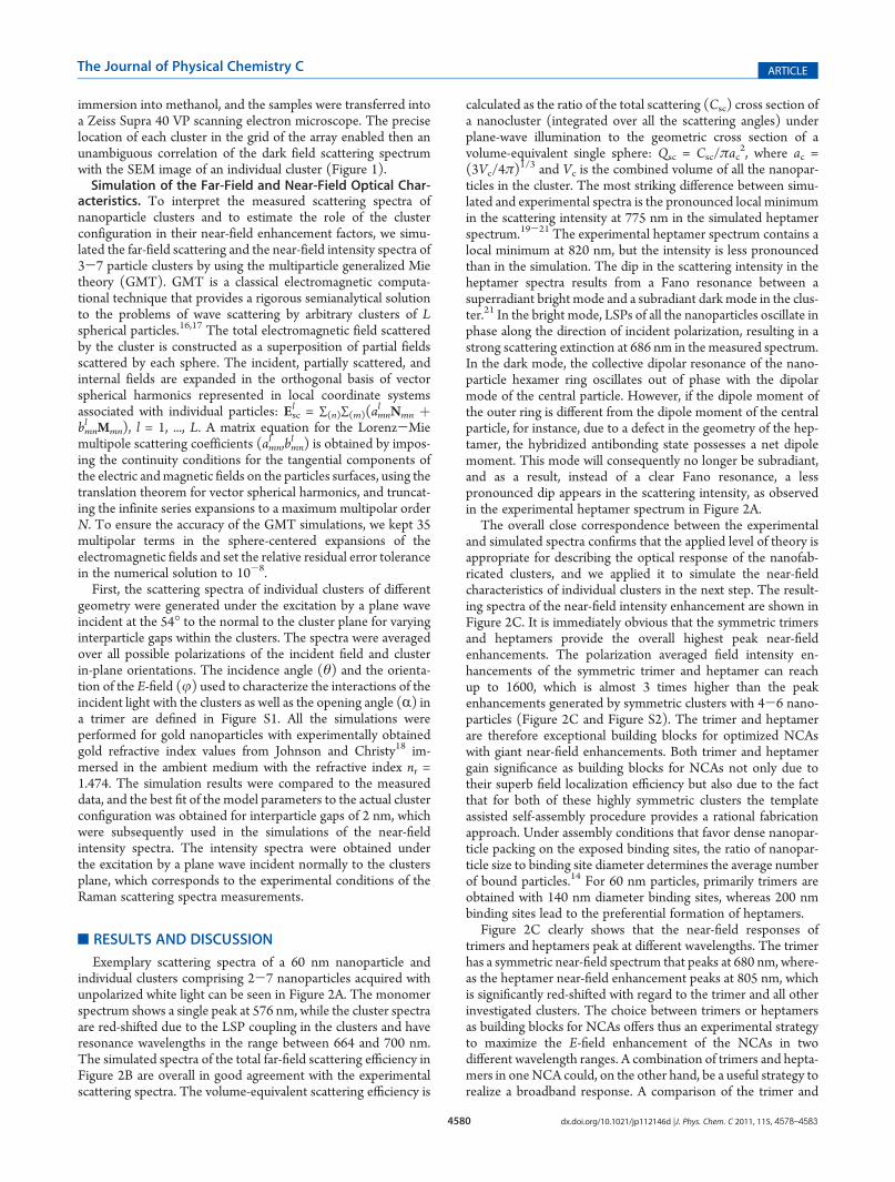
Figure 2. (A) Experimental dark field scattering spectra of nanoparticleclusters with n = 1-7 (see SEM images). (B) Simulated scatteringspectra for these clusters configurations. (C) Calculated E-field intensityenhancement. The incidence angle θwas set to 54� in (B) and 0� in (C).Scale bars in all SEM images are 50 nm.
Figure 1. (A-D) Process flow for generating nanoparticle clusters atpredefined locations on an ITO-glass chip. (E) Dark field image of acluster pattern. The individual clusters are spatially well separated andallow recording of single cluster spectra (inset). (F) SEM image of thecluster pattern. Structural details of the individual clusters becomeapparent at higher magnification (inset). Scale bars in (E) and (F)denote 10 μm.
4580 dx.doi.org/10.1021/jp112146d |J. Phys. Chem. C 2011, 115, 4578–4583
The Journal of Physical Chemistry C ARTICLE
immersion into methanol, and the samples were transferred intoa Zeiss Supra 40 VP scanning electron microscope. The preciselocation of each cluster in the grid of the array enabled then anunambiguous correlation of the dark field scattering spectrumwith the SEM image of an individual cluster (Figure 1).Simulation of the Far-Field and Near-Field Optical Char-
acteristics. To interpret the measured scattering spectra ofnanoparticle clusters and to estimate the role of the clusterconfiguration in their near-field enhancement factors, we simu-lated the far-field scattering and the near-field intensity spectra of3-7 particle clusters by using the multiparticle generalized Mietheory (GMT). GMT is a classical electromagnetic computa-tional technique that provides a rigorous semianalytical solutionto the problems of wave scattering by arbitrary clusters of Lspherical particles.16,17 The total electromagnetic field scatteredby the cluster is constructed as a superposition of partial fieldsscattered by each sphere. The incident, partially scattered, andinternal fields are expanded in the orthogonal basis of vectorspherical harmonics represented in local coordinate systemsassociated with individual particles: Esc
l = ∑(n)∑(m)(amnl Nmn þ
bmnl Mmn), l = 1, ..., L. A matrix equation for the Lorenz-Miemultipole scattering coefficients (amn
l ,bmnl ) is obtained by impos-
ing the continuity conditions for the tangential components ofthe electric andmagnetic fields on the particles surfaces, using thetranslation theorem for vector spherical harmonics, and truncat-ing the infinite series expansions to a maximum multipolar orderN. To ensure the accuracy of the GMT simulations, we kept 35multipolar terms in the sphere-centered expansions of theelectromagnetic fields and set the relative residual error tolerancein the numerical solution to 10-8.First, the scattering spectra of individual clusters of different
geometry were generated under the excitation by a plane waveincident at the 54� to the normal to the cluster plane for varyinginterparticle gaps within the clusters. The spectra were averagedover all possible polarizations of the incident field and clusterin-plane orientations. The incidence angle (θ) and the orienta-tion of the E-field (j) used to characterize the interactions of theincident light with the clusters as well as the opening angle (R) ina trimer are defined in Figure S1. All the simulations wereperformed for gold nanoparticles with experimentally obtainedgold refractive index values from Johnson and Christy18 im-mersed in the ambient medium with the refractive index nr =1.474. The simulation results were compared to the measureddata, and the best fit of the model parameters to the actual clusterconfiguration was obtained for interparticle gaps of 2 nm, whichwere subsequently used in the simulations of the near-fieldintensity spectra. The intensity spectra were obtained underthe excitation by a plane wave incident normally to the clustersplane, which corresponds to the experimental conditions of theRaman scattering spectra measurements.
’RESULTS AND DISCUSSION
Exemplary scattering spectra of a 60 nm nanoparticle andindividual clusters comprising 2-7 nanoparticles acquired withunpolarized white light can be seen in Figure 2A. The monomerspectrum shows a single peak at 576 nm, while the cluster spectraare red-shifted due to the LSP coupling in the clusters and haveresonance wavelengths in the range between 664 and 700 nm.The simulated spectra of the total far-field scattering efficiency inFigure 2B are overall in good agreement with the experimentalscattering spectra. The volume-equivalent scattering efficiency is
calculated as the ratio of the total scattering (Csc) cross section ofa nanocluster (integrated over all the scattering angles) underplane-wave illumination to the geometric cross section of avolume-equivalent single sphere: Qsc = Csc/πac
2, where ac =(3Vc/4π)
1/3 and Vc is the combined volume of all the nanopar-ticles in the cluster. The most striking difference between simu-lated and experimental spectra is the pronounced local minimumin the scattering intensity at 775 nm in the simulated heptamerspectrum.19-21 The experimental heptamer spectrum contains alocal minimum at 820 nm, but the intensity is less pronouncedthan in the simulation. The dip in the scattering intensity in theheptamer spectra results from a Fano resonance between asuperradiant bright mode and a subradiant dark mode in the clus-ter.21 In the bright mode, LSPs of all the nanoparticles oscillate inphase along the direction of incident polarization, resulting in astrong scattering extinction at 686 nm in the measured spectrum.In the dark mode, the collective dipolar resonance of the nano-particle hexamer ring oscillates out of phase with the dipolarmode of the central particle. However, if the dipole moment ofthe outer ring is different from the dipole moment of the centralparticle, for instance, due to a defect in the geometry of the hep-tamer, the hybridized antibonding state possesses a net dipolemoment. This mode will consequently no longer be subradiant,and as a result, instead of a clear Fano resonance, a lesspronounced dip appears in the scattering intensity, as observedin the experimental heptamer spectrum in Figure 2A.
The overall close correspondence between the experimentaland simulated spectra confirms that the applied level of theory isappropriate for describing the optical response of the nanofab-ricated clusters, and we applied it to simulate the near-fieldcharacteristics of individual clusters in the next step. The result-ing spectra of the near-field intensity enhancement are shown inFigure 2C. It is immediately obvious that the symmetric trimersand heptamers provide the overall highest peak near-fieldenhancements. The polarization averaged field intensity en-hancements of the symmetric trimer and heptamer can reachup to 1600, which is almost 3 times higher than the peakenhancements generated by symmetric clusters with 4-6 nano-particles (Figure 2C and Figure S2). The trimer and heptamerare therefore exceptional building blocks for optimized NCAswith giant near-field enhancements. Both trimer and heptamergain significance as building blocks for NCAs not only due totheir superb field localization efficiency but also due to the factthat for both of these highly symmetric clusters the templateassisted self-assembly procedure provides a rational fabricationapproach. Under assembly conditions that favor dense nanopar-ticle packing on the exposed binding sites, the ratio of nanopar-ticle size to binding site diameter determines the average numberof bound particles.14 For 60 nm particles, primarily trimers areobtained with 140 nm diameter binding sites, whereas 200 nmbinding sites lead to the preferential formation of heptamers.
Figure 2C clearly shows that the near-field responses oftrimers and heptamers peak at different wavelengths. The trimerhas a symmetric near-field spectrum that peaks at 680 nm, where-as the heptamer near-field enhancement peaks at 805 nm, whichis significantly red-shifted with regard to the trimer and all otherinvestigated clusters. The choice between trimers or heptamersas building blocks for NCAs offers thus an experimental strategyto maximize the E-field enhancement of the NCAs in twodifferent wavelength ranges. A combination of trimers and hepta-mers in one NCA could, on the other hand, be a useful strategy torealize a broadband response. A comparison of the trimer and

4581 dx.doi.org/10.1021/jp112146d |J. Phys. Chem. C 2011, 115, 4578–4583
The Journal of Physical Chemistry C ARTICLE
heptamer near-field spectra with the other investigated clustersunderlines the strong dependence of the optical response on thecluster geometry in this size range. The D3h trimer and D6h
heptamer clusters are clearly outstanding and represent “magic”configurations that enable eximious cluster properties.
In the next step we want to investigate in more detail how theproperties of “magic” nanoparticle cluster vary when its geometryis continuously changed. For reason of simplicity, we choose forthis purpose the nanoparticle trimer. In addition to themagicD3h
(equilateral triangle) configuration, C2v (kinked triangle) andD¥h (rod) configurations exist for this cluster size. The majordifference between these clusters is the value of the angle (R)formed by the three nanoparticles. In Figure 3 we follow thespectral change that is associated with a continuous increase of Rin a nanoparticle trimer. The opening of the angle converts thecluster configuration from theD3h point group over theC2v to theD¥h point group. We show the SEM images and the correspond-ing far-field scattering spectra for trimers withR = 60�, 70�, 110�,and 180�.
The experimental spectrum of the D3h trimer shows a narrowresonance, which peaks at 665 nm. This band starts to split whenthe symmetry of the cluster is reduced to C2v (R = 70�). Thiscluster shows a main peak at 673 nm and a shoulder at 637 nm.
AsR is further increased to 110�, the resonance splitting becomesmore apparent and two separate features are now clearly distin-guishable: a blue-shifted resonance at 621 nm and a red-shiftedresonance at 698 nm. If R is further increased to 180� to form aD¥h trimer, the spectrum becomes highly symmetric againcontaining a prominent resonance at 712 nm. The simulatedtrimer scattering spectra reproduce the experimental trend(Figure 3B). A symmetric D3h trimer shows a main feature at676 nm. As R increases to 70�, several resonant features emergein the cluster spectrum, with resonances at 556, 610, 668, and713 nm. Further mode splitting can be observed as the angleopens to 110�, with three resonant features at 566, 650, and742 nm in the spectrum.When the three particles align in a chain,only two peaks are visible in the cluster spectrum, at 560 and760 nm. There is a systematic red shift of the peak positions be-tween our simulated and experimental data, which could be dueto the variations in the gaps between nanoparticles and estimationon the refractive index of surrounding environment. Never-theless, the performed GMT calculations overall clearly confirmthe geometry dependent spectral features in the trimer.
To better understand the peak splitting trend resulted fromclusters’ symmetry breaking, we trace the evolution of the mainspectral feature of the trimer with the change of its configurationin Figure 4A. To get a clearer picture of the hybridized modessymmetry evolution, we set the incidence angle to θ = 0� and thepolarization of the E-field along the x-axis (j = 90�). When theopening angle of the trimer (R) changes, the 682 nm feature
Figure 4. (A) Simulated scattering spectra of a monomer and ofindividual trimers with opening angle R = 60�, 70�, 110�, and 180�under illumination by a linearly polarized light incident normally (θ = 0�) tothe cluster plane. Inset shows a typical dipole mode of a monomer for thesame E-field polarization. (B1-B4) Phase maps of the major fieldcomponent Ex of the four trimers at 682, 715, 737, and 755 nm, whichcorrespond to the feature resonances in (A).
Figure 3. (A) Experimental and (B) simulated scattering spectra offour individual trimers with opening angle R = 60�, 70�, 110�, and 180�(see insets). Scale bar denotes 50 nm in all SEM images.

4582 dx.doi.org/10.1021/jp112146d |J. Phys. Chem. C 2011, 115, 4578–4583
The Journal of Physical Chemistry C ARTICLE
corresponding to a hybridized LSP state in the R = 60� trimer,which is degenerate in frequency owing to the cluster symmetry,splits into several modes. The low-energy resonant featurecontinuously red-shifts with increasing R to 715 nm (R = 70�)and 737 nm (R = 110�) to 755 nm (R = 180�). The resonantpeaks corresponding to the split modes on the high energy sidebecome progressively less pronounced and eventually disappearwhen R is increased. To find out the underlying electromagneticinteractions of the hybridized modes in the different clusterconfigurations, we plot the phase maps of the major field com-ponent Ex in the four relevant trimers at the peak wavelengths oftheir low-energy resonances in Figure 4 (B1-B4). The phase of acomplex field component Ex = Ex0 þ iEx00 = |Ex| exp{arg(Ex)} iscalculated as arg(Ex) = arcsin(Ex00/|Ex|) and varies from-π to π.These maps reveal the continuous evolution of the hybridizedLSP mode of the trimer from that of a coupled dipole mode of adimer perturbed by a third monomer in D3h (R = 60�) to alongitudinal mode of the D¥h (R = 180�) linear particle chain,which is consistent with the prediction of the plasmon hybridiza-tion theory.22 We note that, besides the prominent peaks discus-sed thus far, all experimental spectra show another high-energyresonance at around 525 nm with low intensity. We trace theevolution of the hybridized LSP mode that gives rise to this high-energy resonant feature with contribution from in plane and outof plane transverse modes in Figures S3 and S4.
We also analyzed the effect of the excitation field polarizationon the spectral response of different clusters. In Figure 5 weshow experimental (A1-A3) and simulated scattering spectra(B1-B3) of trimers for varying angles of the incident lightpolarization (j), together with the spatial E-field intensity enhance-ment distributions calculated for two orthogonal polarization
angles (C1-C3) and different trimer geometries. The D3h
configuration exhibits the weakest polarization dependence inthe far- and nearfield responses as well as the highest averageE-field intensity enhancement of 1600. This makes the D3h
trimer a good building block for NCAs since it enables efficientelectromagnetic coupling between nanoparticle clusters along alldirections: the clusters do not require a specific alignment.Another cluster configuration that can be reliably obtained withthe same fabrication procedure—the D¥h trimer—provides theintensity enhancement spectrum that is strongly polarizationdependent. In this configuration, the E-field intensity enhance-ment reaches a peak value of 1700 if the excitation polarization isparallel to the¥-fold symmetry axis; however, for excitation lightwith the perpendicular polarization, the enhancement drops to∼20. The lower average enhancement generated under unpolar-ized (or unaligned) conditions makes the D¥h trimer a lessappealing building block for NCAs.
For some applications it might, however, be desirable to havea SERS substrate that can either sustain multiple modes withdistinct resonance peaks23 or enables switching the E-field maxi-mum between predefined wavelengths on one chip.24 The calcu-lated near-field spectrum of the C2v (shown in Figure 5) config-uration shows that this cluster is an interesting candidate forthese applications since it contains two separate resonances, at650 and 740 nm, which provide peak enhancements factors of760 and 1550, respectively. The E-field intensity distribution inFigure 5 (C3) shows that the two LSP modes are spatially colo-calized but are selectively excited at two different wavelengths bythe incident light of two orthogonal polarizations. An array ofaligned C2v trimers would offer the possibility to switch betweenthe two resonances through rotation of the excitation polarization
Figure 5. (A1-A3) Scattering spectra, (B1-B3) calculated E-field intensity enhancement spectra (incidence angle θ = 0�), and (C1-C3) spatialdistributions of the E-field intensity for typical D3h, D¥h, and C2v trimers for different excitation polarization angles.

4583 dx.doi.org/10.1021/jp112146d |J. Phys. Chem. C 2011, 115, 4578–4583
The Journal of Physical Chemistry C ARTICLE
and thus to modulate the SERS substrate sensitivity for differentvibrational bands in the fingerprint region (500-2000 cm-1) onone chip.24,25 Similar double resonances are seen in the scatteringspectra of asymmetric tetramers (Figures S5 and S6), which arealso potential candidates for generating multiresonance substra-tes if their optical responses are systematically studied in thefuture.
’CONCLUSIONS
Individual nanoparticle clusters (n e 7) were constructedfrom 60 nm gold nanoparticles using a template guided self-assembly procedure. We measured the dark field scatteringspectra of these clusters and correlated the optical spectra withthe cluster structures as obtained by SEM. These experimentalstudies were augmented by generalized Mie theory simulations,which provided information about the E-field intensity spectrumas a function of cluster configuration. The performed studiesshow that trimers with D3h and heptamers in D6h geometry aregood building blocks for NCAs due to their high rotationalsymmetry and their outstanding E-field intensity enhancement,which is significantly higher than that for all other investigatedcluster configurations. The D3h trimer and D6h heptamer repre-sent “magic” cluster configurations in the investigated size range.The E-field intensity maxima for the D3h trimer and D6h hepta-mer are of similar magnitude but shifted by ∼125 nm. Thedemonstrated strong size and geometry dependence of the near-and far-field responses of nanoparticle clusters provides newopportunities for a rational design of NCAs with defined near-and far-field responses through choice of the geometry of thebuilding blocks.
’ASSOCIATED CONTENT
bS Supporting Information. Figures S1-S6. This materialis available free of charge via the Internet at http://pubs.acs.org.
’AUTHOR INFORMATION
Corresponding Author*E-mail: [email protected].
’ACKNOWLEDGMENT
The work was partially supported by the National Institutes ofHealth through grant 5R01CA138509-02 and the National ScienceFoundation through grants CBET-0853798 and CBET-0953121.
’REFERENCES
(1) Kelly, K. L.; Coronado, E.; Zhao, L. L.; Schatz, G. C. J. Phys.Chem. B 2003, 107, 668–677.(2) Link, S.; El-Sayed, M. A. Annu. Rev. Phys. Chem. 2003,
54, 331–336.(3) Prodan, E.; Radloff, C.; Halas, N. J.; Nordlander, P. A. Science
2003, 302, 419–422.(4) Lassiter, J. B.; Aizpurua, J.; Hernandez, L. I.; Brandl, D. W.;
Romero, I.; Lal, S.; Hafner, J. H.; Nordlander, P.; Halas, N. J. Nano Lett.2008, 8, 1212–1218.(5) Kinnan, M. K.; Chumanov, G. J. Phys. Chem. C 2010,
114, 7496–7501.(6) Encina, E. R.; Coronado, E. A. J. Phys. Chem. C 2010,
114, 16278–16284.(7) Yang, L.; Wang, H.; Yan, B.; Reinhard, B. J. Phys. Chem. C 2010,
114, 4901–4908.
(8) Lal, S.; Link, S.; Halas, N. J. Nature Photonics 2007, 1, 641–648.(9) Yu, Q.; Guan, P.; Qin, D.; Golden, G.; Wallace, P. M. Nano Lett.
2008, 8, 1923–1928.(10) Chang, W.-S.; Slaughter, L. S.; Khanal, B. P.; Manna, P.;
Zubarev, E. R.; Link, S. Nano Lett. 2009, 9, 1152–1157.(11) Jeanmaire, D. L.; Van Duyne, R. P. J. Electroanal. Chem. 1977,
84, 1–20.(12) Hartstein, A.; Kirtley, J. R.; Tsang, J. C. Phys. Rev. Lett. 1980,
45, 201–204.(13) Yan, B.; Thubagere, A.; Premasiri, R.; Ziegler, L. D.; Dal Negro,
L.; Reinhard, B. M. ACS Nano 2009, 3, 1190–1202.(14) Yang, L.; Yan, B.; Premasiri, R.; Ziegler, L. D.; Dal Negro, L.;
Reinhard, B. M. Adv. Funct. Mater. 2010, 20, 2619–2628.(15) Wang, J.; Yang, L.; Boriskina, S.; Yan, B.; Reinhard, B. Spectro-
scopic Ultra-Trace Detection of Nitroaromatic Gas Vapor on RationallyDesigned Two-Dimensional Nanoparticle Cluster Arrays. Anal. Chem.,published ASAP; DOI: 10.1021/ac103123r.
(16) Xu, Y. L. Appl. Opt. 1995, 34, 4573–4588.(17) Gopinath, A.; Boriskina, S. V.; Premasiri, W. R.; Ziegler, L.;
Reinhard, B. M.; Dal Negro, L. Nano Lett. 2009, 9, 3922–3929.(18) Johnson, P. B.; Christy, R. W. Phys. Rev. B 1972, 6, 4370.(19) Fan, J. A.; Wu, C.; Bao, K.; Bao, J.; Bardhan, R.; Halas, N. J.;
Manoharan, V. N.; Nordlander, P.; Shvets, G.; Capasso, F. Science 2010,328, 1135–1138.
(20) Bao, K.; Mirin, N. A.; Nordlander, P. Appl. Phys. A: Mater. Sci.Process. 2010, 100, 333–339.
(21) Luk’yanchuk, B.; Zheludev, N. I.; Maier, S. A.; Halas, N. J.;Nordlander, P.; Giessen, H.; Chong, C. T. Nature Mater. 2010,9, 707–715.
(22) Brandl, D. W.; Mirin, N. A.; Nordlander, P. J. Phys. Chem. B2006, 110, 12302–12310.
(23) Chu, Y.; Banaee, M. G.; Crozier, K. B. ACS Nano 2010,4, 2804–2810.
(24) Lim, J. K.; Joo, S.-W. Surf. Interface Anal. 2007, 39, 684–690.(25) Li, K.; Clime, L.; Tay, L.; Cui, B.; Geissler, M.; Veres, T. Anal.
Chem. 2008, 80, 4945–4950.