optimizing margin isolation santine e. anderson, … margin isolation santine e. anderson, d.d.s....
TRANSCRIPT
www.dentaladvisor.com
C L I N I C A L C A S E R E P O R T
Optimizing Margin IsolationSantine E. Anderson, D.D.S.
Enspire Dental, Ann Arbor, Michigan
Introduction Digital dentistry is exploding. From digital radiography to digital impressing, we have seen a dramatic shift in the way we capture images. We are using digital technology to capture and visualize data on a much larger scale. Suddenly we have the opportunity to identify and critique the small details that were often lost using traditional final impressions. What has not changed, however, is the essential necessity of maintaining the basic fundamentals of crown and bridge dentistry - comprehensive treatment planning, proper preparation design and soft tissue management along with proper margin placement and identification. As technology evolves, we see that it has become even more critical that we have visual access and identification of the margins prior to scanning. With the traditional impression technique, the injection of a low viscosity material into the sulcus may physically force some of the soft tissue out of the way. Digital scanners can only capture what they see.
For years, retraction of soft tissue has meant packing a cord into the sulcus before taking the final impression. This is often a daunting task as we battle inflamed bleeding gingival tissue. In areas where access is difficult, proper isolation becomes more of a challenge. This report will offer alternative methods for effective ways to optimize clarity and manage soft tissue when using digital impressing.
The most effective way to achieve clear visualization of the margin is to prepare the tooth using supragingival margins. The reality is that teeth that are being prepared for a crown often have old restorations with subgingival margins. The health of tissue in these areas is often compromised. In most cases, there is at least a portion of the prepared tooth that falls below the free gingival margin. When subgingival margins are present, the tissue must be separated from the edge of the preparation to allow clear visualization for the digital scan. Unclear margins may be undetected in traditional impressions; however, with digital scanning the quality of retractions and margin clarity can be identified immediately in a single scan. The clarity of a scan is determined by the quality of retraction and its ability to reveal a clear margin.
In areas of inflammation or subgingival margins, the use of a soft tissue laser may be extremely effective. A soft tissue diode laser such as Odyssey Navigator (Ivoclar Vivadent) provides a simple method to trough the marginal areas. It is an invaluable tool to assist in the removal of unhealthy, inflamed gingival tissue that is covering the margin or creating excessive bleeding. It then stimulates the cells to regrow in a healthier environment. A peroxide solution can be used to remove tissue debris left after using the laser.
EDITORSJohn W. Farah, D.D.S., Ph.D.John M. Powers, Ph.D.
EDITORIAL BOARDJohn A. Molinari, Ph.D. Peter Yaman, D.D.S., M.S. William A. Gregory, D.D.S., M.S. Santine Anderson, D.D.S. Sabiha Bunek, D.D.S. Lori Brown, D.D.S. Alexandra Jacquery, D.D.S., M.S. Brent Kolb, D.D.S. Nizar Mansour, D.D.S. Charles I. McLaren, D.D.S., M.S. Kathy L. O’Keefe, D.D.S., M.S.
Thomas Poirier, D.D.S. William T. Stevenson, D.D.S. Robert J. Stevenson, D.D.S. Victoria Thompson, D.D.S. David Traynor, D.D.S. Gytis Udrys, D.D.S.
EXECUTIVE TEAMJackie Farah, M.A.Ed. Craig MacfarlaneAnnette M. Frederick Heidi L. GraberJennifer KalaszPari Karani, M.S. Craig MacfarlaneTricia G. Price
Nelson Williams, M.S. Mary E. Yakas, B.A., CMC
DIRECTOR OF RESEARCHRon Yapp, M.S.
THE DENTAL ADVISOR3110 West LibertyAnn Arbor, MI 48103Toll free: 800.347.1330E-mail: [email protected] Site: www.dentaladvisor.comCopyright ©2010, Dental Consultants, Inc. All rights reserved. Printed in the U.S.A. (ISSN 0748-4666)
NUMBER 21 • APRIL 2011
“Improving Patient Care Through Research & Education”
www.dentaladvisor.com2
W W W . D E N T A L A D V I S O R . C O M
www.dentaladvisor.com3
C L I N I C A L C A S E R E P O R T
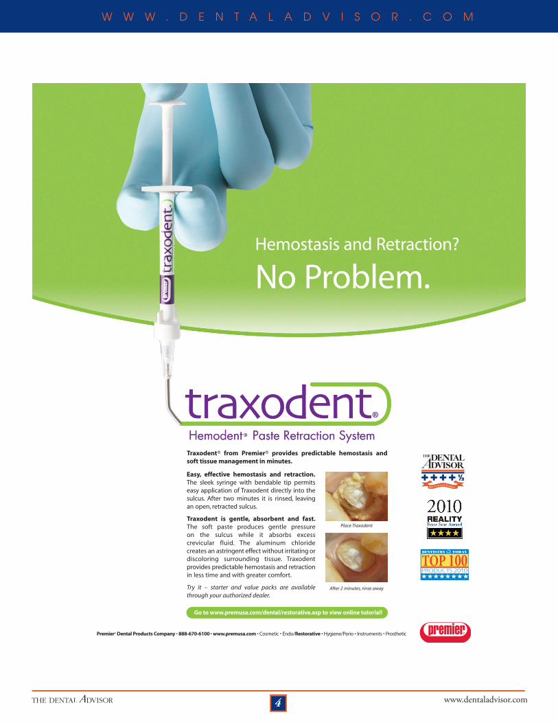
In areas of moderate to minor inflammation, a retraction paste such as the Traxodent Hemodent Paste Retraction System (Premier Dental) is very effective in providing hemostasis and physical separation of the tissue from the margin of the preparation. Traxodent Hemodent Paste Retraction System (Premier Dental) is a viscous paste that is injected into the sulcus and a specially designed, firm cotton roll is placed over the paste. The patient applies firm biting pressure to the cotton for 2 to 5 minutes and the paste is easily rinsed from the tissue and tooth structure. A clear view of the margin is readily achieved.
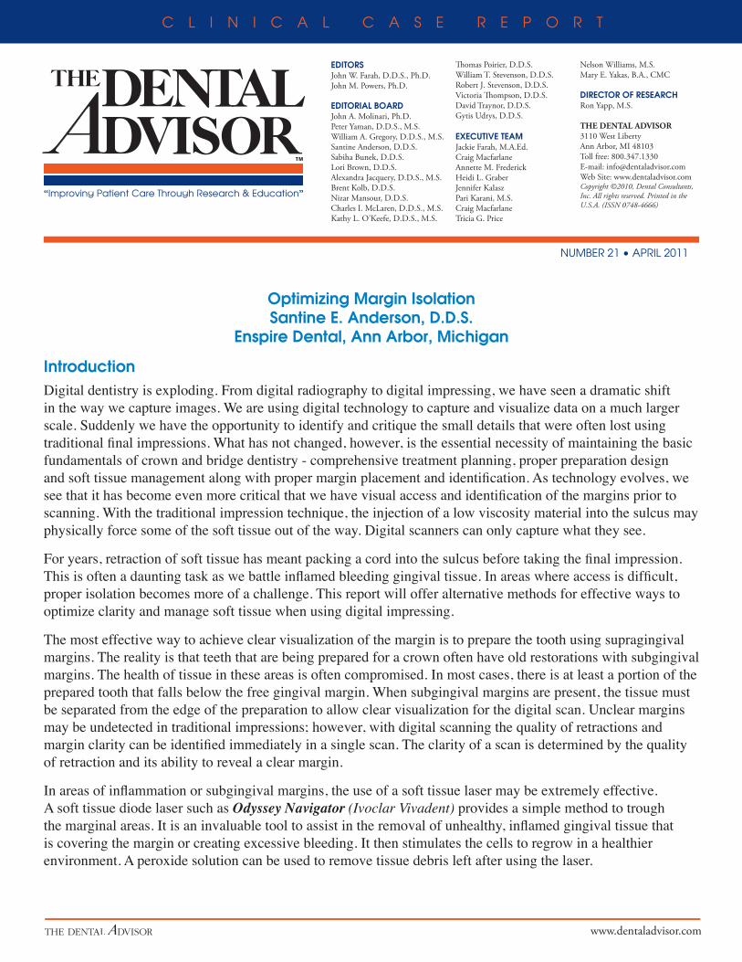
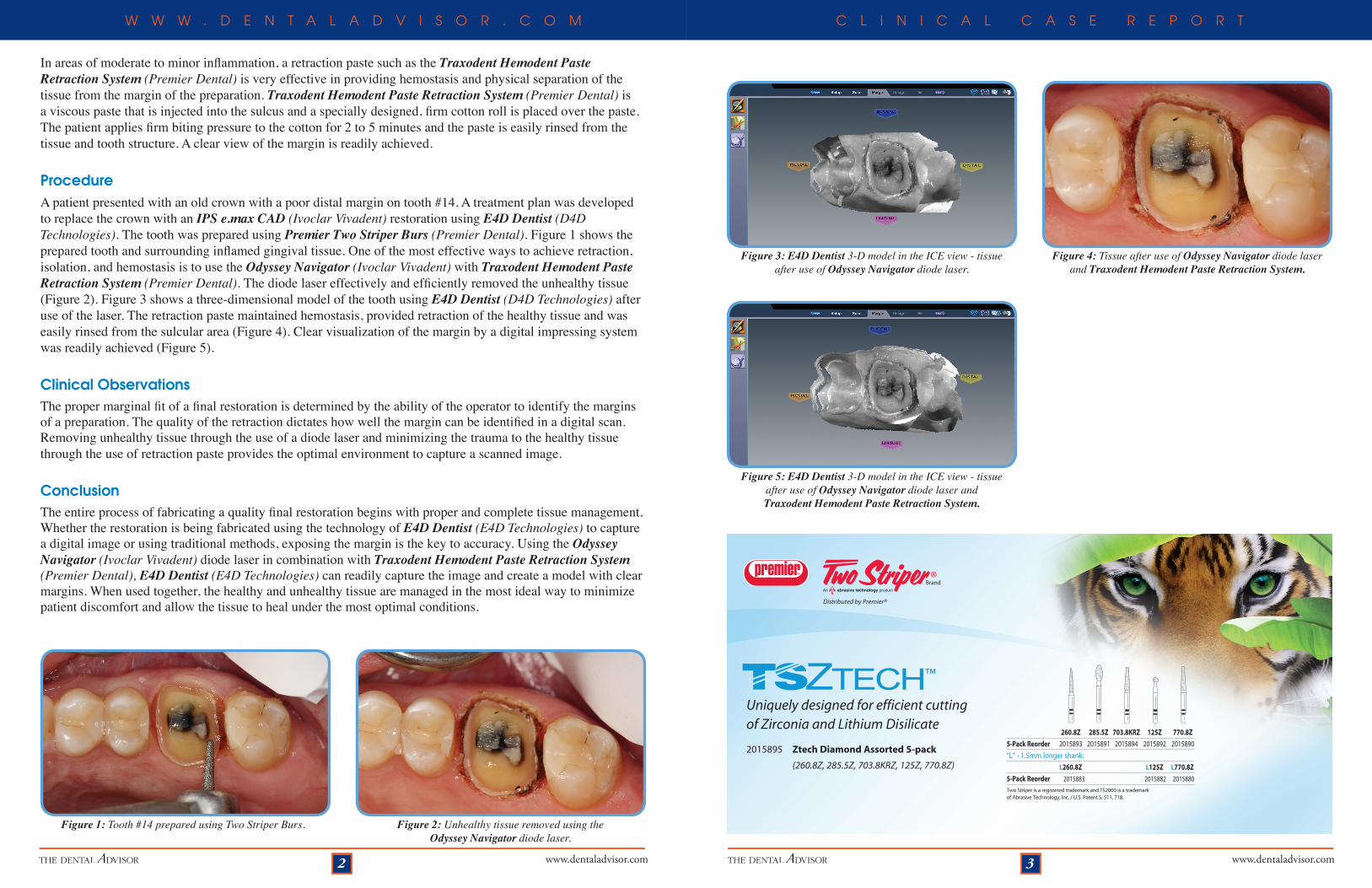
ProcedureA patient presented with an old crown with a poor distal margin on tooth #14. A treatment plan was developed to replace the crown with an IPS e.max CAD (Ivoclar Vivadent) restoration using E4D Dentist (D4D Technologies). The tooth was prepared using Premier Two Striper Burs (Premier Dental). Figure 1 shows the prepared tooth and surrounding inflamed gingival tissue. One of the most effective ways to achieve retraction, isolation, and hemostasis is to use the Odyssey Navigator (Ivoclar Vivadent) with Traxodent Hemodent Paste Retraction System (Premier Dental). The diode laser effectively and efficiently removed the unhealthy tissue (Figure 2). Figure 3 shows a three-dimensional model of the tooth using E4D Dentist (D4D Technologies) after use of the laser. The retraction paste maintained hemostasis, provided retraction of the healthy tissue and was easily rinsed from the sulcular area (Figure 4). Clear visualization of the margin by a digital impressing system was readily achieved (Figure 5).
Clinical Observations The proper marginal fit of a final restoration is determined by the ability of the operator to identify the margins of a preparation. The quality of the retraction dictates how well the margin can be identified in a digital scan. Removing unhealthy tissue through the use of a diode laser and minimizing the trauma to the healthy tissue through the use of retraction paste provides the optimal environment to capture a scanned image.
Conclusion The entire process of fabricating a quality final restoration begins with proper and complete tissue management. Whether the restoration is being fabricated using the technology of E4D Dentist (E4D Technologies) to capture a digital image or using traditional methods, exposing the margin is the key to accuracy. Using the Odyssey Navigator (Ivoclar Vivadent) diode laser in combination with Traxodent Hemodent Paste Retraction System (Premier Dental), E4D Dentist (E4D Technologies) can readily capture the image and create a model with clear margins. When used together, the healthy and unhealthy tissue are managed in the most ideal way to minimize patient discomfort and allow the tissue to heal under the most optimal conditions.
Figure 2: Unhealthy tissue removed using the Odyssey Navigator diode laser.
Figure 1: Tooth #14 prepared using Two Striper Burs.
Figure 4: Tissue after use of Odyssey Navigator diode laser and Traxodent Hemodent Paste Retraction System.
Figure 3: E4D Dentist 3-D model in the ICE view - tissue after use of Odyssey Navigator diode laser.
Figure 5: E4D Dentist 3-D model in the ICE view - tissue after use of Odyssey Navigator diode laser and Traxodent Hemodent Paste Retraction System.
Uniquely designed for efficient cutting of Zirconia and Lithium Disilicate
2015895 Ztech Diamond Assorted 5-pack
(260.8Z, 285.5Z, 703.8KRZ, 125Z, 770.8Z)
Distributed by Premier®
260.8Z 285.5Z 703.8KRZ 125Z 770.8Z
5-Pack Reorder 2015893 2015891 2015894 2015892 2015890
“L” - 1.5mm longer shank:
L260.8Z L125Z L770.8Z
5-Pack Reorder 2015883 2015882 2015880
Two Striper is a registered trademark and TS2000 is a trademark of Abrasive Technology, Inc. / U.S. Patent 5, 511, 718.
www.dentaladvisor.com4
W W W . D E N T A L A D V I S O R . C O M
Traxodent® from Premier® provides predictable hemostasis and soft tissue management in minutes.
Easy, effective hemostasis and retraction. The sleek syringe with bendable tip permitseasy application of Traxodent directly into thesulcus. After two minutes it is rinsed, leaving an open, retracted sulcus.
Traxodent is gentle, absorbent and fast.The soft paste produces gentle pressure on the sulcus while it absorbs excess crevicular fluid. The aluminum chloride creates an astringent effect without irritating ordiscoloring surrounding tissue. Traxodent provides predictable hemostasis and retractionin less time and with greater comfort.
Try it – starter and value packs are availablethrough your authorized dealer.
Premier® Dental Products Company • 888-670-6100 • www.premusa.com • Cosmetic • Endo/Restorative • Hygiene/Perio • Instruments • Prosthetic
Hemostasis and Retraction?
No Problem.
Place Traxodent
After 2 minutes, rinse away
Go to www.premusa.com/dental/restorative.asp to view online tutorial!
1
2011 Preferred Products