optochemogeneticstimulationoftransplantedips-npcs ... · npcs) after stroke in animal models shows...
TRANSCRIPT

Neurobiology of Disease
Optochemogenetic Stimulation of Transplanted iPS-NPCsEnhances Neuronal Repair and Functional Recovery afterIschemic Stroke
Shan Ping Yu,1,5 Jack K. Tung,3* X Zheng Z. Wei,1,5* Dongdong Chen,1* X Ken Berglund,3,5 Weiwei Zhong,1,5
James Y. Zhang,1 Xiaohuan Gu,1,5 Mingke Song,1 X Robert E. Gross,3 Shinn Z. Lin,4 and Ling Wei1,2
Departments of 1Anesthesiology, 2Neurology, 3Neurosurgery, Emory University School of Medicine, Atlanta, Georgia 30322, 4Department of Neurosurgery,Tzu Chi Hospital, Tzu Chi University, Buddhist Tzu Chi Bioinnovation Center, Tzu Chi Foundation, Hualien, Taiwan 970, and 5Center for Visual andNeurocognitive Rehabilitation, Atlanta Veterans Affairs Medical Center, Decatur, Georgia 30033
Cell transplantation therapy provides a regenerative strategy for neural repair. We tested the hypothesis that selective excitation oftransplanted induced pluripotent stem cell-derived neural progenitor cells (iPS-NPCs) could recapitulate an activity-enriched microen-vironment that confers regenerative benefits for the treatment of stroke. Mouse iPS-NPCs were transduced with a novel optochemoge-netics fusion protein, luminopsin 3 (LMO3), which consisted of a bioluminescent luciferase, Gaussia luciferase, and an opsin, VolvoxChannelrhodopsin 1. These LMO3-iPS-NPCs can be activated by either photostimulation using light or by the luciferase substratecoelenterazine (CTZ). In vitro stimulations of LMO3-iPS-NPCs increased expression of synapsin-1, postsynaptic density 95, brain derivedneurotrophic factor (BDNF), and stromal cell-derived factor 1 and promoted neurite outgrowth. After transplantation into the ischemiccortex of mice, LMO3-iPS-NPCs differentiated into mature neurons. Synapse formation between implanted and host neurons wasidentified using immunogold electron microscopy and patch-clamp recordings. Stimulation of transplanted cells with daily intranasaladministration of CTZ enhanced axonal myelination, synaptic transmission, improved thalamocortical connectivity, and functionalrecovery. Patch-clamp and multielectrode array recordings in brain slices showed that CTZ or light stimulation facilitated synaptictransmission and induced neuroplasticity mimicking the LTP of EPSPs. Stroke mice received the combined LMO3-iPS-NPC/CTZ treat-ment, but not cell or CTZ alone, showed enhanced neural network connections in the peri-infarct region, promoted optimal functionalrecoveries after stroke in male and female, young and aged mice. Thus, excitation of transplanted cells via the noninvasive optochemo-genetics treatment provides a novel integrative cell therapy with comprehensive regenerative benefits after stroke.
Key words: functional recovery; iPS cells; neuronal repair; optochemogenetics; optogenetics; stroke
IntroductionStroke is a leading cause of human death and long-term disabil-ity. While clinical trials have failed to recapitulate therapeutic
benefits of neuroprotective treatments, regenerative treatments,such as cell transplantation, have provided a promising strategyfor tissue repair and functional recovery after brain injuries (L.
Received Aug. 6, 2018; revised Feb. 23, 2019; accepted June 11, 2019.Author contributions: S.P.Y., J.K.T., Z.Z.W., K.B., W.Z., R.E.G., S.Z.L., and L.W. designed research; S.P.Y., J.K.T.,
Z.Z.W., D.C., K.B., W.Z., J.Y.Z., X.G., M.S., and L.W. performed research; S.P.Y., K.B., R.E.G., and L.W. contributed
unpublished reagents/analytic tools; S.P.Y., J.K.T., Z.Z.W., D.C., K.B., W.Z., J.Y.Z., X.G., M.S., S.Z.L., and L.W. analyzeddata; S.P.Y. and J.K.T. wrote the first draft of the paper; S.P.Y., Z.Z.W., K.B., R.E.G., S.Z.L., and L.W. edited the paper;S.P.Y. and W.Z. wrote the paper.
Significance Statement
Neural network reconnection is critical for repairing damaged brain. Strategies that promote this repair are expected to improvefunctional outcomes. This study pioneers the generation and application of an optochemogenetics approach in stem cell trans-plantation therapy after stroke for optimal neural repair and functional recovery. Using induced pluripotent stem cell-derivedneural progenitor cells (iPS-NPCs) expressing the novel optochemogenetic probe luminopsin (LMO3), and intranasally deliveredluciferase substrate coelenterazine, we show enhanced regenerative properties of LMO3-iPS-NPCs in vitro and after transplanta-tion into the ischemic brain of different genders and ages. The noninvasive repeated coelenterazine stimulation of transplantedcells is feasible for clinical applications. The synergetic effects of the combinatorial cell therapy may have significant impacts onregenerative approach for treatments of CNS injuries.
The Journal of Neuroscience, August 14, 2019 • 39(33):6571– 6594 • 6571
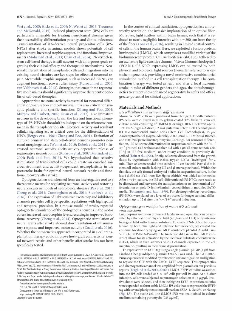
Wei et al., 2005; Hicks et al., 2009; N. Wei et al., 2013; Trounsonand McDonald, 2015). Induced pluripotent stem (iPS) cells areparticularly amenable for treating neurological diseases giventheir accessibility, differentiation potency, and clinical relevance.Transplantation of iPS-derived neural progenitor cells (iPS-NPCs) after stroke in animal models shows potentials of cellreplacement, increased trophic support, and functional improve-ments (Mohamad et al., 2013; Chau et al., 2014). Nevertheless,stem cell-based therapy is still nascent with ambiguous goals re-garding their clinical efficacy and therapeutic mechanisms. Neu-ronal differentiation of transplanted cells and integration into theexisting neural circuitry are key steps for effectual neuronal re-pair. Meanwhile, trophic support, such as increased BDNF, canaugment functional recovery after injury (Kurozumi et al., 2005;van Velthoven et al., 2013). Strategies that enact these regenera-tive mechanisms should significantly improve therapeutic bene-fits of cell-based therapies.
Appropriate neuronal activity is essential for neuronal differ-entiation/maturation and cell survival; it is also critical for syn-aptic plasticity and specific functions (Zhang and Poo, 2001;Murphy and Corbett, 2009; Duan et al., 2017). Like immatureneurons in the developing brain, the fate and functional pheno-type of iPS-NPCs in the adult brain depend on the microenviron-ment in the transplantation site. Electrical activity and resultantcellular signaling act as critical cues for the differentiation ofNPCs (Bergey et al., 1981; Zhang and Poo, 2001). Excitation ofcultured primary and stem cell-derived neurons promotes neu-ronal morphogenesis (Wan et al., 2010; Kobelt et al., 2014). In-creased neuronal activity elicits activity-dependent release ofregenerative neurotrophins (Kolarow et al., 2007; Matsuda et al.,2009; Park and Poo, 2013). We hypothesized that selectivestimulation of transplanted cells could create an enriched mi-croenvironment and activity-dependent neuroplasticity in thepoststroke brain for optimal neural network repair and func-tional recovery after stroke.
Optogenetics has transformed from an interrogative tool to atherapeutic means for regulating neuronal activity and restoringneural circuits in models of neurological diseases (Paz et al., 2013;Cheng et al., 2014; Cunningham et al., 2014; Steinbeck et al.,2016). The expression of light sensitive excitatory and inhibitorychannels provides cell type-specific regulations with high spatialand temporal precision. In a mouse model of stroke, repeatedoptogenetic stimulation of the endogenous neurons in the motorcortex increased neurotrophin levels, resulting in improved func-tional recovery (Cheng et al., 2014). Optogenetic stimulation ofneural grafts after stroke showed downregulation of inflamma-tory response and improved motor activity (Daadi et al., 2016).Whether the optogenetics approach incorporated in a cell trans-plantation therapy could improve host– graft connectivity, neu-ral network repair, and other benefits after stroke has not beenspecifically tested.
In the contest of clinical translation, optogenetics face a note-worthy restriction: the invasive implantation of an optical fiber.Moreover, light scatters within brain tissues, such that it is re-duced to nearly negligible intensity within �200 �m from the tipof the fiber (Yona et al., 2016), resulting in limited spatial controlof cells in the human brain. Here, we exploited a fusion protein,luminopsin 3 (LMO3), which comprises a modified variant of thebioluminescent protein, Gaussia luciferase (sbGLuc), tethered toan excitatory light-sensitive channel, Volvox Channelrhodopsin 1(VChR1). iPS-NPCs expressing LMO3 can be excited by bothphysical and biological light sources (hereafter referred to as op-tochemogenetics), providing a novel noninvasive combinatorialstimulation method in a cell transplantation therapy. The com-bination therapy was tested in vitro and after a focal ischemicstroke in mice of different genders and ages, the optochemoge-netics treatment show enhanced regenerative benefits and offer agreater potential for clinical applications.
Materials and MethodsiPS cell cultures and neuronal differentiationMouse WP5 iPS cells were purchased from Stemgent. UndifferentiatediPS cells were cultured in 0.1% gelatin-coated T25 flasks in stem cellculture media consisting of DMEM (Corning), 10% FBS (Invitrogen),10% NCS (Sigma-Aldrich), 2 mM glutamine (Stem Cell Technologies),0.1 mM nonessential amino acids (Stem Cell Technologies), 55 �M
2-mercaptoethanol (Sigma-Aldrich), 2000 U/ml LIF (Miltenyi Biotec),and 100 U/ml penicillin/streptomycin (Corning). For neuronal differen-tiation, iPS cells were differentiated in suspension culture with the “4�/4�” protocol (4 d without and then 4 d with 1 �M all-trans retinoic acid[RA] in LIF-free medium) under rotary condition as previously de-scribed (Bain et al., 1995). Briefly, cells were dissociated from the growthflasks by trypsinization with 0.25% trypsin-EDTA (Invitrogen) for 2min. Then cells were seeded onto standard 10 cm bacterial Petri dishes instem cell culture media lacking LIF and �-mercaptoethanol. Within thefirst day, the cells formed embryoid bodies in suspension culture. In thelast 4 d, 500 nM of all-trans RA(Sigma-Aldrich) was added to the media.After 4�/4� culture, the iPS cell-differentiated iPS-NPCs were ready tobe dissociated and harvested for transplantation or in vitro terminal dif-ferentiation on poly-D-lysine/laminin-coated dishes in modified SATOmedia (Bottenstein and Sato, 1979). For electrophysiology recordings,iPS-NPCs were plated on a layer of astrocytes for longer terminal differ-entiation up to 12 d after the “4�/4�” neural induction.
Optogenetics gene modification of mouse iPS cells andvirus infectionsLuminopsins are fusion proteins of luciferase and opsin that can be acti-vated by either extrinsic physical light (i.e., laser and LED) or by intrinsicbiological light with chemical substrate. To enable iPS-NPCs to be depo-larized by both blue light and intrinsic luminescence, we created theepisomal backbone carrying an LMO3 construct ( pLenti-CAG-sbGLuc-VChR1-EYFP-IRES-PuroR). The luciferase sbGLuc in the LMO3 con-struct allows for its activation by the luciferase substrate coelenterazine(CTZ), which in turn activates VChR1 channels expressed in the cellmembrane, resulting in membrane depolarization.
To express with an EYFP tag using a single plasmid, pEGIP (a gift fromLinzhao Cheng; Addgene, plasmid #26777) was used. The GFP-IRES-Puro sequence was modified by restriction enzyme digestion and ligationto replace the GFP with the LMO3-EYFP sequence. This optogeneticsand chemogenetics channel was amplified from plasmids in our previousreports (Berglund et al., 2013, 2016b). LMO3-EYFP lentivirus was addedinto the iPS cells seeded at 5 � 10 5 cells per well in vitro. At 4 d afterinfection, cells were subjected to puromycin selection at 15 �g/�l. Posi-tive clones were selected, and then the highest EYFP-expression colonieswere expanded to form stable LMO3-iPS cells that coexpressed the EYFPtag with several pluripotent stem cell markers SSEA-1, Oct 3/4, or Nanog(Fig. 1A). The stable cell line (LMO3-iPS) was maintained in culturemedium containing puromycin (0.5 �g/ml).
This work was supported by National Institutes of Health Grants NS085568 to L.W., S.P.Y., and R.E.G., NS091585to L.W., NS079268 to R.E.G., NS079757 to R.E.G., NS086433 to J.K.T., VA Merit Award RX000666, RX001473 to S.P.Y.,National Science Foundation CBET-1512826 to K.B. and R.E.G., American Heart Association Predoctoral FellowshipPRE31230001 to J.Y.Z., and Postdoctoral Fellowships POST12080252 to M.S. and POST25710112/CDA34110317 toZ.Z.W. The Viral Vector Core of Emory Neuroscience National Institute of Neurological Disorders and Stroke CoreFacilities was supported by National Institutes of Health Grant P30NS055077. We thank Dr. Michael Jiang, Dr. MylesR. McCrary, and Dupe Loye for help in proofreading and editing the manuscript; and Samuel I. Kim for help in CTZadministration and double-blind data analysis in behavioral tests.
The authors declare no competing financial interests.*J.K.T., Z.Z.W., and D.C. contributed equally to this work.Correspondence should be addressed to Ling Wei at [email protected]://doi.org/10.1523/JNEUROSCI.2010-18.2019
Copyright © 2019 the authors
6572 • J. Neurosci., August 14, 2019 • 39(33):6571– 6594 Yu et al. • Optochemogenetics for Cell Stroke Therapy

Figure 1. Expression of LMO3 in iPS-NPCs and functional activities of derived neurons. A, A stable iPS cell line expressing luciferase (LMO3-iPS cell line) was generated by transduction of themodified LMO3 construct (pLenti-CAG-sbGLuc-VChR1-EYFP-IRES-PuroR) and subsequent puromycin selection. Immunocytochemistry showed that LMO3-iPS cells express pluripotent stem cellmarkers SEEA-1, Oct 3/4, and Nanog, which colabeled with fluorescent protein EYFP tagged to the LMO3 construct. B, Differentiated iPS-NPCs showed neuronal morphology of neurites and axons.Green is from EYFP fluorescence in transfected cells. C, In whole-cell recording of current clamp on LMO3-iPS-NPC-derived neuron-like cells, current injection of 30 and 60 pA induced membranedepolarization and repetitive action potentials. D, Demonstration of blue light induced trains of action potential in LMO3-iPS-NPC-derived neurons. E, Bath application of CTZ (6 �M) significantlyincreased the firing frequency in LMO3-expressing cortical neurons compared with vehicle control. The firing rate in the cell increased from 2.6�0.9 Hz to 4.3�0.5 Hz ( p�0.05, t test, n�5 each).F, In voltage-clamp recording, blue laser light (473 nm, 33.6 mW) stimuli evoked inward currents resembling EPSCs in LMO3-iPS-NPC-derived neurons. Light stimulation effect in the same cell wascompletely blocked by the AMPA and NMDA receptor antagonists CNQX and AP5. Different cells were recorded in D–F.
Yu et al. • Optochemogenetics for Cell Stroke Therapy J. Neurosci., August 14, 2019 • 39(33):6571– 6594 • 6573

The point mutation, D248A, as well as a new AfeI restriction site, wasintroduced into the VChR1 moiety of LMO3 using a commercial mu-tagenesis kit following the manufacturer’s instructions and their onlineprimer design software (QuikChange XL; Agilent Technologies). First,the LMO3 cassette in the pcDNA3.1/CAG vector (Berglund et al., 2016a)was moved into the pGP/CMV vector (Addgene, plasmid #40753, a giftfrom Douglas Kim) using the unique NheI/BsrGI restriction sites, andthen site-directed mutagenesis was performed. The correct sequence wasconfirmed by an AfeI restriction enzyme analysis as well as DNA se-quencing. The mutated LMO3/D248A cassette was put back to theoriginal pcDNA3.1/CAG vector using the same restriction sites andtransfection-grade plasmids were prepared using a commercial kit(PureLink Midiprep Kit; Invitrogen). For electrophysiological record-ings and a plate reader assay in vitro, human embryonic kidney 293 cellswere transiently transfected via lipofection following the conventionalmethod (Berglund et al., 2016b) and manufacturer’s instructions (Lipo-fectamine 2000; Invitrogen). For in vivo experiments, a stably transfectediPS cell line was established by lipofection followed by antibiotic selectionusing the neomycin resistance gene in the pcDNA3.1/CAG vector. Inanother control experiment, iPS cells were transduced with a recombi-nant adeno-associated viral (AAV) vectors pseudotyped with AAV2/9carrying the channelrhodopsin 2 (ChR2) gene from the pAAV/CaMKII�-hChR2/H134R-mCherry-WPRE plasmid in Emory ViralCore. ChR2 alone lacked luciferase, serving as a negative control for CTZadministration.
For the expression of LMO3 in naive animals, AAV vectors carryingthe LMO3 gene under control of the human synapsin-1 promoter weresimilarly produced and injected into the sensorimotor/barrel cortex(�2.54 mm AP, 3.5 mm ML, 0.4 mm DV, 30° angle) using a Hamiltonsyringe on a stereotaxic platform (1 �l or �1 � 10 7 viral genomes).Three to 4 weeks after infection, animals were subjected to in vivo testsand killed by overdose of isoflurane and decapitated for brain dissectionfollowed by tissue and cellular examinations.
Isolation of total RNA and RT-PCRTotal RNA from LMO3-iPS-derived neurons was isolated according tothe manufacturer’s instructions (Invitrogen). In brief, the RNA samples(1 �g) were reverse transcribed in 20 �l of a reaction mixture containing2� RT buffer and 20� RT enzyme mix at 37°C for 60 min. The sampleswere then incubated at 95°C for 5 min and transferred to 4°C. RT product(1 �l) was subjected to PCR amplification with 10 pmol primer, 10�standard Taq reaction buffer, 10 mM dNTP, and 0.625 unit Taq polymer-ase in 25 �l PCR buffer (New England Biolabs). PCR primers were usedas follows (5-3): stromal cell-derived factor 1� (SDF-1�): GCATAAA-GACACTCCGCCAT (forward) and TGAGGAGAATGGGGATGAAG(reverse); synapsin-1: CAGCACAACATACCCTATGC (forward) andGGTCTTCCAGTTACCCGACA (reverse); postsynaptic density 95(PSD95): GGCACCGACTACCCCACAG (forward) and AACACCATT-GACCGACAGGA (reverse); 18s: GACTCAACACGGGAAACCTC (for-ward) and ATGCCAGAGTCTCGTTCGTT (reverse). PCR mixtureswere heated to 95°C for 10 min and cycled 30 –37 times for each primer;cycles consisted of 95°C for 15 s, 60°C for 1 min, and 72°C for 30 s. Afteradditional incubation at 72°C for 10 min, the PCR samples were trans-ferred to 4°C. PCR products were subjected to electrophoresis in 2%agarose gel with ethidium bromide. Relative intensity of a PCR band wasanalyzed using InGenius3 manual gel documentation systems (Syngene).
Immunocytochemical and immunohistochemical stainingCultured cells were fixed with 4% PFA in PBS and postfixed with 2:1mixture of enthanol:acetic acid for 5 min, then permeabilized with 0.2%Triton X-100 and blocked with 1% fish gelatin. Cells were incubatedovernight at 4°C with primary antibodies against GFP (1:200; NovusBiologicals; used to stain for the EYFP tag in LMO3), NeuN (1:400;Millipore), �-III tubulin (Tuj-1, 1:400; Covance), synapsin-1 (1:400;Millipore), neurofilament (NF, 1:400; Millipore), CaMKII (1:400; Milli-pore), FoxG-1 (1:200; Abcam), SEEA-1 (1:200; Abcam), Nanog (1:100;Santa Cruz Biotechnology), Oct 3/4 (1:100; Santa Cruz Biotechnology),vesicular glutamate transporter 1 (VGLUT1, 1:200; Abcam), and vesicu-lar glutamate transporter 2 (VGLUT2, 1:200; Abcam). For the secondary
antibodies, either AlexaFluor-488 (1:100; Invitrogen) or Cy-3 (1:400;Jackson ImmunoResearch Laboratories) conjugated antibody againstthe respective IgG was used. DAPI within the DAPI-Vectashield (VectorLabs) was used to stain cell nuclei.
For immunohistochemistry, animals were subjected to cardiac perfu-sion with PBS followed by 4% PFA in PBS. Fixed brains were sectioned at10-�m-thick coronal sections with a cryostat (Leica Microsystems).Brain sections were postfixed in 10% buffered formalin for 10 min fol-lowed by 2:1 mixture of enthanol:acetic acid for 5 min. Standard stainingprotocols were then followed for GFP (1:200; Novus Biologicals), NeuN(1:400; Millipore), synapsin-1 (1:200; Millipore), glucose transporter-1(Glut-1, 1:400; Millipore), and myelin basic protein (MBP, 1:400; Milli-pore). The GFP antibody was used to stain for the EYFP tag in LMO3. ForBrdU (1:400; AbD Serotec) staining, slides were fixed by cold methanolfollowed by hydrochloric acid (HCl) treatment for 1 h. Then standardstaining procedures were followed. Pictures were taken by using a fluo-rescence microscope (BX61; Olympus) or a laser scanning confocal mi-croscope (Carl Zeiss) along the length of the penumbra region definedmorphologically as the region just outside the stroke core. The colabelingwas confirmed using the confocal microscope (FV1000, Olympus). Forsystematic random sampling in design-based stereological cell counting,six coronal brain sections per mouse were selected, spaced 90 �m apartacross the same ROI in each animal. For multistage random sampling, sixfields per brain section were randomly chosen in the peri-infarct/penum-bra region of the brain. The fluorescence density was measured usingNational Institutes of Health ImageJ software.
Western blot analysisProteins were extracted from iPS-NPC-derived neurons using the lysisbuffer containing the protease inhibitor mixture containing AEBSF,aprotinin, bestatin, E-64, leupeptin, and pepstatin A (1:100; Sigma-Aldrich). Protein concentrations were quantified by the BCA assay. Pro-tein (30 �g) from each sample were loaded into a gradient gel and run atconstant current until protein markers had adequately separated. Theywere transferred onto PVDF membranes that were then probed by usingstandard protocols. Primary antibodies, synapsin-1 (1:1000; Cell Signal-ing Technology), PSD95 (1:500; Cell Signaling Technology), BDNF (1:500; Santa Cruz Biotechnology), and mouse �-actin antibody (1:6000;Sigma-Aldrich) were applied overnight at 4°C. Alkaline phosphatase-conjugated secondary antibodies were applied for 1–2 h at room temper-ature. Alkaline phosphatase-conjugated antibodies were developed byusing NBT-BCIP solution. The intensity of each band was measured andsubtracted by the background using National Institutes of Health ImageJsoftware. The expression ratio of each target protein was then normalizedagainst �-actin.
Calcium imaging in cultured iPS-NPCs and neuronsiPS-NPCs were loaded with the fluorescent Ca 2� dye, fura-2, in amembrane-permeable (AM) form (Invitrogen; 5 �M in 100 �l HEPES-buffered solution), inside a CO2 incubator. fura-2 fluorescence was al-ternately excited at 340 and 380 nm light, and emission ratio to the340/380 nm excitation was determined (BX51; Olympus).
Lactate dehydrogenase (LDH) release of cell death assayThe potential effect of optogenetic/chemogenetic stimulation on celldeath was measured using the LDH released assay. LDH release from cellswas detected using the Cytotoxicity Detection Kit (Roche Diagnostics)and a fluorometric plate reader (FL-600; Bio-Tek Instruments) followingthe manufacturer’s instruction manual.
Immunoelectron microscopy of brain sectionsOne to 1.5 months after transplantation, animals were killed and per-fused transcardially with 4% PFA in 0.1 M PB, pH 7.2–7.4. Immunoelec-tron microscopy was performed at Emory Electron Microscopy Coreaccording to previous published method for immunogold-silver labelingat ultrastructural level (Yi et al., 2001). Perfusion fixed brains were fur-ther fixed with 4% PFA in PB overnight and then sectioned coronally at50 �m using a cryostat vibratome (Ultrapro 5000). The sections werewashed thoroughly with PB and then placed in PB containing 0.1% so-dium borohydride to inactivate residual aldehyde groups in the tissue
6574 • J. Neurosci., August 14, 2019 • 39(33):6571– 6594 Yu et al. • Optochemogenetics for Cell Stroke Therapy

sections. Sections were washed with PB several times until the solutionwas devoid of bubbles. To improve antibody penetration, the sectionswere treated with PB containing 0.05% Triton X-100 for 20 min beforeincubating in a blocking solution containing 5% BSA, and 0.1% coldwater fish skin gelatin. After blocking, sections were rinsed twice withPBS containing 0.2% acetylated BSA (PBS/BSA-c; pH 7.4) and then in-cubated overnight at 4°C in a mixture of goat anti-GFP (5 �g/ml) andmouse anti-tubulin (1:100), or goat anti-GFP and mouse anti-neuronalnuclei (both at 5 �g/ml) primary antibodies diluted in PBS/BSA-c. Theneuronal GFP immunolabeling helped the identification of neuronalcells derived from transplanted cells in electron microscopy (EM) im-ages. After washes with PBS/BSA-c, sections were incubated overnight at4°C in the secondary antibody conjugate, ultrasmall gold-conjugateddonkey anti-goat IgG diluted 1:100 with PBS/BSA-c. To remove un-bound secondary antibody, sections were washed thoroughly with PBS/BSA-c and then with PB.
Following PB washes, sections were washed with enhancement condi-tioning solution and then placed in R-Gent SE-EM silver enhancementsolution for 90 min. Silver enhancement was terminated using 0.03 M
sodium thiosulfate in enhancement conditioning solution. After the firstsilver enhancement, sections were washed with PB. Sections were incu-bated overnight at 4°C again with the secondary antibody conjugate,ultrasmall gold-conjugated donkey anti-mouse IgG diluted 1:100 in PBS/BSAc. Sections were washed with PBS/BSAc and then PB. Before thesecond silver enhancement of 60 min, sections were fixed with 2.5%glutaraldehyde in PB. All immunoincubations were done with gentleagitation 4°C, and all immunoEM reagents were purchased from Elec-tron Microscopy Sciences.
Following fixation, sections were washed with PB, fixed with 0.5%osmium tetroxide for 15 min, dehydrated, and flat-embedded in Eponate12 resin (Ted Pella) between two sheets of Aclar film. After resin polym-erization, small pieces of flat-embedded sections were dissected from thecortex, mounted on plastic stubs, and sectioned en face at 80 nm with anultramicrotome (Leica Microsystems, UC6rt). Sections were thenstained with 5% uranyl acetate and 2% lead citrate. Imaging was done ona transmission electron microscope (JEM-1400, JEOL) that is equippedwith a 4 million-pixel charge-coupled device camera (US1000, Gatan).
In vitro neurite/axon morphology and outgrowth analysisAn axon isolation device (Axon investigation system, AXIS, Millipore)was used to measure the process length and distribution of iPS-NPC-derived neurons. The AXIS device has a two-chamber system, which wasseparated by a set of microgrooves, allowing for the separation of cellbody and axons/dendrites and allow only for axon/dendrite crossing.The measurement followed the manufacturer’s instruction. Briefly, afterthe “4�/4�” RA neural differentiation, LMO3-expressing iPS-NPCswere dissociated and plated into one of the culture chambers in SATOmedium, and the other culture chamber was filled with SATO mediumwithout cells. The cells in the photostimulation group received blue lightstimuli as described below for 5 consecutive days starting from 1 d afterplating. Cells in the CTZ group received CTZ treatment (1.5 �M bathapplication for 15 min, 3 times a day for 5 d). Cells in the control groupreceived the medium change without CTZ. On day 6 after plating, thecells were fixed and stained for the neurite marker Tuj-1. The Tuj-1-positive neurites were imaged using confocal microscopy. The distanceand diameter of processes extended from the microgrooves were mea-sured using National Institutes of Health ImageJ. The diameter of eachprocess was measured at five random areas, and the average diameter waspresented in this report. The number and length of processes that crossedthe microgroove barrier into the axonal chamber were counted and com-pared among groups.
The Sholl analysis of major process sprouting was performed usingImageJ software (National Institutes of Health). Briefly, processes 20�m originating from the branch point were counted. Concentric circleswith 15 �m differences in diameter were drawn in the end of micro-grooves, and the number of processes crossing each circle was manuallycounted. The investigators were blind to the treatment groups.
Experimental designLight and CTZ stimulation of iPS-NPCs in vitro. In vitro experiments wereperformed on LMO3-iPS-NPCs. The total number of iPS cell culturedishes tested was 51 cultures dishes/plates from different batches. Start-ing 1 d after the “4�/4�” differentiation and plating, cells were subjectedto blue light stimulation (473 nm, 36 mW for 5 ms at 10 Hz for 12 sfollowed by a 48 s off period repeated 15 times; 3 times a day with aminimum of 2 h interval) for 5– 6 consecutive days. The optical fiber of200 �m core diameter, connected to a laser source (BL473T3–100FC,SLOC), was placed 5 mm below the culture dish. The peak irradiance atthe level of the specimen (i.e., at the bottom of culture dish) was deter-mined by an optical power meter console (ThorLabs) to be �1mW/mm 2.
The same laser light source or wide-field illumination through a greenfilter cube in an epi-fluorescent unit in a microscope (440 –520 nm; 2.1mW/mm 2) was used for all in vitro light stimulation experiments. Cellsin the CTZ group received CTZ exposure (1.5 �M) for 15 min, 3 times aday with a minimum of 2 h interval for 5– 6 consecutive days (n � 12cultures). Cells in the control group received medium changes that con-tained vehicle for CTZ following the same schedule (n � 9 cultures).
Focal ischemic stroke model of the mouse. A total of 442 adult maleC57BL/6 mice (2- to 3-month-old; body weight � 24.1 � 0.7 g for male,22.3 � 0.8 g for female) and 80 aged mice (18- to 20-month-old, 30.7 �2.1 for male, 27.1 � 2.0 for female) (Charles River Laboratories) wereused in stroke experiments. The animal number in each group was de-termined based on the mortality of the distal middle cerebral artery isch-emic surgery (�5% for young adult and �10% for aged animals), thevariables in measurements and our previous data. Randomization wasperformed, and the sample size was further determined using poweranalysis (Power and Precision 4; Biostat).
Focal cerebral ischemia was induced and housed at Emory UniversityAnimal Facility in standard cages in 12 h light/12 h dark cycles. Theischemic stroke in the sensorimotor cortex was induced based on thebarrel cortex stroke model, with modified artery occlusion procedures(L. Wei et al., 1995; Choi et al., 2012). Briefly, anesthesia was inducedusing 3.5% isoflurane followed by the maintenance dose of 1.5% isoflu-rane. Both the tail and paws of the animal were pinch-tested for anes-thetic depth. The right middle cerebral artery was permanently ligatedusing a 10 – 0 suture (Surgical Specialties), accompanied by bilateralcommon carotid artery ligations for 7 min. This modified ischemic pro-cedure is suitable and sufficient for the induction of focal ischemia in themouse cortex, resulting in specific infarct formation in the right sensori-motor cortex. During surgery and recovery periods, body temperaturewas monitored and maintained at 37.0 � 0.5°C using a temperaturecontrol unit and heating pads. The focal ischemic surgery caused lowmortality rate (�5% of 3 d survival). All animal experiments and surgeryprocedures were approved by the Institutional Animal Care and UseCommittee and met the National Institutes of Health standard.
Transplantation of iPS-NPCs into the postischemic brain. Cell trans-plantation was performed at 7 d after the focal ischemic stroke. Threeexperimental groups were stroke control, iPS-NPC transplantation withvehicle, and iPS-NPC transplantation stimulated with CTZ (at least 10animals in each group; specific numbers are mentioned in figure legendsof each experiment of different figures). Animals were randomly as-signed to the groups. After the “4�/4�” neural induction, 5 � 10 5
LMO3-iPS derived LMO3-iPS-NPCs were resuspended in 4 �l SATOmedium and transplanted to the core and peri-infarct regions using aHamilton syringe with injections of 1 �l each at 4 locations (site 1: AP �1.6, L � �4.0, V � �6.5; site 2: AP � 0.6, L � �3.0, V � �5.5; site 3:AP � �0.6, L � �4.5, V � �7.0; and site 4: AP � �2.6, L � �6.5, V ��4.5) in the peri-infarct cortex. A 2 min waiting period at the end ofinjection allowed the cells to settle before needle removal. Stroke controlanimals received vehicle injection (4 �l of SATO medium). In someexperiments, iPS-NPCs were prelabeled with Hoechst 33342 (1:10,000v/v) for 1 h, which facilitated tracking of these cells after transplantation.To enhance cell survival and regenerative property after transplantation,all iPS-NPCs were exposed to hypoxic preconditioning (1% O2) for 8 hbefore transplantation (Theus et al., 2008; Yu et al., 2013). To label pro-liferating cells in the brain, animals received daily administration of
Yu et al. • Optochemogenetics for Cell Stroke Therapy J. Neurosci., August 14, 2019 • 39(33):6571– 6594 • 6575

BrdU (50 mg/kg; i.p.) starting the day of transplantation until the day ofdeath.
Intranasal CTZ administration in vivo. For delivery of CTZ in vivo, weused the nasal route that allows reagents to bypass the blood– brain bar-rier and achieve brain targeted drug delivery (Hanson and Frey, 2008;Chen et al., 2015; Meredith et al., 2015). Intranasal administration ofCTZ followed previous procedures at a dosage of 50 �g per animal (�2mg/kg, dissolved in sterile saline) each time (Chen et al., 2015). Animalsreceived two CTZ treatments per day for 2 weeks (at least 10 animals ineach group; specific numbers are mentioned in figure legends of eachexperiment).
In vivo bioluminescence imaging. We performed the in vivo biolumi-nescence imaging using a chemiluminescence reader (LAS-3000;Fujifilm), which is equipped with a charge-coupled device camera forhigh-sensitivity detection of bioluminescent emission. For each experi-ment, the animals were anesthetized with the ketamine/xylazine mixture(ketamine 80 –100 mg/kg, xylazine 10 –12.5 mg/kg, i.p.) and given CTZ(50 �g) intranasally followed by imaging (n � 3 animals). Using thechemiluminescence mode, the images were acquired as cumulative emis-sion intensities every 4 min over the course of 60 –90 min. Thebackground-corrected luminescence intensity in an ROI was measured.
Electrophysiological recordings in cultured cells and brain slices. Whole-cell patch-clamp recording in dissociated cells in cultures was performedusing an inverted microscope (IX71; Olympus) equipped with an epi-fluorescent unit (Olympus), an electromechanical shutter (Uniblitz;Vincent), a mercury lamp (Olympus), and a scientific CMOS camera(OptiMOS; QImaging) on iPS cell-derived neurons 7–12 d after neuralinduction, which were growing on top of an astrocytes layer. Whole-cellrecording was also done on acute brain slices of naive mice expressingLMO3 using an upright microscope (LABOPHOT; Nikon). The mem-brane currents or potentials were collected using an amplifier (EPC9;HEKA Elektronik) at room temperature (�22°C). The external solutioncontained 135 mM NaCl, 5 mM KCl, 2 mM MgCl2, 1 mM CaCl2, 10 mM
HEPES, and 10 mM glucose, pH 7.4. The internal solution consisted of120 mM KCl, 2 mM MgCl2, 1 mM CaCl2, 2 mM Na2ATP, 10 mM EGTA, and10 mM HEPES, pH 7.2. Recording electrodes pulled from borosilicateglass pipettes (P-97; Sutter Instruments) had a tip resistance between 5and 7 M� when filled with the internal solution. Series resistance wascompensated by 75%– 85%. Linear leak and residual capacitance cur-rents were subtracted online using a P/6 protocol. Action potentials wererecorded under current-clamp mode using patch-clamp software(PatchMaster; HEKA Elektronik). Data were filtered at 3 kHz and digi-tized at sampling rates of 20 kHz.
In brain slice patch-clamp recordings, the forebrain was dissected andimmediately placed in sucrose-enriched cutting solution containing 220mM sucrose, 1.9 mM KCl, 6 mM MgCl2, 0.5 mM CaCl2, 1.2 mM NaH2PO4,33 mM NaHCO3, and 10 mM D-glucose. The solution was ice-cold andbubbled with 95% O2 balanced with 5% CO2, pH 7.40. Coronal brainsections (200 or 400 �m thickness) containing the sensorimotor/barrelcortex were obtained using a vibratome sectioning device (1000 Plus;Vibratome) and then recovered for at least 60 min in the aCSF beforerecordings.
For poststroke recordings, whole-cell voltage clamp was performed onbrain slices containing the ischemic core and peri-infarct regions. Thepipette puller was used to pull the patch pipettes with the tip resistancesof 3–5 M�. During recording, slices were maintained at 34°C and per-fused with oxygenated aCSF containing 124 mM NaCl, 3 mM KCl, 2 mM
MgCl2, 2 mM CaCl2, 1.3 mM NaH2PO4, 26 mM NaHCO3, and10 mM
D-glucose at a rate of 2 ml/min. The pipette was filled with a solutioncontaining 130 mM K-gluconate, 10 mM KCl, 10 mM HEPES, 2 mM Mg-ATP, 0.3 mM Na-GTP, and 0.4 mM EGTA, pH 7.3. Recorded signals wereamplified with a patch-clamp amplifier (Axopatch 200B; Molecular De-vices), digitized at 10 kHz, filtered at 1 kHz, and collected with dataacquisition software (Clampex 8.2; Molecular Devices). Only the neu-rons with membrane potential more negative than �40 mV and actionpotential 65 mV were accepted for further experiments. EPSCs wererecorded as inward currents at the holding potential of �70 mV. Theelectrophysiological data were analyzed using commercial software(Mini Analysis 6.0.7; Synaptosoft).
Microelectrode array (MEA) recordings in brain slices. A high-resolutionMEA2100-system (MultiChannel Systems) was used to perform simul-taneous extracellular recordings at multiple locations in brain slices. Atotal of 31 animals were tested in MEA brain slice recordings. The MEAchamber (60pMEA200/30iR-Ti, MultiChannel Systems) in the experi-ments was composed of a 6-mm-high glass ring and an 8 � 8 titaniumnitride electrode grid, including 60 electrodes (59 electrodes and 1 inter-nal reference electrode) that cover a recording area of 5 mm 2. The elec-trode diameter was 30 �m, and electrodes were separated by 200 �mdistance. The brain slice was placed in the MEA chamber that was per-fused with oxygenated aCSF at a rate of 7 ml/min and maintained at34°C. A stabilization time of 5–10 min was given before formal recording.The collected data were analyzed using Multi Channel Analyzer version2.6.0 (MultiChannel Systems).
In vivo electrophysiological recording. Acute recordings were per-formed with a custom-built tetrode that allowed for recordings of bothsingle-unit and local field potentials (n � 5 for normal and stroke con-trol, respectively, and n � 8 for experimental groups, respectively). Acraniotomy was made 1.3 mm posterior and 1.7 mm lateral to thebregma, and recording was made 3.4 mm inferior to bregma to recordfrom the ventroposteromedial (VPM) nucleus. Extracellular recordingswere sampled at 25 kHz using our custom-built NeuroRighter data ac-quisition system (Rolston et al., 2009). Local field potentials triggered bywhisker stimulation were bandpass-filtered (1–500 Hz) from the rawsignal and analyzed offline using custom MATLAB scripts and theChronux toolbox (Bokil et al., 2010). Single units were detected from thebandpass-filtered (0.5–5 kHz) signal and sorted offline using superpara-magnetic clustering (Wave Clus) scripts developed by Quiroga et al.(2004). The contralateral whiskers were stimulated at �5 Hz for 10 s thatalternated with ipsilateral whisker stimulation trials as a control for atleast 5 repeated trials each during a recording session.
Behavioral testsAdhesive removal test. To evaluate sensorimotor function, time for amouse to remove adhesive pads from the paws was measured as previ-ously described (Bouet et al., 2009; Z. Z. Wei et al., 2015). In brief, a smalladhesive dot was placed on forepaws, and the time needed to contact andremove the sticker from each forepaw was recorded. Mice were trainedthree times before stroke surgery, and the average time was used in dataanalysis. Animals with response time of 120 s were considered insensi-tive to the tactile stimulus and were excluded from further examinations.Sham control, stroke control, and stroke plus CTZ groups contained 10or 12 animals in the control groups and 12 or 16 animals in experimentalgroups. The same animals were also tested in the following tests.
Reach and grasp test. This reaching and grasping performance tests thecoordination of forelimb motor functions in rodents. Mice were placedin a cage of 11.4 � 6.4 � 3.8 cm. In the front wall of each cubicle, there isa hole of 9 mm and through which a feeding plane can be accessed. On thefeeding plane, small food pellets (2–3 mm diameter) were placed 1.5 cmfrom the wall in such a way that the mice can withdraw the food only byreaching out one of its forepaws. Before the experiment, the animals weresubjected to mild fasting with 12 h diet withdrawal. The animal’s attemptto reach food pellets and successful retrieval attempts were counted dur-ing a 2 min period after placing the food pellets. The successful ratio offood grasping was then calculated and compared between groups.
Corner test. The corner test was performed 1 d before ischemia and 3 dafter ischemia, as described previously (Zhang et al., 2002; Choi et al.,2012). Two cardboard plates (30 cm � 20 cm � 0.3 cm) were attached ata 30° angle from each other in a home cage. Each subject mouse wasplaced between the two plates and allowed to freely move to the corner.The number of right and left turns was counted. Twenty trials/tests wereperformed for each mouse.
Cylinder test. The mice were placed in a glass cylinder (9.5 cm diameterand 11 cm height), and the number of times each forelimb or bothforelimbs were used to support the body on the wall of the cylinder wascounted for 5 min. The animals were evaluated at different days afterstroke. Two mirrors were placed behind the cylinder to view all direc-tions. The number of impaired and nonimpaired forelimb contacts wascalculated as a percentage of total contacts.
6576 • J. Neurosci., August 14, 2019 • 39(33):6571– 6594 Yu et al. • Optochemogenetics for Cell Stroke Therapy

Open field test. In the open-field test, mice were allowed to freely move(25 cm � 30 cm � 25 cm) during the dark cycle. Mild stress was inducedby exposure to an open-field animal cage-like box. The container wasdivided into 30 equal 5 � 5 squares, and the number of line crossings andduration of stay by each animal were recorded and calculated during thefirst exposure measured for 2 min. The middle 12 squares were consid-ered the inner space, whereas the other 18 squares were considered theboarder space. The calculation for the relative stress-induced anxiety wasperformed in double-blinded manner using the total stay duration inboarder space in each 2 min.
Whisker-touching behavior. Repetitive whisker-touching behavior is areflection of the intact neuronal controls of the barrel cortex-thalamus-whisker pathway. In stroke animals repaired with LMO3 cell transplan-tation, activation of the LMO3 protein and the whisker-touchingreaction can be achieved by acute application of intranasal CTZ. CTZ (2mg/kg) was intranasally delivered, and the duration of whisker-touchingbehaviors was counted 5 min after CTZ application. This delay helps toavoid counting false-positive reactions due to the animal’s reaction to thelocal sensation on the nose. The counting lasts for 5 min. The ratio of thetouching activity before and after CTZ is calculated and expressed.
Statistical analysisFor comparison between two groups, we used the Student’s two-tailed ttest. Prism version 5.0 (GraphPad) was used to make graphs and toperform statistical analysis. Multiple comparisons were done using one-way or two-way ANOVA followed by Tukey’s test or Bonferroni’s cor-rection for multiple pairwise comparisons. Nonparametric Mann–Whitney’s U test was applied to electrophysiological and behavioralexaminations of multiple comparisons. The F test was performed toverify an F distribution under the null hypothesis in ANOVA tests ofmultiple groups and/or time points. All the tests were two-tailed, and thesignificance level ( p) was set at 0.05. Statistical values, such as t, F, and q,are reported in Results and/or figure legends. The numbers in the text arereported in the form of mean � SEM.
ResultsNeuronal differentiation of LMO3-iPS cells and responses tolight and CTZ stimulationWe established a mouse iPS-cell line that stably expressed theLMO3 fusion protein (LMO3-iPSs). The expression of LMO3and pluripotency of the cell line were confirmed by the EYFP tagin LMO3 and iPS-cell markers (SSEA-1, Oct 3/4, and Nanog),respectively (Fig. 1A). After the “4�/4�” RA neural inductionprotocol (Bain et al., 1995), these LMO3-iPS cells gained neuro-nal morphology in EYFP fluorescence-positive cells (Fig. 1B). Wecharacterized electrophysiological properties of LMO3-iPS-derived neurons 7–12 d after neural induction. In patch-clamprecordings, membrane depolarization caused the firing of repet-itive action potentials in a current injection-dependent manner(Fig. 1C). Similarly, stimulation by both light and the luciferasesubstrate, CTZ, elicited trains of spikes (Fig. 1D,E). In thevoltage-clamp mode at the �60 mV holding potential, photo-stimulation by blue laser light increased occurrence of fast inwardcurrents, resembling EPSCs (Fig. 1F). The activated neuronswere glutamatergic as they were sensitive to block by the gluta-mate receptor antagonists, CNQX and D-AP5 (Fig. 1F). We alsoconducted Ca 2� imaging in LMO3-iPS-derived neurons loadedwith the fluorescent Ca 2� indicator, fura-2, using ultraviolet ex-citation light (Fig. 2A–C). Stimulation with blue light or CTZincreased the intracellular Ca 2� concentration, reminiscent ofthat induced by high potassium (15 mM KCl) medium (�ratio �0.0056 � 0.0026, 0.0079 � 0.0024, and 0.0133 � 0.0028 for CTZ,light, and 15 mM K�, respectively; p 0.2 among groups; one-way ANOVA; F(2,12) � 1.53; n � 3, 8, and 4 assessments, eachincluded �30 cells) (Fig. 2D–F).
Excessive Ca 2� increases may cause excitotoxicity. In LDHrelease assays, neither of the applied light or CTZ stimuli caused asignificant change in cell viability (one-way ANOVA; F(2,6) �7.057, p � 0.0265, n � 8 –10 assessments in each condition; q(6) �3.654, p 0.09 for control vs laser; q(6) � 1.513, p 0.5 forcontrol vs CTZ) (Fig. 2G). These experiments indicate thatLMO3-iPS cells can be differentiated into functional neuronsresponsive to both light and CTZ stimulations with increasedCa 2� and action potentials, yet without causing excitotoxicity.
In a control study, cells were transduced with LMO3 or with apoint mutation analogous of nonfunctional mutation in the poreregion of a channelrhodopsin chimera (D292A in C1/C2) (Katoet al., 2012) that corresponds to D248A in the VChR1 moiety ofLMO3. Judged by the YFP fluorescent intensity, the expressionlevel of these channels in cells was similar (Fig. 2H). A largeinward photocurrent was recorded in patch-clamp recordingsduring photostimulation from cells expressing LMO3 (Fig. 2I),whereas cells expressing LMO3/D248A exhibited negligible pho-tocurrent to the same photostimulation (Fig. 2I–L).
Expression of neuronal markers in differentiatedLMO3-iPS cellsNeuronal differentiation of LMO3-iPS-NPCs was verified withneuronal fluorescent markers 7–12 d after neural induction.Transfected cells were labeled with EYFP, and their neuronaldifferentiation was identified by specific markers, including NF,synapsin-1, and NeuN (Fig. 3). Cell counting assays confirmedthat 80% of total cells expressed NeuN and/or NF, indicatingtheir neuronal differentiation (Fig. 3D). The forebrain markerFoxG-1 was identified in these cells (Fig. 3E). We observed glu-tamatergic markers, VGLUT1, localized with the EYFP tag ofLMO3 in most differentiated cells (Fig. 3H), consistent with ourelectrophysiological recording of glutamatergic currents (Fig.1F). This was in line with previous reports that the 4�/4� RAdifferentiation protocol primarily induced glutamatergic neurallineage cells (Bibel et al., 2007; Boissart et al., 2013).
Light and CTZ stimulation promoted neurite growth ofLMO3-iPS-derived neurons in vitroThe axon/neurite outgrowth of differentiating LMO3-iPS-NPCswas examined in an axon isolation device that contained com-partments of cell bodies and neurites divided by microgrooves.After neural induction, LMO3-iPS-NPCs were plated into the cellbody compartment for terminal differentiation. One day afterplating, cells were subjected to photostimulation or CTZ expo-sure for 5 consecutive days. Blue laser light (10 Hz, 15 min/ses-sion, 3 sessions/d) was applied through fiber optics, while CTZ(1.5 �M) was added into the media for 15 min (3 times/d). Thecontrol group contained cells treated with regular medium andwithout photostimulation. Six days later, Tuj-1 staining identi-fied significantly more axons/neurites that extended cross themicrogrooves in the group that received laser light or CTZ treat-ment (Fig. 4A). The number of neurites extending cross the mi-crogrooves in the AXIS chamber was significantly increased byeither light or CTZ stimuli (Fig. 4B) (n � 4 in each condition;one-way ANOVA followed by Tukey’s pairwise comparisons;F(2,3) � 14.26, p � 0.029, control vs laser and p � 0.0319 controlvs CTZ). The light and CTZ stimuli also significantly increasedthe length of processes extending beyond the microgrooves (Fig.4C) (F(2,3) � 18.15, p � 0.0211; q(3) � 7.146, p � 0.0302 forcontrol vs laser; q(3) � 7.593, p � 0.0256 for control vs CTZ).Moreover, cells stimulated by light exhibited significantly largerprocess diameters (Fig. 4D) (q(27) � 6.797, p � 0.0001 for control
Yu et al. • Optochemogenetics for Cell Stroke Therapy J. Neurosci., August 14, 2019 • 39(33):6571– 6594 • 6577

Figure 2. Activation of LMO3 channels, Ca 2� influx, and cell viability. LMO3 activation-induced cellular consequences, including intracellular Ca 2� increases, and the influence on cell viabilitywere examined in cultures 10 –12 d after neural induction of LMO3-iPS-NPCs. A–C, Intracellular free Ca 2� concentration was measured using the Ca 2� dye fura-2 AM and 340/380 ratio analysis.The expression of LMO3 was demonstrated in cultured cells with the yellow fluorescence of EYFP (A). B, fura-2 image of these cells during light stimulation. C, Overlay of A and B. D–F, The time courseof intracellular Ca 2� increases induced by bath-applied CTZ (15 �M, 4 –5 min), light stimulation (1 Hz pulse for 4 min), and 15 mM KCl solution (4 –5 min), respectively. All three manipulationscaused significant Ca 2� influx and intracellular accumulation. D, The binding of CTZ to the LMO3 luciferase, as its substrate, caused the emission of bioluminescence (red) followed by activation ofthe LMO3 and Ca 2� influx event. G, LMO3-iPS-NPC-derived neurons were subjected to 5 d treatments of blue light stimulation (10 Hz, 15 min/session, 3 times per day) or CTZ exposure (1.5 �M, 15min/session, 3 times per day), and cell viability was measured using the LDH release assay 12 h after the last stimulation session. There was no significant difference among all three groups (n �8 –10 per group). H, A control experiment of expressing LMO3 or LMO-D248A proteins. The latter gene has a point mutation (D248A), which made the channel moiety nonfunctional. The EYFPfluorescence intensity measurement verified similar expression levels in transfected cells. n � 6 wells each. I, J, In whole-cell patch-clamp recording, a large inward current was observed duringwide-field photostimulation with blue light. Photocurrent to the same photostimulation was negligible in the cell expressing mutant LMO3/D248A. Peak and steady-state photocurrents werevirtually absent with LMO3-D248A. N � 5 cells each. K, L, Summery of the patch-clamp data, showing the CTZ-induced peak and steady-state currents in different cells. ***p � 0.001 versus LMO3cells (Student’s t test).
6578 • J. Neurosci., August 14, 2019 • 39(33):6571– 6594 Yu et al. • Optochemogenetics for Cell Stroke Therapy

Figure 3. Neuronal differentiation of LMO3-iPS cells. LMO3-iPS-NPCs were subjected to the “4�/4�” RA differentiation protocol. Immunohistochemical staining was performed for neuronalmarkers 7–12 d later. A, The LMO3 expression was verified by EYFP tagged to the virus construct. NF labeling (NF) (red) shows the neurite growth from differentiated cells marked with the EYFPfluorescence (green). The merged image demonstrates neuronal differentiation of transfected cells. B, C, Enlarged images from the frame in A. C, The overlay of the three images in B, showingneuronally differentiated cells. D, Quantified data demonstrate 80% of cells expression the neuronal marker NF and NeuN (see G). E, The confocal image illustrates that LMO3 expressing cellsexpress the forebrain marker FoxG-1. F, The three high-magnification images illustrate the presynapse protein synapsin-1 in a differentiated iPS cell. G, Many differentiated cells (EYFP, green) werepositive to NF as well as the mature neuronal marker NeuN (blue). The merged image represents a complete overlay of the three markers. H, These neurons also expressed glutamatergic neuronalmarkers, including vesicular glutamate transporter 1 (VGLUT1) and VGLUT2 (data not shown due to space limitation).
Yu et al. • Optochemogenetics for Cell Stroke Therapy J. Neurosci., August 14, 2019 • 39(33):6571– 6594 • 6579

Figure 4. Light and CTZ stimulation promoted neurite growth of LMO3-iPS-NC-derived cells. A, LMO3-iPS-NPCs were plated in AXIS axon isolation device after “4�/4�” differentiation. One dayafter plating, cells in the laser group received blue light (10 Hz, 15 min) three times a day for 5 consecutive days. Cells in the CTZ group were exposed to CTZ (1.5 �M, 15 min) three times a day for 5 d.Cells in the control group received CTZ-free medium changes and also served as the light off control. Six days after plating, cells were stained with TUJ-1. Dashed lines in lower-magnification imagesoutline the microgrooves in the AXIS device, which separated the cell body and processes. Higher-magnification images represent samples of processes in each group. B–E, Quantified neuriteanalysis shows the number of neurites extending across the microgrooves in the AXIS chamber (B), the average distance that neurites extend beyond the microgrooves (C), the average diameter ofneurites (D), and the number of process sprouts (E). Either blue light stimulation or CTZ exposure significantly increased neurite growth and maturation compared with those in the control group.n � 4 independent assays. For detailed statistical results, see text. F, Results of the Sholl analysis using the ImageJ software. The number of neurites that extended and crossed the microgroovebarrier into the axonal chamber was counted at different distances from microgrooves, and their length was measured. In the control group, fewer crossings took place compared with laser and CTZgroups. Neurites in the control group grew shorter than neurites in laser light and CTZ groups. *p � 0.05 versus controls (two-way ANOVA). #p � 0.05 versus the maximal length of control axons(two-way ANOVA). n � 4 per group.
6580 • J. Neurosci., August 14, 2019 • 39(33):6571– 6594 Yu et al. • Optochemogenetics for Cell Stroke Therapy

Figure 5. Light and CTZ stimulation increased gene expression in LMO3-iPS-derived neurons in vitro. Cultured LMO3-iPS-derived cells received chemogenetic and optogenetic stimulation from1 to 5 d after neural induction, RT-PCR, and Western blot were performed at 6 d after induction. A, Representative images of RT-PCR gels indicate the expression of synaptic maker synapsin-1 (SYN-1),PSD95, and growth factor SDF-1 in each group. The mRNA level of each marker was normalized against 18S. Cells that received photostimulation (blue light, 10 Hz, 15 min, three times a day for 5consecutive days) in the laser group and those in the CTZ treatment group (1.5 �M, 15 min, three times a day for 5 d) had significantly higher mRNA levels of SYN-1, PSD95, and SDF-1 (for statisticalanalysis, see text). B, Represented images of Western blot bands of SYN-1, PSD95, and BDNF. Quantified data show that blue light in the laser group or CTZ (Figure legend continues.)
Yu et al. • Optochemogenetics for Cell Stroke Therapy J. Neurosci., August 14, 2019 • 39(33):6571– 6594 • 6581

vs light; q(27) � 4.855, p � 0.0053 for con-trol vs CTZ) as well as increased numberof sprouts (Fig. 4E) (nonparametric Man-n–Whitney test, p � 0.0001 laser light offcontrol vs light; p � 0.0072 control vsCTZ). There were no statistical differ-ences between light and CTZ groups.Consistent with above analyses, the Shollanalysis validated that the number of pro-cesses at 200 �M from microgroves andthe maximal length of processes was sig-nificantly greater in two treatment groupscompared with controls (Fig. 4F).
Light and/or CTZ stimulationupregulated synaptic proteins andBDNF in LMO3-iPS-NPCsTwelve hours after stimulation with laserlight or CTZ (6 d into the terminal differ-entiation), RT-PCR showed that light-stimulated LMO3-iPS-NPCs expressedmore mRNA of synapsin-1 (q(18) � 4.025,p � 0.0276, n � 4; one-way ANOVA fol-lowed by Tukey’s pairwise comparisons),PSD95 (q(18) � 4.556, p � 0.0125, n � 4),and SDF-1 (q(18) � 5.405, p � 0.0034, n � 4) (Fig. 5A). CTZ-treated LMO3-iPS-NPCs also showed significantly higher mRNAlevels of synapsin-1 (q(18) � 6.982, p � 0.0003, n � 4), PSD95(q(18) � 7.578, p � 0.0001), and SDF-1 (q(18) � 8.72, p � 0.0001)(Fig. 5A). Western blot analysis verified higher protein levels ofsynapsin-1 (q(17) � 7.334, p � 0.0003 for control vs laser; q(17) �5.213, p � 0.0059 for control vs CTZ), PSD95 (q(17) � 6.027, p �0.0014 for control vs laser; q(17) � 3.685, p � 0.0462 for control vsCTZ), and BDNF (q(14) � 9.6, p � 0.0001 for control vs laser;q(14) � 8.868, p � 0.0001 for control vs CTZ) in LMO3-iPS-NPCsthat received either photostimulation or CTZ stimulation com-pared with sham controls (n � 4 each group) (Fig. 5B). In cellswithout the expression of LMO3 channels, the same CTZ appli-cations did not show significant influence on these factors (Fig.5C,D). Examined in additional controls in vivo studies, CTZtreatment in stroke animals received iPS-NPCs of no LMO3 ex-pression failed to induce significant influence on tested factors(Fig. 5E,F).
Validation of intranasal delivery of CTZ with in vivobioluminescence imagingFor the translational potential of the optochemogenetic ap-proach, we tested noninvasive brain delivery of CTZ via the well-defined nasal-brain route. Normal mice received stereotaxicinjection of AAV vector carrying the LMO3 gene into the right
barrel cortex. Postmortem histology confirmed proper targetingof the barrel cortex with LMO3 as shown in the EYFP expressionsurrounding the injection site (Fig. 6B). After intranasal admin-istration of CTZ (50 �g, or 2.0 mg/kg), bioluminescence wasobserved in the area of LMO3 expression and decayed over thenext hour (Fig. 6A,C).
In adult stroke mice of focal cerebral ischemia targeting theright sensorimotor cortex, LMO3-iPS-NPCs were transplantedinto the core and peri-infarct regions 7 d after stroke (Fig. 6D).Fourteen days later, transplanted cells and functional expressionof LMO3 in these cells were confirmed with in vivo biolumines-cence imaging (Fig. 6E). Bioluminescence was detected in the cellgraft area and remained visible for �1 h after intranasal CTZadministration (Fig. 6F).
CTZ stimulation of grafted LMO3-iPS-NPCs promotedneuronal differentiation, axonal remyelination, and long-term survival in the ischemic brainIn the ischemic stroke mice receiving cell transplantation or con-trol medium, repeated stimulation of transplanted LMO3-iPS-NPCs was achieved by intranasal CTZ treatment (2 mg/kg, 10 �l)twice a day for 2 weeks starting 1 d after transplantation. Threeweeks after stroke (2 weeks after cell transplantation), graftedLMO3-iPS-NPCs were identified by EYFP or mCherry tag ofLMO3 (Fig. 7A–C). Colocalization with NeuN was observed inmany EYFP- or mCherry-positive cells, indicating neuronal dif-ferentiation (Figs. 7A–C, 8A). Stroke animals showed a signifi-cant reduction in the expression of MBP in the peri-infarct region(q(20) � 15.48, p � 0.0001 for sham vs stroke) (Fig. 7D,E).The MBP expression was noticeably greater in mice of theStroke�LMO3-iPS�CTZ group (one-way ANOVA, q(20) �7.679, p � 0.0003 for Stroke vs Stroke�LMO3-iPS�CTZ; q(20) �4.521, p � 0.026 for Stroke�LMO3-iPS vs Stroke�LMO3-iPS�CTZ; n � 5– 8 per group) (Fig. 7E). Consistent with theseobservations, 1 month after stroke (3 weeks after cell transplanta-tion), EYFP-positive cells colabeled with the intact nucleus ofHoechst 33342 were identified as surviving transplanted cells. In cellcounting assays, stroke animals in the Stroke�LMO3-iPS�CTZ
4
(Figure legend continued.) exposure increased the protein levels of these factors (for non-parametric statistical analysis, see text). n � 4/group independent assays. C, D, Western blotanalysis on the expression levels of BDNF, SDF-1, synapsin-1, and PSD-95 in the cortical cultureswith no LMO-3 transduction. This test served as a negative control for possible off-target actionsof CTZ. There was no significant difference among saline and CTZ groups (unpaired Student’s ttest, p 0.05, n � 4 cultures per group). E, F, Western blot analysis of the expression of BDNF,synapsin-1, PSD-95, and SDF-1 in the peri-infarct region 7 d after stroke. CTZ (intranasal deliveryof 2 mg/kg/d, starting from 8 d after stroke) or vehicle treatment for 14 consecutive days. Therewas no significant difference of the expression of BDNF, SDF-1, synapsin-1, and PSD-95 in theperi-infarct region between the two groups at the end of the treatments. n � 5/group. p 0.05 (unpaired Student’s t test). *p � 0.05 versus controls (one-way ANOVA).
Figure 6. The kinetics of LMO3 bioluminescence after nasal to brain delivery. To demonstrate the effectiveness and biokineticsof LMO3 activation induced by intranasal administration of CTZ, the LMO3 luminescence was measured in the normal brainexpressing LMO3 and in the stroke brain receiving LMO3-iPS-NPC transplantation. A, Intranasal administration of CTZ (2 mg/kg)induced luminescence in the barrel cortex area receiving LMO3 infection. B, AAV carrying the LMO3 construct was injected ot thebarrel cortex of a normal mouse. The EYFP expression appeared in the sensorimotor cortex. C, The time course of CTZ inducedluminescence in the brain, which lasted approximately 1 h. D–F, Seven days after focal ischemic stroke in adult mice, LMO3-iPS-NPCs were transplanted to the core and peri-infarct region of the right sensorimotor cortex. The 2,3,5-triphenyltetrazoliumchloride staining shows the infarct area (white). Diagram illustrates the location of the cell implantation (D). E, Intranasal admin-istration of CTZ (2 mg/kg) induced bioluminescence in the cell-grafted area. F, The intensity of luminescence verified that theCTZ-induced luminescence lasted �1 h in the poststroke brain.
6582 • J. Neurosci., August 14, 2019 • 39(33):6571– 6594 Yu et al. • Optochemogenetics for Cell Stroke Therapy

group showed increased surviving trans-planted cells compared with animals re-ceived LMO3-iPSs but no CTZ treatment(unpaired Student’s t test, p � 0.0034 forStroke�LMO3-iPS vs Stroke�LMO3-iPS�CTZ; F � 2.569, n � 30 and 24 ani-mals, respectively) (Fig. 7F).
There were increased numbers ofBrdU/NeuN double-positive cells (one-way ANOVA followed by Tukey’s pair-wise comparisons; q(25) � 8.257, p �0.0001) and BrdU/Glut-1 double-positivecells (q(20) � 6.081, p � 0.0018) afterstroke compared with that in the shamgroup, indicating pronounced neurogen-esis and angiogenesis (Fig. 8). Strokeanimals receiving cell transplantation(Stroke�LMO3-iPS) or a combination oftransplantation and CTZ stimulation(Stroke�LMO3-iPS�CTZ) showed evengreater numbers of BrdU/NeuN andBrdU/Glut-1 double-positive cells com-pared with those in the Stroke-only group(NeuN: q(25) � 4.129, p � 0.0519 forStroke vs Stroke�LMO3-iPS; q(25) �7.635, p � 0.0001 for Stroke vs Stroke�LMO3-iPS�CTZ; Glut-1: q(20) � 4.079,p � 0.0419 for Stroke vs Stroke�LMO3-iPS; q(20) � 4.06, p � 0.0431 for Stroke vsStroke�LMO3-iPS�CTZ) (Fig. 8C,E),indicating that poststroke regenerationcould be promoted by LMO3-iPS-NPCs.CTZ treatment after LMO3-iPS-NPC trans-plantation showed a trend of further increas-ing BrdU/NeuN-positive cells (Fig. 8C).
Synapse formation of transplantedLMO3-iPS-NPCs and synaptic activity/plasticity induced by light or CTZstimulation on brain slicesWhole-cell patch-clamp recordings inbrain slices 4 weeks after stroke showedcurrent injection evoked trains of actionpotentials in LMO3-expressing cellsmarked with EYFP (Fig. 9A). Photostimu-lation with blue laser light (473 nm, 33.6mW) also induced membrane depolariza-tion and action potentials (Fig. 9B). Bathapplication of CTZ (300 �M) significantlyincreased the firing frequency of spikescompared with vehicle (Fig. 9C,D).
Immunogold EM was performed toidentify synapses. LMO3-expressing cellswere visualized with electron-dense goldparticles by immunostaining against theEYFP tag of LMO3. The ultrastructuralimaging on the brain section 4 weeks afterstroke revealed synapses between imm-unogold-positive transplanted cells andimmunogold-negative host neurons.Transplanted cells were identified eitheras presynaptic (Fig. 9E) or postsynapticwith host cells (Fig. 9F) as well as between
Figure 7. Neuronal differentiation of transplanted LMO3-iPS-NPCs and CTZ treatment promoted remyelination in the ischemicbrain. Transplantation of LMO3-iPS-NPCs was performed 7 d after stroke. At various later time points, immunohistochemicalstaining examined the fate of transplanted cells in the ischemic cortex. A, Twenty-one days after transplantation, immunostainingof the ischemic core and peri-infarct cortical regions identified transplanted cells positive with mCherry tagged LMO3. The threeimages show mCherry-positive (red) and NeuN-positive (green) cells at different magnifications. B, Enlarged images of the framein the last image in A. Right, 3D image represents the overlay of mCherry and NeuN staining. C, In confocal imaging, EYFPfluorescence was from the EYFP tagged to the LMO3 construct; its colocalization with NeuN in the peri-infarct region verified againthe survival and neuronal differentiation of grafted LMO3-iPS-NPCs 3 weeks after transplantation. D, In stroke mice that receivedLMO3-iPS-NPC transplantation, CTZ (2 mg/kg) was intranasally administered 1 d after cell transplantation for 14 consecutive days.Three weeks later, the expression of MBP (red) in the peri-infarct region was examined for myelination. In this assay, Hoechst33342 counterstaining (blue) was used to label all cells in the peri-infarct region. E, Bar graph represents the relative expression ofMBP per field. Stroke alone caused a significant reduction in MBP expression compared with that in sham group. More MBP stainingappears in the LMO3-iPS-NPC and LMO3-iPS-NPC plus CTZ groups. n � 5– 8/group. *p � 0.05 versus sham control (one-wayANOVA). #p � 0.05 versus stroke (one-way ANOVA). �p � 0.05 versus stroke � LMO3 cells (one-way ANOVA). For detailstatistical analysis, see text. F, Long-term survival of transplanted cells in the poststroke brain. Cell counting was performed 1month after stroke (3 weeks after cell transplantation). EYFP-positive cells colabeled with the intact nucleus of Hoechst 33342 wereidentified as surviving transplanted cells. For each animal, 30 brain sections spanning the ipsilateral cortex were counted. Cellcounting was performed in the ischemic core and peri-infarct transplanted regions. *p � 0.0034 (paired t test, F � 2.569, DFn �23, Dfd � 29). n � 30 animals in the LMO3-iPS-NPC group (no CTZ control) and 24 animals in the LMO3-iPS-NPC plus CTZtreatment group.
Yu et al. • Optochemogenetics for Cell Stroke Therapy J. Neurosci., August 14, 2019 • 39(33):6571– 6594 • 6583

transplanted cells (Fig. 9G). Presynapticimmunogold was primarily found in Type1 synapses with characteristic asymmetricpostsynaptic density (Palay, 1956), con-sistent with the identity of transplantedcells as glutamatergic neurons.
Optochemogenetic stimulation ofgrafted LMO3-iPS-NPCs triggeredsynaptic plasticity in theischemic cortexTo validate integration of transplantedLMO3-iPS-NPCs into neuronal networks,spontaneous EPSCs (sEPSCs) were re-corded from LMO3-negative residentneurons under whole-cell voltage clamp.The frequency of sEPSCs significantly in-creased by �2-fold during bath applicationof CTZ (300 �M) with no significant changein the amplitude, indicating that CTZcaused neurotransmitter release from sur-rounding LMO3-iPS-NPCs (Fig. 10A–E).
Stroke in the sensorimotor cortexdamages connections of the barrel cortex-thalamus pathway and causes secondarycell death in the thalamus (Carmichael etal., 2001; L. Wei et al., 2004). Brain slicesfrom stroke animals that received LMO3-iPS-NPCs (4 weeks after stroke) were ex-amined using the MEA containing 59recording electrodes. The electrode tem-plate was placed in the area covering thesomatosensory cortex, including part ofthe ischemic core and peri-infarct regions(Fig. 10F). One of the 59 electrodes lo-cated 200 – 400 �m distance from the edgeof the ischemic core was the stimulationelectrode. Field EPSPs (fEPSPs) wereevoked by electric pulses (�1500 mV, 0.1ms, once every 30 s) and were simultane-ously monitored in different brain areas.Among all recording locations, fEPSPscould usually be captured in 18 areas ofthe peri-infarct and nonischemic regions.Certain fEPSPs significantly enhancedduring bath application of CTZ (100 �M,5 min; 7 slices from 4 mice) (Fig. 10G).Heat maps showed that CTZ significantlyincreased fEPSP responses at several loca-tions (Fig. 10H, I). In the response heatmap generated from MEA recordings,neurons in the cortical layer II/III and Vin the same cortical/barrel column re-sponded strongly and showed CTZ-induced LTP (Fig. 10H). In addition,neurons at a 400 – 600 �m distance in adifferent cortical/barrel column also dis-played strong response and augmentedLTP (Fig. 10H). Marked potentiation of fEPSPs persisted, evenafter washing out of CTZ, and progressed for at least 30 min at theend of recordings (Fig. 10J). In the same or different brain slices,photostimulation (473 mm, 10 Hz for 10 s, separated by 10 sintervals, 4 min total) triggered similar potentiation effects dur-
ing and after light application (Fig. 10K). At the end of the 30 minrecording after CTZ or light stimulation, the slope of fEPSPs wasdoubled compared with the baseline level (Fig. 10L). On the con-trary, fEPSPs in control recordings with drug-free solution orlow-frequency photostimulation were stable throughout the re-
Figure 8. Neurogenesis and angiogenesis in the poststroke brain received different treatments. Immunohistochemical stainingof neuron or vessel markers and BrdU incorporation were applied to detect newly formed neuronal and vascular cells in the corticalperi-infarct region 21 d after stroke with different treatments. BrdU (50 mg/kg, i.p.) was injected 1 d after cell transplantation for14 d. A, The 3D image shows the colocalization of transplanted cell marker YFP and the neuronal marker NeuN, indicating neuronaldifferentiation and maturation of LMO3-iPS-NPCs. B, C, NeuN and BrdU double-positive cells illustrate new neurons. C, Bar graphrepresents that the ischemic stroke was an insult and trigger inducing neurogenesis. Both cell transplantation and the cell/CTZtreatments further increased the number of newly formed neurons. CTZ showed a trend of greater effect than cell alone, althoughit was not statistically significant. *p � 0.05 versus sham control (one-way ANOVA). #p � 0.05 versus stroke control (one-wayANOVA). n � 10 –12 per group. D, E, Angiogenesis was measured using the vessel marker Glut-1 colabeled with BrdU. E, Bar graphrepresents that angiogenesis was induced after stroke, whereas both two treatments further increased the number of new vascularstructures. *p � 0.05 versus sham control (one-way ANOVA). #p � 0.05 versus stroke control (one-way ANOVA). n � 10 –12 pergroup.
6584 • J. Neurosci., August 14, 2019 • 39(33):6571– 6594 Yu et al. • Optochemogenetics for Cell Stroke Therapy

cording (Fig. 10G, J,K). These results verified that facilitatedtransmitter release and a synaptic LTP-like effect were induced byexcitation of transplanted cells that had been conditioned by theactivation of LMO3 channels. The involved neurons were gluta-matergic since their synaptic events were completely blocked byglutamate receptor antagonists NBQX (10 �M) and D-AP5 (5�M) (Fig. 10 J,K). Thus, transplanted LMO3-iPS-NPCs differen-tiated into glutamatergic neurons and integrated into existingcircuits within and between cortical column structures, leadingto enforced intrabarrel and interbarrel connectivity and neuronalsynaptic plasticity.
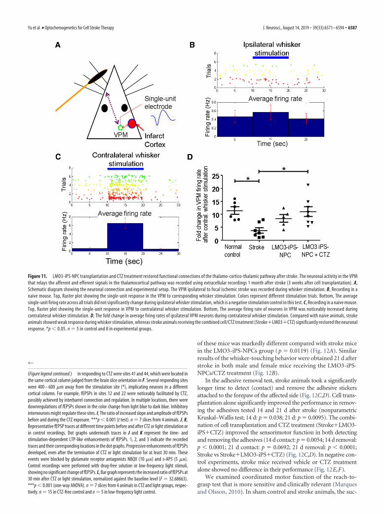
Intranasally delivered CTZ stimulation of grafted LMO3-iPS-NPCs restored thalamocortical connections after strokeTo examine the thalamo-cortico-thalamic connectivity impairedafter stroke, electrophysiological recordings were performed invivo in the VPM nucleus of the thalamus to assess neuronal ac-tivities in the thalamo-cortico-thalamic pathway (Fig. 11A). Onemonth after stroke, the single-unit recording detected neuronalactivity in the VPM barreloid neurons (Fig. 11A–D), an indica-tion of thalamo-cortico-thalamic connectivity involving afferentinformation from the brainstem and efferent signals from thebarrel cortex (Temereanca and Simons, 2003). The frequency ofthis thalamo-cortico-thalamic activity was selectively increasedby mechanical stimulation of contralateral whiskers, and this in-
crease in firing rate was much reduced by stroke (Fig. 11B,C) (L.Wei et al., 2004). In stroke mice of the combined treatment ofLMO3-iPS-NPC plus CTZ, the firing rate of VPM neurons dur-ing whisker stimulation was protected (nonparametric Mann–Whitney’s U test; mean rank difference � �11.05, p � 0.0315,n � 5– 8 per group) (Fig. 11D). This was not seen in the celltransplantation alone group, suggesting that CTZ stimulation ofLMO3 in transplanted cells effectively restored the connectivityand propagation of neuronal signals along the whisker-thalamus-barrel cortex pathway. During the course of experimental proce-dures, no epileptic behavior/activity was observed in animalsreceiving LMO3-iPS-NPC transplantation and/or CTZ stimula-tion by eye surveillance or the MEA recording in brain slices.
Intranasally delivered CTZ stimulated grafted LMO3-iPS-NPCs and promoted functional/behavioral recovery afterstrokeOne month after stroke, mice that received LMO3-iPS-NPCs re-sponded to intranasal CTZ administration with increasedwhisker-touching behaviors (nonparametric Mann–Whitneytest, p � 0.0437 vs saline control, n � 8 and 6, respectively) (Fig.12A). In contrast, stroke mice received iPS-NPCs expressing theChR2 protein or LMO3-D248A-iPS-NPCs showed no responseto CTZ administrations (p � 0.3056 vs saline control, n � 8 pergroup) (Fig. 12A). The CTZ triggered whisker-touching behavior
Figure 9. Regulation of action potential firing in LMO3-iPS-NPC-derived neurons and formation of synapses. A, Whole-cell recording in a brain slice 3 weeks after infection with the LMO3lentivirus. Current injection via the recording electrode evoked action potentials in EYFP-positive cells. B, Blue laser light stimuli (473 nm, 33.6 mW) induced membrane depolarization and actionpotential in EYFP-labeled neurons. C, D, Whole-cell recordings in brain slices expressing the LMO3 protein demonstrate a marked increase in firing frequency upon application of CTZ (300 �M)compared with vehicle controls. The firing frequency increased from 2.8 � 1.1 HZ in control cells to 19.5 � 3.3 Hz upon the application of CTZ (t test, p � 0.05); n � 5. E–G, Immunoelectronmicroscopy micrographs of synapses in the peri-infarct region 28 d after stroke (21 d after cell transplantation). The characteristic electron-dense structure postsynaptic density (an �30-nm-thickdark band located between the presynaptic and postsynaptic neurons). Clusters of synaptic vesicles indicate presynapses (*). Large black dot (arrowhead) labeled GFP/EYFP-positive neurons isderived from transplanted LMO3-iPS-NPCs. Smaller black dot (arrow) reveals the expression of microtubules as a neuronal marker. E, Micrograph represents two synapses, and the synapse on theright is formed between the transplanted cell labeled with black dots as a presynaptic neuron and a host neuron (no black dot) as the postsynaptic neuron. F, Synapse formed between a presynaptichost neuron and a transplanted postsynaptic neuron. G, Synapse likely formed between two transplanted cells because large and small black dots existed in both sides of the synapse. Representativeof brain samples from 7 animals.
Yu et al. • Optochemogenetics for Cell Stroke Therapy J. Neurosci., August 14, 2019 • 39(33):6571– 6594 • 6585

Figure 10. Neuronal circuit repair in the peri-infarct region of the barrel cortex. Stroke mice received LMO3-iPS-NPCs at 7 d after stroke, followed by intranasal CTZ (2 mg/kg) daily treatment for14 d. A–E, Three weeks after stroke, whole-cell voltage clamp was performed in brain slices to characterize the sEPSCs in endogenous neurons surrounding the fluorescence-positive LMO3 cells.Exposure to 300 �M CTZ in the bath solution significantly increased the frequency of sEPSCs by�2-fold, and no significant difference was found in the amplitude. The glutamate receptor antagonistsNBQX (10 �M) and D-AP5 (5 �M) completely blocked the occurrence of sEPSCs. n � 5 brain slices from 3 animals in each group. F, MEA in brain slices recorded the fEPSPs. The distance between twoelectrodes was 200 �m. Image represents the distribution of electrodes against the coronal brain section containing the ischemic core (white dotted line) of the barrel cortex and peri-infarct areas.Black dotted line outlines the edge of the slice. *The location of the electric stimulation electrode, which was 200 – 400 �m away from the ischemic core. fEPSPs were triggered by electric pulse(�1500 mV, 0.1 ms, once every 30 s) and were stable in control recordings. After stabilization of fEPSP baseline recordings for at least 5 min, the brain slice was exposed to CTZ (100 �M) for 5 minor light (473 mm, 10 Hz for 10 s, separated by 10 s intervals) for 4 min. fEPSPs were continuously monitored in 58 electrodes (minus one stimulation electrode and one reference electrode from total60). Eighteen of 58 locations detected fEPSPs. Red plus sign represents reaction sites where fEPSPs were markedly upregulated by CTZ application. Green sign represents no-response sites (seecorresponding heat map in H). G, Examples of fEPSP traces during CTZ exposure. The initial up and down sharp spikes were the stimulation artifacts, followed by the fEPSP response mediated bysynaptic transmission. H, The fEPSP heat map shows the locations of changes of fEPSPs with CTZ exposure. The cortical regions showed strongest rEPSP enhancements (Figure legend continues.)
6586 • J. Neurosci., August 14, 2019 • 39(33):6571– 6594 Yu et al. • Optochemogenetics for Cell Stroke Therapy
of these mice was markedly different compared with stroke micein the LMO3-iPS-NPCs group (p � 0.0119) (Fig. 12A). Similarresults of the whisker-touching behavior were obtained 21 d afterstroke in both male and female mice receiving the LMO3-iPS-NPCs/CTZ treatment (Fig. 12B).
In the adhesive removal test, stroke animals took a significantlylonger time to detect (contact) and remove the adhesive stickersattached to the forepaw of the affected side (Fig. 12C,D). Cell trans-plantation alone significantly improved the performance in remov-ing the adhesives tested 14 and 21 d after stroke (nonparametricKruskal–Wallis test; 14 d: p � 0.038; 21 d: p � 0.0095). The combi-nation of cell transplantation and CTZ treatment (Stroke�LMO3-iPS�CTZ) improved the sensorimotor function in both detectingand removing the adhesives (14 d contact: p � 0.0054; 14 d removal:p � 0.0001; 21 d contact: p � 0.0692; 21 d removal: p � 0.0001;Stroke vs Stroke�LMO3-iPS�CTZ) (Fig. 12C,D). In negative con-trol experiments, stroke mice received vehicle or CTZ treatmentalone showed no difference in their performance (Fig. 12E,F).
We examined coordinated motor function of the reach-to-grasp test that is more sensitive and clinically relevant (Marquesand Olsson, 2010). In sham control and stroke animals, the suc-
4
(Figure legend continued.) in responding to CTZ were sites 41 and 44, which were located inthe same cortical column judged from the brain slice orientation in F. Several responding siteswere 400 – 600 �m away from the stimulation site (*), implicating neurons in a differentcortical column. For example, fEPSPs in sites 12 and 22 were noticeably facilitated by CTZ,possibly achieved by interbarrel connection and regulation. In multiple locations, there weredownregulations of fEPSPs shown in the color change from light blue to dark blue. Inhibitoryinterneurons might regulate these sites. I, The ratio of increased slope and amplitude of fEPSPsbefore and during the CTZ exposure. ***p � 0.001 (t test). n � 7 slices from 4 animals. J, K,Representative fEPSP traces at different time points before and after CTZ or light stimulation orin control recordings. Dot graphs underneath traces in J and K represent the time- andstimulation-dependent LTP-like enhancements of fEPSPs. 1, 2, and 3 indicate the recordedtraces and their corresponding locations in the dot graphs. Progressive enhancements of fEPSPsdeveloped, even after the termination of CTZ or light stimulation for at least 30 min. Theseevents were blocked by glutamate receptor antagonists NBQX (10 �M) and D-AP5 (5 �M).Control recordings were performed with drug-free solution or low-frequency light stimuli,showing no significant change of fEPSPs. L, Bar graph represents the increased ratio of fEPSPs at30 min after CTZ or light stimulation, normalized against the baseline level (F � 32.68663).***p � 0.001 (one-way ANOVA). n � 7 slices from 4 animals in CTZ and light groups, respec-tively. n � 15 in CTZ-free control and n � 5 in low-frequency light control.
Figure 11. LMO3-iPS-NPC transplantation and CTZ treatment restored functional connections of the thalamo-cortico-thalamic pathway after stroke. The neuronal activity in the VPMthat relays the afferent and efferent signals in the thalamocortical pathway was recorded using extracellular recordings 1 month after stroke (3 weeks after cell transplantation). A,Schematic diagram showing the neuronal connection and experimental setup. The VPM ipsilateral to focal ischemic stroke was recorded during whisker stimulation. B, Recording in anaive mouse. Top, Raster plot showing the single-unit response in the VPM to corresponding whisker stimulation. Colors represent different stimulation trials. Bottom, The averagesingle-unit firing rate across all trials did not significantly change during ipsilateral whisker stimulation, which is a negative stimulation control in this test. C, Recording in a naive mouse.Top, Raster plot showing the single-unit response in VPM to contralateral whisker stimulation. Bottom, The average firing rate of neurons in VPM was noticeably increased duringcontralateral whisker stimulation. D, The fold change in average firing rates of ipsilateral VPM neurons during contralateral whisker stimulation. Compared with naive animals, strokeanimals showed weak response during whisker stimulation, whereas stroke animals receiving the combined cell/CTZ treatment (Stroke�LMO3�CTZ) significantly restored the neuronalresponse. *p � 0.05. n � 5 in control and 8 in experimental groups.
Yu et al. • Optochemogenetics for Cell Stroke Therapy J. Neurosci., August 14, 2019 • 39(33):6571– 6594 • 6587

Figure 12. LMO3 activation improved functional recovery after ischemic stroke. A, One month after stroke, animals in different groups were examined for the whisker-touching behaviortriggered by intranasally administered CTZ, which is a specific acute examination of the functional integrity of the whisker-thalamus-barrel cortex neuronal pathway. CTZ (2 mg/kg) was intranasallydelivered, and the time of the animals spent in touching their whiskers using forepaws was counted. The count was started 5 min after the administration of CTZ and ended 5 min later to avoidcounting the animal reaction to a possible local skin assault. Only the animals that received LMO3-iPS-NPC transplantation responded with increased whisker-touching behavior. Stroke micereceived iPS-NPCs expressing the optogenetic gene ChR2, which does not bind to CTZ or the mutant LMO3-D248A protein; both failed to show any response to CTZ administration. *p � 0.05 versusall groups (nonparametric Mann–Whitney test). #p � 0.0119 versus LMO3 group (nonparametric Mann–Whitney test). n � 6 – 8 per group. For detailed values in statistical analysis, see text. B,The responsive whisker-touching behavior in adult male and female mice of sham control and after stroke. Sham and stroke control mice (28 d after stroke) show no whisker-touching behavior afterthe CTZ delivery. Stroke mice (male and female) receiving LMO cells plus the daily CTZ treatment showed increased whisker-touching activities in responding to the acute application of CTZ. n � 16for sham control group. n � 13 for stroke controls of male and female groups. n � 18 and n � 16 for stroke plus LMO cells and daily CTZ treatments of male and female, respectively. F(4,71) � 9.944.*p � 0.05 compared with stroke vehicle controls (male or female). C, D, The sensorimotor function of animals in experimental groups was evaluated using adhesive removal test at 14 and 21 d afterstroke. Stroke animals took a longer time to detect and remove the sticky dot on their left paw compared with sham animals. Stroke animals receiving the (Figure legend continues.)
6588 • J. Neurosci., August 14, 2019 • 39(33):6571– 6594 Yu et al. • Optochemogenetics for Cell Stroke Therapy

cess rate of fetching the food pellet was 60% and 30%, respec-tively. Cell transplantation showed a trend of improving thesuccess rate, whereas cell transplantation combined with CTZtreatment significantly improved the motor coordination to thenormal level (one-way ANOVA with Bonferroni’s correction;p � 0.0013 vs stroke control; p � 0.032 vs cell only group; nodifference from sham controls, p � 0.0865) (Fig. 12G). In theopen field test of anxiety behaviors, animals with stroke had thepreference of staying in corners, suggesting increased anxiety andstress (Fig. 12H). This behavior was corrected by the cell/CTZcombination therapy so animals spent less time in the corner area(p � 0.0013 vs stroke controls; p � 0.1008 vs sham controls) (Fig.12H).
Functional recovery after stroke in male, female, young adult,and aged miceThe above in vivo experiments were performed in young adultmale mice. Next, we examined gender and age factors in respond-ing to the optochemogenetics therapy. The corner test is a widelyused functional assessment for unilateral sensorimotor corticaldamage (Zhang et al., 2002; Choi et al., 2012). Normal mice makeabout equal left and right turns in their exploratory turning be-havior. After the ischemic insult to the right sensorimotor cortex,both male and female animals showed biased turns consistentwith the side of their brain damage (Fig. 13A,B). In male strokemice of young adult (2 months old) receiving no cell transplan-tation, the CTZ intranasal multiday deliveries showed no effect ofcorrecting this functional deficit, whereas LMO3 cells plus CTZdaily treatment restored the functional behavior to the normallevel (one-way ANOVA and Bonferroni correction, p � 0.010 vsstroke control, p � 0.0386 vs cell only group) (Fig. 13A). In youngfemale stroke mice, CTZ stimulation appeared to have some ben-efits, but only the combined cell/CTZ treatment significantly im-proved the behavior back to normal (p � 0.038 vs stroke control,p � 0.045 vs cell only) (Fig. 13A). The same functional benefits ofLMO3 cell plus CTZ treatment were verified in aged male andfemale stroke mice (Fig. 13B; for statistics, see the figure legend).
A unilateral injury to the motor cortex results in an asymme-try in the forelimb used for support during rearing, which can bemeasured using the cylinder test (Lee et al., 2016). Two weeksafter stroke, the focal cerebral ischemia in male young adult miceinduced significantly reduced use of the affected forelimb; CTZalone did not show any effect on the deficit (p 0.999 vs strokecontrol) (Fig. 13C). In female animals, the motor deficit wasobvious after stroke; CTZ alone appeared to induce some im-provement compared with stroke controls (p � 0.021 vs strokecontrol). The LMO3 cell transplantation plus CTZ group showeda complete improvement in the forelimb function (p � 0.0146 vsstroke control); the effect was significantly better compared withthe CTZ group (p � 0.008 vs cell alone) (Fig. 13C). The sameexperiments were repeated in aged (18-month-old) animals tounderstand whether the therapeutic benefit was age dependent.Again, both male and female mice showed reduced use of theiraffected limbs. Mice in the CTZ alone group did not have signif-icant improvement, whereas the LMO3-cell plus CTZ treatmentpartly recovered the motor deficit in aged male mice (Fig. 13D).In aged female mice, the combined treatment noticeably pro-moted the use of affected forelimb, whereas CTZ alone had nobenefit (Fig. 13D; for statistics, see the figure legend).
DiscussionThe present investigation created a stable iPS cell line expressingthe novel optochemogenetic probe LMO3 of a bioluminescentluciferase and an excitatory opsin. Membrane depolarization ofcells expressing LMO3 could be induced by either physical pho-tostimulation or bioluminescence with the luciferase substrate,CTZ. This unique feature allows for optochemogenetic controlsof iPS-derived cells in vitro as well as after transplantation into thebrain via intranasally delivered CTZ. Stimulations of LMO3-iPS-NPCs created an enriched microenvironment of increasedgrowth/trophic factors, leading to enhanced neuronal differenti-ation and neural network repair. In mice receiving LMO3-iPS-NPC transplantation after stroke, the CTZ stimulation of thesecells resulted in activity-dependent benefits, including the estab-lishment of synaptic connections, increased axonal outgrowthand myelination, formation of functional neuronal pathways,augmented neuronal plasticity, and improvements of functional/behavioral recovery after stroke. The MEA recording in brainslices and in vivo single-unit recordings affords novel evidencefor reestablishments of cortical connections and the thalamo-cortico-thalamic pathway after stroke. The neuroplasticity ofLTP-like modulation endorsed by the cell/CTZ therapy and acuteCTZ stimulation implies an underlying mechanism for improvedsensorimotor and behavioral functions after stroke as proposedin a working model of this study (Fig. 14).
Luminopsins possess unique features. The greatest advantageis the activation of the opsin by bioluminescence upon catalyzingits substrate. It is thus able to circumvent the invasive and bulkyhardware requirements (optical fiber and light source) in opto-genetics (Berglund et al., 2013, 2016a, b; Tung et al., 2015). Fur-thermore, the pharmacological approach provides neuralexcitation of transplanted cells in larger brain regions. This ad-vantage is particularly important with the fact that transplantedcells are mobile and capable of migrating long distances (Kelly etal., 2004). Luminopsins retain the sensitivity to physical light,useful for interrogation of functional integration of transplantedcells (Boyden et al., 2005). Up to now, the only optochemogenet-ics report was a study of controlling “caged” tamoxifen ana-logs in gene expression (Lu et al., 2012). The term“optochemogenetics” is used in our investigation to distin-
4
(Figure legend continued.) cell/CTZ combined treatment performed significantly faster com-pared with stroke control and stroke plus cell groups. *p � 0.05 versus sham (nonparametricKruskal–Wallis test). #p � 0.05 versus stroke alone (nonparametric Kruskal–Wallis test). �p �0.05 versus stroke plus LMO3-iPS-NPC transplantation group (nonparametric Kruskal–Wallistest). For detailed analysis values, see text. n � 12–16 per group. E, F, The adhesive removaltest was repeated in stroke animals receiving no cell transplantation. The same CTZ daily treat-ment in those animals showed no benefit in this negative control experiment. n � 8 per group.Performed at 7, 14, and 21 d after stroke, the two stroke groups with vehicle or CTZ aloneperformed similarly, verifying that CTZ by itself did not have any functional benefit. *p � 0.05versus sham controls (one-way ANOVA). p 0.05 between vehicle and CTZ groups (one-wayANOVA). G, The reach and grasp test examined the coordinative motor function of accuratecontrol of reaching and grasping food pellets placed 1.5 cm away from the animal. Strokesignificantly reduced the success rate of the activity. Cell transplantation alone did not improvethis challenging behavior, whereas cell/CTZ combined treatment brought the performanceback to the normal level. n � 14 for sham controls. n � 10 for stroke controls. n � 12 for strokeplus LMO cells. n � 11 for stroke plus LMO cell/CTZ group. F(3,40) � 6.0. *p � 0.05 versus shamcontrol (one-way ANOVA). #p � 0.05 versus stroke control (one-way ANOVA). &p � 0.05versus cell alone (one-way ANOVA). H, The open field test was used to detect anxiety-likebehavior that chronically develops in stroke models. Stroke animals exhibited enhanced anxietybehavior of staying in the corner. Again, the LMO cell/CTZ combined treatment reduced theanxiety level back to normal. n�6 for sham group. n�18 for stroke controls. n�12 for strokeplus LMO cells. n � 10 for LMO cell/CTZ group. F(3,34) � 5.0. *p � 0.05 versus sham control(one-way ANOVA). #p � 0.05 versus stroke control (one-way ANOVA). &p � 0.05 versus cellalone (one-way ANOVA).
Yu et al. • Optochemogenetics for Cell Stroke Therapy J. Neurosci., August 14, 2019 • 39(33):6571– 6594 • 6589

guish from the conventional optogenet-ics as well as from chemogenetics, whichoften refers to the designer receptors ex-clusively activated by designer drugs(DREADDs) (Chen et al., 2016). Themechanism of action of DREADDs is differ-ent from luminopsin-induced activation.DREADDs use G-protein-coupled recep-tor signaling pathways that have wide-spread effects. The optochemogenetic, onthe other hand, provides a unique tooland potential clinical treatments withhighly selective control of neuronal cells.The present investigation is the first suc-cessful attempt to develop optochemoge-netics tools in iPS transplantation therapyfor the treatment of a brain disorder.
In our in vivo experiments, the LMO3activation was achieved by the noninva-sive intranasal delivery of CTZ. The nasal-brain route has been used for deliveringneuropeptides, trophic factors, and stemcells to the brain (Lioutas et al., 2015; Z. Z.Wei et al., 2015). Because of the noninva-sive nature and the feasibility of repeatedadministrations, intranasal drug deliveryreduces risks associated with exposures toanesthesia, surgical procedures, inflam-mation/infection, and stress. So far, intra-nasal delivery has been used clinically for a
Figure 13. Therapeutic effects of the optochemogenetics treatment in stroke mice of different genders and ages. The LMO3-iPS-NPC transplantation and CTZ combination therapy were further examined in male and female of young adult (2-month-old)and aged (18-month-old) mice. After the focal ischemic stroke, animals received the cell transplantation at 7 d after stroke and 7 dCTZ intranasal treatment. A, B, The corner test 14 d after stroke demonstrated sensorimotor deficits in both male and female ofyoung adult and aged mice (biased turning behavior deviated from 50% of right and left turns) (for young male: p �0.015 vs shamcontrols; for young female: p � 0.006 vs sham controls; for aged male: p � 0.009 vs sham controls; for aged female: p � 0.010 vssham controls). CTZ administration alone could not improve the functional deficit (for young male: p 0.999 vs stroke controls; foryoung female: p 0.999 vs stroke controls; for aged male: p � 0.416 vs stroke controls; for aged female: p 0.999 vs strokecontrols), whereas the LMO3-cell plus CTZ treatment recovered the functional level to the normal equal turns in both young adult
4
and aged mice (for young male: p � 0.010 vs stroke controls,p � 0.038 vs stroke plus CTZ; for young female: p � 0.038 vsstroke controls, p � 0.045 vs stroke plus CTZ; for aged male:p � 0.031 vs stroke controls, p � 0.039 vs stroke plus CTZ; foraged female: p 0.999 vs stroke controls, p 0.999 vs strokeplus CTZ; but p 0.999 vs sham control). n � 10 for eachgroup, except male stroke only control (n � 14). One-wayANOVA followed by Bonferroni correction: young male group:F(3,40) � 1.309; young female group: F(3,36) � 3.520; agedmale group: F(3,48) � 3.164; aged female group: F(3,62) �6.432. *p � 0.05 versus sham. #p � 0.05 versus stroke con-trol. �p � 0.05 versus CTZ alone. C, D, The cylinder test de-tected the motor deficit of affected limb of stroke mice. Strokeinduced much less use of the affected limb in 2-month-oldmale and female mice. CTZ showed no effect in male mice( p 0.999 vs stroke controls) but some improvement in fe-male mice ( p � 0.021 vs stroke controls). Stroke mice re-ceived LMO-3 cells and CTZ treatment performed normally asshame controls (male: p � 0.016 vs stroke controls; female:0.015 vs stroke controls), which was significantly better thanCTZ alone (male: p�0.033 vs stroke plus CTZ; female: 0.008 vsstroke plus CTZ). In aged mice of 18-month-old mice, CTZ orcell plus CTZ showed variable results, whereas the combina-tion treatment promoted the use of the affected limb in agedfemale mice (aged male: p 0.999 vs stroke controls, p 0.999 vs stroke plus CTZ; aged female: p � 0.049 vs strokecontrols, p � 0.014 vs stroke plus CTZ). One-way ANOVA fol-lowed by Bonferroni correction: young male: F(3,41) � 14.67;young female: F(3,36) � 7.311; aged male: F(3,47) � 4.432;aged female: F(3,54) � 2.383. n � 11 for sham and strokecontrol groups. n � 13 for stroke with CTZ alone group. n �10 for stroke plus LMO-cells and CTZ. *p � 0.05 versus sham.#p � 0.05 versus stroke control. �p � 0.05 versus CTZ alone.
6590 • J. Neurosci., August 14, 2019 • 39(33):6571– 6594 Yu et al. • Optochemogenetics for Cell Stroke Therapy

Figure 14. Illustration of the mechanisms and benefits of optochemogenetics stem cell therapy for ischemic stroke. Graph represents the experimental design in the investigation. It also outlinesthe hypothetical mechanisms and main therapeutic benefits induced by optochemogenetic stem cell therapy.
Yu et al. • Optochemogenetics for Cell Stroke Therapy J. Neurosci., August 14, 2019 • 39(33):6571– 6594 • 6591

variety of brain disorders, such as epilepsy and neurodegenera-tive diseases (Freiherr et al., 2013; Tayebati et al., 2013; Novak etal., 2014; Spetter and Hallschmid, 2015).
Direct shining of laser light could open ChR2 or LMO3 chan-nels within millisecond ranges, whereas CTZ application acti-vates the LMO3 channel in cultured cells in seconds and evenminutes after in vivo administration. While millisecond resolu-tion is important for correlating stimulus with fast response, thechemogenetic paradigm in a slow pace is better suited for a re-generative treatment. The LMO3 protein provides the capabili-ties for the cells to be activated either in a way mimickingneuronal activities of fast activation or manipulations of thechannel/cell in clinical treatments. The greater sensitivity ofVChR1 to blue light shows higher coupling efficiency withGLuc and cyan bioluminescence, evoking more photocurrentscompared with the same luciferase paired with ChR2 (Ber-glund et al., 2013; Berglund et al., 2016b). Using the cell linewe engineered, activation of grafted iPS-NPCs shows signifi-cant synergistic benefits in neuronal differentiation and re-generative activities.
While optogenetic and chemogenetic activations possess mul-tiple differences, the ultimate physiological effects converge onthe same signaling pathways. Ca 2� imaging showed increasedintracellular Ca 2� following LMO3 activation. Ca 2� can activatea series of Ca 2�-dependent molecules, such as CaMKII, cAMP,and protein kinase A, and trigger the expression and release ofprosurvival/proregenerative genes, such as BDNF and NGF(Lonze and Ginty, 2002; Park and Poo, 2013), which shows ben-eficial paracrine and synaptogenesis effects (Park and Poo, 2013).BDNF is an activity-dependent factor that promotes axonalsprouting and extension (Mamounas et al., 2000). Stimulationthrough LMO3 upregulated presynaptic and postsynaptic mark-ers, synapsin-1 and PSD95, suggesting the formation and stabili-zation of new synaptic contacts. Our electrophysiologicalrecordings revealed robust synaptic activities in differentiatedcells, indicating an activity-dependent maturation and synapto-genesis of iPS-derived neurons.
In the whisker-barrel cortex pathway, sensory signals are sentfrom the contralateral vibrissae to the primary somatosensorybarrel cortex (S1) through the brainstem and the thalamus,which is in turn routed to the primary motor (M1) cortex toregulate whisker movements (Simons and Woolsey, 1979; Pe-tersen, 2007). The tested stroke model allows for evaluation ofcircuit damage and repair through multiple approaches, includ-ing morphological, electrophysiological alterations, sensorimo-tor deficits determined by the adhesive removal task, andwhisker-touching behaviors. Following focal injury in the barrelcortex, the reverberating secondary effects leads to remote dis-ruption of thalamocortical activity (L. Wei et al., 1995; Mohamadet al., 2013; Paz et al., 2013; Fornito et al., 2015) and even retro-grade neuronal degeneration of neurons in the VPM nucleus (L.Wei et al., 2004). Compared with naive animals, neuronal firinginduced by whisker stimulation in the VPM ipsilateral to thestroke site showed diminished responses. This neurophysiologi-cal deficit was significantly ameliorated by the combined cell/CTZ therapy, suggesting a novel cell-based rehabilitationstrategy, particularly for restoration of damaged neuronal con-nectivity. Overall, the present investigation supports and success-fully demonstrates the importance of activity in the integrity andfunctional repair of neuronal connections in sensorimotor recov-ery after brain injury (Alawieh et al., 2017).
We also addressed gender and age factors in the optochemo-genetics therapy. Our data show that iPS-NPC transplantation
and the combined cell/CTZ treatment are effective in most mea-surements of functional tests, although some beneficial effectsmay be less robust in aged male animals. Our rigorous controlexperiments in vitro and in animals provide compelling evidencethat it is the selective LMO3 activation in iPS-NPCs responsiblefor the observed activities and benefits. Interestingly, in a fewfunctional assessments, female animals seemed to show more orless response to CTZ alone (statistically significant in the cornertest and cylinder test of young female mice). The observationneeds to be verified in additional experiments of more functionaltests; if it can be confirmed as a persistent phenomenon, a possi-ble sex difference and underlying mechanism should bedelineated.
It is important to achieve optimal functional recovery afterstroke. To verify this issue, we examined functions that are essen-tial and meaningful for daily activities of stroke patients. In thesesubacute and chronic assessments, the functionality of stroke an-imals recovered to normal levels after the cell/CTZ combinatorytreatment. Clinical practice shows that even slight functional im-provements, such as regaining movement of the palm/finger(s)or slow walking ability, can greatly improve the life quality ofstroke patients. Accordingly, the minimal clinically importantdifference standard indicates that even 10% improvement inmovement control can improve quality of life for patients withupper or lower limb impairments (Page, 2014; Pandian et al.,2016). In this regard, the functional recovery demonstrated inboth genders and different ages is promising and of clinicalsignificance.
In conclusion, the novel optochemogenetics approach pro-vides a translational solution to take the advantage of selectiveactivation of transplanted cells in a clinically feasible way to en-hance regeneration, neural network repair, and functional recov-ery after stroke.
ReferencesAlawieh A, Tomlinson S, Adkins D, Kautz S, Feng W (2017) Preclinical and
clinical evidence on ipsilateral corticospinal projections: implication formotor recovery. Transl Stroke Res 8:529 –540.
Bain G, Kitchens D, Yao M, Huettner JE, Gottlieb DI (1995) Embryonicstem cells express neuronal properties in vitro. Dev Biol 168:342–357.
Bergey GK, Fitzgerald SC, Schrier BK, Nelson PG (1981) Neuronal matura-tion in mammalian cell culture is dependent on spontaneous electricalactivity. Brain Res 207:49 –58.
Berglund K, Birkner E, Augustine GJ, Hochgeschwender U (2013) Light-emitting channelrhodopsins for combined optogenetic and chemical-genetic control of neurons. PLoS One 8:e59759.
Berglund K, Tung JK, Higashikubo B, Gross RE, Moore CI, Hochgesch-wender U (2016a) Combined optogenetic and chemogenetic control ofneurons. Methods Mol Biol 1408:207–225.
Berglund K, Clissold K, Li HE, Wen L, Park SY, Gleixner J, Klein ME, Lu D,Barter JW, Rossi MA, Augustine GJ, Yin HH, Hochgeschwender U(2016b) Luminopsins integrate opto- and chemogenetics by using phys-ical and biological light sources for opsin activation. Proc Natl Acad SciU S A 113:E358 –E367.
Bibel M, Richter J, Lacroix E, Barde YA (2007) Generation of a defined anduniform population of CNS progenitors and neurons from mouse em-bryonic stem cells. Nat Protoc 2:1034 –1043.
Boissart C, Poulet A, Georges P, Darville H, Julita E, Delorme R, Bourgeron T,Peschanski M, Benchoua A (2013) Differentiation from human pluri-potent stem cells of cortical neurons of the superficial layers amenable topsychiatric disease modeling and high-throughput drug screening. TranslPsychiatry 3:e294.
Bokil H, Andrews P, Kulkarni JE, Mehta S, Mitra PP (2010) Chronux: aplatform for analyzing neural signals. J Neurosci Methods 192:146 –151.
Bottenstein JE, Sato GH (1979) Growth of a rat neuroblastoma cell line inserum-free supplemented medium. Proc Natl Acad Sci U S A 76:514 –517.
6592 • J. Neurosci., August 14, 2019 • 39(33):6571– 6594 Yu et al. • Optochemogenetics for Cell Stroke Therapy

Bouet V, Boulouard M, Toutain J, Divoux D, Bernaudin M, Schumann-BardP, Freret T (2009) The adhesive removal test: a sensitive method to as-sess sensorimotor deficits in mice. Nat Protoc 4:1560 –1564.
Boyden ES, Zhang F, Bamberg E, Nagel G, Deisseroth K (2005) Millisecond-timescale, genetically targeted optical control of neural activity. Nat Neu-rosci 8:1263–1268.
Carmichael ST, Wei L, Rovainen CM, Woolsey TA (2001) New patterns ofintracortical projections after focal cortical stroke. Neurobiol Dis8:910 –922.
Chau MJ, Deveau TC, Song M, Gu X, Chen D, Wei L (2014) iPSC transplan-tation increases regeneration and functional recovery after ischemicstroke in neonatal rats. Stem Cells 32:3075–3087.
Chen D, Lee J, Gu X, Wei L, Yu SP (2015) Intranasal delivery of Apelin-13 isneuroprotective and promotes angiogenesis after ischemic stroke in mice.ASN Neuro 7:1759091415605114.
Chen Y, Xiong M, Dong Y, Haberman A, Cao J, Liu H, Zhou W, Zhang SC(2016) Chemical control of grafted human PSC-derived neurons in amouse model of Parkinson’s disease. Cell Stem Cell 18:817– 826.
Cheng MY, Wang EH, Woodson WJ, Wang S, Sun G, Lee AG, Arac A, FennoLE, Deisseroth K, Steinberg GK (2014) Optogenetic neuronal stimula-tion promotes functional recovery after stroke. Proc Natl Acad Sci U S A111:12913–12918.
Choi KE, Hall CL, Sun JM, Wei L, Mohamad O, Dix TA, Yu SP (2012) Anovel stroke therapy of pharmacologically induced hypothermia afterfocal cerebral ischemia in mice. FASEB J 26:2799 –2810.
Cunningham M, Cho JH, Leung A, Savvidis G, Ahn S, Moon M, Lee PK, HanJJ, Azimi N, Kim KS, Bolshakov VY, Chung S (2014) hPSC-derived ma-turing GABAergic interneurons ameliorate seizures and abnormal behav-ior in epileptic mice. Cell Stem Cell 15:559 –573.
Daadi MM, Klausner JQ, Bajar B, Goshen I, Lee-Messer C, Lee SY, Winge MC,Ramakrishnan C, Lo M, Sun G, Deisseroth K, Steinberg GK (2016) Op-togenetic stimulation of neural grafts enhances neurotransmission anddownregulates the inflammatory response in experimental stroke model.Cell Transplant 25:1371–1380.
Duan J, Fu H, Zhang J (2017) Activation of parvalbumin-positive neuronsin both retina and primary visual cortex improves the feature-selectivityof primary visual cortex neurons. Neurosci Bull 33:255–263.
Fornito A, Zalesky A, Breakspear M (2015) The connectomics of brain dis-orders. Nat Rev Neurosci 16:159 –172.
Freiherr J, Hallschmid M, Frey WH 2nd, Brunner YF, Chapman CD,Holscher C, Craft S, De Felice FG, Benedict C (2013) Intranasal insulinas a treatment for Alzheimer’s disease: a review of basic research andclinical evidence. CNS Drugs 27:505–514.
Hanson LR, Frey WH 2nd (2008) Intranasal delivery bypasses the blood–brain barrier to target therapeutic agents to the central nervous systemand treat neurodegenerative disease. BMC Neurosci 9 [Suppl 3]:S5.
Hicks A, Schallert T, Jolkkonen J (2009) Cell-based therapies and functionaloutcome in experimental stroke. Cell Stem Cell 5:139 –140.
Kato HE, Zhang F, Yizhar O, Ramakrishnan C, Nishizawa T, Hirata K, Ito J,Aita Y, Tsukazaki T, Hayashi S, Hegemann P, Maturana AD, Ishitani R,Deisseroth K, Nureki O (2012) Crystal structure of the channelrhodop-sin light-gated cation channel. Nature 482:369 –374.
Kelly S, Bliss TM, Shah AK, Sun GH, Ma M, Foo WC, Masel J, Yenari MA,Weissman IL, Uchida N, Palmer T, Steinberg GK (2004) Transplantedhuman fetal neural stem cells survive, migrate, and differentiate in isch-emic rat cerebral cortex. Proc Natl Acad Sci U S A 101:11839 –11844.
Kobelt LJ, Wilkinson AE, McCormick AM, Willits RK, Leipzig ND (2014)Short duration electrical stimulation to enhance neurite outgrowth andmaturation of adult neural stem progenitor cells. Ann Biomed Engin42:2164 –2176.
Kolarow R, Brigadski T, Lessmann V (2007) Postsynaptic secretion ofBDNF and NT-3 from hippocampal neurons depends on calcium-calmodulin kinase II signaling and proceeds via delayed fusion pore open-ing. J Neurosci 27:10350 –10364.
Kurozumi K, Nakamura K, Tamiya T, Kawano Y, Ishii K, Kobune M, Hirai S,Uchida H, Sasaki K, Ito Y, Kato K, Honmou O, Houkin K, Date I, HamadaH (2005) Mesenchymal stem cells that produce neurotrophic factorsreduce ischemic damage in the rat middle cerebral artery occlusionmodel. Mol Ther 11:96 –104.
Lee JH, Wei ZZ, Cao W, Won S, Gu X, Winter M, Dix TA, Wei L, Yu SP(2016) Regulation of therapeutic hypothermia on inflammatory cyto-
kines, microglia polarization, migration and functional recovery afterischemic stroke in mice. Neurobiol Dis 96:248 –260.
Lioutas VA, Alfaro-Martinez F, Bedoya F, Chung CC, Pimentel DA, Novak V(2015) Intranasal insulin and insulin-like growth factor 1 as neuropro-tectants in acute ischemic stroke. Transl Stroke Res 6:264 –275.
Lonze BE, Ginty DD (2002) Function and regulation of CREB family tran-scription factors in the nervous system. Neuron 35:605– 623.
Lu X, Agasti SS, Vinegoni C, Waterman P, DePinho RA, Weissleder R (2012)Optochemogenetics (OCG) allows more precise control of genetic engi-neering in mice with CreER regulators. Bioconjug Chem 23:1945–1951.
Mamounas LA, Altar CA, Blue ME, Kaplan DR, Tessarollo L, Lyons WE(2000) BDNF promotes the regenerative sprouting, but not survival, ofinjured serotonergic axons in the adult rat brain. J Neurosci 20:771–782.
Marques JM, Olsson IA (2010) Performance of juvenile mice in a reach-to-grasp task. J Neurosci Methods 193:82– 85.
Matsuda N, Lu H, Fukata Y, Noritake J, Gao H, Mukherjee S, Nemoto T,Fukata M, Poo MM (2009) Differential activity-dependent secretion ofbrain-derived neurotrophic factor from axon and dendrite. J Neurosci29:14185–14198.
Meredith ME, Salameh TS, Banks WA (2015) Intranasal delivery of proteinsand peptides in the treatment of neurodegenerative diseases. AAPS J17:780 –787.
Mohamad O, Drury-Stewart D, Song M, Faulkner B, Chen D, Yu SP, Wei L(2013) Vector-free and transgene-free human iPS cells differentiate intofunctional neurons and enhance functional recovery after ischemic strokein mice. PLoS One 8:e64160.
Murphy TH, Corbett D (2009) Plasticity during stroke recovery: from syn-apse to behaviour. Nat Rev Neurosci 10:861– 872.
Novak V, Milberg W, Hao Y, Munshi M, Novak P, Galica A, Manor B, Rob-erson P, Craft S, Abduljalil A (2014) Enhancement of vasoreactivity andcognition by intranasal insulin in type 2 diabetes. Diabetes Care 37:751–759.
Page P (2014) Beyond statistical significance: clinical interpretation of reha-bilitation research literature. Int J Sports Phys Ther 9:726 –736.
Palay SL (1956) Synapses in the central nervous system. J Biophys BiochemCytol 2:193–202.
Pandian S, Arya KN, Kumar D (2016) Minimal clinically important differ-ence of the lower-extremity Fugl-Meyer Assessment in chronic-stroke.Top Stroke Rehabil 23:233–239.
Park H, Poo MM (2013) Neurotrophin regulation of neural circuit devel-opment and function. Nat Rev Neurosci 14:7–23.
Paz JT, Davidson TJ, Frechette ES, Delord B, Parada I, Peng K, Deisseroth K,Huguenard JR (2013) Closed-loop optogenetic control of thalamus as atool for interrupting seizures after cortical injury. Nat Neurosci 16:64 –70.
Petersen CC (2007) The functional organization of the barrel cortex. Neu-ron 56:339 –355.
Quiroga RQ, Nadasdy Z, Ben-Shaul Y (2004) Unsupervised spike detectionand sorting with wavelets and superparamagnetic clustering. NeuralComput 16:1661–1687.
Rolston JD, Gross RE, Potter SM (2009) A low-cost multielectrode systemfor data acquisition enabling real-time closed-loop processing with rapidrecovery from stimulation artifacts. Front Neuroeng 2:12.
Simons DJ, Woolsey TA (1979) Functional organization in mouse barrelcortex. Brain Res 165:327–332.
Spetter MS, Hallschmid M (2015) Intranasal neuropeptide administrationto target the human brain in health and disease. Mol Pharm 12:2767–2780.
Steinbeck JA, Jaiswal MK, Calder EL, Kishinevsky S, Weishaupt A, Toyka KV,Goldstein PA, Studer L (2016) Functional connectivity under optoge-netic control allows modeling of human neuromuscular disease. CellStem Cell 18:134 –143.
Tayebati SK, Nwankwo IE, Amenta F (2013) Intranasal drug delivery to thecentral nervous system: present status and future outlook. Curr PharmDes 19:510 –526.
Temereanca S, Simons DJ (2003) Local field potentials and the encoding ofwhisker deflections by population firing synchrony in thalamic barre-loids. J Neurophysiol 89:2137–2145.
Theus MH, Wei L, Cui L, Francis K, Hu X, Keogh C, Yu SP (2008) In vitrohypoxic preconditioning of embryonic stem cells as a strategy of promot-ing cell survival and functional benefits after transplantation into theischemic rat brain. Exp Neurol 210:656 – 670.
Yu et al. • Optochemogenetics for Cell Stroke Therapy J. Neurosci., August 14, 2019 • 39(33):6571– 6594 • 6593

Trounson A, McDonald C (2015) Stem cell therapies in clinical trials: prog-ress and challenges. Cell Stem Cell 17:11–22.
Tung JK, Gutekunst CA, Gross RE (2015) Inhibitory luminopsins:genetically-encoded bioluminescent opsins for versatile, scalable, andhardware-independent optogenetic inhibition. Sci Rep 5:14366.
van Velthoven CT, Sheldon RA, Kavelaars A, Derugin N, Vexler ZS, WillemenHL, Maas M, Heijnen CJ, Ferriero DM (2013) Mesenchymal stem celltransplantation attenuates brain injury after neonatal stroke. Stroke44:1426 –1432.
Wan L, Xia R, Ding W (2010) Low-frequency electrical stimulation im-proves neurite outgrowth of dorsal root ganglion neurons in vitro viaupregulating Ca 2�-mediated brain-derived neurotrophic factor expres-sion. Neural Regen Res 5:1256 –1260.
Wei L, Rovainen CM, Woolsey TA (1995) Ministrokes in rat barrel cortex.Stroke 26:1459 –1462.
Wei L, Ying DJ, Cui L, Langsdorf J, Yu SP (2004) Necrosis, apoptosis andhybrid death in the cortex and thalamus after barrel cortex ischemia inrats. Brain Res 1022:54 – 61.
Wei L, Cui L, Snider BJ, Rivkin M, Yu SS, Lee CS, Adams LD, Gottlieb DI,Johnson EM Jr, Yu SP, Choi DW (2005) Transplantation of embryonicstem cells overexpressing bcl-2 promotes functional recovery after tran-sient cerebral ischemia. Neurobiol Dis 19:183–193.
Wei N, Yu SP, Gu X, Taylor TM, Song D, Liu XF, Wei L (2013) Delayedintranasal delivery of hypoxic-preconditioned bone marrow mesenchy-mal stem cells enhanced cell homing and therapeutic benefits after isch-emic stroke in mice. Cell Transplant 22:977–991.
Wei ZZ, Gu X, Ferdinand A, Lee JH, Ji X, Ji XM, Yu SP, Wei L (2015)Intranasal delivery of bone marrow mesenchymal stem cells improvedneurovascular regeneration and rescued neuropsychiatric deficits afterneonatal stroke in rats. Cell Transplant 24:391– 402.
Yi H, Leunissen J, Shi G, Gutekunst C, Hersch S (2001) A novel procedurefor pre-embedding double immunogold-silver labeling at the ultrastruc-tural level. J Histochem Cytochem 49:279 –284.
Yona G, Meitav N, Kahn I, Shoham S (2016) Realistic numerical and ana-lytical modeling of light scattering in brain tissue for optogenetic appli-cations. eNeuro 3:ENEURO.0059 –15.2015.
Yu SP, Wei Z, Wei L (2013) Preconditioning strategy in stem cell transplan-tation therapy. Transl Stroke Res 4:76 – 88.
Zhang LI, Poo MM (2001) Electrical activity and development of neuralcircuits. Nat Neurosci 4 [Suppl]:1207–1214.
Zhang L, Schallert T, Zhang ZG, Jiang Q, Arniego P, Li Q, Lu M, Chopp M(2002) A test for detecting long-term sensorimotor dysfunction in themouse after focal cerebral ischemia. J Neurosci Methods 117:207–214.
6594 • J. Neurosci., August 14, 2019 • 39(33):6571– 6594 Yu et al. • Optochemogenetics for Cell Stroke Therapy