oral radiology clinic ucla school of dentistry 10833 le ... report.pdf · 1 of 1 oral radiology...
TRANSCRIPT

1 of 1
Oral Radiology Clinic UCLA School of Dentistry 10833 Le Conte Avenue Los Angeles, CA 90095 310 825-5634 [email protected] www.orad.ucla.edu
PATIENT: Joe Bruin
DATE OF BIRTH: 01-01-2020
EXAM DATE: 01-01-2020
REFERRED BY: Dr. Dentist
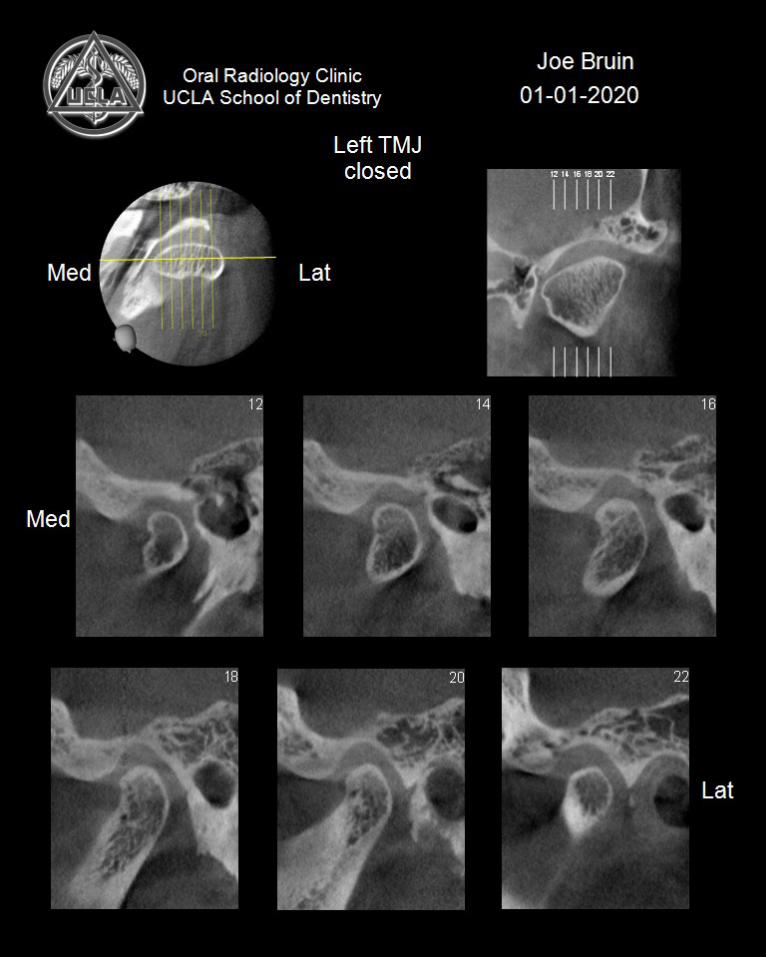
Clinical Indications TMJ evaluation Radiographic Examination PANORAMIC RADIOGRAPH AND CONE-BEAM CT VOLUMETRIC SCANS OBTAINED WITH THE MORITA ACCUITOMO 170 SCANNER AND THE MORITA VERAVIEW SCANNER UTILIZING 4 X 4 CM FIELDS OF VIEW Image Quality: Optimal for diagnosis Radiographic findings TMJ: Right Side The right TMJ demonstrates markedly altered morphology. The condylar head is enlarged, and demonstrates concave contours at its medial and lateral aspects. There is extensive sclerosis and a multiple erosions of the cortical and trabecular bone. Nodular opacities are observed circumferentially around the condylar head. No significant alteration of the morphology of the glenoid fossa and articular eminence is noted. Overall, the circumferential opacities and extensive degenerative changes involving the condylar head are consistent with synovial chondromatosis. In the closed position, the condylar head is seated in a significantly protruded position within the glenoid fossa. Upon opening, there is minimal translation of the condylar head to approximately 6 mm short of the tip of the articular eminence. TMJ: Left Side The superior cortical outline of the left condylar head, glenoid fossa and articular eminence is sclerotic. Minimal subchondral sclerosis is seen of the underlying trabecular bone. Mild flattening of the superior surface of the condylar head is seen at its medial pole. In the closed position, the condylar head is centrally positioned within the glenoid fossa. Upon opening, the condylar head translates to approximately 6 mm short of the tip of the articular eminence. On the panoramic radiograph, a small dome shaped radiopacity is observed along the floor of the right maxillary sinus, consistent with a mucous retention phenomenon. The remainder of the facial bones, paranasal sinuses, and airway where visualized are within normal limits. Impressions:
1. Circumferential, nodular radiopacities around the right condylar head consistent with synovial chondromatosis.
2. Severe degenerative changes and bone remodeling of the right condylar head. Clinical correlation with patient symptomatology is recommended.
3. A protruded position of the right condylar head and reduced translatory movement suggestive of internal derangement of the articular disc in the right TMJ.
___________________________
Sanjay Mallya, BDS, MDS, PhD Diplomate, American Board
of Oral & Maxillofacial Radiology