oral surgery original article - ijcdr.netijcdr.net/admin/php/uploads/80_pdf.pdf · oral surgery...
TRANSCRIPT
Oral Surgery Original Article
International Journal of Clinical And Diagnostic Research ISSN 2395-3403
Volume 5, Issue 1, Jan-Feb 2017
© Glorigin Lifesciences Private Limited.
Intl. J. Clin. Diag. Res. 2017;5(1):II www.ijcdr.net
A CLINICO-RADIOGRAPHIC EVALUATION OF WOUND HEALING AND BONE
FORMATION AFTER SURGICAL REMOVAL OF IMPACTED THIRD MOLARS
USING PLATELET RICH FIBRIN (PRF) AND PRF IMPREGNATED WITH
2% METRONIDAZOLE
R. Dilip Kumar, Shashant Kumar Gupta, HP. Raghuveer, Shobha ES, Prashanth NT,
VinodRangan, Archana G. Panicker
Abstract
Aim and objective: The aim of the study was to compare the efficacy of Platelet Rich Fibrin with
or without 2% Metronidazole in mandibular third molar surgery by assessing the postoperative
pain, swelling, wound healing (by wound infection and dehiscence ) comparing the quantity of
bone healing at various time intervals
Materials and Methods: Twenty patients aged 20-40 years who visited the Department of Oral
and Maxillofacial Surgery, DayanandaSagar College of Dental Sciences were randomly selected
and divided into a Control group and a Study group of ten patients each. Clinical assessment was
done for both groups for wound dehiscence, wound infection, swelling (by using a modification of
the Scultz-Mosgau method) and pain (by Visual Analog Scale) after 3rd
post-operative day, 7th
post-operative day and 14th
post-operative day. The radiographic assessment of mean bone height
was done by using Orthopantomograph and evaluated preoperatively, immediate post-operatively
and 3rd month post-operatively.
Results: The VAS score of Study group (6.1 ±0.9) was lesser than the Control group (6.8 ±1.03)
but the result was not statistically significant (p value was 0.14) on the 3rd
post-operative day.
Swelling on 3rd post-operative day (3.48±0.5), 7th post-operative day (1.90±0.5) and 14th post-
operative day (0.50±0.5) on the study group were consistently lesser than the control group on 3rd
post-operative day (4.71±1.2), 7th
post-operative day (2.54±1.2) and 14th post-operatively day
(0.81±1.2) but statistical significance value was found to be only on 3rd day (p value was 0.008).

Dilip kumar et al., A Clinico-Radiographic evaluation of……..
Intl. J. Clin. Diag. Res. 2017;5(1):II www.ijcdr.net
The mean bone height post-operatively after 3rd month in study group was higher than the control
group and the result was statistically significant (p value was < 0.001).
Conclusion: The Platelet Rich Fibrin with 2% Metronidazole group recorded reduced pain,
swelling as well as enhanced and faster bone healing compared with those in the control group.
Author Affiliations:
Department of Oral and Maxillofacial Surgery, Dayananda Sagar College of Dental Sciences,
Bangalore, Karnataka
Keywords: Wound healing, Third molar surgery, Platelet Rich Fibrin, 2% Metronidazole
*Corresponding Author:
Dr. R. Dilip Kumar, Professor, Department of Oral and Maxillofacial Surgery, Dayananda Sagar
College of Dental Sciences, Bangalore, Karnataka.
INTRODUCTION
Removal of the lower third molars is one of
the most common procedures in oral and
maxillofacial surgery and it is often attended
by complications which are distressing to
patients[1]
. The incidence of infectious and
inflammatory post-operative sequelae
following an impacted mandibular third molar
surgery varies between 0% and 45%,
according to different published literature[2,3]
.
Healing of extraction socket is a coordinated
sequence of biochemical, physiologic,
cellular, and molecular responses involving
various cell types, growth factors, hormones,
cytokines, and other proteins, which is
intended to restore the tissue integrity and
functional capacity after injury[4]
.
Various methods have been suggested to
enhance socket healing and reduce the
postoperative wound healing complications
after third molar surgery. Developments of
bioactive surgical additives are one of the
most promising clinical researches which have
been used to regulate inflammation and
increase the speed of the healing process. It is
known that platelet plays a crucial role not
only in the hemostasis, but also in the process
Dilip kumar et al., A Clinico-Radiographic evaluation of……..
Intl. J. Clin. Diag. Res. 2017;5(1):II www.ijcdr.net
of wound healing. Autogenous Platelet Rich
Growth Factors (PRGF) are successfully used
in stimulating bone regeneration and promotes
healing after the surgical removal of third
molar tooth[5- 11]
.
The various in vivo and in vitro studies of the
release of growth factors from Platelet Rich
Fibrin has shown that PRF yields better results
over PRP and thus helped in optimising its
clinical application. It has been proved that
there is a slower release of growth factors and
better healing properties with PRF than
compared to PRP. It has been observed and
shown that the cells are able to migrate from
fibrin scaffold; while some authors
demonstrated the PRF as a supportive matrix
for bone morphogenetic protein as well[12]
.
Metronidazole has shown the greatest promise
in randomized double-blind studies in third
molar surgery. It has a narrower spectrum and
targets primarily anaerobes, therefore reducing
the chance of bacterial resistance as well as
being associated with fewer side effects than
other antibiotics[13, 14]
. However, there is
dearth of literature regarding the use of
biological dressing impregnated with
antibiotics in the mandibular third molar
surgery. Hence, we definitely believe that
there is a need for study to compare the role of
PRF along with Metronidazole for enhanced
wound healing as well as post-extraction
complications and bone regeneration after
mandibular third molar surgery.
MATERIALS AND METHODS
The study was done on twenty outpatients of
the age group of 20-40 years who visited the
Department of Oral & Maxillofacial Surgery,
DayanandaSagar College of Dental Sciences,
Bangalore, for surgical removal of impacted
mandibular third molars.. They were divided
into two equal groups at random, i.e. Control
group, in which Platelet Rich Fibrin was
placed into the extraction socket and a Study
group, in which Platelet Rich Fibrin with 2%
Metronidazole was placed in the extraction
socket. A custom made case sheet was
designed for the study to record the case
history of the patients pre-operatively. All
patients were operated by a single operator
and all types of impacted teeth were selected
for the study. Prior to surgery, patients were
examined for fitness to undergo the surgery by
a thorough case history, clinical examination
and routine investigations. Medically
compromised patients and cases where the
surgical site was associated with active
infection or local pathology of hard and soft
tissue were excluded from the study.
Dilip kumar et al., A Clinico-Radiographic evaluation of……..
Intl. J. Clin. Diag. Res. 2017;5(1):II www.ijcdr.net
Clinical Assessment was performed pre-
operatively and at the 3rd
, 7th
and 14th
day
postoperatively for wound healing with
respect to pain, swelling, wound infection,
wound dehiscence and bone height. Pain was
evaluated using a 10 point Visual Analog
Scale. Swelling was calculated by using a
modification of the method of Scultz-Mosgau
et al and this required measuring the distances
from the Tragus to the soft tissue Pogonion
and corner of the mouth to the Tragus. The
arithmetic sum of the two measurements was
used to determine the facial swelling. The
percentage of facial swelling was calculated
from the difference of the measurements made
in the preoperative and postoperative i.e.
(Swelling Postoperatively – Swelling
Preoperatively) / Swelling Preoperatively X
100). The criteria used to assess wound
infection were presence of suppuration,
increase in pain, erythema, local warmth,
swelling and purulent discharge at the
operated site. Wound Dehiscence (if present)
was clinically evaluated on 3rd
, 7th and 14th
day post-operatively. For radiological
assessment, pre-operative, immediate post-
operative and 12th
week postoperative OPG
was taken and measurements were done to
evaluate the quantity of bone using listed
standard landmarks on the radiograph. Bone
height from the distal cement-enamel junction
of second molar to a point on the superior
border of inferior alveolar canal, was
measured by the digital VernierCaliper in a
straight line.
After taking all the aseptic precautions, 10ml
of intravenous blood was drawn from the
antecubital region through 10 ml sterile
syringe which will be transferred to
centrifugal test tubes. The blood was
centrifuged using a tabletop centrifuge for 10
minutes at 3000 rpm and Platelet Rich Fibrin
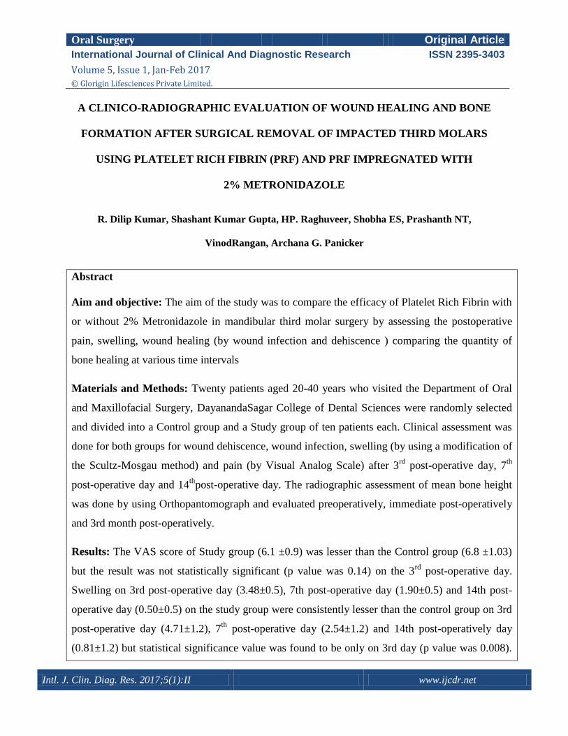
is obtained in the middle layer. (Fig.1)
Fig 1: Preparation of Platelet Rich Fibrin,
A: Withdrawal of 10 ml blood for preparation
of PRF,
B: Blood taken in the test tube,
C: Preparation of PRF for 10 minutes at
3000rpm,
D: Platelet Rich Fibrin matrix present in the
middle layer
Dilip kumar et al., A Clinico-Radiographic evaluation of……..
Intl. J. Clin. Diag. Res. 2017;5(1):II www.ijcdr.net
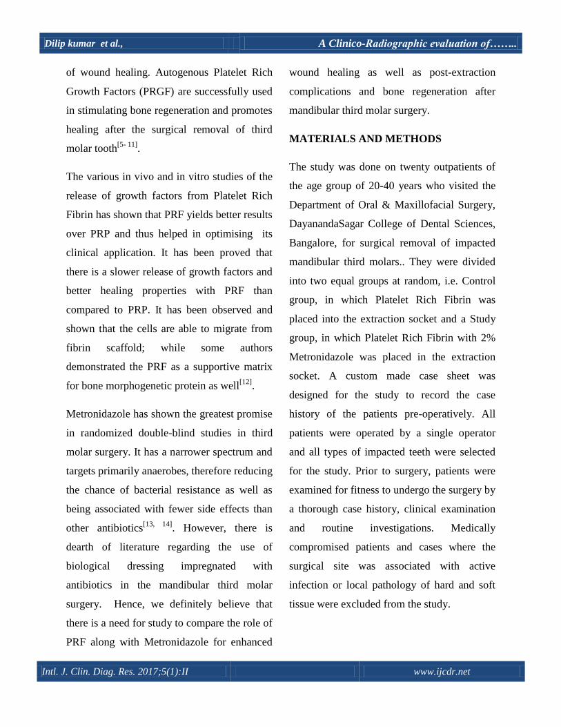
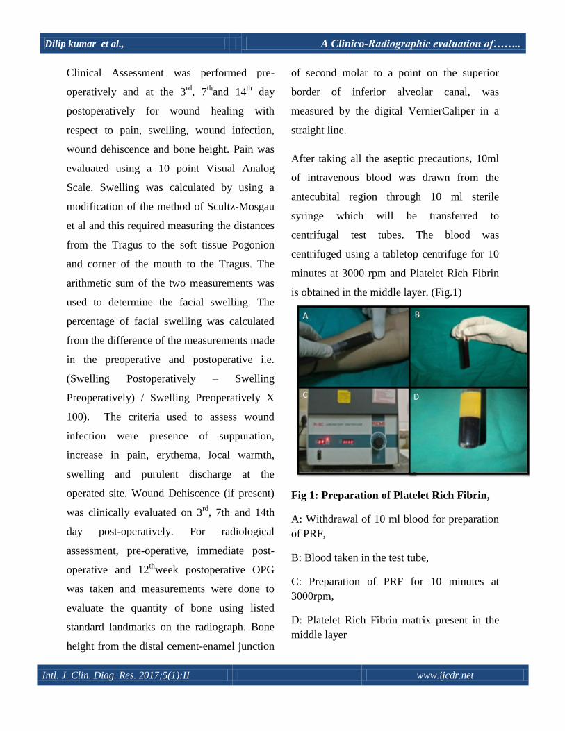
Under sterile aseptic conditions, 2 grams of
Metronidazole powder was dissolved in 100ml
of distilled water to get the prepared solution
of 2% Metronidazole. Platelet Rich Fibrin was
added to the prepared 2% metronidazole
solution before the surgical procedure, kept in
petridish till the surgical procedure was
completed and then placed in extracted third
molar socket. (Fig.2 and Fig.3) The surgical
procedure was standardized in all cases. PRF
(in case of Control group) or PRF with 2%
Metronidazole (in case of Study group) was
placed into the socket with the help of
Adson’s non toothed forceps. Sutures were
placed with round body needle 3-0 black silk.
All the patients were instructed to take anti-
inflammatory analgesic medication.
Statistical Analysis
Student t test (two tailed, independent) has
been used to find the significance of study
parameters on continuous scale between two
groups, inter group analysis on metric
parameters. P value: 0.01<P≤0.05 showed
moderate significance and P value ≤0.01 was
considered strongly significant.
Fig.2: PRF matrix without 2%
metronidazole solution
Fig.3: PRF matrix placed in 2%
Metronidazole solution
RESULTS
The mean age of the patients in the Control
group was 28.50± 3.8 years and the study
group was 28.90± 2.2 years. The control
group consisted of 8 male and 2 female
Dilip kumar et al., A Clinico-Radiographic evaluation of……..
Intl. J. Clin. Diag. Res. 2017;5(1):II www.ijcdr.net
patients and study group consisted of 5 male
and female patients each. (Table 1) The VAS
score of Study group (6.1 ±0.9) was lesser
than the Control group (6.8 ±1.03) but the
result was not statistically significant (p value
was 0.14) on the 3rd
post-operative day. At the
7th
day post-operatively the mean pain score
was similar in both the groups study group
was as compared to the control group was the
results were not statistically significant. At the
14th
day post-operatively the mean pain score
was similar in both the groups and the p value
was not comparable (Table 2). On the 3rd
, 7th
and the 14th
post-operative days, the mean
swelling score were lesser for the Study group
as compared to the Control group (Table 3).
Wound dehiscence and wound infection was
absent on the 3rd
, 7th
and 14th
post-operative
day in both the groups which shows that the
results was not comparable. The mean score
of bone height in the study group post-
operatively 3rd
month was (18.5 ±1.8) and
found to be higher than the control group post-
operatively 3rd
month (13.91±1.6), which was
highly statistically significant.(Table 4)
DISCUSSION
In regular tooth extraction procedures,
eventual contamination of the wound site by
food residue and oral bacteria is a common
finding as both the blood clot as well as the
wound is directly exposed to the oral cavity.
The primary blood clot plays a vital role in
the growth of new tissue at the early stages
of wound healing and the disruption/ loss of
this primary clot often results in reduced rate
of growth in the mucous epithelium into the
extraction socket owing to the lack of
support from the blood clot that markedly
retards the process of healing, further
causing an infection from contamination that
results in discomfort and pain [15]
.

Dilip kumar et al., A Clinico-Radiographic evaluation of……..
Intl. J. Clin. Diag. Res. 2017;5(1):II www.ijcdr.net
Table 1: Distribution of participants according to gender
Gender
Participants
Total
n (%)
Control
n (%)
Study
n (%)
Male
Female
8 (61.5) 5 (38.5) 13 (100)
2 (28.6) 5 (71.4) 7 (100)
Total 10 (50) 10 (50) 20 (100)
Table 2: Mean (SD) pain scores on VAS (n = 10 in each group)
Table 3: Mean (SD) swelling (n = 10 in each group)
Post-operative
day
Study PRF with
2%
Metronidazole
Control (PRF) P value
3 3.48 (0.5) 4.71 (1.2) 0.008**
7 1.90 (0.5) 2.54 (1.23) 0.15
14 0.50 (0.5) 0.81 (0.5) 0.17
**Statistically Significant
Post-operative
day
Study PRF with
2%
Metronidazole)
Control (PRF) P value
3 6.1 (0.9) 6.8 (1.03) 0.14
7 1.4 (0.5) 1.4 (0.5) 1.0
14 0 (0) 0 (0) -

Dilip kumar et al., A Clinico-Radiographic evaluation of……..
Intl. J. Clin. Diag. Res. 2017;5(1):II www.ijcdr.net
Table 4: Mean (SD) Bone height: - from the distal CEJ of 2nd molar to a point on superior
border of inferior alveolar canal in a straight line.
**Statistically Significant
DISCUSSION
In regular tooth extraction procedures,
eventual contamination of the wound site by
food residue and oral bacteria is a common
finding as both the blood clot as well as the
wound is directly exposed to the oral cavity.
The primary blood clot plays a vital role in the
growth of new tissue at the early stages of
wound healing and the disruption/ loss of this
primary clot often results in reduced rate of
growth in the mucous epithelium into the
extraction socket owing to the lack of support
from the blood clot that markedly retards the
process of healing, further causing an
infection from contamination that results in
discomfort and pain [15]
.
Several mechanisms have to be in sync with
the formation of the clot and its sustenance.
Within the framework of the Fibrinolytic
theory of Alveolar Osteitis (Dry Socket), clot
lysis and loss may be categorically viewed as
multifactorial in origin. The various
contributing mechanism include Plasminogen-
mediated fibrinolysis caused by physiologic
tissue-type plasminogen activators that get
locally liberated by surgical trauma,
Study (PRF with
2% Metronidazole) Control (PRF) P value
Bone height pre-op
15.62 (2.4)
11.89 (1.3) 0.0004**
Bone height IMM
PO 15.32 (2.3) 11.89 (1.3) 0.0004**
Bone height 3rd
month PO
18.5 (1.8)
13.91 (1.6)
<0.001**

Dilip kumar et al., A Clinico-Radiographic evaluation of……..
Intl. J. Clin. Diag. Res. 2017;5(1):II www.ijcdr.net
Non-physiologic activators elaborated by oral
bacteria present in or introduced into the
extraction wound, Non-Plasminogen-mediated
fibrinolysis mediated by other mechanisms
such as bacterial liberated substances as well
as by a leukocyte-mediated fibrinolysis (either
Plasminogen-mediated or independent)
occurring as a result of an acute local
inflammatory response to surgical trauma,
manipulation or by local bacterial
challenges[16]
.
Having understood the probable causes of
Clot failure as stated, relevant literature has
addressed various topical applications of
pharmacological agents that are used to
stabilize the primary clot at the extraction site.
The advantages of these pharmacological
substances is that they allow a greater local
concentration such that a stable fibrin clot
develops and this is more than that normally
expected from systemic administration and
further minimizes the potential adverse effects
and sensitization that accompanies this route
of administration [17]
.
The efficacy of localized (intra-alveolar)
antibiotic therapy placed immediately post-
operative has always been a debate.
Antibiotics have been used both as a single
regimen as well as multi-regimen in various
formulations and dosages whilst some may be
administered via numerous carriers in an
attempt to alleviate Alveolar Osteitis and its
associated symptoms. Several authors suggest
the use of the same in order to prevent post-
operative complications. Prophylactic anti-
bacterials administered pre-operative either
systemically or locally include Penicillins,
Tetracyclines, Clindamycin, Erythromycin,
Tinidazole as well as Metronidazole. These
pharmacological agents have shown better
results when used pre-operatively rather than
post-operatively. Of all the discussed agents,
Metronidazole showed superior results in
randomized double-blinded studies that
showed its effectiveness in reducing the
incidence of Alveolar Osteitis, especially
owing to its narrow anti-bacterial spectrum
that is more anaerobicidal and that it is
associated with fewer and more infrequent
adverse effects than those with high resistance
developing secondary to administration of
Penicillin and Erythromycin as well as
Pseudomembranous colitis inducing
Clindamycin [18,19, 20]
.
An important consideration about the use of
Metronidazole is in the placement of the drug
at the right concentration such that maximum
efficacy of the drug may be obtained at the
local wound site. The placement of
Metronidazole dressing directly into the
extraction-site/ socket is aimed primarily to
Dilip kumar et al., A Clinico-Radiographic evaluation of……..
Intl. J. Clin. Diag. Res. 2017;5(1):II www.ijcdr.net
destroy anaerobic microorganisms [17, 18]
.
Further, the use of pre-operative antibiotics
prophylactically showed marked reduction in
the density of anaerobes in the oral cavity and
aided in the reduction of post-operative
infections. With recent advances and an in-
depth understanding of wound healing, newer
approaches are being developed which play a
direct active role in the healing while
eliminating the potential downside of non-
vital materials [13,19]
.
A promising mode of investigation involves
the use of Platelet Rich Plasma (PRP) to aid
the wound healing process. Platelet rich
plasma is an autologous preparation of a
concentration of platelets and plasma that can
be created by centrifuging the patient’s own
blood. Several publications have cited the
advantages and the uses of platelet rich plasma
as an adjunct in the peri and post-operative
management following Periodontal as well as
Oral Surgical applications [21]
.
Platelet rich Fibrin (PRF) is a second-
generation platelet concentrate defined by
Choukron as an autologous leukocyte and
platelet-rich fibrin biomaterial[13]
. Platelet rich
fibrin has been used as a close alternate to the
use of platelet rich plasma and represents a
new modality that involves the use of a
platelet gel therapeutic concept with
simplified processing without any artificial
biochemical modifications.15
Unlike other
platelet concentrates, the technique does not
necessarily require anticoagulants or bovine
thrombin ; rendering it a pure preparation of
centrifuged blood without the use of any
additives making it a more advantageous
choice [15, 16]
. Platelet rich fibrin can be used in
an array of clinical situations such as to
improve soft tissue healing, protection of bone
graft material over the surgical area, bone
remodeling procedures or even as an osteo-
conductive filling material that aids bone
formation following a Maxillary sinus lift
procedure [14]
.
Platelet rich fibrin has several advantages to
its credit in terms of ease of preparation, less
technique sensitive, lack of biochemical
handling of blood and its autologous nature
over otherwise traditionally prepared platelet
rich plasma gels or bovine thrombin used in
the preparation of platelet rich plasma gel that
may lead to development of antibodies to
clotting factors V, IX and Thrombin resulting
in the risk of life-threatening coagulopathies
[16,22].
In the present study, we have advocated the
use of Platelet Rich Fibrin as a biological
surgical additive that offers and aids optimum
soft tissue wound healing as well as Hard
Dilip kumar et al., A Clinico-Radiographic evaluation of……..
Intl. J. Clin. Diag. Res. 2017;5(1):II www.ijcdr.net
tissue regeneration, along with the
incorporation of 2% Metronidazole in order to
assess whether the use of this amalgamation is
beneficial after third molar surgery with
regard to the wound healing and bone
formation. The chosen mode of radiographic
investigation was with the use of an
Orthopantomogram (OPG) and bone height
measurements were compared and done 3
months post operatively.
Our study showed that in the Study group
(Platelet rich fibrin with 2% Metronidazole),
the patients showed better wound healing,
decreased post-operative sequelae such as pain
and fswelling while the bone height measured
was higher than that of the control group
(Platelet rich fibrin without 2%
Metronidazole). The mean post-operative pain
was considerably lower in the study group as
compared with the control group at all times,
however, the result was not statistically
significant [p=0.14] (Table: 2). The mean
percentage of facial swelling in the study
group was lower than that of the control group
during the entire time period but the
differences were statistically significant only
on the third day [p value was 0.008](Table: 3).
The mean bone-height in the study group was
also found to be higher than the control group
and the results were highly significant with the
p value <0.001. (Table: 4).
Our findings support the use of Platelet Rich
Fibrin impregnated with 2% Metronidazole
and has a superior advantage clinically in the
healing of extraction sockets following third
molar surgery that was further confirmed
radiographically. The study however, had a
limitation of a small sample size and a short
duration of follow-up. We acknowledge that a
larger sample size with a longer period of
follow-up would be essential to obtain a more
statistically significant result.
CONCLUSION
Platelet Rich Fibrin is an autologous
preparation which is found to be clinically
effective and economical than any other of the
plethora of available regenerative materials
and is more productive when used along with
2% Metronidazole solution. However, long
term, multicentre randomized, controlled
clinical trial will be required to know its
clinical and radiographic effect over bone
regeneration. Hence, it can be inferred that the
use of PRF impregnated with 2%
Metronidazole solution effectively proves to
be beneficial in the healing during the post-
operative period of tooth sockets following
third molar surgery.
Dilip kumar et al., A Clinico-Radiographic evaluation of……..
Intl. J. Clin. Diag. Res. 2017;5(1):II www.ijcdr.net
REFERENCES
1. Osunde OD, Adebola RA, Omeje UK.
Management of inflammatory complications
in third molar surgery: A review of the
literature. Afr Health Sci. 2011; 11(3): 530–
537.
2. Bui CH, Seldin EB, Dodson TB. Types,
frequencies, and risk factors for complications
after third molar extraction. J Oral Maxillofac
Surg. 2003; 61(12):1379–89.
3. Benediktsdóttir IS, Wenzel A, Petersen JK,
Hintze H. Mandibular third molar removal:
Risk indicators for extended operation time,
postoperative pain, and complications. Oral
Surg Oral Med Oral Pathol Oral
RadiolEndod.2004; 97:438-46.
4. Saravanakumar B, Julius A, Sarumathi T,
Aarthinisha V, Manisundar N. Therapeutic
Effects and Concepts in the Use of Platelet-
Rich Fibrin (PRF) on Alveolar Bone Repair-A
Literature Review. Middle-East J. Sci. Res.
2014; 19 (5): 669-673
5. Figueiredo R, Castello´n EV, B-Ayte´s L,
Gay-Escoda C. Incidence and clinical features
of delayed-onset infections after extraction of
lower third molars. Oral Surg Oral Med Oral
Pathol Oral Radiol Endod.2005; 99: 265-9.
6. Del Fabbro M, Bortolin M, Taschieri S. Is
autologous platelet concentrate beneficial for
post-extraction socket healing? A systematic
review.Int J Oral Maxillofac. Surg. 2011; 40:
891–900.
7. Simon BI, Gupta P, Tajbakhsh S.
Quantitative evaluation of extraction socket
healing following the use of autologous
platelet-rich fibrin matrix in humans. Int J
Periodontics Restorative Dent. 2011;
31(3):285-95.
8. Ogundipe O K, Ugboko V I, Owotade F J.
Can Autologous Platelet-Rich Plasma Gel
Enhance Healing After Surgical Extraction of
Mandibular Third Molars? J Oral
MaxillofacSurg 69:2305-2310, 2011.
9. Simon BI, Zatcoff AL, Kong JJ, O’Connell
SM. Clinical and histological comparison of
extraction socket healing following the use of
autologous platelet-rich fibrin matrix (PRFM)
to ridge preservation procedures employing
demineralized freeze dried bone allograft
material and membrane. Open Dent J. 2009; 3:
92–99.
10. Noroozi AR, Philbert RF. Modern
concepts in understanding and management of
the ―dry socket syndrome: comprehensive
review of the literature. Oral Surg Oral Med

Dilip kumar et al., A Clinico-Radiographic evaluation of……..
Intl. J. Clin. Diag. Res. 2017;5(1):II www.ijcdr.net
Oral Pathol Oral RadiolEndod.2009; 107: 30-
35.
11. DohanEhrenfest DM, de Peppo GM,
Doglioli P, Sammartino G. Slow release of
growth factors and thrombospondin-1 in
Choukroun's platelet-rich fibrin (PRF): a gold
standard to achieve for all surgical platelet
concentrates technologies. Growth Factors.
2009; 27(1):63-9.
12. Choukroun J, Diss A, Simonpieri A,
Girard MO, Schoeffler C, Dohan SL, Dohan
AJ, Mouhyi J, Dohan DM. Platelet-rich fibrin
(PRF): a second-generation platelet
concentrate. Part IV: clinical effects on tissue
healing.Oral Surg Oral Med Oral Pathol Oral
Radiol Endod. 2006; 101: E56-60.
13. Choukroun J, Adda F, Schoeffler
C. Vervelle A. An opportunity in perio-
implantology: The PRF (in French).
Implantodontie 2001; 42: 55-62. .
14. Lauritano D, Avantaggiato A, Candotto V,
Zollino I, Carinci F. Is platelet rich fibrin
really useful in oral and maxillofacial surgery?
Lights and shadows of this new technique.Ann
Oral Maxillofac Surg. 2013; 1: 25-29.
15. Li YQ, Shann ZC. Initial study on
facilitating wound healing after tooth
extraction by using microbial fiber membrane-
flagyl. American association of oral and
maxillofacial surgery, J Oral MaxillofacSurg
69:994-1002, 2011
16. Lauritano D, Avantaggiato A, Candotto
V, Zollino I, Carinci F. Platelet rich fibrin
(PRF) is really useful in oral and maxillofacial
surgery: Lights and shadows. Ann Oral
Maxillofac Surg. 2013; 1(3): 25.
17. Lucarelli E, Beretta R, Dozza B, Tazzari
PL, O'Connel SM, Ricci F, Pierini M,
Squarzoni S, Pagliaro PP, Oprita EI, Donati D.
A recently developed bifacial platelet-rich
fibrin matrix.Eur Cell Mater.2010; 20: 13-23.
18. Rood J.P, Murgatroyd J. Metronidazole in
the prevention of Dry Socket. Br J Oral Surg.
1979; 17(1): 62-70.
19. Bergdahl M, Hedström L. Metronidazole
for the prevention of dry socket after removal
of partially impacted mandibular third molar:
a randomised controlled trial. Br J Oral
Maxillofac Surg. 2004; 42(6):555–558.
20. Poor M R. Reduction in the Incidence of
Alveolar Osteitis in Patients Treated With the
SaliCept Patch, Containing Acemannan
Hydrogel. American Association of Oral and
Maxillofacial Surgeons, J Oral axillofacSurg
60:374-379, 2002.
21. Singh J, Bharti V, Laterally positioned
flap-revised technique along with platelet rich
Dilip kumar et al., A Clinico-Radiographic evaluation of……..
Intl. J. Clin. Diag. Res. 2017;5(1):II www.ijcdr.net
fibrin in the management of miller class II
gingival recession, Dent Res J (Isfahan), 2013;
10(2): 268-273.
22. Srivastava A, Gupta KK, Agarwal K,
Kumar N, The role of growth factors in
autologous platelet concenterate for treatment
of periodontal bone defect with platelet rich
fibrin: a case report, Innovative journal of
medical and health science, 2013; 3(3): 113-
116.