orbit imaging anatomy
TRANSCRIPT

ORBIT Imaging anatomy

Imaging Recommendations
Radiography
Ultrasound• First line modality for intraocular lesions• Noninvasive, readily available
CT & MR are complementary techniques; both are indicated for evaluation of complex lesions
CT• Excellent evaluation of orbit aided by natural contrast.
between fat, bone, air & soft tissues.• Easily detects calcifications.

MR
• Optimal soft tissue contrast for globe, optic nerve, orbital structures, and intracranial findings.
• Stronger gradients, faster sequences, surface coils, routine use of fat suppression ± gadolinium improve image quality

RadiographyWATERS PROJECTION
CM- canthomeatal line; CR- central ray.(a, frontal sinus; b, medial orbital wall; c, innominate line; d, inferior orbital rim; e, orbital floor; f, maxillary antrum; g, superior orbital fissure; h, zygomatic-frontal suture; i, zygomatic arch)

CALDWELL PROJECTION
a, frontal sinus; b, innominate line; c, inferior orbital rim; d, posterior orbital floor; e, superior orbital fissure; f, greater wing of sphenoid;g, ethmoid sinus; h, medial orbital wall; i, petrous ridge; j, zygomatic-frontal suture

LATERAL PROJECTION
a, orbital roof; b, frontal sinus; c, ethmoid sinus; d, anterior clinoid process; e, sella turcica; f, planum sphenoidale

BASAL PROJECTION (SUBMENTO-VERTEX)
a, zygomatic arch; b, orbit; c, lateral orbital wall; d, posterior wall of maxillary sinus; e, pterygoid plate; f, sphenoid sinus
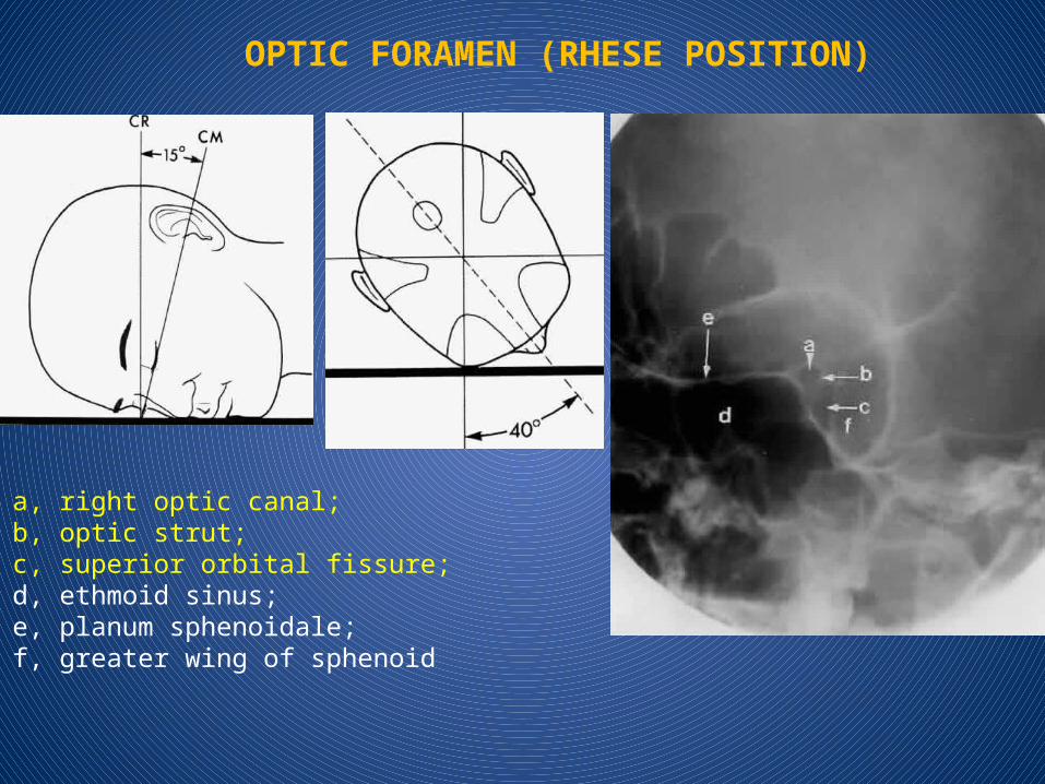
OPTIC FORAMEN (RHESE POSITION)
a, right optic canal; b, optic strut; c, superior orbital fissure; d, ethmoid sinus; e, planum sphenoidale; f, greater wing of sphenoid
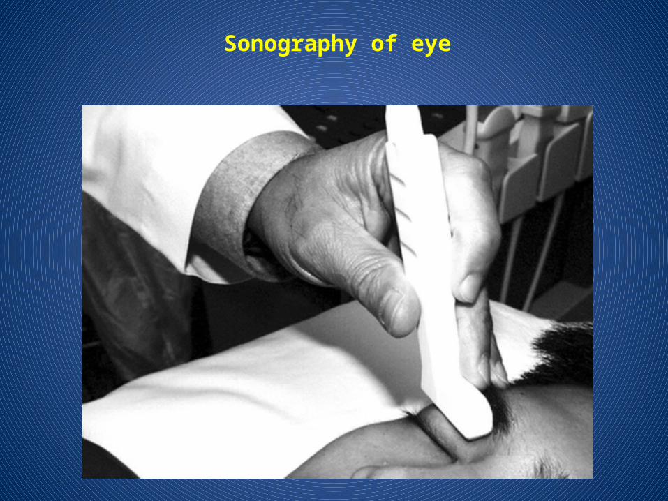
Sonography of eye

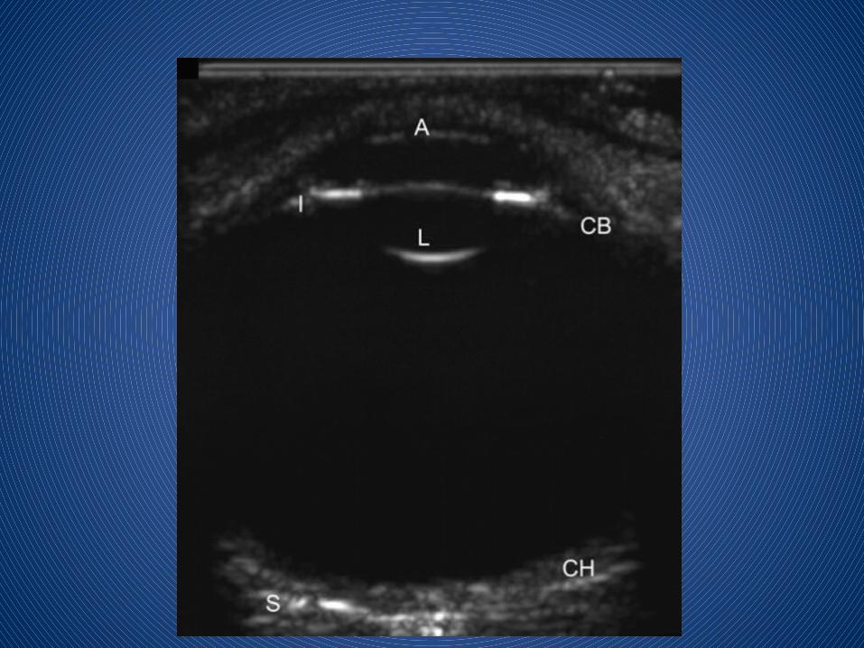
B-scan probe orientations1. Axial scan is obtained by placing the probe face directly over
the center of the cornea. The resulting B-scan image includes the lens and is bisected
by the optic nerve.


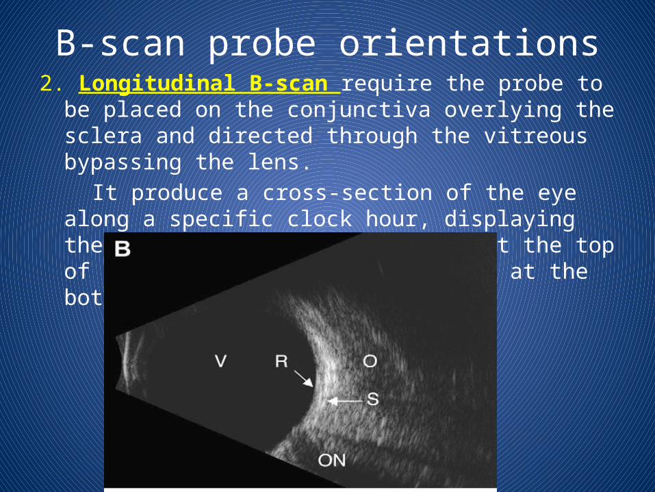
B-scan probe orientations2. Longitudinal B-scan require the probe to be placed on the
conjunctiva overlying the sclera and directed through the vitreous bypassing the lens.
It produce a cross-section of the eye along a specific clock hour, displaying the anterior portion of the eye at the top of the screen and the optic nerve at the bottom.

Imaging protocols
CT• Axial + coronal planes; thin-sections (~ 2 mm),Multislice acquisition • Soft tissue algorithm• Contrast for masses or inflammatory disease• Noncontrast only when in conjunction with MR

Bones of the Orbit

Bones of the Orbit
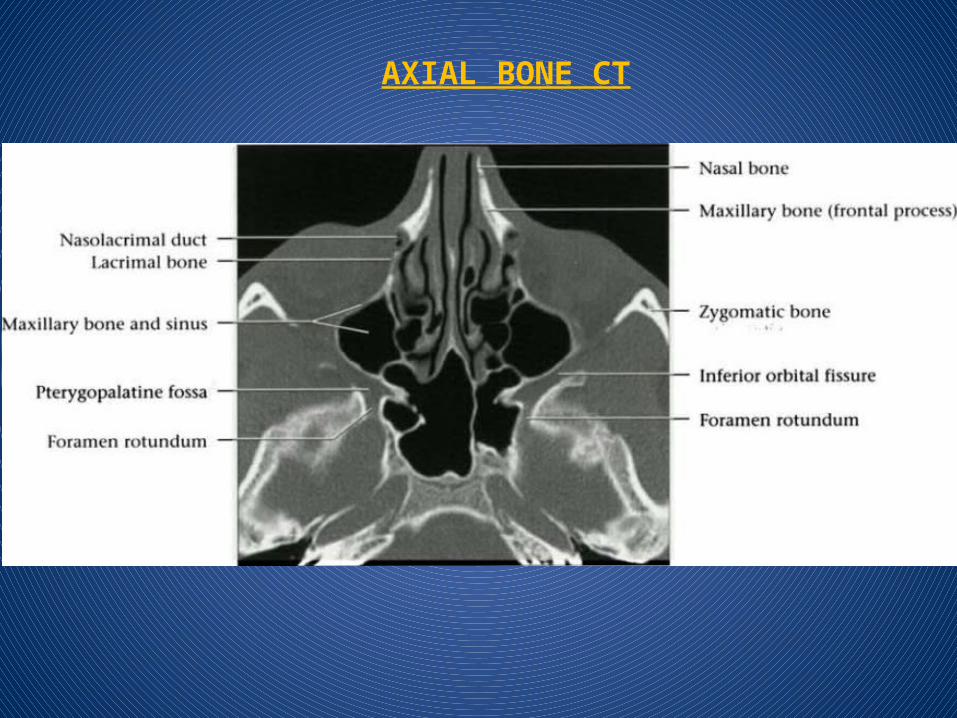
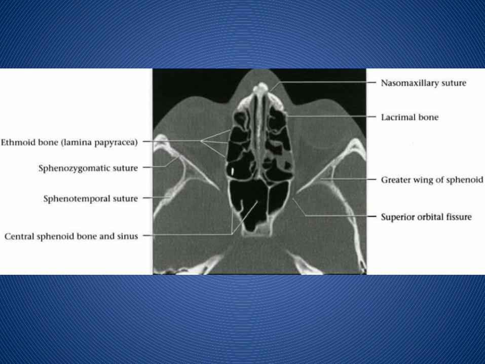
AXIAL BONE CT


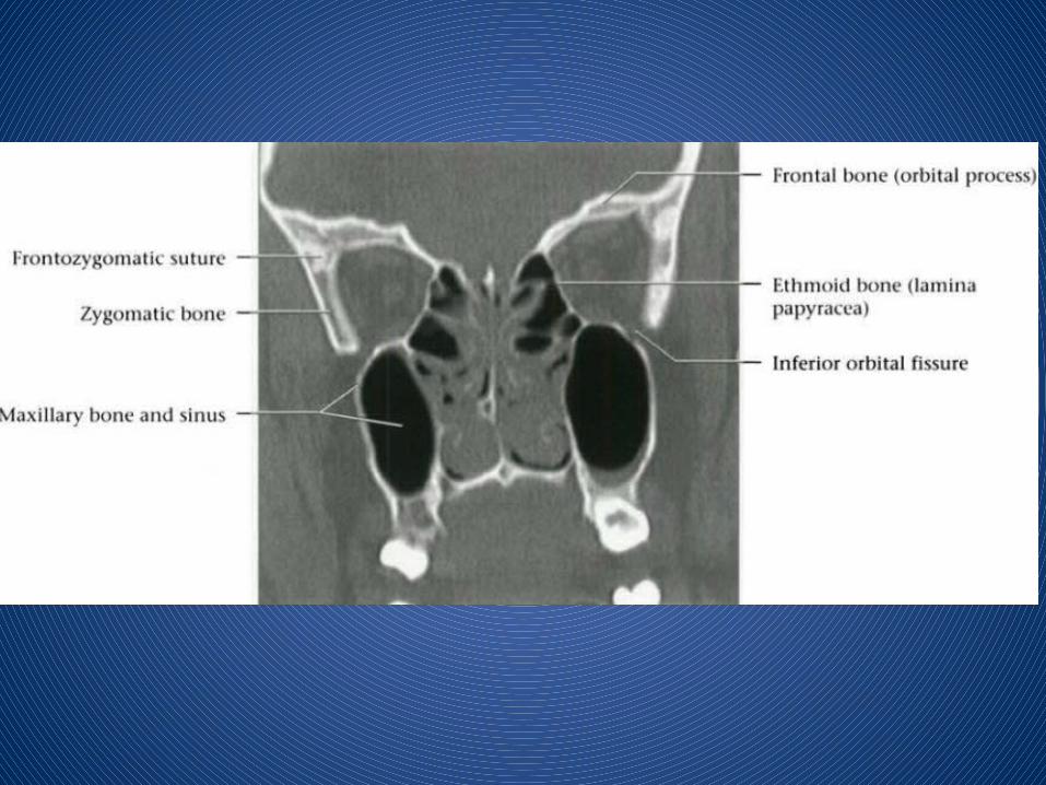
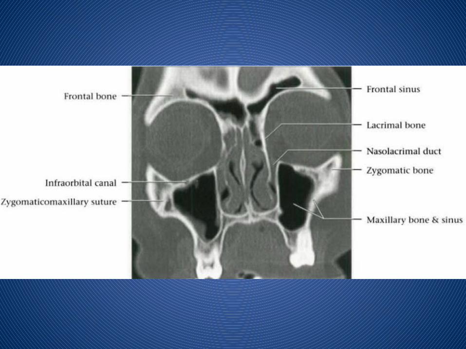
CORONAL BONE CT

MR• Axial: Above orbital roof to orbital floor• Coronal: Back of pons through globe• Thin-section (3-4 mm); small FOV (12-16 cm)• T1 pre-contrast (axial + coronal)• STIR or T2 FSE fat-saturation (axial + coronal)• T1C+ with fat-saturation (axial+coronal)
•Sagittal oblique for optic nerves

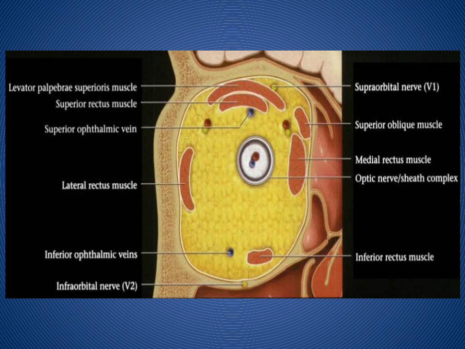


CORONAL T1 MR


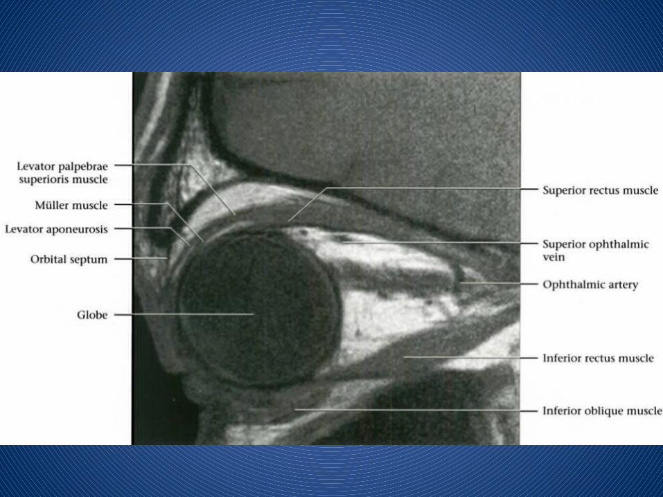
OBLIQUE SAGITTAL T1 MR
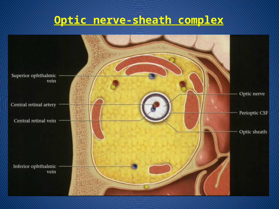
Optic nerve-sheath complex


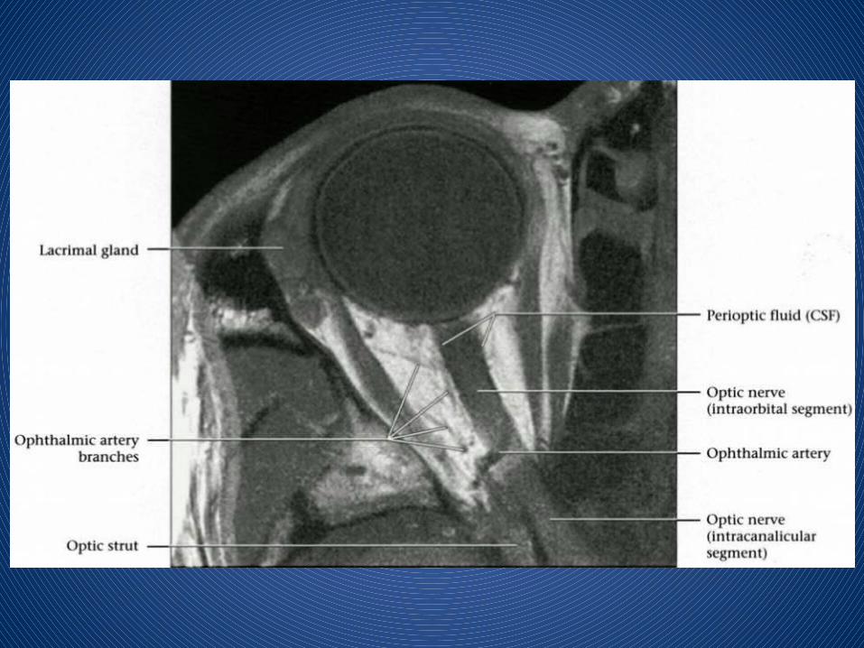
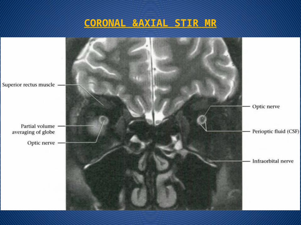
CORONAL &AXIAL STIR MR


GLOBE


THANK YOU