organization and transcription analyses of the
TRANSCRIPT
Organization and transcription analyses of the
immunoglobulin genes in cattle and horses
Dissertation
to obtain the Ph.D. degree
in the Ph.D. Program for Agricultural Sciences in Göttingen (PAG)
at the Faculty of Agricultural Sciences,
Georg-August-University Göttingen, Germany
presented by
Stefanie Walther
born in Sangerhausen, Germany
Göttingen, May 2016

D7
1. Name of supervisor: Prof. Dr. Dr. Claus-Peter Czerny
2. Name of co-supervisor: Prof. Dr. Sven König
Date of dissertation: May 12, 2016

Table of Contents
I
Table of Contents Summary ..................................................................................................................... 1
General Introduction .................................................................................................. 4
References ............................................................................................................... 6
Chapter 1: Immunoglobulins ..................................................................................... 7
General Structure of immunoglobulins ...................................................................... 8
Immunoglobulin heavy chains ..............................................................................10
Immunoglobulin light chains.................................................................................13
Development of the immunoglobulin repertoire ........................................................16
References ..............................................................................................................20
Chapter 2: Equine Immunoglobulins .......................................................................25
Equine immunoglobulins and organization of immunoglobulin genes .................26
Abstract ...................................................................................................................27
1. Introduction ..........................................................................................................28
2. Immunoglobulins in equine offspring ....................................................................30
3. The equine immunoglobulin heavy chain gene locus ...........................................31
4. Equine immunoglobulin light chain gene loci ........................................................44
5. Transcription analyses of heavy and light chain genes ........................................55
6. New allotypic variants of IGLC .............................................................................58
7. Future Directions .................................................................................................60
Acknowledgments ....................................................................................................61
References ..............................................................................................................62
Transcriptional analysis of equine λ-light chains in the horse breeds Rhenish-German Coldblood and Hanoverian Warmblood ....................................................69
Abstract ...................................................................................................................70
1. Introduction ..........................................................................................................71
2. Material and methods ..........................................................................................73
3. Results ................................................................................................................78
4. Discussion ...........................................................................................................97
Acknowledgment ................................................................................................... 101
Appendix A. Supplementary data ........................................................................... 101
References ............................................................................................................ 103

Table of Contents
II
Chapter 3: Bovine Immunoglobulins ..................................................................... 107
Bovine immunoglobulin heavy and light chains ................................................... 108
Bovine immunoglobulin heavy chain gene locus .................................................... 108
The bovine immunoglobulin light chain gene loci ................................................... 115
Development of B cells and the bovine antibody repertoire .................................... 119
References ............................................................................................................ 122
Exceptionally Long CDR3H Are Not Isotype Restricted in Bovine Immunoglobulins .................................................................................................... 128
Abstract ................................................................................................................. 129
Introduction ............................................................................................................ 130
Materials and Methods........................................................................................... 132
Results .................................................................................................................. 136
Discussion ............................................................................................................. 144
Supporting Information........................................................................................... 147
Acknowledgments .................................................................................................. 164
References ............................................................................................................ 165
Development of a bioinformatics framework for the detection of gene conversion and the analysis of combinatorial diversity in immunoglobulin heavy chains in four cattle breeds .................................................................................................... 169
Abstract ................................................................................................................. 170
Author Summary .................................................................................................... 171
Introduction ............................................................................................................ 172
Results .................................................................................................................. 175
Discussion ............................................................................................................. 197
Material and Methods ............................................................................................ 202
Acknowledgments .................................................................................................. 209
References ............................................................................................................ 210
Supporting information ........................................................................................... 214
Chapter 4: General Discussion .............................................................................. 231
General Discussion ................................................................................................ 232
Conclusions ........................................................................................................... 244
Future Prospects ................................................................................................... 246
References ............................................................................................................ 248
Chapter 5: Appendix ............................................................................................... 254
Curriculum Vitae .................................................................................................... 255

Summary
1
Summary
Initial studies on genetic aspects of immunoglobulins were performed on humans and
mice but were successfully applied to various other animals such as chicken, rabbit,
swine, cattle, and horses, too. Especially in cattle and horses, fundamental research in
immunoglobulin genetics still needs more attention to complete previous information
such as the number of available gene segments, gene families, and allotypes of
different isotypes of the immunoglobulin heavy and light chains. Results will enable the
analysis and generation of synthetic recombinant antibodies, as well as an alternating
treatment of infectious diseases to prevent resistance to antibiotics.
As reviewed in the first publication, the understanding of the organization of equine
immunoglobulin genes has increased significantly in the recent years. For equine
heavy chains, 52 immunoglobulin heavy chain variable gene segments (IGHV),
40 immunoglobulin heavy chain diversity gene segments (IGHD), 8 immunoglobulin
heavy chain joining gene segments (IGHJ) and 11 immunoglobulin heavy chain
constant region genes (IGHC) are present. Seven of these IGHCs are gamma chain
genes. Sequence diversity is increasing between fetal, neonatal, foal and adult age.
The kappa light chain contains 60 immunoglobulin kappa light chain variable gene
segments (IGKV), 5 immunoglobulin kappa light chain joining gene segments (IGKJ)
and 1 immunoglobulin kappa light chain constant region gene (IGKC), whereas there
are 144 immunoglobulin lambda light chain variable gene segments (IGLV),
7 immunoglobulin lambda light chain joining gene segments (IGLJ), and
7 immunoglobulin lambda light chain constant region genes (IGLC) for the lambda light
chain, which is expressed predominantly in horses. A decrease in IGLVs is noted
during age development, although nucleotide diversity and significant differences in
gene usage increased. A standardization of the existing nomenclature of
immunoglobulin genes is suggested.
The first experimental study focused on the identification of allotypic variants of equine
IGLC and differences in the expression of IGLV within and between the two horse
breeds Rhenish-German Coldblood (RGC) and Hanoverian Warmblood (HW). The two
breeds differ in stud book size and breeding goals. After PCR amplification of cDNA
and subcloning, 120 samples per breed were isolated and sequenced. Statistical
analysis of transcription frequencies were performed applying non-parametric tests.
The significant majority of the sequences represented IGLC6/7 in both breeds,

Summary
2
whereas IGLC1, IGLC4, and IGLC5 occurred in significant different frequencies per
breed. Five allotypic IGLC1 variants, four allotypic IGLC5 variants, and three allelic as
well as two allotypic IGLC6/7 variants were identified in breed specific proportions.
Eleven out of 144 known IGLV segments were transcribed of which IGLV15 and
IGLV17 were preferred significantly. IGLV25 displayed significant differences in the
rearrangement between both breeds. In addition, the pseudogenes IGLV101ψ and
IGLV74ψ were also identified. Rearrangements with IGLC genes showed significant
differences for IGLV15 in both breeds, whereas IGLV25 also revealed significant
differences between the breeds. The transcriptional orientation of the functional
segments had no influence on the occurrence of the IGLV.
The second experimental study carried out in cattle dealt with two main topics. On the
one hand it focused on the third complementarity determining region of the bovine
heavy chain (CDR3H) whose exceptional length previously was described as a
specificity of bovine IgG and IgM. On the other hand, the genomic organization of the
immunoglobulin heavy chain locus was analyzed with a special focus on the number of
IGHV. After isotype-specific cDNA-PCR, subcloning of 20 DNA plasmids per
immunoglobulin isotype and sequence analyzes of the variable regions, we proved the
existence of exceptionally long CDR3H in all five bovine isotypes. The sequences of
CDR3H belong to three distinct groups and possess ≤10, 12 to 31 or ≥48 amino acid
residues. Hydrophilic amino acid residues dominated in long and intermediate long
CDR3H, while short CDR3H possessed hydrophobic amino acid residues, too. All
sequences with exceptionally long CDR3H were related to the germline IGHV10.
Further, the germline IGHD2, with 148 bp in size, contributes to those CDR3H.
The genomic organization of the bovine immunoglobulin heavy-chain locus was
analyzed using the current genome assembly, Bos taurus NCBI build 6.1. A main locus
was identified on BTA21. Additional exons coding for immunoglobulin heavy chain
variable (IGHV), diversity (IGHD), and joining (IGHJ) segments, as well as for the
constant regions of different isotypes, were localized on BTA7, BTA8, BTA20, and on
unplaced contigs, too. Altogether, 36 IGHV were detected of which 13 are putatively
functional. For the phylogenetic analysis, the complete nucleotide sequences of the 36
bovine IGHV segments were aligned with one member of the human IGHV families 1 to
7. Results proved the existence of two bovine IGHV families (boVH1, boVH2). The
boVH1 comprises all functional segments. This study substantially improved the
understanding of the generation of immunoglobulin diversity in cattle.

Summary
3
The third study aimed to gain more insight into the combinatorial diversity, somatic
hypermutations and putative gene conversions of IgG in the four cattle breeds Aubrac,
German Simmental, German Black Pied, and Holstein Friesian. For the more detailed
analysis of rearranged bovine heavy chain immunoglobulin variable regions, a new
bioinformatics framework was developed by combining and refining widely used
alignment algorithms. Immunoglobulin heavy chains possessing exceptionally long
CDR3Hs can now be analyzed specifically, as well as the dominantly transcribed
IGHV, IGHD, and IGHJ segments and their recombination.
The use of 15 different IGHV segments, 21 IGHD segments, and 2 IGHJ segments
was investigated with significant different transcription levels within the breeds. There
are preferred rearrangements within the 3 groups of CDR3H lengths. In sequences of
group 1 (≤10 aa) and 3 (≥48 aa) a lower number of recombinations were observed than
in sequences of group 2 (11-47 aa). The combinatorial diversity revealed 162
significantly different rearrangements of germline IGHV, IGHD, and IGHJ segments.
The few preferably rearranged gene segments within group 3 CDR3H regions are
supposed to indicate specialized antibodies because this length is unique in cattle.
The main result of this study enabled by the new bioinformatics framework, is the
strong evidence for gene conversion as a rare event using pseudogenes fulfilling all
definitions for this particular diversification mechanism.
In conclusion, this thesis contributes to a more detailed understanding of the expressed
immunoglobulin repertoire in cattle and horses. Breed and husbandry conditions are
supposed to influence the repertoire significantly. This thesis also highlights that the
bovine heavy chain diversity is not restricted to the use of a limited number of germline
genes although there are preferred rearrangements within the three groups of CDR3H
lengths. These results will be of future importance in analyzing seroconversion data
after infection or vaccination, as well as determining breed specific differences to select
healthy, robust animals.

General Introduction
4
General Introduction Immunoglobulin genetics focuses on the special genetic aspects of immunoglobulins.
As relatively few research groups work on immunogenetics, fundamental research is
still necessary. Consequently no definite numbers of germline gene segments leading
to immunoglobulins, gene families and allotypic variants are known in many species.
Nevertheless, in the last few decades, this field of research has attracted higher
attention because of its increasing importance in the regional or global eradication and
monitoring of several infectious diseases. The availability of new high throughput
technologies and descending prices facilitate and advance the experimental work flow
in analyzing the entire immunoglobulin repertoire. Initial studies were performed on
humans and mice but successful techniques were applied to various other animals
such as chicken, rabbit, cattle, and horses, too. Especially in cattle and horses,
investigation in immunoglobulin genetics still needs more attention to complete
previous information about fundamentals such as the number of available gene
segments, gene families, and allotypes of different isotypes of the immunoglobulin
heavy and light chains. Results will contribute to the analysis and generation of
synthetic recombinant species-specific antibodies. The production of antibodies from
animals may be replaced and first of all, the results will enable an alternating treatment
of infectious diseases to avoid antibiotics and resulting resistance of antigens. Side
effects of conventional therapeutics might be excluded and higher antigen specificity
will be achieved. Therefore, recombinant antibodies and antibody fragments are
important tools for research, diagnostics and therapy (Hust et al. 2002; Hust and Dubel
2004). A widely used method for the selection of recombinant antibody fragments is the
phage display (Smith 1985; Taussig et al. 2007). Further, monoclonal antibodies have
been used successfully for the therapeutic treatment of many disorders, including
inflammatory and putative autoimmune diseases as they bind to cell-specific antigens
and mediate immune response (Hohlfeld and Wekerle 2005). By adding an appropriate
constant domain, a promising antibody fragment is converted into any antibody isotype,
for example IgG from different species (Moutel et al. 2009).
This present thesis was performed to contribute to a more detailed understanding of
immunoglobulin diversity in cattle and horses. One of the major scopes was to
investigate the bovine germline heavy chain gene segments in silico, which represent
the theoretical immunoglobulin repertoire. The expressed repertoire was then
General Introduction
5
investigated in four different cattle breeds by transcriptional analyses and statistical
methods. Further, the bovine specific mechanism of exceptionally long CDR3H
contributing to diversity was proven in all five immunoglobulin isotypes. Gene
conversion using pseudogenes was indicated for the first time in bovine heavy chains.
In addition, gene segments of transcribed equine lambda light chains were evaluated
statistically in two different horse breeds. The previous findings of horse
immunoglobulins were collated and reviewed.
The objectives of this dissertation were:
1. Equine immunoglobulins and organization of immunoglobulin genes
2. Transcriptional analysis of equine λ light chains in the horse breeds Rhenish-
German Coldblood and Hanoverian Warmblood
3. Exceptionally long CDR3H are not isotype restricted in bovine immunoglobulins
4. Development of a bioinformatics framework for the detection of gene conversion and
the analysis of combinatorial diversity in immunoglobulin heavy chains in four cattle
breeds

General Introduction
6
References
Hohlfeld R, Wekerle H. 2005. Drug insight: using monoclonal antibodies to treat multiple sclerosis. Nature clinical practice Neurology 1: 34-44.
Hust M, Dubel S. 2004. Mating antibody phage display with proteomics. Trends in biotechnology 22: 8-14.
Hust M, Maiss E, Jacobsen HJ, Reinard T. 2002. The production of a genus-specific recombinant antibody (scFv) using a recombinant potyvirus protease. Journal of virological methods 106: 225-233.
Moutel S, El Marjou A, Vielemeyer O, Nizak C, Benaroch P, Dubel S, Perez F. 2009. A multi-Fc-species system for recombinant antibody production. BMC biotechnology 9: 14.
Smith GP. 1985. Filamentous fusion phage: novel expression vectors that display cloned antigens on the virion surface. Science 228: 1315-1317.
Taussig MJ, Stoevesandt O, Borrebaeck CA, Bradbury AR, Cahill D, Cambillau C, de Daruvar A, Dubel S, Eichler J, Frank R et al. 2007. ProteomeBinders: planning a European resource of affinity reagents for analysis of the human proteome. Nature methods 4: 13-17.

Chapter 1: Immunoglobulins
7
Chapter 1:
Immunoglobulins

Chapter 1: Immunoglobulins
8
General Structure of immunoglobulins
The three-dimensional structure of immunoglobulins was determined by crystallization.
Immunoglobulins (Ig) are described as a Y-shaped tetramer composed of four
polypeptide chains containing two identical heavy chains (IGH) and two identical light
chains (IGL) that are covalently connected by disulfide bonds (Figure 1) (Edelman
1973). In addition, non-covalent interactions contribute to the connection of IGH and
IGL. While an IGH has a molecular mass of about 50 kDa, the molecular mass of an
IGL is of about 25 kDa.
Both the IGH and IGL chains are further divided functionally and genetically into
variable and constant domains that show a similar structural folding (Marchalonis et al.
2002; Ramsland and Farrugia 2002). Each chain possesses one variable domain
(IGHV, IGLV), while the number of constant domains depends on the chain type and
isotype. The variable domains of both chains are located at the arms of the Y-shaped
structure (Figure 1). The variability, mediated by the first 110 amino-terminal amino
acids, accounts for competent and highly specific antigen binding, whereby both the
heavy and light chain variable region work together (Ramsland and Farrugia 2002;
Torres et al. 2007). At the carboxyl terminus of the IGH, at least two constant regions
(IGHC2 and IGHC3) are connected to the arms of the Y shaped structure. The IGHC3
regions of both IGH interact, but steric hindrance by carbohydrate side chains inhibits
the interaction of the IGHC2 (Ramsland and Farrugia 2002).
In addition to flexible regions or hinge regions between the IGHC1 and IGHC2
connection between variable and constant domain is adjustable, too. Both torsion and
bending are possible and enable simultaneous binding of antigen structures with
different distances, as well as the interaction with signal proteins to enable effector
mechanisms (Porter 1973; Ramsland and Farrugia 2002).
The proteases papain and pepsin cleave the polypeptide chain at specific amino acids
generating fragments of different sizes (Porter 1973). While cleavage with papain
occurs at the carboxyl terminal side of the disulfide bonds within the hinge region of an
IgG resulting in two fragments, cleavage at the amino terminal side of the disulfide
bonds using papain generates three fragments. The resulting fragments are named
according to their characteristic features such as antigen binding (F(ab‘)2 fragment) and
the ability to crystallize (Fc fragment) (Yamaguchi et al. 1995). F(ab‘)2 fragments
contain the complete IGL, as well as the variable domain and the first constant region
(IGHC1) of the heavy chain. They may still bind antigens. IGHC2 and IGHC3 belong to

Chapter 1: Immunoglobulins
9
the easy crystallizable Fc fragment. These regions mediate effector mechanisms after
antigen binding.
Figure 1: Structure of an immunoglobulin G (IgG) The heavy chains are shown in blue, the light chains are red. IGLV and IGHV designate the variable regions of light and heavy chain. White boxes within IGLV and IGHV show the complementarity determining regions. The constant regions of light and heavy chain are marked by IGLC and IGHC1 to IGHC3. The hinge region is shown in black, disulfide bonds are colored in yellow. The antigen binding fragments are named Fab and Fc denominates the easy crystallizable fragment. Carbohydrate side chains are colored in green.
The secondary and tertiary structure of all domains is similar but there are differences
between variable and constant domains (Ramsland and Farrugia 2002). Both domains
consist of seven stacked antiparallel beta-strands that form a beta-barrel (Figure 2). At
the end of each beta-strand and change of direction flexible turns are generated.
Variable domains possess one more turn than constant domains. The three turns of
each the IGH and IGL variable domain generate the area for antigen binding and
consist of hypervariable regions contributing to the diversity of the immunoglobulin
repertoire as these regions are characterized by extraordinary variability. They are
located at restricted areas at the tip of the arms of the Y-shaped molecule (Figure 1, 2).
The three-dimensional structure of their amino acid motif is complementary to the
three-dimensional structure of the antigen epitope and is called complementarity
determining region (CDR1-3, Figure 2) (Wu and Kabat 1970; Decanniere et al. 2000;
Ramsland and Farrugia 2002). The length of the CDRs varies. Especially the CDR3 of
the heavy chain is highly variable as described in cattle (Walther et al. 2013).
Fab Fab
Fc
IG
HC
2
IGH
C2
IG
HC
3
IGH
C3

Chapter 1: Immunoglobulins
10
Depending on the amino acid sequence, electrostatic interactions, hydrogen bonds,
Van-der-Waals-forces, and hydrophobic interactions also contribute to antigen binding
(Braden and Poljak 1995; Braden et al. 1998). The complete specificity for antigens is
generated by the combination of IGH and IGL.
The three hypervariable regions are interspersed by four less variable parts called
framework regions (FR1-4). Their amino acid sequences within the variable and
constant domain are very similar and they are responsible for stability and structure
(Ramsland and Farrugia 2002).
Figure 2: Secondary structure of the constant (left) and variable (right) domains There are three hypervariable loops (CDR1-3) within the IGHV and the IGLV domains that account for most of the structural variability of the binding site. The CDRs are colored in red. The β-strands build the framework. The insertion of two β-strands (3b and 3c) linked by a loop containing the CDR2 in the variable domain is also marked red. (Branden 1991)
Immunoglobulin heavy chains
One immunoglobulin heavy chain is composed of one variable domain and a varying
number of constant domains. The variable region is composed of three different gene
segments that were randomly joined together during B-cell development and finally
possesses around 110 amino acid residues. These gene segments are the variable
(IGHV), diversity (IGHD), and joining (IGHJ) segments existing in multiple copies at the
heavy chain locus (Figure 3) (Taussig 1988). Numbers of the gene segments are
species specific. For instance in human, 123-129 IGHV, 27 IGHD, and 9 IGHJ
segments are known, whereby not all of these segments are functional (Lefranc 2001).
Non-functional gene segments are called pseudogenes. Mutations leading to
premature stop codons prevent the formation of functional proteins. Further, changes
within sequence regions necessary for gene recombination such as promotor or

Chapter 1: Immunoglobulins
11
recombination signal sequences lead to potentially functional gene segments, named
open reading frame (ORF) (Lefranc 1998).
Figure 3: Genomic organization and recombination of the heavy chain gene segments The immunoglobulin heavy chain variable (V1-x, light red), diversity (D1-x, orange), and joining (J1-x, dark yellow) gene segments, as well as the constant region genes (C1-2, blue) are organized in separated clusters on the genome. The recombination of single gene segments occurs at random in multiple steps and results in the deletion of gene segments between the recombined ones.
In general, the immunoglobulin heavy chain gene segments are located on a single
chromosome. However, for instance in cattle there is evidence for gene segments
located outside the major locus which are called orphan genes (Walther et al. 2013).
Each IGHV is preceded by a leader sequence and they are grouped together upstream
of an IGHD cluster. Downstream of IGHD, a separate cluster of IGHJ follows. The
constant region genes are located 3’ of the IGHJ cluster (Figure 3). Each IGHC
corresponds to a different isotype.
The different variable gene segments are divided into several families where the
members show sequence identities of at least 80% as recommended for mice (Brodeur
and Riblet 1984). Families are further combined to clans. Families within one clan are
more similar than families of different clans (Kirkham et al. 1992; Ota and Nei 1994).
The variable gene segments of reptiles, amphibians, and mammals belong to the same
three clans. During evolution, gene duplication and diversifications led to the variable
gene segments known so far.
The number of constant domains in the immunoglobulin molecule depends on the
isotype of which five different ones are described in humans, as well as in i.e. mice,
cattle, and horses (Berens et al. 1997). These isotpyes are known as IgM, IgD, IgG,

Chapter 1: Immunoglobulins
12
IgE, and IgA (Figure 4), whereby they are encoded by μ, δ, γ, ε, and α genes (Woof
and Burton 2004). IgM and IgD are coexpressed in naïve B-cells due to alternative
splicing of mRNA or class switch recombination in artiodactyls. Activation by antigen
contact leads to a switch of isotypes which is also called class switch recombination.
Figure 4: The 5 immunoglobulin isotypes and their multimers The immunoglobulin variable domains are shown in light red, the constant region genes are shown in blue. The white boxes within the variable domains indicate the complementarity determining regions. Green spots symbolize carbohydrate side chains. Disulfide bonds are shown in yellow and the hinge region is black. The red triangle indicates an additional 15 kDa polypeptide chain contributing to polymerization.
Heavy chains of the α-, δ-, and γ-isotype possess a constant region composed of three
domains (IGHC1, IGHC2, IGHC3) whereas the μ- and ε-isotypes have an additional
fourth constant region (IGHC4). While IgA, IgD, and IgG possess a flexible hinge
region, IgM and IgE achieve flexibility by bending of the antibody binding fragments
(Mousavi et al. 1998; Janeway 2001). Furthermore, isotypes vary in the number of
disulfide bonds between the chains, connected oligosaccharids, and length of the hinge
region. Different sizes and compositions are characteristic for each isotpye.
Consequently, α and γ possess 450 amino acids, whereas δ contains approximately
500 amino acids, and μ and ε are composed of 550 amino acids. Further, the antibody
classes may occur as monomers (membrane bound in all isotypes), dimers, or
multimers (Fudenberg and Warner 1970; Mestecky 1972). Secreted IgM and IgA may
Chapter 1: Immunoglobulins
13
occur as pentamers in plasma or dimers in mucous secretions, respectively (Figure 4).
The IGHCs are responsible for complement activation, Fc receptor binding, serum
half-life, and flexibility or stabilization of the variable region (Ravetch and Kinet 1991;
Woof and Burton 2004).
Immunoglobulin light chains
Light chains contribute to antigen binding and enlarge variability of antibodies. They
enable the expression of the heavy chains in pre-B-cells and therefore are responsible
for the expression of B-cell receptors, as well as of secreted antibodies (Meffre et al.
2001). Immunoglobulin light chain constant regions support antigen recognition,
stabilize the variable region, and are associated to the first constant region of heavy
chain isotypes by specific amino acid residues that form an interdomain interface and
contribute to non-covalent binding, as well as they contribute to covalent binding due to
disulfide bonds (Padlan et al. 1986; Chen et al. 2008).
In mammals, two isotypes of IGL exist, which are called kappa (κ) and lambda (λ)
(Korngold and Lipari 1956). As there is no shared origin for the light chain isotypes,
they are polyphyletic (Sitnikova and Su 1998). The ratio of the isotypes depends on the
species. A κ:λ ratio of 2:1 is found in human and swine, mice possess a ratio of 20:1,
whereas in cattle and horse ratios of 1:20 and 1:13 are described. Consequently, in
cattle and horses λ-light chains are predominantly expressed (Home et al. 1992; Arun
et al. 1996; Butler 1998). Exceptions from these immunoglobulins with either κ- or
λ-light chain are found in chicken, camel, and shark where solely λ-light chains are
expressed or antibodies without any light chains and heavy chain homodimers were
found (Ford et al. 1994; Wernery 2001; Saini et al. 2003). In Xenopus laevis an
additional IGL of σ-isotype was described (Klein et al. 2002).The light chain isotypes
can be distinguished by specific conserved amino acid motifs (Das et al. 2008). Hence,
this isotype occurs in more than 90% of horse serum antibodies (Gibson 1974).
The light chain isotypes comprises of 211 to 217 amino acid residues (Janeway 2001).
Characteristic amino acid motifs within the framework regions of the variable domain
allow differentiation of the three IGL isotypes (Das et al. 2008). Distinctive features are
the additional three amino acids in FR3 in σ-isotype compared to κ- and λ-light chains,
22 amino acids within FR1 in λ-isotypes and 23 amino acids building FR1 in the
κ-isotypes. Further, amino acids Ser and Thr are distinguished at position 7 using the
Ensembl annotation which is based on the IMGT nomenclature (Das et al. 2008). Also

Chapter 1: Immunoglobulins
14
the amino acid residue at position 53 differentiates κ- and λ-light chains (κ: Phe/Tyr vs.
λ: Ala/Gly). A conserved amino acid motif in λ-isotype is Asp/Glu/Ala/Asp, which is
missing in the σ- and κ-isotypes. Beside the differences within the variable gene
segments, differences within the joining gene segments are described (σ: Ser 4, Ser 7;
λ and κ: Gly 4, Thr 7; κ: Thr 2, Glu-Ile-Lys/Glu-Leu-Lys 10-12; λ and σ: Thr-Val-
Leu/Thr-Val-Thr und Ile-Val-Thr 10-12). Specific amino acids at the positions 14, 32,
34, 79, and 91 using the Ensembl annotation enable to discriminate the constant
regions of σ-light chains from κ- and λ-light chains, while particular amino acids at
positions 17, 56, 60, 65, and 102 are responsible for the differentiation of κ- and
λ-isotypes (Das et al. 2008).
Genes coding for light chain isotypes are located on different chromosomes. While
κ- and λ-light chains show similar differences in their sequences compared to the
σ-light chains, the genomic organization of joining gene segments and constant region
genes of σ- and κ-light chains is analogical and differs from the organization found for λ
(Das et al. 2008). Joining gene segments and constant region genes in σ- and κ-loci
have an own cluster, whereas in the λ-locus joining gene segments and constant
region genes cluster pairwise (Figure 5).
Hitherto no functional differences were described between the light chain isotypes
although they appear in connection to specific diseases (Das et al. 2008). For instance,
allotypic markers of human light chains were associated with the susceptibility to
different infectious diseases (Pandey et al. 1995; Pandey 2000; Giha et al. 2009).

Chapter 1: Immunoglobulins
15
Figure 5: Genomic organization and recombination of the light chain gene segments The immunoglobulin light chain variable (V1-x, light red), diversity (D1-x, orange), and joining (J1-x, dark yellow) gene segments, as well as the constant region genes (C/ Cx, blue) are organized in separated clusters on the genome. (A) Lambda light chain genes, the joining and constant region genes occur pairwise. (B) Kappa light chain genes, joining gene segments and the constant region gene are separated. The recombination of single gene segments occurs at random in multiple steps and results in the deletion of gene segments between the recombined ones.
A
B

Chapter 1: Immunoglobulins
16
Development of the immunoglobulin repertoire
The vertebrate immune system is able to produce a large diversity of antibodies with
different specificities from a relatively modest number of gene segments (Parng et al.
1996). Therefore, immunoglobulins are a major component of the humoral immunity
(Saini et al. 2007). Immunoglobulins are produced by B-cells, whereas each of these
cells is specific for one antigen. Consequently, the immunoglobulin repertoire at a
certain point in time is restricted by the number of B-cells and depends on antigen
contacts (Janeway CA Jr 2001).
In one individual, the repertoire of immunoglobulins is immense. Immunoglobulins are
produced by B-lymphocytes and plasma cells and may be membrane-bound (B-cell
receptors) or secreted proteins (antibodies), which become diversified additionally. The
development of the whole immunoglobulin repertoire depends on different primary and
secondary mechanisms. The original repertoire is generated by the combinatorial
diversity due to heavy and light chain pairing, random gene rearrangements, as well as
the junctional diversity, which introduces insertions and deletions of nucleotides at the
recombination site. Antigen contact and effector functions of the expressed antibodies
further increase the primary repertoire. This secondary part of diversification is based
on somatic hypermutations, gene conversion, and class switch recombination.
During the early development of lymphatic progenitor B-cells, variable and constant
domains are joined together by somatic recombinations of separate heavy and light
chain variable (IGHV, IGLV), diversity (IGHD), joining (IGHJ, IGLJ), and constant
(IGHC, IGLC) germline components (Tonegawa 1983). In B-lymphocytes, the heavy
chain rearrangement precedes the rearrangement of the light chains (Alt et al. 1981).
Thus, recombination process starts within the heavy chain locus in the pro-B-cells. The
separate gene segments rearrange together to form one complete variable domain
exon. This process depends on recombination signal sequences (RSS) consisting of
two conserved parts, the heptamer and the nonamer that are separated by a 12 or
23 bp spacer. The heptamer is directly connected to the gene segment. The spacer
length is specific for the segment type e.g. the spacer following the IGLV of the λ-light
chain (IGVL) has 23 bp, whereas the nonamer and the heptamer of the joining
segment (IGJL) are separated by a 12 bp spacer. Spacer lengths between heptamer
and nonamer of the IGLV and IGLJ of the κ-light chains (IGVK and IGKJ) possess 12
bp and 23 bp, respectively. Due to the spacer length, the heptamer and nonamer bind
to the protein complex catalyzing the somatic recombination (Kim et al. 2015;

Chapter 1: Immunoglobulins
17
Lapkouski et al. 2015). This process follows the 12/23 rule and uses gene segments
located on the same chromosome. During the process of recombination, the DNA
located between the two joined segments is deleted (Sakano et al. 1979; Akira et al.
1987). After effective rearrangement, a µ-chain is expressed and associates with
surrogate light chains. These surrogate light chains simulate the variable and constant
region of the light chains that are not expressed yet at this step in B-cell development.
Their expression is caused by the transcription factors E2A and EBF. This first
checkpoint induces the completion of the heavy chain rearrangement and results in
allelic exclusion (Loffert et al. 1996; Melchers et al. 1999; Vettermann and Schlissel
2010). Consequently, only one of the two alleles is expressed in one pro-B-cell.
Subsequently, pro-B-cells divide and result in a large number of pro-B-cells that
contain the same heavy chain and develop into pre-B-cells. The rearrangement of the
light chain genes starts and is repeated until a productive light chain emerges. In case
of unsuccessful recombinations of one light chain isotype, the rearrangement may also
switch to the second light chain isotype. This process is called light chain rescue to
prevent cell death. Also during rearrangements in the light chain genes, allelic
exclusion and isotype exclusion occur. Therefore, just one light chain isotype is
transcribed in one B-cell (Arakawa et al. 1996; Loffert et al. 1996; Melchers et al.
1999). Finally, the associated µ- and light chains are expressed as B-cell receptors on
the surface of immature B-cells. Before these immature cells leave the bone marrow
for the periphery, they undergo several types of negative selection, such as clonal
deletion (Nemazee and Burki 1989), receptor editing (Gay et al. 1993; Tiegs et al.
1993), clonal anergy (Goodnow et al. 1988), or apoptosis to avoid autoreactivity
(Levine et al. 2000).
Beside the random combination of different variable, diversity, and joining gene
segments a junctional diversity occurs during rearrangements by the insertion or
deletion of nucleotides within the joining area of IGHV-IGHD, IGHD-IGHJ, IGKV-IGKJ,
and IGLV-IGLJ, respectively. The inserted nucleotides are called N- and P-nucleotides
that are characterized by the addition of non-encoded (N-) or palindromic (P-)
nucleotides catalyzed by the enzymes terminal deoxynucleotidyltransferase, as well as
RAG-proteins and the artemis enzyme complex. Further, exonucleases may delete
nucleotides. These mechanisms result in an increased variability of nucleotides and
amino acid residues within the CDR3 of both heavy and light chains, as well as in
length differences. Following, the identification of the originating IGHD is sometimes
difficult or may even be impossible in some cases.

Chapter 1: Immunoglobulins
18
The secondary diversification mechanisms, somatic hypermutation, gene conversion,
and class switch recombination introduce changes into the sequence of functional,
secreted antibodies (Figure 6). All these mechanisms are initiated on single stranded
DNA by the activation induced cytidine deaminase (AID) (Di Noia and Neuberger 2002;
Petersen-Mahrt et al. 2002; Bransteitter et al. 2003; Chaudhuri et al. 2003).
Figure 6: Secondary diversification mechanisms
AID: activation induced cytidine deaminase; UNG: uracil DNA glycosylase; APE1: apurinic/ apyrimidinic endonuclease 1 (modified from “Janeway Immunologie”, (Murphy 2009))
Deamination of cytidine to uracil by AID results in transition mutations, one kind of
somatic hypermutation. A second kind of somatic hypermutation, transversion
mutation, is generated by the base excision enzyme uracil DNA glycosylase (UNG),
which deletes the uracil generated by AID. Somatic hypermutation occurs in B-cells
located in peripheric lymphoid tissues after stimulation by an antigen and generates
point mutations within the complete exon of the variable region of both the heavy and
light chains (Muramatsu et al. 2000). While silent mutations accumulate also in FRs,
mutations affecting amino acid substitutions and protein structure are mainly found in
the CDRs (Maizels 2005; Neuberger 2008). Certain major hotspots targeting somatic
hypermutation are known. For instance, a cytosine (C) residue is more likely to be
mutated if it is part of a WRCY motif (W= A or T, R= A or G, Y= T or C) and also WA
motifs (Li et al. 2004; Wang et al. 2010). If this leads to improved affinity for antigens,
the resulting affinity maturation of cells expressing such immunoglobulins leads to
Chapter 1: Immunoglobulins
19
further expansion. After deamination of cytidine by AID and deletion of uracil by UNG
an abasic residue exists. This abasic residue is exised by the apurinic/ apyrimidinic
endonuclease 1 (APE1) leading to a single strand break which is assumed to result in
matrix-based replication and gene conversion or shifted double strand brakes and
class switch recombination, respectively. Gene conversion also affects the complete
variable regions. Parts of pseudogene sequences replace the original sequence, which
is assumed to be a homology based repair mechanism characteristically found in
chicken and rabbits. This mechanism increases antibody diversification in species with
small number of germline gene segments for the variable region such as chicken,
sheep, rabbits, cattle, and is assumed in horses (Reynaud et al. 1985; Reynaud et al.
1987; Reynaud et al. 1989; Becker and Knight 1990; Reynaud et al. 1995; Parng et al.
1996; Sun et al. 2010). Class switch recombination concerns the constant region.
Hence, the same heavy chain variable region associates with different IGHC during
one immune response. Once a B-cell was stimulated by an antigen, CD40 and toll like
receptors provide the activation for B cells to undergo class switch recombination. For
this purpose, toll like receptors on the surface of major B cells respond to microbial
products such as lipopolysaccharides and CpG-enriched DNA. Both ligands
(lipopolysaccharides and CpG DNA) of toll like receptors stimulate cell proliferation,
AID expression and class switch recombination, as well as differentiation into antibody
secreting cells by signals transduced through the toll like receptors (Edry et al. 2008;
Pone et al. 2012). During class switch recombination, the primary IgM is replaced by
an alternative IGHC isotype resulting in an increased functional diversity of the
immunoglobulin molecule. This process is directed by repetitive nucleotide sequences
(switch-regions). For instance, common elements are GGGGT, GGGCT, or GAGCT
within the introns upstream of the IGHC and downstream of the IGHJ. Switch regions
possess tandem repeats of short consensus elements that function as hotspot target
for the AID. Class switches are supposed to occur by a non-homologous end joining
mechanism. Cytokines produced by T-helper cells and dendritic cells regulate this
intrachromosomal deletional recombination by inducing transcription form promotors
located upstream to the acceptor switch region. Consequently, cytokines target the
class switch recombination to a specific isotype (Stavnezer et al. 2008). In mice, IL-4
induces the switch to IgG1 and IgE, while TGF-β induces the switch to IgG2b and IgA
(Stavnezer and Amemiya 2004). Class switch recombination always results in
functional immunoglobulins.
Chapter 1: Immunoglobulins
20
References
Akira S, Okazaki K, Sakano H. 1987. Two pairs of recombination signals are sufficient to cause immunoglobulin V-(D)-J joining. Science 238: 1134-1138.
Alt F, Rosenberg N, Lewis S, Thomas E, Baltimore D. 1981. Organization and reorganization of immunoglobulin genes in A-MULV-transformed cells: rearrangement of heavy but not light chain genes. Cell 27: 381-390.
Arakawa H, Shimizu T, Takeda S. 1996. Re-evaluation of the probabilities for productive arrangements on the kappa and lambda loci. International immunology 8: 91-99.
Arun SS, Breuer W, Hermanns W. 1996. Immunohistochemical examination of light-chain expression (lambda/kappa ratio) in canine, feline, equine, bovine and porcine plasma cells. Zentralblatt fur Veterinarmedizin Reihe A 43: 573-576.
Becker RS, Knight KL. 1990. Somatic diversification of immunoglobulin heavy chain VDJ genes: evidence for somatic gene conversion in rabbits. Cell 63: 987-997.
Berens SJ, Wylie DE, Lopez OJ. 1997. Use of a single VH family and long CDR3s in the variable region of cattle Ig heavy chains. International immunology 9: 189-199.
Braden BC, Goldman ER, Mariuzza RA, Poljak RJ. 1998. Anatomy of an antibody molecule: structure, kinetics, thermodynamics and mutational studies of the antilysozyme antibody D1.3. Immunological reviews 163: 45-57.
Braden BC, Poljak RJ. 1995. Structural features of the reactions between antibodies and protein antigens. FASEB journal : official publication of the Federation of American Societies for Experimental Biology 9: 9-16.
Branden CT, J. 1991. Introduction to Protein Structure. Garland Publishing. Bransteitter R, Pham P, Scharff MD, Goodman MF. 2003. Activation-induced cytidine
deaminase deaminates deoxycytidine on single-stranded DNA but requires the action of RNase. Proceedings of the National Academy of Sciences of the United States of America 100: 4102-4107.
Brodeur PH, Riblet R. 1984. The immunoglobulin heavy chain variable region (Igh-V) locus in the mouse. I. One hundred Igh-V genes comprise seven families of homologous genes. European journal of immunology 14: 922-930.
Butler JE. 1998. Immunoglobulin diversity, B-cell and antibody repertoire development in large farm animals. Rev Sci Tech 17: 43-70.
Chaudhuri J, Tian M, Khuong C, Chua K, Pinaud E, Alt FW. 2003. Transcription-targeted DNA deamination by the AID antibody diversification enzyme. Nature 422: 726-730.
Chen L, Li M, Li Q, Yang X, An X, Chen Y. 2008. Characterization of the bovine immunoglobulin lambda light chain constant IGLC genes. Veterinary immunology and immunopathology 124: 284-294.
Das S, Nikolaidis N, Klein J, Nei M. 2008. Evolutionary redefinition of immunoglobulin light chain isotypes in tetrapods using molecular markers. Proceedings of the National Academy of Sciences of the United States of America 105: 16647-16652.
Decanniere K, Muyldermans S, Wyns L. 2000. Canonical antigen-binding loop structures in immunoglobulins: more structures, more canonical classes? Journal of molecular biology 300: 83-91.
Di Noia J, Neuberger MS. 2002. Altering the pathway of immunoglobulin hypermutation by inhibiting uracil-DNA glycosylase. Nature 419: 43-48.

Chapter 1: Immunoglobulins
21
Edelman GM. 1973. Antibody structure and molecular immunology. Science 180: 830-840.
Edry E, Azulay-Debby H, Melamed D. 2008. TOLL-like receptor ligands stimulate aberrant class switch recombination in early B cell precursors. International immunology 20: 1575-1585.
Ford JE, Home WA, Gibson DM. 1994. Light chain isotype regulation in the horse. Characterization of Ig kappa genes. Journal of immunology 153: 1099-1111.
Fudenberg HH, Warner NL. 1970. Genetics of immunoglobulins. Advances in human genetics 1: 131-209.
Gay D, Saunders T, Camper S, Weigert M. 1993. Receptor editing: an approach by autoreactive B cells to escape tolerance. The Journal of experimental medicine 177: 999-1008.
Gibson D. 1974. Structural studies on normal horse immunoglobulin light chains. Detection of k-type N-terminal sequences. Biochemistry 13: 2776-2785.
Giha HA, Nasr A, Iriemenam NC, Arnot D, Troye-Blomberg M, Theander TG, Berzins K, ElGhazali G, Pandey JP. 2009. Antigen-specific influence of GM/KM allotypes on IgG isotypes and association of GM allotypes with susceptibility to Plasmodium falciparum malaria. Malaria journal 8: 306.
Goodnow CC, Crosbie J, Adelstein S, Lavoie TB, Smith-Gill SJ, Brink RA, Pritchard-Briscoe H, Wotherspoon JS, Loblay RH, Raphael K et al. 1988. Altered immunoglobulin expression and functional silencing of self-reactive B lymphocytes in transgenic mice. Nature 334: 676-682.
Home WA, Ford JE, Gibson DM. 1992. L chain isotype regulation in horse. I. Characterization of Ig lambda genes. Journal of immunology 149: 3927-3936.
Janeway CA Jr TP, Walport M, et al. . 2001. The generation of diversity in immunoglobulins. . in Immunobiology: The Immune System in Health and Disease New York: Garland Science.
Janeway CA, Jr. 2001. How the immune system protects the host from infection. Microbes and infection / Institut Pasteur 3: 1167-1171.
Kim MS, Lapkouski M, Yang W, Gellert M. 2015. Crystal structure of the V(D)J recombinase RAG1-RAG2. Nature 518: 507-511.
Kirkham PM, Mortari F, Newton JA, Schroeder HW, Jr. 1992. Immunoglobulin VH clan and family identity predicts variable domain structure and may influence antigen binding. EMBO J 11: 603-609.
Klein SL, Strausberg RL, Wagner L, Pontius J, Clifton SW, Richardson P. 2002. Genetic and genomic tools for Xenopus research: The NIH Xenopus initiative. Dev Dyn 225: 384-391.
Korngold L, Lipari R. 1956. Multiple-myeloma proteins. III. The antigenic relationship of Bence Jones proteins to normal gammaglobulin and multiple-myeloma serum proteins. Cancer 9: 262-272.
Lapkouski M, Chuenchor W, Kim MS, Gellert M, Yang W. 2015. Assembly Pathway and Characterization of the RAG1/2-DNA Paired and Signal-end Complexes. The Journal of biological chemistry 290: 14618-14625.
Lefranc M-PaL, G. 2001. The Immunoglobulin FactsBook, Academic Press. Lefranc MP. 1998. IMGT (ImMunoGeneTics) locus on focus. A new section of
Experimental and Clinical Immunogenetics. Experimental and clinical immunogenetics 15: 1-7.
Levine MH, Haberman AM, Sant'Angelo DB, Hannum LG, Cancro MP, Janeway CA, Jr., Shlomchik MJ. 2000. A B-cell receptor-specific selection step governs immature to mature B cell differentiation. Proceedings of the National Academy of Sciences of the United States of America 97: 2743-2748.

Chapter 1: Immunoglobulins
22
Li Z, Woo CJ, Iglesias-Ussel MD, Ronai D, Scharff MD. 2004. The generation of antibody diversity through somatic hypermutation and class switch recombination. Genes Dev 18: 1-11.
Loffert D, Ehlich A, Muller W, Rajewsky K. 1996. Surrogate light chain expression is required to establish immunoglobulin heavy chain allelic exclusion during early B cell development. Immunity 4: 133-144.
Maizels N. 2005. Immunoglobulin gene diversification. Annu Rev Genet 39: 23-46. Marchalonis JJ, Jensen I, Schluter SF. 2002. Structural, antigenic and evolutionary
analyses of immunoglobulins and T cell receptors. Journal of molecular recognition : JMR 15: 260-271.
Meffre E, Milili M, Blanco-Betancourt C, Antunes H, Nussenzweig MC, Schiff C. 2001. Immunoglobulin heavy chain expression shapes the B cell receptor repertoire in human B cell development. The Journal of clinical investigation 108: 879-886.
Melchers F, ten Boekel E, Yamagami T, Andersson J, Rolink A. 1999. The roles of preB and B cell receptors in the stepwise allelic exclusion of mouse IgH and L chain gene loci. Seminars in immunology 11: 307-317.
Mestecky J. 1972. Structure of antibodies. Journal of oral pathology 1: 288-300. Mousavi M, Rabbani H, Pilstrom L, Hammarstrom L. 1998. Characterization of the
gene for the membrane and secretory form of the IgM heavy-chain constant region gene (C mu) of the cow (Bos taurus). Immunology 93: 581-588.
Muramatsu M, Kinoshita K, Fagarasan S, Yamada S, Shinkai Y, Honjo T. 2000. Class switch recombination and hypermutation require activation-induced cytidine deaminase (AID), a potential RNA editing enzyme. Cell 102: 553-563.
Murphy KT, P.; Walport, M. 2009. Janeway Immunologie, Spektrum Akademischer Verlag Heidelberg
Nemazee DA, Burki K. 1989. Clonal deletion of B lymphocytes in a transgenic mouse bearing anti-MHC class I antibody genes. Nature 337: 562-566.
Neuberger MS. 2008. Antibody diversification by somatic mutation: from Burnet onwards. Immunol Cell Biol 86: 124-132.
Ota T, Nei M. 1994. Divergent evolution and evolution by the birth-and-death process in the immunoglobulin VH gene family. Molecular biology and evolution 11: 469-482.
Padlan EA, Cohen GH, Davies DR. 1986. Antibody Fab assembly: the interface residues between CH1 and CL. Molecular immunology 23: 951-960.
Pandey JP. 2000. Immunoglobulin GM and KM allotypes and vaccine immunity. Vaccine 19: 613-617.
Pandey JP, Elson LH, Sutherland SE, Guderian RH, Araujo E, Nutman TB. 1995. Immunoglobulin kappa chain allotypes (KM) in onchocerciasis. The Journal of clinical investigation 96: 2732-2734.
Parng CL, Hansal S, Goldsby RA, Osborne BA. 1996. Gene conversion contributes to Ig light chain diversity in cattle. Journal of immunology 157: 5478-5486.
Petersen-Mahrt SK, Harris RS, Neuberger MS. 2002. AID mutates E. coli suggesting a DNA deamination mechanism for antibody diversification. Nature 418: 99-103.
Pone EJ, Xu Z, White CA, Zan H, Casali P. 2012. B cell TLRs and induction of immunoglobulin class-switch DNA recombination. Front Biosci (Landmark Ed) 17: 2594-2615.
Porter RR. 1973. Structural studies of immunoglobulins. Science 180: 713-716. Ramsland PA, Farrugia W. 2002. Crystal structures of human antibodies: a detailed
and unfinished tapestry of immunoglobulin gene products. Journal of molecular recognition : JMR 15: 248-259.
Ravetch JV, Kinet JP. 1991. Fc receptors. Annual review of immunology 9: 457-492.

Chapter 1: Immunoglobulins
23
Reynaud CA, Anquez V, Dahan A, Weill JC. 1985. A single rearrangement event generates most of the chicken immunoglobulin light chain diversity. Cell 40: 283-291.
Reynaud CA, Anquez V, Grimal H, Weill JC. 1987. A hyperconversion mechanism generates the chicken light chain preimmune repertoire. Cell 48: 379-388.
Reynaud CA, Dahan A, Anquez V, Weill JC. 1989. Somatic hyperconversion diversifies the single Vh gene of the chicken with a high incidence in the D region. Cell 59: 171-183.
Reynaud CA, Garcia C, Hein WR, Weill JC. 1995. Hypermutation generating the sheep immunoglobulin repertoire is an antigen-independent process. Cell 80: 115-125.
Saini SS, Farrugia W, Muthusamy N, Ramsland PA, Kaushik AK. 2007. Structural evidence for a new IgG1 antibody sequence allele of cattle. Scandinavian journal of immunology 65: 32-38.
Saini SS, Farrugia W, Ramsland PA, Kaushik AK. 2003. Bovine IgM antibodies with exceptionally long complementarity-determining region 3 of the heavy chain share unique structural properties conferring restricted VH + Vlambda pairings. International immunology 15: 845-853.
Sakano H, Huppi K, Heinrich G, Tonegawa S. 1979. Sequences at the somatic recombination sites of immunoglobulin light-chain genes. Nature 280: 288-294.
Sitnikova T, Su C. 1998. Coevolution of immunoglobulin heavy- and light-chain variable-region gene families. Mol Biol Evol 15: 617-625.
Stavnezer J, Amemiya CT. 2004. Evolution of isotype switching. Seminars in immunology 16: 257-275.
Stavnezer J, Guikema JE, Schrader CE. 2008. Mechanism and regulation of class switch recombination. Annual review of immunology 26: 261-292.
Sun Y, Wang C, Wang Y, Zhang T, Ren L, Hu X, Zhang R, Meng Q, Guo Y, Fei J et al. 2010. A comprehensive analysis of germline and expressed immunoglobulin repertoire in the horse. Developmental and comparative immunology 34: 1009-1020.
Taussig MJ. 1988. Molecular genetics of immunoglobulins. Immunol Suppl 1: 7-15. Tiegs SL, Russell DM, Nemazee D. 1993. Receptor editing in self-reactive bone
marrow B cells. The Journal of experimental medicine 177: 1009-1020. Tonegawa S. 1983. Somatic generation of antibody diversity. Nature 302: 575-581. Torres M, Fernandez-Fuentes N, Fiser A, Casadevall A. 2007. The immunoglobulin
heavy chain constant region affects kinetic and thermodynamic parameters of antibody variable region interactions with antigen. The Journal of biological chemistry 282: 13917-13927.
Vettermann C, Schlissel MS. 2010. Allelic exclusion of immunoglobulin genes: models and mechanisms. Immunological reviews 237: 22-42.
Walther S, Czerny CP, Diesterbeck US. 2013. Exceptionally long CDR3H are not isotype restricted in bovine immunoglobulins. PloS one 8: e64234.
Wang M, Rada C, Neuberger MS. 2010. Altering the spectrum of immunoglobulin V gene somatic hypermutation by modifying the active site of AID. The Journal of experimental medicine 207: 141-153.
Wernery U. 2001. Camelid immunoglobulins and their importance for the new-born--a review. Journal of veterinary medicine B, Infectious diseases and veterinary public health 48: 561-568.
Woof JM, Burton DR. 2004. Human antibody-Fc receptor interactions illuminated by crystal structures. Nature reviews Immunology 4: 89-99.
Wu TT, Kabat EA. 1970. An analysis of the sequences of the variable regions of Bence Jones proteins and myeloma light chains and their implications for antibody complementarity. The Journal of experimental medicine 132: 211-250.
Chapter 1: Immunoglobulins
24
Yamaguchi Y, Kim H, Kato K, Masuda K, Shimada I, Arata Y. 1995. Proteolytic fragmentation with high specificity of mouse immunoglobulin G. Mapping of proteolytic cleavage sites in the hinge region. Journal of immunological methods 181: 259-267.

Chapter 2: Equine immunoglobulins
25
Chapter 2:
Equine Immunoglobulins The nature and extent of my contribution to the work was the following: 1. Equine immunoglobulins and organization of immunoglobulin genes
Nature of contribution Extent of contribution
1. Scientific design 70%
2. Laboratory work -
3. Evaluation 70%
4. Scientific Writing 80%
2. Transcriptional analysis of equine λ light chains in the horse breeds Rhenish-
German Coldblood and Hanoverian Warmblood
Nature of contribution Extent of contribution
1. Scientific design 50%
2. Laboratory work -
3. Evaluation 70%
4. Scientific Writing 70%

Chapter 2: Equine immunoglobulins and organization of immunoglobulin genes
26
Equine immunoglobulins and organization of immunoglobulin genes
Stefanie Walthera, Tamara V. Rusitzkaa, Ulrike S. Diesterbeckb, Claus-Peter Czernya* aDepartment of Animal Sciences, Institute of Veterinary Medicine, Division of
Microbiology and Animal Hygiene, Faculty of Agricultural Sciences, Georg-August University Göttingen, Burckhardtweg 2, 37077 Göttingen, Germany
bBuilding 33, 33 North Drive, Bethesda, MD 20892
*Corresponding author: Claus-Peter Czerny Tel.: +49 551 3933375 Fax: +49 551 3913513 E-mail: [email protected] Postal address: Prof. Claus-Peter Czerny, DVM, PhD Department of Animal Sciences, Georg-August University Göttingen Burckhardtweg 2 37077 Göttingen, Germany Published in “Developmental and Comparative Immunology”
DOI: 10.1016/j.dci.2015.07.017 URL: http://www.sciencedirect.com/science/article/pii/S0145305X15300227

Chapter 2: Equine immunoglobulins and organization of immunoglobulin genes
27
Abstract
Our understanding of how equine immunoglobulin genes are organized has increased
significantly in recent years. For equine heavy chains, 52 IGHV, 40 IGHD, 8 IGHJ and
11 IGHC are present. Seven of these IGHCs are gamma chain genes. Sequence
diversity is increasing between fetal, neonatal, foal and adult age. The kappa light
chain contains 60 IGKV, 5 IGKJ and 1 IGKC, whereas there are 144 IGLV, 7 IGLJ, and
7 IGLC for the lambda light chain, which is expressed predominantly in horses.
Significant transcriptional differences for IGLV and IGLC are identified in different
breeds. Allotypic and allelic variants are observed for IGLC1, IGLC5, and IGLC6/7, and
two IGLV pseudogenes are also transcribed. During age development, a decrease in
IGLVs is noted, although nucleotide diversity and significant differences in gene usage
increased. The following paper suggests a standardization of the existing nomenclature
of immunoglobulin genes.
Keywords: horse, immunoglobulin genes, immunoglobulin heavy and light chains,
allotype, diversity, equine developmental stages

Chapter 2: Equine immunoglobulins and organization of immunoglobulin genes
28
1. Introduction
Equine immunoglobulins have played a key role throughout the history of human and
veterinary immunology. Since the 19th century, horses have been important blood
donors for serum therapy in heterospecific hosts. In the early 20th century, common
horse serum or serum from immunized horses was produced, especially for the
treatment and prophylaxis of diphtheria in humans (Bingel, 1918; Daniels, 1921). Later,
horses assisted with the production of sera not only for the treatment of diphtheria but
also for the treatment of other human infectious diseases such as tuberculosis,
tetanus, and pneumonia (Behring von, 1918; Cole and Moore, 1917; Glatman-
Freedman and Casadevall, 1998; Winau and Winau, 2002). Albert Calmette employed
this method to produce antivenoms against poisonous bites from snakes and spiders
(Calmette, 1896; Hawgood, 1999). Today, equine immunoglobulins are also used to
support immunosuppression after organ or stem cell transplantation, or to manage
autoimmune diseases in humans (Leleu et al., 2006; Zand, 2006). However, repeated
systemic injections of hyperimmune sera or polyclonal and monoclonal antibodies from
different species resulted in serum sickness and therefore, were not a feasible option
for repeated therapy (Lang et al., 2000; Theakston et al., 2003). Early studies on the
structure and function of equine immunoglobulins have already generated a
considerable amount of data on the characterization of equine immunoglobulin genes
and their genomic organization (Helms and Allen, 1970; Hill and Cebra, 1965; Pahud
and Mach, 1972; Rockey, 1967; Sandor et al., 1964a; Vaerman et al., 1971; Wagner,
2006; Weir et al., 1966; Zolla and Goodman, 1968). As is known for humans and mice
(Edelman, 1973), and nearly all jawed vertebrates, equine immunoglobulins are
heterotetramers with two identical heavy and light chains. Both of them can be divided
functionally and genetically into a variable region and a constant region. The variable
regions are created by the random fusion of germline variable (V), diversity (D), and
joining gene segments (J) that are combined with a constant region gene (Tonegawa,
1983), which are found in species-specific numbers in the genome. The
immunoglobulin diversity depends on several processes of combinatorial and junctional
diversity due to the imprecise joining of the single gene segments and non-templated
or palindromic nucleotide insertions between two adjacent gene segments, as well as
somatic hypermutations. In addition, several secondary mechanisms such as gene
conversion and isotype switch may increase diversity further. The resulting variable
regions of the heavy and light chains together are responsible for antigen binding.

Chapter 2: Equine immunoglobulins and organization of immunoglobulin genes
29
Highly specialized, complementary determining regions form a perfect counterpart of
the antigen epitope and are stabilized by conserved framework regions (Kabat and Wu,
1991). The repertoire of immunoglobulins in one individual is immense because they
are produced by B-lymphocytes and plasma cells and may be membrane-bound (B-cell
receptors) or secreted proteins (antibodies), which additionally become diversified. In
B-lymphocytes, the heavy chain rearrangement precedes the rearrangement of the
light chains (Alt et al., 1981). In mammals there are two types of light chains – the
lambda and kappa light chains – which are expressed in species-specific ratios. In
contrast to humans and mice, where the kappa isotype dominates in serum antibodies,
the lambda isotype is predominantly found in cattle and horses (Almagro et al., 1998;
Arun et al., 1996). In addition to what is already known about the general structural
features of equine immunoglobulins and their function, most recent studies on diversity
provide the scientific basis for the production of highly specific and effective
recombinant antibodies or antibody libraries.

Chapter 2: Equine immunoglobulins and organization of immunoglobulin genes
30
2. Immunoglobulins in equine offspring
Evidently, as early as in the equine fetus, an initial B-cell repertoire is developed
despite the lack of exogenous antigenic stimulation (Tallmadge et al., 2009).
Corresponding antibodies such as IgM, IgG1, and IgG4/7 are detectable on a limited
scale at birth. Nevertheless, newborn foals are immunocompetent but do not possess
an effective humoral immunity to infections and, therefore, depend on the absorption of
maternal colostral immunoglobulins by specialized cells lining the small intestine
(Jenvey et al., 2012). During the first 6 h after birth, absorption is highest and
decreases gradually within 24 h (Franz et al., 1998). After this time, absorption of
antibodies is no longer possible (McGuire and Crawford, 1973) leading to the rapid
decrease of IgA and IgG levels in the mare´s milk for the first days after parturition.
Failure of passive transfer (FPT) – meaning the insufficient transfer of immunoglobulins
via the mare´s colostrum in the first 12-24 h after birth – results in a considerably higher
risk of sepsis, bacteremia and localized infections (Haas et al., 1996; Koterba et al.,
1984). Both the foal and the dam may suffer from FPT, which could be attributed to
poor colostrum quality, lack of colostrum ingestion, poor intestinal absorption or a
combination of these factors (Drogoul et al., 2008). At least 60 g of Ig/l are regarded as
a sufficient quality colostrum (Drogoul et al., 2008). Nevertheless 23% to 32% of mares
produce colostrum of low qualities (LeBlanc et al., 1992). With up to 70%, IgG is the
main isotype present (Turtinen and Allen, 1982). An average of about 100 g IgG is
secreted per lactation. Furthermore, colostrum comprises 20% IgA that shows limited
absorption but has a local protective local function within the digestive tract of newborn
foals (Sedlinska et al., 2006). Similarly, horse type and breed are known to have an
influence on the overall Ig concentration. Arabian and Quarter Horse mares show
better colostrum qualities than Thoroughbred and Standardbred (Leblanc and Tran,
1987). Six hours after delivery, Haflinger exhibit higher whey protein amounts than
Arabian and Trotter, but these quantities decrease more rapidly (Civardi et al., 2002).
Age and rank of lactation, nutrition and body condition of mares, as well as vaccination
programs, season, and temperature may be further variables concerning colostrum
quality (Drogoul et al., 2008).
After maternal colostrum antibodies disappear and the appropriate antigenic
stimulation is provided, the active antibody synthesis begins in the foal. The immune
system starts the production of IgM followed by the other Ig classes (Wagner, 2006).

Chapter 2: Equine immunoglobulins and organization of immunoglobulin genes
31
Endogenous IgG and IgA synthesis begins within the first four weeks of life. Stable
levels were reached by 8 weeks of age (IgGa), 12 weeks of age (IgG(T) and IgA), and
51 weeks of age (IgGb) (Holznagel et al., 2003).
3. The equine immunoglobulin heavy chain gene locus
In silico analyses identified the heavy chain locus on two unplaced contigs, which are
called Un0011 and Un0038 (Fig. 1a and b, Tables 1-3). The nomenclature used in
previous studies to denominate Ig heavy and light chain gene segments varied,
although they conformed to current definitions by the international ImMunoGeneTics
(IMGT) information system, as well as to the WHO–IUIS Nomenclature Subcommittee
for immunoglobulins and T-cell receptors (Hara et al., 2012; Lefranc, 2001b, 2007; Sun
et al., 2010; Tallmadge et al., 2014; Tallmadge et al., 2013). Nevertheless, in the
designation system (Tallmadge et al., 2014) used most recently, pseudogenes and
open reading frames are not indicated precisely. This led us to propose a
supplemented taxonomical designation for all known Ig heavy and light chain genes
investigated in the most recent studies (Sun et al., 2010; Tallmadge et al., 2013;
Wagner et al., 2006), which is shown in Tables 1-10 and in Figs 1-3. The gene
segments IGHV/IGLV/IGKV, IGHD, IGHJ/IGLJ/IGKJ, IGHC, IGLC, and IGKC (without
superscript letters) are potentially functional variable gene segments. Superscript ORF
was used to indicate variable gene segments with open reading frames that have either
a defect in splicing sites, recombination signal sequence (RSS) and/or regulatory
elements, and/or changes to the conserved amino acids, and therefore have been
suggested to lead to incorrect folding (Lefranc, 1998). Superscript P indicates pseudo-
variable gene segments. The genes were named according to the subgroup they
belong to (Sun et al., 2010) and their number within this subgroup. The former ‘VH1’
was renamed ‘IGHV1S1’ to designate sequence 1 of subgroup 1. Based on >75%
nucleotide identity 28 subgroups were established for the 40 IGHD genes and 2
subgroups were established for the 8 IGHJ genes. The classification of variable genes
followed previous research. In Sun et al. (2010) and Tallmadge et al. (2013) sequences
with at least 75% identity were grouped to the same family (Giudicelli and Lefranc,
1999). Their genes were named accordingly (Tallmadge et al., 2013). However, for
future analyses we suggest to use 80% nucleotide identity as already recommended in
1984 for mouse immunoglobulin genes (Brodeur and Riblet, 1984). The IGHC

Chapter 2: Equine immunoglobulins and organization of immunoglobulin genes
32
nomenclature also conforms to IMGT. Positions identified on several contigs are listed
as well. The contigs are Un0011/NW_001876796, *Un0388/NW_001871527,
**NW_001869767, and ***NW_001872990. The variable gene segments that were not
classified into subgroups because they were too divergent or truncated are marked
with n.c. (Sun et al., 2010).

Chapter 2: Equine immunoglobulins and organization of immunoglobulin genes
33
Fig. 1: Map of the equine heavy chain gene segments (A) Within the scaffold Un0011 (NW_001876796), IGHV, IGHD and IGHJ gene clusters span a 510 kb region. (B) Scaffold Un0388 (NW_001871527) contains two potentially functional IGHVs (IGHV4S5 and IGHV2S4, according to the supposed designation), one IGHV ORF (IGHV1S4ORF) and one IGHVᴪ (IGHV4S16P). (C) Map of the IGHC region of the horse, indicating the order of the eleven IGHC genes (adapted from Wagner et al., 2004). Boxes indicate IGHC genes. This map is adapted from Wagner et al. (2004) and Sun et al. (2010) to ensure that the newly proposed nomenclature (Tables 1-4) is designated to the respective positions on the locus. IGHV without superscript letters are potentially functional variable gene segments. Superscript ORF indicates variable gene segments with open reading frames that either have a defect in splicing sites, RSS and/or regulatory elements, and/or changes to the conserved amino acids, and therefore have been suggested to lead to incorrect folding (Lefranc, 1998). Superscript P indicates pseudo-variable gene segments.
The heavy chain contig Un0011 contains 50 variable gene segments (IGHV), 40
diversity gene segments (IGHD), as well as eight joining gene segments (IGHJ).
Twelve out of 50 IGHV were described as functional, whereas 33 IGHV were defined
as pseudogenes. Five open reading frames (ORF) were also described by Sun and
coworkers. Two additional functional IGHV, as well as one ORF and one pseudogene
were identified on Un0038 (Sun et al., 2010). All IGHV, IGHJ, and IGHC show the
same transcriptional orientation (Fig. 1a, b). Most of the IGHV segments are flanked by
23 bp-spaced RSSs at their 3’ends, except IGHV7S1, IGHVxS1P, IGHVxS2P,
IGHVxS7P, IGHVxS8P, IGHV3S7P, and IGHV1S5P, which either lack the nonamer or
carry spacers shorter than 23 bp. The segments IGHV1S6 and IGHV4S17 were
identified on the unplaced contigs NW_001869767 and NW_001872990, respectively
(Table 1) (Tallmadge et al., 2013). IGHJ2S1 and IGHJ1S4 have 22 bp-spaced RSSs at
their 5’ends. The remaining 6 IGHJs show 23 bp-spaced RSSs. All the IGHDs have 12
bp-spaced RSSs on both sides (Sun et al., 2010). With 40 IGHD identified, horses
belong the mammalian species that possess the most IGHD. For instance, in guinea
pig and the African elephant 41 and 87 IGHD gene segments were identified so far
(Guo et al., 2011; Guo et al., 2012).
The equine immunoglobulin heavy chain constant region gene locus was localized on
chromosome 24 (ECA24qter) and comprises 11 genes. All five isotypes known from
humans were also identified in horses (Fig. 1c, Table 4). The entire equine IGHC
region is located on a minimum of two overlapping clones from the CHORI-241 Horse
Bacterial Artificial Chromosome library, suggesting that the size of the IGHC region is
250–350 kb (Wagner et al., 2004).

Chapter 2: Equine immunoglobulins and organization of immunoglobulin genes
34
Table 1: Nomenclature of the equine IGHV gene segments
IGHV subgroup
Designation Sun et al. 2010
Designation Tallmadge et al. 2013
Proposed designation
Genome Position
1 VH1 IGHV1S1 IGHV1S1 349128-349423
1 VH6 IGHV1S2 IGHV1S2 211197-211492
1 VH7 IGHV1S3 IGHV1S3 175891-176186
1 VHORF6 IGHV1S4 IGHV1S4ORF 7876-8169*
1 pVH24 IGHV1S5 IGHV1S5P 126998-127283
1 Novel Vha IGHV1S6 IGHV1S6 38420-38913**
2 VH2 IGHV2S1 IGHV2S1 322988-323280
2 VH4 IGHV2S2 IGHV2S2 254382-254674
2 VH5 IGHV2S3 IGHV2S3 237777-238067
2 VH14 IGHV2S4 IGHV2S4 19736-20028*
3 VHORF1 IGHV3S1 IGHV3S1ORF 283617-283909
3 VHORF4 IGHV3S2 IGHV3S2ORF 132488-132804
3 VHORF5 IGHV3S3 IGHV3S3ORF 110030-110316
3 pVH8 IGHV3S4 IGHV3S4P 330696-330991
3 pVH9 IGHV3S5 IGHV3S5P 303932-304217
3 pVH14 IGHV3S6 IGHV3S6P 227389-227680
3 pVH17 IGHV3S7 IGHV3S7P 203930-204210
3 pVH21 IGHV3S8 IGHV3S8P 172321-172616
3 pVH23 IGHV3S9 IGHV3S9P 147144-147434
3 pVH27 IGHV3S10 IGHV3S10P 79642-79940
4 VH3 IGHV4S1 IGHV4S1 280198-280496
4 VH9 IGHV4S2 IGHV4S2 156258-156556
4 VH11 IGHV4S3 IGHV4S3 100632-100927
4 VH12 IGHV4S4 IGHV4S4 75777-76072
4 VH13 IGHV4S5 IGHV4S5 4967-5265*
4 VHORF2 IGHV4S6 IGHV4S6ORF 232063-232361
4 pVH4 IGHV4S7 IGHV4S7P 370921-371206
4 pVH5 IGHV4S8 IGHV4S8P 344891-345187
4 pVH12 IGHV4S9 IGHV4S9P 266901-267198
4 pVH13 IGHV4S10 IGHV4S10P 248659-248968
4 pVH15 IGHV4S11 IGHV4S11P 223950-224266
4 pVH22 IGHV4S12 IGHV4S12P 169462-169754
4 pVH26 IGHV4S13 IGHV4S13P 87064-87357
4 pVH28 IGHV4S14 IGHV4S14P 59569-59865
4 pVH29 IGHV4S15 IGHV4S15P 54540-54838
4 pVH34 IGHV4S16 IGHV4S16P 13567-13866*
4 Novel VHb IGHV4S17 IGHV4S17 11760-12258***
5 VHORF3 IGHV5S1 IGHV5S1ORF 165239-165534
6 VH8 IGHV6S1 IGHV6S1 159437-159729

Chapter 2: Equine immunoglobulins and organization of immunoglobulin genes
35
IGHV subgroup
Designation Sun et al. 2010
Designation Tallmadge et al. 2013
Proposed designation
Genome Position
7 VH10 IGHV7S1 IGHV7S1 141209-141509
n.c. pVH1 IGHVxS1P 400324-400623
n.c. pVH2 IGHVxS2P 385845-386119
n.c. pVH3 IGHVxS3P 375741-376041
n.c. pVH6 IGHVxS4P 339915-340211
n.c. pVH7 IGHVxS5P 333740-334025
n.c. pVH10 IGHVxS6P 301302-301579
n.c. pVH11 IGHVxS7P 277258-277526
n.c. pVH16 IGHVxS8P 217225-217491
n.c. pVH18 IGHVxS9P 200550-200817
n.c. pVH19 IGHVxS10P 188124-188419
n.c. pVH20 IGHVxS11P 185173-185466
n.c. pVH25 IGHVxS12P 89740-89991
n.c. pVH30 IGHVxS13P 47308-47605
n.c. pVH31 IGHVxS14P 35699-35979
n.c. pVH32 IGHVxS15P 24565-24859
n.c. pVH33 IGHVxS16P 19976-20184
Un0011 NW_001876796 *Un0388=NW_001871527 **NW_001869767 ***NW_001872990

Chapter 2: Equine immunoglobulins and organization of immunoglobulin genes
36
Table 2: Nomenclature of the equine IGHD gene segments
IGHD subgroup
Designation Sun et al. 2010
Designation Tallmadge et al. 2013
Proposed designation
Genome Position
1 DH1 IGHD1S1 IGHD1S1 415757-415787
2 DH2 IGHD2S1 IGHD2S1 417275-417293
3 DH3 IGDH3S1 IGDH3S1 417450-417497
4 DH4 IGHD4S1 IGHD4S1 419955-419986
5 DH5 IGHD5S1 IGHD5S1 422080-422110
5 DH10 IGHD5S2 IGHD5S2 430784-430814
5 DH15 IGHD5S3 IGHD5S3 441806-441836
5 DH21 IGHD5S4 IGHD5S4 453012-453042
5 DH25 IGHD5S5 IGHD5S5 459989-460019
5 DH28 IGHD5S6 IGHD5S6 466318-466348
6 DH6 IGHD6S1 IGHD6S1 423268-423286
7 DH7 IGHD7S1 IGHD7S1 423733-423761
7 DH17 IGHD7S2 IGHD7S2 445696-445724
8 DH8 IGHD8S1 IGHD8S1 426759-426777
8 DH16 IGHD8S2 IGHD8S2 443012-443030
8 DH19 IGHD8S3 IGHD8S3 447486-447504
9 DH9 IGHD9S1 IGHD9S1 428333-428363
10 DH11 IGHD10S1 IGHD10S1 433564-433589
11 DH12 IGHD11S1 IGHD11S1 435422-435455
12 DH13 IGHD12S1 IGHD12S1 436872-436890
12 DH37 IGHD12S2 IGHD12S2 486726-486744
13 DH14 IGHD13S1 IGHD13S1 439594-439631
14 DH18 IGHD14S1 IGHD14S1 446390-446423
15 DH20 IGHD15S1 IGHD15S1 450401-450433
15 DH27 IGHD15S2 IGHD15S2 463956-463987
16 DH22 IGHD16S1 IGHD16S1 454814-454843
17 DH23 IGHD17S1 IGHD17S1 456420-456444
17 DH26 IGHD17S2 IGHD17S2 461177-461201
18 DH24 IGHD18S1 IGHD18S1 457832-457861
19 DH29 IGHD19S1 IGHD19S1 467519-467540
20 DH30 IGHD20S1 IGHD20S1 471601-471618
20 DH40 IGHD20S2 IGHD20S2 490706-490721
21 DH31 IGHD21S1 IGHD21S1 475562-475586
22 DH32 IGHD22S1 IGHD22S1 477123-477153
23 DH33 IGHD23S1 IGHD23S1 478925-478957
24 DH34 IGHD24S1 IGHD24S1 480174-480192
25 DH35 IGHD25S1 IGHD25S1 483056-483084
26 DH36 IGHD26S1 IGHD26S1 485482-485512
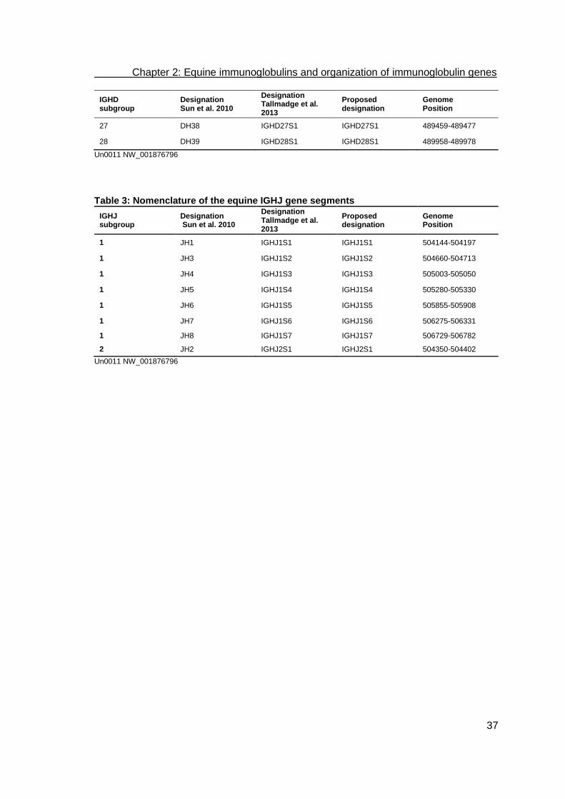
Chapter 2: Equine immunoglobulins and organization of immunoglobulin genes
37
IGHD subgroup
Designation Sun et al. 2010
Designation Tallmadge et al. 2013
Proposed designation
Genome Position
27 DH38 IGHD27S1 IGHD27S1 489459-489477
28 DH39 IGHD28S1 IGHD28S1 489958-489978
Un0011 NW_001876796
Table 3: Nomenclature of the equine IGHJ gene segments
IGHJ subgroup
Designation Sun et al. 2010
Designation Tallmadge et al. 2013
Proposed designation
Genome Position
1 JH1 IGHJ1S1 IGHJ1S1 504144-504197
1 JH3 IGHJ1S2 IGHJ1S2 504660-504713
1 JH4 IGHJ1S3 IGHJ1S3 505003-505050
1 JH5 IGHJ1S4 IGHJ1S4 505280-505330
1 JH6 IGHJ1S5 IGHJ1S5 505855-505908
1 JH7 IGHJ1S6 IGHJ1S6 506275-506331
1 JH8 IGHJ1S7 IGHJ1S7 506729-506782
2 JH2 IGHJ2S1 IGHJ2S1 504350-504402
Un0011 NW_001876796

Chapter 2: Equine immunoglobulins and organization of immunoglobulin genes
38
Table 4: Nomenclature of the equine IGHC genes
Original nomen-clature
Current desig-nation
Gene Allel Genome Position on NW_001876796
IgM IgM IGHM M_02 519420-524201
514993-518108
M_01 516522-517025
515374-515717
IgD IgD IGHD
531198-534510
526129-527334
IgGa IgG1 IGHG1
576506-576800
577273-577607
577685-578019
G1_01 576506-578019
IgG2 IGHG2
608720-610787
625782-627849
IgG(T) IgG3 IGHG3 G3_01 651171-651465
651958-652292
652361-652695
G3_02 650866-653171
IgG7 IGHG7 G7_01 674467-676688
G7_02 674945-676422
IgGb IgG4 IGHG4 G4_02 674954-676422
651171-651701
G4_01 674827-676691
IgGc IgG6 IGHG6 698724-700204
651171-651723
IgG(T) IgG5 IGHG5 730626-732347
IgGB
IgE IgE IGHE E_01 754864-756464
E_02 756129-756464
755315-755639
755732-756058
754864-755155
E_03 756129-756585
755732-756058
E_04 756129-756551
755732-756058
755315-755639
754858-755155
IgA IgA IGHA 766888-768383
768662-769379
Chapter 2: Equine immunoglobulins and organization of immunoglobulin genes
39
3.1. IgM
IgM is the first isotype that appears phylogenetically, as well as ontogenetically. It is the
first immunoglobulin produced by fetuses. This isotype is responsible for the primary
immune response. IgM may be bound to the membrane as a monomer or secreted as
a pentameric molecule with high avidity. IgM, as well as IgG, is present in the plasma
(Tallmadge et al., 2009).
The equine IGHM locus spans 1472 bp coding for 451 amino acid residues (Fig. 1c,
Table 4). This isotype exhibits six potential N-glycosylation sites. Similar to IgE, no
separated hinge region was identified. To enable flexibility of this istopye, the second
constant region may act as hinge. The third and fourth constant regions of IgM are
conserved even between different species, which indicates a specific role of IgM in
complement activation (Schrenzel et al., 1997).
3.2. IgD
Although the importance of this isotype remains unclear and a receptor-like function is
supposed, constant region genes coding for IgD are not restricted to primates and
rodents. After successful description of the IgD isotype in cattle, sheep and swine, IgD
was also identified in horses (Wagner et al., 2004; Zhao et al., 2002). The equine IgD
locus was identified 5 kb downstream of the genes coding for IgM (Fig. 1c, Table 4).
The entire genomic locus spans about 9.1 kb and contains eight exons that are: CH1,
two exons coding for the hinge region, CH2, CH3, one exon for the secreted molecule,
as well as two exons for the transmembrane anchor; the first of which contains a
specific splicing site. For IgD, the secreted form consists of 391 amino acid residues
and the transmembrane form of 418 amino acid residues. The coding nucleotide
sequences of CH1 to CH3 exons show sequence identities of 64% to human and
porcine IgD genes and 61% to bovine and ovine IgD genes. In analogy to the human
IgD, there is no switch-region between the genes coding for IgM and IgD, indicating
that there is an alternative splice mechanism (Wagner et al., 2004). In contrast, a
c-switch region has only been identified for cattle and pig (Zhao et al., 2002; Zhao et
al., 2003).

Chapter 2: Equine immunoglobulins and organization of immunoglobulin genes
40
3.3. IgG
The IgG isotype is considered to be the most versatile immunoglobulin in all mammals
and birds. Only in the porcine immune system has a similarly high number of eleven
genomic constant region genes representing six putative subclasses been described
(Fig. 1c, Table 4) (Butler et al., 2009). IgG performs all functions of a typical
immunoglobulin molecule such as antigen binding, complement fixation, and binding to
various cells like phagocytic cells, lymphocytes, platelets, mast cells, and basophils. It
contributes about 75% of the antibodies. The isotype was defined at a biochemical and
serological level and the different genomic elements were characterized. The
subclasses IgGa, IgGb, IgGc, IgG(T), and IgG(B) were classified (Klinman et al., 1965;
Rockey, 1967; Rockey et al., 1964; Sandor et al., 1964b; Sheoran and Holmes, 1996;
Widders et al., 1986) according to their antigenic differences and their serological and
electrophoretic properties. Restriction enzyme analyses of the genomic DNA of horse
lambda phage clones, deletion analyses of heterohybridomas, and nucleotide
sequencing of individual IGHC genes revealed six constant region genes in the equine
haploid genome (Overesch et al., 1998; Wagner et al., 2006). The characterization of
the complete IGHC region and the analysis of 34 overlapping genomic clones from the
CHORI-241 BAC library revealed seven constant region genes of equine IgG (Table 4)
and constituted the current repertoire of IgG IGHCs based on their sequences,
phylogeny, and homologous structures (Wagner et al., 2002; Wagner et al., 2004).
Multiple duplications, gene conversions, and crossovers are supposed to explain these
seven IgG isotypes, which are unique in all species investigated. IgG4 and IgG7 seem
to be the latest duplication events in evolution as their sequences show 96% identity.
As available reagents are insufficient for differentiation, isotypes IgG2 and IgG7 could
not yet be diversified in serum and other body fluids (Wagner et al., 2006). The
individual subclasses possess specific roles in protective immunity. Thus, the FcγR and
complement-binding isotypes IgG1, IgG4, and IgG7 (former IgGa and IgGb) contribute
to the protection against several equine pathogens such as equine influenza virus
(Breathnach et al., 2006; Nelson et al., 1998), Streptococcus equi (Sheoran et al.,
1997) and Rhodococcus equi (Lopez et al., 2002). In addition, systemic and mucosal
IgG responses were described to play an important role in limiting the spread and
severity of equine herpes virus 1 (Kydd et al., 2006). Although IgG4 and IgG7
represent the dominating serum antibodies, these isotypes are rarely found in clinically
healthy horses (Keggan et al., 2013).

Chapter 2: Equine immunoglobulins and organization of immunoglobulin genes
41
All these seven IgG subclasses were expressed in vivo (Wagner et al., 2004). Based
on these insights, the first recombinant versions of all seven equine IgG subclasses
(mouse -light chains, horse IGHG1-7) were expressed in Chinese Hamster Ovary
cells to analyze their individual physical and biological properties (Lewis et al., 2008). In
contrast to these first recombinant equine immunoglobulins, which were expressed for
functional analyses, seven distinct monoclonal equine antibodies (IgM, IgG1, IgG3,
IgG4/7, IgG5, IgG6, and IgE) were produced in equine-murine heterohybridomas to be
used for quantification of isotypes in diagnostic testing and immunological research
(Keggan et al., 2013).
All recombinant IgGs were N-glycosylated and maintained the affinity for their antigen.
Heavy and light chains assembled in intact IgG were stabilized by interchain disulfide
bridges except in a small proportion of IgG4 and 7. Complete IgG molecules
possessed molecular weights of approximately 150 kDa. Nevertheless, differences in
complement activation, Fc-receptor binding and the bacterial protein binding capacity
between the IgG isotypes were observed. IgG3 showed O-glycosylation in addition to
N-glycans. As its hinge region is rich in Proline at position -1 and +3 relative to
Threonine residues, which are known sites for O-glycan attachment, O-glycans may be
attached to the hinge region of the equine IgG3 (Lewis et al., 2008; Wilson et al.,
1991). In IgG4, IgG6, and IgG7 the Cystein equivalent to the human Cys131 was
missing within the CH1 region. Disulfide bond formation – with a Cystein at position
220 in the upper hinge region – is assumed to support the formation of intact IgG.
Amino acid substitutions at key positions (234-239, 251-154, 270, 311, 314, 318, 320,
322, 329, 331, 380, 382, 428, 432-436, 438) of the IgG molecule, which is responsible
for binding to protein A and G, were described (Lewis et al., 2008). IgG1, IgG3, IgG4,
IgG5, and IgG7 elicited strong respiratory burst activity in equine peripheral blood
leukocytes (PBL), indicating an efficient interaction with FcγR. In contrast, IgG2 and
IgG6 induced little or no response. Apparently, they are unable to interact with FcγR.
An amino acid residue motif at the N-terminus of the CH2 domain (Leu234-Leu235-
Gly236-Gly237-Pro238-Ser239) seemed to be involved in Fc-receptor binding. In
complement activation, IgG1, IgG3, IgG4, and IgG7 showed positive reactions,
whereas IgG2, IgG5, and IgG6 failed. Responsible C1q binding sites appeared not to
be conserved between different mammalian species, but there was a Lysine
corresponding to human and mouse Lysine at position 322, which was found in all
complement activating equine IgG, as well as in IgG6, but Lys322 was replaced by
Serine in IgG2 and IgG5 (Keggan et al., 2013; Lewis et al., 2008).
Chapter 2: Equine immunoglobulins and organization of immunoglobulin genes
42
Evidently, IgG plays an important role both in serum and mucosal compartments in
horses. Therefore, the summarized considerations appear to be relevant to systemic
and mucosal vaccination strategies. IgG antibodies of the subclasses IgG1, IgG3,
IgG4/IgG7 should be induced by vaccines to achieve maximal protection via FcγR and
complement-mediated elimination mechanisms. Less effective protection is provided by
vaccines that trigger only IgG2, IgG5 or IgG6 antibodies (Lewis et al., 2008).
3.4. IgE
IgE is the least common serum immunoglobulin isotype (Oethinger et al., 1997).
Besides the immune response against parasites, IgE is also known to be responsible
for type 1 allergies and resulting diseases like summer eczema, urticaria, COPD
(recurrent airway obstruction) or head shaking (Wagner et al., 2006). For primary
investigations, equine IgE was prepared from the serum of foals suffering from
endoparasitic infections (Suter and Fey, 1981). Compared to humans, horses revealed
a high concentration of IgE in serum (Wagner et al., 2006). The constant region gene is
located between the constant region genes coding for IgG5 and IgA, and it contains
four exons. Similar to IgM, there is no separated hinge region for IgE and the second
constant region enables flexibility. The switch region is located 2-4 kb upstream of the
constant region gene. Three haplotypes are described differing in two SNPs, which
result in two amino acid residue substitutions (Fig. 1c, Table 4) (Navarro et al., 1995;
Wagner et al., 2006). The polymorphic character of the IgE genes may influence
effector functions, binding mast cells and basophils, and finally the level of allergic
reactions.
3.5. IgA
IgA dominates in the mucosal immune response of respiratory, gastrointestinal and
genitourinary tracts (Souza et al., 2010). In body fluids such as milk, nasal secretions
and saliva, it appears as a dimeric secreted molecule and is called secretory IgA. In
serum it shows itself in monomeric form (Sheoran et al., 1997; Wagner et al., 2003).
IgA produced by B1 lymphocytes represents a T-lymphocyte independent source of
IgA, which recognizes commensal bacteria. IgA produced by B2 lymphocytes in the

Chapter 2: Equine immunoglobulins and organization of immunoglobulin genes
43
mucosal-associated lymphoid tissues represents T-dependent secreted IgA, e.g.
directed against exotoxins (Macpherson et al., 2000).
The IgA locus possesses three exons, two introns, and one hinge region. This hinge
region is an extension of the CH2 sequence. Within the equine IGHC region, IGHA is
the most 3’located gene (Fig. 1c, Table 4). The switch region is located 1.4 kb
upstream of the constant region genes coding for IgA. Phylogenetic comparisons show
a common cluster with the IgA genes of humans, dogs, swine, cattle, and sheep. In
contrast to rabbits possessing 13 subclasses of IgA and humans with two subclasses
of IgA, horses have only one IgA class (Burnett et al., 1989; Mestecky et al., 2004;
Wagner et al., 2003).
Investigations concerning the concentration of IgA in serum showed significantly lower
IgA quantities in sport horses when compared to extensively reared horses (Souza et
al., 2010). Comparable amounts of IgA were found in the colostrum of Warmblood and
Standardbred mares after parturition (Kohn et al., 1989). In milk, the reduced IgA
concentration is likely due to passively transferred IgA, which was observed in a similar
way for IgG (Jenvey et al., 2012). Once the window for absorbing maternal IgA has
passed, IgA levels in the mare’s milk persisted but the significance of this observation
is yet to be fully established. However, investigations in human neonates demonstrated
that IgA and other factors within breast milk may provide an ongoing contribution to
local passive protection and immunological development (Corthesy, 2007). Equine
serum and secretory IgA was quantified in Shetland ponies 182 days after parturition.
While IgG levels decreased with time after parturition, IgA levels increased until IgA
was the predominant immunoglobulin (McGuire and Crawford, 1972). In contrast, the
quantity of IgA in colostrum and milk from Thoroughbred mares rapidly decreased
during the first 19 days after parturition, and fluctuated at this level until day 60 p.p.
(Jenvey et al., 2012).

Chapter 2: Equine immunoglobulins and organization of immunoglobulin genes
44
4. Equine immunoglobulin light chain gene loci
As known from other mammals, horses also express two isotypes of light chains ( and
) differentiated by isotype-specific amino acid residue motifs, antigenic properties, and
chemical structure. In contrast to other mammals, 95% of the equine serum antibodies
combine -light chains with the heavy chains, although the reason for this phenomenon
is not yet clear (Gibson, 1974; Home et al., 1992). The assumption that amino acid
residue substitutions in IGKC result in structures with reduced binding efficiency to the
heavy chains was not verified (Ford et al., 1994; Sun et al., 2010). Therefore, a
correlation between the number of IGLV gene segments and the preferred usage of a
particular isotype is supposed. The -light chain locus was mapped on equine
autosome 8 (ECA8) and the gene segments of the -light chain were located on
ECA15 (Das et al., 2008).
First studies on the organization of the equine light chain loci revealed a restricted
number of variable gene segments for both isotypes. The assembly of variable, joining,
and constant region genes was also found to be similar to other species (Ford et al.,
1994; Home et al., 1992). Today 144 IGLV, 7 IGLJ and 7 IGLC are identified within
1310 kb on ECA8 (Fig. 2, Tables 5-7). Each of the 7 IGLJ is preceded by one IGLC
and there are two clusters of IGLV possessing different transcriptional orientations.
Within each IGLV cluster there are functional genes and pseudogenes (Sun et al.,
2010). Similarly, pseudogenes are described in different species such as chicken,
where it is already known that pseudogenes are used for gene conversion (Reynaud et
al., 1985).
As there were differences in the nomenclature, we also suggest a supplemented
designation for the light chain gene segments (Tables 5-7). The name of Ig lambda
light chain variable (IGLV) and joining (IGLJ) gene segments were assigned according
to IMGT nomenclature guidelines; the international ImMunoGeneTics information
system (www.imgt.org). The genes were named according to the subgroup they belong
to (Sun et al., 2010) and their number within this subgroup. Furthermore, to denote
open reading frames or pseudogenes, sequence names were complemented by
superscript ORF and P, respectively. The former ‘Vλ1’ was renamed ‘IGLV1S1’ to
designate sequence 1 of subgroup 1. In analogy, IGLJ and IGLC genes were
designated IGLJ1S1 through IGLJ7S1 and IGLC1S1 through IGLC7S1, instead of
‘‘Jλ1’’ and ‘‘Cλ1’’, which is consistent with the human nomenclature of Ig lambda genes

Chapter 2: Equine immunoglobulins and organization of immunoglobulin genes
45
(Hara et al., 2012; Lefranc, 2001a; Sun et al., 2010; Tallmadge et al., 2014). Positions
identified on several contigs are also listed. The contigs are NW_001867428.1 and
chromosome 8 (Sun et al., 2010). Superscript P indicates pseudo-variable gene
segments. The variable gene segments that were not classified to subgroups because
they were too divergent or truncated were marked with n.c. (Sun et al., 2010).
According to the supposed nomenclature, which is in analogy to the heavy and -light
chain, IGLJ5S1, IGLJ6S1, and IGLJ7S1 show identical sequences. IGLC2S1P is
defined as pseudogene due to premature stop codons, as well as IGLJ2S1P,
IGLJ3S1P, and IGLJ4S1(P) (Sun et al., 2010). Among the 144 IGLV segments identified
by Sun et al. (2010), 108 have RSS heptamers and nonamers, 9 lack conserved
nonamers and the remaining segments lack both heptamers and nonamers. IGLV3S1,
IGLV9S1, IGLV9S2, and IGLV9S3ORF have 22-spaced RSS and the remaining
potentially functional and ORF-IGLVs carry 23-spaced RSS. The spacer length is more
varied among the 76 IGLVP segments, which have the intact RSSs. As many as 55
IGLVP segments maintain 23 bp-spaced RSSs, whereas 16 IGLVP segments carry 22
bp-spaced RSSs. The IGLVxS36P, IGLV9S4P and IGLVxS59P have spacer lengths of
21, 21 and 19 bp, respectively. All 7 IGLJ segments have 12 bp-spaced RSSs at their
5’end.

Chapter 2: Equine immunoglobulins and organization of immunoglobulin genes
46
Fig. 2: Map of the equine lambda light chain gene locus The lambda light chain genes are located on chromosome 8 spanning about 1310 kb. The IGLJ-IGLC pairs and the IGLVs upstream of the (IGLJ-IGLC)7 show opposite transcriptional orientation to the IGLVs downstream of the (IGLJ-IGLC)7 cluster. This physical map is adapted from Sun et al. (2010) to ensure that the newly proposed nomenclature (table 4-7) is designated to the respective positions on the locus. IGLV without superscript letters are potentially functional variable gene segments. Superscript ORF indicates variable gene segments with open reading frames that either have a defect in splicing sites, RSS and/or regulatory elements, and/or changes to the conserved amino acids, and therefore have been suggested to lead to incorrect folding (Lefranc, 1998). Superscript P indicates pseudo-variable gene segments.

Chapter 2: Equine immunoglobulins and organization of immunoglobulin genes
47
Table 5: Nomenclature of the equine IGLV gene segments
IGLV subgroup Designation Sun et al. 2010
Designation Tallmadge et al. 2013
Proposed designation
Genome Position
1 Vλ1 IGLV1-38 IGLV1S1 4171705-4171992
2 Vλ2 IGLV2-41 IGLV2S1 4195092-4195381
3 Vλ3 IGLV3-44 IGLV3S1 4210612-4210896
4 Vλ4 IGLV4-61 IGLV4S1 4330800-4331090
4 Vλ5 IGLV4-66 IGLV4S2 4366543-4366833
4 Vλ6 IGLV4-69 IGLV4S3 4381050-4381340
4 Vλ7 IGLV4-75 IGLV4S4 4411343-4411633
4 VλORF1 IGLV4-92 IGLV4S5ORF 4529112-4529396
4 pVλ13 IGLV4-50 IGLV4S6P 4245996-4246283
4 pVλ16 IGLV4-53 IGLV4S7P 4267110-4267396
4 pVλ18 IGLV4-55 IGLV4S8P 4287903-4288193
4 pVλ39 IGLV4-80 IGLV4S9P 4447087-4447374
4 pVλ42 IGLV4-83 IGLV4S10P 4458683-4458970
4 pVλ48 IGLV4-90 IGLV4S11P 4502561-4502854
5 Vλ8 IGLV5-87 IGLV5S1 4489461-4489759
5 Vλ9 IGLV5-95 IGLV5S2 4554617-4554901
6 Vλ10 IHLV6-98 IHLV6S1 4571250-4571546
6 Vλ11 IGLV6-101 IGLV6S2 4590316-4590612
6 Vλ12 IGLV6-109 IGLV6S3 4648068-4648364
6 Vλ13 IGLV6-111 IGLV6S4 4660775-4661062
6 VλORF2 IGLV6-104 IGLV6S5ORF 4614091-4614386
6 pVλ54 IGLV6-99 IGLV6S7P 4574617-4574913
6 pVλ55 IGLV6-100 IGLV6S8P 4584807-4585103
6 pVλ57 IGLV6-103 IGLV6S9P 4607689-4607985
6 pVλ59 IGLV6-106 IGLV6S10P 4624835-4625128
6 pVλ61 IGLV6-108 IGLV6S11P 4643773-4644068
6 pVλ62 IGLV6-110 IGLV6S12P 4654126-4654437
7 Vλ14 IGLV7-116 IGLV7S1 4763155-4763451
7 pVλ67 IGLV7-117 IGLV7S2P 4766554-4766835
8 Vλ15 IGLV8-122 IGLV8S1 4855083-4855381
8 Vλ17 IGLV8-128 IGLV8S2 4916905-4917203
8 Vλ18 IGLV8-133 IGLV8S3 4944794-4945086
8 Vλ19 IGLV8-137 IGLV8S4 4975350-4975639
8 Vλ22 IGLV8-28 IGLV8S5 3994214-3994503
8 Vλ23 IGLV8-26 IGLV8S6 3976833-3977125
8 Vλ25 IGLV8-24 IGLV8S7 3942211-3942500
8 Vλ26 IGLV8-20 IGLV8S8 3902029-3902327
8 Vλ27 IGLV8-12 IGLV8S9 3799404-3799702
8 pVλ74 IGLV8-125 IGLV8S10P 4878404-4878692

Chapter 2: Equine immunoglobulins and organization of immunoglobulin genes
48
IGLV subgroup Designation Sun et al. 2010
Designation Tallmadge et al. 2013
Proposed designation
Genome Position
8 pVλ89 IGLV8-32 IGLV8S11P 4023156-4023449
8 pVλ101 IGLV8-14 IGLV8S12P 3838136-3838430
8 pVλ105 IGLV8-9 IGLV8S13P 3781419-3781717
9 Vλ16 IGLV9-126 IGLV9S1 4904149-4904473
9 Vλ24 IGLV9-25 IGLV9S2 3948638-3948962
9 VλORF4 IGLV9-16 IGLV9S3ORF 3870582-3870906
9 pVλ69 IGLV9.119 IGLV9S4P 4780685-4780996
9 pVλ73 IGLV9-124 IGLV9S5P 4866218-4866534
9 pVλ76 IGLV9-129 IGLV9S6P 4922628-4922938
9 pVλ78 IGLV9-131 IGLV9S7P 4929757-4930046
9 pVλ80 IGLV9-134 IGLV9S8P 4950548-4950857
9 pVλ81 IGLV9-136 IGLV9S9P 4968911-4969235
9 pVλ82 IGLV9-138 IGLV9S10P 4981125-4981435
9 pVλ85 IGLV9-142 IGLV9S11P 4999541-4999848
9 pVλ91 IGLV9-30 IGLV9S12P 4006944-4007268
9 pVλ94 IGLV9-23 IGLV9S13P 3936435-3936744
9 pVλ95 IGLV9-22 IGLV9S14P 3914834-3915157
9 pVλ97 IGLV9-19 IGLV9S15P 4780685-4780996
9 pVλ99 IGLV9-17 IGLV9S16P 4866218-4866534
9 pVλ102 IGLV9-13 IGLV9S17P 3812261-3812584
9 pVλ103 IGLV9-11 IGLV9S18P 3793651-3793937
9 pVλ104 IGLV9-10 IGLV9S19P 3788296-3788612
10 VλORF3 IGLV10-135 IGLV10S1ORF 4954678-4954976
10 pVλ77 IGLV10-130 IGLV10S2P 4926370-4926670
10 pVλ87 IGLV10-144 IGLV10S3P 5012699-5012989
10 pVλ98 IGLV10-18 IGLV10S4P 3892471-3892771
10 pVλ110 IGLV10-3 IGLV10S5P 3723141-3723424
11 pVλ20 IGLV11-140 IGLV11S1P 4300066-4300348
11 pVλ21 IGLV11-33 IGLV11S2P 4316629-4316892
11 VλORF5 IGLV11-5 IGLV11S3ORF 3730193-3730489
n.c. pVλ1 IGLVxS1P 4157587-4157847
n.c. pVλ2 IGLVxS2P 4160537-4160835
n.c. pVλ3 IGLVxS3P 4166903-4167176
n.c. pVλ4 IGLVxS4P 4178429-4178741
n.c. pVλ5 IGLVxS5P 4188046-4188311
n.c. pVλ6 IGLVxS6P 4199645-4199909
n.c. pVλ7 IGLVxS7P 4205575-4205866
n.c. pVλ8 IGLVxS8P 4216947-4217233
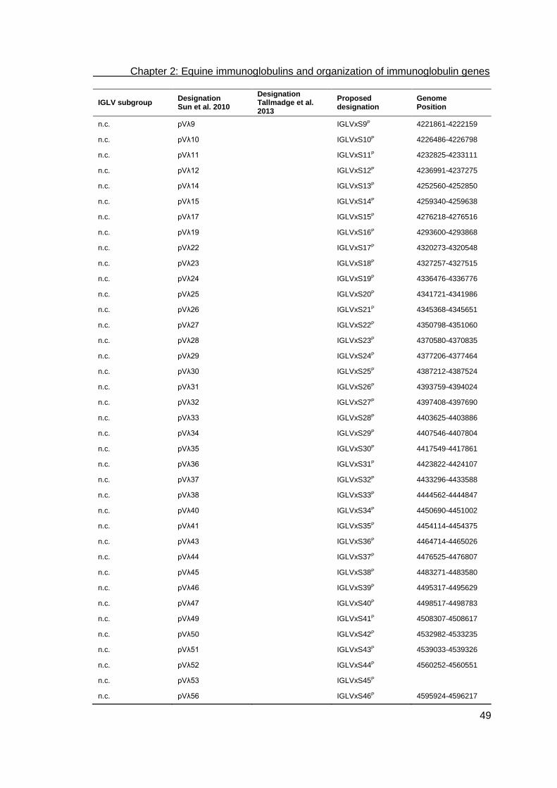
Chapter 2: Equine immunoglobulins and organization of immunoglobulin genes
49
IGLV subgroup Designation Sun et al. 2010
Designation Tallmadge et al. 2013
Proposed designation
Genome Position
n.c. pVλ9 IGLVxS9P 4221861-4222159
n.c. pVλ10 IGLVxS10P 4226486-4226798
n.c. pVλ11 IGLVxS11P 4232825-4233111
n.c. pVλ12 IGLVxS12P 4236991-4237275
n.c. pVλ14 IGLVxS13P 4252560-4252850
n.c. pVλ15 IGLVxS14P 4259340-4259638
n.c. pVλ17 IGLVxS15P 4276218-4276516
n.c. pVλ19 IGLVxS16P 4293600-4293868
n.c. pVλ22 IGLVxS17P 4320273-4320548
n.c. pVλ23 IGLVxS18P 4327257-4327515
n.c. pVλ24 IGLVxS19P 4336476-4336776
n.c. pVλ25 IGLVxS20P 4341721-4341986
n.c. pVλ26 IGLVxS21P 4345368-4345651
n.c. pVλ27 IGLVxS22P 4350798-4351060
n.c. pVλ28 IGLVxS23P 4370580-4370835
n.c. pVλ29 IGLVxS24P 4377206-4377464
n.c. pVλ30 IGLVxS25P 4387212-4387524
n.c. pVλ31 IGLVxS26P 4393759-4394024
n.c. pVλ32 IGLVxS27P 4397408-4397690
n.c. pVλ33 IGLVxS28P 4403625-4403886
n.c. pVλ34 IGLVxS29P 4407546-4407804
n.c. pVλ35 IGLVxS30P 4417549-4417861
n.c. pVλ36 IGLVxS31P 4423822-4424107
n.c. pVλ37 IGLVxS32P 4433296-4433588
n.c. pVλ38 IGLVxS33P 4444562-4444847
n.c. pVλ40 IGLVxS34P 4450690-4451002
n.c. pVλ41 IGLVxS35P 4454114-4454375
n.c. pVλ43 IGLVxS36P 4464714-4465026
n.c. pVλ44 IGLVxS37P 4476525-4476807
n.c. pVλ45 IGLVxS38P 4483271-4483580
n.c. pVλ46 IGLVxS39P 4495317-4495629
n.c. pVλ47 IGLVxS40P 4498517-4498783
n.c. pVλ49 IGLVxS41P 4508307-4508617
n.c. pVλ50 IGLVxS42P 4532982-4533235
n.c. pVλ51 IGLVxS43P 4539033-4539326
n.c. pVλ52 IGLVxS44P 4560252-4560551
n.c. pVλ53 IGLVxS45P
n.c. pVλ56 IGLVxS46P 4595924-4596217

Chapter 2: Equine immunoglobulins and organization of immunoglobulin genes
50
IGLV subgroup Designation Sun et al. 2010
Designation Tallmadge et al. 2013
Proposed designation
Genome Position
n.c. pVλ58 IGLVxS47P 4621790-4622080
n.c. pVλ60 IGLVxS48P 4629113-4629400
n.c. pVλ63 IGLVxS49P 4670998-4671281
n.c. pVλ64 IGLVxS50P 4684017-4684281
n.c. pVλ65 IGLVxS51P 4753071-4753353
n.c. pVλ66 IGLVxS52P 4760021-4760299
n.c. pVλ68 IGLVxS53P 4775528-4775824
n.c. pVλ70 IGLVxS54P 4807475-4807769
n.c. pVλ71 IGLVxS55P 4808989-4809266
n.c. pVλ72 IGLVxS56P 4860864-4861172
n.c. pVλ75 IGLVxS57P 4910505-4910786
n.c. pVλ79 IGLVxS58P 4938347-4938661
n.c. pVλ83 IGLVxS59P 4985429-4985745
n.c. Vλ20 IGLVxS60P 4994608-4994903
n.c. pVλ84 IGLVxS61P 4998412-4998621
n.c. pVλ86 IGLVxS62P 5010496-5010800
n.c. pVλ88 IGLVxS63P 4033346-4033647
n.c. Vλ21 IGLVxS64P 4031616-4031911
n.c. pVλ90 IGLVxS65P 4017794-4018083
n.c. pVλ92 IGLVxS66P 4000617-4000857
n.c. pVλ93 IGLVxS67P 3988326-3988631
n.c. pVλ96 IGLVxS68P 3908433-3908722
n.c. pVλ100 IGLVxS69P 3864249-3864531
n.c. pVλ106 IGLVxS70P 3745800-3746092
n.c. pVλ107 IGLVxS71P 3742171-3742365
n.c. pVλ108 IGLVxS72P 3738392-3738672
n.c. pVλ109 IGLVxS73P 3726480-3726695
n.c. pVλ111 IGLVxS74P 3719722-3720011
n.c. pVλ112 IGLVxS75P 3703267-3703501
IGLV: Sun et al., 2010, Chromosom 8, NW_001867428.1

Chapter 2: Equine immunoglobulins and organization of immunoglobulin genes
51
Table 6: Nomenclature of the equine IGLJ gene segments
IGLJ subgroup
Designation Sun et al. 2010
Designation Tallmadge et al. 2013
Proposed designation
Genome Position
1 Jλ1 IGLJ1 IGLJ1S1 4146337-4146302 a
2 Jλ2 IGLJ2 IGLJ2S1P 4141033-4140996 a
3 Jλ3 IGLJ3 IGLJ3S1P 4135576-4135541 a
4 Jλ4 IGLJ4 IGLJ4S1(P) 4132448-4132410 a
5 Jλ5 IGLJ5 IGLJ5S1 4129086-4129050 a
6 Jλ6 IGLJ6 IGLJ6S1 4125743-4125707 a
7 Jλ7 IGLJ7 IGLJ7S1 4122272-4122236 a
a Contig_ NW_001867428.1
Table 7: Nomenclature of the equine IGLC genes
IGLC subgroup
Designation Sun et al. 2010
Designation Tallmadge et al. 2013
Proposed designation
Genome Position
1 Cλ1 IGLC1 IGLC1S1 4144802-4144483 a
2 pCλ2 IGLC2 IGLC2S1P 4139692-4139371 a
3 Cλ3 IGLC3 IGLC3S1 4134292-4133973 a
4 Cλ4 IGLC4 IGLC4S1 4131021-4130702 a
5 Cλ5 IGLC5 IGLC5S1 4127666-4127347 a
6 Cλ6 IGLC6 IGLC6S1 4124320-4124002 a
7 Cλ7 IGLC7 IGLC7S1 4120851-4120531 a
a Contig_ NW_001867428.1
The -light chain locus shows a unique feature of two IGLV clusters compared to this
locus in all other species analyzed, whereas the -light chain genes are arranged as
usual in one cluster. In the 820 kb locus, there is one cluster of 60 IGKV, one cluster of
five IGKJ, and a single IGKC (Fig. 3, Tables 8-10) (Sun et al., 2010). Moreover, the
functional and nonfunctional IGKV are located within one locus. All IGKV segments
have 12 bp-spaced RSSs at their 3’ends except IGKV1S21P, which possesses an
11 bp spacer and additional 8 IGKVP segments where RSSs are lacking completely.
IGKJ1S1, IGKJ2S1, and IGKJ4S1 have 23 bp-spaced RSSs at their 5’ends in contrast
to IGKJ3S1, which has a 20 bp-spaced RSS. IGKJ5S1P is nonfunctional as there is no
conserved heptamer within the RSS (Sun et al., 2010). Transcriptional orientations in
both directions are found without grouping. The single IGKC is separated from the
IGKJ by a 2926 bp intron. IGKJ5S1P has no heptamer in the recombination signal
sequence (RSS) and is therefore described as pseudogene. In IGKJ3S1, the spacer of
the RSS shows 20 bp instead of 23 bp, which does not result in reduced recombination
frequency (Ford et al., 1994; Sun et al., 2010).

Chapter 2: Equine immunoglobulins and organization of immunoglobulin genes
52
Fig. 3: Physical map of the equine kappa light chain gene locus 820 kb of the kappa light chain genes are located on chromosome 15. The potentially functional IGKVs, 2 ORFs and 15 pseudogenes show opposite transcriptional orientation to the (IGKJ)5-IGKC cluster. Arrows below the vertical lines directed 5’ indicate transcriptional orientation. This physical map is adapted from Sun et al. (2010) to ensure that the newly proposed nomenclature (table 3) is designated to the respective positions on the locus. IGKV without superscript letters are potentially functional variable gene segments. Superscript ORF indicates variable gene segments with open reading frames that either have a defect in splicing sites, RSS and/or regulatory elements, and/or changes to the conserved amino acids, and therefore have been suggested to lead to incorrect folding (Lefranc, 1998). Superscript P indicates pseudo-variable gene segments.

Chapter 2: Equine immunoglobulins and organization of immunoglobulin genes
53
Table 8: Nomenclature of the equine IGKV gene segments
IGKV subgroup
Designation Sun et al. 2010
Proposed designation
Genome Position
1 Vκ1 IGKV1S1 17071497-17071783
1 Vκ2 IgKV1S2 17057542-17057828
1 Vκ3 IGKV1S3 17023418-17023704
1 Vκ4 IGKV1S4 17010212-17010498
1 Vκ5 IGKV1S5 16958378-16958664
1 Vκ6 IGKV1S6 16941855-16942141
1 Vκ7 IGKV1S7 16933657-16933943
1 Vκ8 IGKV1S8 16855771-16856057
1 VκORF1 IGKV1S9ORF 16739610-16739895
1 VκORF2 IGKV1S10ORF 16560248-16560552
1 pVκ2 IGKV1S11P 17046559-17046846
1 pVκ4 IGKV1S12P 16998516-16998803
1 pVκ6 IGKV1S13P 16917360-16917646
1 pVκ7 IGKV1S14P 16912894-16913180
1 pVκ8 IGKV1S15P 16868993-16869281
1 pVκ9 IGKV1S16P 16849772-16850060
1 pVκ10 IGKV1S17P 16835919-16836203
1 pVκ11 IGKV1S18P 16789755-16790042
1 pVκ12 IGKV1S19P 16778531-16778817
1 pVκ13 IGKV1S20P 16728653-16728934
1 pVκ14 IGKV1S21P 16720475-16720760
1 pVκ19 IGKV1S22P 16659096-16659390
1 pVκ20 IGKV1S23P 16626800-16627104
2 Vκ9 IGKV2S1 16700635-16700951
2 pVκ24 IGKV2S2P 16557308-16557612
3 Vκ10 IGKV3S1 16599670-16599971
3 Vκ11 IGKV3S2 16540025-16540326
3 Vκ14 IGKV3S3 16470843-16471144
3 Vκ16 IGKV3S4 16407261-16407562
3 Vκ17 IGKV3S5 16383431-16383732
3 Vκ18 IGKV3S6 16360142-16360443
3 pVκ17 IGKV3S7P 16676005-16676305
3 pVκ21 IGKV3S8P 16608233-16608528
3 pVκ22 IGKV3S9P 16591859-16592160
3 pVκ28 IGKV3S10P 16461689-16461983
3 pVκ29 IGKV3S11P 16450171-16450478
3 pVκ30 IGKV3S12P 16441110-16441414
3 pVκ34 IGKV3S13P 16351930-16352220
4 Vκ12 IGKV4S1 16502696-16502982
4 pVκ18 IGKV4S2P 16672345-16672638
5 Vκ13 IGKV5S1 16484542-16484828
6 Vκ15 IGKV6S1 16431518-16431804
7 Vκ19 IGKV7S1 16313053-16313339

Chapter 2: Equine immunoglobulins and organization of immunoglobulin genes
54
IGKV subgroup
Designation Sun et al. 2010
Proposed designation
Genome Position
n.c. pVκ1 IGKVxS1P 17053257-17053545
n.c. pVκ3 IGKVxS2P 17004266-17004536
n.c. pVκ5 IGKVxS3P 16927468-16927746
n.c. pVκ15 IGKVxS4P 16709612-16709911
n.c. pVκ16 IGKVxS5P 16689071-16689357
n.c. pVκ23 IGKVxS6P 16581856-16582113
n.c. pVκ25 IGKVxS7P 16550552-15550840
n.c. pVκ26 IGKVxS8P 16534333-16534598
n.c. pVκ27 IGKVxS9P 16520693-16520979
n.c. pVκ31 IGKVxS10P 16434887-16435177
n.c. pVκ32 IGKVxS11P 16413133-16413356
n.c. pVκ33 IGKVxS12P 16370408-16370629
n.c. pVκ35 IGKVxS13P 16348990-16349239
n.c. pVκ36 IGKVxS14P 16342798-16343043
n.c. pVκ37 IGKVxS15P 16338745-16339014
n.c. pVκ38 IGKVxS16P 16332882-16333152
n.c. pVκ39 IGKVxS17P 16305235-16305453
IGKV: Sun et al., 2010, Chromosom 15, NW_001867379.1
Table 9: Nomenclature of the equine IGKJ gene segments
IGKJ subgroup
Designation Sun et al. 2010
Proposed designation
Genome Position
1 Jκ1 IGKJ1S1 13727254-13727217*
2 Jκ2 IGKJ2S1 13727624-13727586*
3 Jκ3 IGKJ3S1 13727931-13727894*
4 Jκ4 IGKJ4S1 13728269-13728232*
5 pJκ5 IGKJ5S1P 13728570-13728534*
*Contig: NW_001867379.1
Table 10: Nomenclature of the equine IGKC genes
IGKC subgroup
Designation Sun et al. 2010
Proposed designation
Genome Position
1 Cκ1 IGKC1S1 13731499-13731821*
*Contig: NW_001867379.1

Chapter 2: Equine immunoglobulins and organization of immunoglobulin genes
55
5. Transcription analyses of heavy and light chain genes
5.1. Heavy chain genes
Newborn foals are unable to mount an effective humoral immune response despite
many features of the equine immune system having been developed during fetal life.
Immunohistochemical experiments showed the expression of essential B-cell genes
during equine gestation. Active gene recombination and isotype switching were
suggested and underpinned by limited but detectable production of IgM and IgG at birth
(Tallmadge et al., 2009). After demonstrating that equine neonatal B-cells produce all
known five Ig isotypes, usage frequencies of the equine variable, diversity, and joining
gene segments of the heavy and light chains were first investigated in two unrelated
horses (Sun et al., 2010). Both animals showed a similar usage pattern of IGHV, IGHD,
and IGHJ. Out of 50 IGHV, four (IGHV4S1, IGHV2S2, IGHV2S3, and IGHV4S5) were
expressed but IGHV2S2 and IGHV2S3 were used preferably. Six out of eight IGHJ
were recombined. IGHJ1S1 and IGHJ2S1 were not detected, whereas IGHJ1S5 was
mainly rearranged. The analysis of the usage frequencies of the 40 germline IGHD
revealed the dominant frequent use of IGHD18S1. The transcription of five additional
IGHDs (IGDH3S1, IGHD12S1, IGHD8S2, IGHD8S3, IGHD28S1) was not observed.
Eight clones indicated the incorporation of more than one IGHD into the IGHV-IGHD-
IGHJ rearrangement resulting in IGHV-IGHD-IGHD-IGHJ. The second incorporated
IGHD was supposed to be located 5’ of the first IGHD in the germline locus (Sun et al.,
2010). In addition to these studies of immunoglobulin diversity in adult horses, the level
of Ig heavy chain variable region diversity was analyzed from fetal spleen, neonatal,
foal, and adult horse mesenteric lymph node samples (Tallmadge et al., 2009).
Interestingly, combinatorial and junctional levels of VDJ sequences in equine fetus
were comparable to those of adult horses and a similar set of variable gene segments
was used during fetal and post-natal life stages (Tallmadge et al., 2009; Tallmadge et
al., 2013). During fetal life stage, 7 IGHV genes were identified with nucleotide
identities of at least 97% compared to the donor germline sequences. Besides the
gene segments IGHV2S2, IGHV2S3, IGHV1S3, IGHV4S2, IGHV4S5 and IGHV2S4, a
newly identified IGHV4S17 was recognized. A reduced repertoire was observed in
neonates where only 4 segments were used: IGHV2S2, IGHV2S3, IGHV2S4, and a
novel IGHV1S6. In foals and adult horses, 5 and 4 IGHV were transcribed, respectively
(foal: IGHV2S2, IGHV2S3, IGHV2S4, IGHV1S3, IGHV4S2; adult: IGHV2S2, IGHV2S3,

Chapter 2: Equine immunoglobulins and organization of immunoglobulin genes
56
IGHV2S4, IGHV4S2). The segments IGHV2S2, IGHV2S3, and IGHV2S4 were used
predominantly at all ages. Between the expressed gene segments, there are 250,000
bases suggesting that the entire IGHV locus is available throughout equine life
(Tallmadge et al., 2013). Similarly, in fetal piglets the IGHV usage was described to be
independent of the genome position although there is a limited number of IGHV
representing the Ig repertoire, too (Butler et al., 2011; Eguchi-Ogawa et al., 2010). In
contrast, the biased usage of germline IGHV in the mouse fetus was explained with
different accessible positions within the IGHV locus (Jeong et al., 1988).
Analysis of the IGHD gene segments revealed that 13, 17, 15, and 20 different IGHDs
were used in fetuses, neonates, foals, and adult horses. The predominant IGHD was
IGHD18S1 at all ages. Four out of 8 IGHJ had an influence on diversity in all life
stages, whereas IGHJ1S5 was the most common (Tallmadge et al., 2013).
VDJ diversity increased mainly during the last two thirds of gestation. Overall, there
was no biased usage of a single IGHV, IGHD, or IGHJ gene segment at any age. In
foals, first variations in the length of CDR2H were found. Sequence diversity and length
variation further increased in CDR1H and CDR2H, and framework regions of adult
horses in accordance to somatic hypermutation (Tallmadge et al., 2013). Non-
templated nucleotide insertions of about 8 nucleotides were observed at the IGHV-
IGHD junction in fetuses and neonates, although there was no significant increase in
CDR3H length, varying from 7 to 21 amino acids in contrast to cattle (Walther et al.,
2013). The length of the IGHV-IGHD and IGHD-IGHJ junctions increased significantly
until birth and then remained similar in foals and adult horses. At least 5 amino acid
residues were observed within the CDR3H of an adult horse, whereas 25 amino acid
residues were counted in the longest CDR3H, which was identified in an equine
neonate (Tallmadge et al., 2013). In comparison, recent studies showed the existence
of very short CDR3H (5-10 amino acids), midlength CDR3H (11-31 amino acids) and
exceptionally long CDR3H (more than 47 amino acids) in both bovine fetuses and adult
cattle. This length heterogeneity is not isotype restricted and very long CDR3H
contribute to diversity by uniquely folded small domains (Koti et al., 2010; Saini et al.,
1999; Saini and Kaushik, 2002; Shojaei et al., 2003; Walther et al., 2013; Wang et al.,
2013).

Chapter 2: Equine immunoglobulins and organization of immunoglobulin genes
57
5.2. Light chain genes
For the IGKV, a preferential usage pattern was also observed. IGKV1S1, IGKV1S4,
and IGKV2S1 were used most often. These gene segments showed an opposite
transcriptional orientation to the IGKJ and IGKC cluster. All four functional IGKJ were
expressed (Sun et al., 2010).
Within the transcribed -light chains, IGLVs belonging to subgroup 8 were preferred.
IGLV from subgroup 6 and 4 were also transcribed but less frequently. The recombined
IGLVs – especially IGLV8S2 and IGLV8S7 – were located within the IGLV cluster
downstream of the IGLJ-IGLC cluster and were oriented in opposite transcriptional
polarity. In both animals, examined by Sun and coworkers, genomic IGLJ-IGLC were
amplified, whereas IGLJ-IGLC5 could not be amplified and IGLJ-IGLC7 was amplified
successfully in one of these animals. IGLJ4S1(P) was found to be functional but without
the single C-insertion, which is present in the database sequence of the equine
genome project (Sun et al., 2010). Subsequent investigations of the transcription
frequencies of -light chain genes in ten animals of Rhenish-German Coldblood (RGC)
and Hanoverian Warmblood (HW), respectively, showed similar results (Hara et al.,
2012). Eleven out of 144 IGLV were identified; all members of subgroup 8 as caused
by the applied primers. In both breeds analyzed, the preferred use of two IGLV
(IGLV8S1 and IGLV8S2) was observed. In addition, significant differences in usage
frequencies of the IGLV (both within and between the breeds) were noted. Two
pseudogenes were also recombined (IGLV8S10P and IGLV8S12P). The transcriptional
orientation of the IGLV had no influence on the transcription frequencies. All four
functional IGLC were also transcribed. IGLC6S1/7S1 was predominantly used in both
breeds. These two constant region genes could not be distinguished due to high
sequence similarity. Isotypes IGLC1S1 and IGLC5S1 revealed significantly higher
transcription frequencies than IGLC4S1 in RGC and showed significant usage
differences in HW. New allotypic variants were observed for IGLC1S1, IGLC5S1 and
IGLC6S1/7S1 (Hara et al., 2012; Tallmadge et al., 2014).
The constant gene usage was also investigated in fetal spleen, neonatal, foal, and
adult horse mesenteric lymph node tissues (Tallmadge et al., 2014). In equine fetuses,
IGLC1S1, IGLC4S1 and IGLC5S1 were expressed. This feature changed in neonates,
where IGLC1S1 and IGLC7S1 were transcribed. Both of these IGLC were still
expressed in foal and adult horses but they also showed IGLC4S1. IGLC1S1, solely

Chapter 2: Equine immunoglobulins and organization of immunoglobulin genes
58
found in all four developmental stages, was not the predominantly used constant region
gene.
Furthermore, the usage and diversity of the equine IGLV was analyzed in fetal spleen
and mesenteric lymph nodes of neonates, foals, and adult horses (Tallmadge et al.,
2014). Analysis of the transcribed IGLV revealed usage of 13 IGLV in equine fetuses;
14 IGLV were used in neonates; 11 IGLV were identified in foals; and 9 IGLV were
expressed in adult horses. Out of 27 functional genomic IGLVs, 18 different were
identified in this study. In addition, the pseudogene IGLV8S10P (Table 4) was also
transcribed, as it had already been observed in Hanoverian Warmblood and Rhenish-
German Coldblood horses (Hara et al., 2012). Interestingly, five sequences of equine
fetus and neonate were not assigned to the currently defined germline IGLVs. These
new IGLVs belonged to subgroups 4, 6, and 8, whereas the already known expressed
IGLVs belonged to subgroups 2, 3, and 11. Within each age group, subgroup 8 was the
most prevalent. Furthermore, the first allelic variants in horse IGLV were identified.
Nine IGLV genes were found in only one developmental stage and 5 IGLV genes were
identified in all life stages investigated. More and varying IGLVs were used during
equine fetal life than in later stages. Nucleotide diversity increased significantly with
time (Tallmadge et al., 2014).
6. New allotypic variants of IGLC
It is well known today that immunoglobulin light chains increase antibody variability and
contribute to antigen binding. In addition to the use of non-functional variable gene
segments due to gene conversion, genes coding for the constant regions also influence
antibody diversity (Emorine et al., 1983; Moxley and Gibbs, 1992). Allotypic variants
from allels of IGHCs and IGLCs originate from single nucleotide polymorphisms (SNPs)
and amino acid residue substitutions. Resulting epitope variability can improve immune
responses as already shown for human and cattle. Allotypic markers of human light
chains were associated with the susceptibility of infectious diseases caused by
Plasmodium falciparum malaria, Haemophilus influenza, and Meningococcus
polysaccharides for example (Giha et al., 2009; Granoff et al., 1984; Pandey et al.,
1979). For allotypes of the bovine IgG2, correlations regarding complement activation,
age-dependent expression, and influences on the effector function were reported in

Chapter 2: Equine immunoglobulins and organization of immunoglobulin genes
59
binding of Haemophilus somnus immunoglobulin binding protein (IgBP) and cleavage
by Tritrichomonas foetus extracellular cysteine proteinase (Bastida-Corcuera et al.,
2000; Bastida-Corcuera et al., 1999a; Bastida-Corcuera et al., 1999c; Corbeil et al.,
1997). Allelic variations of the bovine light chain have been described for different cattle
breeds (Diesterbeck et al., 2012; Stein et al., 2012). Similarly, the constant region
genes of the -light chains of the horse breeds RGC and HW showed five allotypic
IGLC1S1 variants (IGLC1S1a-e), four allotypic IGLC5S1 variants (IGLC5S1a-d), and
three allelic, as well as two allotypic IGLC6S1/7S1 variants (IGLC6S1/7S1a1-a3,b). In
RGC, IGLC1S1b,d, IGLC5S1c,d and IGLC6S1/7S1a3, b were observed, whereas
IGLC1S1c and IGLC5S1b were detected in HW. Amino acid residue substitutions were
identified inside the molecule, as well as on the solvent accessible surface. They were
shown by homology-based 3D modeling. Conservative amino acid residue
substitutions as well as changes of charge or hydrophobicity were identified.
Replacements adjacent to the interface and within the interface to IGHC1 were
supposed to potentially influence the stable connection between both the heavy and
light chains (Hara et al., 2012). These investigations of the Ig lambda constant region
genes in adult horses of two different breeds were complemented by the analyses of
the Ig lambda joining gene segments in combination with the constant genes at
different equine developmental stages (Tallmadge et al., 2014). In fetuses, the usage
of germline IGLJ1S1-IGLC1S1, IGLJ4S1(P)-IGLC4S1 and IGLJ5S1-IGLC5S1 was
found. New allelic variants were identified for IGLJ1S1, IGLC1S1, IGLJ4S1(P) and
IGLC4S1. The previously described isotype IGLC5S1b was also expressed. Neonatal
sequences showed the transcription of IGLJ1S1-IGLC1S1, as well as IGLJ7S1-
IGLC7S1, whereas there were two allelic variants for IGLJ7S1-IGLC7S1. The same
allelic IGLJ7S1-IGLC7S1 variants were also expressed in addition to one version of
IGLJ1S1-IGLC1S1 in neonates. Both in foals and adult horses, IGLJ1S1-IGLC1S1,
IGLJ4S1(P)-IGLC4S1, and IGLJ7S1-IGLC7S1 were transcribed. Two alleles were
identified for IGLC7S1 in both developmental stages. IGLC4S1 revealed two allelic
versions in foal. Only IGLC1S1 was detected at all four life stages, but it was not the
most frequently used gene at any stage (Tallmadge et al., 2014).

Chapter 2: Equine immunoglobulins and organization of immunoglobulin genes
60
7. Future Directions
Since the late 19th century, horse antibodies in terms of anti-sera were closely
connected with the medication of human diseases. Today equine antibody applications
come more and more to the fore in order to prevent or treat equine infectious diseases.
Amongst others they can be regarded as potential alternatives to antibiotic therapy in
the near future.
Starting with the germline repertoire, it is, therefore, necessary to investigate the
fundamental immunoglobulin genetics underlying the equine immunoglobulin immune
response.
Recent analyzes already revealed individual and breed specific differences in the
genomic fundament, as well as the gene usage at discrete life stages and an increased
variability throughout life (Hara et al., 2012; Sun et al., 2010; Tallmadge et al., 2014;
Tallmadge et al., 2013). However, more detailed studies are required to substantiate
allelic or haplotypic differences for both variable and constant region genes and link
them to individuals or breeds. Such detailed maps of the germline repertoire are
currently available for human and mice. In vitro analyses of bovine IgG already
determined the influences of genetic variations on the immune response (Bastida-
Corcuera et al., 2000; Bastida-Corcuera et al., 1999a; Bastida-Corcuera et al., 1999c;
Corbeil et al., 1997). Similar studies are still missing in horses as well as analyzes of
their in vivo impact.
Subsequent investigations in genetic, structural, and configurational properties of
equine immunoglobulins might offer opportunities for the development of new antibody-
based immunotherapeutics. For instance, recombinant antibodies or antibody-fragment
related products are among the fastest growing new therapeutics worldwide (Dübel,
2010). However, only few engineered species-specific antibodies have been developed
against veterinary pathogens (Koti et al., 2014). The most promising outcome from
latest equine immunoglobulin research might be the construction of tailor-designed
antibodies based on the format of single chain fragments variable (scFv). In these
antibodies VH and VL domains are linked together by, e.g., a (Gly4Ser)3 linker. When
naïve or immunized immunoglobulin libraries are constructed for this reason by PCR
amplification with well established primer sets for the equine VH and VL domains,
highly affine and neutralizing recombinant scFv molecules can be assembled by phage
display against any desired viral, bacterial, and parasitic antigenic site, as well as
toxins. To elongate the short half-life time and clearance of scFvs in vivo, and to

Chapter 2: Equine immunoglobulins and organization of immunoglobulin genes
61
achieve effector functions, such as opsonization, complement-dependent cytotoxicity
(CDC), and antibody-dependent cellular cytotoxicity (ADCC), equine scFv-molecules
can be enhanced in the same way as established for human antibodies to scFv-
FCGRT (FcRn) fusion proteins (Kontermann, 2011; Koti et al., 2014) or to complete
IgG antibodies through genetic manipulation.
Meanwhile, X-ray crystal structure analyses of antigen/antibody binding are increasing
(Saini et al., 1999; Saini and Kaushik, 2002; Wang et al., 2013) and together with
immunoglobulin sequencing data, even from high-throughput next generation
sequencing, they will give valuable information on antigen-/antibody interactions.
Subsequent amino acid replacement, especially in the CDR-regions, may contribute to
enhanced binding efficiency of equine recombinant antibody molecules.
Consequently, future investigations of the basic biology once might lead into translation
of new prophylaxis and treatment options of diseases.
Acknowledgments
We are grateful to Lynne Riddles for completing the critical review of this manuscript.

Chapter 2: Equine immunoglobulins and organization of immunoglobulin genes
62
References
Almagro, J.C., Hernandez, I., Ramirez, M.C., Vargas-Madrazo, E., 1998. Structural differences between the repertoires of mouse and human germline genes and their evolutionary implications. Immunogenetics 47, 355e363.
Alt, F., Rosenberg, N., Lewis, S., Thomas, E., Baltimore, D., 1981. Organization and reorganization of immunoglobulin genes in A-MULV-transformed cells: rearrangement of heavy but not light chain genes. Cell 27, 381e390.
Arun, S.S., Breuer, W., Hermanns, W., 1996. Immunohistochemical examination of light-chain expression (lambda/kappa ratio) in canine, feline, equine, bovine and porcine plasma cells. Zent. Vet. A 43, 573e576.
Bastida-Corcuera, F., Butler, J.E., Heyermann, H., Thomford, J.W., Corbeil, L.B., 2000. Tritrichomonas foetus extracellular cysteine proteinase cleavage of bovine IgG2 allotypes. J. Parasitol. 86, 328e332.
Bastida-Corcuera, F.D., Butler, J.E., Yahiro, S., Corbeil, L.B., 1999a. Differential complement activation by bovine IgG2 allotypes. Vet. Immunol. Immunopathol. 71, 115e123.
Bastida-Corcuera, F.D., Nielsen, K.H., Corbeil, L.B., 1999c. Binding of bovine IgG2a and IgG2b allotypes to protein A, protein G, and Haemophilus somnus IgBPs. Vet. Immunol. Immunopathol. 71, 143e149.
Behring von, E., 1918. Epidemiologie, Aetiologie und Bek€ampfung der Diphtherie. In: Beiträge zur experimentellen Therapie. Springer.
Bingel, A., 1918. Über Behandlung der Diphtherie mit gew€ohnlichem Pferdeserum. F. C. W. Vogel, Leipzig.
Breathnach, C.C., Clark, H.J., Clark, R.C., Olsen, C.W., Townsend, H.G., Lunn, D.P., 2006. Immunization with recombinant modified vaccinia Ankara (rMVA) constructs encoding the HA or NP gene protects ponies from equine influenza virus challenge. Vaccine 24, 1180e1190.
Brodeur, P.H., Riblet, R., 1984. The immunoglobulin heavy chain variable region (Igh- V) locus in the mouse. I. One hundred Igh-V genes comprise seven families of homologous genes. Eur. J. Immunol. 14, 922e930.
Burnett, R.C., Hanly, W.C., Zhai, S.K., Knight, K.L., 1989. The IgA heavy-chain gene family in rabbit: cloning and sequence analysis of 13 C alpha genes. EMBO J. 8, 4041e4047.
Butler, J.E., Sun, X., Wertz, N., Lager, K.M., Chaloner, K., Urban Jr., J., Francis, D.L., Nara, P.L., Tobin, G.J., 2011. Antibody repertoire development in fetal and neonatal piglets XXI. Usage of most VH genes remains constant during fetal and postnatal development. Mol. Immunol. 49, 483e494.
Butler, J.E., Wertz, N., Deschacht, N., Kacskovics, I., 2009. Porcine IgG: structure, genetics, and evolution. Immunogenetics 61, 209e230.
Calmette, A., 1896. Venin des Serpents. Physiologie de l`Envenimation Traitment des Morsures Venimeuses par le Seárum des Animaux Vaccineás. Socieáte ád`Eáditions Scienti ques, Paris.
Civardi, G., Curadi, M.C., Orlandi, M., Cattaneo, M., Giangiacomo, R., 2002. Capillar electrophoresis (CE) applied to analysis of mare's milk. Milchwissenschaft 57, 515e517.
Cole, R., Moore, H.F., 1917. The production of Antipneumococcic serum. J. Exp. Med. 26, 537e561.
Corbeil, L.B., Gogolewski, R.P., Kacskovics, I., Nielsen, K.H., Corbeil, R.R., Morrill, J.L., Greenwood, R., Butler, J.E., 1997. Bovine IgG2a antibodies to Haemophilus somnus and allotype expression. Can. J. Vet. Res. 61, 207e213.

Chapter 2: Equine immunoglobulins and organization of immunoglobulin genes
63
Corthesy, B., 2007. Roundtrip ticket for secretory IgA: role in mucosal homeostasis? J. Immunol. 178, 27e32.
Daniels, J., 1921. Über die prophylaktische Wirkung des Diphtherieheilserums und des normalen antitoxinfreien Pferdeserums auf die Diphtherieinfektion des Meerschweinchens. Feller, Neubrandenburg.
Das, S., Nikolaidis, N., Klein, J., Nei, M., 2008. Evolutionary redefinition of immunoglobulin light chain isotypes in tetrapods using molecular markers. Proc. Natl. Acad. Sci. U. S. A. 105, 16647e16652.
Diesterbeck, U.S., Aboelhassan, D.M., Stein, S.K., Czerny, C.P., 2012. Detection of new allotypic variants of bovine lambda-light chain constant regions in different cattle breeds. Dev. Comp. Immunol. 36, 130e139.
Drogoul, C., Cl_ement, F., Ventorp, M., Curadi, M.C., Orlandi, M., 2008. Euine passive immune transfer trough colostrum. Horse Health Nutrition. In: 4th European Nutrition & Health Congress, vol. 18e19, pp. 23e27.
Dübel, S., 2010. Handbook of Therapeutic Antibodies. Weiley-Blackwell, Weinheim, Germany.
Edelman, G.M., 1973. Antibody structure and molecular immunology. Science 180, 830e840.
Eguchi-Ogawa, T., Wertz, N., Sun, X.Z., Piumi, F., Uenishi, H., Wells, K., Chardon, P., Tobin, G.J., Butler, J.E., 2010. Antibody repertoire development in fetal and neonatal piglets. XI. The relationship of variable heavy chain gene usage and the genomic organization of the variable heavy chain locus. J. Immunol. 184, 3734e3742.
Emorine, L., Dreher, K., Kindt, T.J., Max, E.E., 1983. Rabbit immunoglobulin kappa genes: structure of a germline b4 allotype J-C locus and evidence for several b4- related sequences in the rabbit genome. Proc. Natl. Acad. Sci. U. S. A. 80, 5709e5713.
Ford, J.E., Home, W.A., Gibson, D.M., 1994. Light chain isotype regulation in the horse. Characterization of Ig kappa genes. J. Immunol. 153, 1099e1111.
Franz, L.C., Landon, J.C., Lopes, L.A., Marinho, L.A., Sarma, C., Bruemmer, J., Squires, E.L., 1998. Oral and intravenous immunglobulin therapy in neonatal foals. Equine Vet. Sci. 18, 742e748.
Gibson, D., 1974. Structural studies on normal horse immunoglobulin light chains. Detection of k-type N-terminal sequences. Biochemistry 13, 2776e2785.
Giha, H.A., Nasr, A., Iriemenam, N.C., Arnot, D., Troye-Blomberg, M., Theander, T.G., Berzins, K., ElGhazali, G., Pandey, J.P., 2009. Antigen-specific influence of GM/KM allotypes on IgG isotypes and association of GM allotypes with susceptibility to Plasmodium falciparum malaria. Malar. J. 8, 306.
Giudicelli, V., Lefranc, M.P., 1999. Ontology for immunogenetics: the IMGTONTOLOGY. Bioinformatics 15, 1047e1054.
Glatman-Freedman, A., Casadevall, A., 1998. Serum therapy for tuberculosis revisited: reappraisal of the role of antibody-mediated immunity against Mycobacterium tuberculosis. Clin. Microbiol. Rev. 11, 514e532.
Granoff, D.M., Pandey, J.P., Boies, E., Squires, J., Munson Jr., R.S., Suarez, B., 1984. Response to immunization with Haemophilus influenzae type b polysaccharidepertussis vaccine and risk of Haemophilus meningitis in children with the Km(1) immunoglobulin allotype. J. Clin. Investig. 74, 1708e1714.
Guo, Y., Bao, Y.,Wang, H., Hu, X., Zhao, Z., Li, N., Zhao, Y., 2011. A preliminary analysis of the immunoglobulin genes in the african elephant (Loxodonta africana). PLoS One 6, e16889.
Chapter 2: Equine immunoglobulins and organization of immunoglobulin genes
64
Guo, Y., Bao, Y., Meng, Q., Hu, X., Meng, Q., Ren, L., Li, N., Zhao, Y., 2012. Immunoflobulin genomics in the guinea pig (Cavia porcellus). PLoS One 7, e39298.
Haas, S.D., Bristol, F., Card, C.E., 1996. Risk factors associated with the incidence of foal mortality in an extensively managed mare herd. Can. Vet. J. 37, 91e95.
Hara, S., Diesterbeck, U.S., Konig, S., Czerny, C.P., 2012. Transcriptional analysis of equine lambda-light chains in the horse breeds Rhenish-German Coldblood and Hanoverian Warmblood. Vet. Immunol. Immunopathol. 145, 50e65.
Hawgood, B.J., 1999. Doctor Albert Calmette 1863e1933: founder of antivenomous serotherapy and of antituberculous BCG vaccination. Toxicon 37, 1241e1258.
Helms, C.M., Allen, P.Z., 1970. Studies on equine immunoglobulins, II. Antigenic interrelationships among horse IgG(T) and antipneumococcal gamma-1- component. J. Immunol. 105, 1253e1263.
Hill, W.C., Cebra, J.J., 1965. Horse anti-SI immunoglobulins. I. Properties of gamma- M-antibody. Biochemistry 4, 2575e2584.
Holznagel, D.L., Hussey, S., Mihalyi, J.E., Wilson, W.D., Lunn, D.P., 2003. Onset of immunoglobulin production in foals. Equine Vet. J. 35, 620e622.
Home, W.A., Ford, J.E., Gibson, D.M., 1992. L chain isotype regulation in horse. I. Characterization of Ig lambda genes. J. Immunol. 149, 3927e3936.
Jenvey, C., Caraguel, C., Howarth, G.B., Riley, C.B., 2012. Identification of periparturient mare and foal associated predictors of post parturient immunoglobulin A concentrations in thoroughbred foals. Equine Vet. J. Suppl. 73e77.
Jeong, H.D., Komisar, J.L., Kraig, E., Teale, J.M., 1988. Strain-dependent expression of VH gene families. J. Immunol. 140, 2436e2441.
Kabat, E.A.,Wu, T.T., 1991. Identical V region amino acid sequences and segments of sequences in antibodies of different specificities. Relative contributions of VH and VL genes, minigenes, and complementarity-determining regions to binding of antibody-combining sites. J. Immunol. 147, 1709e1719.
Keggan, A., Freer, H., Rollins, A., Wagner, B., 2013. Production of seven monoclonal equine immunoglobulins isotyped by multiplex analysis. Vet. Immunol. Immunopathol. 153, 187e193.
Klinman, N.R., Rockey, J.H., Karush, F., 1965. Equine antihapten antibody Ii. The gamma-G(7s-gamma) components and their specific interaction. Int. J. Cancer Suppl. 47, 51e60.
Kohn, C.W., Knight, D., Hueston,W., Jacobs, R., Reed, S.M., 1989. Colostral and serum IgG, IgA, and IgM concentrations in Standardbred mares and their foals at parturition. J. Am. Vet. Med. Assoc. 195, 64e68.
Kontermann, R.E., 2011. Strategies for extended serum half-life of protein therapeutics. Curr. Opin. Biotechnol. 22, 868e876.
Koterba, A.M., Brewer, B.D., Tarplee, F.A., 1984. Clinical and clinicopathological characteristics of the septicaemic neonatal foal: review of 38 cases. Equine Vet. J. 16, 376e382.
Koti, M., Kataeva, G., Kaushik, A.K., 2010. Novel atypical nucleotide insertions specifically at VH-DH junction generate exceptionally long CDR3H in cattle antibodies. Mol. Immunol. 47, 2119e2128.
Koti, M., Saini, S.S., Sachan, A., Kaushik, A., 2014. Engineered bovine antibodies in the development of novel therapeutics, immunomodulators and vaccines. Antibodies 3, 205e214.
Kydd, J.H., Townsend, H.G., Hannant, D., 2006. The equine immune response to equine herpesvirus-1: the virus and its vaccines. Vet. Immunol. Immunopathol. 111, 15e30.
Chapter 2: Equine immunoglobulins and organization of immunoglobulin genes
65
Lang, J., Kamga-Fotso, L., Peyrieux, J.C., Blondeau, C., Lutsch, C., Forrat, R., 2000. Safety and immunogenicity of a new equine tetanus immunoglobulin associated with tetanus-diphtheria vaccine. Am. J. Trop. Med. Hyg. 63, 298e305.
Leblanc, M.M., Tran, T., 1987. Relationship among colostral electrolytes, colostral IgG concentration and absortion of colostral IgG by foals. J. Reprod. Fertil. 35, 735e736.
LeBlanc, M.M., Tran, T., Baldwin, J.L., Pritchard, E.L., 1992. Factors that influence passive transfer of immunoglobulins in foals. J. Am. Vet. Med. Assoc. 200, 179e183.
Lefranc, M.P., 1998. IMGT (ImMunoGeneTics) locus on focus. A new section of experimental and clinical immunogenetics. Exp. Clin. Immunogenet. 15.
Lefranc, M.P., 2001a. IMGT, the international ImMunoGeneTics database. Nucleic Acids Res. 29, 207e209.
Lefranc, M.P., 2001b. Nomenclature of the human immunoglobulin lambda (IGL) genes. Exp. Clin. Immunogenet. 18, 242e254.
Lefranc, M.P., 2007. WHO-IUIS nomenclature subcommittee for immunoglobulins and T cell receptors report. Immunogenetics 59, 899e902.
Leleu, X., Terriou, L., Duhamel, A., Moreau, A.S., Andrieux, J., Dupire, S., Coiteux, V., Berthon, C., Micol, J.B., Guieze, R., Facon, T., Bauters, F., 2006. Long-term outcome in acquired aplastic anemia treated with an intensified dose schedule of horse antilymphocyte globulin in combination with androgens. Ann. Hematol. 85, 711e716.
Lewis, M.J., Wagner, B., Woof, J.M., 2008. The different effector function capabilities of the seven equine IgG subclasses have implications for vaccine strategies. Mol. Immunol. 45, 818e827.
Lopez, A.M., Hines, M.T., Palmer, G.H., Alperin, D.C., Hines, S.A., 2002. Identification of pulmonary T-lymphocyte and serum antibody isotype responses associated with protection against Rhodococcus equi. Clin. Diagn. Lab. Immunol. 9, 1270e1276.
Macpherson, A.J., Gatto, D., Sainsbury, E., Harriman, G.R., Hengartner, H., Zinkernagel, R.M., 2000. A primitive T cell-independent mechanism of intestinal mucosal IgA responses to commensal bacteria. Science 288, 2222e2226.
McGuire, T.C., Crawford, T.B., 1972. Identification and quantitation of equine serum and secretory immunoglobulin A. Infect. Immun. 6, 610e615.
McGuire, T.C., Crawford, T.B., 1973. Passive immunity in the foal: measurement of immunoglobulin classes and specific antibody. Am. J. Vet. Res. 34, 1299e1303.
Mestecky, J., Jackson, S., Moldoveanu, Z., Nesbit, L.R., Kulhavy, R., Prince, S.J., Sabbaj, S., Mulligan, M.J., Goepfert, P.A., 2004. Paucity of antigen-specific IgA responses in sera and external secretions of HIV-type 1-infected individuals. AIDS Res. Hum. Retrovir. 20, 972e988.
Moxley, G., Gibbs, R.S., 1992. Polymerase chain reaction-based genotyping for allotypic markers of immunoglobulin kappa shows allelic association of Km with kappa variable segment. Genomics 13, 104e108.
Navarro, P., Barbis, D.P., Antczak, D., Butler, J.E., 1995. The complete cDNA and deduced amino acid sequence of equine IgE. Mol. Immunol. 32, 1e8.
Nelson, K.M., Schram, B.R., McGregor, M.W., Sheoran, A.S., Olsen, C.W., Lunn, D.P., 1998. Local and systemic isotype-specific antibody responses to equine influenza virus infection versus conventional vaccination. Vaccine 16, 1306e1313.
Oethinger, M., Wand-Wurttenberger, A., Hengel, H., 1997. Mirkobiologie und Immunologie. Gustav Fischer Verlag, Ulm.

Chapter 2: Equine immunoglobulins and organization of immunoglobulin genes
66
Overesch, G.,Wagner, B., Radbruch, A., Leibold,W., 1998. Organisation of the equine immunoglobulin constant heavy chain genes. II. Equine cgamma genes. Vet. Immunol. Immunopathol. 66, 273e287.
Pahud, J.J., Mach, J.P., 1972. Equine secretory IgA and secretory component. Int. Arch. Allergy Appl. Immunol. 42, 175e186.
Pandey, J.P., Fudenberg, H.H., Virella, G., Kyong, C.U., Loadholt, C.B., Galbraith, R.M., Gotschlich, E.C., Parke Jr., J.C., 1979. Association between immunoglobulin allotypes and immune responses to Haemophilus influenzae and Meningococcus polysaccharides. Lancet 1, 190e192.
Reynaud, C.A., Anquez, V., Dahan, A., Weill, J.C., 1985. A single rearrangement event generates most of the chicken immunoglobulin light chain diversity. Cell 40, 283e291.
Rockey, J.H., 1967. Equine antihapten antibody. The subunits and fragments of antibeta-lactoside antibody. J. Exp. Med. 125, 249e275.
Rockey, J.H., Klinman, N.R., Karush, F., 1964. Equine antihapten antibody. I. 7s Beta- 2a- and 1os Gamma-1- globulin components of purified anti-beta-lactoside antibody. J. Exp. Med. 120, 589e609.
Saini, S.S., Allore, B., Jacobs, R.M., Kaushik, A., 1999. Exceptionally long CDR3H region with multiple cysteine residues in functional bovine IgM antibodies. Eur. J. Immunol. 29, 2420e2426.
Saini, S.S., Kaushik, A., 2002. Extensive CDR3H length heterogeneity exists in bovine foetal VDJ rearrangements. Scand. J. Immunol. 55, 140e148.
Sandor, G., Korach, S., Mattern, P., 1964a. 7 S globulin, immunologically identical to 19 S gamma-1-(beta-2-)-M-globulin, a new protein of horse serum. Nature 204, 795e796.
Schrenzel, M.D., King, D.P., McKnight, M.L., Ferrick, D.A., 1997. Characterization of horse (Equus caballus) immunoglobulin mu chain-encoding genes. Immunogenetics 45, 386e393.
Sedlinska, M., Krejci, J., Vyskocil, M., Kudiackova, H., 2006. Postnatal development of blood serum concentrations of immunoglobulin IgG, IgA and IgM isotypes of suckling foals. Acta Vet. Brno 75, 175e182.
Sheoran, A.S., Holmes, M.A., 1996. Separation of equine IgG subclasses (IgGa, IgGb and IgG(T)) using their differential binding characteristics for staphylococcal protein A and streptococcal protein G. Vet. Immunol. Immunopathol. 55, 33e43.
Sheoran, A.S., Sponseller, B.T., Holmes, M.A., Timoney, J.F., 1997. Serum and mucosal antibody isotype responses to M-like protein (SeM) of Streptococcus equi in convalescent and vaccinated horses. Vet. Immunol. Immunopathol. 59, 239e251.
Shojaei, F., Saini, S.S., Kaushik, A.K., 2003. Unusually long germline DH genes contribute to large sized CDR3H in bovine antibodies. Mol. Immunol. 40, 61e67.
Souza, C.M., Miotto, B.A., Bonin, C.P., Camargo, M.M., 2010. Lower serum IgA levels in horses kept under intensive sanitary management and physical training. Animal 4, 2080e2083.
Stein, S.K., Diesterbeck, U.S., Aboelhassan, D.M., Czerny, C.P., 2012. Comparison of joining and constant kappa-light chain regions in different cattle breeds. Anim. Genet. 43, 776e780.
Sun, Y., Wang, C., Wang, Y., Zhang, T., Ren, L., Hu, X., Zhang, R., Meng, Q., Guo, Y., Fei, J., Li, N., Zhao, Y., 2010. A comprehensive analysis of germline and expressed immunoglobulin repertoire in the horse. Dev. Comp. Immunol. 34, 1009e1020.
Suter, M., Fey, H., 1981. Isolation and characterization of equine IgE. Zent. Vet. B 28, 414e420.

Chapter 2: Equine immunoglobulins and organization of immunoglobulin genes
67
Tallmadge, R.L., McLaughlin, K., Secor, E., Ruano, D., Matychak, M.B., Flaminio, M.J., 2009. Expression of essential B cell genes and immunoglobulin isotypes suggests active development and gene recombination during equine gestation. Dev. Comp. Immunol. 33, 1027e1038.
Tallmadge, R.L., Tseng, C.T., Felippe, M.J., 2014. Diversity of immunoglobulin lambda light chain gene usage over developmental stages in the horse. Dev. Comp. Immunol. 46, 171e179.
Tallmadge, R.L., Tseng, C.T., King, R.A., Felippe, M.J., 2013. Developmental progression of equine immunoglobulin heavy chain variable region diversity. Dev. Comp. Immunol. 41, 33e43.
Theakston, R.D., Warrell, D.A., Griffiths, E., 2003. Report of a WHO workshop on the standardization and control of antivenoms. Toxicon 41, 541e557.
Tonegawa, S., 1983. Somatic generation of antibody diversity. Nature 302, 575e581. Turtinen, L.W., Allen, G.P., 1982. Identification of the envelope surface glycoproteins of
equine herpesvirus type 1. J. Gen. Virol. 63, 481e485. Vaerman, J.P., Querinjean, P., Heremans, J.F., 1971. Studies on the IgA system of the
horse. Immunology 21, 443e454. Wagner, B., 2006. Immunoglobulins and immunoglobulin genes of the horse. Dev.
Comp. Immunol. 30, 155e164. Wagner, B., Greiser-Wilke, I., Antczak, D.F., 2003. Characterization of the horse
(Equus caballus) IGHA gene. Immunogenetics 55, 552e560. Wagner, B., Greiser-Wilke, I., Wege, A.K., Radbruch, A., Leibold, W., 2002. Evolution
of the six horse IGHG genes and corresponding immunoglobulin gamma heavy chains. Immunogenetics 54, 353e364.
Wagner, B., Miller, D.C., Lear, T.L., Antczak, D.F., 2004. The complete map of the Ig heavy chain constant gene region reveals evidence for seven IgG isotypes and for IgD in the horse. J. Immunol. 173, 3230e3242.
Wagner, B., Miller, W.H., Morgan, E.E., Hillegas, J.M., Erb, H.N., Leibold, W., Antczak, D.F., 2006. IgE and IgG antibodies in skin allergy of the horse. Vet. Res. 37, 813e825.
Walther, S., Czerny, C.P., Diesterbeck, U.S., 2013. Exceptionally long CDR3H are not isotype restricted in bovine immunoglobulins. PLoS One 8, e64234.
Wang, F., Ekiert, D.C., Ahmad, I., Yu, W., Zhang, Y., Bazirgan, O., Torkamani, A., Raudsepp, T., Mwangi, W., Criscitiello, M.F., Wilson, I.A., Schultz, P.G., Smider, V.V., 2013. Reshaping antibody diversity. Cell 153, 1379e1393.
Weir, R.C., Porter, R.R., Givol, D., 1966. Comparison of the C-terminal amino-acid sequence of two horse immunoglobulins IgG and IgG(T). Nature 212, 205e206.
Widders, P.R., Stokes, C.R., Bourne, F.J., 1986. Investigation of the antigenic relationship between equine IgG and IgGT. Vet. Immunol. Immunopathol. 13, 255e259.
Wilson, I.B., Gavel, Y., von Heijne, G., 1991. Amino acid distributions around O-linked glycosylation sites. Biochem. J. 275 (Pt 2), 529e534.
Winau, F.,Winau, R., 2002. Emil von Behring and serum therapy. Microbes Infect. 4, 185e188.
Zand, M.S., 2006. B-cell activity of polyclonal antithymocyte globulins. Transplantation 82, 1387e1395.
Zhao, Y., Kacskovics, I., Pan, Q., Liberles, D.A., Geli, J., Davis, S.K., Rabbani, H., Hammarstrom, L., 2002. Artiodactyl IgD: the missing link. J. Immunol. 169, 4408e4416.
Zhao, Y., Kacskovics, I., Rabbani, H., Hammarstrom, L., 2003. Physical mapping of the bovine immunoglobulin heavy chain constant region gene locus. J. Biol. Chem. 278, 35024e35032.

Chapter 2: Equine immunoglobulins and organization of immunoglobulin genes
68
Zolla, S., Goodman, J.W., 1968. An aggregating immunoglobulin in hyperimmune equine anti-pneumococcal sera. J. Immunol. 100, 880e897.

Chapter 2: Transcriptional analysis of equine lambda light chains
69
Transcriptional analysis of equine λ-light chains in the horse breeds Rhenish-German Coldblood and Hanoverian Warmblood
Stefanie Hara1, Ulrike S. Diesterbeck1*, Sven König2, Claus-Peter Czerny1
1Department of Animal Sciences, Institute of Veterinary Medicine, Division of Microbiology and Animal Hygiene, Faculty of Agricultural Sciences, Georg-August University Göttingen, Burckhardtweg 2, 37077 Göttingen, Germany 2Department of Animal Breeding, University of Kassel, Nordbahnhofstr. 1a, 37213 Witzenhausen, Germany *Corresponding author: U. S. Diesterbeck Tel.: +49 551 393375 Fax: +49 551 3913513 E-mail: [email protected] Postal address: Dr. Ulrike Diesterbeck
Department of Animal Sciences, Georg-August University Göttingen
Burckhardtweg 2 37077 Göttingen, Germany Published in “Veterinary Immunology and Immunopathology”
DOI: 10.1016/j.vetimm.2011.10.006 URL: http://www.sciencedirect.com/science/article/pii/S0165242711004132

Chapter 2: Transcriptional analysis of equine lambda light chains
70
Abstract
The present study analyzed equine λ-light chain genes (IGLV and IGLC) transcribed in
the horse breeds Rhenish-German Coldblood (RGC) and Hanoverian Warmblood
(HW). Primers were generated for the major expressed IGLV subgroup 8. The
significant majority of the sequences represented IGLC6/7. In RGC, IGLC1 and IGLC5
were observed in significant higher frequencies than IGLC4. In HW, significant
differences were obtained for the transcription of IGLC1 and IGLC5. IGLC4 was not
determined in this breed. Five allotypic IGLC1 variants, four allotypic IGLC5 variants,
and three allelic as well as two allotypic IGLC6/7 variants were identified. IGLC1b, d,
IGLC5c, d, and IGLC6/7a3, b were detected in RGC while IGLC1c and IGLC5b were solely
found in HW. Furthermore, 11 out of 144 known IGLV-segments were transcribed of
which IGLV15 and IGLV17 were preferred significantly. IGLV25 displayed significant
differences in the rearrangement between both breeds. The classified pseudogenes
IGLV101ψ and IGLV74ψ were also identified. Rearrangements with IGLC-genes
showed significant differences for IGLV15 in both breeds, whereas IGLV25 also
revealed significant differences between the breeds. The transcriptional orientation of
the functional segments has no influence on the occurrence of the IGLV.
Keywords
Equus caballus; horse; immunoglobulin; immunoglobulin light chains; allotype

Chapter 2: Transcriptional analysis of equine lambda light chains
71
1. Introduction
In jawed vertebrates, immunoglobulins (Ig) are usually described as a Y-shaped
tetramer composed of two identical heavy chains (IGH) and two identical light chains
(IGL) (Edelman, 1973). Both chains are comprised of variable and constant domains.
During differentiation of B-lymphocytes they are joined together by combinatorial
assembly of separate heavy and light chain variable (VH+L), diversity (DH), joining (JH+L),
and constant (CH+L) germline components (Tonegawa, 1983). While IGH are encoded
by a single locus, mammals express 2 isotypes of IGL, which are called kappa (κ) and
lambda (λ) (Korngold and Lipari, 1956). In comparison to IGH, evolution of IGL seems
to be more complex. Sequences similar to κ light chains described in different species,
such as mammals and fish, gave rise to this assumption (Sitnikova and Su, 1998). In
Xenopus laevis an additional IGL of σ-isotype is present (Klein et al., 2002). The light
chain isotypes can be distinguished by specific conserved amino acid motifs as well as
by distinct species specific usage frequencies (Das et al., 2008). For instance, in cattle,
sheep, and horses λ-light chains are predominantly expressed (Arun et al., 1996;
Home et al., 1992). This isotype occurs in more than 90% of horse serum antibodies
(Gibson, 1974).
In several genetic investigations equine IGH constant regions (IGHC) as well as the
germline VH, DH, and JH segments were mapped to Equus caballus autosome (ECA) 24
and the genomic scaffold UN0011. Eleven IGHC, 50 VH, 40 DH, and eight JH segments
were described (Sun et al., 2010; Wagner, 2006). The genomic organization of equine
IGL is characterized by 144 variable λ-light chain segments (IGLV) and 60 variable
κ-light chain segments (IGKV). While IGKV and IGKJ-segments are organized, as it is
the case, for example, in humans and mice, IGLV-segments are distributed into two
clusters located up- and downstream of the IGLJ-IGLC-cluster (Kawasaki et al., 2001;
Roschenthaler et al., 2000; Sun et al., 2010). The genetic information for equine λ-light
chains was found to be located on ECA8. Genes for equine κ-light chains are located
on ECA15. The number of genes is supposed to be responsible for the dominant λ-light
chain expression (Almagro et al., 2006; Sun et al., 2010). 112 IGLV-pseudogenes
(IGLVψ) were described for the λ-locus (Sun et al., 2010). Beside IGLV, seven IGLC
were described for the equine genomic λ-locus. Each was preceded by an
IGLJ-segment. Genomic analyses revealed that three IGLC were functional, while in
transcriptional analyses four IGLC were determined to be functional (Das et al., 2008;
Sun et al., 2010).

Chapter 2: Transcriptional analysis of equine lambda light chains
72
In general, light chains enlarge antibody variability and contribute to antigen binding.
Both can be enhanced by mechanisms of somatic hypermutation and gene conversion
(Arakawa and Buerstedde, 2009; Parng et al., 1996; Winstead et al., 1999; Zhou et al.,
2004). Beside gene conversion utilizing nonfunctional V-gene segments, genes for the
constant regions can also contribute to antibody diversity (Emorine et al., 1983; Moxley
and Gibbs, 1992). Allotypic variants emerging from alleles of the constant regions of
IGH and IGL as well as the resulting epitope variability can improve immune responses
(Bastida-Corcuera et al., 2000; Bastida-Corcuera et al., 1999a; Bastida-Corcuera et al.,
1999b; Oli et al., 2004). Similar allelic variations of the light chains have already been
described for different cattle breeds (Diesterbeck et al., 2012) but not yet for horses. In
addition, no structural analyses of the inter-domain interface between the first constant
IGH and IGL regions are available. Comparable data were obtained by analyses of the
crystal structures of two murine and two human Fabs (Padlan et al., 1986).
We investigated two horse breeds (Rhenish-German Coldblood, RGC, and Hanoverian
Warmblood, HW) with different stud book sizes and breeding goals. Our interest was a
comparative analysis of IGLC sequences and the identification of different alleles and
putative allotypes. The locations of allotype-specific amino acid residue substitutions
within the molecule were examined using comparative 3D modeling with known crystal
structures. Furthermore, the usage frequency and rearrangement of IGLV-segments
were examined for comparison of the λ-light chain repertoire in HW and RGC.

Chapter 2: Transcriptional analysis of equine lambda light chains
73
2. Material and methods
2.1. Breed selection, isolation of lymphocytes and cDNA-synthesis
For the analysis of breed specific expression of the immunoglobulin λ-light chain the
equine breeds Rhenish-German Coldblood (RGC) and Hanoverian Warmblood (HW)
were chosen as these breeds differ in stud book size and breeding goals. In RGC the
stud book is closed resulting in a true-breeding population. For HW the stud book is
open and the breeding concept is more flexible (Hartmann et al., 2006).
Peripheral blood samples were collected from 10 randomly chosen animals per breed.
All animals of one breed were located in one stable. Peripheral blood mononuclear
cells (PBMC) were isolated from 20 ml EDTA-blood using Ficoll gradients (GE
Healthcare Europe GmbH, Germany) according to the manufacturer’s protocol. After
staining with trypan blue viable PBMC were counted and total RNA was extracted from
1x10⁷ cells by using the RNeasy® Mini Kit (QIAGEN, Germany). For synthesis of cDNA
from 3 µg of total RNA in a total volume of 20 µl pd(N)6-primers were used (First-Strand
cDNA Synthesis Kit, GE Healthcare Europe GmbH, Germany).
2.2. PCR amplification of the expressed immunoglobulin λ-light chain repertoire
Complete λ-light chains were amplified using primers binding in the 5’UTR
(eIgL_5’UTR_for: 5’-GCCACAGAAGGCAGGACTCGG-3’) and the 3’UTR
(eIgL_3’UTR_rev1: 5’-AGGGGTCCCGTGACAGCAGG-3’). Primer eIgL_5’UTR_for
hybridizes at positions 2-22 in the reference sequence L07565 (Home et al., 1992).
Primer eIgL_3’UTR_rev is complementary to positions 764-783 in the same reference
sequence. The expected product size of the λ-light chain including leader, variable, and
constant region was 782 bp. By using the eIgL_5’UTR_for primer, subgroup 8-IGLV
were amplified.
A total reaction volume of 50 µl contained 2 µl of cDNA, 200 µM dNTPs (Bioline,
Germany), 5 µl of 10x PCR buffer (75 mM Tris HCl pH 9.0; 2 mM MgCl2; 50 mM KCl;
20 mM (NH4)2SO4), 0.4 µM of each primer, and 2 units DNA polymerase (Biotools,
Spain). Thermal cycling was carried out at 95°C for 5 min, 95°C for 1 min, 65°C for 1
min, 72°C for 2 min for 35 cycles with initial denaturation at 95°C for 5 min and terminal
elongation at 72°C for 10 min. The length and purity of the PCR products was
evaluated by agarose gel electrophoresis.

Chapter 2: Transcriptional analysis of equine lambda light chains
74
2.3. PCR amplification of the germline IGLJ-IGLC pairs
To examine the presence of all predicted germline IGLJ-IGLC pairs, they were
amplified from genomic DNA, which was isolated from EDTA-blood (QIAamp® DNA
Blood Mini Kit, QIAGEN, Germany). Primers for the functional and transcribed IGLC
were based on the breed specific sequence information derived from the analyses of
the expressed λ-light chains. To generate primers for nonfunctional IGLC, sequences
from Acc. No. NW_001867428.1 were used as reference. The IGLJ-IGLC pair specific
primers are shown in Table 1 including sequences, start and stop positions within the
reference as well as product sizes. For IGLJ5-IGLC5 and IGLJ7-IGLC7 identical
reverse primers were used. IGLJ6-IGLC6 was not amplified particularly because only
one difference to IGLJ7-IGLC7 concerning one nucleotide could be identified. Product
size varies from 1614 bp to 1870 bp.
Table 1 Primers for amplification of germline IGLJ-IGLC pairs. Binding positions for primers amplifiying IGLJ1-IGLC1 to IGLJ7-IGLC7 and referring to NW_001867428.1 in the equine genome assembly EquCab2.0.
Primers Sequences
5‘ > 3‘ Product
Size Binding positions
eIgL_J1-C1L_for eIgL_J1-C1L_rev
AGAACACTCTGAGGGGGACAGT
TGAGACTCAGTCACCGTGGT 1870 bp
4144485…4144506 4146335…4146355
eIgL_J2-C2L_for eIgL_J2-C2L_rev
CCTCTGAGGGGGACACTTTCT
TGTGTCACAGTGTTCATAGTTCAC 1667 bp
4139378…4139399 4141021…4141045
eIgL_J3-C3L_for eIgL_J3-C3L_rev
AGCACACTCTGAGGGGGACACTTT
CTGTGTCACTGTGTCCTGTGC 1614 bp
4133976…4134000 4135569…4135590
eIgL_J4-C4L_for eIgL_J4-C4L_rev
AAGGACACTCTGAGGGGGACAGT
TGCTGTATTCGGCGGAGGCAC 1743 bp
4130704…4130727 4132426…4132447
eIgL_C5L_for eIgL_J7-C5L_rev
TTCCACTCTGTGGAGGTCCGTG
ACCTGACCATCGCAGGTGAGTC 1635 bp
4127429…4127450 4129063…4129042
eIgL_C7L_for eIgL_J7-C5L_rev
TTCCACTGTGCGGAAGTCCGC
ACCTGACCATCGCAGGTGAGTC 1637 bp
4120613…4120633 4122249…4122228
A total reaction volume of 50 µl contained 2 µl of cDNA, 200 µM dNTPs (Bioline,
Germany), 5 µl of 10x PCR buffer (75 mM Tris-HCl pH 9.0; 2 mM MgCl2; 50 mM KCl;
20 mM (NH4)2SO4), 3% of DMSO; 0.4 µM of each primer, and 2 units of DNA
polymerase (Biotools, Spain). Thermal cycling was performed at 95°C for 5 min, 95°C
for 1 min, 60°C for 1 min, 72°C for 2 min for 35 cycles with initial denaturation at 95°C
for 5 min and terminal elongation at 72°C for 10 min. Length and purity of the PCR
products were evaluated by agarose gel electrophoresis.

Chapter 2: Transcriptional analysis of equine lambda light chains
75
2.4. Purification and cloning of PCR products
For reasons of purity and amount, PCR-products were either purified by gel extraction
or they were concentrated. For gel extraction the MiniElute Gel Extraction Kit was used
(QIAGEN, Germany) according to the manufacturer’s protocol. The QG buffer was
replaced by a QX1 buffer. Samples were eluted with 13 µl of 10 mmol Tris/HCl; pH 8.5.
To increase the concentration of the PCR-products the DNA Clean & Concentrator™
Kit was used (Zymo Research Corporation, CA92867 U.S.A) in accordance to the
manufacturer’s protocol. Purified products were sub-cloned into the pCR®2.1-TOPO®
3.9 kb TA vector and transformed into chemically competent One Shot TOP 10 E. coli
cells (Invitrogen™, Germany). After blue-white selection, 12 randomly chosen white
transformants, which had been generated individually from each horse, were grown in
5 ml LB-ampicillin broth. Plasmid isolation occurred using a MiniPrep Kit (QIAGEN,
Germany). In order to verify the correct insert size, plasmids were cleaved with EcoRI
(New England Biolabs, Germany).
2.5. Sequencing and sequence analysis of PCR products
A definite number of 12 clones per animal were sequenced according to the
chain-termination method (Sanger et al., 1977). The M13 (-20) Forward and M13
Reverse (Invitrogen, Germany) vector specific primers as well as the corresponding
gene specific primers eIgL_5’UTR_for/ eIgL_3’UTR_rev were used for sequencing. The
sequences were analyzed with the DNAstar program (GATC Biotech AG, Germany)
and aligned by ClustalW (Thompson et al., 1994) to the nucleotide sequences from the
genomic IGLV and IGLC published by Sun et al. (2010). The nomenclature for the
transcribed IGLV and IGLC corresponds to this reference. The IMGT numbering
system was used for numbering of amino acid residues (Lefranc et al., 2005). In order
to determine the different isotypes, specific amino acid residue motifs were identified.
Unique features in IGLC1 are SWK at positions 40-42, KSSSSV at positions 97-102,
and VTH at positions 106-108. IGLC6/7 can only be distinguished from IGLC5 due to
the combination of special motifs. In IGLC6/7 STPS, DAVTT, and TRTSAQW are
characteristic at positions 1.1-3, 45-45.4, and 90-96. In contrast, IGLC5 has SAPS,
DAVTN, and TRTSTEW at these positions. Identities between IGLC5 and IGLC4 are
KSYSSV and VKH (positions 97-102 and 106-108). IGLC4 and IGLC1 share SWK,
GAATT, and PLTPTQW at positions 40-42, 45-45.4, and 90-96. In addition,
phylogenetic trees as well as sequence identity of at least 80% were used for grouping
the transcribed with the genomic IGLC and IGLV. New alleles of IGLC were defined if

Chapter 2: Transcriptional analysis of equine lambda light chains
76
substitutions were detected in at least two clones sequenced from one animal.
Furthermore, these substitutions had to occur in at least two animals in order to
exclude sequencing errors derived from reverse transcriptase and DNA polymerase
amplification. Nucleotide substitutions found in more than four sequences of one
animal were also defined as new allele. All alleles were compared with the equine
ESTs (expressed sequence tags) database (gp/9796.11760/9796_est; 37199
sequences) using BLASTN. Breed information derived from sequences with 100%
coverage and identity were also considered. In sequences received from the two horse
breeds RGC and HW combinations of IGLV with IGLC-isotypes were analyzed with
regard to occurrence and frequency.
2.6. Homology-based modeling of the λ-constant region
The amino acid sequences of the transcribed IGLC were deduced from the
corresponding nucleotide sequences. By using the PHYRE server (Kelley and
Sternberg, 2009) the protein sequences were aligned with known crystal structures of
λ-light chains. Due to this alignment the generation of a three-dimensional (3D) model
was possible. The analysis included the identification of amino acid residue positions
which putatively form the connection to the constant region of the heavy chain
according to Padlan et al. (1986) and the position of detected amino acid residue
substitutions within the 3D-molecule. Graphical analyses and figure preparation were
compiled with vmd (Humphrey et al., 1996). To demonstrate the accessible surface
area, additional 1.4Å were added to the radii of van der Waals forces (Lee and
Richards, 1971).
2.7. Statistical analyses
Frequencies of individual IGLC and IGLV genes as well as combinations of both within
one breed and among the breeds have been compared by applying non-parametric
tests. Such test procedures, i.e. the Chi2-test for analyzing IGLC and IGLV
independently and Fisher's exact test for the gene segment x breed contingency table,
are implemented in the software package SAS, Version 9.2.
The association between the count variable usage frequency of IGLV and the effects of
transcriptional orientation including either all classified IGLV or only the putative
functional IGLV was analyzed separately by breed as well as by combining both breeds
via mixed model theory. The association study was carried out by defining the usage
frequency of IGLV as a dependent variable as well as by modelling transcriptional

Chapter 2: Transcriptional analysis of equine lambda light chains
77
orientation and functionality as fixed effects. Application of linear mixed models
considering both fixed and random effects assumes a Gaussian distribution for the
variable to be analyzed, whereas the count variable usage frequency of IGLV follows a
Poisson-like distribution. Hence, as an extension of linear mixed models a generalized
linear mixed model (GLMM), that can be used to analyze data with a Poisson-like
distribution, was used for this specific part of the statistical analysis. The main feature
of a GLMM is a link function that allows the mean of a population to depend on a linear
predictor. The link function between the linear predictor and the observations
used for these type of count data was a log link defined as . The
statistical model was defined as follows:
log = r = + s + δt
where yrst = value for usage frequency of IGLV r; = overall mean effect; s = fixed
effect of transcriptional orientation s; δt = fixed effect of functionality t.
if ih iy
)(log ieif h=
yrst[ ]

Chapter 2: Transcriptional analysis of equine lambda light chains
78
3. Results
3.1. Amplification of germline IGLJ-IGLC pairs
To examine the presence of all predicted germline IGLJ-IGLC pairs, specific primers
were generated. For functional IGLJ-IGLC pairs, primers based on the sequences of
the transcribed λ-light chain repertoire (IGLJ1-IGLC1, IGLJ4-IGLC4, IGLJ5-IGLC5,
IGLJ6/7-IGLC6/7). The genomic contig NW_001867428.1 was used for primer
generation of the nonfunctional IGLJ2-IGLC2 and IGLJ3-IGLC3 pairs. Nomenclature of
the IGLC-genes refers to Sun et al. (2010). DNA of two RGC animals were not
available. All six IGLJ-IGLC pairs were amplified in two RGC animals and one HW
animal. IGLJ1-IGLC1 was demonstrated in all RGC animals as well as in eight animals
of the breed HW. IGLJ2-IGLC2 was proven in all animals analyzed in RGC, whereas in
HW eight animals showed the presence of this IGLJ-IGLC pair. IGLJ3-IGLC3 was
amplified in five animals of the breed RGC and in six HW animals, respectively.
IGLJ4-IGLC4, which seemed to be nonfunctional due to a single C-insertion within the
IGLJ4 exon (Sun et al., 2010), was demonstrated in eight and seven animals of RGC
and HW. In four RGC as well as in five HW animals IGLJ5-IGLC5 was amplified.
IGLJ6/7-IGLC6/7 was amplified in all RGC and HW animals analyzed. IGLJ6 and
IGLJ7 as well as IGLC6 and IGLC7 were not distinguished, because primer
hybridization sites were identical due to the high sequence similarities.
3.2. Transcriptional use of IGLC-genes
A total of 12 sequences per animal were analyzed to establish potential differences in
usage frequencies of IGLC-genes in the breeds RGC and HW. IGLC were
differentiated by specific amino acid residue motifs. The analysis of 120 sequences per
breed revealed that IGLC-genes were transcribed in different frequencies (Fig. 1a). The
isotype IGLC6/7 was predominantly used in both horse breeds examined and was
identified in 55% (n=66) of the 120 sequences analyzed in each breed. IGLC5 had a
lower frequency in RGC (n=25, 20.8%) than in HW (n=42, 35.0%). Twenty-seven
sequences (22.5%) of RGC were classified to IGLC1, whereas in HW 12 sequences
(10%) were identified as IGLC1. IGLC4 was transcribed in two RGC animals (1.7%),
but not in the breed HW. In the breed RGC highly significant differences (P<0.001) in
the transcription were confirmed by the Chi² test between IGLC1 and IGLC4, IGLC1
and IGLC6/7, IGLC4 and IGLC5, IGLC4 and IGLC6/7, as well as IGLC5 and IGLC6/7.
The same P-values were obtained for the statistical comparison of IGLC1 and IGLC5,

Chapter 2: Transcriptional analysis of equine lambda light chains
79
as well as IGLC1 and IGLC6/7 in HW. With P≤0.05 IGLC5 and IGLC6/7 were
significantly different in this breed (Fig. 1a). Statistical analysis did not show significant
differences of IGLC usage between the breeds.
In most of the animals more than one IGLC-gene was transcribed (Fig. 1b) but without
a significant preference of a specific combination. The isotype combinations
IGLC1/IGLC5/IGLC6/7 (four animals) as well as IGLC1/IGLC6/7 (three animals) were
preferred in RGC. The combinations of IGLC1/IGLC4/IGLC5 and
IGLC1/IGLC4/IGLC5/IGLC6/7 were exhibited by one RGC animal. Exclusive
transcription of IGLC6/7 was detected in four animals of the breed HW but in only one
RGC animal. Other IGLC-genes transcribed simultaneously were detected in two HW
animals (IGLC5/IGLC6/7, IGLC1/IGLC5, IGLC1/IGLC5/IGLC6/7; Fig 1b).

Chapter 2: Transcriptional analysis of equine lambda light chains
80
Fig. 1 Transcription of IGLC-genes in the horse breeds Rhenish-German Coldblood (RGC) and Hanoverian Warmblood (HW). a Usage frequency of IGLC-genes in RGC and HW. Significant differences between transcriptional frequency of the four IGLC are indicated by asterisks (*P≤0.05, **P≤0.01, ***P≤0.001). b Concurrently transcribed IGLC-genes in RGC- and HW animals. IGLC1, IGLC4, IGLC5, and IGLC6/7 describe immunoglobulin λ-light chain constant regions that were transcribed in this study. Nomenclature of the IGLC-genes corresponds to a previous study (Sun et al., 2010).
3.3. Transcriptional use of IGLV-segments
In analogy to the IGLC-genes, the 120 sequences available per breed were analyzed
for transcriptional use of IGLV-segments. With a threshold of at least 80% sequence
identity, all sequences were classified to 11 of 144 genomic IGLV-segments. Those 11
germline IGLV-segments were assigned to subgroup 8 of 11 known IGLV subgroups
as previously described (Sun et al., 2010). All eleven IGLV-segments found were
transcribed in RGC, whereas nine IGLV-segments were transcribed in HW (Fig. 2).
a
0
10
20
30
40
50
60
70
RGC HW
Num
ber
of
Sequences
IGLC1
IGLC4
IGLC5
IGLC6/7
***
***
ns ***
***
***
***
***
*
b
0
1
2
3
4
IGLC6/7 IGLC1 +IGLC5
IGLC1 +IGLC6/7
IGLC5 +IGLC6/7
IGLC1 +IGLC4 +IGLC5
IGLC1 +IGLC5 +IGLC6/7
IGLC1 +IGLC4 +IGLC5 +IGLC6/7
Num
ber
of
Hors
es
RGC
HW

Chapter 2: Transcriptional analysis of equine lambda light chains
81
Fig. 2 Usage frequency of IGLV in Rhenish-German Coldblood (RGC) and Hanoverian Warmblood (HW) resulting from transcription analysis. IGLV describe the numbers of immunoglobulin λ-light chain variable gene segments as designated in a previous study (Sun et al., 2010). Significant differences between transcriptional frequency of the IGLV are indicated by asterisks (*P≤0.05, **P≤0.01, ***P≤0.001). For IGLV25, a significant difference was calculated between the two breeds (P=0.0233).
In both breeds examined, IGLV17 was rearranged preferably (RGC: n=53, 44.2%;
HW: n=51, 42.5%). Similarly, IGLV15 was used with high frequencies in RGC (n=25,
20.8%) as well as in HW (n=21, 17.5%). In both breeds the Chi2 statistics revealed
significant differences of the transcriptional use of IGLV15 and IGLV17 in comparison
to the other transcribed IGLVs. P-values ≤0.001 were obtained for the transcription of
IGLV17 compared to IGLV19, IGLV18, IGLV74ψ, IGLV22, IGLV25, IGLV26, and
IGLV27 within the both breeds examined. Comparing IGLV17 and IGLV15, we
received a P-value ≤0.01 for RGC and ≤0.001 for HW. Very high significances
(P≤0.001) were observed for the use IGLV15 in comparison to IGLV19, IGLV74ψ, and
IGLV26 in RGC and HW animals. The same P-values were calculated for IGLV15 and
IGLV23 as well as for IGLV15 and IGLV101ψ in RGC. Differences in the significance
level were obtained between IGLV15 and IGLV18 (PHW=0.0004, PRGC=0.0015) and
IGLV15 and IGLV27 (PHW=0.0017, PRGC=0.0326). Beside these similarities in
transcriptional use of IGLV15 and IGLV17, both breeds differed in usage of further
0
10
20
30
40
50
60
RGC HW
Num
ber
of
Sequences
IGLV19
IGLV18
IGLV17
IGLV74ψ
IGLV15
IGLV22
IGLV23
IGLV25
IGLV26
IGLV101ψ
IGLV27
*
*
**
**
**
** ***
***
***
***
***
***
***
*** ***
*** ***
***
***
***
** *
*
*
*
**
**
**
**
*** ***
*** ***
***
*** ***
*** ***
***
***
***
***
***
***
***

Chapter 2: Transcriptional analysis of equine lambda light chains
82
IGLV-segments. While HW preferred IGLV25 as a third major IGLV-segment (n=22,
18.3%), RGC favored IGLV27 (n=12, 10%) as verified by Chi². In HW significant
differences (P≤0.001) in transcription frequency were calculated for IGLV25 compared
to IGLV19, IGLV74ψ, and IGLV26. Comparing IGLV25-IGLV18 (P≤0.05), IGLV22, and
IGLV27 (P≤0.01) additional significant differences were calculated. In RGC IGLV27
revealed significant differences in transcriptional use to IGLV19, IGLV74ψ, IGLV101ψ
(P≤0.01), IGLV23, and IGLV26 (P≤0.05). Statistical analysis further revealed significant
differences in the expression of IGLV25 between the breeds (P=0.0233).
Moreover, it is noteworthy that two pseudogenes (IGLV101ψ and IGLV74ψ) were
completely transcribed. For IGLV74ψ we identified nucleotide variations in CDR1
sequences that started at nucleotide position 96 of IGLV and neutralized the stop
codons. These nucleotide variations within CDR1 resulted in a frame shift by what the
deduced amino acid sequence changed and replaced the first stop codon by A, while
S, K, I, or R substituted the second stop codon.
In order to describe the impact of the transcriptional orientation on the usage of
IGLV-segments two assumptions were examined. The first calculation was based on all
classified IGLV independently of their functionality. The second analysis included only
the putative functional segments. Both statistical analysis showed that the F-ratios
provided in the analysis of variance are identical to the Wald/rank(K) F-statistics as
defined by Littell et al. (2004). Those F-statistics have been used to identify the impact
of fixed effects on transcriptional orientation and functionality. For both breeds, RGC
and HW, the transcriptional orientation has no influence on the transcription frequency
of the IGLV-segments.
3.4. Rearrangement of IGLV- and IGLC-genes
In order to investigate the λ-light chain repertoire in the breeds RGC and HW, we
analyzed rearrangement frequencies of the transcribed IGLV- and IGLC-genes. In
addition, we used non-parametric tests for the analysis of significant differences within
the rearrangements in the breeds as well as between the two breeds. The most
frequently transcribed IGLC6/7 was rearranged with all of the transcribed
IGLV-segments. In RGC, IGLV19, IGLV18, IGLV74ψ, and IGLV101ψ were only
combined with IGLC6/7 (Fig. 3a), while IGLV26 was combined solely with IGLC6/7 in
HW (Fig. 3b).

Chapter 2: Transcriptional analysis of equine lambda light chains
83
Fig. 3 Frequency of rearrangement of IGLV and IGLC genes in Rhenish-German Coldblood (RGC; a) and Hanoverian Warmblood (HW; b). IGLC1, IGLC4, IGLC5, and IGLC6/7 describe immunoglobulin λ-light chain constant regions that were transcribed in this study. IGLV describe the numbers of immunoglobulin λ-light chain variable gene segments as designated in a previous study (Sun et al., 2010). Ψ indicates completely transcribed pseudogenes. Arrows below annotation of the horizontal axis designate transcriptional orientation of IGLV. Arrows pointing to the right hand side indicate the same transcriptional orientation as germline IGLJ-IGLC cluster. Asterisks show significant differences in IGLC usage within this IGLV (*P≤0.05, **P≤0.01, ***P≤0.001). For IGLV15 there is a significant difference between the breeds (P=0.011).
0
10
20
30
40
50
Num
ber
of
Sequences
IGLC1
IGLC4
IGLC5
IGLC6/7
a ***
*
b
0
10
20
30
40
50
Num
ber
of
Sequences
IGLC1
IGLC4
IGLC5
IGLC6/7
***
*

Chapter 2: Transcriptional analysis of equine lambda light chains
84
Furthermore, in RGC IGLV22 and IGLV26 were rearranged either with IGLC1/IGLC6/7
or IGLC5/IGLC6/7, respectively. The IGLV-segments IGLV23, IGLV25, and IGLV27
were rearranged with IGLC1, IGLC5, and IGLC6/7, whereas in RGC IGLV15 and
IGLV17 were rearranged with all four transcribed IGLC-genes, wherein in both IGLV
IGLC4 was rearranged once (Fig. 3a). 44.0% of IGLV15 sequences in RGC were
rearranged with IGLC1, 24.0% were joined to IGLC5, and 28.0% were joined to
IGLC6/7. In HW IGLC5 was used preferentially (57.1%) for recombination with IGLV15.
IGLC6/7 (38.1%) and IGLC1 (4.8%) were recombined less frequently with IGLV15.
Analyses of the sequence numbers showed significant differences (P=0.011) of
rearrangements of IGLV15 with the IGLC-genes between both breeds examined.
These analyses also revealed significant differences of IGLC rearrangements within
the breeds (PRGC=0.0436; PHW=0.0119). IGLV17 preferred recombination with IGLC6/7
(RGC: 62.3%; HW: 70.6%) followed by IGLC5 (RGC: 20.8%; HW: 23.5%) and IGLC1
(RGC: 15.1%; HW: 5.9%). For IGLV17 statistical analyses showed highly significant
differences of IGLC rearrangements within the two breeds examined (PRGC<0.0001;
PHW<0.0001).
In comparison to RGC, in HW IGLV19, IGLV18, IGLV74ψ, IGLV22, and IGLV27 were
rearranged with either IGLC1/IGLC6/7 or IGLC5/IGLC6/7, whereas IGLV17, IGLV15,
and IGLV25 were combined with IGLC1, IGLC5, and IGLC6/7 (Fig. 3b).
3.5. Allelic variants of IGLC1
In the breeds analyzed, single nucleotide polymorphisms (SNPs) of IGLC1 were
detected (Table 2, Fig. 4a). The analyses of the deduced amino acid sequences
revealed five allotypic variants denominated as IGLC1a, IGLC1b, IGLC1c, IGLC1d, and
IGLC1e. Germline IGLC1 was set as allotype a. The corresponding sequence can be
found in the contig NW_001867428.1 (nucleotides 4144483-4144802) of the equine
genome assembly EquCab2.0. SNPs were compared to the positions within this exon.
Deduced amino acid residue substitutions were referred to the IMGT nomenclature
(Lefranc et al., 2005; Table 2, Fig. 4a). The homology-based modeling showed the
closest model for all IGLC1 allotypic variants to the crystal structures of either Fab KOL
or Fab NEW derived from human mAbs (IGLC1a, e PDB code 2FB4, IGLC1b-d PDB
code 1AQK; Faber et al., 1998; Kratzin et al., 1989). Identities of at least 63% were
calculated.

Chapter 2: Transcriptional analysis of equine lambda light chains
85
Table 2 Allotypic variants of IGLC1. Nucleotide substitutions of allotypes are shown as SNP at corresponding nucleotide positions of IGLC. Resulting amino acid residue substitutions are shown as follows: initial amino acid residue, position, and replacing amino acid residue. Numbering of amino acid residue positions occurred in accordance with the IMGT numbering system for constant domains (Lefranc et al., 2005).
Isotype Allotype/ allele Position: SNPa Position
amino acid substitution
IGLC1
b
7: C > A P 1.3 T 10: A > C T1.2 P 146: T > C I 45.3 T 149: G > C S 45.4 T 156: A > C - 189: T > C - 201: G > A - 204: T > C - 217: A > C T 90 S 229: G > A
A 94 S 230: C > G 316: T > C S 127 P
c
10: A > C T 1.2 L
11: C > T 16: G > C A 1 P 62: C > T A 16 V 146: T > C I 45.3 T 149: G > C S 45.4 T 156: A > C - 170: A > G K 82 R 189: T > C - 201: G > A - 204: T > C - 217: A > C T 90 P 229: G > A A 94 T 316: T > C S 127 P
d
16: G > A A 1 T 146: T > C I 45.3 T 149: G > C S 45.4 T 156: A > C - 189: T > C - 201: G > A - 204: T > C - 217: A > C T 90 S 229: G > A
A 94 S 230: C > G 316: T > C S 127 P
e
149: G > C S 45.4 T 170: A > G K 82 R 217: A > C T 90 P 229: G > A A 94 T 316: T > C S 127 P
a Single nucleotide polymorphism.

Chapter 2: Transcriptional analysis of equine lambda light chains
86
The allotypic variant IGLC1b differed from IGLC1a in two conservative as well as four
non-conservative amino acid residue substitutions represented by P1.3T, T1.2P,
S45.4T, T90S, A94S, and S127P which are located at the solvent accessible surface
(Table 2, Fig. 4b). A fifth non-conservative amino acid residue substitution namely
I45.3T was located inside the molecule. The adjoining residues 1.3 and 1.2 are located
at the linking area to IGLV. At position 1.3 the hydrophobic P was replaced by the
hydrophilic T. Due to the reverse substitution at position 1.2 the polarity in this molecule
part was converted. At position 94 the replacement of A by S also led to a polar side
chain. Residue 90 was adjacent to the putative interface to CH1, whereas positions
45.4 and 94 were located at the outer side of the molecule as determined by homology-
based 3D modeling (Fig. 4b). Another non-conservative amino acid residue substitution
represented by S127P was located at the C-terminal end of IGLC1, leading to the
replacement of the neutral and hydrophilic amino acid residue by the stabilizing
heterocyclic and hydrophobic P. Altogether 12 sequences of five RGC horses were
assigned to allotypic variant IGLC1b (JN228100). Three of these five animals
transcribed this allotype solely, two were heterozygous with IGLC1d. In HW amino acid
residue substitutions regarding IGLC1b were not observed (Table 3).

Chapter 2: Transcriptional analysis of equine lambda light chains
87

Chapter 2: Transcriptional analysis of equine lambda light chains
88
Fig. 4 Comparison of IGLC1 and its allotypic variants. a Alignment of isotype IGLC1 by ClustalW (Thompson et al., 1994) using detected allotypes. Sequences were numbered according to the IMGT numbering system for constant domains (Lefranc et al., 2005). A, B, C, D, E, F, and G correspond to the sandwich fold beta strands, whereas AB, BC, CD, DE, EF, and FG classify the turns and loops of constant domain immunoglobulin structure. Dots indicate identical nucleotides and dashes indicate gaps. The asterisk marks the stop codon. Amino acid residues providing the contact to CH1 on the interface between IGHC1 and IGLC in the crystal structure of human mAb NEW are indicated by bold letters and in the crystal structure of human mAb KOL they are indicated by italic letters (Padlan et al., 1986). Bold and italic letters demonstrate interface residues in both of the mAbs. b Homology-based modeling of IGLC isotype IGLC1 and its allotypes. For the predicted 3D structures solvent accessible surfaces are shown from two opposite sides. The images at the bottom are turned by 180° over the vertical axis compared to those at the top. The putative interface to CH1 is green. Locations of amino acid residue differences to the basic allotype IGLC1a within the interface are marked in orange, whereas the remaining substitutions are marked in magenta. VL indicates the location of the corresponding variable region. Description of the amino acid residues followed the IMGT nomenclature (Lefranc et al., 2005).
IGLC1c showed nine amino acid residue substitutions in comparison to IGLC1a
(Table 2, Fig. 4b). Seven of them occurred in residues exposed to the surface whereas
only four were shown in figure 4a due to sequence predictions of the homology-based
PDB-file. Amino acid residue substitutions at positions 16 and 45.3 were placed inside
the molecule. While amino acid residues T45.4 and T94 were adjacent to the putative
interface to CH1, R82 and P90 were located at the outer side of the molecule. The
amino acid residue substitutions at positions 45.4 and 82 were conservative, while at
position 90 the hydrophilic T was replaced by the hydrophobic P and the hydrophilic T
was incorporated instead of hydrophobic A at position 94. There was no change of
charge beside the change of hydrophobicity. Allotype IGLC1c (JN228101) was detected
in one HW animal as well as in one RGC animal that were heterozygous and also
exhibited IGLC1e (Table 3).
The fourth allotypic variant IGLC1d also revealed two conservative amino acid residue
substitutions resulting in T45.4 and S90, whereas the hydrophilic S at position 94
replaced the hydrophobic A. These positions were located at the surface of the
molecule. IGLC1d also differed from IGLC1a by T45.3. An additional amino acid residue
substitution enabling the differentiation of IGLC1d from the other four allotypic variants
of IGLC1 was located at amino acid residue position 1. Here, A was replaced by T
exhibiting a polar side chain (Table 2, Fig. 4a, b). IGLC1d (JN228102) was found in
three animals of the breed RGC but was not detected in HW. In two animals this
allotype was transcribed heterozygously together with IGLC1b, while it was transcribed
homozygously in one animal (Table 3).

Chapter 2: Transcriptional analysis of equine lambda light chains
89
Table 3 Transcriptional occurrence and frequencies of the allotypic variants in the horse breeds Rhenish-German Coldblood (RGC) and Hanoverian Warmblood (HW). Homozygous and heterozygous transcription is indicated by the combination of allotypic variants. Only transcribed allotypic variants are shown in the table.
Allotypes/ Alleles Horse breeds
RGC HW
IGLC1
b/b 3 0
b/d 2 0
c/e 1 1
d/d 1 0
e/e 2 3
∑ 9 4
Frequencies
b 0.44 0
c 0.06 0.13
d 0.22 0
e 0.28 0.87
∑ 1 1
IGLC5
a/a 2 5
a/b 0 1
a/c 1 0
a/d 1 0
d/d 2 0
∑ 6 6
Frequencies
a 0.5 0.92
b 0 0.08
c 0.08 0
d 0.42 0
∑ 1 1
IGLC6/7
a1/a1 1 0
a1/a2 3 7
a1/a3 1 0
a2/a2 0 1
a2/a3 1 0
a3/a3 1 0
a2/b 1 0
a3/b 1 0
∑ 9 8
Frequencies
a1 0.33 0.44
a2 0.28 0.56
a3 0.28 0
b 0.11 0
∑ 1 1

Chapter 2: Transcriptional analysis of equine lambda light chains
90
IGLC1e revealed five amino acid residue substitutions compared to IGLC1a (S45.5T,
K82R, T90P, A94T, S127P; Table 2, Fig. 4a). In accordance to IGLC1b-d, IGLC1e differs
in S127P IGLC1a. This stabilizing amino acid substitution is located at the C-terminal
end of IGLC1 and also had an influence on hydrophobicity. In HW, three horses were
homozygous for IGLC1e as well as two animals of RGC (RGC: JN228103,
HW: JN228104). In each breed, one animal showed heterozygous transcription
together with IGLC1c (Table 3).
In silico analyses of the ESTs database gave evidence to one nucleotide sequence
coding for IGLC1d in a Dartmore Pony (CD470899). For further allotypes no entries
were observed matching 100% sequence identity and coverage. Alleles IGLC1a,
IGLC1b, IGLC1c, IGLC1d, and IGLC1e were at least 96.0% identical to each other
(Online Resource 1).
3.6. Allelic variants of IGLC5
For IGLC5, four allotypic variants were identified and were denominated as IGLC5a,
IGLC5b, IGLC5c, and IGLC5d. The germline sequence of the equine genome assembly
EquCab2.0 (NW_001867428, nucleotide no. 4127347-4127666) was set IGLC5a. The
allotypic variants were distinguished by the deduced amino acid residue substitutions
resulting from 10 different SNPs (Table 4, Fig. 5a). For IGLC5, the deduced amino acid
residues of the allotypic variants were aligned and 3D structures of the accessible
surfaces were calculated (Fig. 5b). IGLC5a shared the best homology with the PDB
code 2FB4 (Kratzin et al., 1989). The closest model for IGLC5b-d was PDB code 1AQK
(Faber et al., 1998). Homology to selected PDB codes was at least 63%.
For IGLC5a five of ten HW animals were homozygous (JN228106) and one animal also
transcribed IGLC5b. In RGC four of 10 animals transcribed this allotype (JN228105). In
one of these animals IGLC5a was transcribed heterozygously together with IGLC5c. A
second RGC animal transcribed IGLC5d (Table 3).
IGLC5b differed from IGLC5a in three amino acid residue substitutions (Table 4,
Fig. 5b). A1P and K107T represented conservative amino acid residue substitutions
concerning charge and hydrophobicity. In spite of that, the long amino acid side chain
of K107 was replaced by the shorter side chain of T and a steric influence on the
molecule has to be considered. The replacement T1.2L resulted in a neutral amino acid
with hydrophobic side chain which was located at the opposite site of the interface to
CH1. Allotype IGLC5b (JN228107) was only identified in five sequences of one HW
animal. In this animal IGLC5a was also determined (Table 3).

Chapter 2: Transcriptional analysis of equine lambda light chains
91

Chapter 2: Transcriptional analysis of equine lambda light chains
92
Fig. 5 Comparison of IGLC5 and its allotypic variants a Alignment of isotype IGLC5 by ClustalW (Thompson et al., 1994) using detected allotypes. Sequences were numbered according to the IMGT numbering system for constant domains (Lefranc et al., 2005). A, B, C, D, E, F, and G correspond to the sandwich fold beta strands, whereas AB, BC, CD, DE, EF, and FG classify the turns and loops of constant domain immunoglobulin structure. Dots indicate identical nucleotides and dashes indicate gaps. The asterisk marks the stop codon. Amino acid residues providing the contact to CH1 on the interface between IGHC1 and IGLC in the crystal structure of human mAb NEW are indicated by bold letters and in the crystal structure of human mAb KOL they are indicated by italic letters (Padlan et al., 1986). Bold and italic letters demonstrate interface residues in both of the mAbs. b Homology-based modeling of IGLC isotype IGLC5 and its allotypes. For the predicted 3D structures solvent accessible surfaces are shown from two opposite sides. The images at the bottom are turned by 180° over the vertical axis compared to those at the top. The putative interface to CH1 is green. Locations of amino acid residue differences to the basic allotype IGLC5a within the interface are marked in orange, whereas the remaining substitutions are marked in magenta. VL indicates the location of the corresponding variable region. Description of the amino acid residues followed the IMGT nomenclature (Lefranc et al., 2005).
IGLC5c (JN228108) revealed a single SNP located near the putative interface to CH1,
which resulted in the conservative amino acid residue substitution A1P (Table 4,
Fig. 5b). This substitution was solely observed in one RGC animal which also
expressed IGLC5a (Table 3).
Three non-conservative amino acid residue substitutions at positions 1, 77, and 83 as
well as one conservative amino acid residue substitution at position 45.4 characterized
IGLC5d (Table 4, Fig. 5a). Due to the amino acid residue substitution P83S a new
hydrophilic amino acid residue was exposed to the accessible surface. In addition,
R77G converts the charge at this position into neutral as the hydrophobic G was
incorporated instead of the hydrophilic R. Position 77 was adjacent to the putative
interface to CH1. Amino acid residue substitutions at positions 45.4 and 107 were
located at the opposite site to the interface to CH1. As IGLC5c, allotypic variant IGLC5d
(JN228109) was only found in one RGC animal that also transcribed IGLC5a. IGLC5d
was represented by one sequence in two further animals of RGC (Table 3).
For allotypic variants of IGLC5 no EST was observed with 100% identity and coverage.
Alleles IGLC5a, IGLC5b, IGLC5c, and IGLC5d showed at least 96.9% identity to each
other (Online Resource 2).

Chapter 2: Transcriptional analysis of equine lambda light chains
93
Table 4 Allotypic variants of IGLC5. Nucleotide substitutions of further allotypes are shown as SNP at corresponding nucleotide positions of IGLC. Resulting amino acid residue substitutions are shown as follows: initial amino acid residue, position, and replacing amino acid residue. Numbering of amino acid residue positions occurred in accordance with the IMGT numbering system for constant domains (Lefranc et al., 2005).
Isotype Allotype/ allele Position: SNPa Position
amino acid substitution
IGLC5
b
10: A > C T 1.2 L
11: C > T 16: G > C A 1 P 129: A > G - 269: A > C K 107 T
c 16: G > C A 1 P
d
16: G > A A 1 T 112: T > C - 129: A > G - 149: A > C N 45.4 T 154: C > G R 77 G 172: C > T P 83 S
a Single nucleotide polymorphism.
3.7. Allelic variants of IGLC6/7
The analyses of the predominantly expressed isotypes revealed two allotypic variants
(IGLC6/7a, IGLC6/7b). For IGLC6/7a three allelic variants were differentiated
(IGLC6/7a1, IGLC6/7a2, IGLC6/7a3; Table 5, Fig. 6a). The germline sequence of the
equine genome assembly EquCab2.0 was defined as IGLC6/7a1. The corresponding
nucleotides are presented by positions 4120531-4120851 coding for IGLC7 in the
contig NW_001867428. IGLC6 and IGLC7 were not distinguished from each other as
their sequences differed only in nucleotide position 16 within their genes corresponding
to positions 4124306 (G) and 4120837 (A) (NW_001867428). This SNP resulted in
different motifs at amino acid positions 1.1 to 3 (SAPS for IGLC6, STPS for IGLC7). In
all of our sequences analyzed nucleotide A and therefore amino acid residue motif
STPS was determined similarly to germline IGLC7.

Chapter 2: Transcriptional analysis of equine lambda light chains
94
Table 5 Allelic and allotypic variants of IGLC6/7. Nucleotide substitutions of the variants are shown as SNP at corresponding nucleotide positions of IGLC. Resulting amino acid residue substitutions are shown as follows: initial amino acid residue, position, and replacing amino acid residue. Numbering of amino acid residue positions occurred in accordance with the IMGT numbering system for constant domains (Lefranc et al., 2005).
Isotype Allotype/ allele Position: SNPa Position
amino acid substitution
IGLC6/ IGLC7
a2 93: T > C -
a3 112: C > T -
b 32: T > C L 6 P 93: T > C - 167: C > T T 81 I
a Single nucleotide polymorphism.
Alignments of the deduced amino acid sequences to the crystal structures of human
mAb KOL and NEW derived Fabs enabled calculation of the accessible surfaces
(Fig. 6b). For IGLC6/7a, b the closest model was PDB code 1AQK (Faber et al., 1998).
Homology to this PDB code was at least 63%.
The three alleles were distinguished by SNPs at nucleotide positions 4120760 with
T>C (IGLC6/7a2) and 4120741 with C>T (IGLC6/7a3) within the gene (NW_001867428,
Table 5, Fig. 6a). Only one animal of the breeds analyzed showed homozygous
transcription of IGLC6/7a1 (RGC: JN228110, HW: JN228111). IGLC6/7a2 was found in
five RGC animals (JN228112) and in eight horses of HW (JN228113). One HW animal
was homozygous for this allele. All remaining animals were heterozygous and
transcribed additionally either IGLC6/7a1, IGLC6/7a3, or IGLC6/7b. IGLC6/7a3
(JN228114) was transcribed in four animals of the breed RGC (Table 3).
In comparison to IGLC6/7a, IGLC6/7b is characterized by the two amino acid residue
exchanges L6P and T81I. At position six a neutral and hydrophobic amino acid side
chain was replaced by an aliphatic and hydrophobic side chain. The amino acid residue
substitution resulting in a hydrophobic side chain at position 81 affected one of the
conserved residues that were responsible for the interaction between CL and CH1
(Fig. 6b). In comparison to IGLC6/7a2 the same SNP like at nucleotide position
4,120,760 caused a silent mutation in IGLC6/7b.
IGLC6/7b was only detected in two animals of the breed RGC (JN228115). In these two
animals either IGLC6/7a2 or IGLC6/7a3 were observed additionally. For all allotypic
variants of IGLC6/7 no EST showed 100% identity and coverage. The four alleles of
IGLC6/7 were at least 98.4% identical to each other (Online Resource 3).

Chapter 2: Transcriptional analysis of equine lambda light chains
95

Chapter 2: Transcriptional analysis of equine lambda light chains
96
Fig. 6 Comparison of IGLC6/7 and its allotypic variants a Alignment of isotype IGLC6/7 by ClustalW (Thompson et al., 1994) using detected allotypes. Sequences were numbered according to the IMGT numbering system for constant domains (Lefranc et al., 2005). A, B, C, D, E, F, and G correspond to the sandwich fold beta strands, whereas AB, BC, CD, DE, EF, and FG classify the turns and loops of constant domain immunoglobulin structure. Dots indicate identical nucleotides and dashes indicate gaps. The asterisk marks the stop codon. Amino acid residues providing the contact to CH1 on the interface between IGHC1 and IGLC in the crystal structure of human mAb NEW are indicated by bold letters and in the crystal structure of human mAb KOL they are indicated by italic letters (Padlan et al. 1986). Bold and italic letters demonstrate interface residues in both of the mAbs. b Homology-based modeling of IGLC isotype IGLC6/7 and its allotypes. For the predicted 3D structures solvent accessible surfaces are shown from two opposite sides. The images at the bottom are turned by 180° over the vertical axis compared to those at the top. The putative interface to CH1 is green. Locations of amino acid residue differences to the basic allotype IGLC7a within the interface are marked in orange, whereas the remaining substitutions are marked in magenta. VL indicates the location of the corresponding variable region. Description of the amino acid residues followed the IMGT nomenclature (Lefranc et al., 2005).

Chapter 2: Transcriptional analysis of equine lambda light chains
97
4. Discussion
Our study is the first detailed molecular genetic description of allotypic variants within
the equine λ-light chain locus. The present analyses revealed the existence of five
IGLC1, four IGLC5, and two IGLC6/7 allotypic variants. For IGLC6/7a three alleles were
observed. The amino acid residue variations at positions 1.3, 1.2, 1, 45.3, 45.4, 81, 83,
90, 94, 95, and 107 were located at the accessible surface as confirmed by
homology-based predicted 3D structural analysis. These modifications are assumed to
possess distinct serological properties resulting in allotypic variants.
The transcriptional use of IGLC6/7 was significantly preferred in both breeds analyzed
as confirmed by Chi² statistics. There were highly significant differences in the
transcription frequency of the four functional IGLC isotypes within the two breeds.
IGLC6 and IGLC7 were distinguished by only one amino acid residue substitution
which was caused by one SNP. Due to the similarity of our corresponding sequences it
was not obvious whether they were different alleles of IGLC6 or IGLC7 or allotypic
variants of one gene. It is noteworthy that the sequences analyzed resembled IGLC7.
All allotypic variants identified showed distinct amino acid residue substitutions in all
seven β-sheets according to the IMGT nomenclature (Lefranc et al., 2005). Most of the
substitutions were located within the accessible surface area (Padlan et al., 1986).
Seven of these substitutions (A1T, R77G, T81I, 82R, P83S, T90S/P) were positioned
within the interface between the constant region of the light chain and the first constant
region of the heavy chain. These changes might influence the stable connection
between both the heavy and light chains. Due to crystal structures of Fabs derived from
human mAbs KOL and NEW, six amino acids (F7, E12, E13, T20, V22, and T81) were
defined as conserved residues. They are expected for the main interaction of IGLC and
IGHC1. The remaining interface residues may be variable (Padlan et al., 1986).
Regarding these facts, T81I affected one of the conserved residues for the contact to
IGHC1 leading to the conclusion, that the assembly of heavy and light chains could be
changed. Otherwise we do not have any information about allelic changes in CH1 of
the heavy chains and this conclusion remains to be verified. In previous studies IGHC1
was related to antibody affinity (Pritsch et al., 1996). IGLC and IGHC1 were found to
have an influence on the whole immunoglobulin molecule due to time differences in the
assembly of full size antibodies and in the kinetics of antigen binding (Montano and
Morrison, 2002).

Chapter 2: Transcriptional analysis of equine lambda light chains
98
Amino acid residue substitutions located at the solvent accessible surface area might
be distinguished by specific sera and therefore might be used as markers. Specific
markers differing in frequencies within human populations were observed in λ- and
κ-light chains as well as in heavy chains of IgG and IgA (Calderon et al., 2007;
Matsumoto et al., 1984; Schanfield et al., 2008). Due to either direct association or
linkage disequilibrium with the causative gene, allotypic markers of human
immunoglobulin chains could be related to susceptibility of different infectious diseases
(Giha et al., 2009; Granoff et al., 1984; Pandey, 2000; Pandey et al., 1995; Pandey et
al., 1979). Influences of bovine allotypes on effector functions, complement activation,
and age dependent expression have also been pointed out in previous studies
(Bastida-Corcuera et al., 2000; Bastida-Corcuera et al., 1999a; Bastida-Corcuera et al.,
1999b; Corbeil et al., 1997). As investigations on these issues are missing in horses,
we suppose heterozygotic horses to be advantageous to the humoral immune
response.
In addition to IGLC1, IGLC5, and IGLC6/7, a fourth IGLC-gene (IGLC4) was found in
one sequence of two RGC horses. Both of them varied from each other as well as from
the genomic sequence in the genome assembly EquCab2.0. Since our set criteria for
the definition of a new allotype were not fulfilled, this isotype was not analyzed further.
This constant region was described as potentially nonfunctional due to a single
C-insertion within the corresponding joining gene segment IGLJ4. We observed
transcription without the C-insertion within the IGLJ-segment as mentioned in previous
studies (Sun et al., 2010).
In our investigation we also analyzed the presence of all predicted germline
IGLJ-IGLC pairs. Each of the six distinguishable IGLJ-IGLC pairs was amplified from
two RGC and one HW animal. IGLJ5-IGLC5 was detected in four animals of RGC and
five animals of HW. In contrast, in previous investigations germline IGLJ5-IGLC5 as
well as IGLJ7-IGLC7 were not amplified. In the same study germline IGLC6 and IGLC7
were distinguished by PCR-products amplified with primers that anneal within the
genomic introns (Sun et al., 2010). As we used exon specific primer pairs to minimize
genomic sequence variability and to avoid false-negative results we could not
differentiate between IGLC6 and IGLC7.
Beside the presence of predicted germline IGLJ-IGLC pairs and the transcription
frequency of IGLC-genes, we had a closer look at the usage of IGLV-segments of
subgroup 8 because this subgroup contains the highest number of functional IGLV. In
addition, Sun et al. (2010) showed this subgroup is transcribed preferentially. As there

Chapter 2: Transcriptional analysis of equine lambda light chains
99
are nine functional IGLV in subgroup 8, we sequenced 12 clones per animal to find
subgroup 8-IGLV transcribed at low frequency in horses, too. Finally, there were two
variable segments, IGLV15 and IGLV17, which were preferred in both breeds. For
IGLV15, statistical analyses showed significant differences of the rearrangements with
the four IGLC-isotypes transcribed. While rearrangement of IGLV17 was highly
significant different from the other IGLV-segments in both breeds examined,
rearrangement of IGLV15 showed P-values of 0.0119 in HW and 0.0436 in RGC.
Between both breeds analyzed, only IGLV15 differed significantly in rearrangements
with the IGLC-genes (P=0.011). In HW a third variable segment, IGLV25, was
transcribed at high frequency. For this segment Chi2 revealed significant differences of
transcription frequency within both breeds examined (P=0.0233). These results
indicate that the V-domain repertoire is dominated by two breed independent IGLV
genes and a third breed specific IGLV gene that are effectively used for combinatorial
joining of IGLJ-IGLC-genes and thus for antibody production. Furthermore, two putative
pseudogenes (IGLV74ψ, IGLV101ψ) were transcribed. For IGLV74ψ a frameshift in
CDR1 resulting in two in frame stop codons in FR3 and CDR3 was described
(Sun et al., 2010). In addition, no TATA-box was identified. As known for humans and
Saccharomyces cerevisiae, the TATA-box is not essential for accurate transcription
initiation (Bjornsdottir and Myers, 2008; Yang et al., 2007). Consequently the
premature stop codons are seen as reason for nonfunctionality. The isolated
sequences did not show premature termination. We identified variations in CDR1
sequences that were introduced by the deletion of C, which was present at nucleotide
position 96 of germline IGLV74ψ. This deletion resulted in a frame shift replacing the
first stop codon by A, and the second one either by S, K, I, or R. As cDNA sequences
were identified as IGLV74ψ due to sequence identities between 82.5% and 90.1%, we
suppose individual mutations within the breeds to enable the transcriptional use of
pseudogenes. Further we have to consider that germline sequences are prepared from
a Thoroughbred, which is a very conserved breed founded by few stallions. So
mutations resulting in pseudogenes may be spread within this breed but can be
eliminated in other breeds. In RGC IGLV101ψ, also caused by a frame shift within the
CDR1 of the germline sequence, was identified in two sequences. Transcription of
pseudo V-gene segments has already been described for cattle, chicken, and rabbits
(Arakawa and Buerstedde, 2009; Parng et al., 1996; Winstead et al., 1999). In these
species usage of pseudogenes occurs by gene conversion which is a process
contributing to antibody diversity. Our results indicated presence of similar mechanisms

Chapter 2: Transcriptional analysis of equine lambda light chains
100
in horses. In addition, we have to take into account that the spacer sequences of the
recombination signal sequences shown in Sun et al. (2010) of the transcribed IGLVs
seem to be conserved. The spacer sequence in IGLV101ψ is identical to that in
IGLV17 and IGLV15, which are the mainly used IGLV. As shown in Nadel et al. (1998),
spacer sequences contribute to rearrangement of human IGKV segments. In analogy
these identical sequences of the spacers are an additional option for the transcription
of potential pseudogenes. Moreover, transcriptional use of IGLV19 is limited in
comparison to the other IGLV. The spacer of this segment shows three nucleotide
substitutions relative to IGLV17 at positions 3, 10, and 12 of the spacer which also
indicate the spacer to be a determinant factor for rearrangements.
As analyses took place in two breeds with different stud book sizes and breeding goals,
some of the results could be linked to breeding. RGC is a quiet conserved breed with
approximately 1348 mares and 153 stallions presently in Germany. Animals of this
breed are mainly used for agricultural purposes and breed representations (Bremond
and Balzer, 2011). In contrast, animals of the breed HW participate in all disciplines of
equestrian sports and are internationally well placed. This breed influenced other horse
breeds and it presently is one of the biggest horse breeds with 19492 registered mares
and more than 522 stallions (Bremond and Balzer, 2011). As there was a large number
of founder animals of genetically different breeds, the permission to choose stallions
from different sire lines, and modest migration of sires from other breeds is also
granted. The inbreeding coefficient in the Hanoverian population is at low level (1.33%)
than those of smaller populations like the Rhenish German Coldblood (1.73%;
Biedermann et al., 2002; Hamann and Distl, 2008). Nevertheless, effective
management of breeding and therefore restricted use of single stallions is necessary to
prevent the random loss of alleles caused by large genetic contributions of few
individuals over a long time period (bottleneck effect). Genetic drift and homogenization
of genetic information may also occur and have to be controlled as the rate of
inbreeding has increased in Hanoverians in the last ten years (Constans et al., 1985;
Hamann and Distl, 2008; Simianer and Kohn, 2010).
Our study aimed to characterize genetic and transcriptional differences of λ-light chain
in two horse breeds. Distinct alleles and putative allotypic variants were described for
the first time in horses. Future investigations should evaluate the distribution of allotypic
markers of immunoglobulin chains in further horse breeds. Moreover, the linkage to
infectious diseases such as in humans and cattle should be examined.

Chapter 2: Transcriptional analysis of equine lambda light chains
101
Acknowledgment
We are grateful to Jessica Olbricht for critical review of the manuscript and thank Tina
Henkel as well as Caroline Bierschenk for excellent technical assistance.
Appendix A. Supplementary data
Supplementary data associated with this article can be found, in the online version, at
doi:10.1016/j.vetimm.2011.10.006.
Legends to the Supplemental tables
Supplemental table 1 Percent identity (upper triangle) and divergence (lower triangle)
of the detected allotypes of IGLC1 were divided by black boxes. Database entries were
indicated by their accession number, calculations were based on the pairwise
alignment using ClustalW (Thompson et al., 1994). Percent identity was compared to
sequence pairs without regard to their phylogenetic relationships. Divergence was
calculated in relation to the phylogeny.
Percent Identity
Div
erg
en
ce
1 2 3 4 5 6
1 96.3 96.0 96.3 97.5 96.3 1 IGLC1a
2 3.8 97.5 98.8 97.8 98.8 2 IGLC1b
3 4.2 2.5 97.2 98.4 97.2 3 IGLC1c
4 3.8 1.3 2.9 97.8 100.0 4 IGLC1d
5 2.5 2.2 1.6 2.2 97.8 5 IGLC1e
6 3.8 1.3 2.9 0.0 2.2 6 CD470899
1 2 3 4 5 6
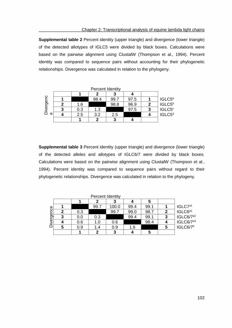
Chapter 2: Transcriptional analysis of equine lambda light chains
102
Supplemental table 2 Percent identity (upper triangle) and divergence (lower triangle)
of the detected allotypes of IGLC5 were divided by black boxes. Calculations were
based on the pairwise alignment using ClustalW (Thompson et al., 1994). Percent
identity was compared to sequence pairs without accounting for their phylogenetic
relationships. Divergence was calculated in relation to the phylogeny.
Percent Identity
Div
erg
en
c
e
1 2 3 4
1 98.4 99.7 97.5 1 IGLC5a
2 1.6 98.8 96.9 2 IGLC5b
3 0.3 1.3 97.5 3 IGLC5c
4 2.5 3.2 2.5 4 IGLC5d
1 2 3 4
Supplemental table 3 Percent identity (upper triangle) and divergence (lower triangle)
of the detected alleles and allotypes of IGLC6/7 were divided by black boxes.
Calculations were based on the pairwise alignment using ClustalW (Thompson et al.,
1994). Percent identity was compared to sequence pairs without regard to their
phylogenetic relationships. Divergence was calculated in relation to the phylogeny.
Percent Identity
Div
erg
en
ce
1 2 3 4 5
1 99.7 100.0 99.4 99.1 1 IGLC7a1
2 0.3 99.7 99.0 98.7 2 IGLC6a1
3 0.0 0.3 99.4 99.1 3 IGLC6/7a2
4 0.6 1.0 0.6 98.4 4 IGLC6/7a3
5 0.9 1.4 0.9 1.6 5 IGLC6/7b
1 2 3 4 5

Chapter 2: Transcriptional analysis of equine lambda light chains
103
References
Almagro, J.C., Martinez, L., Smith, S.L., Alagon, A., Estevez, J., Paniagua, J., 2006. Analysis of the horse V(H) repertoire and comparison with the human IGHV germline genes, and sheep, cattle and pig V(H) sequences. Mol. Immunol. 43, 1836-1845.
Arakawa, H., Buerstedde, J.M., 2009. Activation-induced cytidine deaminase-mediated hypermutation in the DT40 cell line. Philos. Trans. R. Soc. Lond. B. Biol. Sci. 364, 639-644.
Arun, S.S., Breuer, W., Hermanns, W., 1996. Immunohistochemical examination of light-chain expression (lambda/kappa ratio) in canine, feline, equine, bovine and porcine plasma cells. Zentralbl. Veterinarmed. A. 43, 573-576.
Bastida-Corcuera, F., Butler, J.E., Heyermann, H., Thomford, J.W., Corbeil, L.B., 2000. Tritrichomonas foetus extracellular cysteine proteinase cleavage of bovine IgG2 allotypes. J. Parasitol. 86, 328-332.
Bastida-Corcuera, F.D., Butler, J.E., Yahiro, S., Corbeil, L.B., 1999a. Differential complement activation by bovine IgG2 allotypes. Vet. Immunol. Immunopathol. 71, 115-123.
Bastida-Corcuera, F.D., Nielsen, K.H., Corbeil, L.B., 1999b. Binding of bovine IgG2a and IgG2b allotypes to protein A, protein G, and Haemophilus somnus IgBPs. Vet. Immunol. Immunopathol. 71, 143-149.
Biedermann, G., Clar, U., Finke, A., Bickel, M., 2002. Analyse der Population des Rheinisch-Deutschen Kaltbluts. Züchtungskunde 74, 237-249.
Bjornsdottir, G., Myers, L.C., 2008. Minimal components of the RNA polymerase II transcription apparatus determine the consensus TATA box. Nucleic Acids Res. 36, 2906-2916.
Bremond, J., Balzer, B., 2011. Zentrale Dokumentation Tiergenetischer Ressourcen in Deutschland (TGRDEU).
Calderon, R., Lodeiro, R., Varela, T.A., Farina, J., Ambrosio, B., Guitard, E., Gonzalez-Martin, A., Dugoujon, J.M., 2007. GM and KM immunoglobulin allotypes in the Galician population: new insights into the peopling of the Iberian Peninsula. BMC Genet. 8, 37.
Constans, J., Hazout, S., Garruto, R.M., Gajdusek, D.C., Spees, E.K., 1985. Population distribution of the human vitamin D binding protein: anthropological considerations. Am. J. Phys. Anthropol. 68, 107-122.
Corbeil, L.B., Gogolewski, R.P., Kacskovics, I., Nielsen, K.H., Corbeil, R.R., Morrill, J.L., Greenwood, R., Butler, J.E., 1997. Bovine IgG2a antibodies to Haemophilus somnus and allotype expression. Can. J. Vet. Res. 61, 207-213.
Das, S., Nikolaidis, N., Klein, J., Nei, M., 2008. Evolutionary redefinition of immunoglobulin light chain isotypes in tetrapods using molecular markers. Proc. Natl. Acad. Sci. U. S. A. 105, 16647-16652.
Diesterbeck, U.S., Aboelhassan, D.M., Stein, S.K., Czerny, C.-P., 2011 in press. Detection of new allotypic variants of bovine λ-light chain constant regions in different cattle breeds. Dev. Comp. Immunol.
Edelman, G.M., 1973. Antibody structure and molecular immunology. Science 180, 830-840.
Emorine, L., Dreher, K., Kindt, T.J., Max, E.E., 1983. Rabbit immunoglobulin kappa genes: structure of a germline b4 allotype J-C locus and evidence for several b4-related sequences in the rabbit genome. Proc. Natl. Acad. Sci. U. S. A. 80, 5709-5713.

Chapter 2: Transcriptional analysis of equine lambda light chains
104
Faber, C., Shan, L., Fan, Z., Guddat, L.W., Furebring, C., Ohlin, M., Borrebaeck, C.A., Edmundson, A.B., 1998. Three-dimensional structure of a human Fab with high affinity for tetanus toxoid. Immunotechnology 3, 253-270.
Gibson, D., 1974. Structural studies on normal horse immunoglobulin light chains. Detection of k-type N-terminal sequences. Biochemistry 13, 2776-2785.
Giha, H.A., Nasr, A., Iriemenam, N.C., Arnot, D., Troye-Blomberg, M., Theander, T.G., Berzins, K., ElGhazali, G., Pandey, J.P., 2009. Antigen-specific influence of GM/KM allotypes on IgG isotypes and association of GM allotypes with susceptibility to Plasmodium falciparum malaria. Malar. J. 8, 306.
Granoff, D.M., Pandey, J.P., Boies, E., Squires, J., Munson, R.S., Jr., Suarez, B., 1984. Response to immunization with Haemophilus influenzae type b polysaccharide-pertussis vaccine and risk of Haemophilus meningitis in children with the Km(1) immunoglobulin allotype. J. Clin. Invest. 74, 1708-1714.
Hamann, H., Distl, O., 2008. Genetic variability in Hanoverian warmblood horses using pedigree analysis. J. Anim. Sci. 86, 1503-1513.
Hartmann, O., Bruns, E., Hertsch, B., Schade, W., 2006. Pferdezucht. Eugen Ulmer KG, Stuttgart (Hohenheim).
Home, W.A., Ford, J.E., Gibson, D.M., 1992. L chain isotype regulation in horse. I. Characterization of Ig lambda genes. J. Immunol. 149, 3927-3936.
Humphrey, W., Dalke, A., Schulten, K., 1996. VMD: visual molecular dynamics. J. Mol. Graph. 14, 33-38, 27-38.
Kawasaki, K., Minoshima, S., Nakato, E., Shibuya, K., Shintani, A., Asakawa, S., Sasaki, T., Klobeck, H.G., Combriato, G., Zachau, H.G., Shimizu, N., 2001. Evolutionary dynamics of the human immunoglobulin kappa locus and the germline repertoire of the Vkappa genes. Eur. J. Immunol. 31, 1017-1028.
Kelley, L.A., Sternberg, M.J., 2009. Protein structure prediction on the Web: a case study using the Phyre server. Nat. Protoc. 4, 363-371.
Klein, S.L., Strausberg, R.L., Wagner, L., Pontius, J., Clifton, S.W., Richardson, P., 2002. Genetic and genomic tools for Xenopus research: The NIH Xenopus initiative. Dev. Dyn. 225, 384-391.
Korngold, L., Lipari, R., 1956. Multiple-myeloma proteins. III. The antigenic relationship of Bence Jones proteins to normal gammaglobulin and multiple-myeloma serum proteins. Cancer 9, 262-272.
Kratzin, H.D., Palm, W., Stangel, M., Schmidt, W.E., Friedrich, J., Hilschmann, N., 1989. [The primary structure of crystallizable monoclonal immunoglobulin IgG1 Kol. II. Amino acid sequence of the L-chain, gamma-type, subgroup I]. Biol. Chem. Hoppe Seyler 370, 263-272.
Lee, B., Richards, F.M., 1971. The interpretation of protein structures: estimation of static accessibility. J. Mol. Biol. 55, 379-400.
Lefranc, M.P., Pommie, C., Kaas, Q., Duprat, E., Bosc, N., Guiraudou, D., Jean, C., Ruiz, M., Da Piedade, I., Rouard, M., Foulquier, E., Thouvenin, V., Lefranc, G., 2005. IMGT unique numbering for immunoglobulin and T cell receptor constant domains and Ig superfamily C-like domains. Dev. Comp. Immunol. 29, 185-203.
Littell, R.C., Milliken, G.A., Stroup, W.W., Wolfinger, R., 2004. SASⓇ system for Mixed Models, 6th Edition. SAS Institute Inc, Cary, NC, USA.
Matsumoto, H., Miyazaki, T., Rychkov, Y.G., Zhukova, O., Lebedeva, I.A., Kondik, V.M., Udina, I.G., Spitzin, V.A., Batsuur, J., Shnader, Y.V., et al., 1984. Studies on the human immunoglobulin allotypes in five populations in the USSR. Jinrui Idengaku Zasshi 29, 105-111.
Montano, R.F., Morrison, S.L., 2002. Influence of the isotype of the light chain on the properties of IgG. J. Immunol. 168, 224-231.

Chapter 2: Transcriptional analysis of equine lambda light chains
105
Moxley, G., Gibbs, R.S., 1992. Polymerase chain reaction-based genotyping for allotypic markers of immunoglobulin kappa shows allelic association of Km with kappa variable segment. Genomics 13, 104-108.
Nadel, B., Tang, A., Escuro, G., Lugo, G., Feeney, A.J., 1998. Sequence of the spacer in the recombination signal sequence affects V(D)J rearrangement frequency and correlates with nonrandom Vkappa usage in vivo. J. Exp. Med. 187, 1495-1503.
Oli, M.W., Rhodin, N., McArthur, W.P., Brady, L.J., 2004. Redirecting the humoral immune response against Streptococcus mutans antigen P1 with monoclonal antibodies. Infect. Immun. 72, 6951-6960.
Padlan, E.A., Cohen, G.H., Davies, D.R., 1986. Antibody Fab assembly: the interface residues between CH1 and CL. Mol. Immunol. 23, 951-960.
Pandey, J.P., 2000. Immunoglobulin GM and KM allotypes and vaccine immunity. Vaccine 19, 613-617.
Pandey, J.P., Elson, L.H., Sutherland, S.E., Guderian, R.H., Araujo, E., Nutman, T.B., 1995. Immunoglobulin kappa chain allotypes (KM) in onchocerciasis. J. Clin. Invest. 96, 2732-2734.
Pandey, J.P., Fudenberg, H.H., Virella, G., Kyong, C.U., Loadholt, C.B., Galbraith, R.M., Gotschlich, E.C., Parke, J.C., Jr., 1979. Association between immunoglobulin allotypes and immune responses to Haemophilus influenzae and Meningococcus polysaccharides. Lancet 1, 190-192.
Parng, C.L., Hansal, S., Goldsby, R.A., Osborne, B.A., 1996. Gene conversion contributes to Ig light chain diversity in cattle. J. Immunol. 157, 5478-5486.
Pritsch, O., Hudry-Clergeon, G., Buckle, M., Petillot, Y., Bouvet, J.P., Gagnon, J., Dighiero, G., 1996. Can immunoglobulin C(H)1 constant region domain modulate antigen binding affinity of antibodies? J. Clin. Invest. 98, 2235-2243.
Roschenthaler, F., Hameister, H., Zachau, H.G., 2000. The 5' part of the mouse immunoglobulin kappa locus as a continuously cloned structure. Eur. J. Immunol. 30, 3349-3354.
Sanger, F., Nicklen, S., Coulson, A.R., 1977. DNA sequencing with chain-terminating inhibitors. Proc. Natl. Acad. Sci. U. S. A. 74, 5463-5467.
Schanfield, M.S., Ferrell, R.E., Hossaini, A.A., Gerald Sandler, S., Stevenson, J.C., 2008. Immunoglobulin allotypes in Southwest Asia: populations at the crossroads. Am. J. Hum. Biol. 20, 671-682.
Simianer, H., Kohn, F., 2010. Genetic management of the Gottingen Minipig population. J. Pharmacol. Toxicol. Methods 62, 221-226.
Sitnikova, T., Su, C., 1998. Coevolution of immunoglobulin heavy- and light-chain variable-region gene families. Mol. Biol. Evol. 15, 617-625.
Sun, Y., Wang, C., Wang, Y., Zhang, T., Ren, L., Hu, X., Zhang, R., Meng, Q., Guo, Y., Fei, J., Li, N., Zhao, Y., 2010. A comprehensive analysis of germline and expressed immunoglobulin repertoire in the horse. Dev. Comp. Immunol. 34, 1009-1020.
Thompson, J.D., Higgins, D.G., Gibson, T.J., 1994. CLUSTAL W: improving the sensitivity of progressive multiple sequence alignment through sequence weighting, position-specific gap penalties and weight matrix choice. Nucleic Acids Res. 22, 4673-4680.
Tonegawa, S., 1983. Somatic generation of antibody diversity. Nature 302, 575-581. Wagner, B., 2006. Immunoglobulins and immunoglobulin genes of the horse. Dev.
Comp. Immunol. 30, 155-164. Winstead, C.R., Zhai, S.K., Sethupathi, P., Knight, K.L., 1999. Antigen-induced somatic
diversification of rabbit IgH genes: gene conversion and point mutation. J. Immunol. 162, 6602-6612.

Chapter 2: Transcriptional analysis of equine lambda light chains
106
Yang, C., Bolotin, E., Jiang, T., Sladek, F.M., Martinez, E., 2007. Prevalence of the initiator over the TATA box in human and yeast genes and identification of DNA motifs enriched in human TATA-less core promoters. Gene 389, 52-65.
Zhou, J., Lottenbach, K.R., Barenkamp, S.J., Reason, D.C., 2004. Somatic hypermutation and diverse immunoglobulin gene usage in the human antibody response to the capsular polysaccharide of Streptococcus pneumoniae Type 6B. Infect. Immun. 72, 3505-3514.

Chapter 3: Bovine Immunoglobulins
107
Chapter 3:
Bovine Immunoglobulins The nature and extent of my contribution to the work was the following:
3. Exceptionally long CDR3H are not isotype restricted in bovine
immunoglobulins
Nature of contribution Extent of contribution
1. Scientific design 50%
2. Laboratory work 80%
3. Evaluation 70%
4. Scientific Writing 65%
4. Development of a bioinformatics framework for the detection of gene
conversion and the analysis of combinatorial diversity in immunoglobulin heavy
chains in four cattle breeds
Nature of contribution Extent of contribution
1. Scientific design 50%
2. Laboratory work 50%
3. Evaluation 70%
4. Scientific Writing 65%

Chapter 3: Bovine Immunoglobulins
108
Bovine immunoglobulin heavy and light chains
Bovine immunoglobulin heavy chain gene locus
The bovine immunoglobulin heavy chain gene (IGH) locus was assigned to the Bos
taurus autosome (BTA) 21 and spans approximately 250 kb (Miller et al. 1992; Niku et
al. 2012). Southern blot analyses indicated the existence of two IGHV families (boVH1
and boVH2) in the bovine germline repertoire. Before the study of Walther et al. (2013),
the expression of only boVH1 genes has been observed and boVH2 was found
comprising only pseudogenes (Tutter and Riblet 1989; Berens et al. 1997; Saini et al.
1997; Sinclair et al. 1997; Lopez et al. 1998). The family boVH1 offers a restricted set
of functional genes, which shares homologies to the murine Q52 family and human
VHII family. The definite number and organization of IGHV has not been determined
yet. The latest in silico analysis using blastn on all bovine genome assemblies revealed
IGHV on the contigs NW_003104530.1 and NW_003104538.1 on Bos taurus
chromosome 21 (BTA21; AC_000178.1), NW_003064289.1, NW_003064290.1,
NW_003064296.1, NW_003064297.1, NW_003064298.1, and NW_003064299.1 on
BTA7 (AC_000164.1), as well as on several unplaced contigs (Walther et al. 2013). In
total, 36 IGHV are identified in the bovine genome assembly. On BTA 21, IGHV1 and
IGHV2 were found at the centromeric region in NW_003104530.1. Eight IGHV located
at the telomeric region were described within about 146 kb on NW_003104538.1
(IGHV3, IGHV4, IGHV5, IGHV6, IGHV7, IGHV8, IGHV9, and IGHV10). The
contigs NW_003064289.1, NW_003064290.1, NW_003064296.1, NW_003064297.1,
NW_003064298.1, and NW_003064299.1 were annotated to 144 kb of the centromeric
region on BTA7 possessing nine IGHV (IGHV11, IGHV12, IGHV13, IGHV14,
IGHV15, IGHV16(ORF), IGHV17(ORF), IGHV18, and IGHV19). Thirteen out of
the 36 IGHV segments are putatively functional. Eleven IGHV segment pairs shared
sequence identity of 100% (IGHV3/33, IGHV10/34, IGHV9/35, IGHV4/32,
IGHV7/22, IGHV2/26, IGHV1/27, IGHV18/30, IGHV16(ORF)/25,
IGHV14Ψ/23Ψ, and IGHV36/29(F)). Phylogenetic analyses revealed a relation of all
functional IGHV to huIGHV2-05, whereas this group corresponds to the bovine IGHV
family 1 (boVH1) (Berens et al. 1997; Saini et al. 1997; Sinclair et al. 1997).
In cattle, ten IGHD genes are classified into four families that are organized in sub-
clusters (Koti et al. 2008; Koti et al. 2010). Further, the IGHD exons reveal huge size

Chapter 3: Bovine Immunoglobulins
109
differences (Koti et al. 2008). On the telomeric end of BTA21, the bovine IGHD8 and
IGHD4 were localized between IGHV6 and IGHV7 (Walther et al. 2013). Within the
contig NW_003064411.1 on BTA7 one IGHD cluster of 1131 bp, encoding
IGHD1(ORF) to IGHD3(ORF), was identified. On BTA8 a third location enclosing about
43 kb possesses five IGHD segments, a δ chain pseudogene, a µ chain gene, and six
IGHJ segments. In general, loci for the IGHD were observed on three chromosomes
(Walther et al. 2013).
By hybridization experiments, six joining segments (IGHJ) were detected on BTA11q23
(Hosseini et al. 2004). The number of IGHJ segments preceding the IGHC genes was
described by screening a bovine BAC and Cosmid library. Only two out of six IGHJ are
functional (IGHJ1, IGHJ2). IGHJ1 is recombined predominantly while IGHJ2 is
rearranged at low frequency (Zhao et al. 2003; Hosseini et al. 2004).
Figure 1: Physical map of the bovine heavy chain locus The locus is shown in 3’-5’ direction on chromosome 21. This figure is adapted from current literature (Zhao et al. 2003; Zimin et al. 2009; Koti et al. 2010; Niku et al. 2012; Walther et al. 2013; Pasman 2014).

Chapter 3: Bovine Immunoglobulins
110
The CDR3H encoded by IGHV-IGHD-IGHJ recombinations varies from 3 to 67 amino
acid residues (Berens et al. 1997; Saini et al. 1999; Saini and Kaushik 2002; Saini et al.
2003; Kaushik et al. 2009; Larsen and Smith 2012). These exceptionally long CDR3H
are specific for cattle and possess characteristic hydrophilic Glycine and Tyrosine
residues. The 148 bp long IGHD2 contributes to those CDR3H (Berens et al. 1997;
Shojaei et al. 2003; Koti et al. 2008). Further, a high number of Cysteine residues was
described and is demonstrated to promote intra-CDR3H disulfide bonds that contribute
to the “knob” domain in exceptionally long CDR3H (Sinclair et al. 1997). In contrast,
inter CDR disulfide bonds may exist in mid-length CDR3H due to one to three Cys
residues in CDR3H and one Cys within CDR2H (Saini et al. 1999; Ramsland et al.
2001). Additional mechanisms contributing to the unusual length of bovine CDR3H
were studied because the imprecise junction of germline encoded IGHV, IGHD, and
IGHJ cannot fully explain the existence of exceptionally long CDR3H in cattle. The
insertion of conserved short nucleotide sequences of 13 to 18 nucleotides specifically
into the IGHV and IGHD junction was found to contribute to a further elongation of the
CDR3H. So far, this mechanism is unique for cattle (Koti et al. 2010).
The genomic organization of the bovine IGHC locus was described by screening a
bovine BAC and Cosmid library. All heavy chain isotype classes that have been
detected in other mammals were also described for cattle (Knight and Becker 1987;
Zhao et al. 2002). Three sub-classes comprise the γ-isotype, namely γ1, γ2, and γ3
(Knight and Becker 1987; Symons et al. 1989). The IGHC locus spans approximately
150 kb enclosing the genes in the following order: 5’-JH–7 kb–μ–5 kb–δ–33 kb–γ3–20
kb–γ1–34 kb–γ2–20 kb–ε–13 kb–α-3’ (Zhao et al. 2003).
Bovine immunoglobulin M (IgM)
IgM is the major serum antibody in the primary immune response as it provides the first
antibody-mediated host defense (Saini and Kaushik 2001; Woof and Burton 2004). IgM
combats septicaemia when administered passively to calves and it is an important
bactericidal antibody directed against mastitis in cattle and other ruminants (Mousavi et
al. 1998). High amounts of IgM are described in colostrum and milk (3 mg/ml) (Butler
1995).
Two loci comprising functional IGHM genes were identified on BTA21 and BTA11q23
by in situ hybridization, whereas the latter is currently assigned to BTA8 (Tobin-Janzen
and Womack 1992; Hayes and Petit 1993). The IGHM gene comprises four exons

Chapter 3: Bovine Immunoglobulins
111
(IGHM1-4) encoding the constant domains, as well as two additional exons encoding
the transmembrane domain (TM1, TM2) (Mousavi et al. 1998). Three bovine IgM
allotypes are described, designated as IgMa, IgMb, and IgMc. While these allotypes
are classified by amino acid substitutions within the exons, further IgM variants may
result via alternative splicing e.g. inserting in frame codons at the IGHM1 and IGHM2
junction. IGHM2 possesses few proline residues and functions as a hinge restricting
flexibility and antigen binding ability of IgM (Saini and Kaushik 2001; Pasman 2014).
Bovine immunoglobulin D (IgD)
On BTA7 (contig NW_003064411.1), a cluster of 1131 bp was identified encoding
exons for IGHD1(ORF) to IGHD3(ORF) and a pseudogene for a δ chain. The exons 1
and 2 of the IGHD pseudogene are fragmented and exon 6 is missing (codes for the
secretory region). Nevertheless, IgD is also found in a secreted form although showing
lower concentrations than IgG, IgA, and IgM (Preud'homme et al. 2000; Zhao et al.
2002). Further, frame shifts were identified. Genomic information for IGHD4 to 7 and a
δ chain pseudogene were found on the contig NW_001503306.1, while IGHD4 and 8
were detected on NW_001504477.2. A locus involving a putative functional δ chain
gene was detected on NT_186572. In addition to NW_003100387.1, including a δ
chain pseudogene locus, a putative functional δ chain gene was noted on
NW_003100112.1 (Walther et al. 2013).
The bovine, ovine, and porcine germline IgD genes are transcriptionally active at low
levels and show structural similarities to the human IgD due to the three constant
domains and the hinge region. As phylogenetic analyses revealed the Cδ gene
duplicated from the Cμ gene more than 300 million years ago, the exon of the first
constant domain of the IgD (IGHD1) is very similar to the IGHM1 exon. Further, unlike
other species, the bovine IGHD has a switch region that may permit class switch
recombination (Zhao et al. 2002).
Bovine immunoglobulin G (IgG)
Immunoglobulin G is the most abundant isotype in bovine serum as it constitutes more
than 75% of serum immunoglobulins (Saini et al. 2007). It is also found in blood, lymph,
peritoneal fluid, and cerebrospinal fluid. Furthermore, IgG contributes to different
effector functions. For instance, it is involved in the enhancement of phagocytosis,
Chapter 3: Bovine Immunoglobulins
112
antibody-dependent cellular cytotoxicity, the regulation of cytokine and antibody
production, antigen processing and presentation, as well as in the selective transfer via
the FcRn-like receptor on alveolar epithelial cells of mammary glands (Burg and
Pillinger 2001; Kacskovics 2004; Mestecky 2005; Pentsuk and van der Laan 2009).
Three IgG subclasses (IgG1, IgG2, and IgG3) were identified in cattle, whereas two
subclasses (IgG1 and IgG2) were found in sheep, seven IgG subclasses (IgG1-7) were
detected in horses, four subclasses were described in humans, five in swine, and one
in rabbits (Knight et al. 1988; Symons et al. 1989; Clarkson et al. 1993; Kacskovics et
al. 1995; Kacskovics and Butler 1996; Rabbani et al. 1997; Wagner et al. 2004;
Pasman 2014). Depending on characterization methods, two different types of
designation exist. IgG1, IgG2a, and IgG2b were used in serological analyses, whereas
IgG1, IgG2, and IgG3 were used in molecular analyses, respectively (Knight et al.
1988). Serum concentrations of IgG1 and IgG2 reveal 10 mg/ml and the concentration
of IgG1 can exceed 60 mg/ml in hyperimmunized animals. In colostrum, IgG1
concentration was also found to exceed 100 mg/ml (Butler 1995). The bovine IgG3
shows low serum concentrations, which may be caused by the longer hinge region that
is a preferred proteolytic site (Rabbani et al. 1997).
These three bovine Cγ genes (γ1, γ2, and γ3) have been mapped to chromosome
21q24 and are located between the δ and ε genes of the heavy chain locus (Gu et al.
1992; Tobin-Janzen and Womack 1992; Chowdhary et al. 1996; Saini et al. 2007).
Interestingly, in an in silico study, on the bovine genome assembly BTA20 revealed the
genomic information for a γ2 chain. A genomic order of γ3, γ2, and γ1 was identified on
NT_185580.1. Genes coding for γ1 and γ3 were found on NW_003100065.1 and
NW_003099305.1 (Walther et al. 2013). Gene duplication of bovine IGHG1 led to
IGHG2 and IGHG3. Sequence similarities of 85.1% and 83.4% exist between the
homologues IGHG1 and IGHG3, as well as IGHG1 and IGHG2, respectively (Zhao et
al. 2003). For IgG1, four allotypic variants were described (IgG1a-d) (Symons et al.
1989; Jackson et al. 1992; Kacskovics and Butler 1996; Saini et al. 2007). The bovine
IgG2 and IgG3 reveal the two allotypic variants IgG2a and IgG2b (Kacskovics and
Butler 1996) and IgG3a and IgG3b (Rabbani et al. 1997), respectively. Significant
structural differences within the hinge regions and the CH3 domain result in different
biological effector functions of IGHG1, IGHG2, and IGHG3. Amino acid residue
substitutions were also described within the CH1 and CH2 domains of these three
genes (Saini et al. 2007).
The four known IgG1 allotypic variants are characterized by individual amino acid

Chapter 3: Bovine Immunoglobulins
113
differences at the positions 190 and 192 (CH1), 218, 224, 225, and 226 (hinge region),
as well as 281 (CH2), and 402 (CH3), which were designated using the EU numbering
system. IgG1a showed the amino acids Gly190, Thr192, Arg218, Thr224, and Thr226,
Asp281, Gly402, as well as a deletion of the amino acid at position 225. The most
similar allelic variants IgGb and IgGd possess Gly190, Thr192, Thr218, Pro224,
Ser225, Pro226, and Asp281. They solely differ by Gly and Ser at position 402. The
allotypic variant IgGc shows the amino acid residues Ala190, Ser192, Arg218, Arg224,
Pro226, Asn281, Ser402 and a deletion at position 225. Three-dimensional modeling
revealed that all substitutions are located on the surface of the IgG molecule and
therefore may be recognized by antisera and effector molecules such as cellular
adhesion molecules (Symons et al. 1989; Saini et al. 2007). Additionally, the repeating
motif Pro-Ala-Ser-Ser at the positions 189–192 and 205–208 within the CH1 domain of
IgG1c may stabilize the core immunoglobulin fold or promote interactions with
fibronectin or similar adhesion molecules. The replacement of Thr with Arg residues
within the hinge region reduces the number of O-linked glycosylation sites and
increases the susceptibility to degradation by protease secreting bacteria (Saini et al.
2007).
The allotypic variants IgG2a and IgG2b differed in amino acid residues at 19 positions.
These amino acid residue substitutions were found in the three constant region
domains (CH1, CH2, and CH3) but also in the hinge region. In CH1, IgG2a and IgG2b
differed by Ser and Ala at position 129 (Ser/Ala129), Cys/Ser131, Lys/Thr135,
Gly/Ala190, and Thr/Ser192. Within the hinge region Ser/Ile219, Pro/Cys224,
Asn/His225, and His/Asn228 were observed. In CH2, Thr/Ser290, Ile/Asn326, and
Ser/Pro332 differentiate IgG2a and IgG2b. Val/Leu364, Ile/Thr370, Asp/Ala378,
Asp/Asn384, Ala/Thr402, Arg/Glu419, and Thr/Ala422 are the characteristic amino
acids described for IgG2a and IgG2b, respectively. These amino acid differences result
in different immune responses of the two IgG2 allotypes (Kacskovics and Butler 1996;
Corbeil et al. 1997). In comparison to IgG2a, IgG2b activates the complement more
than twice (Bastida-Corcuera et al. 1999a).
Previous studies indicated that IgG3a is expressed at a lower level than IgG3b. These
two allotypic variants differ in six positions. The amino acids Ser/Arg131, Arg/Leu174,
and Ser/Thr192 are located in the CH1 domain, whereas Pro/Gln237 and Lys/Glu238
were found in the hinge region, and Lys/Arg4431 characterizes individual amino acid
residues of IgG3a and IgG3b in the CH3 domain (Rabbani et al. 1997).

Chapter 3: Bovine Immunoglobulins
114
The allotypic variants of IgG provide polymorphic immunoglobulin genetic markers with
specific abilities. In particular, modified qualities in complement activation, age-
dependent expression, and influences on the effector function against Haemophilus
somnus and Tritrichomonas foetus were made for the allotypic variants of bovine IgG2
(Corbeil et al. 1997; Bastida-Corcuera et al. 1999b; Bastida-Corcuera et al. 2000; Saini
et al. 2007).
Bovine immunoglobulin E (IgE)
Whereas IgE is found in low amounts in the serum, IgE is mainly found during immune
responses against parasite infections mediated by basophiles and mast cells.
Furthermore, the presence of IgE gives rise to type I hypersensitivity reactions such as
asthma, conjunctivitis, and rhinitis (Mousavi et al. 1997). The bovine IgE also shows
heat labile skin sensitizing ability as the human IgE does (Hammer et al. 1971).
The latest in silico analysis revealed genomic information for an ε chain on
NT_185723.1 (Walther et al. 2013). This finding is consistent with previous studies,
where only a single copy of the bovine IgE was identified (Knight et al. 1988). IgE has
four exons (IGHE1-4) showing homologies of 87% to sheep IGHE, 58% to horse IGHE,
55% to human IGHE, and 52% to mouse IGHE. Due to the additional fourth constant
domain, IGHE possesses a higher molecular weight than other Ig isotypes. The
additional domain is supposed to be important for high affinity binding to Fc receptors
on mast cells (Mousavi et al. 1997).
Bovine immunoglobulin A (IgA)
Immunoglobulin A is the dominating isotype on mucosal surfaces such as mammary
glands with an important part in the immune response against microbial infections.
A single IGHA gene was identified in the bovine genome and was assigned to the
contig NT_185723.1 (Knight et al. 1988; Walther et al. 2013). IgA possesses three
IGHA exons (Brown et al. 1997). Two allelic variants were identified by restrictions
fragment length polymorphism and serological analyses but were not confirmed by
genomic DNA analyses of 50 Swedish cattle (De Benedictis G 1984; Brown et al. 1997;
Pasman 2014).

Chapter 3: Bovine Immunoglobulins
115
The bovine immunoglobulin light chain gene loci
In cattle, two distinct immunoglobulin light chain isotypes (lambda λ and kappa κ) have
been described (Pilstrom 2002; Das et al. 2008). In serum antibodies, bovine λ-light
chains are predominantly expressed in a proportion of 95% (Arun et al. 1996). About
5% of the heavy chains in serum antibodies are connected with functional κ-light chains
(Arun et al. 1996; Aitken et al. 1999). Similarly, the light chain repertoire is dominated
by λ-light chains in horses (Ford et al. 1994) and sheep (Foley and Beh 1992; Griebel
and Ferrari 1994; Broad et al. 1995), although a functional kappa system was
described, too (Home et al. 1992). In contrast, human and mice serum antibodies
dominantly associate κ-light chains (60% and 95%) (Chen et al. 2008). Genomic
conditions such as genomic complexity of the loci, recombination signal sequences, the
ordered rearrangement of κ- and λ-light chains, as well as antigen selection are
supposed to influence differences in the κ:λ ratio across species (Pasman et al. 2010).
Bovine lambda light chain genes
The bovine variable, joining, and constant region genes of the λ-light chain (IGL) are
located on Bos taurus autosome 17 (BTA17) (Tobin-Janzen and Womack 1992). The
locus spans 412 kb and contains 25 to 31 variable gene segments (IGLV) whereof 14
to 17 are potentially functional (Ekman et al. 2009). A total of 63 IGLV were identified
on ten scaffolds in the current genome assembly (Btau_3.1). Out of these 63 IGLV, 32
were identified on unplaced contigs and 11 of them are also potentially functional
(Ekman et al. 2009). The IGLV locus possessed both transcriptional orientations. All
IGLV are organized in three clusters separated by 126.8 kb and 138.3 kb introns. The
three IGLV clusters also define separate IGLV families designated as IGLV1, IGLV2,
and IGLV3. The predominantly expressed IGLV1 genes are subdivided into five
subfamilies (IGLV1a, IGLV1b, IGLV1c, IGLV1d, IGLV1x) (Sinclair et al. 1995; Saini et
al. 2003). These IGLV1 are located within the two 5’ subclusters, whereas the minor
expressed IGLV2 and IGLV3 genes were identified within the IGLJ-proximal cluster.
The subfamilies IGLV1a and IGLV1b are mainly expressed, whereas IGLV1c, IGLV1e,
and IGLV1x specifically pair with heavy chains possessing CDR3H with at least 50
amino acid residues (Saini et al. 1999; Saini et al. 2003). Analyses of genomic
sequences of the cattle breed Herford revealed 17 IGLV1 genes of which ten are
described functional. Further, three out of four IGLV2 genes as well all four IGLV3
genes were designated functional (Pasman et al. 2010). Phylogenetic comparisons
revealed that four of six ovine subgroups cluster with the bovine ones, which are

Chapter 3: Bovine Immunoglobulins
116
supposedly ruminant specific genes. Consequently, due to sequence identities of
92.6% and 88.9%, the bovine IGLV1 and IGLV2 families are closely related with the
ovine families IGLVI and IGLVII. The bovine IGLV3 family is related to the unclassified
ovine IGLV family (Pasman et al. 2010; Pasman 2014). In addition, the CDR1L length
varies from 6, 8, or 9 codons. The length of CDR2L is restricted to 3 or 7 codons. The
number of unique CDR1L–CDR2L combinations is lower than in mice and humans
(Ekman et al. 2009).
Figure 2: Physical map of the bovine lambda light chain locus The locus is shown in 5’-3’ direction on chromosome 17. This figure is adapted from (Pasman et al. 2010; Pasman 2014).
In cattle, four λ-light chain constant region (IGLC) genes (IGLC1, IGLC2, IGLC3, and
IGLC4) were identified yet, each preceded by a joining gene segment (IGLJ1-4) (Parng
et al. 1995; Parng et al. 1996; Chen et al. 2008; Ekman et al. 2009; Pasman et al.
2010). Only IGLC2 and IGLC3 are functional whereas IGLC3 is preferentially
expressed during rearrangements. IGLC1 and IGLC4 are pseudogenes (Chen et al.
2008; Ekman et al. 2009; Pasman et al. 2010). A fifth bovine IGLC gene was detected
but could not be located on a chromosome until now (Ekman et al. 2009). In
comparison, in sheep only two IGLC genes were described (IGLC1 and IGLC2). The

Chapter 3: Bovine Immunoglobulins
117
ovine IGLC1 is functional and in the IGLC2 gene possess a premature stop codon
(Jenne et al. 2003). Furthermore, in previous studies seven equine IGLC genes were
identified. Three of these genes are functional, whereas the others were described to
be pseudogenes (Home et al. 1992; Das et al. 2008; Sun et al. 2010).
Transcription analyses in several cattle breeds revealed single nucleotide
polymorphisms (SNPs) for both the bovine IGLC2 and IGLC3 genes resulting in silent
mutations or amino acid residue substitutions (Diesterbeck et al. 2012). Within the
breeds German Simmental (GS) and Aubrac (A), single nucleotide polymorphisms of
IGLC2 were identified. Analyses in the breeds German Black Pied (GBP) and Holstein
Friesian (HF) revealed the already known allele and allotype IGLC2a. Two additional
alleles, IGLC2b and IGLC2c, were detected and represent putative new allotypes.
Amino acid residue substitutions were either conservative or led to charge changes.
They were located within the molecule by homology based three-dimensional
modeling. Most amino acid residue substitutions were found on the outer side of the
molecule in the solvent accessible surface area, within or near the putative interface to
IGHC1. Additionally, by in silico analyses IGLC2a was also identified in other cattle
breeds such as Angus (DY170709) and Hereford (EH173018). IGLC2a and IGLC2b
showed 98.4% sequence identity while IGLC2c was 97.8% identical to IGLC2a and
96.9% identical to IGLC2b (Diesterbeck et al. 2012). Similar to IGLC2, five allelic
variants were identified for the predominantly expressed IGLC3 (IGLC3a, IGLC3b,
IGLC3c, IGLC3d, IGLC3e) within the same cattle breeds. Amino acid residue
substitutions were also determined on the accessible surface of the molecule, as well
as in and next to the interface to IGHC1. Reversed charges were observed due to
several amino acid residue replacements. Further, nucleotide sequences coding for
IGLC3a were found in Hereford (EV679232) and Angus (DY173535), while IGLC3b
was only found in Angus (DY149783). IGLC3c was identified both in Holstein Friesian
crossbreed (DY145594) and purebred (CK950153). Angus and Holstein Friesian
crossbreeds (EV608839, DY216512), as well as Holstein Friesian purebred
(CK979405) and Hereford (DT858283) exhibited IGLC3d (Diesterbeck et al. 2012).
Bovine kappa light chain genes
The bovine κ-light chain genes are located on Bos taurus autosome 11 (BTA11)
(Ekman et al. 2009) and span approximately 280 kb, although the complete Ig κ-light
chain (IGK) locus encompasses a size of 412 kb (Pasman et al. 2010). The analysis of
the bovine κ-light chain locus on Herford genome sequence revealed 22 IGKV, 3 IGKJ,

Chapter 3: Bovine Immunoglobulins
118
and 1 IGKC (Ekman et al. 2009). The IGKV genes are organized in a cluster that
covers 150 kb and which is followed by the three IGKJ genes and the IGKC gene. Out
of 22 IGKV, only eight are functional (Ekman et al. 2009). They are present in both
transcriptional orientations (Sitnikova and Nei 1998). The 22 IGKV genes are classified
into four subfamilies. Twenty-one of them are closely related to the ovine gene families
1, 2, and 4. Seven of the eight functional segments belong to bovine subgroup 2
(IGKV2), which therefore is supposed to be mainly expressed. This subgroup is
phylogenetically related to sheep (91%), killer whale (84%), and dolphin (84%). Bovine
IGKV vary from six, ten, or 11 codons within CDR1L and differs from human, mice, and
sheep CDR1L possessing 11 codons, whereby CDR2L constantly possesses three
codons (Ekman et al. 2009; Ehrenmann et al. 2010; Pasman 2014).
The three IGKJ genes (IGKJ1, IGKJ2, IGKJ3) are clustered within 800 bp on the
genome and are followed by one IGKC gene. Although all IGKJ genes are functional,
they were all transcribed in different frequencies. IGKJ1 showing the conserved 23 bp
RSS, was transcribed predominantly in animals of the breeds GS, HF, A, and GBP
(Stein et al. 2012). IGKJ2 was detected in two animals of GS despite of the shortened
spacer with a size of 22 bp instead of 23 bp (Ekman et al. 2009). IGKJ3 was also
described in each of the four cattle breeds. The distinct cladistic protein motif ‘EIK’ was
found changed in IGKJ3 to ‘EIK’ (Das et al. 2008).
Figure 3: Physical map of the bovine kappa light chain locus The locus is shown in 5’-3’ direction on chromosome 11. The locus is identical to both BTAU 4.6 and UMD 3.1 assembly of the Hereford cow genome. This figure is adapted from Pasman and Kaushik (2014) and Ekman et al. (2009).

Chapter 3: Bovine Immunoglobulins
119
Transcriptional analysis of IGKC within four cattle breeds revealed three alleles
distributed in different frequencies (Stein et al. 2012). In animals of HF, GS, and A, all
three alleles were identified, whereas in GBP solely IGKCa was found. Additional in
silico analyses of the ESTs database revealed allele 1 and 2 of IGKCa in Hereford
Shorthorns (CF763228, CF768037). The three alleles differed at four nucleotide
positions with two of them leading to amino acid residue substitutions at positions 100
and 116 within the constant region resulting in the putative allotypic variants of IGKCa
and IGKCb. The two amino acid residue replacements caused either a change in
polarity or a change in electric charge of the amino acid side chain. The amino acid
residue substitutions were located at the solvent accessible surface. Also for humans
and rabbits three and five allotypes were described, respectively (Emorine et al. 1983;
Moxley and Gibbs 1992). In humans, an improvement of immune responses by IGKC
allotypes was observed (Pandey et al. 1979; Granoff et al. 1984). Due to the similar
substitutions in bovine allotypes, different serological properties are assumed in cattle
allotypes, too (Stein et al. 2012).
Development of B cells and the bovine antibody repertoire
The development of immunoglobulin producing cells already starts in the bovine fetus.
While IgM-bearing B-cells have been detected at day 59 of gestation (Schultz et al.
1973), recombinations of heavy and light chain gene segments were observed in
splenic B-cells at day 125 of gestation and serum immunoglobulins were detectable at
day 145 of gestation (Saini and Kaushik 2002). The perinatal diversification occurs in
the ileal Peyer’s patches (Yasuda et al. 2004; Yasuda et al. 2006) but λ-light chain
diversification was observed in bovine fetal spleen prior to the diversification in the
ileum (Lucier et al. 1998; Aida 2014). Pre-B like cells were also noted in the fetal bone
marrow and lymph node indicating B lymphopoieses in parallel to ileal Peyer’s patches
(Ekman et al. 2012; Aida 2014).
The generation of antibody diversity in vertebrates depends on several mechanisms
that are very similar in all vertebrate species. Such processes are the recombination of
separated germline gene segments (IGHV, IGHD, and IGHJ; IGLV/IGKV and
IGLJ/IGKJ), the imprecise junction of these gene segments due to nucleotide deletions
or additions (N, P), the combination of two identical heavy and light chains (Alt and
Baltimore 1982; Kurosawa and Tonegawa 1982; Tonegawa 1983; Desiderio et al.
1984), as well as somatic hypermutations (Reynaud et al. 1995; Wagner and

Chapter 3: Bovine Immunoglobulins
120
Neuberger 1996; Berens et al. 1997). The available pool of the germline IGHV, IGHD,
and IGHJ segments differs between species. Whereas humans and mice possess a
high number of IGHV-IGHD-IGHJ genes, in livestock such as chicken (Reynaud et al.
1989), pigs (Sun et al. 1994), sheep (Dufour et al. 1996), and cattle (Berens et al.
1997; Saini et al. 1997; Sinclair et al. 1997) relatively few genes were detected
resulting in a restricted combinatorial diversity. Therefore, several diversifying
mechanisms dominate or additional options are employed in such species. For
instance, in chicken and bovine λ-light chains gene conversion is a post-
recombinatorial strategy for the generation of the preimmune antibody repertoire
(Reynaud et al. 1995; Arakawa et al. 2004). Further, this mechanism is discussed in
horses (Sun et al. 2010).
Today, differences in gene usage of bovine fetus and adult animals are known. In fetal
IGHV-IGHD-IGHJ recombinations two IGHV are preferentially used (Saini and Kaushik
2002). Furthermore, IGHD5 and IGHD7 dominate both in bovine fetus and adult
animals in contrast to IGHDQ52 (Koti et al. 2010). Within the CDRs transition
nucleotide substitutions predominate over transversion mutations and in framework
region three somatic hypermutations lead to isotype dependent degree of
diversification (Kaushik et al. 2009).
In both bovine fetal and adult heavy chains, exceptionally long CDR3H regions
contribute to higher variability, which is restricted by the use of only one VH-family
(Armour et al. 1994; Saini et al. 1999; Saini and Kaushik 2002; Wang et al. 2013).
Those long CDR3H regions were first described in IgM but were also detected in IgG
and in all other isotypes by now and occur in 8-10% of circulating B cells (Saini et al.
1997; Sinclair et al. 1997; Saini et al. 1999; Saini and Kaushik 2002; Saini et al. 2003;
Kaushik et al. 2009; Larsen and Smith 2012; Walther et al. 2013). The length of
CDR3H varies by class. Consequently, the bovine CDR3H has an average length of
22.7 ± 3.2 amino acids, whereas in IgM there are 21.7 ± 1.8 and in IgG there are 18.2 ±
1.3 amino acids (Almagro et al. 2006; Kaushik et al. 2009; Aida 2014). While the
CDR3H region in human and mice encodes 12 amino acid residues, in cattle it extends
up to 67 amino acid residues (Sun et al. 1994; Walther et al. 2013) and up to 30 in
chicken (McCormack and Thompson 1990) where this phenomenon is caused by the
usage of two IGHD segments within on CDR3H region. Therefore the length of the
CDR3H region is 50% greater than in human, mice, swine, and rabbit sequences
(Butler 1997). The extensive size heterogeneity in CDR3H together with intrachain
disulfide bridges results in a configurational diversity of this region (Saini and Kaushik

Chapter 3: Bovine Immunoglobulins
121
2002; Wang et al. 2013). Crystallization of such bovine antibodies revealed a unique
‘stalk and knob’ structure (Wang et al. 2013). Although both fetal and adult
exceptionally long CDR3H originate from identical IGHV-IGHD-IGHJ rearrangements,
there are structural differences. In the bovine fetus, there is no addition of CSNS at the
IGHV-IGHD junction resulting in a shorter or non-existent stalk. Due to a structural
support, bovine heavy chains with exceptionally long CDR3H always pair with λ-light
chains that possess a conserved Ser at position 90 (Wang et al. 2013).
In contrast to human and mice bovine antibody diversity increases by somatic
hypermutation, which may also occur without previous antigen contact (Tomlinson et
al. 1996; Berens et al. 1997; Lopez et al. 1998). Furthermore, diversity already exists at
the foetal stage as a result of somatic hypermutation.
In bovine light chains, gene conversion and somatic hypermutation are the major
mechanisms leading to diversification (Parng et al. 1995; Parng et al. 1996). Initial
diversification of λ-light chains was identified in the early fetal spleen before the
establishment of a diverse repertoire in the ileum in cattle and sheep (Lee and
Richards 1971). Gene conversion is supposed to be less significant in sheep than in
cattle as there is a higher number of IGLV pseudogenes in the bovine genome (Parng
et al. 1996; Ekman et al. 2009). This mechanism is also highly relevant regarding light
chain diversification in chicken (Arakawa and Buerstedde 2009), as well as in rabbits
using gene conversion to diversify Ig heavy chains (Weinstein et al. 1994). In addition,
allotypes diversify the immunoglobulin repertoire, which has already been described in
species like humans and rabbits. For instance, in humans three allotypic variants of the
so called Km 1, Km 1.2, and Km 3 (Moxley and Gibbs 1992) are described. Similarly,
rabbits possess five allotypic variants of the κ-light chain constant region gene called
b4 (a+b), b5, b6, and b9 (Emorine et al. 1983). In cattle, allotypes have been identified
for both the heavy and light chains (Bastida-Corcuera et al. 1999a; Bastida-Corcuera et
al. 1999b; Diesterbeck et al. 2012; Stein et al. 2012).

Chapter 3: Bovine Immunoglobulins
122
References
Aida Y, Takeshima, S.-N., Baldwin, C. L. and Kaushik, A. K. 2014. Bovine Immunogenetics. in The Genetics of Cattle (ed. DJaR Garrick, A.). CAB International, CAB International.
Aitken R, Hosseini A, MacDuff R. 1999. Structure and diversification of the bovine immunoglobulin repertoire. Veterinary immunology and immunopathology 72: 21-29.
Almagro JC, Martinez L, Smith SL, Alagon A, Estevez J, Paniagua J. 2006. Analysis of the horse V(H) repertoire and comparison with the human IGHV germline genes, and sheep, cattle and pig V(H) sequences. Molecular immunology 43: 1836-1845.
Alt FW, Baltimore D. 1982. Joining of immunoglobulin heavy chain gene segments: implications from a chromosome with evidence of three D-JH fusions. Proc Natl Acad Sci U S A 79: 4118-4122.
Arakawa H, Buerstedde JM. 2009. Activation-induced cytidine deaminase-mediated hypermutation in the DT40 cell line. Philosophical transactions of the Royal Society of London Series B, Biological sciences 364: 639-644.
Arakawa H, Saribasak H, Buerstedde JM. 2004. Activation-induced cytidine deaminase initiates immunoglobulin gene conversion and hypermutation by a common intermediate. PLoS Biol 2: E179.
Armour KL, Tempest PR, Fawcett PH, Fernie ML, King SI, White P, Taylor G, Harris WJ. 1994. Sequences of heavy and light chain variable regions from four bovine immunoglobulins. Molecular immunology 31: 1369-1372.
Arun SS, Breuer W, Hermanns W. 1996. Immunohistochemical examination of light-chain expression (lambda/kappa ratio) in canine, feline, equine, bovine and porcine plasma cells. Zentralblatt fur Veterinarmedizin Reihe A 43: 573-576.
Bastida-Corcuera F, Butler JE, Heyermann H, Thomford JW, Corbeil LB. 2000. Tritrichomonas foetus extracellular cysteine proteinase cleavage of bovine IgG2 allotypes. The Journal of parasitology 86: 328-332.
Bastida-Corcuera FD, Butler JE, Yahiro S, Corbeil LB. 1999a. Differential complement activation by bovine IgG2 allotypes. Veterinary immunology and immunopathology 71: 115-123.
Bastida-Corcuera FD, Nielsen KH, Corbeil LB. 1999b. Binding of bovine IgG2a and IgG2b allotypes to protein A, protein G, and Haemophilus somnus IgBPs. Veterinary immunology and immunopathology 71: 143-149.
Berens SJ, Wylie DE, Lopez OJ. 1997. Use of a single VH family and long CDR3s in the variable region of cattle Ig heavy chains. International immunology 9: 189-199.
Broad TE, Burkin DJ, Cambridge LM, Jones C, Lewis PE, Morse HG, Geyer D, Pearce PD, Ansari HA, Maher DW. 1995. Six loci mapped on to human chromosome 2p are assigned to sheep chromosome 3p. Animal genetics 26: 85-90.
Brown WR, Rabbani H, Butler JE, Hammarstrom L. 1997. Characterization of the bovine C alpha gene. Immunology 91: 1-6.
Burg ND, Pillinger MH. 2001. The neutrophil: function and regulation in innate and humoral immunity. Clinical immunology 99: 7-17.
Butler JE. 1995. Antigen receptors, their immunomodulation and the immunoglobulin genes of cattle and swine. Livest Prod Sci 42: 105-121.
Butler JE. 1997. Immunoglobulin gene organization and the mechanism of repertoire development. Scandinavian journal of immunology 45: 455-462.

Chapter 3: Bovine Immunoglobulins
123
Chen L, Li M, Li Q, Yang X, An X, Chen Y. 2008. Characterization of the bovine immunoglobulin lambda light chain constant IGLC genes. Veterinary immunology and immunopathology 124: 284-294.
Chowdhary BP, Fronicke L, Gustavsson I, Scherthan H. 1996. Comparative analysis of the cattle and human genomes: detection of ZOO-FISH and gene mapping-based chromosomal homologies. Mammalian genome : official journal of the International Mammalian Genome Society 7: 297-302.
Clarkson CA, Beale D, Coadwell JW, Symons DB. 1993. Sequence of ovine Ig gamma 2 constant region heavy chain cDNA and molecular modelling of ruminant IgG isotypes. Molecular immunology 30: 1195-1204.
Corbeil LB, Gogolewski RP, Kacskovics I, Nielsen KH, Corbeil RR, Morrill JL, Greenwood R, Butler JE. 1997. Bovine IgG2a antibodies to Haemophilus somnus and allotype expression. Canadian journal of veterinary research = Revue canadienne de recherche veterinaire 61: 207-213.
Das S, Nikolaidis N, Klein J, Nei M. 2008. Evolutionary redefinition of immunoglobulin light chain isotypes in tetrapods using molecular markers. Proceedings of the National Academy of Sciences of the United States of America 105: 16647-16652.
De Benedictis G CP, Dragone A. 1984. Identification of an allotypic IgA in cattle serum. Comp Immunol Microbiol Infect Dis 7: 35-42.
Desiderio SV, Yancopoulos GD, Paskind M, Thomas E, Boss MA, Landau N, Alt FW, Baltimore D. 1984. Insertion of N regions into heavy-chain genes is correlated with expression of terminal deoxytransferase in B cells. Nature 311: 752-755.
Diesterbeck US, Aboelhassan DM, Stein SK, Czerny CP. 2012. Detection of new allotypic variants of bovine lambda-light chain constant regions in different cattle breeds. Developmental and comparative immunology 36: 130-139.
Dufour V, Malinge S, Nau F. 1996. The sheep Ig variable region repertoire consists of a single VH family. J Immunol 156: 2163-2170.
Ehrenmann F, Kaas Q, Lefranc MP. 2010. IMGT/3Dstructure-DB and IMGT/DomainGapAlign: a database and a tool for immunoglobulins or antibodies, T cell receptors, MHC, IgSF and MhcSF. Nucleic acids research 38: D301-307.
Ekman A, Ilves M, Iivanainen A. 2012. B lymphopoiesis is characterized by pre-B cell marker gene expression in fetal cattle and declines in adults. Developmental and comparative immunology 37: 39-49.
Ekman A, Niku M, Liljavirta J, Iivanainen A. 2009. Bos taurus genome sequence reveals the assortment of immunoglobulin and surrogate light chain genes in domestic cattle. BMC immunology 10: 22.
Emorine L, Dreher K, Kindt TJ, Max EE. 1983. Rabbit immunoglobulin kappa genes: structure of a germline b4 allotype J-C locus and evidence for several b4-related sequences in the rabbit genome. Proceedings of the National Academy of Sciences of the United States of America 80: 5709-5713.
Foley RC, Beh KJ. 1992. Analysis of immunoglobulin light chain loci in sheep. Animal genetics 23: 31-42.
Ford JE, Home WA, Gibson DM. 1994. Light chain isotype regulation in the horse. Characterization of Ig kappa genes. Journal of immunology 153: 1099-1111.
Granoff DM, Pandey JP, Boies E, Squires J, Munson RS, Jr., Suarez B. 1984. Response to immunization with Haemophilus influenzae type b polysaccharide-pertussis vaccine and risk of Haemophilus meningitis in children with the Km(1) immunoglobulin allotype. The Journal of clinical investigation 74: 1708-1714.

Chapter 3: Bovine Immunoglobulins
124
Griebel PJ, Ferrari G. 1994. Evidence for a stromal cell-dependent, self-renewing B cell population in lymphoid follicles of the ileal Peyer's patch of sheep. European journal of immunology 24: 401-409.
Gu F, Chowdhary BP, Andersson L, Harbitz I, Gustavsson I. 1992. Assignment of the bovine immunoglobulin gamma heavy chain (IGHG) gene to chromosome 21q24 by in situ hybridization. Hereditas 117: 237-240.
Hammer DK, Kickhofen B, Schmid T. 1971. Detection of homocytotropic antibody associated with a unique immunoglobulin class in the bovine species. European journal of immunology 1: 249-257.
Hayes HC, Petit EJ. 1993. Mapping of the beta-lactoglobulin gene and of an immunoglobulin M heavy chain-like sequence to homoeologous cattle, sheep, and goat chromosomes. Mamm Genome 4: 207-210.
Home WA, Ford JE, Gibson DM. 1992. L chain isotype regulation in horse. I. Characterization of Ig lambda genes. Journal of immunology 149: 3927-3936.
Hosseini A, Campbell G, Prorocic M, Aitken R. 2004. Duplicated copies of the bovine JH locus contribute to the Ig repertoire. Int Immunol 16: 843-852.
Jackson T, Morris BA, Sanders PG. 1992. Nucleotide sequences and expression of cDNAs for a bovine anti-testosterone monoclonal IgG1 antibody. Molecular immunology 29: 667-676.
Jenne CN, Kennedy LJ, McCullagh P, Reynolds JD. 2003. A new model of sheep Ig diversification: shifting the emphasis toward combinatorial mechanisms and away from hypermutation. Journal of immunology 170: 3739-3750.
Kacskovics I. 2004. Fc receptors in livestock species. Veterinary immunology and immunopathology 102: 351-362.
Kacskovics I, Butler JE. 1996. The heterogeneity of bovine IgG2--VIII. The complete cDNA sequence of bovine IgG2a (A2) and an IgG1. Molecular immunology 33: 189-195.
Kacskovics I, Wittum TE, Butler JE, Littledike ET. 1995. The heterogeneity of bovine IgG2. VII. The phenotypic distribution of the A1 and A2 allotypes of IgG2a among beef cows with known clinical history. Veterinary immunology and immunopathology 48: 89-96.
Kaushik AK, Kehrli ME, Jr., Kurtz A, Ng S, Koti M, Shojaei F, Saini SS. 2009. Somatic hypermutations and isotype restricted exceptionally long CDR3H contribute to antibody diversification in cattle. Veterinary immunology and immunopathology 127: 106-113.
Knight KL, Becker RS. 1987. Isolation of genes encoding bovine IgM, IgG, IgA and IgE chains. Vet Immunol Immunopathol 17: 17-24.
Knight KL, Suter M, Becker RS. 1988. Genetic engineering of bovine Ig. Construction and characterization of hapten-binding bovine/murine chimeric IgE, IgA, IgG1, IgG2, and IgG3 molecules. Journal of immunology 140: 3654-3659.
Koti M, Kataeva G, Kaushik AK. 2008. Organization of D(H)-gene locus is distinct in cattle. Developments in biologicals 132: 307-313.
Koti M, Kataeva G, Kaushik AK. 2010. Novel atypical nucleotide insertions specifically at VH-DH junction generate exceptionally long CDR3H in cattle antibodies. Molecular immunology 47: 2119-2128.
Kurosawa Y, Tonegawa S. 1982. Organization, structure, and assembly of immunoglobulin heavy chain diversity DNA segments. J Exp Med 155: 201-218.
Larsen PA, Smith TP. 2012. Application of circular consensus sequencing and network analysis to characterize the bovine IgG repertoire. BMC immunology 13: 52.
Lee B, Richards FM. 1971. The interpretation of protein structures: estimation of static accessibility. Journal of molecular biology 55: 379-400.

Chapter 3: Bovine Immunoglobulins
125
Lopez O, Perez C, Wylie D. 1998. A single VH family and long CDR3s are the targets for hypermutation in bovine immunoglobulin heavy chains. Immunological reviews 162: 55-66.
Lucier MR, Thompson RE, Waire J, Lin AW, Osborne BA, Goldsby RA. 1998. Multiple sites of V lambda diversification in cattle. Journal of immunology 161: 5438-5444.
McCormack WT, Thompson CB. 1990. Somatic diversification of the chicken immunoglobulin light-chain gene. Advances in immunology 48: 41-67.
Mestecky J. 2005. Mucosal immunology, Amsterdam; Boston. Miller JR, Thomsen PD, Dixon SC, Tucker EM, Konfortov BA, Harbitz I. 1992. Synteny
mapping of the bovine IGHG2, CRC and IGF1 genes. Animal genetics 23: 51-58.
Mousavi M, Rabbani H, Hammarstrom L. 1997. Characterization of the bovine epsilon gene. Immunology 92: 369-373.
Mousavi M, Rabbani H, Pilstrom L, Hammarstrom L. 1998. Characterization of the gene for the membrane and secretory form of the IgM heavy-chain constant region gene (C mu) of the cow (Bos taurus). Immunology 93: 581-588.
Moxley G, Gibbs RS. 1992. Polymerase chain reaction-based genotyping for allotypic markers of immunoglobulin kappa shows allelic association of Km with kappa variable segment. Genomics 13: 104-108.
Niku M, Liljavirta J, Durkin K, Schroderus E, Iivanainen A. 2012. The bovine genomic DNA sequence data reveal three IGHV subgroups, only one of which is functionally expressed. Developmental and comparative immunology 37: 457-461.
Pandey JP, Fudenberg HH, Virella G, Kyong CU, Loadholt CB, Galbraith RM, Gotschlich EC, Parke JC, Jr. 1979. Association between immunoglobulin allotypes and immune responses to Haemophilus influenzae and Meningococcus polysaccharides. Lancet 1: 190-192.
Parng CL, Hansal S, Goldsby RA, Osborne BA. 1995. Diversification of bovine lambda-light chain genes. Annals of the New York Academy of Sciences 764: 155-157.
Parng CL, Hansal S, Goldsby RA, Osborne BA. 1996. Gene conversion contributes to Ig light chain diversity in cattle. Journal of immunology 157: 5478-5486.
Pasman Y, Saini SS, Smith E, Kaushik AK. 2010. Organization and genomic complexity of bovine lambda-light chain gene locus. Veterinary immunology and immunopathology 135: 306-313.
Pasman YK, A. 2014. Bovine Immunoglobulin Genetics: Novel Phylogenetic Perspective. in Comparative Immunoglobulin Genetics (ed. AaP Kaushik, Y.). Apple Academic Press, Toronto, Canada.
Pentsuk N, van der Laan JW. 2009. An interspecies comparison of placental antibody transfer: new insights into developmental toxicity testing of monoclonal antibodies. Birth defects research Part B, Developmental and reproductive toxicology 86: 328-344.
Pilstrom L. 2002. The mysterious immunoglobulin light chain. Developmental and comparative immunology 26: 207-215.
Preud'homme JL, Petit I, Barra A, Morel F, Lecron JC, Lelievre E. 2000. Structural and functional properties of membrane and secreted IgD. Molecular immunology 37: 871-887.
Rabbani H, Brown WR, Butler JE, Hammarstrom L. 1997. Polymorphism of the IGHG3 gene in cattle. Immunogenetics 46: 326-331.
Ramsland PA, Kaushik A, Marchalonis JJ, Edmundson AB. 2001. Incorporation of long CDR3s into V domains: implications for the structural evolution of the antibody-combining site. Exp Clin Immunogenet 18: 176-198.

Chapter 3: Bovine Immunoglobulins
126
Reynaud CA, Dahan A, Anquez V, Weill JC. 1989. Somatic hyperconversion diversifies the single Vh gene of the chicken with a high incidence in the D region. Cell 59: 171-183.
Reynaud CA, Garcia C, Hein WR, Weill JC. 1995. Hypermutation generating the sheep immunoglobulin repertoire is an antigen-independent process. Cell 80: 115-125.
Saini SS, Allore B, Jacobs RM, Kaushik A. 1999. Exceptionally long CDR3H region with multiple cysteine residues in functional bovine IgM antibodies. European journal of immunology 29: 2420-2426.
Saini SS, Farrugia W, Muthusamy N, Ramsland PA, Kaushik AK. 2007. Structural evidence for a new IgG1 antibody sequence allele of cattle. Scandinavian journal of immunology 65: 32-38.
Saini SS, Farrugia W, Ramsland PA, Kaushik AK. 2003. Bovine IgM antibodies with exceptionally long complementarity-determining region 3 of the heavy chain share unique structural properties conferring restricted VH + Vlambda pairings. International immunology 15: 845-853.
Saini SS, Hein WR, Kaushik A. 1997. A single predominantly expressed polymorphic immunoglobulin VH gene family, related to mammalian group, I, clan, II, is identified in cattle. Molecular immunology 34: 641-651.
Saini SS, Kaushik A. 2001. Origin of bovine IgM structural variants. Molecular immunology 38: 389-396.
Saini SS, Kaushik A. 2002. Extensive CDR3H length heterogeneity exists in bovine foetal VDJ rearrangements. Scandinavian journal of immunology 55: 140-148.
Schultz RD, Dunne HW, Heist CE. 1973. Ontogeny of the bovine immune response. Infection and immunity 7: 981-991.
Shojaei F, Saini SS, Kaushik AK. 2003. Unusually long germline DH genes contribute to large sized CDR3H in bovine antibodies. Mol Immunol 40: 61-67.
Sinclair MC, Gilchrist J, Aitken R. 1995. Molecular characterization of bovine V lambda regions. Journal of immunology 155: 3068-3078.
Sinclair MC, Gilchrist J, Aitken R. 1997. Bovine IgG repertoire is dominated by a single diversified VH gene family. Journal of immunology 159: 3883-3889.
Sitnikova T, Nei M. 1998. Evolution of immunoglobulin kappa chain variable region genes in vertebrates. Molecular biology and evolution 15: 50-60.
Stein SK, Diesterbeck US, Aboelhassan DM, Czerny CP. 2012. Comparison of joining and constant kappa-light chain regions in different cattle breeds. Animal genetics 43: 776-780.
Sun J, Kacskovics I, Brown WR, Butler JE. 1994. Expressed swine VH genes belong to a small VH gene family homologous to human VHIII. Journal of immunology 153: 5618-5627.
Sun Y, Wang C, Wang Y, Zhang T, Ren L, Hu X, Zhang R, Meng Q, Guo Y, Fei J et al. 2010. A comprehensive analysis of germline and expressed immunoglobulin repertoire in the horse. Developmental and comparative immunology 34: 1009-1020.
Symons DB, Clarkson CA, Beale D. 1989. Structure of bovine immunoglobulin constant region heavy chain gamma 1 and gamma 2 genes. Molecular immunology 26: 841-850.
Tobin-Janzen TC, Womack JE. 1992. Comparative mapping of IGHG1, IGHM, FES, and FOS in domestic cattle. Immunogenetics 36: 157-165.
Tomlinson IM, Walter G, Jones PT, Dear PH, Sonnhammer EL, Winter G. 1996. The imprint of somatic hypermutation on the repertoire of human germline V genes. Journal of molecular biology 256: 813-817.
Tonegawa S. 1983. Somatic generation of antibody diversity. Nature 302: 575-581.
Chapter 3: Bovine Immunoglobulins
127
Tutter A, Riblet R. 1989. Conservation of an immunoglobulin variable-region gene family indicates a specific, noncoding function. Proceedings of the National Academy of Sciences of the United States of America 86: 7460-7464.
Wagner B, Miller DC, Lear TL, Antczak DF. 2004. The complete map of the Ig heavy chain constant gene region reveals evidence for seven IgG isotypes and for IgD in the horse. Journal of immunology 173: 3230-3242.
Wagner SD, Neuberger MS. 1996. Somatic hypermutation of immunoglobulin genes. Annu Rev Immunol 14: 441-457.
Walther S, Czerny CP, Diesterbeck US. 2013. Exceptionally long CDR3H are not isotype restricted in bovine immunoglobulins. PloS one 8: e64234.
Wang F, Ekiert DC, Ahmad I, Yu W, Zhang Y, Bazirgan O, Torkamani A, Raudsepp T, Mwangi W, Criscitiello MF et al. 2013. Reshaping antibody diversity. Cell 153: 1379-1393.
Weinstein PD, Anderson AO, Mage RG. 1994. Rabbit IgH sequences in appendix germinal centers: VH diversification by gene conversion-like and hypermutation mechanisms. Immunity 1: 647-659.
Woof JM, Burton DR. 2004. Human antibody-Fc receptor interactions illuminated by crystal structures. Nature reviews Immunology 4: 89-99.
Yasuda M, Fujino M, Nasu T, Murakami T. 2004. Histological studies on the ontogeny of bovine gut-associated lymphoid tissue: appearance of T cells and development of IgG+ and IgA+ cells in lymphoid follicles. Developmental and comparative immunology 28: 357-369.
Yasuda M, Jenne CN, Kennedy LJ, Reynolds JD. 2006. The sheep and cattle Peyer's patch as a site of B-cell development. Veterinary research 37: 401-415.
Zhao Y, Kacskovics I, Pan Q, Liberles DA, Geli J, Davis SK, Rabbani H, Hammarstrom L. 2002. Artiodactyl IgD: the missing link. Journal of immunology 169: 4408-4416.
Zhao Y, Kacskovics I, Rabbani H, Hammarstrom L. 2003. Physical mapping of the bovine immunoglobulin heavy chain constant region gene locus. The Journal of biological chemistry 278: 35024-35032.
Zimin AV, Delcher AL, Florea L, Kelley DR, Schatz MC, Puiu D, Hanrahan F, Pertea G, Van Tassell CP, Sonstegard TS et al. 2009. A whole-genome assembly of the domestic cow, Bos taurus. Genome biology 10: R42.

Chapter 3: Exceptionally long CDR3H in bovine immunoglobulins
128
Exceptionally Long CDR3H Are Not Isotype Restricted in Bovine Immunoglobulins
Stefanie Walther1, Claus-Peter Czerny1, Ulrike S. Diesterbeck1*
1Department of Animal Sciences, Institute of Veterinary Medicine, Division of Microbiology and Animal Hygiene, Faculty of Agricultural Sciences, Georg-August University Göttingen, Burckhardtweg 2, 37077 Göttingen, Germany *Corresponding author: U. S. Diesterbeck Tel.: +49 551 393375 Fax: +49 551 3913513 E-mail: [email protected] Postal address: Dr. Ulrike Diesterbeck
Department of Animal Sciences, Georg-August University Göttingen
Burckhardtweg 2 37077 Göttingen, Germany Published in “PlosOne”
DOI: 10.1371/journal.pone.0064234 URL: http://journals.plos.org/plosone/article?id=10.1371/journal.pone.0064234

Chapter 3: Exceptionally long CDR3H in bovine immunoglobulins
129
Abstract
Exceptionally long third complementarity determining regions of the heavy chain
(CDR3H) were previously described as a specificity of bovine IgG and IgM
immunoglobulins. In addition, the genomic organization of the immunoglobulin heavy
chain locus remains to be elucidated with a special focus on the number of variable
segments (IGHV).
By analyzing the variable regions according to the isotype-specific PCR using cDNA-
PCR, we were able to prove the existence of exceptional long CDR3H in all bovine
isotypes. The corresponding sequences of three distinct amplicons were grouped
according to the length of the CDR3H. Sequences of CDR3H possessed 5 to 10, 12 to
31 or at least 48 amino acid residues. Long and mid-length CDR3H were composed of
mainly hydrophilic amino acid residues, while short CDR3H also contained
hydrophobic amino acid residues. All sequences with long CDR3H were related to the
germline variable segment 10.
Using the current genome assembly, Bos taurus NCBI build 6.1, the genomic
organization of the bovine immunoglobulin heavy-chain locus was analyzed. A main
locus was investigated on BTA21. Exons coding for variable, diversity, and joining
segments, as well as for the constant regions of different isotypes, were also localized
on BTA7, BTA8, and BTA20. Together with the information from unplaced contigs, 36
IGHV were detected of which 13 are putatively functional. Phylogenetic analysis
revealed two bovine IGHV families (boVH1, boVH2). Thus, the existence of the two
bovine families suggested was demonstrated, where boVH1 comprises all functional
segments.
This study substantially improves the understanding of the generation of
immunoglobulin diversity in cattle.

Chapter 3: Exceptionally long CDR3H in bovine immunoglobulins
130
Introduction
The generation of antibody diversity in vertebrates is subjected to a sequence of steps
such as the recombination of separated germline gene segments for both heavy (V, D,
and J) and light (V and J) chains. Furthermore, the imprecise junction of the germline
gene segments occurs as a result of nucleotide deletions or additions (N, P),
introduced by the terminal deoxynucleotidyl transferase during the recombination
process. The assembly of two identical heavy and light chains completes the tetrameric
molecule [1,2,3,4]. In addition, somatic hypermutations contribute to antibody diversity
– dependent or independent of antigen contact [5,6,7]. While these general processes
of diversification are very similar in all vertebrate species, considerable differences
were found in the available pool of the germline V, D, and J segments. Although
humans and mice possess a large pool of VDJ genes [8], livestock such as chicken [9],
pigs [10], sheep [11], and cattle [6,12,13] are relatively restricted in the generation of
combinatorial diversity. Therefore, species-dependent mechanisms dominate the
different diversification steps or additional options are employed. For instance, in
chicken gene conversion, the use of pseudogene sequences is a frequent post-
recombinatorial strategy for the generation of the preimmune antibody repertoire [5,14].
This mechanism was confirmed for λ-light chains in cattle [15] and is discussed in
horses [16].
All heavy-chain isotype classes detected in other mammals were also described for
cattle [17,18], whereas the γ-isotype encompasses three sub-classes, namely γ1, γ2,
and γ3 [18,19]. The bovine IGH locus was assigned to the Bos taurus autosome (BTA)
21 [20] and localized on the q23-q24 bands [21] or on the q24 band respectively
[22,23]. An IgM-like chain was assigned to BTA11q23 by hybridization [24,25], which
was supported by the detection of six IGHJ segments on the same chromosome [26].
By screening a bovine BAC and Cosmid library, the genomic organization of the IGHC
locus was described, as well as the number of the preceding joining segments (IGHJ).
Only two out of six IGHJ were classified as functional – of which only one seems to be
involved predominantly in the recombination process [21,26]. The IGHV itself codes for
the complementarity determining regions 1 and 2 (CDR1H, CDR2H) and for the N-
terminal part of the complementarity determining region 3 (CDR3H). Bovine IGHV offer
a restricted set of genes related to one family (boVH1), which shares homologies to the
murine Q52 family and human VHII family. Southern blot analyses indicated one
additional IGHV family in the germline repertoire but only expression of boVH1 has

Chapter 3: Exceptionally long CDR3H in bovine immunoglobulins
131
been observed yet [6,12,13,27,28]. The definite number and organization of IGHV
remains under further investigation.
Another peculiarity is the organization of the bovine IGHD locus. Ten IGHD genes
classified into four families are organized in sub-clusters [29,30]. A comparison of the
IGHD exons revealed huge size differences [29]. Cattle antibodies provide
exceptionally long CDR3H consisting of up to 62 amino acid residues (aa)
[6,31,32,33,34,35]. IGHD2, with 148 bp in size, contributes to those CDR3H and
encodes the characteristic hydrophilic Glycine and Tyrosine residues [6,29,36]. The
high number of Cysteine residues detected is supposed to promote intra-CDR3H
disulfide bonds [13]. Mid-length CDR3H – containing one to three Cys residues – were
almost always accompanied by one Cys residue found in the CDR2H, which may result
in intra CDR disulfide bond formation [31,37]. The germline encoded IGHV, IGHD, and
IGHJ and their imprecise junction during rearrangement cannot fully explain the
remarkable length of the CDR3H. Conserved short nucleotide sequences of 13 to 18
nucleotides are specifically inserted into the IGHV and IGHD junction, leading to a
further extension of the CDR3H. This mechanism is unique for cattle [30].
To date, these exceptionally long CDR3H have been attributed exclusively to the γ1-3-
and µ-isotype [33,35]. In our study, we demonstrate the expression of exceptionally
long CDR3H in all bovine immunoglobulin isotypes. We were able to observe three
distinct groups of CDR3H sizes, which were related to their genomic origin. Loci of
IGHV were determined on BTA7, BTA21 and seven unplaced contigs.

Chapter 3: Exceptionally long CDR3H in bovine immunoglobulins
132
Materials and Methods
In silico Analysis of the Bovine IGHV Segments
A sequence search was performed with blastn on Bos taurus in the Reference genomic
sequences (refseq_genomic) database using the leader and variable region of one
mRNA sequence (accession number AY145128). On the identified contigs, the IGHV
and their respective leader were annotated together with the recombination signal
sequences (RSS). The octamers, TATA boxes, and splicing sites were also noted. The
nucleotide sequences of the bovine IGHD1 to 8 and Q52 [30], as well as all IGHJ
coding sequences (AY158087, AY149283), were used for a similar alignment
approach. The detected IGHV, IGHD, and IGHJ were used in the further analysis of the
amplified immunoglobulin sequences. To annotate the constant region locus, IgM
(U63637), IgD (AF411240, AF515672), IgG1 (X16701), IgG2 (S82407, X16702), IgG3
(U63638), IgE (AY221098), and IgA (AF109167) bovine coding sequences were
applied. Missing transmembrane regions were determined in bovine ESTs (expressed
sequence tags). Based on the available sequence data, functionality was defined
according to Lefranc [38]. In brief, functional sequences exhibited an open reading
frame (ORF) without stop codon, and no defects in the splicing sites, RSS, or in the
regulatory elements. If sequence information was missing due to end of contigs or N’s
introduced in the sequence but the available sequence offered putative functionality,
genes were marked with (F). Classification to ORF included either alterations in the
splicing site, RSS, regulatory elements, substitutions of conserved amino acid residues
(Cys23, Trp41, Leu89, Cys104 within IGHV or IGHC and a Phe/Trp118-Gly119-X120-
Gly121 motif within IGHJ [39]) or orphons ((ORF)). In this case, orphons are located
outside of BTA21 [20,21,22,23]. Pseudogenes () were characterized by the presence
of stop codons or frameshifts. Fragmented loci were also defined as pseudogenes.
Functional recombination assays revealed the spacer lengths, the first three
nucleotides of the heptamer as well as three consecutive adenosine residues within the
nonamer to be crucial for efficient recombination [40,41].
For the purpose of phylogenetic analysis, the complete nucleotide sequences of bovine
IGHV segments were aligned with one member of the human IGHV families 1 to 7,
respectively, using the ClustalW algorithm with the ClustalX 2.1 interface [42]. The
phylogenetic tree was calculated using the neighbor-joining method, with the exclusion
of gaps. The confidence values were compiled with 1000 bootstrap replicates [43]. To

Chapter 3: Exceptionally long CDR3H in bovine immunoglobulins
133
root the tree, the sequence of one IGHV segment of the horned shark (accession
number X13449) and little skate (X15124) were defined as an outgroup, similar to the
method performed by Sitnikova and Su [44] and Almagro et al. [45]. Visualization of the
phylogenetic tree was performed using the program NJplot [46].
Ethical Statement
To collect B-lymphocytes, 20 ml of EDTA blood were taken from the tail vein of a
German Simmental bull kept by the Division of Microbiology and Animal Hygiene for
demonstrations in claw-treatment within student courses and to study the clinical
development of Mycobacteria avium spp. paratuberculosis infection. The bull was
owned, because he had acquired a natural infection of MAP and showed positive
antibody-titers already with an age of 18 months. Similar to other cattle herds, he has
to be tested for cattle diseases periodically. The blood sample was taken from the tail
vein during regular investigation of infectious diseases in the bull. The plasma was
applied e.g. in an indirect ELISA testing for antibodies against Mycobacteria avium spp.
paratuberculosis or BHV-1. Therefore, no specific approval is required.
Isolation of PBMCs and cDNA Synthesis
Peripheral blood mononuclear cells (PBMCs) were isolated using Ficoll gradients (GE
Healthcare Europe GmbH, Germany) according to the manufacturer’s protocol. Viable
B cells were stained with trypan blue and counted. Total RNA was isolated from 1x10⁷
cells using the RNeasy Mini Kit (Qiagen, Germany). The first-strand cDNA was
synthesized using pd(N)6 primers from 3 µg of total RNA in a total volume of 20 µl
(SuperScript™III First-Strand Synthesis SuperMix, Life Technologies GmbH,
Germany).
Amplification of Immunoglobulin Heavy-chain Isotype Restricted Variable
Regions
To amplify the variable region restricted to each isotype, a primer set was generated
with one primer hybridizing in the leader region, and individual primers with binding
sites in the constant region (CH) of the immunoglobulin heavy chains. The primers
were based on database entries and their own sequence information (data not shown).
For α, γ1-3, and ε isotypes primers anneal to the CH1. For δ and µ isotypes, primers
bind within the CH2 (Table 1). To monitor the integrity and purity of the cDNA, 527 bp

Chapter 3: Exceptionally long CDR3H in bovine immunoglobulins
134
of the bovine GAPDH (Glycerinaldehyde 3-phosphate dehydrogenase) were amplified
as a positive control. A no template control served as a negative control for the PCR.
Table 1. Primer for the isotype-specific amplification of the complete variable regions.
Forward primer Reverse primer Primersequence 5’-3’ Approximated
product size in bp
BoLH_BACK ACCCACTGTGGACCCTCCTC BoIgMCH2_FOR TGCCGTCACCAGAGAGGCTGT 795 BoIgDCH2_FOR TGCGTGCTGACCGCCTTGTT 805 BoIgG1-3CH1_FOR GGCACCCGAGTTCCAGGTCA 536 BoIgECH1_FOR GCCCAGCCTTACACGGGCTT 467 BoIgACH1_FOR GCCAGCACGGCAGGGAAGTT 574
GAPDH_for TGGTCACCAGGGCTGCT
GAPDH_rev GGAGGGGCCATCCACAGTCT 527
One universal forward primer was used for annealing within the leader region. For each isotype, a reverse primer was generated for specific amplification. The annealing sites were selected in the first constant region (IGCH1), with the exception of IgM and IgD. Both isotypes share high homologies in the IGCH1 and therefore, specific reverse primers were generated for binding in the second constant region. The IgG subtypes were not distinguished further. Primers for bovine GAPDH served as cDNA quality control.
The total reaction volume of 50 µl included 1 µl of cDNA, 200 µM dNTPs (Bioline,
Germany), 5 µl of 10x PCR buffer (75 mM Tris HCl pH 9.0; 2 mM MgCl2; 50 mM KCl;
20 mM (NH4)2SO4), 5% DMSO (Dimethyl sulfoxide), 0.4 µM of each primer pair, and 2
units of DNA polymerase (Biotools, Spain). PCR was performed under cycling
conditions of 95oC for 5 min, followed by 35 cycles of 95oC for 1 min, 58°C for 1 min,
72oC for 2 min, and terminated with elongation at 72°C for 10 min. The length and
purity of the PCR products were evaluated by means of electrophoresis on 1% agarose
gels.
Cloning and Sequencing of the PCR Products
The PCR products were purified and concentrated using the DNA Clean &
Concentrator Kit (Zymo Research, USA). Purified products were cloned into the pCR
2.1-TOPO 3.9 Kb TA vector (InvitrogenTM, Karlsruhe, Germany) and transformed into
chemically competent One Shot TOP10 E. coli cells (InvitrogenTM, Karlsruhe,
Germany). Transformants were plated on LB agar containing 0.3 mM ampicillin, 40 µl
2.44 µM X-gal (5-bromo-4-chloro-3-indolyl-beta-D-galactopyranoside), and 40 µl 1 M
IPTG (Isopropyl β-D-1-thiogalactopyranoside) for blue/white selection. After incubation
at 37°C, overnight cultures of randomly selected white transformants were grown in a
5 ml LB-ampicillin broth. Plasmids were isolated using the MiniPrep Kit (Qiagen,
Chapter 3: Exceptionally long CDR3H in bovine immunoglobulins
135
Germany). In order to assess the insert size, plasmid DNA was cleaved with EcoRI
(New England Biolabs, Germany) and DNA sizes were confirmed by agarose gel
electrophoresis.
Twenty plasmids of each PCR product were sequenced according to the chain-
termination method [47]. The M13 (-20) Forward and M13 Reverse (Invitrogen,
Germany) vector-specific primers, as well as the corresponding gene specific primers,
were used for sequencing.
Nucleotide and Amino Acid Sequence Analyses
The genetic information of the VDJ recombinations was used for further analysis. The
amplified part of the constant regions served as verification of the respective isotype.
The sequences were analyzed using the DNAStar program (GATC Biotech AG,
Germany) and aligned by ClustalW [48].
The deduced amino acid residues of the variable parts were aligned to the IMGT
nomenclature [39] using the IMGT/DomainGapAlign [49,50]. Framework regions, as
well as CDRs, were identified and analyzed with regard to their biochemical properties
such as the hydrophobicity, polarity, and charge of the amino acid residues
incorporated. The CDR3H regions were classified according to their number of amino
acid residues. The amino acid compositions of CDR2H and CDR3H were examined for
their numbers of Tyr, Gly, aromatic amino acid residues and Cys, since some bovine
CDR3Hs are characterized by exceptional length and preferred amino acid residues.

Chapter 3: Exceptionally long CDR3H in bovine immunoglobulins
136
Results
Annotation of the Bovine Germline Immunoglobulin Heavy-chain Locus
For the identification of germline IGHV, a search using blastn on all bovine genome
assemblies was performed. The contigs NW_003104530.1 and NW_003104538.1
were identified on Bos taurus chromosome 21 (BTA21; AC_000178.1).
NW_003104530.1 was located at the centromeric region with two IGHV (IGHV1 and
IGHV2). A region of about 146 kb on NW_003104538.1, located at the telomeric
region, comprised eight IGHV: IGHV3, IGHV4, IGHV5, IGHV6, IGHV7, IGHV8,
IGHV9, and IGHV10. Upstream of them, the exons coding for the µ, ε and α chains,
were identified. Two IGHD (8 and 4) were localized between IGHV6 and IGHV7
(Figure 1A).
The contigs NW_003064289.1, NW_003064290.1, NW_003064296.1,
NW_003064297.1, NW_003064298.1, and NW_003064299.1 were localized to the
centromeric region on BTA7 (AC_000164.1) involving nine IGHV (IGHV11,
IGHV12, IGHV13, IGHV14, IGHV15, IGHV16(ORF), IGHV17(ORF), IGHV18,
and IGHV19). This cluster spans approximately 144 kb. Downstream on BTA7, a
cluster of 1131 bp, with exons encoding IGHD1(ORF) to IGHD3(ORF), was identified
on contig NW_003064411.1, followed by a pseudogene for a δ chain. The latter one is
characterized by fragmented exons 1 and 2, a deleted exon 6 (codes for the secretory
region), and frame shifts (Figure 1B).
A third location comprising five IGHD segments, a δ chain pseudogene, a µ chain
gene, and six IGHJ segments in about 43 kb was detected on BTA8. Two loci for the
IGHD were observed. IGHDQ52(ORF) was the most downstream segment on contig
NW_003066919.1, whereas IGHD4(ORF) to 7(ORF) were found to be the most
upstream on contig NW_003066918.1 (Figure 1C). Interestingly, BTA20 revealed the
genomic information for a γ2 chain (Figure 1D).

Chapter 3: Exceptionally long CDR3H in bovine immunoglobulins
137

Chapter 3: Exceptionally long CDR3H in bovine immunoglobulins
138
Figure 1. Chromosomal organization of variable (IGHV), diversity (IGHD), joining (IGHJ) segments, and the constant regions of the heavy chains. The physical map displays the order of functional segments (F), pseudogenes (Ψ), and open reading frames (ORF). Classification to “functional” includes an ORF without stop and exhibition of conserved amino acid residues as well as no defects in splicing signals, recombination signal sequences (RSS) or regulatory elements. ORF are defined by alterations in the splicing signals, recombination signal sequences, and/or regulatory elements. In addition, changes to conserved amino acid residues, which may lead to misfolding were included in the ORF classification. Functional elements on orphon localizations are highlighted with ORF in parenthesis (ORF) [38,39]. Pseudogenes possessed stop codons, frameshifts or mutations of the spacer lengths within the first three nucleotides of the heptamer as well as in three consecutive adenosines residues within the nonamer abolish the recombination [40,41]. In addition, fragmented loci were also defined as pseudogenes. Arrows indicate the transcription direction. In addition, unplaced contigs (NT_182448.1, NT_182449.1, NT_183109.1,
NT_185036.1, NT_185907.1, NT_186922.1, and NW_003100762.1) were discovered
to contain IGHV segments. Likewise, IGHD1(ORF) and 2 genes were localized on
NT_186153.1. NW_001494075.1 includes a gene coding for IGHDQ52 downstream of
an IGHJ1 to 6 locus. Genes coding for a µ chain and a δ chain pseudogene were found
most upstream in this contig. The contig NW_001503306.1 comprises the genomic
information for IGHD4 to 7 and a δ chain pseudogene while IGHD4 and 8 were
detected on NW_001504477.2. A locus involving IGHJ4ORF to 6ORF and a putative
functional δ chain gene was detected on NT_186572. Additional genomic information
for α and ε chains were discovered on NT_185723.1. A genomic order of γ3, γ2, and
γ1 was identified on NT_185580.1. Furthermore, a gene coding for γ1 and γ3 was
found on NW_003100065.1 and NW_003099305.1, respectively. Along with
NW_003100387.1, including a δ chain pseudogene locus, a putative functional δ chain
gene was noted on NW_003100112.1.
Thirteen out of the 36 IGHV segments identified are putatively functional
(Supplemental Table S1). Eleven IGHV segment pairs shared sequence identity of
100%, namely IGHV3/33, IGHV10/34, IGHV9/35, IGHV4/32, IGHV7/22,
IGHV2/26, IGHV1/27, IGHV18/30, IGHV16(ORF)/25, IGHV14Ψ/23Ψ, and
IGHV36/29(F). Since the human IGHV locus has been fully investigated [51], one
member of each family was chosen for phylogenetic analysis. The sequences
clustered into two distinct branches either with huIGHV2-05 or with huIGHV4-04 and
huIGHV6-1. All functional IGHV were phylogenetically related to huIGHV2-05 (Figure
2). This group corresponds to the bovine IGHV family 1 (boVH1) described previously
[6,12,13]. Multiple alignments revealed two distinct families with identities of at least
80.5% for boVH1 and 79.5% for boVH2. IGHV5Ψ and IGHV8Ψ represent fragmented
loci consisting of 77 bp. They share the highest identity of 70.1% with IGHV1Ψ,

Chapter 3: Exceptionally long CDR3H in bovine immunoglobulins
139
IGHV18Ψ, IGHV27Ψ, and IGHV30Ψ which are members of boVH2. The highest
identity between members of both families was calculated to be 69.7%.
Figure 2. Neighbor-joining phylogenetic tree of the genomic bovine IGHV segments. The complete sequences of the bovine IGHV segment (boIGHV), and one representative sequence of each human family (huIGHV1 to huIGHV7), were used for the comparison. The reliability of the tree was estimated using 1000 bootstrap replicates [43]. Numbers at each node are the percentage bootstrap value and are indicated only when greater than 50%. Arrows mark the respective node. The Roman numerals I, II, and III describe the clans [64]. Two clusters of bovine IGHV were visible and corresponded to two families. The bovine IGHV family 1 (boVH1) comprises all functional segments, whereas boVH2 consists only of pseudogenes. IGHV5Ψ and IGHV8Ψ present fragmented loci, which consist of only 77 bp. They share 70.1% sequence identity with IGHV1Ψ, IGHV18Ψ, IGHV27Ψ, and IGHV30Ψ. We would therefore propose to assign IGHV5Ψ and IGHV8Ψ to boVH2. Horned shark (heIGHV from accession number X13449) and little skate (raIGHV; X15124) represent the outgroup in this analysis, similar to that performed by Sitnikova and Su [44] and Almagro et al. [45]. The scale bar indicates the number of nucleotide substitutions per site.

Chapter 3: Exceptionally long CDR3H in bovine immunoglobulins
140
Amplification of Isotype-specific Variable Regions and Sequence Analyses
The immunoglobulin heavy chains were amplified by PCR for each bovine isotype.
Three distinct bands became visible following agarose gel electrophoresis of the
amplicons of the µ, δ, γ1 to 3, ε, and α isotypes. The expected product sizes of 467 bp
(IgE) to 805 bp (IgD) corresponded with the lowest band. The isotypes γ1 to 3, ε, and α
revealed the lowest band, always approximately 100 bp below the middle band, which
was again 100 bp smaller than the largest band (Figure 3). The dominant product was
observed in the middle band. In contrast, a faint lower band was noticed in the
products of IgM and IgD – also with differences in size of 100 bp. The middle and
upper bands showed strong amplification. The three bands observed per amplified
isotype should allow for grouping of the respective CDR3H lengths. After sub-cloning of
the purified products, 20 sequences per isotype were evaluated.
Figure 3. PCR products of the bovine µ, δ, γ1 to 3, ε, and α isotypes. The amplicons of the heavy chain variable domain of each bovine isotype were resolved by 2.5% agarose gel electrophoresis and revealed three distinct products. Lanes 1 and 11: 1kb ladder, lanes 2 and 10: 100 bp ladder. Lane 1: product of the GAPDH positive control; Lanes 4 and 5: in the products of µ and δ, a faint lower band was noticed also with differences in size of 100 bp. The middle and upper bands showed strong amplification. Lanes 6 to 8: the dominant product of isotypes γ1 to 3, ε, and α was observed in the middle with a size difference of about 100 bp compared to both the upper and the lower band. Lane 9 is the no template negative control of the PCR.

Chapter 3: Exceptionally long CDR3H in bovine immunoglobulins
141
The deduced amino acid sequences were aligned to the IMGT nomenclature for
variable domains [39]. The size of the variable regions varied between 111 and 173
amino acid residues (Table 2). The Cysteine residues, forming the intra-chain disulfide
bond, were always observed at position 23 and 104. Trp41 and Leu89 were also
conserved. These amino acid residue positions are coded by the V segment [39]. A Trp
at position 118 and subsequently a Gly119, coded by the J segment, were also found
in most sequences analyzed. In four sequences, amino acid residue substitutions were
observed at these positions. Two SNPs resulted in the replacement of Gly119 by Asp
(SNP: GGC>GAC, in one sequence) or Ser (SNP: GGC>AGC, in two sequences). In
one sequence, we found Trp118 substituted by Ser, which was caused by the SNP
TTG>TCG.
The CDR1H always comprised eight amino acid residues, whereas CDR2H possessed
seven amino acid residues. They consisted of both hydrophobic and hydrophilic amino
acid residues.
Table 2. Analysis of the complement determining regions (CDR) of the different isotypes.
Isotype Length of the
variable region
Length of the
CDR1H
Length of the
CDR2H
Length of the
CDR3H
IgM 131.65 ±14.62 8 ±0 7 ±0 25.65 ±14.62
IgD 138.70 ±17.54 8 ±0 7 ±0 32.70 ±17.54
IgG1-3 138.70 ±19.39 8 ±0 7 ±0 32.70 ±19.39
IgE 136.55 ±17.97 8 ±0 7 ±0 30.55 ±17.97
IgA 127.05 ±12.17 8 ±0 7 ±0 21.05 ±12.17
The average amino acid residue lengths of the complete variable region and the CDR regions, according to the IMGT nomenclature [39], are provided with their standard deviations. The size of the variable regions ranged from 111 to 173 amino acid residues. In total, 20 sequences per isotype were analyzed.

Chapter 3: Exceptionally long CDR3H in bovine immunoglobulins
142
Composition of the CDR3H with Different Lengths
The three distinct sizes of the CDR3H were noted in all bovine immunoglobulin
isotypes. A specific composition of amino acid residues was found within the CDR3H,
which seems to correlate with the different lengths of CDR3H. The shortest group
(group 1) – comprising five to ten amino acid residues according to Lopez et al. [27] –
was characterized by hydrophobic, as well as hydrophilic amino acid residues, without
Cysteines (Figure 4). Twelve to 31 amino acid residues formed the middle size
(group 2). Within this group, hydrophilic amino acid residues were incorporated
predominantly into the CDR3H. The Gly, Tyr, and Cys-rich, long CDR3H included more
than 47 amino acid residues (group 3). Gly and Tyr were frequently found within the
CDR3H, whereas Gly dominated in most sequences analyzed. Within the exceptionally
long CDR3H, four, six, seven or eight Cys were detected, which accumulated in the
middle of the CDR3H. Group 1 and 2 only possessed none, one, or two Cys. In two
sequences of IgE, with one Cys in the mid-length CDR3H, one additional Cys was
found in CDR2H. Gly, Pro and multiple Cys, as well as aromatic residues, were
observed in long and intermediate CDR3H.
Figure 4. Three distinct sizes of CDR3H in all isotypes. The lengths of CDR3H of all sequences analyzed was divided into three groups. These groups were identified in each isotype, marked in different colors. The numbers of amino acid residues (aa) in CDR3H are indicated on the horizontal axis, whereas the number of sequences possessing each number of aa is provided on the vertical axis.

Chapter 3: Exceptionally long CDR3H in bovine immunoglobulins
143
The IGHV, IGHD, and IGHJ segments were related to their genomic origin by
phylogenetic alignment. In general, IGHJ1 was related to all sequences and heavy-
chain isotypes were analyzed. Although IGHJ2 is also functional, we did not identify
this segment. In addition to the exclusive use of one IGHJ segment, six IGHV
segments were recombined. For group 1, segment IGHV3 was detected in six out of
seven sequences. One sequence showed IGHV6. The segments IGHV2 (1 sequence),
IGHV3 (5 sequences), IGHV6 (18 sequences), IGHV10 (6 sequences), IGHV17(ORF)
(35 sequences), and IGHV36 (9 sequences) were identified within group 2. These
IGHV segments were distributed between the isotypes in similar proportions. Only the
segment IGHV10 was identified in all group 3 sequences. The phylogenetic alignment
of IGHD segments revealed dissimilarities between recombined and genomic
sequences, which resulted in low sequence identities. In addition, there were
homologies between the germline IGHD segments ranging from 81.4% (IGHD2 and
IGHD8) up to 97.6% (IGHD1 and IGHD6). Therefore, it was not possible to
unambiguously annotate all CDR3H regions of the sequences analyzed. Nevertheless,
IGHD2 was preferentially recombined in sequences possessing exceptionally long
CDR3H. At the IGHV-IGHD junctions, conserved short nucleotide sequences (CSNS)
were identified, which are rich in A nucleotides. The inclusion of N and P nucleotides
was noted within the IGHD-IGHJ junction. In addition to the presence of CSNS in
sequences possessing very long CDR3H, these CSNS were also discovered in
sequences with CDR3H of mid-length. IGHD4 was used most often in these group 2
sequences. All other known IGHD segments were recombined less frequently in this
group. The short IGHD segments IGHDQ52 and IGHD4(1 and 2) – possessing 15 and
36 nucleotides – were identified in very short CDR3H regions.

Chapter 3: Exceptionally long CDR3H in bovine immunoglobulins
144
Discussion
The in silico analyses of the genomic organization of the heavy-chain locus revealed
differences from previous mapping and annotation results. The functional locus was
mapped to BTA21q23-q24 [21,22,23], where we also detected ten variable and two
diversity segments together with the exons coding for IgM(F), IgE, and IgA(F). The
order of the respective segments highlighted deviations from other fully described
mammalian loci [16,52]. Zhao et al. [21] have already described the constant heavy-
chain locus by means of BAC clone analysis, as well as the joining segments
organized upstream of IgM. These BAC clones were not introduced into the genomic
assembly. Since the UMD2 assembly used mapping data, synteny with the human
genome, and paired-end sequence information [53], we did not expect heavy-chain loci
on BTA7, BTA8, and BTA20. Upstream of IGHV11, the bovine
contig-NW_003064289 on BTA7-harbors the genes for FBXL12 (F-box and
leucine-rich repeat protein 12), UBL5 (ubiquitin-like 5), PIN1 (peptidylprolyl cis/trans
isomerase, NIMA-interacting 1), and OLFM2 (olfactomedin 2) which share homology
with HSA19. The human gene following downstream is COL5A3 (collagen, type 5,
alpha 3). No variable segment was found between OLFM2 and COL5A3 on
HSA19p13.2. The bovine pseudo δ-chain locus on BTA7 revealed no human
equivalent. The human 5pter part is inversely syntenic to BTA20qter [54]. On contig
NW_003104522.1, we found the gene ADCY2 (adenylat cyclase 2) as an anchor. The
bovine IgG2(ORF) (IGCGAMMA) gene, annotated between AHRR (aryl-hydrocarbon
receptor repressor) and PDCD6 (programmed cell death 6), has no homologue on the
HSA5pter, although it has been noted that AHRR and PDCD6 overlap on HSA5pter.
For IGHD4 to 7, and pseudo δ-chain on BTA8, no specific genes were identified.
Human syntenic groups on BTA8 were described for HSA8p and HSA9 [54]. No
additional genes were determined on contig NW_003066919, with a complete IgDΨ,
IgM(ORF) gene and IGHJ1(ORF) to 6(ORF), as well as IGHDQ52(ORF). Hybridization
investigations assigned an IgM-like chain (probe IGHML1) to the syntenic group U16;
corresponding to HSA9q [24,55]. Later, IGHML1 was assigned to BTA11q23 by
hybridization [24,25], which was supported by the detection of six IGHJ segments on
the same chromosome using BAC clone and locus-specific PCR analysis [26]. We
were not able to identify an IgM-like locus on BTA11. Based on these results and the
fact that we observed a transcribed IGHV from a putative orphon, we concluded an
incorrect and incomplete annotation of the bovine immunoglobulin heavy-chain locus,

Chapter 3: Exceptionally long CDR3H in bovine immunoglobulins
145
which may be solved by the re-sequencing of the described localizations and
underpinned by different authors and methods.
Previous studies have already classified bovine IGHV segments into clan II, with the
closest homology to the human VH2 family [6,12,13,44]. Quite in contrast to the
statement made by Tutter and Riblet [28] and Berens et al. [6], none of the genomic
sequences clustered with the human clan III family VH3. All of the functional bovine
IGHV segments are most closely related to the human VH2 family represented by
IGHV2-05, which explains the exclusive transcription of only one bovine VH family
[6,13,56]. The comparison with human sequences was performed by Saini et al. [12]
using one VH4 family sequence only. The second bovine VH family described here
consisted only of pseudogenes and clustered with the human VH4 and VH6 family. We
were now able to describe the second bovine VH family (boVH2) previously proposed
[6], but we had no indications of the possible gene conversions using boVH2 segments
in the sequences investigated, as shown for the bovine λ-light chains [15]. We would
propose that IGHV5Ψ and IGHV8Ψ should be assigned to the boVH2 family as they
are fragmented loci and share 70.1% sequence identity with IGHV1Ψ, IGHV18Ψ,
IGHV27Ψ, and IGHV30Ψ.
With regard to the description of unusually long CDR3H in bovine IgM, long and short
IGHD segments have already been described. Independent of nucleotide addition
during rearrangement they contribute directly to CDR3H length heterogeneity.
Nevertheless, the genomic IGHD segments showed high homologies among
themselves, which resulted in the complicated annotation of the transcribed IGHD. In
particular, many hypermutations within the recombined IGHD segments led to low
sequence identities. Intrinsic hot spots as targets for somatic hypermutations within
CDR1H, CDR2H, and CDR3H were already found in a bovine fetus. Furthermore,
CDR3H length heterogeneity, junctional flexibility, and somatic hypermutation are
thought to contribute solely to IgM antibody diversification in both bovine fetus and
adult cattle [32]. As we found CDR3H length heterogeneity in all isotypes, exceptionally
long CDR3H are apparently not primarily generated to compensate the restricted
flexibility of IgM caused by reduced Pro numbers within Cµ2. The most recent study on
the IgG repertoire in calves also showed very long CDR3H [35]. Therefore, antigen
selection of variable domains and class switch recombination seem to be of higher
impact. We did not observe any evidence to suggest that the combination of two
different IGHD segments enhances diversity, which was the case for horses [16]. The
exceptionally long CDR3H were generated by the direct fusion of a single IGHV

Chapter 3: Exceptionally long CDR3H in bovine immunoglobulins
146
segment (IGHV10), the longest IGHD segment (IGHD2), and one functional IGHJ
segment (IGHJ1), as described previously [30]. In very short CDR3H, we noted a
preferred use of the short IGHD segments, IGHDQ52 and IGHD4. In sequences of
CDR3H group 1 and 2, no predominant use of one special IGHV segment was
determined. According to structural analyses, spatial distances are not thought to
contribute to preferred IGHV-IGHD-IGHJ rearrangements, as there are conformational
changes of chromatin resulting in the repositioning of the IGHD cluster and the merging
of proximal and distal IGHV regions during early B cell development [57]. All IGHV
segments identified were found to be functional. Thus, there is no evidence for gene
conversion in bovine immunoglobulin heavy chains, which is already known to
contribute to the diversity of chicken immunoglobulin heavy chains and bovine λ-light
chains [15,58]. Conserved short nucleotide sequences (CSNS), which were inserted
into the IGHV-IGHD junction, were found both in intermediate length and very long
CDR3H. This novel mechanism, which contributes to antibody diversification, is neither
restricted to immunoglobulin heavy chains with exceptionally long CDR3H, nor is it
isotype restricted. As the insertion of CSNS is supposed to directly follow antigen
exposure during the development of an immune response [30], we conclude class
switch recombination to be responsible for isotype-independent, long CDR3H in cattle.
In addition, exceptionally long CDR3H protrude from the variable domain with support
from the λ-light chains. Thus, there is no conventional combining site and the other two
CDRH do not contribute to antigen binding. Instead, this function is undertaken by side
chains that are exclusively contained within long CDR3H regions, as investigated by
structural comparisons with protein toxins [37].
Transcribed CDR1H, CDR2H, and short CDR3H sequences showed both hydrophilic
and hydrophobic amino acid residues. In long CDR3H, hydrophilic amino acid residues
were represented mainly by repetitive Gly, Tyr, and Ser, whereas Gly dominated in
most of the sequences analyzed – which is consistent with previous findings [27]. The
major usage of the hydrophilic reading frame was already described in humans [59],
mice [60], chicken [58], and rabbits [61]. Their occurrence in antigen-binding loops is
thought to enhance flexibility and recruit somatic hypermutations for advantageous
antigen binding [59].
Moreover, in accordance with descriptions provided previously, we identified multiple
and mainly even numbered Cys within exceptionally long CDR3H, which were
accumulated in the middle of the CDR3H [31,33]. These Cys are predicted to form intra
and inter CDRH disulfide bonds, rigidifying the combining site or helping to stabilize

Chapter 3: Exceptionally long CDR3H in bovine immunoglobulins
147
long CDR3H, as demonstrated in the crystallized human Fab Kol [62] and the camel
cAb-Lys3 single domain antibody [63]. In this context, additional Cys in CDR2H were
noticed before, when there were only one or three Cys in CDR3H [31]. Concerning the
IMGT numbering system, we solely identified one additional Cys in CDR2H, when one
Cys in CDR3H was found in two sequences of IgE with middle-length CDR3H. When
numbering the amino acids according to Kabat et al. [8], ten sequences classified in
group 2 possessed one Cys in CDR3H and showed an additional Cys in CDR2H. We
did not note analogues for sequences from group 1 or group 3. In contrast to the
findings that there is at least one Cys in CDR3H regions containing more than 12
amino acid residues, and that there is no Cys if the CDR3H possessed less than 10
amino acid residues [27], we also observed CDR3H sequences of intermediate length
without Cys residue in CDR3H.
We annotated the bovine immunoglobulin heavy-chain locus, demonstrated the
expression of unusually long CDR3H in the five bovine immunoglobulin isotypes, and
specified their genomic origin. Thus, this study reviewed the opinion that exceptionally
long CDR3H are a unique feature of bovine IgG1-3 and IgM.
Note
Sequences can be found under GenBank Acc.No. KC471523 to KC471622.
Supporting Information
Table S1 Genomic annotation of the bovine immunoglobulin heavy-chain locus.
For a functional gene (F), the complete coding sequence, octamer motif, TATA box,
splicing signals or recombination signal sequences (RSS), and poly A motif were
identified. Putative functional genes ((F)) lacked some of the described parts due to
end of the contig or N's introduced into gaps for example. ORFs are classified by
alterations in the splicing signals, RSS or regulatory elements. The extension (ORF)
describes fully functional genes in putative orphons. Pseudogenes (Ψ) revealed stop
codons, frameshifts or mutations within the RSS, which lead to abolition of
recombination. Fragmented loci were also defined as Ψ [38,39,40,41].
(XLS)

Chapter 3: Exceptionally long CDR3H in bovine immunoglobulins
148

Chapter 3: Exceptionally long CDR3H in bovine immunoglobulins
149

Chapter 3: Exceptionally long CDR3H in bovine immunoglobulins
150

Chapter 3: Exceptionally long CDR3H in bovine immunoglobulins
151
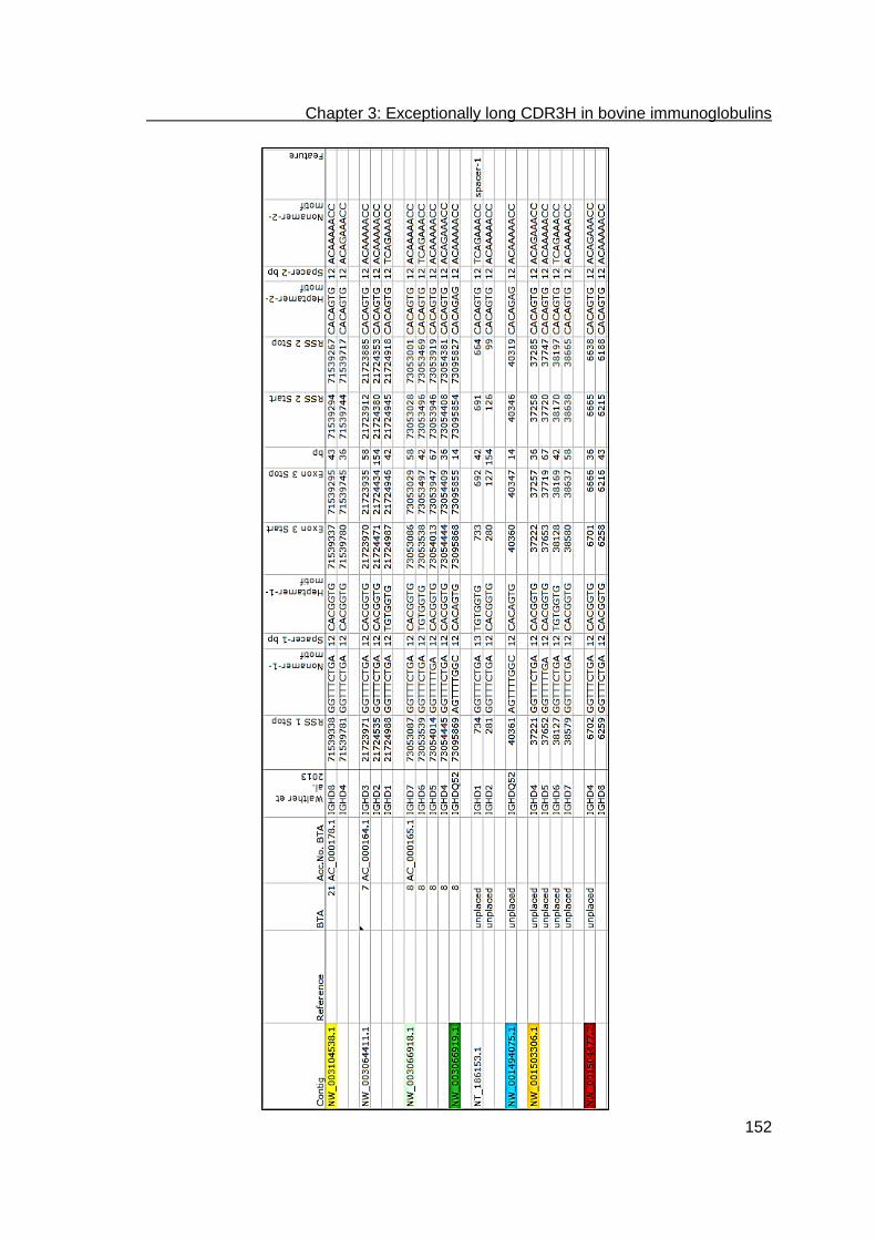
Chapter 3: Exceptionally long CDR3H in bovine immunoglobulins
152

Chapter 3: Exceptionally long CDR3H in bovine immunoglobulins
153

Chapter 3: Exceptionally long CDR3H in bovine immunoglobulins
154

Chapter 3: Exceptionally long CDR3H in bovine immunoglobulins
155

Chapter 3: Exceptionally long CDR3H in bovine immunoglobulins
156

Chapter 3: Exceptionally long CDR3H in bovine immunoglobulins
157

Chapter 3: Exceptionally long CDR3H in bovine immunoglobulins
158

Chapter 3: Exceptionally long CDR3H in bovine immunoglobulins
159

Chapter 3: Exceptionally long CDR3H in bovine immunoglobulins
160

Chapter 3: Exceptionally long CDR3H in bovine immunoglobulins
161

Chapter 3: Exceptionally long CDR3H in bovine immunoglobulins
162
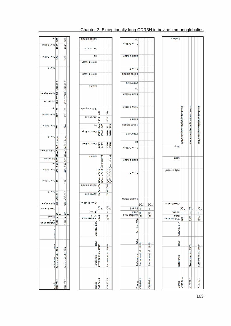
Chapter 3: Exceptionally long CDR3H in bovine immunoglobulins
163

Chapter 3: Exceptionally long CDR3H in bovine immunoglobulins
164
Acknowledgments
We are grateful to Lynne Armstrong for critical review of the manuscript. We also thank
the Göttingen Genomics Laboratory for sample sequencing and Caroline Bierschenk
for her excellent technical assistance.

Chapter 3: Exceptionally long CDR3H in bovine immunoglobulins
165
References
1. Tonegawa S (1983) Somatic generation of antibody diversity. Nature 302: 575-581. 2. Desiderio SV, Yancopoulos GD, Paskind M, Thomas E, Boss MA, et al. (1984)
Insertion of N regions into heavy-chain genes is correlated with expression of terminal deoxytransferase in B cells. Nature 311: 752-755.
3. Alt FW, Baltimore D (1982) Joining of immunoglobulin heavy chain gene segments: implications from a chromosome with evidence of three D-JH fusions. Proc Natl Acad Sci U S A 79: 4118-4122.
4. Kurosawa Y, Tonegawa S (1982) Organization, structure, and assembly of immunoglobulin heavy chain diversity DNA segments. J Exp Med 155: 201-218.
5. Reynaud CA, Garcia C, Hein WR, Weill JC (1995) Hypermutation generating the sheep immunoglobulin repertoire is an antigen-independent process. Cell 80: 115-125.
6. Berens SJ, Wylie DE, Lopez OJ (1997) Use of a single VH family and long CDR3s in the variable region of cattle Ig heavy chains. Int Immunol 9: 189-199.
7. Wagner SD, Neuberger MS (1996) Somatic hypermutation of immunoglobulin genes. Annu Rev Immunol 14: 441-457.
8. Kabat EA, Wu TT, Perry HM, Gottesmann KS, Foeller C (2006) Sequences of proteins of immunological interest. Bethesda, MD: U.S. Department of Health and Human Services, Public Health Service, National Institutes of Health.
9. Reynaud CA, Dahan A, Anquez V, Weill JC (1989) Somatic hyperconversion diversifies the single Vh gene of the chicken with a high incidence in the D region. Cell 59: 171-183.
10. Sun J, Kacskovics I, Brown WR, Butler JE (1994) Expressed swine VH genes belong to a small VH gene family homologous to human VHIII. J Immunol 153: 5618-5627.
11. Dufour V, Malinge S, Nau F (1996) The sheep Ig variable region repertoire consists of a single VH family. J Immunol 156: 2163-2170.
12. Saini SS, Hein WR, Kaushik A (1997) A single predominantly expressed polymorphic immunoglobulin VH gene family, related to mammalian group, I, clan, II, is identified in cattle. Mol Immunol 34: 641-651.
13. Sinclair MC, Gilchrist J, Aitken R (1997) Bovine IgG repertoire is dominated by a single diversified VH gene family. J Immunol 159: 3883-3889.
14. Arakawa H, Saribasak H, Buerstedde JM (2004) Activation-induced cytidine deaminase initiates immunoglobulin gene conversion and hypermutation by a common intermediate. PLoS Biol 2: E179.
15. Parng CL, Hansal S, Goldsby RA, Osborne BA (1996) Gene conversion contributes to Ig light chain diversity in cattle. J Immunol 157: 5478-5486.
16. Sun Y, Wang C, Wang Y, Zhang T, Ren L, et al. (2010) A comprehensive analysis of germline and expressed immunoglobulin repertoire in the horse. Dev Comp Immunol 34: 1009-1020.
17. Zhao Y, Kacskovics I, Pan Q, Liberles DA, Geli J, et al. (2002) Artiodactyl IgD: the missing link. J Immunol 169: 4408-4416.
18. Knight KL, Becker RS (1987) Isolation of genes encoding bovine IgM, IgG, IgA and IgE chains. Vet Immunol Immunopathol 17: 17-24.
19. Symons DB, Clarkson CA, Beale D (1989) Structure of bovine immunoglobulin constant region heavy chain gamma 1 and gamma 2 genes. Mol Immunol 26: 841-850.
20. Miller JR, Thomsen PD, Dixon SC, Tucker EM, Konfortov BA, et al. (1992) Synteny mapping of the bovine IGHG2, CRC and IGF1 genes. Anim Genet 23: 51-58.

Chapter 3: Exceptionally long CDR3H in bovine immunoglobulins
166
21. Zhao Y, Kacskovics I, Rabbani H, Hammarstrom L (2003) Physical mapping of the bovine immunoglobulin heavy chain constant region gene locus. J Biol Chem 278: 35024-35032.
22. Hayes H, Di Meo GP, Gautier M, Laurent P, Eggen A, et al. (2000) Localization by FISH of the 31 Texas nomenclature type I markers to both Q- and R-banded bovine chromosomes. Cytogenet Cell Genet 90: 315-320.
23. Gu F, Chowdhary BP, Andersson L, Harbitz I, Gustavsson I (1992) Assignment of the bovine immunoglobulin gamma heavy chain (IGHG) gene to chromosome 21q24 by in situ hybridization. Hereditas 117: 237-240.
24. Tobin-Janzen TC, Womack JE (1992) Comparative mapping of IGHG1, IGHM, FES, and FOS in domestic cattle. Immunogenetics 36: 157-165.
25. Hayes HC, Petit EJ (1993) Mapping of the beta-lactoglobulin gene and of an immunoglobulin M heavy chain-like sequence to homoeologous cattle, sheep, and goat chromosomes. Mamm Genome 4: 207-210.
26. Hosseini A, Campbell G, Prorocic M, Aitken R (2004) Duplicated copies of the bovine JH locus contribute to the Ig repertoire. Int Immunol 16: 843-852.
27. Lopez O, Perez C, Wylie D (1998) A single VH family and long CDR3s are the targets for hypermutation in bovine immunoglobulin heavy chains. Immunol Rev 162: 55-66.
28. Tutter A, Riblet R (1989) Conservation of an immunoglobulin variable-region gene family indicates a specific, noncoding function. Proc Natl Acad Sci U S A 86: 7460-7464.
29. Koti M, Kataeva G, Kaushik AK (2008) Organization of D(H)-gene locus is distinct in cattle. Dev Biol (Basel) 132: 307-313.
30. Koti M, Kataeva G, Kaushik AK (2010) Novel atypical nucleotide insertions specifically at VH-DH junction generate exceptionally long CDR3H in cattle antibodies. Mol Immunol 47: 2119-2128.
31. Saini SS, Allore B, Jacobs RM, Kaushik A (1999) Exceptionally long CDR3H region with multiple cysteine residues in functional bovine IgM antibodies. Eur J Immunol 29: 2420-2426.
32. Saini SS, Kaushik A (2002) Extensive CDR3H length heterogeneity exists in bovine foetal VDJ rearrangements. Scand J Immunol 55: 140-148.
33. Kaushik AK, Kehrli ME, Jr., Kurtz A, Ng S, Koti M, et al. (2009) Somatic hypermutations and isotype restricted exceptionally long CDR3H contribute to antibody diversification in cattle. Vet Immunol Immunopathol 127: 106-113.
34. Saini SS, Farrugia W, Ramsland PA, Kaushik AK (2003) Bovine IgM antibodies with exceptionally long complementarity-determining region 3 of the heavy chain share unique structural properties conferring restricted VH + Vlambda pairings. Int Immunol 15: 845-853.
35. Larsen PA, Smith TP (2012) Application of circular consensus sequencing and network analysis to characterize the bovine IgG repertoire. BMC Immunol 13: 52.
36. Shojaei F, Saini SS, Kaushik AK (2003) Unusually long germline DH genes contribute to large sized CDR3H in bovine antibodies. Mol Immunol 40: 61-67.
37. Ramsland PA, Kaushik A, Marchalonis JJ, Edmundson AB (2001) Incorporation of long CDR3s into V domains: implications for the structural evolution of the antibody-combining site. Exp Clin Immunogenet 18: 176-198.
38. Lefranc MP (1998) IMGT (ImMunoGeneTics) locus on focus. A new section of Experimental and Clinical Immunogenetics. Exp Clin Immunogenet 15: 1-7.
39. Lefranc MP, Pommie C, Ruiz M, Giudicelli V, Foulquier E, et al. (2003) IMGT unique numbering for immunoglobulin and T cell receptor variable domains and Ig superfamily V-like domains. Dev Comp Immunol 27: 55-77.

Chapter 3: Exceptionally long CDR3H in bovine immunoglobulins
167
40. Akamatsu Y, Tsurushita N, Nagawa F, Matsuoka M, Okazaki K, et al. (1994) Essential residues in V(D)J recombination signals. J Immunol 153: 4520-4529.
41. Hesse JE, Lieber MR, Mizuuchi K, Gellert M (1989) V(D)J recombination: a functional definition of the joining signals. Genes Dev 3: 1053-1061.
42. Larkin MA, Blackshields G, Brown NP, Chenna R, McGettigan PA, et al. (2007) Clustal W and Clustal X version 2.0. Bioinformatics 23: 2947-2948.
43. Felsenstein J (1985) Confidence limits on phylogenies: an approach using the bootstrap. Evolution 39: 783-791.
44. Sitnikova T, Su C (1998) Coevolution of immunoglobulin heavy- and light-chain variable-region gene families. Mol Biol Evol 15: 617-625.
45. Almagro JC, Martinez L, Smith SL, Alagon A, Estevez J, et al. (2006) Analysis of the horse V(H) repertoire and comparison with the human IGHV germline genes, and sheep, cattle and pig V(H) sequences. Mol Immunol 43: 1836-1845.
46. Perriere G, Gouy M (1996) WWW-query: an on-line retrieval system for biological sequence banks. Biochimie 78: 364-369.
47. Sanger F, Nicklen S, Coulson AR (1977) DNA sequencing with chain-terminating inhibitors. Proc Natl Acad Sci U S A 74: 5463-5467.
48. Thompson JD, Higgins DG, Gibson TJ (1994) CLUSTAL W: improving the sensitivity of progressive multiple sequence alignment through sequence weighting, position-specific gap penalties and weight matrix choice. Nucleic Acids Res 22: 4673-4680.
49. Ehrenmann F, Kaas Q, Lefranc MP (2010) IMGT/3Dstructure-DB and IMGT/DomainGapAlign: a database and a tool for immunoglobulins or antibodies, T cell receptors, MHC, IgSF and MhcSF. Nucleic Acids Res 38: D301-307.
50. Ehrenmann F, Lefranc MP (2011) IMGT/DomainGapAlign: IMGT standardized analysis of amino acid sequences of variable, constant, and groove domains (IG, TR, MH, IgSF, MhSF). Cold Spring Harb Protoc 2011: 737-749.
51. Matsuda F, Ishii K, Bourvagnet P, Kuma K, Hayashida H, et al. (1998) The complete nucleotide sequence of the human immunoglobulin heavy chain variable region locus. J Exp Med 188: 2151-2162.
52. Matsuda F, Honjo T (1996) Organization of the human immunoglobulin heavy-chain locus. Adv Immunol 62: 1-29.
53. Zimin AV, Delcher AL, Florea L, Kelley DR, Schatz MC, et al. (2009) A whole-genome assembly of the domestic cow, Bos taurus. Genome Biol 10: R42.
54. Everts-van der Wind A, Kata SR, Band MR, Rebeiz M, Larkin DM, et al. (2004) A 1463 gene cattle-human comparative map with anchor points defined by human genome sequence coordinates. Genome Res 14: 1424-1437.
55. Heuertz S, Hors-Cayla MC (1978) [Bovine chromosome mapping with the cell hybridization technic. Localization on the x chromosome of glucose-6-phosphate dehydrogenase, phosphoglycerate kinase, alpha-galactosidase A and hypoxanthine phosphoribosyltransferase]. Ann Genet 21: 197-202.
56. Sinclair MC, Aitken R (1995) PCR strategies for isolation of the 5' end of an immunoglobulin-encoding bovine cDNA. Gene 167: 285-289.
57. Jhunjhunwala S, van Zelm MC, Peak MM, Cutchin S, Riblet R, et al. (2008) The 3D structure of the immunoglobulin heavy-chain locus: implications for long-range genomic interactions. Cell 133: 265-279.
58. Reynaud CA, Bertocci B, Dahan A, Weill JC (1994) Formation of the chicken B-cell repertoire: ontogenesis, regulation of Ig gene rearrangement, and diversification by gene conversion. Adv Immunol 57: 353-378.
59. Corbett SJ, Tomlinson IM, Sonnhammer EL, Buck D, Winter G (1997) Sequence of the human immunoglobulin diversity (D) segment locus: a systematic analysis

Chapter 3: Exceptionally long CDR3H in bovine immunoglobulins
168
provides no evidence for the use of DIR segments, inverted D segments, "minor" D segments or D-D recombination. J Mol Biol 270: 587-597.
60. Kaartinen M, Mäkelä O (1985) Reading of D genes in variable frames as a source of antibody diversity. Immunol Today 6: 324-327.
61. Friedman ML, Tunyaplin C, Zhai SK, Knight KL (1994) Neonatal VH, D, and JH gene usage in rabbit B lineage cells. J Immunol 152: 632-641.
62. Schmidt WE, Jung HD, Palm W, Hilschmann N (1983) [Three-dimensional structure determination of antibodies. Primary structure of crystallized monoclonal immunoglobulin IgG1 KOL, I]. Hoppe Seylers Z Physiol Chem 364: 713-747.
63. Desmyter A, Transue TR, Ghahroudi MA, Thi MH, Poortmans F, et al. (1996) Crystal structure of a camel single-domain VH antibody fragment in complex with lysozyme. Nat Struct Biol 3: 803-811.
64. Ota T, Nei M (1994) Divergent evolution and evolution by the birth-and-death process in the immunoglobulin VH gene family. Mol Biol Evol 11: 469-482.

Chapter 3: Gene conversion and combinatorial diversity in bovine heavy chains
169
Development of a bioinformatics framework for the detection of gene conversion and the analysis of combinatorial diversity in immunoglobulin heavy chains in four cattle breeds
Stefanie Walther1¶, Manfred Tietze2¶, Claus-Peter Czerny1, Sven König2, Ulrike S. Diesterbeck1* 1Department of Animal Sciences, Institute of Veterinary Medicine, Division of Microbiology and Animal Hygiene, Faculty of Agricultural Sciences, Georg-August University Goettingen, Goettingen, Germany 2Department of Animal Breeding, University of Kassel, Witzenhausen, Germany *Corresponding author: Email: [email protected] (USD) ¶These authors contributed equally to this work. Submitted to “PloSOne”, manuscript under review

Chapter 3: Gene conversion and combinatorial diversity in bovine heavy chains
170
Abstract
We have developed a new bioinformatics framework for the analysis of rearranged
bovine heavy chain immunoglobulin (Ig) variable regions by combining and refining
widely used alignment algorithms. This bioinformatics framework allowed us to
investigate alignments of heavy chain framework regions (FRHs) and the separate
alignments of FRHs and heavy chain complementarity determining regions (CDRHs) to
determine their germline origin in the 4 cattle breeds Aubrac, German Black Pied,
German Simmental, and Holstein Friesian. Now it is also possible to specifically
analyze Ig heavy chains possessing exceptionally long CDR3Hs.
In order to gain more insight into breed specific differences in Ig combinatorial diversity,
somatic hypermutations and putative gene conversions of IgG, we compared the
dominantly transcribed variable (IGHV), diversity (IGHD), and joining (IGHJ) segments
and their recombination in the 4 cattle breeds.
The analysis revealed the use of 15 different IGHV segments, 21 IGHD segments, and
2 IGHJ segments with significant different transcription levels within the breeds.
Furthermore, there are preferred rearrangements within the 3 groups of CDR3H
lengths. In the sequences of group 2 (CDR3H lengths (L) of 11-47 amino acid residues
(aa)) a higher number of recombination was observed than in sequences of group 1
(L≤10 aa) and 3 (L≥48 aa). The combinatorial diversity of germline IGHV, IGHD, and
IGHJ-segments revealed 162 rearrangements that were significantly different. The few
preferably rearranged gene segments within group 3 CDR3H regions may indicate
specialized antibodies because this length is unique in cattle.
The most important finding of this study, which was enabled by using the bioinformatics
framework, is the discovery of strong evidence for gene conversion as a rare event
using pseudogenes fulfilling all definitions for this particular diversification mechanism.

Chapter 3: Gene conversion and combinatorial diversity in bovine heavy chains
171
Author Summary
The humoral immune response is an integral part in the specific pathogen defense.
Beside universal processes for the generation of the immunoglobulin diversity applying
for all higher vertebrates, species-specific mechanisms exist. Cattle possess less
functional gene segments available for combinatorial diversity if compared with human
and mice.
Thus, we hypothesized that cattle use pseudogenes for a genetically exchange, the so-
called gene conversion. Beside the difficulty of the incomplete annotated bovine heavy
chain immunoglobulin locus, we were able to verify gene conversion events fulfilling all
conditions like clusters of nucleotide exchanges, at least triplets, origin of the
pseudogene upstream of the rearranged segment and a high degree of flanking
homology.
In addition, we analyzed the combinatorial diversity and hot spots for hypermutations in
four different cattle breeds: Aubrac, German Black Pied, German Simmental, and
Holstein Friesian. Here we observed a significant non-random distribution in the usage
of the segments as well as in their combinations in Aubrac and German Simmental.
The highest variability within the variable region was always observed in the transition
of framework regions to complementarity determining regions (CDRH) if compared to
the middle of the CDRHs.

Chapter 3: Gene conversion and combinatorial diversity in bovine heavy chains
172
Introduction
The basic genetic mechanism in developing immunoglobulin diversity is similar in all
jawed vertebrates. Immunoglobulins (Ig) are Y-shaped hetero-tetramers consisting of
two identical heavy chains (IGH) and two identical light chains, either or in
mammals (IGK, IGL) [1]. Both chains are functionally divided into variable and constant
domains that are combined during B-cell development. The variable domain is
rearranged by separate heavy and light chain variable (IGHV, IGKV, IGLV), diversity
(IGHD), and joining (IGHJ, IGKJ, IGLJ) germline gene segments [2]. In addition, the
imprecise junction of the germline gene segments and somatic hypermutations
contribute to antibody diversity [3-5].
Species differences were primarily found in the number of germline IGHV/IGKV/IGLV,
IGHD, and IGHJ/IGKJ/IGLJ segments. In livestock species with restricted
combinatorial germline diversity such as chicken [4], pigs [6], sheep [7], and cattle [5,
8, 9], species-dependent mechanisms dominate the different diversification steps. For
instance, the use of pseudogene sequence parts is a frequent post-recombinatorial
strategy for the generation of the preimmune antibody repertoire in chicken, sheep, and
rabbit [4, 10-13]. This phenomenon, called gene conversion, was also confirmed for
IGLs in cattle [14] and is assumed to be operative in horses [15].
Gene conversions are difficult to detect especially within a large number of sequences
e.g. like those obtained from high throughput sequencing. Gene conversion in
immunoglobulins is characterized by clusters of nucleotide changes [14], sometimes
only triplets [11], originating from upstream genes of the rearranged segment [4, 13].
High degree of flanking homology of the conversion region ensures the genetic
exchange [13], whereby 3 to 5 nucleotides seem to be the minimal overlapping
requirement [11].
Detection of gene conversion in bovine IGHV is complicated due to the incomplete IGH
locus annotation. The main bovine IGH locus was assigned to the Bos taurus
autosome (BTA) 21 but exons coding for variable, diversity, and joining segments were
also found on BTA7, BTA8, and BTA20 [16-18]. Thirty-six IGHVs were identified of
which 13 are functional and belong phylogenetically to the bovine IGHV family 1
(boVH1). The second bovine IGHV family consists solely of non-functional IGHVs that
have not been identified in expression analyses yet. Eleven IGHV segment pairs
shared 100% sequence identity, whereas two of these pairs contain a functional
segment and either an ORF or a putative functional segment, respectively [17]. The

Chapter 3: Gene conversion and combinatorial diversity in bovine heavy chains
173
high proportion of pseudogene segments leads to the assumption of their use in gene
conversion events. Two IGHJ loci possessing six IGHJ segments were detected on
BTA11 by BAC clone and locus-specific PCR analysis and were found to rearrange at
low frequency while those located on BTA21 rearrange at high frequency. Only two out
of these six IGHJ were classified as functional whereas one is involved predominantly
in the recombination process [19, 20]. Fifteen IGHD genes were detected and revealed
a sub-cluster organization. IGHD are classified into four families and the IGHD exons
revealed huge size differences [21, 22]. The organization of the actual bovine germline
repertoire and its possible allelic variants is incomplete and needs to be investigated in
more detail [17]. Since, even the organization of the extensively studied human
immunoglobulin germline repertoire is questioned and requires ongoing analyses [23].
In all rearranged bovine immunoglobulin isotypes, exceptionally long complementarity
determining region 3 of the heavy chain (CDR3H) possessing up to 67 aa were
described [17]. Together with IGHD2 and IGHJ1, the germline IGHV10 segment was
found to be the only variable segment rearranged in these exceptionally long CDR3Hs
[17, 24]. This mechanism is also not isotype restricted [17, 25].
An additional bovine specific mechanism for antibody diversification is the insertion of
conserved short nucleotide sequences into the IGHV-IGHD junction, which was found
in intermediate and exceptionally long CDR3Hs [24].
Currently available programs like IMGT/Junction Analysis [26], IMGT/V-QUEST [27,
28] and IMTG/HIGHV-QUEST [29], VBASE2 [30], JoinSolver [31], iHMMun-align [32],
and IgBLAST [33] allow the annotation of only the entire IGHV sequence to germline
IGHV segments. Differentiated analysis of single parts is not directly possible. Most of
the databases are focusing on mouse and human immunoglobulin genes (VBASE2,
human, [30]). For cattle and other livestock or companion animals, separate databases
have to be created (IgBLAST, [33]). Furthermore, the IMGT numbering system does
not provide numbering for CDR3H larger than 31 aa. Placing of bovine intermediate as
well as of exceptional long CDR3H in this numbering system is therefore not possible
and consequently does not allow correct analysis of the rearrangement in those
immunoglobulins. In addition, only IgBLAST allows the adjustment of parameters for
IGHD identification.
Detailed genetic analysis of the pre- and post-immunization humoral immune response
is important to describe the developing diversity and the effectiveness of vaccines and
to detect possible individual and breed related differences including non-responders.

Chapter 3: Gene conversion and combinatorial diversity in bovine heavy chains
174
As a conclusion those analyses help to develop fast recombinant antibodies for
passive vaccination, therapy or diagnostic by genetically pre-selection of newly
developed or abundant sequences.
As a first attempt to gain more insight into bovine breed specific differences, we
compared the dominantly transcribed and the combinatorial diversity of germline IGHV,
IGHD, and IGHJ segments as well as somatic hypermutations and putative gene
conversions of IgG in the four cattle breeds Aubrac, German Simmental, German Black
Pied and Holstein Friesian, by using a newly developed Bioinformatics framework. This
new bioinformatics framework combines and extends several analysis tools and takes
into account the unique specificities of bovine immunoglobulin sequences of
exceptionally long CDR3Hs. In addition, it allows for the adaptation of alignment
parameters for the single segments and enables the selective analysis of the different
functional regions of the variable domain (namely framework regions and CDRs) to
determine putative gene conversions. This new tool should facilitate a fast and detailed
analysis of data sets generated by high throughput sequencing.

Chapter 3: Gene conversion and combinatorial diversity in bovine heavy chains
175
Results
For sequence analysis, we developed a new bioinformatics framework using MUSCLE
[34, 35] for the initial fast and accurate multiple nucleotide sequence alignment.
Subsequently, the sequence distances were calculated with ClustalW [36]. For
nucleotide alignments of IGHV and IGHJ, default values of MUSCLE were used. To
improve the biological significance of the assignment of germline and sample IGHDs,
we tested three different procedures using different parameters. To determine the
germline origin using the new bioinformatics framework, only the FRHs were aligned to
avoid interference with the highly diversified CDRH [18]. To analyze possible gene
conversion events, FR1-3Hs and CDR1-3Hs were extracted and aligned separately to
the corresponding regions of the IGHV reference sequences to find the most similar
germline segment.
Using our bioinformatics tool, we established a sample sequence set for use in detailed
analysis of the transcribed bovine immunoglobulin repertoire. Blood samples were
taken from 10 animals per cattle breed: Aubrac (A), German Simmental (GS), German
Black Pied (GBP), and Holstein Friesian (HF).
In total, 160 IgG heavy chain sequences per breed (n=640 sequences) were
investigated as described above. The variable regions were identified and extracted at
the 5’ end (N-terminal end) using the motif GCCTCCACC/AST (nt/AA) marking the
start of the first constant region of all bovine IgGs. Due to premature Stop-codons or
incompletely amplified variable regions, 131 sequences were excluded from further
analyses. Consequently, 509 sequences were left: 137 in A, 116 in GS, 111 in GBP,
and 145 in HF. Sequences analyzed are published under accession numbers
KT761498-KT762006.

Chapter 3: Gene conversion and combinatorial diversity in bovine heavy chains
176
Transcriptional analyses, assignment of germline gene segments
Assigning the FR1H to FR3H of transcribed IGHV segments to their germline
origin
Ig heavy chain gene usage and identity to germline gene segments was determined by
comparing the transcribed sequences with the germline Ig heavy chain genes
described by Walther et al. [17] and Liljavirta et al. [22]. We found six of the germline
IGHVs possess 100% sequence identity (presented as IGHVx/y) up to the 3’ end of
FR3H. Analysis of transcribed IGHV segments (here: comprising FR1-3H) revealed
germline gene usage of IGHV3/33, IGHV6, IGHV10/34, IGHV36/29(F),
IGHV17(ORF)/31(F), IGHV1S26, IGHV1S28, IGHV1S32, IGHV1S33, IGHV1S34,
IGHV1S35, IGHV1S37, IGHV1S38, IGHV1S39 as well as IGHV1S40 (Fig 1).
Fig 1. Transcription frequencies of IGHV in four cattle breeds. Transcribed IGHV are shown on the horizontal axis, their relative usage frequencies are indicated on the vertical axis. Each breed is marked by the following color code: Aubrac: white, German Simmental: light grey, Holstein Friesian: black, German Black Pied: dark grey

Chapter 3: Gene conversion and combinatorial diversity in bovine heavy chains
177
We were not able to unambiguously assign ten of 116 analyzed GS sequences to one
germline IGHV; this was also true for 14 out of 111 GBP sequences, 12 out of 145 HF
sequences, and 19 out of 137 analyzed sequences of A.
These samples showed equal divergence to at least two germline IGHV which included
combinations of IGHV3/33, IGHV6, IGHV10/34, IGHV17(ORF)/31(F), IGHV1S28, and
IGHV1S32-40. The most frequent multiple assignment affected IGHV6 and IGHV1S34,
followed by IGHV6 and IGHV1S35 in all A, GS, and HF. IGHV6, IGHV1S34, and
IGHV17(ORF)/31(F) could not be differentiated in two sequences of GS, one sequence
of GBP and two sequences of HF. Ambiguously assigned germline IGHVs and multiple
assignments are listed in Table 1.
Table 1. IGHV assigned ambiguously and their frequency. Ambiguous sequences can be assigned to more than one germline IGHV with the same distance. Breed A GBP GS HF
Total No. of animals 10 10 10 10
No. of animals with ambiguous sequences 9 5 4 6
Total No. of sequences analyzed 137 111 116 145
No. of ambiguous sequences 19 14 10 12
Total No. of putative IGHV recombinations 167 140 130 158
Ambiguous IGHV
IGHV3/IGHV1S33 0 0 1 0
IGHV3/IGHV1S39 0 0 0 2
IGHV6/IGHV17 1 0 0 0
IGHV6/IGHV1S34 3 3 3 5
IGHV6/IGHV1S35 3 0 1 1
IGHV6/IGHV36 0 1 1 1
IGHV6/IGHV1S39 1 0 1 0
IGHV1S28/IGHV1S32 0 0 0 1
IGHV1S33/IGHV1S39 1 1 0 0
IGHV1S33/IGHV1S40 1 0 0 0
IGHV1S37/IGHV1S38 0 0 0 1
IGHV1S39/IGHV17 0 1 0 0
IGHV1S39/IGHV1S40 1 0 0 0
IGHV3/IGHV10/IGHV1S39 2 0 0 0
IGHV6/IGHV17/IGHV1S34 0 2 2 1
IGHV6/IGHV17/IGHV1S40 1 0 0 0
IGHV6/IGHV1S32/IGHV1S34 1 0 1 0
IGHV6/IGHV1S33/IGHV1S35 1 0 0 0
IGHV3/IGHV6/IGHV17/IGHV1S34 0 2 0 0
IGHV6/IGHV10/IGHV1S34/IGHV1S39 1 0 0 0
IGHV6/IGHV17/IGHV1S28/IGHV1S39 0 1 0 0
IGHV6/IGHV17/IGHV1S34/IGHV1S39 1 1 0 0
IGHV6/IGHV36/IGHV1S34/IGHV1S39 0 1 0 0
IGHV3/IGHV6/IGHV17/IGHV1S34/IGHV1S39 0 1 0 0
IGHV6/IGHV17/IGHV1S33/IGHV1S34/IGHV1S35 1 0 0 0

Chapter 3: Gene conversion and combinatorial diversity in bovine heavy chains
178
Including the multiple assignments as described above, a total number of 595 possible
transcribed germline IGHV were observed. Overall, the most frequent variable gene
segment was IGHV1S39; this was identified in 17.65% of all sequences. This IGHV
was used in 51 A sequences (n=167, 30.54%), in 19 sequences of GBP (n=140,
13.57%), in four sequences of GS (n=130, 3.08%), and 31 sequences of HF (n=158,
19.62%) (Fig 1, Table 2). IGHV3/33 was represented by 14.29% of all sequences. The
number of transcribed IGHV3/33 varied from 15 in GS (11.54%) to 32 in HF (20.25%)
(Fig 1, Table 2). In similar proportions of 13.45% and 12.27% germline IGHV36/29(F)
and IGHV1S40 were used. IGHV1S28, IGHV1S34, and IGHV6 were transcribed in
proportions of 8.07%, 8.57%, and 9.24%. The other transcribed IGHVs were identified
in minor proportions of 0.17% to 5.04%, respectively. These rarely used IGHVs were
identified once or twice in GS and HF but up to ten times in A (Fig 1). Very high
significant differences were calculated for IGHV usage within the breeds and between
the breeds (P<0.0001).
Table 2. Percentage of IGHV assigned to sample sequences of four cattle breeds.
IGHV A
(%; n=167) GS
(%; n=130) GBP
(%; n=158) HF
(%; n=140)
IGHV1S26 0.00 0.77 0.00 0.00
IGHV1S28 6.59 8.46 8.57 8.86
IGHV1S32 0.00 2.31 0.00 1.27
IGHV1S33 3.59 1.54 0.71 0.00
IGHV1S34 5.99 10.00 13.57 5.70
IGHV1S35 2.99 11.54 1.43 1.27
IGHV1S37 0.00 0.00 0.00 1.27
IGHV1S38 0.00 0.77 0.00 0.63
IGHV1S39 30.54 3.08 13.57 19.62
IGHV1S40 8.38 15.38 15.71 10.76
IGHV3 11.98 11.54 12.86 20.25
IGHV6 10.78 7.69 9.29 8.86
IGHV10 5.99 6.92 1.43 5.70
IGHV17 5.39 3.08 6.43 1.90
IGHV36 7.78 16.92 16.43 13.92

Chapter 3: Gene conversion and combinatorial diversity in bovine heavy chains
179
Separated analyses of FRH 1-3 and CDRH 1-2 to detect gen conversion events
In livestock such as chicken, rabbit, and cattle the use of pseudogene segments is
known to contribute to immunoglobulin diversity [4, 10, 11, 13, 14, 37, 38]. Gene
conversion in immunoglobulins is characterized by clusters of nucleotide changes [14],
sometimes only triplets [11], originating from upstream genes of the rearranged
segment [4, 13]. A high degree of flanking homology of the conversion region ensures
the genetic exchange [13], whereby three to five nucleotides have been shown to be
the minimal overlapping requirement [11].
Consequently, FR1-3H and CDR1-2H were analyzed separately to identify mutations
within the FR1-3H and CDR1-2H that would indicate possible gene conversion events.
The current genomic annotation of germline immunoglobulin segments in cattle makes
a correct prediction of 5’ donor segments difficult. Nonetheless, larger contigs have
been identified [17] and enabled us to show gene conversion events in bovine
immunoglobulin heavy chains variable regions.
For instance, the calculated putative originating germline genes for nucleotide changes
identified solely in the CDR2H region of KT761864 were IGHV4Ψ, IGHV9Ψ, and
IGHV18Ψ (divergence 0.190). In the calculation covering the whole V-region and using
only FR1-3H, IGHV6 was identified as the originating gene for KT761864 (divergence
0.075). All genes but IGHV18Ψ are located on BTA21, whereas IGHV4Ψ is located
upstream of IGHV6 and is therefore most likely used for the gene conversion. There
are two triplets in KT761864 and IGHV4Ψ that are different from IGHV6 due to a
transversion mutation in the first changed triplet (from AAT in IGHV6 to TAT in
KT761864 and IGHV4Ψ) and a transition mutation in the second changed triplet (from
GAT in IGHV6 to AAT in KT761864 and IGHV4Ψ; Table 3).
Table 3. Possible gene conversion in KT761864.
Sample/Gene Sequence in CDR2H Location Divergence
KT761864 ATT AAT TAT AAT … GGA GAC ACC BTA21
IGHV4ᴪ ATA TAT TAT AAT … GGT GAC ACT BTA21 0.190
IGHV9ᴪ ATA TAT TAT AAT … GGT GAC ACT BTA21 0.190
IGHV18ᴪ ATA TAT TAT AAT … GGT GAC ACT BTA7 0.190
IGHV61 ATA GAT AAT GAT … GGA GAC ACA BTA21 0.075 1 Divergence in calculations covering the whole V-region and using only FR1-3H. This gene was not observed in the analysis of the separated CDR2H.

Chapter 3: Gene conversion and combinatorial diversity in bovine heavy chains
180
Due to high sequence similarities and because we assumed preferable transcription of
functional germline gene segments, we first concentrated on pseudogenes that were
calculated as unique origin of the sample sequences considering the particular regions.
In all breeds, IGHV13Ψ and IGHV17(ORF) were used most often in CDR2H (3.34%
and 1.77%) as well as in FR2H and FR3H (IGHV17(ORF) both 1.38%) and CDR2H
(IGHV13Ψ 1.77%), respectively. IGHV15Ψ was found in 0.59% of CDR2H in
sequences of A and HF. IGHV11Ψ, IGHV12Ψ, and IGHV21Ψ always showed the
same divergence from GBP and HF sequences but were not solely identified as the
potential origin of the sample sequence. Similarly, IGHV4Ψ, IGHV9Ψ, and IGHV18Ψ
were identified in triplet as possible originating gene segments but only together with
functional germline genes. In one sequence of GBP and 2 sequences of GS both in
CDR1H and CDR2H pseudogenes were calculated as possible parental IGHVs. In one
A sequence only a pseudogene showed lowest divergence from the sample in FR2H
and CDR2H (Fig 2).
Fig 2. Possible gene conversion in FRH and CDRH. The absolute number of possible gene conversion events is shown for pseudogenes assigned unambiguously to CDR1H, FR2H, and CDR2H in the four cattle breeds Aubrac (A), German Black Pied (GBP), German Simmental (GS), and Holstein Friesian (HF).

Chapter 3: Gene conversion and combinatorial diversity in bovine heavy chains
181
Our analyses revealed a less distinct assignment to the germline gene segments.
Nevertheless, the majority of identified and putative originating germline sequences
were IGHV3/33, IGHV6, IGHV10/34, IGHV2/26, IGHV29(F)/36, IGHV16(ORF)/25,
IGHV17(ORF)/31(F), and IGHV19Ψ, as well as IGHV1S19, IGHV1S23-28, IGHV1S30,
IGHV1S32-40 [22] within FRHs and CDRHs. Although these are the same gene
segments as determined for the complete IGHV segment, calculations indicate
exchanges between the IGHV gene segments within FRHs and CDRHs. Additional
pseudogenes (IGHV1Ψ, IGHV4Ψ, IGHV7Ψ, IGHV9Ψ, IGHV11Ψ, IGHV12Ψ,
IGHV13Ψ, IGHV15Ψ, IGHV18Ψ, IGHV21Ψ) were identified by the program and
concentrated on CDR1H and CDR2H. IGHV13Ψ, IGHV15Ψ, and IGHV19Ψ showed
little divergence in FR1H and FR2H from sample sequences, too. Within these FRHs,
different functional IGHV were possible originating germline sequences (Table 4).
Table 4. Possible gene conversions in FRH and CDRH.
A GBP GS HF Sum Percent
a1 u2 a1 u2 a1 u2 a1 u2 a1 u2 u2
IGHV1Ψ CDR1H 1 0 0 0 0 0 1 0 2 0 0.00
CDR2H 1 1 1 0 1 0 2 0 5 1 0.20
IGHV4Ψ CDR1H 1 0 0 0 0 0 1 0 2 0 0.00
CDR2H 4 0 1 0 2 0 2 0 9 0 0.00
IGHV7Ψ CDR2H 2 1 1 0 1 1 4 0 8 2 0.39
IGHV9Ψ CDR1H 1 0 0 0 0 0 1 0 2 0 0.00
CDR2H 4 0 1 0 2 0 2 0 9 0 0.00
IGHV11Ψ CDR2H 0 0 0 0 0 0 2 0 2 0 0.00
IGHV12Ψ CDR2H 0 0 0 0 0 0 2 0 2 0 0.00
IGHV13Ψ CDR1H 18 0 11 0 9 0 19 0 57 0 0.00
FR2H 1 0 0 0 0 0 0 0 1 0 0.00
CDR2H 16 2 20 5 17 5 22 5 75 17 3.34
IGHV17Ψ FR1H 80 0 58 0 63 0 75 0 276 0 0.00
CDR1H 20 0 10 0 2 0 10 0 42 0 0.00
FR2H 8 4 4 1 4 0 6 2 22 7 1.38
CDR2H 12 5 7 1 3 0 14 3 36 9 1.77
FR3H 9 5 10 1 9 0 8 1 36 7 1.38
IGHV15Ψ CDR1H 7 2 3 0 3 0 4 1 17 3 0.59
FR2H 0 0 0 0 0 0 0 0 0 0 0.00
CDR2H 3 1 9 0 1 0 9 2 22 3 0.59
IGHV18Ψ CDR1H 1 0 0 0 0 0 1 0 2 0 0.00
CDR2H 4 0 1 0 2 0 2 0 9 0 0.00
IGHV19Ψ FR1H 80 0 58 0 63 0 75 0 276 0 0.00
CDR1H 25 0 13 0 27 0 20 0 85 0 0.00
FR2H 6 1 1 0 0 0 4 0 11 1 0.20
CDR2H 9 0 15 0 9 0 25 0 58 0 0.00
IGHV21Ψ CDR2H 0 0 0 0 0 0 2 0 2 0 0.00 1 ambiguously assigned 2 unambiguously assigned

Chapter 3: Gene conversion and combinatorial diversity in bovine heavy chains
182
CDR3H length distribution
In all four cattle breeds the program identified very short CDR3Hs (less or equal 10 aa,
group 1), CDR3Hs of intermediate length (11-47 aa, group 2) as well as exceptionally
long CDR3Hs (at least 48 aa, group 3) (Fig 3, Table 5). Very high significant
differences were calculated for the number of sequences within the 3 groups of lengths
(P<0.0001) when they were compared between all breeds but also within the breeds
(GS, GBP, A, HF: P<0.0001). Fourty-four (7.37%) sequences possessed a CDR3H
length with 10 or less amino acid (aa) residues. The highest amount of sequences
within this group were identified in breed A (12.57%), followed by HF (7.55%), GBP
(5.00%) and GS (3.05%). In the breeds A, GS, and HF one and four sequences
possessed only four amino acid residues within the CDR3H. Five amino acid residues
were the shortest CDR3H in sequences of GBP.
Fig 3. Length distribution of CDR3H in the four cattle breeds. The number of amino acid residues making up the CDR3H lengths identified are shown on the horizontal axis, their relative usage frequencies are indicated on the vertical axis. Each breed is marked by the following color code: Aubrac: white, German Simmental: light grey, Holstein Friesian: black, German Black Pied: dark grey

Chapter 3: Gene conversion and combinatorial diversity in bovine heavy chains
183
Table 5. Length distribution of the CDR3Hs.
Number of amino acids within CDR3H
All breeds (%; n=597)
GS (%; n=131)
GBP (%; n=140)
HF (%; n=159)
A (%; n=167)
4 1.01 0.76 0.00 0.63 2.40
5 2.68 0.00 2.14 2.52 5.39
6 1.68 1.53 1.43 2.52 1.20
7 1.17 0.76 1.43 0.63 1.80
8 0.17 0.00 0.00 0.63 0.00
9 0.34 0.00 0.00 0.63 0.60
10 0.34 0.00 0.00 0.00 1.20
11 1.17 0.00 0.71 1.89 1.80
12 1.01 0.00 3.57 0.00 0.60
13 1.01 0.76 0.00 2.52 0.60
14 1.51 2.29 2.14 0.63 1.20
15 3.18 4.58 2.86 1.89 3.59
16 4.36 0.00 6.43 8.18 2.40
17 2.85 2.29 5.00 1.89 2.40
18 4.86 3.82 5.00 4.40 5.99
19 3.85 3.82 0.71 5.66 4.79
20 5.70 9.16 3.57 5.66 4.79
21 6.70 12.21 3.57 6.29 5.39
22 10.55 12.21 8.57 10.69 10.78
23 8.38 6.11 10.71 6.29 10.18
24 8.04 10.69 6.43 8.81 6.59
25 6.87 4.58 7.86 7.55 7.19
26 6.20 9.16 5.71 4.40 5.99
27 7.20 6.11 10.71 6.29 5.99
28 2.51 1.53 5.71 2.52 0.60
29 1.68 0.76 1.43 3.14 1.20
30 1.17 0.00 2.14 0.63 1.80
31 0.34 0.76 0.71 0.00 0.00
49 0.17 0.76 0.00 0.00 0.00
50 0.17 0.00 0.71 0.00 0.00
52 0.17 0.00 0.00 0.00 0.60
53 0.34 0.76 0.00 0.63 0.00
54 0.17 0.00 0.71 0.00 0.00
56 0.34 0.76 0.00 0.00 0.60
57 0.17 0.00 0.00 0.63 0.00
58 0.17 0.00 0.00 0.00 0.60
59 0.17 0.00 0.00 0.00 0.60
60 0.50 1.53 0.00 0.63 0.00
61 0.17 0.00 0.00 0.63 0.00
63 0.34 0.76 0.00 0.63 0.00
64 0.17 0.00 0.00 0.00 0.60
65 0.50 1.53 0.00 0.00 0.60

Chapter 3: Gene conversion and combinatorial diversity in bovine heavy chains
184
Complementarity determining regions of group 2 were identified in 532 of all
sequences analyzed (89.11%). 93.57% of GBP sequences were found to use 11 up to
47 aa. In HF, 89.31% of the sequences were attributed to this group as well as 83.83%
of A and 90.84% of GS sequences. The most frequent CDR3H length was 22 aa,
which was found in 10.78% of A sequences, in 8.57% of GBP, in 12.21% of GS, and in
10.69% HF sequences. Simultaneously, this length was identified preferably in CDR3H
of A and HF. Nevertheless, in GS CDR3H with a length of 21 aa was identified as often
as a length of 22 aa (12.21%). In GBP, CDR3Hs with a length of 23 and 27 aa
dominated (10.71%).
CDR3Hs of group 3 were identified in 21 sequences of all four breeds (9.22%). The
breed GS showed the highest number of these sequences (6.11%) followed by A
(3.59%). German Black Pied and HF sequences possessed smaller proportions of the
exceptionally long CDR3H with 3.14% and 1.43%, respectively. Whereas in GS and
HF sequences with 65 aa were the longest CDR3Hs (1.53%, 0.6%), 63 and 54 aa were
counted in the longest CDR3Hs of A (0.63%) and GBP (0.71%), respectively.
Assigning IGHD to their germline origin using 3 different procedures
We tested three different procedures to assign germline and sample IGHDs in order to
improve the biological significance. At first, we applied the default values of MUSCLE,
in procedure 2 we changed the penalties for gap opening (=-4) and gap extention
(=-0.3), and in procedure 3 we additionally incorporated a modified scoring matrix
(match +2) to evaluate transversion and transition mutations.
In all three procedures, the assignment of germline and transcribed IGHD revealed
clear results for the sequences analyzed. Nevertheless, using procedure 3 we obtained
results matching short and long sample IGHD sequences best to germline short
CDR3H and exceptionally long CDR3H, respectively, whereas procedure 1 and 2
assigned a major number of group 3 CDR3H to germline IGHD of moderate length.
Twenty-one different germline IGHD were transcribed, whereby IGHD8 located on
BTA21, IGHD3 located on BTA7, IGHD5 as well as the very short IGHDQ52 located on
BTA8; these were preferred in all breeds (Fig 4, Table 6). While IGHDQ52, IGHDS10
and 14 [22] were solely transcribed in sense orientation, germline IGHD1 to IGHD8
gene segments were transcribed in antisense direction in 38 sequences distributed
over all four breeds investigated. IGHD4 (antisense (as), [21]) was identified the most
often, followed by IGHD1 (as, [39]), IGHD3 (as, [39]), IGHD2 (as, [39]), IGHD5 (as,
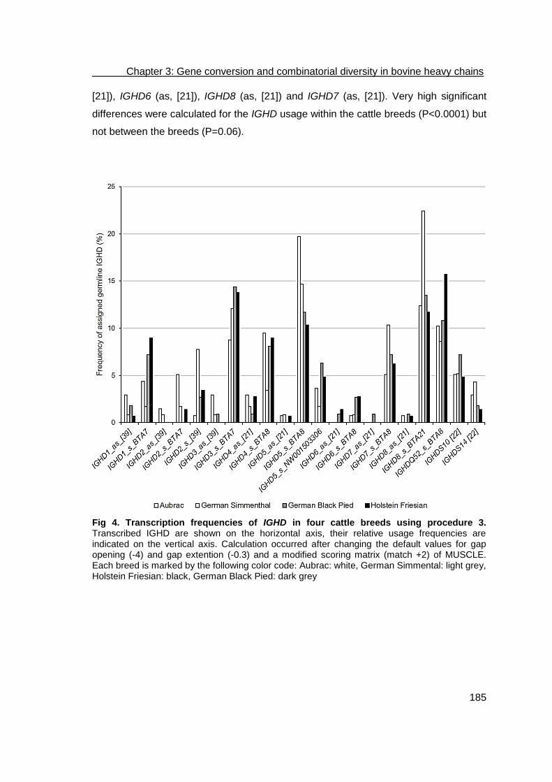
Chapter 3: Gene conversion and combinatorial diversity in bovine heavy chains
185
[21]), IGHD6 (as, [21]), IGHD8 (as, [21]) and IGHD7 (as, [21]). Very high significant
differences were calculated for the IGHD usage within the cattle breeds (P<0.0001) but
not between the breeds (P=0.06).
Fig 4. Transcription frequencies of IGHD in four cattle breeds using procedure 3. Transcribed IGHD are shown on the horizontal axis, their relative usage frequencies are indicated on the vertical axis. Calculation occurred after changing the default values for gap opening (-4) and gap extention (-0.3) and a modified scoring matrix (match +2) of MUSCLE. Each breed is marked by the following color code: Aubrac: white, German Simmental: light grey, Holstein Friesian: black, German Black Pied: dark grey

Chapter 3: Gene conversion and combinatorial diversity in bovine heavy chains
186
Table 6. Percentage of IGHD assigned to sample sequences of four cattle breeds.
IGHD A
(%; n=137) GS
(%; n=116) GBP
(%; n=111) HF
(%; n=145)
IGHD1_as1_[39] 2.92 0.86 1.80 0.69
IGHD1_s2_BTA7 4.38 1.72 7.21 8.97
IGHD2_as_[39] 1.46 0.86 0.00 0.00
IGHD2_s_BTA7 5.11 1.72 0.00 1.38
IGHD2_s_[39] 0.73 7.76 2.70 3.45
IGHD3_as_[39] 2.92 0.86 0.90 0.00
IGHD3_s_BTA7 8.76 12.07 14.41 13.79
IGHD4_as_[21] 2.92 1.72 0.90 2.76
IGHD4_s_BTA8 9.49 3.45 8.11 8.97
IGHD5_as_[21] 0.73 0.86 0.00 0.69
IGHD5_s_BTA8 19.71 14.66 11.71 10.34
IGHD5_s_NW001503306 3.65 1.72 6.31 4.83
IGHD6_as_[21] 0.00 0.00 0.90 1.38
IGHD6_s_BTA8 0.73 0.86 2.70 2.76
IGHD7_as_[21] 0.00 0.00 0.90 0.00
IGHD7_s_BTA8 5.11 10.34 7.21 6.21
IGHD8_as_[21] 0.73 0.00 0.90 0.69
IGHD8_s_BTA21 12.41 22.41 13.51 11.72
IGHDQ52_s_BTA8 10.22 8.62 10.81 15.71
IGHDS10_[22] 5.11 5.17 7.21 4.83
IGHDS14_[22] 2.92 4.31 1.80 1.38 1 antisense 2 sense
In comparison, 20 different germline IGHD were transcribed using default conditions of
MUSCLE for alignments (procedure 1), whereby IGHD8 being located on BTA21 was
the preferred IGHD in all breeds. The very short IGHDQ52 located on BTA8 was
transcribed in high frequencies in the breeds A, GBP, and HF (S1 Fig, S1 Table). While
IGHDQ52, IGHDS10 and 14, and IGHD6 were solely transcribed in sense orientation,
germline IGHD1 to IGHD8 gene segments were transcribed in antisense direction in 23
sequences that were distributed over all four breeds. IGHD4 (as, [21]) was identified
the most often. Very high significant differences were calculated for the IGHD usage
within the cattle breeds (P<0.0001) but not between the breeds (P=0.1630).

Chapter 3: Gene conversion and combinatorial diversity in bovine heavy chains
187
After changing penalties for gap opening and gap extension (procedure 2), 17 different
germline IGHD were transcribed, whereby IGHD8 located on BTA21, was the most
observed IGHD in all breeds (S2 Fig, S2 Table). Germline IGHD1, IGHD4, IGHD6, and
IGHD8 gene segments were transcribed in as direction in 24 sequences that were
distributed over all four breeds. Again, IGHD4 (as, [21]) was identified the most often.
Very high significant differences were calculated for the IGHD usage within the cattle
breeds (P<0.0001) but not between the breeds (P=0.6654).
Assigning the FR4H and IGHJ to their germline origin
Located on BTA21, IGHJ1 and IGHJ6 [22] were identified as origin to the transcribed
gene segments, which defines the FR4H within the samples analyzed. IGHJ1 was
transcribed preferably in the sequences investigated in all breeds (98.83%). IGHJ6 [22]
(1.17%) was detected in only one sequence of each A, GS, and GBP (0.73%, 0.85%,
0.90%), as well as in three sequences of HF (2.05%) animals. One sequence of GS
and HF, respectively, showed IGHJ1 and IGHJ6 as a possible originating germline
segment (Fig 5, Table 7). Statistical analysis revealed very high significant differences
(P<0.0001) for the usage of IGHJ1 and IGHJ6 [22] within all cattle breeds. No
significant differences were calculated between the four breeds.
Fig 5. Transcription frequencies of IGHJ in four cattle breeds. Transcribed IGHJ are shown on the horizontal axis, their relative usage frequencies are indicated on the vertical axis. Each breed is marked by the following color code: Aubrac: white, German Simmental: light grey, Holstein Friesian: black, German Black Pied: dark grey

Chapter 3: Gene conversion and combinatorial diversity in bovine heavy chains
188
Table 7. Percentage of IGHJ assigned to sample sequences of four cattle breeds.
IGHJ A
(%; n=137) GS
(%; n=117) GBP
(%; n=111) HF
(%; n=146)
IGHJ1 99.27 99.15 99.10 97.95
IGHJ6 [22] 0.73 0.85 0.90 2.05
Recombination of IGHV, IGHD, and IGHJ in different cattle breeds using
procedure 3
Recombined IGHV, IGHD, and IGHJ were identified for each sequence and all possible
frequencies were analyzed statistically within and between the breeds examined. In
total, 597 recombinations were analyzed including double assigned germline origins for
IGHV and IGHJ.
Applying procedure 3 for the IGHD assignment, 162 different combinations of IGHV,
IGHD, and IGHJ were revealed (Fig 6, S3 Table). Most combinations occurred in less
than ten sequences. Calculations revealed very high significant differences in usage
frequencies of the rearranged gene segments between the breeds investigated
(P<0.0001). Eleven rearrangements were observed in ten to 27 sequences. They were
observed in sequences of all four breeds. These major rearrangements were: IGHV36-
IGHD8 (sense (s), BTA21)-IGHJ1 (AY158087) (4.52%), IGHV1S40-IGHD5 (s, BTA8)-
IGHJ1 (AY158087) (3.02%), IGHV1S39-IGHD5 (s, BTA8)-IGHJ1 (AY158087) (2.85%),
IGHV1S40-IGHD3 (s, BTA7)-IGHJ1 (AY158087) (2.35%), IGHV1S39-IGHD8 (s,
BTA21)-IGHJ1 (AY158087) (2.18%), IGHV3-IGHD5 (s, BTA8)-IGHJ1 (AY158087)
(2.01%), IGHV3-IGHDQ52 (s, BTA8)-IGHJ1 (AY158087) (1.84%), IGHV36-IGHDQ52
(s, BTA8)-IGHJ1 (AY158087) (1.84%), IGHV1S39-IGHD4 (s, BTA8)-IGHJ1
(AY158087) (1.68%), IGHV1S39-IGHD7 (s, BTA8)-IGHJ1 (AY158087) (1.68%),
IGHV3-IGHD8 (s, BTA21)-IGHJ1 (AY158087) (1.68%). Beside these preferred
combinations, 52 minor recombinations were identified solely in one sequence
distributed with 19 sequences in A, 14 in GS, six in GBP, and 13 in HF. Six variations
of rearranged IGHJ6 were also identified one and two-times. These appear distributed
over all four breeds: IGHV3-IGHD3 (s, BTA7)-IGHJ6 [22], IGHV1S28-IGHD4 (s,
BTA8)-IGHJ6 [22], IGHV1S39-IGHD3 (s, BTA7)-IGHJ6 [22], IGHV1S40-IGHDS10-
IGHJ6 [22], IGHV3-IGHD8 (as, [21])-IGHJ6 [22], as well as IGHV10-IGHD5 (as, [21])-
IGHJ6 [22].
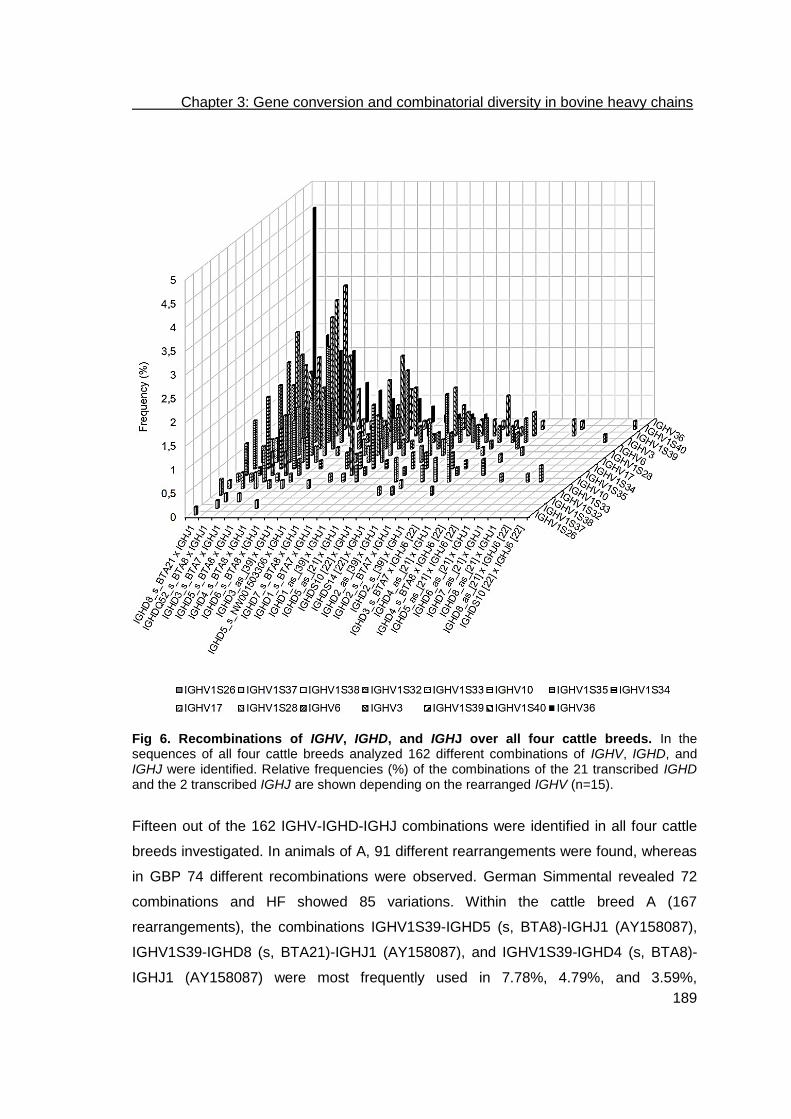
Chapter 3: Gene conversion and combinatorial diversity in bovine heavy chains
189
Fig 6. Recombinations of IGHV, IGHD, and IGHJ over all four cattle breeds. In the sequences of all four cattle breeds analyzed 162 different combinations of IGHV, IGHD, and IGHJ were identified. Relative frequencies (%) of the combinations of the 21 transcribed IGHD and the 2 transcribed IGHJ are shown depending on the rearranged IGHV (n=15).
Fifteen out of the 162 IGHV-IGHD-IGHJ combinations were identified in all four cattle
breeds investigated. In animals of A, 91 different rearrangements were found, whereas
in GBP 74 different recombinations were observed. German Simmental revealed 72
combinations and HF showed 85 variations. Within the cattle breed A (167
rearrangements), the combinations IGHV1S39-IGHD5 (s, BTA8)-IGHJ1 (AY158087),
IGHV1S39-IGHD8 (s, BTA21)-IGHJ1 (AY158087), and IGHV1S39-IGHD4 (s, BTA8)-
IGHJ1 (AY158087) were most frequently used in 7.78%, 4.79%, and 3.59%,

Chapter 3: Gene conversion and combinatorial diversity in bovine heavy chains
190
respectively. Chi square calculations revealed high significant differences in usage
frequencies of the recombinations in the breed A (P=0.0004). In animals of HF (159
rearranged sample sequences), the dominant rearrangements were IGHV3-IGHD8 (s,
BTA21)-IGHJ1 (AY158087) (4.40%), IGHV3-IGHDQ52 (s, BTA8)-IGHJ1 (AY158087)
(3.77%), IGHV36-IGHDQ3 (s, BTA7)-IGHJ1 (AY158087) (3.77%), IGHV3-IGHD5 (s,
BTA8)-IGHJ1 (AY158087) (3.14%), and IGHV3-IGHD1 (s, BTA7)-IGHJ1 (AY158087)
(3.14%). The Chi square test did not show significant usage differences (P=0.6689) of
the different rearranged gene segments in this breed. Very high significant differences
were calculated for the recombined IGHV-IGHD-IGHJ in the breeds GS (P<0.0001),
while in GBP no significant differences were found (P=0.8870). In GS animals
rearranged IGHV36-IGHD8 (s, BTA21)-IGHJ1 (AY158087) (10.69%), IGHV1S40-
IGHD3 (s, BTA7)-IGHJ1 (AY158087) (4.58%), IGHV1S40-IGHD5 (s, BTA8)-IGHJ1
(AY158087) (4.58%), IGHV1S34-IGHD8 (s, BTA21)-IGHJ1 (AY158087) (3.82%), and
IGHV1S35-IGHD7 (s, BTA8)-IGHJ1 (AY158087) (3.82%) were observed in at least five
sequences (131 rearranged samples). Four rearrangements were preferred in GBP
(140 rearranged sequences): IGHV36-IGHD8 (s, BTA21)-IGHJ1 (AY158087) (5.00%),
IGHV1S40-IGHD5 (s, BTA8)-IGHJ1 (AY158087) (3.57%), and IGHV36-IGHDQ52 (s,
BTA8)-IGHJ1 (AY158087) (3.57%), and IGHV1S34-IGHD1 (s, BTA7)-IGHJ1
(AY158087) (3.57%).
Recombination of IGHV, IGHD, and IGHJ in different cattle breeds using
procedure 1
With the alignment conditions applied in procedure 1 and 2, different predominantly
used recombinations were observed as expected due to the different identified IGHDs.
Using the default values of MUSCLE (procedure 1), 147 different combinations of
IGHV, IGHD, and IGHJ were found (S3 Fig, S4 Table). Most combinations occurred in
less than ten sequences. We found very high significant differences in the usage
frequencies of the rearranged gene segments between the breeds investigated
(P<0.0001). We also observed rearrangements observed in a quantity of ten to 21
sequences that also occurred in sequences spanning of all four breeds. The major
rearrangement was IGHV36-IGHD8 (s, BTA21)-IGHJ1 (AY158087) (3.52%). Beside
this preferred combination, 49 were identified solely in one sequence, whereby 12 were
identified in A, 15 in GS, nine in GBP, and 13 in HF. We also identified seven variations
rearranging IGHJ6 over all animals.
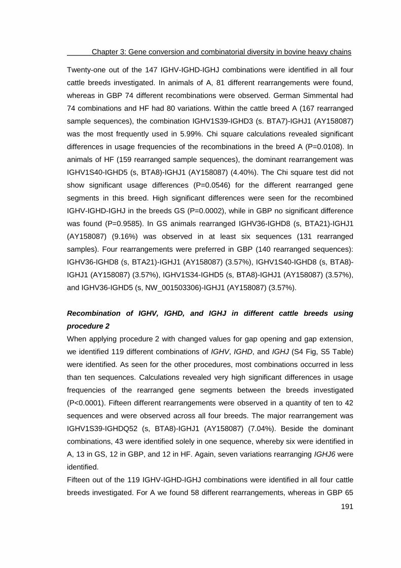
Chapter 3: Gene conversion and combinatorial diversity in bovine heavy chains
191
Twenty-one out of the 147 IGHV-IGHD-IGHJ combinations were identified in all four
cattle breeds investigated. In animals of A, 81 different rearrangements were found,
whereas in GBP 74 different recombinations were observed. German Simmental had
74 combinations and HF had 80 variations. Within the cattle breed A (167 rearranged
sample sequences), the combination IGHV1S39-IGHD3 (s. BTA7)-IGHJ1 (AY158087)
was the most frequently used in 5.99%. Chi square calculations revealed significant
differences in usage frequencies of the recombinations in the breed A (P=0.0108). In
animals of HF (159 rearranged sample sequences), the dominant rearrangement was
IGHV1S40-IGHD5 (s, BTA8)-IGHJ1 (AY158087) (4.40%). The Chi square test did not
show significant usage differences (P=0.0546) for the different rearranged gene
segments in this breed. High significant differences were seen for the recombined
IGHV-IGHD-IGHJ in the breeds GS (P=0.0002), while in GBP no significant difference
was found (P=0.9585). In GS animals rearranged IGHV36-IGHD8 (s, BTA21)-IGHJ1
(AY158087) (9.16%) was observed in at least six sequences (131 rearranged
samples). Four rearrangements were preferred in GBP (140 rearranged sequences):
IGHV36-IGHD8 (s, BTA21)-IGHJ1 (AY158087) (3.57%), IGHV1S40-IGHD8 (s, BTA8)-
IGHJ1 (AY158087) (3.57%), IGHV1S34-IGHD5 (s, BTA8)-IGHJ1 (AY158087) (3.57%),
and IGHV36-IGHD5 (s, NW_001503306)-IGHJ1 (AY158087) (3.57%).
Recombination of IGHV, IGHD, and IGHJ in different cattle breeds using
procedure 2
When applying procedure 2 with changed values for gap opening and gap extension,
we identified 119 different combinations of IGHV, IGHD, and IGHJ (S4 Fig, S5 Table)
were identified. As seen for the other procedures, most combinations occurred in less
than ten sequences. Calculations revealed very high significant differences in usage
frequencies of the rearranged gene segments between the breeds investigated
(P<0.0001). Fifteen different rearrangements were observed in a quantity of ten to 42
sequences and were observed across all four breeds. The major rearrangement was
IGHV1S39-IGHDQ52 (s, BTA8)-IGHJ1 (AY158087) (7.04%). Beside the dominant
combinations, 43 were identified solely in one sequence, whereby six were identified in
A, 13 in GS, 12 in GBP, and 12 in HF. Again, seven variations rearranging IGHJ6 were
identified.
Fifteen out of the 119 IGHV-IGHD-IGHJ combinations were identified in all four cattle
breeds investigated. For A we found 58 different rearrangements, whereas in GBP 65

Chapter 3: Gene conversion and combinatorial diversity in bovine heavy chains
192
different recombinations were observed. German Simmental had 57 combinations and
HF had 62 variations. Within A (167 rearrangements), the combination IGHV1S39-
IGHDQ52 (s, BTA8)-IGHJ1 (AY158087) was the most frequently used and was seen in
11.38% of the time. Chi square calculations showed significant differences in the usage
frequencies of the recombinations for breed A (P=0.0108). In animals of HF (159
rearranged sample sequences), the dominant rearrangement was IGHV1S39-
IGHDQ52 (s, BTA8)-IGHJ1 (AY158087) (8.81%). The Chi square test did not show
significant usage differences (P=0.0546) for the different rearranged gene segments in
this breed. High significant differences were seen for the recombined IGHV-IGHD-IGHJ
in the breed GS (P=0.0002), while in GBP no significant differences were observed
(P=0.9585). In GS animals rearranged IGHV36-IGHDQ52 (s, BTA8)-IGHJ1
(AY158087) (11.45%) was observed predominantly (131 rearranged sequences). The
rearrangement IGHV1S40-IGHDQ52 (s, BTA8)-IGHJ1 (AY158087) (9.29%) was
preferred in GBP (140 rearranged sequences).
Recombination of IGHV, IGHD, and IGHJ with different length of CDR3H
Within the three groups of length of CDR3H we identified different preferably
expressed recombinations of IGHV, IGHD, and IGHJ. Very high significant differences
regarding identified rearrangements within these groups were calculated (procedure 1:
among the breeds, GS, A, HF: P<0.0001; GBP: P=0.0016; procedure 2: among the
breeds, GS, A, HF, GBP: P<0.0001; procedure 3: among the breeds, A, GBP:
P<0.0001; GS: P=0.0003; HF: P=0.0085).
Using changed penalties for gap opening and gap extension and a new scoring matrix
(procedure 3) rearrangements of IGHV3 and IGHJ1 (AY158087) together with IGHD1
(s, BTA7), IGHD2 (as, [39]), IGHDQ52 (s, BTA8), and IGHDS10 [22] dominated (0.5-
0.84%) in sequences with group 1 CDR3H if breed was not taken into account. The
single breeds showed different major recombinations. In A, IGHV6-IGHD2 (s, BTA7)-
IGHJ1 was calculated for 1.8% of the sequences, whereas in GS IGHV3-IGHDQ52 (s,
BTA8)-IGHJ1 was identified in the same frequencies as IGHV3-IGHD2 (as, [39])-
IGHJ1, IGHV3-IGHD1 (as, [39])-IGHJ1, and IGHV1S35-IGHD8 (s, BTA21)-IGHJ1
(0.76%), in GBP IGHV3-IGHDS10 [22]-IGHJ1, IGHV3-IGHDS14 [22]-IGHJ1, IGHV3-
IGHD3 (s, BTA7)-IGHJ1, IGHV3-IGHD1 (s, BTA7)-IGHJ1, IGHV1S34-IGHDS10 [22]-
IGHJ1, IGHV1S34-IGHDS14 [22]-IGHJ1 and IGHV6-IGHDS10 [22]-IGHJ1 made up

Chapter 3: Gene conversion and combinatorial diversity in bovine heavy chains
193
0.71%, and in HF IGHV3-IGHDQ52 (s, BTA8)-IGHJ1 was found the most often
(2.52%).
In samples of all breeds possessing CDR3Hs of group 2 the rearrangement of
IGHV36-IGHD8 (s, BTA21)-IGHJ1 was found most often (4.52%). This is congruent
with our findings from the cattle breeds GS and GBP (10.69%, 5.00%). In A sequences
with rearrangement of IGHV1S39-IGHD5 (s, BTA8)-IGHJ1 were used with the highest
frequency (7.19%) whereas in HF IGHV3-IGHD8 (s, BTA21)-IGHJ1 and IGHV36-
IGHD3 (s, BTA7)-IGHJ1 dominated (3.77%).
The recombination of IGHV10-IGHD2 (s, [39])-IGHJ1 was identified in 0.84% of all
sequences with group 3 CDR3Hs. This combination was also dominant in sequences
possessing an exceptionally long CDR3H in HF (1.26%). In A, the combination
IGHV10-IGHD2 (s, BTA7)-IGHJ1 was preferred (1.2%), whereas in GS, IGHV10-
IGHDS10 [22]-IGHJ1 was found the most often (2.29%). In GBP IGHV10-IGHD8 (as,
[21])-IGHJ1 and IGHV10-IGHD7 (as, [21])-IGHJ1 were identified (0.71%).
If only new values for gap opening and gap extension were applied (procedure 2),
rearrangements of IGHV3 and IGHJ1 (AY158087) together with IGHD1 (as, [39])
dominated (1.01%) in sequences with a very short CDR3H if the breed was not taken
into account. The single breeds showed different major recombinations. In A, IGHV6-
IGHD1 (s, BTA7)-IGHJ1 was calculated for 2.4% of the sequences, whereas in GS
IGHV3-IGHDQ52 (s, BTA8)-IGHJ1 was identified in the same frequencies as IGHV3-
IGHD4 (as, [21])-IGHJ1, IGHV3-IGHD1 (s, BTA7)-IGHJ1, and IGHV1S35-IGHD1 (s,
BTA7)-IGHJ1 (0.76%), in GBP IGHV3-IGHD1 (as, [39])-IGHJ1 made up 1.43%,
whereas in HF IGHV3-IGHD1 (as, [39])-IGHJ1 and IGHV3-IGHD4 (s, BTA8)-
IGHJ1were found the most often (1.26%).
In samples of all breeds possessing group 2 CDR3Hs, the rearrangement of
IGHV1S39-IGHDQ52 (s, BTA8)-IGHJ1 was found the most often (7.04%). This is
congruent with the findings in the cattle breeds A and HF (11.38%, 8.81%). In GS
sequences showing the rearrangement IGHV36-IGHDQ52 (s, BTA8)-IGHJ1 were used
with the highest frequency (11.45%) whereas in GBP IGHV1S40-IGHDQ52 (s, BTA8)-
IGHJ1 dominated (9.29%).
The recombination of IGHV10-IGHD8 (s, BTA21)-IGHJ1 was identified in 1.17% of all
sequences with group 3 CDR3Hs. This combination was also dominant in sequences
possessing exceptionally long CDR3H in A (2.4%) and GBP (0.71%). In GBP IGHV10-

Chapter 3: Gene conversion and combinatorial diversity in bovine heavy chains
194
IGHDS10-IGHJ1 was also found at this frequency. In GS, the combinations IGHV10-
IGHD1 (s, BTA7)-IGHJ1 and IGHV10-IGHD4 (s, BTA8)-IGHJ1 were preferred (2.29%),
whereas in HF, IGHV10-IGHD3 (s, BTA7)-IGHJ1 was found most often (1.26%).
Using default values (procedure 1) rearrangements of IGHV3 and IGHJ1 (AY158087)
together with IGHDQ52 (s, BTA8) as well as IGHV6-IGHD1 (s, BTA7) dominated (1.51
and 0.5%) in sequences with a short CDR3H if the breed was not taken into account.
But again the single breeds showed different major recombinations. In A, IGHV3-
IGHDQ52 (s, BTA8)-IGHJ1 and IGHV6-IGHD1 (s, BTA7)-IGHJ1 were found for 1.8%
of the sequences, whereas in GS IGHV1S35-IGHD4 (s, BTA8)-IGHJ1 was identified at
the same frequency as IGHV3-IGHD2 (s, BTA8)-IGHJ1, IGHV3-IGHD1 (as, [39])-
IGHJ1, and IGHV3-IGHD8 (as, [21])-IGHJ1 (0.76%), in GBP IGHV3-IGHD1 (s, BTA7)-
IGHJ1, IGHV3-IGHD2 (as, [39])-IGHJ1, IGHV3-IGHD5 (s, BTA8)-IGHJ1, IGHV3-
IGHDQ52 (s, BTA8)-IGHJ1, IGHV1S34-IGHD5 (s, BTA8)-IGHJ1, IGHV1S34-IGHD5 (s,
NW_001503306.)-IGHJ1 and IGHV6-IGHD5 (s, BTA8)-IGHJ1 made up 0.71%, and in
HF IGHV3-IGHDQ52 (s, BTA8)-IGHJ1 was found most often (3.14%).
In samples of all breeds possessing CDR3Hs of group 2 the rearrangement of
IGHV36-IGHD8 (s, BTA21)-IGHJ1 was found most often (3.52%). This is congruent
with the findings in the cattle breed GS (9.16%). In HF sequences showing the
rearrangements IGHV1S39-IGHD8 (s, BTA21)-IGHJ1 and IGHV1S40-IGHD5 (s,
BTA8)-IGHJ1 were used with the highest frequency (4.4%) whereas in A IGHV1S39-
IGHD3 (s, BTA7)-IGHJ1 dominated (5.99%) as well as IGHV1S40-IGHD8 (s, BTA21)-
IGHJ1, IGHV36-IGHD5 (s, NW_001503306)-IGHJ1, and IGHV36-IGHD8 (s, BTA21)-
IGHJ1 in the cattle breed GBP (3.57%).
The recombination of IGHV10-IGHD8 (s, BTA21)-IGHJ1 was identified in 1.01% of all
sequences with exceptionally long CDR3Hs. In sequences possessing those group 3
CDR3H in HF the combinations IGHV10-IGHD4 (s, BTA8)-IGHJ1 and IGHV10-IGHD8
(s, BTA21)-IGHJ1 (1.26%) dominated. In A, the combination IGHV10-IGHD4 (s,
BTA8)-IGHJ1 was preferred (1.2%), whereas in GS, IGHV10-IGHD2 (s, BTA7)-IGHJ1
and IGHV10-IGHD8 (s, BTA21)-IGHJ1 were found most often (1.53%), and in GBP
only the two recombinations IGHV10-IGHD8 (s, BTA21)-IGHJ1 and IGHV10-IGHD4
(as, [21])-IGHJ1 were identified (0.71%).

Chapter 3: Gene conversion and combinatorial diversity in bovine heavy chains
195
Variability based on amino acid substitutions
We then counted the amino acid substitutions at each position to calculate variability as
described by Wu and Kabat [40]. The results are shown in the variability plots for each
breed separately in Figs 8 a-d. The amino acid positions were numbered in accordance
to the IMGT numbering systems [41]. Therefore, FRHs and CDRHs are defined by the
following amino acid positions: FR1H: 1…26, CDR1H: 27…38, FR2H: 39…55,
CDR2H: 56…65, FR3H: 66…104, CDR3H: 105…117, FR4H: 118…128. In cattle there
are no amino acids assigned to positions 10, 31-34, 60-62, and 73.
Fig 7. Variability plots of the heavy chain variable regions in the cattle breeds Aubrac, German Simmental, Holstein Friesian, and German Black Pied. The complete variable region is displayed on the horizontal axis. Positions are numbered in accordance to the IMGT numbering system. Within the FR1-4H, little variability is discernable whereas in CDR1-3H increase in variability is observed. (A) Aubrac, (B) German Simmental, (C) Holstein Friesian, (D) German Black Pied
(A) (B)
(C) (D)

Chapter 3: Gene conversion and combinatorial diversity in bovine heavy chains
196
Within the FR1-4H little variability is discernable whereas in CDR1-3H an increase in
variability is observed as expected. A maximum of variability in FRHs is calculated for
position 96 in FR3H. Variability at this position is 23.7 in A, in HF 19.0, whereas in GS
and GBP variability is 12.7 and 17.5, respectively. In CDRHs, variability increases from
CDR1H to CDR3H in all breeds. In CDR1H breed A showed the highest variability. In
CDR2H variability varies between 58 in GS and 93 in GBP. GBP also showed highest
variability in CDR3H. The lowest variability within CDR3H was found in GS. Both HF
and A showed moderate variability of CDR2H and CDR3H in comparison to GBP and
GS. Overall, variability was much higher at the transition areas between FRH and CDR
than in the middle of the CDRHs.

Chapter 3: Gene conversion and combinatorial diversity in bovine heavy chains
197
Discussion
This study makes a substantial contribution to the analysis and understanding of the
development of the transcribed bovine immunoglobulin repertoire. We examined
possible gene conversions within the variable region of bovine heavy chains. We
investigated the dominantly transcribed IGHV, IGHD, and IGHJ gene segments and
their combinatorial diversity using a newly developed bioinformatics framework, which
considers the unique specificity of exceptionally long CDR3 group of bovine
immunoglobulin heavy chains. During the development of the program, we applied
different conditions (procedure 1 to 3) to improve the alignments of the single gene
segments. The progress of assigning germline IGHV, IGHD, and IGHJ to sample
sequences is also shown in this study. Unlike previous studies, we complemented our
investigation with the analysis of breed specific differences in the four different cattle
breeds Aubrac, German Simmental, German Black Pied, and Holstein Friesian.
Due to the limited germline sequence divergence recent studies on bovine
immunoglobulin genetics focused on antibody diversification strategies and the
junctional diversity of the antibody repertoire. Specific diversification strategies were
identified such as the generation of exceptionally long CDR3H [17, 42, 43], the
insertion of conserved short nucleotide sequences (CSNS) at the IGHV-IGHD junction
[24], the use of pseudogene fragments in lambda light chains as well as gene
conversions [14], and somatic hypermutations independent of exposure to external
antigens during B-cell development [44]. In addition, more germline gene segments
were determined over the last few years [17, 18, 22]. Since the current bovine genome
assembly is still incomplete, the full germline repertoire remains under active
investigation.
Previous analyses of the bovine immunoglobulin repertoire aimed at identifying
rearranged germline gene segments applied various software tools for sequence
alignments. As these tools are of limited use for detailed analysis of rearranged
immunoglobulin genes due to the difficult and error prone manual assembly of different
genes, specialized software tools have been developed. The most familiar ones are
IMGT/Junction Analysis [26], IMGT/V-QUEST [27, 28], IMTG/HIGHV-QUEST [29],
VBASE2 [30], JoinSolver [31], iHMMun-align [32], and IgBLAST [33]. Only IgBLAST
enables the analysis of both nucleotide and protein sequences for FR/CDR and allows
the user to either apply the numbering system of Kabat or the IMGT system [41, 45].
Matching germline IGHV, IGHD, and IGHJ genes as well as details at rearrangement

Chapter 3: Gene conversion and combinatorial diversity in bovine heavy chains
198
junctions may be analyzed. Searches against germline gene databases and other
databases are possible [33]. Tools other than IgBLAST do not provide simultaneous
database searches or the analysis of protein sequences. All of these immunoglobulin
sequence analysis tools support organisms such as human, mouse, rat, rabbit and
rhesus monkey, but representation of livestock is missing or incomplete. We did not
apply these analysis tools, as they do not consider the bovine specific occurrence of
exceptionally long CDR3H. Therefore, we developed a new bioinformatics framework
to address this specific case.
In contrast to the analysis tools mentioned above, our program not only searches our
updated bovine specific immunoglobulin germline gene database but also is able to
load other pre-designed databases. Matches are identified on the basis of nucleotides
as it is the case for all other tools. For the delineation of FR and CDR, we apply the
IMGT nomenclature that is currently recommended and most widely used. In addition,
we focused on the adjustment of search parameters for IGHV and IGHJ and especially
for the identification of IGHD.
The analysis of Ig heavy chain variable regions in four cattle breeds revealed the
usage of 15 different IGHV segments, 21 IGHD segments, as well as two IGHJ
segments. IGHV1S39 was used most frequently followed by IGHV3/33. Rarely used
IGHV segments were IGHV1S26, IGHV1S32, IGHV1S33, IGHV1S37, and IGHV1S38.
In bovine fetal bone marrow, ileum, and spleen high frequencies of IGHV3/33
(=IGHV1S3) and IGHV1S39 was observed as well as low frequencies of IGHV1S38
and IGHV1S26 [22]. The transcription of IGHV1S32 and S37 has not yet been
described. Among the 20, 17, and 21 transcribed IGHD (regarding procedures 1-3),
IGHDS8, IGHDS5, IGHDS10, and IGHDQ52 (=IGHDS9) were preferred in all breeds.
IGHDS1 to IGHDS8 were also found to be transcribed in antisense direction in the third
calculation procedure but in low numbers. Using the first procedure, IGHDS6 was not
identified in antisense direction, and applying the second procedure IGHDS1, IGHDS4,
IGHDS6, and IGHDS8 were shown to be transcribed in antisense orientation. Previous
studies also elucidated the transcription of 14 IGHD, where the occurrence of IGHDS5
was the most frequent one and was present in 42% of the sequences analyzed in
bovine fetus [22]. The assignment of the FR4H to germline IGHJ revealed the
transcription of IGHJ1, and IGHJ6 [22] with IGHJ1 clearly preferred. In the cattle breed
A, procedure 3 confirmed IGHV1S39-IGHD5-IGHJ1 as the most common
recombination of gene segments which is identical to the most frequent finding in

Chapter 3: Gene conversion and combinatorial diversity in bovine heavy chains
199
bovine fetus [22]. This recombination belongs to immunoglobulins possessing a
CDR3H region of intermediate length. Statistical analyses showed significant different
transcription levels of IGHV, IGHD, and IGHJ segments within the breeds.
The usage of pseudogene segments has already been described for animals such as
chicken [4, 37]. In bovine lambda light chains, fragments of pseudogenes were also
shown to contribute to immunoglobulin diversity in a gene conversion process [14]. In
the current analysis, possible gene conversion events were identified by the
assignments of parental germline IGHV to separate FR1-3H and CDR1-3H. In addition
to the IGHV identified for the complete variable region based exclusively on FR1-3H,
several pseudogenes were assigned as possible originating germline IGHV in the
separation analysis. For instance, the pseudogenes IGHV4Ψ, IGHV9Ψ, and IGHV18Ψ
belong to the boVH2 family [17], but seem to contribute to gene conversion events by
nucleotide substitutions. In particular, IGHV4Ψ, which was mentioned in the example
above meets the criteria for gene conversion such as the location upstream of the
rearranged segment [4, 13] and clusters of nucleotide changes [14]. Further, the
flanking homology of the conversion region supports the genetic exchange [13] and the
separation from IGHV6 by more than 18 kb on the genome allows looping during
rearrangement [17]. In comparison, in chicken the nearest pseudogene is separated by
7 kb [4]. It should be noted that it is difficult to consider the order of gene segments to
evaluate the plausibility of other gene conversions due to the incomplete annotation of
the bovine genome [17, 18]. Finally, our data indicate an exchange between the two
bovine VH families which obviously is rare and which might be an influence of breed or
method of analysis when compared to previous results [18].
The length distribution of CDR3H consists of short CDR3H (group 1), intermediate
length CDR3H (group 2), and exceptionally long CDR3H (group 3) in all four cattle
breeds. In the breed GS the highest percentage of group 3 CDR3H was calculated.
The longest CDR3H with 65 aa were found in GS and HF sequences. The longest ever
detected CDR3H in cattle was 67 aa long using IMGT numbering [17]. In contrast, four
amino acids made up the shortest CDR3H in A animals, GS animals, and HF animals.
The maximum length of group 2 CDR3H was 22 aa.
The combinatorial diversity of germline IGHV, IGHD, and IGHJ-segments is
represented by 162 different rearrangements that were expressed with significant
differences (procedure 3). In comparison, 147 and 119 different recombinations of
IGHV-IGHD-IGHJ were identified using calculation procedures 1 and 2, respectively. In
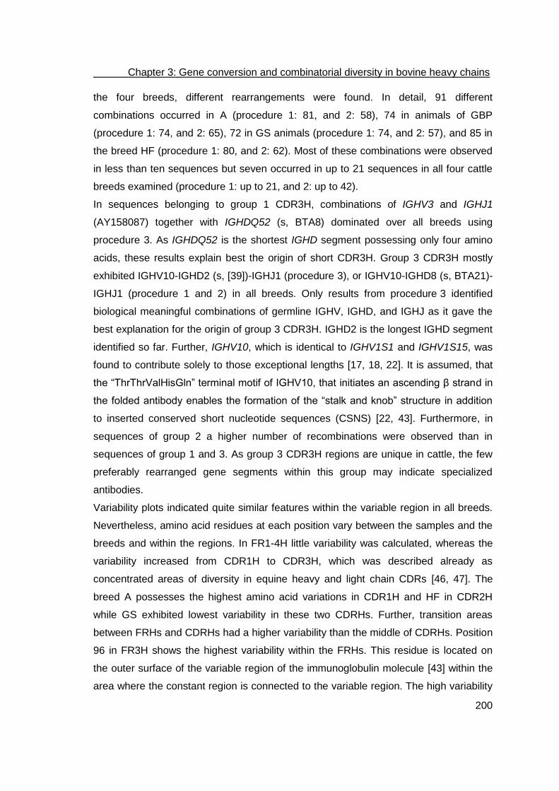
Chapter 3: Gene conversion and combinatorial diversity in bovine heavy chains
200
the four breeds, different rearrangements were found. In detail, 91 different
combinations occurred in A (procedure 1: 81, and 2: 58), 74 in animals of GBP
(procedure 1: 74, and 2: 65), 72 in GS animals (procedure 1: 74, and 2: 57), and 85 in
the breed HF (procedure 1: 80, and 2: 62). Most of these combinations were observed
in less than ten sequences but seven occurred in up to 21 sequences in all four cattle
breeds examined (procedure 1: up to 21, and 2: up to 42).
In sequences belonging to group 1 CDR3H, combinations of IGHV3 and IGHJ1
(AY158087) together with IGHDQ52 (s, BTA8) dominated over all breeds using
procedure 3. As IGHDQ52 is the shortest IGHD segment possessing only four amino
acids, these results explain best the origin of short CDR3H. Group 3 CDR3H mostly
exhibited IGHV10-IGHD2 (s, [39])-IGHJ1 (procedure 3), or IGHV10-IGHD8 (s, BTA21)-
IGHJ1 (procedure 1 and 2) in all breeds. Only results from procedure 3 identified
biological meaningful combinations of germline IGHV, IGHD, and IGHJ as it gave the
best explanation for the origin of group 3 CDR3H. IGHD2 is the longest IGHD segment
identified so far. Further, IGHV10, which is identical to IGHV1S1 and IGHV1S15, was
found to contribute solely to those exceptional lengths [17, 18, 22]. It is assumed, that
the “ThrThrValHisGln” terminal motif of IGHV10, that initiates an ascending β strand in
the folded antibody enables the formation of the “stalk and knob” structure in addition
to inserted conserved short nucleotide sequences (CSNS) [22, 43]. Furthermore, in
sequences of group 2 a higher number of recombinations were observed than in
sequences of group 1 and 3. As group 3 CDR3H regions are unique in cattle, the few
preferably rearranged gene segments within this group may indicate specialized
antibodies.
Variability plots indicated quite similar features within the variable region in all breeds.
Nevertheless, amino acid residues at each position vary between the samples and the
breeds and within the regions. In FR1-4H little variability was calculated, whereas the
variability increased from CDR1H to CDR3H, which was described already as
concentrated areas of diversity in equine heavy and light chain CDRs [46, 47]. The
breed A possesses the highest amino acid variations in CDR1H and HF in CDR2H
while GS exhibited lowest variability in these two CDRHs. Further, transition areas
between FRHs and CDRHs had a higher variability than the middle of CDRHs. Position
96 in FR3H shows the highest variability within the FRHs. This residue is located on
the outer surface of the variable region of the immunoglobulin molecule [43] within the
area where the constant region is connected to the variable region. The high variability

Chapter 3: Gene conversion and combinatorial diversity in bovine heavy chains
201
at this position may indicate an influence on the position of variable and constant
region and their sterical orientation, which may affect light chain pairing as heavy
chains possessing group 3 CDR3H are connected to a special type of lambda light
chains [43, 48].
Further analysis revealed that no amino acids were assigned to the IMGT amino acid
positions 10, 31-34, 60-62, and 73 in cattle. This means, that one amino acid position
within FR1H, four positions in CDR1H, three positions in CDR2H, as well as one
position in FR3H were not filled. Consequently, in cattle 8 out of 12 amino acid
positions within CDR1H are covered. In CDR2H, 10 positions are available and 7 are
covered. Compared to FR-IMGT and CDR-IMGT lengths of functional and ORF IGHV-
genes of human IGHV, mouse IGHV, rat IGHV, arabian camel IGHV, sheep IGHV, and
pig IGHV the missing amino acids within FR1H and FR3H are conserved in all animals
mentioned [49, 50]. Averaged eight to ten amino acids were positioned in CDR1H
whereas in CDR2H six to ten amino acids were placed by the IMGT numbering system
in human, mouse, rat, camel, sheep, and pig IGHV. Therefore, the positions of missing
amino acids are congruent with other species.
In the breed A, the highest number of recombinations and variability were observed
when compared to the other breeds investigated. GS possessed the lowest number of
recombinations and showed less variability except in the middle of the CDR3H region.
This finding indicates the contribution of insertions and deletions to diversity in case of
few rearrangements [25]. It should be noted that A and GS were kept under the same
management in a mixed herd. GBP and HF were kept at different farms. The breeds
kept in different areas were consequently exposed to different antigens. Thus, the
individual number of rearrangements per breed and differences in variability
additionally indicate a specialized immune response as animals on one farm are
challenged with the same environment.
The application of the newly developed bioinformatics framework led to important new
results. Our analyses demonstrated that the bovine heavy chain diversity is not
restricted to the use of a limited number of germline genes although there are preferred
rearrangements within the three groups of CDR3H lengths. We also found strong
evidence for gene conversion using pseudogenes. Despite current advances in the
understanding of bovine immunoglobulin diversification, future investigations of the
germline repertoire are necessary.

Chapter 3: Gene conversion and combinatorial diversity in bovine heavy chains
202
Material and Methods
Detailed analyses of immunoglobulin sequences using a newly developed
bioinformatics framework
For sequence analysis, we developed a new bioinformatics framework using MUSCLE
[34, 35] for the initial fast but accurate multiple nucleotide sequence alignment and
following ClustalW [36] for calculating the sequence distances after deduction of the
amino acid residues. Both programs are available as stand-alone algorithms and were
implemented into our program. The immunogenetics nomenclature (IMGT) was used to
assign framework regions 1-4 (FR1-4H) and the complementarity determining regions
1-3 [41].
Therefore, germline nucleotide sequences were imported in FASTA-format (IGHV,
IGHD, IGHJ [17, 18, 22]). Using the functional IGHV, the nucleotide sequences were
translated into amino acids to number the codons of the functional germline IGHV gene
segments according to the IMGT system from FR1H to FR3H. This required first the
identification of the conserved and preassigned positions of Cys23, Trp41, Leu89, and
Cys104 defined by Lefranc et al. [41]. Following, the nucleotide sequences of germline
pseudo IGHV gene segments were aligned separately using MUSCLE [34, 35] to
obtain the putative open reading frame. The previously defined positions of the codons
using the functional genes were transferred onto the pseudogenes. Insertions as well
as deletions of nucleotides within the pseudo gene segment sequence were discarded.
The last 33 nucleotides of germline IGHJs were then used to define the FR4H. The
region between FR3H and FR4H is defined as CDR3H. This region was later used to
align the sample CDR3H to germline IGHD segments. Defining FRHs and CDRHs
allowed, beside the analyses of the complete transcribed genes, the alignment of
IGHV, IGHD and IGHJ using different parameters to improve the biological significance
as well as the analyses of the single functionally divergent regions to determine
putative gene conversion events in those regions. All functional and pseudo gene
germline segments are referred as reference sequences.
Framework regions and CDRs of transcribed sequences (designated as sample
sequences) were aligned pairwise to the isolated reference sequences after isolation
from the first IgG constant region. Following, minimal divergence was used to identify
the most similar sample sequence and reference sequence pairing.
For nucleotide alignments of IGHV and IGHJ, default values of MUSCLE were used.
We tested three different procedures to assign germline and sample IGHDs to improve

Chapter 3: Gene conversion and combinatorial diversity in bovine heavy chains
203
biological significance. In procedure 1, we applied default values of MUSCLE [34, 35],
in procedure 2 we changed the penalties for gap opening to -4 and for gap extention to
-0.3 [22], and in procedure 3 we additionally incorporated a new scoring matrix with
match = 2, transversion = -1, and transition = 1 to evaluate transversion and transition
mutations, whereby the IUB (international union of biochemistry) code for single and
wobble bases was used.
The three procedures were applied to a set of sample sequences of IgG-derived
variable regions from four different cattle breeds. Each nucleotide sequence of our
sample sequences was aligned separately to the reference sequences to determine
the most similar reference sequence as germline origin. Following, the codons were
translated into amino acids. Sample sequences possessing premature Stop codons or
not covering the full length of the variable region due to incomplete sequencing were
eliminated and were not incorporated in further analyses. The remaining sample
sequences were annotated in accordance to the IMGT nomenclature. For exceptional
long CDR3H no positions are defined in the IMGT system, therefore positions had to
be added as required and designated as 111.1-111.x and 112.y-112.1 in accordance to
the IMGT numbering system [41]. To determine the germline origin, only the FRHs
were aligned to avoid interference with the highly diversified CDRH [18]. To analyze
possible gene conversion events, FR1-3Hs and CDR1-3Hs were extracted and aligned
separately to the corresponding regions of the IGHV reference sequences to find the
most similar one (Fig 8). The results were presented as an html table.

Chapter 3: Gene conversion and combinatorial diversity in bovine heavy chains
204
(A)
(B)
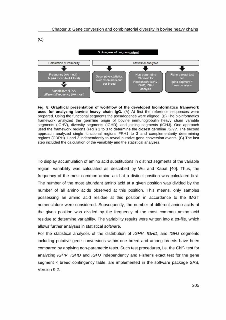
Chapter 3: Gene conversion and combinatorial diversity in bovine heavy chains
205
(C)
Fig. 8. Graphical presentation of workflow of the developed bioinformatics framework used for analyzing bovine heavy chain IgG. (A) At first the reference sequences were prepared. Using the functional segments the pseudogenes were aligned. (B) The bioinformatics framework analyzed the germline origin of bovine immunoglobulin heavy chain variable segments (IGHV), diversity segments (IGHD), and joining segments (IGHJ). One approach used the framework regions (FRH) 1 to 3 to determine the closest germline IGHV. The second approach analyzed single functional regions FRH1 to 3 and complementarity determining regions (CDRH) 1 and 2 independently to reveal putative gene conversion events. (C) The last step included the calculation of the variability and the statistical analyses.
To display accumulation of amino acid substitutions in distinct segments of the variable
region, variability was calculated as described by Wu and Kabat [40]. Thus, the
frequency of the most common amino acid at a distinct position was calculated first.
The number of the most abundant amino acid at a given position was divided by the
number of all amino acids observed at this position. This means, only samples
possessing an amino acid residue at this position in accordance to the IMGT
nomenclature were considered. Subsequently, the number of different amino acids at
the given position was divided by the frequency of the most common amino acid
residue to determine variability. The variability results were written into a txt-file, which
allows further analyses in statistical software.
For the statistical analyses of the distribution of IGHV, IGHD, and IGHJ segments
including putative gene conversions within one breed and among breeds have been
compared by applying non-parametric tests. Such test procedures, i.e. the Chi2- test for
analyzing IGHV, IGHD and IGHJ independently and Fisher's exact test for the gene
segment × breed contingency table, are implemented in the software package SAS,
Version 9.2.

Chapter 3: Gene conversion and combinatorial diversity in bovine heavy chains
206
Generation of the sample sequence set
Breed selection, isolation of PBMCs and cDNA synthesis
For the analysis of the transcription of IgG heavy chain genes, the four cattle breeds
German Black Pied (GBP), German Simmental (GS), Holstein-Friesian (HF), and
Aubrac (A) were chosen. The animals selected from the herd of breed A were
composed of seven French and three German animals, whereas the sample of breed
GS included one Austrian bull. German Black Pied and A represent small populations
and have local importance, whereas HF and GS are commonly used in global
commercial milk and meat production. German Simmental and A are kept on the same
farm under same management conditions and in a mixed herd.
Blood samples were collected from ten randomly chosen animals per breed during
routine blood sampling for mandatory examinations in disease control. Peripheral blood
mononuclear cells (PBMCs) were isolated using Ficoll gradients (GE Healthcare
Europe GmbH, Germany) according to the manufacturer’s protocol. Cells were stained
with trypan blue and viable cells were counted. Total RNA was isolated from 1x10⁷
cells using the RNeasy Mini Kit (Qiagen, Germany). The first-strand cDNA was
synthesized using pd(N)6 primers from 3 µg of total RNA in a total volume of 20 µl
(SuperScript™III First-Strand Synthesis SuperMix, Life Technologies GmbH,
Germany).
Ethical Statement
To collect B-lymphocytes, 20 ml of EDTA blood were taken from the tail vein during
regular investigation of infectious diseases in the cattle herds. The plasma was applied
e.g. in an indirect ELISA testing for antibodies against Mycobacteria avium spp.
paratuberculosis or BHV-1. Therefore, no specific approval is required.
Amplification of immunoglobulin heavy-chain isotype restricted variable regions
To amplify the variable regions restricted to γ1-3 isotype heavy chains, a PCR was
performed with primers binding within the leader region and the 3’UTR (bIgG_leader:
ATG AAC CCA CTG TGG ACC; bIgG_3’UTR: CAG GAG GAA TGC ACA CAG). The
primers were based on database sequence information and assigned to accession
number X62916. The primer boIgG_leader anneals to position 22-39, and the primer
boIgG_3’UTR to position 1518-1535. To monitor the integrity and purity of the cDNA,
527 bp of the bovine GAPDH (Glycerinaldehyde 3-phosphate dehydrogenase) were

Chapter 3: Gene conversion and combinatorial diversity in bovine heavy chains
207
amplified as a positive control. A no- template control served as a negative control for
the PCR. The total reaction volume of 50 µl included 0.67 µl of cDNA, 200 µM dNTPs
(Bioline, Germany), 5 µl of 10x PCR buffer (75 mM Tris-HCl, pH 9.0; 2 mM MgCl2;
50 mM KCl; 20 mM (NH4)2SO4), 0.4 µM of each primer, and 2 units of DNA polymerase
(Biotools, Spain). PCR was performed under cycling conditions of 95oC for 5 min,
followed by 35 cycles of 95oC for 1 min, 59.4°C for 1 min, 72oC for 2 min, and
terminated with elongation at 72°C for 10 min. Length and purity of the PCR products
were evaluated by means of electrophoresis on 1% agarose gels.
Cloning and sequencing of the PCR products
The PCR products were purified and concentrated using the MiniElute Gel Extraction
Kit (Qiagen, Germany) in accordance to the manufacturer’s protocol except QX1 buffer
replaced QG buffer. Samples were eluted with 13 µl EB buffer (10 mM Tris-HCl, pH
8.5) and were stored at 4°C. Purified products were cloned into the pCR 2.1-TOPO
3.9 kb TA vector (InvitrogenTM, Karlsruhe, Germany) and transformed into chemically
competent One Shot TOP10 E. coli cells (InvitrogenTM, Karlsruhe, Germany).
Transformants were plated on LB agar containing 0.3 mM ampicillin, 40 µl 2.44 µM X-
gal (5-bromo-4-chloro-3-indolyl-beta-D-galactopyranoside), and 40 µl 1 M IPTG
(Isopropyl β-D-1-thiogalactopyranoside) for blue-white selection. After incubation at
37°C, overnight cultures of randomly selected white transformants were grown in a
5 ml LB-ampicillin broth. Plasmids were isolated using the MiniPrep Kit (Qiagen,
Germany). In order to assess the insert size, plasmid DNA was cleaved with EcoRI
(New England Biolabs, Germany) or a colony PCR was performed. Therefore a 25 µl
mixture containing 2 µl cell culture, 0.4 µM of vector specific primers M13 (-20) Forward
and M13 Reverse (Invitrogen, Germany), respectively, and one PCR-bead (GE
Healthcare Europe GmbH, Germany) were used in a hot start PCR at 95°C for 5 min,
denaturation at 95°C for 1 min, annealing at 60°C for 1 min, and extension at 72°C for
2 min up to a total of 30 cycles. A final extension at 72°C for 10 min was included after
the final cycle before PCR mixtures were cooled down to 4°C. The size of the resulting
fragments and of the PCR products was confirmed by agarose gel electrophoresis.
Sixteen clones per animal were sequenced according to the chain-termination method
[51]. The M13 (-20) Forward (5’-GTA AAA CGA CGG CCA G-3’) and M13 Reverse (5’-
CAG GAA ACA GCT CTG AC-3’, Invitrogen, Germany) vector-specific primers, as well
as the gene specific primers boIgG_leader, boIgG_3’UTR, boIgG_CH1_for (5’-GCC

Chapter 3: Gene conversion and combinatorial diversity in bovine heavy chains
208
TCC ACC ACA GCC CCG AAA G-3’), boIgG_CH3_rev (5’-GAC CTT GCA CTT GAA
CTC C-3’) and boIgG_CH1_rev (5’-ACG GTC ACC ATG CTG CTG AG-3’) were used
for sequencing.

Chapter 3: Gene conversion and combinatorial diversity in bovine heavy chains
209
Acknowledgments
We gratefully thank Dr. Dalia Aboelhassan, Sina Stein and Caroline Bierschenk for
their technical support in generating the sequences. We also thank Dr. Apostolos Gittis
and Dr. Stephen Dollery for critical review of the manuscript.

Chapter 3: Gene conversion and combinatorial diversity in bovine heavy chains
210
References
1. Edelman GM. Antibody structure and molecular immunology. Science. 1973;180(88):830-40. Epub 1973/05/25. PubMed PMID: 4540988.
2. Tonegawa S. Somatic generation of antibody diversity. Nature. 1983;302(5909):575-81. Epub 1983/04/14. PubMed PMID: 6300689.
3. Wagner SD, Neuberger MS. Somatic hypermutation of immunoglobulin genes. Annu Rev Immunol. 1996;14:441-57. Epub 1996/01/01. doi: 10.1146/annurev.immunol.14.1.441. PubMed PMID: 8962691.
4. Reynaud CA, Dahan A, Anquez V, Weill JC. Somatic hyperconversion diversifies the single Vh gene of the chicken with a high incidence in the D region. Cell. 1989;59(1):171-83. Epub 1989/10/06. doi: 0092-8674(89)90879-9 [pii]. PubMed PMID: 2507167.
5. Berens SJ, Wylie DE, Lopez OJ. Use of a single VH family and long CDR3s in the variable region of cattle Ig heavy chains. Int Immunol. 1997;9(1):189-99. Epub 1997/01/01. PubMed PMID: 9043960.
6. Sun J, Kacskovics I, Brown WR, Butler JE. Expressed swine VH genes belong to a small VH gene family homologous to human VHIII. J Immunol. 1994;153(12):5618-27. Epub 1994/12/15. PubMed PMID: 7989761.
7. Dufour V, Malinge S, Nau F. The sheep Ig variable region repertoire consists of a single VH family. J Immunol. 1996;156(6):2163-70. Epub 1996/03/15. PubMed PMID: 8690905.
8. Saini SS, Hein WR, Kaushik A. A single predominantly expressed polymorphic immunoglobulin VH gene family, related to mammalian group, I, clan, II, is identified in cattle. Mol Immunol. 1997;34(8-9):641-51. Epub 1997/06/01. PubMed PMID: 9393967.
9. Sinclair MC, Gilchrist J, Aitken R. Bovine IgG repertoire is dominated by a single diversified VH gene family. J Immunol. 1997;159(8):3883-9. Epub 1997/10/23. PubMed PMID: 9378976.
10. Reynaud CA, Anquez V, Dahan A, Weill JC. A single rearrangement event generates most of the chicken immunoglobulin light chain diversity. Cell. 1985;40(2):283-91. Epub 1985/02/01. doi: 0092-8674(85)90142-4 [pii]. PubMed PMID: 3917859.
11. Reynaud CA, Anquez V, Grimal H, Weill JC. A hyperconversion mechanism generates the chicken light chain preimmune repertoire. Cell. 1987;48(3):379-88. Epub 1987/02/13. doi: 0092-8674(87)90189-9 [pii]. PubMed PMID: 3100050.
12. Reynaud CA, Garcia C, Hein WR, Weill JC. Hypermutation generating the sheep immunoglobulin repertoire is an antigen-independent process. Cell. 1995;80(1):115-25. Epub 1995/01/13. doi: 0092-8674(95)90456-5 [pii]. PubMed PMID: 7813007.
13. Becker RS, Knight KL. Somatic diversification of immunoglobulin heavy chain VDJ genes: evidence for somatic gene conversion in rabbits. Cell. 1990;63(5):987-97. Epub 1990/11/30. doi: 0092-8674(90)90502-6 [pii]. PubMed PMID: 2124176.
14. Parng CL, Hansal S, Goldsby RA, Osborne BA. Gene conversion contributes to Ig light chain diversity in cattle. J Immunol. 1996;157(12):5478-86. Epub 1996/12/15. PubMed PMID: 8955197.
15. Sun Y, Wang C, Wang Y, Zhang T, Ren L, Hu X, et al. A comprehensive analysis of germline and expressed immunoglobulin repertoire in the horse. Dev Comp Immunol. 2010;34(9):1009-20. Epub 2010/05/15. doi: S0145-305X(10)00103-5 [pii]
10.1016/j.dci.2010.05.003. PubMed PMID: 20466019.

Chapter 3: Gene conversion and combinatorial diversity in bovine heavy chains
211
16. Miller JR, Thomsen PD, Dixon SC, Tucker EM, Konfortov BA, Harbitz I. Synteny mapping of the bovine IGHG2, CRC and IGF1 genes. Anim Genet. 1992;23(1):51-8. Epub 1992/01/01. PubMed PMID: 1349204.
17. Walther S, Czerny CP, Diesterbeck US. Exceptionally long CDR3H are not isotype restricted in bovine immunoglobulins. PLoS One. 2013;8(5):e64234. Epub 2013/05/30. doi: 10.1371/journal.pone.0064234
PONE-D-13-04746 [pii]. PubMed PMID: 23717573; PubMed Central PMCID: PMC3661452.
18. Niku M, Liljavirta J, Durkin K, Schroderus E, Iivanainen A. The bovine genomic DNA sequence data reveal three IGHV subgroups, only one of which is functionally expressed. Dev Comp Immunol. 2012;37(3-4):457-61. doi: 10.1016/j.dci.2012.02.006. PubMed PMID: 22369780.
19. Zhao Y, Kacskovics I, Rabbani H, Hammarstrom L. Physical mapping of the bovine immunoglobulin heavy chain constant region gene locus. J Biol Chem. 2003;278(37):35024-32. Epub 2003/06/28. doi: 10.1074/jbc.M301337200
M301337200 [pii]. PubMed PMID: 12829708. 20. Hosseini A, Campbell G, Prorocic M, Aitken R. Duplicated copies of the bovine JH
locus contribute to the Ig repertoire. Int Immunol. 2004;16(6):843-52. Epub 2004/05/06. doi: 10.1093/intimm/dxh085
dxh085 [pii]. PubMed PMID: 15126417. 21. Koti M, Kataeva G, Kaushik AK. Organization of D(H)-gene locus is distinct in
cattle. Dev Biol (Basel). 2008;132:307-13. Epub 2008/09/27. PubMed PMID: 18817318.
22. Liljavirta J, Niku M, Pessa-Morikawa T, Ekman A, Iivanainen A. Expansion of the preimmune antibody repertoire by junctional diversity in Bos taurus. PLoS One. 2014;9(6):e99808. doi: 10.1371/journal.pone.0099808. PubMed PMID: 24926997; PubMed Central PMCID: PMC4057420.
23. Wang Y, Jackson KJ, Sewell WA, Collins AM. Many human immunoglobulin heavy-chain IGHV gene polymorphisms have been reported in error. Immunol Cell Biol. 2008;86(2):111-5. doi: 10.1038/sj.icb.7100144. PubMed PMID: 18040280.
24. Koti M, Kataeva G, Kaushik AK. Novel atypical nucleotide insertions specifically at VH-DH junction generate exceptionally long CDR3H in cattle antibodies. Molecular immunology. 2010;47(11-12):2119-28. doi: 10.1016/j.molimm.2010.02.014. PubMed PMID: 20435350.
25. Larsen PA, Smith TP. Application of circular consensus sequencing and network analysis to characterize the bovine IgG repertoire. BMC Immunol. 2012;13:52. Epub 2012/09/18. doi: 1471-2172-13-52 [pii]
10.1186/1471-2172-13-52. PubMed PMID: 22978666. 26. Yousfi Monod M, Giudicelli V, Chaume D, Lefranc MP. IMGT/JunctionAnalysis: the
first tool for the analysis of the immunoglobulin and T cell receptor complex V-J and V-D-J JUNCTIONs. Bioinformatics. 2004;20 Suppl 1:i379-85. doi: 10.1093/bioinformatics/bth945. PubMed PMID: 15262823.
27. Brochet X, Lefranc MP, Giudicelli V. IMGT/V-QUEST: the highly customized and integrated system for IG and TR standardized V-J and V-D-J sequence analysis. Nucleic Acids Res. 2008;36(Web Server issue):W503-8. Epub 2008/05/27. doi: gkn316 [pii]
10.1093/nar/gkn316. PubMed PMID: 18503082. 28. Giudicelli V, Brochet X, Lefranc MP. IMGT/V-QUEST: IMGT standardized analysis
of the immunoglobulin (IG) and T cell receptor (TR) nucleotide sequences. Cold Spring Harb Protoc. 2011;2011(6):695-715. doi: 10.1101/pdb.prot5633. PubMed PMID: 21632778.

Chapter 3: Gene conversion and combinatorial diversity in bovine heavy chains
212
29. Alamyar E, Duroux P, Lefranc MP, Giudicelli V. IMGT((R)) tools for the nucleotide analysis of immunoglobulin (IG) and T cell receptor (TR) V-(D)-J repertoires, polymorphisms, and IG mutations: IMGT/V-QUEST and IMGT/HighV-QUEST for NGS. Methods Mol Biol. 2012;882:569-604. doi: 10.1007/978-1-61779-842-9_32. PubMed PMID: 22665256.
30. Retter I, Althaus HH, Munch R, Muller W. VBASE2, an integrative V gene database. Nucleic Acids Res. 2005;33(Database issue):D671-4. doi: 10.1093/nar/gki088. PubMed PMID: 15608286; PubMed Central PMCID: PMC540042.
31. Souto-Carneiro MM, Longo NS, Russ DE, Sun HW, Lipsky PE. Characterization of the human Ig heavy chain antigen binding complementarity determining region 3 using a newly developed software algorithm, JOINSOLVER. J Immunol. 2004;172(11):6790-802. PubMed PMID: 15153497.
32. Gaeta BA, Malming HR, Jackson KJ, Bain ME, Wilson P, Collins AM. iHMMune-align: hidden Markov model-based alignment and identification of germline genes in rearranged immunoglobulin gene sequences. Bioinformatics. 2007;23(13):1580-7. doi: 10.1093/bioinformatics/btm147. PubMed PMID: 17463026.
33. Ye J, Ma N, Madden TL, Ostell JM. IgBLAST: an immunoglobulin variable domain sequence analysis tool. Nucleic Acids Res. 2013;41(Web Server issue):W34-40. doi: 10.1093/nar/gkt382. PubMed PMID: 23671333; PubMed Central PMCID: PMC3692102.
34. Edgar RC. MUSCLE: a multiple sequence alignment method with reduced time and space complexity. BMC Bioinformatics. 2004;5:113. doi: 10.1186/1471-2105-5-113. PubMed PMID: 15318951; PubMed Central PMCID: PMC517706.
35. Edgar RC. MUSCLE: multiple sequence alignment with high accuracy and high throughput. Nucleic Acids Res. 2004;32(5):1792-7. doi: 10.1093/nar/gkh340. PubMed PMID: 15034147; PubMed Central PMCID: PMC390337.
36. Thompson JD, Higgins DG, Gibson TJ. CLUSTAL W: improving the sensitivity of progressive multiple sequence alignment through sequence weighting, position-specific gap penalties and weight matrix choice. Nucleic Acids Res. 1994;22(22):4673-80. Epub 1994/11/11. PubMed PMID: 7984417.
37. Reynaud CA, Bertocci B, Dahan A, Weill JC. Formation of the chicken B-cell repertoire: ontogenesis, regulation of Ig gene rearrangement, and diversification by gene conversion. Adv Immunol. 1994;57:353-78. Epub 1994/01/01. PubMed PMID: 7872160.
38. Thompson CB, Neiman PE. Somatic diversification of the chicken immunoglobulin light chain gene is limited to the rearranged variable gene segment. Cell. 1987;48(3):369-78. Epub 1987/02/13. doi: 0092-8674(87)90188-7 [pii]. PubMed PMID: 3100049.
39. Shojaei F, Saini SS, Kaushik AK. Unusually long germline DH genes contribute to large sized CDR3H in bovine antibodies. Molecular immunology. 2003;40(1):61-7. PubMed PMID: 12909131.
40. Wu TT, Kabat EA. An analysis of the sequences of the variable regions of Bence Jones proteins and myeloma light chains and their implications for antibody complementarity. J Exp Med. 1970;132(2):211-50. Epub 1970/08/01. PubMed PMID: 5508247.
41. Lefranc MP, Pommie C, Ruiz M, Giudicelli V, Foulquier E, Truong L, et al. IMGT unique numbering for immunoglobulin and T cell receptor variable domains and Ig superfamily V-like domains. Dev Comp Immunol. 2003;27(1):55-77. Epub 2002/12/13. doi: S0145305X02000393 [pii]. PubMed PMID: 12477501.

Chapter 3: Gene conversion and combinatorial diversity in bovine heavy chains
213
42. Saini SS, MacGlashan DW, Jr., Sterbinsky SA, Togias A, Adelman DC, Lichtenstein LM, et al. Down-regulation of human basophil IgE and FC epsilon RI alpha surface densities and mediator release by anti-IgE-infusions is reversible in vitro and in vivo. J Immunol. 1999;162(9):5624-30. Epub 1999/05/05. PubMed PMID: 10228046.
43. Wang F, Ekiert DC, Ahmad I, Yu W, Zhang Y, Bazirgan O, et al. Reshaping antibody diversity. Cell. 2013;153(6):1379-93. Epub 2013/06/12. doi: S0092-8674(13)00525-4 [pii]
10.1016/j.cell.2013.04.049. PubMed PMID: 23746848; PubMed Central PMCID: PMC4007204.
44. Lucier MR, Thompson RE, Waire J, Lin AW, Osborne BA, Goldsby RA. Multiple sites of V lambda diversification in cattle. J Immunol. 1998;161(10):5438-44. Epub 1998/11/20. PubMed PMID: 9820519.
45. Kabat EA, Wu TT, Perry HM, Gottesmann KS, Foeller C. Sequences of proteins of immunological interest. 5th ed. Bethesda, MD: U.S. Department of Health and Human Services, Public Health Service, National Institutes of Health; 2006.
46. Tallmadge RL, Tseng CT, Felippe MJ. Diversity of immunoglobulin lambda light chain gene usage over developmental stages in the horse. Dev Comp Immunol. 2014;46(2):171-9. Epub 2014/04/15. doi: S0145-305X(14)00097-4 [pii]
10.1016/j.dci.2014.04.001. PubMed PMID: 24726757; PubMed Central PMCID: PMC4107094.
47. Tallmadge RL, Tseng CT, King RA, Felippe MJ. Developmental progression of equine immunoglobulin heavy chain variable region diversity. Dev Comp Immunol. 2013;41(1):33-43. Epub 2013/04/10. doi: S0145-305X(13)00087-6 [pii]
10.1016/j.dci.2013.03.020. PubMed PMID: 23567345; PubMed Central PMCID: PMC3672396.
48. Saini SS, Farrugia W, Ramsland PA, Kaushik AK. Bovine IgM antibodies with exceptionally long complementarity-determining region 3 of the heavy chain share unique structural properties conferring restricted VH + Vlambda pairings. Int Immunol. 2003;15(7):845-53. Epub 2003/06/17. PubMed PMID: 12807823.
49. Lefranc MP, Giudicelli V, Ginestoux C, Bodmer J, Muller W, Bontrop R, et al. IMGT, the international ImMunoGeneTics database. Nucleic Acids Res. 1999;27(1):209-12. PubMed PMID: 9847182; PubMed Central PMCID: PMC148137.
50. Scaviner D, Barbie V, Ruiz M, Lefranc MP. Protein displays of the human immunoglobulin heavy, kappa and lambda variable and joining regions. Exp Clin Immunogenet. 1999;16(4):234-40. doi: 19115. PubMed PMID: 10575277.
51. Sanger F, Nicklen S, Coulson AR. DNA sequencing with chain-terminating inhibitors. Proc Natl Acad Sci U S A. 1977;74(12):5463-7. Epub 1977/12/01. PubMed PMID: 271968.

Chapter 3: Gene conversion and combinatorial diversity in bovine heavy chains
214
Supporting information
S1 Fig. Transcription frequencies of IGHD in four cattle breeds using procedure
1. Transcribed IGHD are shown on the horizontal axis, their relative usage frequencies
are indicated on the vertical axis. Calculation occurred using the default values for gap
opening and gap extention of MUSCLE. Each breed is marked by the following color
code: Aubrac: white, German Simmental: light grey, Holstein Friesian: black, German
Black Pied: dark grey

Chapter 3: Gene conversion and combinatorial diversity in bovine heavy chains
215
S2 Fig. Transcription frequencies of IGHD in four cattle breeds using procedure
2. Transcribed IGHD are shown on the horizontal axis, their relative usage frequencies
are indicated on the vertical axis. Calculation occurred after changing the default
values for gap opening (-4) and gap extention (-0.3) of MUSCLE. Each breed is
marked by the following color code: Aubrac: white, German Simmental: light grey,
Holstein Friesian: black, German Black Pied: dark grey

Chapter 3: Gene conversion and combinatorial diversity in bovine heavy chains
216
S3 Fig. Recombinations of IGHV, IGHD, and IGHJ over all four cattle breeds
using procedure 1. In the sequences of all four cattle breeds analyzed 147 different
combinations of IGHV, IGHD, and IGHJ were identified. Relative frequencies (%) of the
combinations of the 21 transcribed IGHD and the 2 transcribed IGHJ are shown
depending on the rearranged IGHV (n=15).
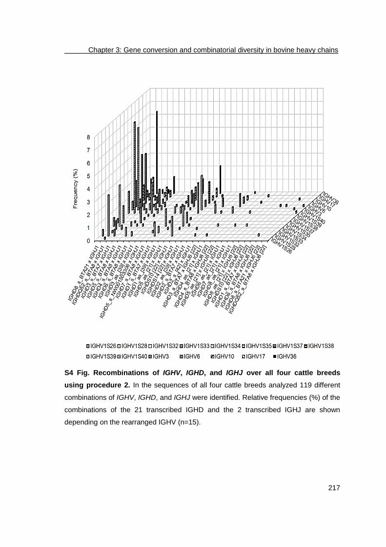
Chapter 3: Gene conversion and combinatorial diversity in bovine heavy chains
217
S4 Fig. Recombinations of IGHV, IGHD, and IGHJ over all four cattle breeds
using procedure 2. In the sequences of all four cattle breeds analyzed 119 different
combinations of IGHV, IGHD, and IGHJ were identified. Relative frequencies (%) of the
combinations of the 21 transcribed IGHD and the 2 transcribed IGHJ are shown
depending on the rearranged IGHV (n=15).

Chapter 3: Gene conversion and combinatorial diversity in bovine heavy chains
218
S1 Table. Transcription frequencies of IGHD in four cattle breeds using
procedure 1.
IGHD A
(%; n=137)
GS
(%; n=116)
GBP
(%; n=111)
HF
(%; n=145)
IGHD1_as1_[39] 2.19 0.86 0.00 0.00
IGHD1_s2_BTA7 6.57 2.59 7.21 5.52
IGHD2_as_[39] 0.00 0.00 0.90 0.69
IGHD2_s_BTA7 2.92 5.17 4.50 2.07
IGHD2_s_[39] 0.73 1.72 2.70 1.38
IGHD3_as_[39] 0.00 0.00 0.00 0.69
IGHD3_s_BTA7 14.60 11.21 9.91 13.79
IGHD4_as_[21] 2.92 0.86 2.70 2.07
IGHD4_s_BTA8 10.95 6.03 2.70 11.72
IGHD5_as_[21] 0.73 0.00 0.00 0.00
IGHD5_s_BTA8 15.33 13.79 17.12 17.24
IGHD5_s_NW001503306 4.38 3.45 12.61 6.90
IGHD6_s_BTA8 0.00 0.86 0.00 0.69
IGHD7_as_[21] 0.00 0.00 0.90 0.00
IGHD7_s_BTA8 9.49 12.93 6.31 4.83
IGHD8_as_[21] 0.73 0.86 0.00 0.69
IGHD8_s_BTA21 11.68 25.86 15.32 15.86
IGHDQ52_s_BTA8 10.95 3.45 9.91 9.66
IGHDS10 [22] 3.65 7.76 4.50 4.14
IGHDS14 [22] 2.19 2.59 2.70 2.07 1 antisense 2 sense

Chapter 3: Gene conversion and combinatorial diversity in bovine heavy chains
219
S2 Table. Transcription frequencies of IGHD in four cattle breeds using
procedure 2.
1 antisense 2 sense
IGHD
A
(%; n=137)
GS
(%; n=116)
GBP
(%; n=111)
HF
(%; n=145)
IGHD1_as1_[39] 2.19 0.86 2.70 2.07
IGHD1_s2_BTA7 9.49 8.62 7.21 6.21
IGHD2_s_BTA7 2.19 0.86 1.80 1.38
IGHD2_s_[39] 0.00 0.00 0.90 0.69
IGHD3_s_BTA7 2.92 2.59 4.50 4.14
IGHD4_as_[21] 3.65 2.59 0.00 2.76
IGHD4_s_BTA8 12.41 12.07 4.50 13.10
IGHD5_s_BTA8 1.46 3.45 4.50 0.69
IGHD5_s_NW001503306 0.00 0.86 1.80 0.69
IGHD6_as_[21] 0.00 0.00 0.90 0.00
IGHD6_s_BTA8 2.19 0.86 2.70 2.07
IGHD7_s_BTA8 0.00 0.86 0.90 2.76
IGHD8_as_[21] 0.00 0.00 0.90 0.00
IGHD8_s_BTA21 8.76 12.07 11.71 8.28
IGHDQ52_s_BTA8 37.96 42.24 39.64 36.55
IGHDS10 [22] 16.79 11.21 15.32 18.62
IGHDS14 [22] 0.00 0.86 0.00 0.00

Chapter 3: Gene conversion and combinatorial diversity in bovine heavy chains
220
S3 Table. Recombinations of IGHV, IGHD, and IGHJ over all four cattle breeds
using procedure 3.
IGHV - IGHD - IGHJ
all
(n=597)
A
(n=167)
GBP
(n=140)
GS
(n=131)
HF
(n=159)
IGHV1S26 - IGHD8_s1_BTA21 - IGHJ1 0.17 0.00 0.00 0.76 0.00
IGHV1S28 - IGHD1_as2_[39] - IGHJ1 0.34 0.00 1.43 0.00 0.00
IGHV1S28 - IGHD1_s_BTA7 - IGHJ1 0.50 1.20 0.00 0.00 0.63
IGHV1S28 - IGHD2_s_[39] - IGHJ1 0.17 0.00 0.00 0.76 0.00
IGHV1S28 - IGHD3_as_[39] - IGHJ1 0.17 0.60 0.00 0.00 0.00
IGHV1S28 - IGHD3_s_BTA7 - IGHJ1 1.01 1.20 2.14 0.00 0.63
IGHV1S28 - IGHD4_s_BTA8 - IGHJ6_[22] 0.17 0.00 0.00 0.00 0.63
IGHV1S28 - IGHD4_s_BTA8 - IGHJ1 1.17 1.20 1.43 0.76 1.26
IGHV1S28 - IGHD5_s_BTA8 - IGHJ1 1.17 1.20 0.71 2.29 0.63
IGHV1S28 - IGHD5_s_NW_001503306 - IGHJ1 0.34 0.60 0.00 0.76 0.00
IGHV1S28 - IGHD6_s_BTA8 - IGHJ1 0.17 0.00 0.71 0.00 0.00
IGHV1S28 - IGHD7_s_BTA8 - IGHJ1 0.84 0.00 1.43 1.53 0.63
IGHV1S28 - IGHD8_s_BTA21 - IGHJ1 0.34 0.00 0.00 0.76 0.63
IGHV1S28 - IGHDQ52_s_BTA8 - IGHJ1 0.84 0.00 0.71 0.76 1.89
IGHV1S28 - IGHDS10 [22] - IGHJ1 0.67 0.00 0.00 0.00 2.52
IGHV1S28 - IGHDS14 [22] - IGHJ1 0.34 0.60 0.00 0.76 0.00
IGHV1S32 - IGHD2_s_[39] - IGHJ1 0.17 0.00 0.00 0.76 0.00
IGHV1S32 - IGHD8_s_BTA21 - IGHJ1 0.34 0.00 0.00 1.53 0.00
IGHV1S32 - IGHDS10 [22] - IGHJ1 0.17 0.00 0.00 0.00 0.63
IGHV1S32 - IGHDS14 [22] - IGHJ1 0.17 0.00 0.00 0.00 0.63
IGHV1S33 - IGHD3_as_[39] - IGHJ1 0.17 0.60 0.00 0.00 0.00
IGHV1S33 - IGHD3_s_BTA7 - IGHJ1 0.34 1.20 0.00 0.00 0.00
IGHV1S33 - IGHD4_s_BTA8 - IGHJ1 0.17 0.00 0.71 0.00 0.00
IGHV1S33 - IGHD5_s_BTA8 - IGHJ1 0.17 0.00 0.00 0.76 0.00
IGHV1S33 - IGHD8_s_BTA21 - IGHJ1 0.17 0.60 0.00 0.00 0.00
IGHV1S33 - IGHDQ52_s_BTA8 - IGHJ1 0.34 1.20 0.00 0.00 0.00
IGHV1S33 - IGHDS14 [22] - IGHJ1 0.17 0.00 0.00 0.76 0.00
IGHV1S34 - IGHD1_as_[39] - IGHJ1 0.17 0.60 0.00 0.00 0.00
IGHV1S34 - IGHD1_s_BTA7 - IGHJ1 1.34 0.60 3.57 0.76 0.63
IGHV1S34 - IGHD2_s_BTA7 - IGHJ1 0.34 0.60 0.00 0.00 0.63
IGHV1S34 - IGHD2_s_[39] - IGHJ1 0.17 0.00 0.00 0.76 0.00
IGHV1S34 - IGHD3_s_BTA7 - IGHJ1 0.84 0.00 2.14 0.76 0.63
IGHV1S34 - IGHD4_s_BTA8 - IGHJ1 0.67 0.60 1.43 0.00 0.63
IGHV1S34 - IGHD5_s_BTA8 - IGHJ1 1.01 0.60 1.43 0.76 1.26
IGHV1S34 - IGHD5_s_NW_001503306 - IGHJ1 0.34 0.00 0.71 0.00 0.63
IGHV1S34 - IGHD6_s_BTA8 - IGHJ1 0.17 0.00 0.00 0.76 0.00
IGHV1S34 - IGHD7_s_BTA8 - IGHJ1 0.34 0.60 0.71 0.00 0.00
IGHV1S34 - IGHD8_s_BTA21 - IGHJ1 1.01 0.00 0.71 3.82 0.00
IGHV1S34 - IGHDQ52_s_BTA8 - IGHJ1 1.51 2.40 2.14 0.76 0.63

Chapter 3: Gene conversion and combinatorial diversity in bovine heavy chains
221
IGHV1S34 - IGHDS10 [22] - IGHJ1 0.34 0.00 0.71 0.00 0.63
IGHV1S34 - IGHDS14 [22] - IGHJ1 0.34 0.00 0.00 1.53 0.00
IGHV1S35 - IGHD1_s_BTA7 - IGHJ1 0.17 0.00 0.00 0.00 0.63
IGHV1S35 - IGHD2_s_[39] - IGHJ1 0.17 0.00 0.00 0.76 0.00
IGHV1S35 - IGHD3_s_BTA7 - IGHJ1 0.50 0.00 0.71 1.53 0.00
IGHV1S35 - IGHD4_as_[21] - IGHJ1 0.34 0.60 0.00 0.76 0.00
IGHV1S35 - IGHD4_s_BTA8 - IGHJ1 0.34 0.60 0.00 0.76 0.00
IGHV1S35 - IGHD5_s_BTA8 - IGHJ1 0.50 0.00 0.00 2.29 0.00
IGHV1S35 - IGHD7_s_BTA8 - IGHJ1 1.01 0.00 0.71 3.82 0.00
IGHV1S35 - IGHD8_s_BTA21 - IGHJ1 0.67 1.20 0.00 1.53 0.00
IGHV1S35 - IGHDQ52_s_BTA8 - IGHJ1 0.17 0.60 0.00 0.00 0.00
IGHV1S35 - IGHDS10 [22] - IGHJ1 0.17 0.00 0.00 0.00 0.63
IGHV1S37 - IGHD4_s_BTA8 - IGHJ1 0.17 0.00 0.00 0.00 0.63
IGHV1S37 - IGHDQ52_s_BTA8 - IGHJ1 0.17 0.00 0.00 0.00 0.63
IGHV1S38 - IGHD3_s_BTA7 - IGHJ1 0.17 0.00 0.00 0.76 0.00
IGHV1S38 - IGHDQ52_s_BTA8 - IGHJ1 0.17 0.00 0.00 0.00 0.63
IGHV1S39 - IGHD1_as_[39] - IGHJ1 0.34 0.60 0.00 0.00 0.63
IGHV1S39 - IGHD1_s_BTA7 - IGHJ1 1.01 1.20 1.43 0.00 1.26
IGHV1S39 - IGHD2_as_[39] - IGHJ1 0.34 1.20 0.00 0.00 0.00
IGHV1S39 - IGHD2_s_BTA7 - IGHJ1 0.34 0.60 0.00 0.00 0.63
IGHV1S39 - IGHD2_s_[39] - IGHJ1 0.84 0.00 1.43 0.76 1.26
IGHV1S39 - IGHD3_s_BTA7 - IGHJ6_[22] 0.17 0.00 0.00 0.00 0.63
IGHV1S39 - IGHD3_s_BTA7 - IGHJ1 1.01 1.20 1.43 0.00 1.26
IGHV1S39 - IGHD4_as_[21] - IGHJ1 0.50 0.60 0.00 0.76 0.63
IGHV1S39 - IGHD4_s_BTA8 - IGHJ1 1.68 3.59 0.71 0.00 1.89
IGHV1S39 - IGHD5_s_BTA8 - IGHJ1 2.85 7.78 0.71 0.00 1.89
IGHV1S39 - IGHD5_s_NW_001503306 - IGHJ1 1.17 1.20 2.86 0.76 0.00
IGHV1S39 - IGHD6_as_[21] - IGHJ1 0.34 0.00 0.00 0.00 1.26
IGHV1S39 - IGHD6_s_BTA8 - IGHJ1 0.34 0.00 0.00 0.00 1.26
IGHV1S39 - IGHD7_s_BTA8 - IGHJ1 1.68 2.40 0.71 0.76 2.52
IGHV1S39 - IGHD8_s_BTA21 - IGHJ1 2.18 4.79 1.43 0.00 1.89
IGHV1S39 - IGHDQ52_s_BTA8 - IGHJ1 1.34 2.99 0.00 0.00 1.89
IGHV1S39 - IGHDS10 [22] - IGHJ1 1.01 1.20 2.86 0.00 0.00
IGHV1S39 - IGHDS14 [22] - IGHJ1 0.50 1.20 0.00 0.00 0.63
IGHV1S40 - IGHD1_s_BTA7 - IGHJ1 0.17 0.00 0.00 0.00 0.63
IGHV1S40 - IGHD2_s_BTA7 - IGHJ1 0.17 0.60 0.00 0.00 0.00
IGHV1S40 - IGHD2_s_[39] - IGHJ1 0.17 0.00 0.00 0.76 0.00
IGHV1S40 - IGHD3_as_[39] - IGHJ1 0.17 0.60 0.00 0.00 0.00
IGHV1S40 - IGHD3_s_BTA7 - IGHJ1 2.35 0.00 2.86 4.58 2.52
IGHV1S40 - IGHD4_as_[21] - IGHJ1 0.17 0.60 0.00 0.00 0.00
IGHV1S40 - IGHD4_s_BTA8 - IGHJ1 0.84 0.60 1.43 0.00 1.26
IGHV1S40 - IGHD5_s_BTA8 - IGHJ1 3.02 2.99 3.57 4.58 1.26
IGHV1S40 - IGHD5_s_NW_001503306 - IGHJ1 0.50 0.60 0.71 0.00 0.63
IGHV1S40 - IGHD6_as_[21] - IGHJ1 0.17 0.00 0.71 0.00 0.00

Chapter 3: Gene conversion and combinatorial diversity in bovine heavy chains
222
IGHV1S40 - IGHD6_s_BTA8 - IGHJ1 0.17 0.60 0.00 0.00 0.00
IGHV1S40 - IGHD7_s_BTA8 - IGHJ1 0.84 0.00 1.43 1.53 0.63
IGHV1S40 - IGHD8_s_BTA21 - IGHJ1 1.34 1.20 2.14 0.76 1.26
IGHV1S40 - IGHDQ52_s_BTA8 - IGHJ1 1.51 0.60 1.43 2.29 1.89
IGHV1S40 - IGHDS10 [22] - IGHJ6_[22] 0.17 0.00 0.71 0.00 0.00
IGHV1S40 - IGHDS10 [22] - IGHJ1 0.50 0.00 0.71 0.76 0.63
IGHV3 - IGHD1_as_[39] - IGHJ1 0.34 0.60 0.00 0.76 0.00
IGHV3 - IGHD1_s_BTA7 - IGHJ1 1.51 0.00 2.14 0.76 3.14
IGHV3 - IGHD2_as_[39] - IGHJ1 0.50 1.20 0.00 0.76 0.00
IGHV3 - IGHD2_s_BTA7 - IGHJ1 0.50 0.60 0.00 0.76 0.63
IGHV3 - IGHD2_s_[39] - IGHJ1 0.34 0.00 0.71 0.00 0.63
IGHV3 - IGHD3_as_[39] - IGHJ1 0.17 0.60 0.00 0.00 0.00
IGHV3 - IGHD3_s_BTA7 - IGHJ1 1.34 1.20 2.14 0.76 1.26
IGHV3 - IGHD3_s_BTA7 - IGHJ6_[22] 0.34 0.00 0.00 0.76 0.63
IGHV3 - IGHD4_as_[21] - IGHJ1 0.50 0.00 0.71 0.00 1.26
IGHV3 - IGHD4_s_BTA8 - IGHJ1 0.50 1.20 0.71 0.00 0.00
IGHV3 - IGHD5_as_[21] - IGHJ1 0.17 0.60 0.00 0.00 0.00
IGHV3 - IGHD5_s_BTA8 - IGHJ1 2.01 1.20 1.43 2.29 3.14
IGHV3 - IGHD5_s_NW_001503306 - IGHJ1 0.50 0.60 0.00 0.00 1.26
IGHV3 - IGHD7_s_BTA8 - IGHJ1 0.50 0.60 1.43 0.00 0.00
IGHV3 - IGHD8_as_[21] IGHJ6_[22] 0.17 0.60 0.00 0.00 0.00
IGHV3 - IGHD8_s_BTA21 - IGHJ1 1.68 0.60 0.71 0.76 4.40
IGHV3 - IGHDQ52_s_BTA8 - IGHJ1 1.84 0.00 0.71 3.05 3.77
IGHV3 - IGHDS10 [22] - IGHJ1 1.01 2.40 0.71 0.76 0.00
IGHV3 - IGHDS14 [22] - IGHJ1 0.50 0.00 1.43 0.76 0.00
IGHV6 - IGHD1_as_[39] - IGHJ1 0.17 0.60 0.00 0.00 0.00
IGHV6 - IGHD1_s_BTA7 - IGHJ1 0.67 0.60 1.43 0.00 0.63
IGHV6 - IGHD2_s_BTA7 - IGHJ1 0.50 1.80 0.00 0.00 0.00
IGHV6 - IGHD2_s_[39] - IGHJ1 0.17 0.00 0.00 0.76 0.00
IGHV6 - IGHD3_as_[39] - IGHJ1 0.34 0.00 0.71 0.76 0.00
IGHV6 - IGHD3_s_BTA7 - IGHJ1 0.84 0.60 1.43 0.00 1.26
IGHV6 - IGHD4_as_[21] - IGHJ1 0.34 0.60 0.00 0.00 0.63
IGHV6 - IGHD4_s_BTA8 - IGHJ1 1.01 1.20 0.00 0.76 1.89
IGHV6 - IGHD5_s_BTA8 - IGHJ1 0.84 0.60 1.43 0.00 1.26
IGHV6 - IGHD5_s_NW_001503306 - IGHJ1 0.50 0.60 0.71 0.76 0.00
IGHV6 - IGHD7_s_BTA8 - IGHJ1 0.50 0.60 0.71 0.00 0.63
IGHV6 - IGHD8_s_BTA21 IGHJ1 1.34 1.80 0.71 3.05 0.00
IGHV6 - IGHDQ52_s_BTA8 - IGHJ1 1.34 1.80 1.43 0.00 1.89
IGHV6 - IGHDS10 [22] - IGHJ1 0.34 0.00 0.71 0.00 0.63
IGHV6 - IGHDS14 [22] - IGHJ1 0.34 0.00 0.00 1.53 0.00
IGHV10 - IGHD2_as_[39] - IGHJ1 0.34 1.20 0.00 0.00 0.00
IGHV10 - IGHD1_s_BTA7 - IGHJ1 0.34 0.60 0.00 0.00 0.63
IGHV10 - IGHD2_s_BTA7 - IGHJ1 0.50 1.20 0.00 0.76 0.00
IGHV10 - IGHD2_s_[39] - IGHJ1 1.01 0.60 0.00 2.29 1.26

Chapter 3: Gene conversion and combinatorial diversity in bovine heavy chains
223
1 sense 2 antisense
IGHV10 - IGHD3_s_BTA7 - IGHJ1 0.67 0.60 0.00 0.76 1.26
IGHV10 - IGHD4_s_BTA8 - IGHJ1 0.17 0.60 0.00 0.00 0.00
IGHV10 - IGHD5_as_[21] - IGHJ6_[22] 0.17 0.00 0.00 0.00 0.63
IGHV10 - IGHD5_s_BTA8 - IGHJ1 0.17 0.00 0.00 0.00 0.63
IGHV10 - IGHD5_s_NW_001503306 - IGHJ1 0.17 0.00 0.00 0.00 0.63
IGHV10 - IGHD7_as_[21] - IGHJ1 0.17 0.00 0.71 0.00 0.00
IGHV10 - IGHD7_s_BTA8 - IGHJ1 0.17 0.00 0.00 0.76 0.00
IGHV10 - IGHD8_as_[21] - IGHJ1 0.34 0.00 0.71 0.00 0.63
IGHV10 - IGHD8_s_BTA21 - IGHJ1 0.17 0.60 0.00 0.00 0.00
IGHV10 - IGHDQ52_s_BTA8 - IGHJ1 0.17 0.60 0.00 0.00 0.00
IGHV10 - IGHDS10 [22] - IGHJ1 0.50 0.00 0.00 2.29 0.00
IGHV17 - IGHD1_s_BTA7 - IGHJ1 0.50 0.60 1.43 0.00 0.00
IGHV17 - IGHD3_s_BTA7 - IGHJ1 1.01 1.80 0.71 1.53 0.00
IGHV17 - IGHD4_as_[21] - IGHJ1 0.17 0.60 0.00 0.00 0.00
IGHV17 - IGHD4_s_BTA8 - IGHJ1 0.34 0.60 0.71 0.00 0.00
IGHV17 - IGHD5_s_BTA8 - IGHJ1 0.50 0.00 1.43 0.00 0.63
IGHV17 - IGHD5_s_NW_001503306 - IGHJ1 0.17 0.00 0.71 0.00 0.00
IGHV17 - IGHD7_s_BTA8 - IGHJ1 0.34 0.00 0.71 0.00 0.63
IGHV17 - IGHD8_s_BTA21 - IGHJ1 0.34 0.00 0.00 1.53 0.00
IGHV17 - IGHDQ52_s_BTA8 - IGHJ1 0.50 1.20 0.00 0.00 0.63
IGHV17 - IGHDS10 [22] - IGHJ1 0.34 0.60 0.71 0.00 0.00
IGHV36 - IGHD1_as_[39] - IGHJ1 0.17 0.60 0.00 0.00 0.00
IGHV36 - IGHD1_s_BTA7 - IGHJ1 0.34 0.60 0.00 0.00 0.63
IGHV36 - IGHD3_as_[39] - IGHJ1 0.50 0.60 0.71 0.76 0.00
IGHV36 - IGHD3_s_BTA7 - IGHJ1 1.51 0.00 2.14 0.00 3.77
IGHV36 - IGHD4_s_BTA8 - IGHJ1 0.84 0.00 0.00 1.53 1.89
IGHV36 - IGHD5_as_[21] - IGHJ1 0.17 0.00 0.00 0.76 0.00
IGHV36 - IGHD5_s_BTA8 - IGHJ1 1.51 1.80 2.86 0.76 0.63
IGHV36 - IGHD5_s_NW_001503306 - IGHJ1 0.50 0.00 0.71 0.00 1.26
IGHV36 - IGHD6_s_BTA8 - IGHJ1 0.67 0.00 1.43 0.00 1.26
IGHV36 - IGHD7_s_BTA8 - IGHJ1 0.50 0.60 0.00 0.76 0.63
IGHV36 - IGHD8_s_BTA21 - IGHJ1 4.52 1.20 5.00 10.69 2.52
IGHV36 - IGHDQ52_s_BTA8 - IGHJ1 1.84 1.80 3.57 0.76 1.26
IGHV36 - IGHDS10 [22] - IGHJ1 0.17 0.00 0.00 0.76 0.00
IGHV36 - IGHDS14 [22] - IGHJ1 0.17 0.60 0.00 0.00 0.00

Chapter 3: Gene conversion and combinatorial diversity in bovine heavy chains
224
S4 Table. Recombinations of IGHV, IGHD, and IGHJ over all four cattle breeds using procedure 1.
IGHV - IGHD - IGHJ all
(n=597)
GS
(n=131)
GBP
(140)
A
(n=167)
HF
(n=159)
IGHV1S26 - IGHDS14 [22] - IGHJ1 0,17 0,76 0,00 0,00 0,00
IGHV1S28 - IGHD1_as1_[39] - IGHJ1 0,17 0,00 0,00 0,60 0,00
IGHV1S28 - IGHD1_s2_BTA7 - IGHJ1 0,34 0,00 0,71 0,60 0,00
IGHV1S28 - IGHD2_s_BTA7 - IGHJ1 0,17 0,00 0,71 0,00 0,00
IGHV1S28 - IGHD2_s_[39] - IGHJ1 0,17 0,00 0,71 0,00 0,00
IGHV1S28 - IGHD3_s_BTA7 - IGHJ1 0,67 0,76 0,00 0,60 1,26
IGHV1S28 - IGHD4_s_BTA8 - IGHJ6_[22] 0,17 0,00 0,00 0,00 0,63
IGHV1S28 - IGHD4_s_BTA8 - IGHJ1 0,50 0,00 0,00 0,60 1,26
IGHV1S28 - IGHD5_s_BTA8 - IGHJ1 2,18 3,82 2,86 0,00 2,52
IGHV1S28 - IGHD5_s_NW_001503306 - IGHJ1 0,84 0,76 0,71 1,20 0,63
IGHV1S28 - IGHD7_s_BTA8 - IGHJ1 0,50 0,76 1,43 0,00 0,00
IGHV1S28 - IGHD8_as_[21] - IGHJ1 0,17 0,00 0,00 0,00 0,63
IGHV1S28 - IGHD8_s_BTA21 - IGHJ1 1,17 1,53 0,71 1,20 1,26
IGHV1S28 - IGHDQ52_s_BTA8 - IGHJ1 0,84 0,76 0,71 1,20 0,63
IGHV1S28 - IGHDS10 [22] - IGHJ1 0,34 0,00 0,00 0,60 0,63
IGHV1S32 - IGHD3_s_BTA7 - IGHJ1 0,17 0,76 0,00 0,00 0,00
IGHV1S32 - IGHD5_s_NW_001503306 - IGHJ1 0,17 0,00 0,00 0,00 0,63
IGHV1S32 - IGHD8_s_BTA21 - IGHJ1 0,34 1,53 0,00 0,00 0,00
IGHV1S32 - IGHDQ52_s_BTA8 - IGHJ1 0,17 0,00 0,00 0,00 0,63
IGHV1S33 - IGHD1_s_BTA7 - IGHJ1 0,17 0,00 0,00 0,60 0,00
IGHV1S33 - IGHD2_s_BTA7 - IGHJ1 0,17 0,00 0,00 0,60 0,00
IGHV1S33 - IGHD3_s_BTA7 - IGHJ1 0,17 0,00 0,00 0,60 0,00
IGHV1S33 - IGHD4_s_BTA8 IGHJ1 0,17 0,76 0,00 0,00 0,00
IGHV1S33 - IGHD5_as_[21] - IGHJ1 0,17 0,00 0,00 0,60 0,00
IGHV1S33 - IGHD5_s_BTA8 - IGHJ1 0,34 0,00 0,71 0,60 0,00
IGHV1S33 - IGHD7_s_BTA8 - IGHJ1 0,17 0,76 0,00 0,00 0,00
IGHV1S33 - IGHDS14 [22] - IGHJ1 0,17 0,00 0,00 0,60 0,00
IGHV1S34 - IGHD1_s_BTA7 - IGHJ1 0,50 0,76 0,71 0,60 0,00
IGHV1S34 - IGHD2_s_BTA7 - IGHJ1 0,17 0,00 0,00 0,60 0,00
IGHV1S34 - IGHD2_s_[39] - IGHJ1 0,34 0,00 0,71 0,60 0,00
IGHV1S34 - IGHD3_s_BTA7 - IGHJ1 0,84 0,76 1,43 0,60 0,63
IGHV1S34 - IGHD4_as_[21] - IGHJ1 0,17 0,00 0,00 0,00 0,63
IGHV1S34 - IGHD4_s_BTA8 - IGHJ1 0,67 0,00 2,14 0,00 0,63
IGHV1S34 - IGHD5_s_BTA8 - IGHJ1 2,01 3,05 3,57 1,20 0,63
IGHV1S34 - IGHD5_s_NW_001503306 - IGHJ1 0,50 0,00 1,43 0,00 0,63
IGHV1S34 - IGHD7_s_BTA8 - IGHJ1 0,84 0,76 0,00 1,20 1,26
IGHV1S34 - IGHD8_s_BTA21 - IGHJ1 1,17 3,82 0,71 0,00 0,63
IGHV1S34 - IGHDQ52_s_BTA8 - IGHJ1 0,67 0,00 2,14 0,60 0,00
IGHV1S34 - IGHDS10 [22] - IGHJ1 0,50 0,76 0,00 0,60 0,63
IGHV1S34 - IGHDS14 [22] - IGHJ1 0,17 0,00 0,71 0,00 0,00

Chapter 3: Gene conversion and combinatorial diversity in bovine heavy chains
225
IGHV1S35 - IGHD2_s_BTA7 - IGHJ1 0,17 0,76 0,00 0,00 0,00
IGHV1S35 - IGHD2_s_[39] - IGHJ1 0,17 0,76 0,00 0,00 0,00
IGHV1S35 - IGHD3_s_BTA7 - IGHJ1 0,50 0,76 0,00 0,60 0,63
IGHV1S35 - IGHD4_s_BTA8 - IGHJ1 0,34 0,76 0,00 0,00 0,63
IGHV1S35 - IGHD5_s_BTA8 - IGHJ1 0,67 1,53 0,00 1,20 0,00
IGHV1S35 - IGHD5_s_NW_001503306 - IGHJ1 0,17 0,76 0,00 0,00 0,00
IGHV1S35 - IGHD7_s_BTA8 - IGHJ1 0,84 3,05 0,71 0,00 0,00
IGHV1S35 - IGHD8_s_BTA21 - IGHJ1 0,34 0,76 0,71 0,00 0,00
IGHV1S35 - IGHDQ52_s_BTA8 - IGHJ1 0,34 0,00 0,00 1,20 0,00
IGHV1S35 - IGHDS10 [22] - IGHJ1 0,50 2,29 0,00 0,00 0,00
IGHV1S37 - IGHDQ52_s_BTA8 - IGHJ1 0,34 0,00 0,00 0,00 1,26
IGHV1S38 - IGHD7_s_BTA8 - IGHJ1 0,17 0,76 0,00 0,00 0,00
IGHV1S38 - IGHDQ52_s_BTA8 - IGHJ1 0,17 0,00 0,00 0,00 0,63
IGHV1S39 - IGHD1_as_[39] - IGHJ1 0,34 0,00 0,00 1,20 0,00
IGHV1S39 - IGHD1_s_BTA7 - IGHJ1 1,84 0,76 2,86 2,40 1,26
IGHV1S39 - IGHD2_s_BTA7 - IGHJ1 0,17 0,00 0,71 0,00 0,00
IGHV1S39 - IGHD2_s_[39] - IGHJ1 0,34 0,00 0,00 0,00 1,26
IGHV1S39 - IGHD3_s_BTA7 - IGHJ6_[22] 0,17 0,00 0,00 0,00 0,63
IGHV1S39 - IGHD3_s_BTA7 - IGHJ1 3,02 0,76 2,14 5,99 2,52
IGHV1S39 - IGHD4_as_[21] - IGHJ1 0,84 0,76 0,00 1,80 0,63
IGHV1S39 - IGHD4_s_BTA8 - IGHJ1 1,34 0,00 0,00 3,59 1,26
IGHV1S39 - IGHD5_s_BTA8 - IGHJ1 2,68 0,00 2,86 5,39 1,89
IGHV1S39 - IGHD5_s_NW_001503306 - IGHJ1 1,01 0,76 1,43 1,20 0,63
IGHV1S39 - IGHD6_s_BTA8 - IGHJ1 0,17 0,00 0,00 0,00 0,63
IGHV1S39 - IGHD7_as_[21] - IGHJ1 0,17 0,00 0,71 0,00 0,00
IGHV1S39 - IGHD7_s_BTA8 - IGHJ1 1,68 0,00 0,00 3,59 2,52
IGHV1S39 - IGHD8_s_BTA21 - IGHJ1 1,68 0,00 0,71 1,20 4,40
IGHV1S39 - IGHDQ52_s_BTA8 - IGHJ1 0,84 0,00 1,43 1,20 0,63
IGHV1S39 - IGHDS10 [22] - IGHJ1 0,67 0,00 0,00 1,20 1,26
IGHV1S39 - IGHDS14 [22] - IGHJ1 0,67 0,00 0,71 1,80 0,00
IGHV1S40 - IGHD1_s_BTA7 - IGHJ1 0,34 0,00 0,00 0,00 1,26
IGHV1S40 - IGHD2_s_BTA7 - IGHJ6_[22] 0,17 0,00 0,71 0,00 0,00
IGHV1S40 - IGHD2_s_BTA7 - IGHJ1 0,17 0,76 0,00 0,00 0,00
IGHV1S40 - IGHD2_s_[39] - IGHJ1 0,17 0,00 0,71 0,00 0,00
IGHV1S40 - IGHD3_s_BTA7 - IGHJ1 1,17 2,29 1,43 0,60 0,63
IGHV1S40 - IGHD4_s_BTA8 - IGHJ1 0,67 0,76 0,00 1,20 0,63
IGHV1S40 - IGHD5_as_[21] - IGHJ1 0,17 0,00 0,00 0,60 0,00
IGHV1S40 - IGHD5_s_BTA8 - IGHJ1 2,35 0,76 1,43 2,40 4,40
IGHV1S40 - IGHD5_s_NW_001503306 - IGHJ1 0,84 0,76 2,14 0,60 0,00
IGHV1S40 - IGHD7_s_BTA8 - IGHJ1 1,68 4,58 1,43 1,20 0,00
IGHV1S40 - IGHD8_s_BTA21 - IGHJ1 2,85 4,58 3,57 1,20 2,52
IGHV1S40 - IGHDQ52_s_BTA8 - IGHJ1 0,84 0,00 2,14 0,60 0,63
IGHV1S40 - IGHDS10 [22] - IGHJ1 0,84 0,76 2,14 0,00 0,63
IGHV3 - IGHD1_as_[39] - IGHJ1 0,17 0,76 0,00 0,00 0,00

Chapter 3: Gene conversion and combinatorial diversity in bovine heavy chains
226
IGHV3 - IGHD1_s_BTA7 - IGHJ1 0,67 0,00 1,43 0,00 1,26
IGHV3 - IGHD2_as_[39] - IGHJ1 0,34 0,00 0,71 0,00 0,63
IGHV3 - IGHD2_s_BTA7 - IGHJ1 0,84 1,53 0,71 0,00 1,26
IGHV3 - IGHD3_s_BTA7 - IGHJ6_[22] 0,17 0,00 0,00 0,00 0,63
IGHV3 - IGHD3_s_BTA7 - IGHJ1 1,17 1,53 2,14 0,60 0,63
IGHV3 - IGHD4_as_[21] - IGHJ6_[22] 0,17 0,00 0,00 0,60 0,00
IGHV3 - IGHD4_as_[21] - IGHJ1 0,67 0,00 1,43 1,20 0,00
IGHV3 - IGHD4_s_BTA8 - IGHJ1 1,84 0,76 0,71 2,40 3,14
IGHV3 - IGHD5_s_BTA8 - IGHJ1 2,01 2,29 2,14 0,00 3,77
IGHV3 - IGHD5_s_NW_001503306 - IGHJ1 0,34 0,00 0,71 0,00 0,63
IGHV3 - IGHD7_s_BTA8 - IGHJ1 0,17 0,76 0,00 0,00 0,00
IGHV3 - IGHD8_as_[21] - IGHJ1 0,34 0,76 0,00 0,60 0,00
IGHV3 - IGHD8_s_BTA21 - IGHJ6_[22] 0,17 0,76 0,00 0,00 0,00
IGHV3 - IGHD8_s_BTA21 - IGHJ1 2,35 0,76 1,43 2,99 3,77
IGHV3 - IGHDQ52_s_BTA8 - IGHJ1 2,18 0,00 1,43 2,99 3,77
IGHV3 - IGHDS10 [22] - IGHJ1 0,67 2,29 0,00 0,60 0,00
IGHV3 - IGHDS14 [22] - IGHJ1 0,17 0,00 0,00 0,00 0,63
IGHV6 - IGHD1_s_BTA7 - IGHJ1 1,01 1,53 0,71 1,80 0,00
IGHV6 - IGHD2_s_BTA7 - IGHJ1 0,17 0,00 0,00 0,60 0,00
IGHV6 - IGHD2_s_[39] - IGHJ1 0,17 0,00 0,00 0,60 0,00
IGHV6 - IGHD3_as_[39] - IGHJ1 0,17 0,00 0,00 0,00 0,63
IGHV6 - IGHD3_s_BTA7 - IGHJ1 1,68 0,00 0,71 2,40 3,14
IGHV6 - IGHD4_as_[21] - IGHJ1 0,34 0,00 0,00 0,00 1,26
IGHV6 - IGHD4_s_BTA8 - IGHJ1 0,50 0,00 1,43 0,00 0,63
IGHV6 - IGHD5_s_BTA8 - IGHJ1 1,34 0,76 2,86 1,20 0,63
IGHV6 - IGHD5_s_NW_001503306 - IGHJ1 0,50 0,76 0,71 0,60 0,00
IGHV6 - IGHD7_s_BTA8 - IGHJ1 0,50 0,00 0,00 0,60 1,26
IGHV6 - IGHD8_s_BTA21 - IGHJ1 1,34 3,82 0,71 0,60 0,63
IGHV6 - IGHDQ52_s_BTA8 - IGHJ1 1,17 0,76 1,43 1,80 0,63
IGHV6 - IGHDS10 [22] - IGHJ1 0,34 0,00 0,71 0,60 0,00
IGHV10 - IGHD1_s_BTA7 - IGHJ1 0,67 0,76 0,00 1,20 0,63
IGHV10 - IGHD2_s_BTA7 - IGHJ1 0,50 1,53 0,00 0,60 0,00
IGHV10 - IGHD3_s_BTA7 - IGHJ1 0,34 0,76 0,00 0,60 0,00
IGHV10 - IGHD4_as_[21] - IGHJ1 0,50 0,00 0,71 1,20 0,00
IGHV10 - IGHD4_s_BTA8 - IGHJ1 0,84 0,76 0,00 1,20 1,26
IGHV10 - IGHD5_s_BTA8 - IGHJ6_[22] 0,17 0,00 0,00 0,00 0,63
IGHV10 - IGHD5_s_BTA8 - IGHJ1 0,17 0,00 0,00 0,60 0,00
IGHV10 - IGHD5_s_NW_001503306 - IGHJ1 0,17 0,00 0,00 0,00 0,63
IGHV10 - IGHD8_s_BTA21 - IGHJ1 1,17 1,53 0,71 0,60 1,89
IGHV10 - IGHDQ52_s_BTA8 - IGHJ1 0,17 0,76 0,00 0,00 0,00
IGHV10 - IGHDS14 [22] - IGHJ1 0,34 0,76 0,00 0,00 0,63
IGHV17 - IGHD1_s_BTA7 - IGHJ1 0,50 0,76 1,43 0,00 0,00
IGHV17 - IGHD3_s_BTA7 - IGHJ1 0,84 0,76 0,71 1,80 0,00
IGHV17 - IGHD4_s_BTA8 - IGHJ1 0,50 0,00 1,43 0,00 0,63

Chapter 3: Gene conversion and combinatorial diversity in bovine heavy chains
227
IGHV17 - IGHD5_s_BTA8 - IGHJ1 0,34 0,00 0,71 0,60 0,00
IGHV17 - IGHD5_s_NW_001503306 - IGHJ1 0,17 0,00 0,71 0,00 0,00
IGHV17 - IGHD7_s_BTA8 - IGHJ1 0,67 0,76 0,00 0,60 1,26
IGHV17 - IGHD8_s_BTA21 - IGHJ1 0,50 0,76 0,00 1,20 0,00
IGHV17 - IGHDQ52_s_BTA8 - IGHJ1 0,50 0,00 0,71 1,20 0,00
IGHV17 - IGHDS14 [22] - IGHJ1 0,17 0,00 0,71 0,00 0,00
IGHV36 - IGHD1_s_BTA7 - IGHJ1 0,34 0,00 0,71 0,00 0,63
IGHV36 - IGHD2_s_BTA7 - IGHJ1 0,50 0,00 0,71 0,60 0,63
IGHV36 - IGHD2_s_[39] - IGHJ1 0,17 0,76 0,00 0,00 0,00
IGHV36 - IGHD3_s_BTA7 - IGHJ1 1,84 0,76 1,43 1,80 3,14
IGHV36 - IGHD4_s_BTA8 - IGHJ1 0,67 1,53 0,00 0,00 1,26
IGHV36 - IGHD5_s_BTA8 - IGHJ1 2,01 0,76 2,86 2,40 1,89
IGHV36 - IGHD5_s_NW_001503306 - IGHJ1 1,84 0,00 3,57 0,60 3,14
IGHV36 - IGHD6_s_BTA8 - IGHJ1 0,17 0,76 0,00 0,00 0,00
IGHV36 - IGHD7_s_BTA8 - IGHJ1 0,50 0,00 1,43 0,60 0,00
IGHV36 - IGHD8_s_BTA21 - IGHJ1 3,52 9,16 3,57 1,80 0,63
IGHV36 - IGHDQ52_s_BTA8 - IGHJ1 1,01 1,53 1,43 0,00 1,26
IGHV36 - IGHDS10 [22] - IGHJ1 0,50 0,76 0,71 0,00 0,63
IGHV36 - IGHDS14 [22] - IGHJ1 0,34 0,76 0,00 0,00 0,63 1 antisense 2 sense

Chapter 3: Gene conversion and combinatorial diversity in bovine heavy chains
228
S5 Table. Recombinations of IGHV, IGHD, and IGHJ over all four cattle breeds
using procedure 2.
IGHV - IGHD - IGHJ all
(n=597)
GS
(n=131)
GBP
(n=140)
A
(n=167)
HF
(n=159)
IGHV1S26 - IGHDQ52_s1_BTA8 - IGHJ1 0,17 0,76 0,00 0,00 0,00
IGHV1S28 - IGHD1_s_BTA7 - IGHJ1 1,01 0,00 2,14 1,20 0,63
IGHV1S28 - IGHD2_s_[39] - IGHJ1 0,17 0,00 0,00 0,00 0,63
IGHV1S28 - IGHD3_s_BTA7 - IGHJ1 0,50 0,76 0,71 0,00 0,63
IGHV1S28 - IGHD4_as2_[21] - IGHJ1 0,17 0,00 0,00 0,60 0,00
IGHV1S28 - IGHD4_s_BTA8 - IGHJ1 0,67 1,53 0,71 0,60 0,00
IGHV1S28 - IGHD6_s_BTA8 - IGHJ6_[22] 0,17 0,00 0,00 0,00 0,63
IGHV1S28 - IGHD6_s_BTA8 - IGHJ1 0,34 0,00 0,71 0,00 0,63
IGHV1S28 - IGHD8_s_BTA21 - IGHJ1 0,50 2,29 0,00 0,00 0,00
IGHV1S28 - IGHDQ52_s_BTA8 - IGHJ1 3,18 3,05 2,86 2,40 4,40
IGHV1S28 - IGHDS10 [22] - IGHJ1 1,51 0,76 1,43 1,80 1,89
IGHV1S32 - IGHD1_s_BTA7 - IGHJ1 0,50 1,53 0,00 0,00 0,63
IGHV1S32 - IGHD7_s_BTA8 - IGHJ1 0,17 0,00 0,00 0,00 0,63
IGHV1S32 - IGHDQ52_s_BTA8 - IGHJ1 0,17 0,76 0,00 0,00 0,00
IGHV1S33 - IGHD1_as_[39] - IGHJ1 0,17 0,00 0,00 0,60 0,00
IGHV1S33 - IGHD2_s_BTA7 - IGHJ1 0,17 0,76 0,00 0,00 0,00
IGHV1S33 - IGHD3_s_BTA7 - IGHJ1 0,34 0,00 0,00 1,20 0,00
IGHV1S33 - IGHDQ52_s_BTA8 - IGHJ1 0,67 0,76 0,71 1,20 0,00
IGHV1S33 - IGHDS10 [22] - IGHJ1 0,17 0,00 0,00 0,60 0,00
IGHV1S34 - IGHD1_as_[39] - IGHJ1 0,17 0,00 0,71 0,00 0,00
IGHV1S34 - IGHD1_s_BTA7 - IGHJ1 1,34 0,76 1,43 2,99 0,00
IGHV1S34 - IGHD4_as_[21] - IGHJ1 0,17 0,00 0,00 0,60 0,00
IGHV1S34 - IGHD4_s_BTA8 - IGHJ1 0,17 0,00 0,71 0,00 0,00
IGHV1S34 - IGHD5_s_BTA8 - IGHJ1 0,34 0,76 0,71 0,00 0,00
IGHV1S34 - IGHD5_s_NW_001503306 - IGHJ1 0,34 0,76 0,71 0,00 0,00
IGHV1S34 - IGHD6_as_[21] - IGHJ1 0,17 0,00 0,71 0,00 0,00
IGHV1S34 - IGHD6_s_BTA8 - IGHJ1 0,17 0,00 0,71 0,00 0,00
IGHV1S34 - IGHD8_as_[21] - IGHJ1 0,17 0,00 0,71 0,00 0,00
IGHV1S34 - IGHD8_s_BTA21 - IGHJ1 0,67 1,53 0,71 0,00 0,63
IGHV1S34 - IGHDQ52_s_BTA8 - IGHJ1 3,18 5,34 2,86 1,80 3,14
IGHV1S34 - IGHDS10 [22] - IGHJ1 1,51 0,00 3,57 0,60 1,89
IGHV1S34 - IGHDS14 [22] - IGHJ1 0,17 0,76 0,00 0,00 0,00
IGHV1S35 - IGHD1_s_BTA7 - IGHJ1 0,17 0,76 0,00 0,00 0,00
IGHV1S35 - IGHD4_s_BTA8 - IGHJ1 1,17 3,82 0,71 0,00 0,63
IGHV1S35 - IGHD5_s_BTA8 - IGHJ1 0,17 0,76 0,00 0,00 0,00
IGHV1S35 - IGHD8_s_BTA21 - IGHJ1 0,34 1,53 0,00 0,00 0,00
IGHV1S35 - IGHDQ52_s_BTA8 - IGHJ1 1,34 3,82 0,00 1,20 0,63
IGHV1S35 - IGHDS10 [22] - IGHJ1 0,84 0,76 0,71 1,80 0,00
IGHV1S37 - IGHD4_s_BTA8 - IGHJ1 0,17 0,00 0,00 0,00 0,63

Chapter 3: Gene conversion and combinatorial diversity in bovine heavy chains
229
IGHV1S37 - IGHDS10 [22] - IGHJ1 0,17 0,00 0,00 0,00 0,63
IGHV1S38 - IGHD4_s_BTA8 - IGHJ1 0,17 0,00 0,00 0,00 0,63
IGHV1S38 - IGHD6_s_BTA8 - IGHJ1 0,17 0,76 0,00 0,00 0,00
IGHV1S39 - IGHD1_s_BTA7 - IGHJ1 0,50 0,76 0,00 1,20 0,00
IGHV1S39 - IGHD2_s_BTA7 - IGHJ1 0,67 0,00 0,71 1,20 0,63
IGHV1S39 - IGHD2_s_[39] - IGHJ1 0,17 0,00 0,71 0,00 0,00
IGHV1S39 - IGHD3_s_BTA7 - IGHJ1 0,84 0,00 1,43 1,20 0,63
IGHV1S39 - IGHD4_as_[21] - IGHJ1 0,50 0,00 0,00 1,80 0,00
IGHV1S39 - IGHD4_s_BTA8 - IGHJ1 2,35 0,00 0,71 5,39 2,52
IGHV1S39 - IGHD5_s_BTA8 - IGHJ1 0,34 0,76 0,00 0,60 0,00
IGHV1S39 - IGHD6_as_[21] - IGHJ1 0,17 0,00 0,71 0,00 0,00
IGHV1S39 - IGHD6_s_BTA8 - IGHJ1 0,34 0,00 0,00 1,20 0,00
IGHV1S39 - IGHD7_s_BTA8 - IGHJ1 0,50 0,00 0,71 0,00 1,26
IGHV1S39 - IGHD8_s_BTA21 - IGHJ1 1,68 0,00 2,14 2,40 1,89
IGHV1S39 - IGHDQ52_s_BTA8 - IGHJ6_[22] 0,17 0,00 0,00 0,00 0,63
IGHV1S39 - IGHDQ52_s_BTA8 - IGHJ1 7,04 1,53 5,00 11,38 8,81
IGHV1S39 - IGHDS10 [22] - IGHJ1 2,35 0,00 1,43 4,19 3,14
IGHV1S40 - IGHD1_as_[39] - IGHJ1 0,17 0,00 0,00 0,60 0,00
IGHV1S40 - IGHD1_s_BTA7 - IGHJ1 0,34 0,00 1,43 0,00 0,00
IGHV1S40 - IGHD3_s_BTA7 - IGHJ1 0,50 1,53 0,71 0,00 0,00
IGHV1S40 - IGHD4_s_BTA8 - IGHJ1 0,50 0,00 0,00 0,00 1,89
IGHV1S40 - IGHD5_s_BTA8 - IGHJ1 0,50 0,76 1,43 0,00 0,00
IGHV1S40 - IGHD6_s_BTA8 - IGHJ1 0,34 0,00 0,71 0,60 0,00
IGHV1S40 - IGHD8_s_BTA21 - IGHJ6_[22] 0,17 0,00 0,71 0,00 0,00
IGHV1S40 - IGHD8_s_BTA21 - IGHJ1 0,67 0,76 0,00 1,80 0,00
IGHV1S40 - IGHDQ52_s_BTA8 - IGHJ1 6,37 6,87 9,29 3,59 6,29
IGHV1S40 - IGHDS10 [22] - IGHJ1 2,68 5,34 1,43 1,80 2,52
IGHV3 - IGHD1_as_[39] - IGHJ1 1,01 0,00 1,43 1,20 1,26
IGHV3 - IGHD1_s_BTA7 - IGHJ6_[22] 0,17 0,76 0,00 0,00 0,00
IGHV3 - IGHD1_s_BTA7 - IGHJ1 0,84 1,53 0,00 0,60 1,26
IGHV3 - IGHD2_s_BTA7 - IGHJ1 0,67 0,76 0,71 0,60 0,63
IGHV3 - IGHD4_as_[21] - IGHJ1 1,01 1,53 0,00 1,20 1,26
IGHV3 - IGHD4_s_BTA8 - IGHJ1 1,34 1,53 0,00 0,60 3,14
IGHV3 - IGHD5_s_BTA8 - IGHJ1 0,17 0,00 0,71 0,00 0,00
IGHV3 - IGHD5_s_NW_001503306 - IGHJ1 0,17 0,00 0,00 0,00 0,63
IGHV3 - IGHD6_s_BTA8 - IGHJ1 0,34 0,00 0,00 0,60 0,63
IGHV3 - IGHD7_s_BTA8 - IGHJ1 0,17 0,76 0,00 0,00 0,00
IGHV3 - IGHD8_s_BTA21 - IGHJ1 2,01 1,53 2,86 0,60 3,14
IGHV3 - IGHDQ52_s_BTA8 - IGHJ6_[22] 0,17 0,00 0,00 0,00 0,63
IGHV3 - IGHDQ52_s_BTA8 - IGHJ1 4,02 2,29 5,71 4,19 3,77
IGHV3 - IGHDS10 [22] - IGHJ6_[22] 0,17 0,00 0,00 0,60 0,00
IGHV3 - IGHDS10 [22] - IGHJ1 2,18 1,53 1,43 1,80 3,77
IGHV6 - IGHD1_s_BTA7 - IGHJ1 1,01 0,00 0,71 2,99 0,00
IGHV6 - IGHD3_s_BTA7 - IGHJ1 0,50 0,00 1,43 0,00 0,63

Chapter 3: Gene conversion and combinatorial diversity in bovine heavy chains
230
IGHV6 - IGHD4_as_[21] - IGHJ1 0,50 0,00 0,00 0,60 1,26
IGHV6 - IGHD4_s_BTA8 - IGHJ1 0,34 0,00 0,00 0,60 0,63
IGHV6 - IGHD5_s_BTA8 - IGHJ1 0,50 0,76 0,71 0,00 0,63
IGHV6 - IGHD6_as_[21] - IGHJ1 0,17 0,00 0,71 0,00 0,00
IGHV6 - IGHD7_s_BTA8 - IGHJ1 0,17 0,00 0,00 0,00 0,63
IGHV6 - IGHD8_s_BTA21 - IGHJ1 0,34 0,00 0,71 0,00 0,63
IGHV6 - IGHDQ52_s_BTA8 - IGHJ1 3,85 6,11 3,57 4,19 1,89
IGHV6 - IGHDS10 [22] - IGHJ1 1,68 0,00 1,43 2,40 2,52
IGHV6 - IGHDS14 [22] - IGHJ1 0,17 0,76 0,00 0,00 0,00
IGHV10 - IGHD1_as_[39] - IGHJ1 0,17 0,00 0,00 0,00 0,63
IGHV10 - IGHD1_s_BTA7 - IGHJ1 1,01 2,29 0,00 1,20 0,63
IGHV10 - IGHD3_s_BTA7 - IGHJ1 0,34 0,00 0,00 0,00 1,26
IGHV10 - IGHD4_as_[21] - IGHJ1 0,34 0,00 0,00 1,20 0,00
IGHV10 - IGHD4_s_BTA8 - IGHJ1 1,01 2,29 0,00 1,20 0,63
IGHV10 - IGHD6_s_BTA8 - IGHJ6_[22] 0,17 0,00 0,00 0,00 0,63
IGHV10 - IGHD8_s_BTA21 - IGHJ1 1,17 1,53 0,71 2,40 0,00
IGHV10 - IGHDQ52_s_BTA8 - IGHJ1 0,50 0,76 0,00 0,00 1,26
IGHV10 - IGHDS10 [22] - IGHJ1 0,34 0,00 0,71 0,00 0,63
IGHV17 - IGHD3_s_BTA7 - IGHJ1 0,34 0,00 0,71 0,60 0,00
IGHV17 - IGHD4_s_BTA8 - IGHJ1 0,50 0,00 0,00 1,80 0,00
IGHV17 - IGHD5_s_BTA8 - IGHJ1 0,34 0,76 0,71 0,00 0,00
IGHV17 - IGHD6_as_[21] - IGHJ1 0,17 0,00 0,71 0,00 0,00
IGHV17 - IGHD8_s_BTA21 - IGHJ1 0,50 0,00 0,71 0,00 1,26
IGHV17 - IGHDQ52_s_BTA8 - IGHJ1 1,51 0,76 2,86 1,80 0,63
IGHV17 - IGHDS10 [22] - IGHJ1 0,67 0,76 0,71 1,20 0,00
IGHV17 - IGHDS14 [22] - IGHJ1 0,17 0,76 0,00 0,00 0,00
IGHV36 - IGHD1_as_[39] - IGHJ1 0,17 0,76 0,00 0,00 0,00
IGHV36 - IGHD1_s_BTA7 - IGHJ1 1,17 0,00 0,71 0,60 3,14
IGHV36 - IGHD3_s_BTA7 - IGHJ1 0,34 0,00 0,71 0,00 0,63
IGHV36 - IGHD4_as_[21] - IGHJ1 0,17 0,76 0,00 0,00 0,00
IGHV36 - IGHD4_s_BTA8 - IGHJ1 1,34 1,53 0,71 0,60 2,52
IGHV36 - IGHD5_s_BTA8 - IGHJ1 0,34 0,00 0,71 0,60 0,00
IGHV36 - IGHD5_s_NW_001503306 - IGHJ1 0,17 0,00 0,71 0,00 0,00
IGHV36 - IGHD8_s_BTA21 - IGHJ1 1,17 1,53 1,43 0,00 1,89
IGHV36 - IGHDQ52_s_BTA8 - IGHJ1 6,37 11,45 7,14 4,19 3,77
IGHV36 - IGHDS10 [22] - IGHJ1 2,18 0,76 4,29 1,80 1,89 1 sense 2 antisense

Chapter 4: General Discussion
231
Chapter 4:
General Discussion
Chapter 4: General Discussion
232
General Discussion
Since the resistance of antigens to antibiotics remains a significant problem in livestock
production due to considerable economic impacts, research in the alternate treatment
of infectious diseases increased. The investigation of special genetic aspects of
immunoglobulins has attracted higher attention during the last few decades. New high
throughput technologies are available that facilitate and advance the experimental
workflow. The price for analyses of the immunoglobulin repertoires is descending.
Initial studies on the immunoglobulin repertoire were performed on humans and mice
but successful techniques were applied to various other animals such as chicken,
rabbit, cattle, horses, and zebrafish, too. Nevertheless, investigation in immunoglobulin
genetics still needs more attention to complete previous information about
fundamentals, such as the number of genomic available gene segments, gene families,
and allotypes of different isotypes of the immunoglobulin heavy and light chains in
cattle and horses.
The primary objective of this thesis was to analyze the genomic available and
transcribed immunoglobulin heavy and light chain gene segments to contribute to a
more detailed understanding of immunoglobulin diversity in cattle and horses.
Similar to most vertebrates, bovine and equine immunoglobulins possess two identical
heavy and light chains that consist of a variable region and a constant region. The
variable regions are created by the random fusion of germline variable
(IGHV/IGLV/IGKV), diversity (IGHD), and joining gene segments (IGHJ/IGLJ/IGKJ) that
are combined with a constant region gene (IGHC/IGLC/IGKC) (Tonegawa 1983). The
gene segments are found in species-specific numbers in the genome. The
immunoglobulin repertoire is generated by several processes of combinatorial and
junctional diversity comprising the imprecise joining of the single gene segments and
non-templated or palindromic nucleotide insertions between two adjacent gene
segments, as well as somatic hypermutations. Secondary mechanisms such as gene
conversion and isotype switch may increase the diversity further. Highly specialized,
complementarity determining regions form a perfect counterpart to the antigen epitope
and are stabilized by conserved framework regions (Kabat and Wu 1991).
In mammals, two types of light chains are described – the lambda and kappa light
chains (Korngold and Lipari 1956). They are expressed in species-specific ratios. In
Chapter 4: General Discussion
233
contrast to humans and mice, where the kappa isotype dominates in serum antibodies,
the lambda isotype is predominantly found in cattle and horses (Arun et al. 1996;
Almagro et al. 1998). In addition to what is already known about the general structural
features of both bovine and equine immunoglobulins and their function, this thesis on
diversity gave some important baseline information and contributes to the scientific
background for the production of highly specific and effective recombinant antibodies
or antibody libraries. The findings will also be of future importance in analyzing
seroconversion data after infection or vaccination, as well as determining breed
specific differences to select healthy, robust animals.
Since the late 19th century, horse antibodies in terms of anti-sera were closely
connected with the treatment of human diseases. Today, equine immunoglobulins are
also used to support immunosuppression after organ or stem cell transplantation, or to
manage autoimmune diseases in humans (Leleu et al. 2006; Zand 2006). In addition,
equine antibody applications gain in importance in order to prevent or treat equine
infectious diseases. Amongst others they can be regarded as potential alternatives to
antibiotic therapy in the near future. Therefore it is necessary to investigate the
fundamental immunoglobulin genetics underlying the equine immunoglobulin immune
response.
The understanding of the organization of equine immunoglobulin genes has increased
significantly in recent years (reviewed in publication 1). For equine heavy chains, 52
IGHV, 40 IGHD, 8 IGHJ and 11 IGHC were determined. With 40 IGHD identified,
horses belong to the mammalian species that possess the most IGHD. Likewise, in
guinea pig and the African elephant 41 and 87 IGHD gene segments are known so far
(Guo et al. 2011; Guo et al. 2012). Seven of the equine IGHCs are gamma chain
genes (IgG). Only in the porcine immune system a similarly high number of eleven
genomic constant region genes representing six putative subclasses has been
described (Butler et al. 2009).
Using biochemical and serological methods, the subclasses IgGa, IgGb, IgGc, IgG(T),
and IgG(B) were identified and characterized (Rockey et al. 1964; Sandor et al. 1964;
Klinman et al. 1965; Rockey 1967; Widders et al. 1986; Sheoran and Holmes 1996).
Later, seven IgG isotypes were identified by the analysis of a BAC library. The previous
subclasses were renamed. Multiple duplications, gene conversions, and crossovers
are supposed to explain these seven IgG isotypes (Wagner et al. 2006). The individual

Chapter 4: General Discussion
234
IgG subclasses possess specific roles in protective immunity. Thus, the FcγR and
complement-binding isotypes IgG1, IgG4, and IgG7 (former IgGa and IgGb) contribute
to the protection against several equine pathogens such as equine influenza virus
(Nelson et al. 1998; Breathnach et al. 2006), Streptococcus equi (Sheoran et al. 1997)
and Rhodococcus equi (Lopez et al. 2002). In addition, systemic and mucosal IgG
responses were described to play an important role in limiting the spread and severity
of equine herpes virus 1 (Kydd et al. 2006). The first recombinant versions of all seven
equine IgG subclasses (mouse -light chains, horse IGHG1-7) were expressed in
Chinese Hamster Ovary cells to analyze their individual physical and biological
properties (Lewis et al. 2008). Further, seven distinct monoclonal equine antibodies
(IgM, IgG1, IgG3, IgG4/7, IgG5, IgG6, and IgE) were produced in equine-murine
heterohybridomas to be used for quantification of isotypes in diagnostic testing and
immunological research (Keggan et al. 2013).
The combinatorial and junctional levels of IGHV-IGHD-IGHJ sequences in equine fetus
were comparable to those of adult horses and a similar set of variable gene segments
was used during fetal and post-natal life stages (Tallmadge et al. 2009; Tallmadge et
al. 2013). Few IGHV segments were used predominantly at all ages. The 250,000
bases between the expressed gene segments suggest that the entire IGHV locus is
available throughout equine life (Tallmadge et al. 2013). Similarly, in fetal piglets the
IGHV usage was described to be independent of the genome position although there is
a limited number of IGHV representing the Ig repertoire, too (Eguchi-Ogawa et al.
2010; Butler et al. 2011). In contrast, the biased usage of germline IGHV in the mouse
fetus was explained with different accessible positions within the IGHV locus (Jeong et
al. 1988). The incorporation of more than one IGHD into the IGHV-IGHD-IGHJ
rearrangement resulting in IGHV-IGHD-IGHD-IGHJ was observed (Sun et al. 2010). In
horses, sequence diversity and length variation further increased in complementarity
determining region 1 (CDR1H) and CDR2H, and framework regions in accordance to
somatic hypermutation (Tallmadge et al. 2013). Within the CDR3H of an adult horse, at
least 5 amino acid residues were observed, whereas 25 amino acid residues were
counted in the longest CDR3H, which was identified in an equine neonate (Tallmadge
et al. 2013). In comparison, recent studies showed the length heterogeneity of very
short CDR3H (5-10 amino acids), midlength CDR3H (11-31 amino acids) and
exceptionally long CDR3H (more than 47 amino acids) in all isotypes in both bovine
fetuses and adult cattle. Very long CDR3H contribute to diversity by uniquely folded

Chapter 4: General Discussion
235
small domains (Saini et al. 1999a; Saini and Kaushik 2002; Shojaei et al. 2003; Koti et
al. 2010; Walther et al. 2013; Wang et al. 2013).
The equine kappa light chain contains 60 IGKV, 5 IGKJ and 1 IGKC, whereas there are
144 IGLV, 7 IGLJ, and 7 IGLC for the lambda light chain. Each of the 7 IGLJ is
preceded by one IGLC and there are two clusters of IGLV possessing different
transcriptional orientations. Within each IGLV cluster there are functional genes and
pseudogenes (Sun et al. 2010). Similarly, pseudogenes are described in different
species such as chicken, where it is already known that pseudogenes are used for
gene conversion (Reynaud et al. 1985). During age development, a decrease in IGLVs
is noted, although nucleotide diversity and significant differences in gene usage
increased. As in all previous studies different methods were used for designation, the
standardization of the existing nomenclature of immunoglobulin genes is suggested.
The study of allotypic variants within the equine λ-light chain locus (publication 2) was
the first detailed molecular genetic description. Five IGLC1, four IGLC5, and two
IGLC6/7 allotypic variants, as well as three alleles for IGLC6/7a were observed.
Especially for the transcriptional use of IGLC6/7, significant differences were calculated
by Chi² tests.
Several amino acid residue variations were located at the accessible surface as
confirmed by homology-based predicted 3D structural analysis. Distinct serological
properties resulting in allotypic variants due to these modifications are assumed. All
allotypic variants showed distinct amino acid residue substitutions in all seven β-sheets
according to the IMGT nomenclature (Lefranc et al. 2005). Most of them were located
within the solvent accessible surface area (Padlan et al. 1986). Seven of these
substitutions were positioned at the interface to the first constant region of the heavy
chain (IGHC1). As one of these amino acid substitutions affected one of the conserved
residues for the main interaction of constant region of the light chain (IGLC) and
IGHC1, the stable assembly of heavy and light chains could be changed. We do not
have any information about allelic changes in IGHC1, thus this assumption remains to
be verified. Amino acid residue substitutions located at the solvent accessible surface
area might be distinguished by specific sera and therefore might be used as markers.
Previously, allotypic markers of human immunoglobulin chains were related to the
susceptibility of different infectious diseases due to either direct association or linkage
disequilibrium with the causative gene (Pandey et al. 1979; Granoff et al. 1984; Pandey

Chapter 4: General Discussion
236
et al. 1995; Pandey 2000; Giha et al. 2009). Furthermore, influences of bovine
allotypes on effector functions like complement activation, and age dependent
expression have also been shown (Corbeil et al. 1997; Bastida-Corcuera et al. 1999a;
Bastida-Corcuera et al. 1999b; Bastida-Corcuera et al. 2000). Specific investigations
on functional differences of allotypic variants are missing in horses, but similar to cattle
heterozygotic horses are thought to possess an advantage in their humoral immune
response.
Beside the presence of predicted germline IGLJ-IGLC pairs and the transcription
frequency of IGLC-genes, the usage of IGLV-segments of subgroup 8 was further
analyzed. This subgroup contains the highest number of functional IGLV and was
shown to be transcribed preferentially (Sun et al. 2010). The variable segments
IGLV15, IGLV17, and IGLV25 were preferably transcribed. Significant differences were
calculated for the rearrangements with the four IGLC-isotypes transcribed within and
among the breeds. Consequently, the V-domain repertoire is dominated by two breed
independent IGLV genes and a third breed specific IGLV gene that are productively
used for combinatorial joining of IGLJ-IGLC-genes and thus for antibody production.
Furthermore, two putative pseudogenes (IGLV74ψ, IGLV101ψ) were transcribed. The
isolated sequences showed variations in CDR1 sequences resulting in a frame shift
replacing the premature stop codons of germline IGLV74ψ. Individual mutations within
the breeds are supposed to enable the transcriptional use of pseudogenes. In addition,
in silico germline sequences are prepared from a Thoroughbred, which is a conserved
breed founded by few stallions. Hence, mutations resulting in pseudogenes may be
segregate within this breed but can be functional in other breeds. The transcription of
pseudo V-gene segments has already been described in species such as cattle,
chicken, and rabbits and occurs by gene conversion (Parng et al. 1996; Winstead et al.
1999; Arakawa and Buerstedde 2009). The results of this study indicated the presence
of similar mechanisms in horses. Moreover, the spacer sequences of the
recombination signal sequences of the transcribed IGLVs seem to be conserved (Sun
et al. 2010). In IGLV101ψ, the spacer sequence is identical to that in IGLV17 and
IGLV15, which are the dominantly used IGLV. These identical sequences of the
spacers are an additional option for the transcription of potential pseudogenes as was
already shown in human IGKV segments (Nadel et al. 1998).
Some of the results could be linked to breeding because two breeds with different stud
book sizes and breeding goals were examined. While Rhenish German Coldblood is a
Chapter 4: General Discussion
237
quiet conserved and small breed mainly used for agricultural purposes and breed
representations (Bremond and Balzer 2011), animals of Hanoverian Warmblood
belong to one of the biggest horse breeds and participate in all disciplines of equestrian
sports (Bremond and Balzer 2011). Although the inbreeding coefficient in the
Hanoverian population (1.33%) is at lower level than those of smaller populations like
the Rhenish German Coldblood (1.73%; (Biedermann et al. 2002; Hamann and Distl
2008), effective management of breeding is necessary to prevent the random loss of
alleles caused by large genetic contributions of few individuals over a long time period
(bottleneck effect). Genetic drift and homogenization of genetic information may also
occur and have to be controlled as the rate of inbreeding has increased in Hanoverians
in the last ten years (Constans et al. 1985; Hamann and Distl 2008; Simianer and Kohn
2010).
This study characterized genetic and transcriptional differences of λ-light chain in two
horse breeds. For the first time distinct alleles and putative allotypic variants were
described in horses. Future investigations should evaluate the distribution of allotypic
markers of immunoglobulin chains in further horse breeds. Moreover, the linkage to
infectious diseases such as in humans and cattle should be examined.
The in silico analyses of the genomic organization of the bovine heavy-chain locus
revealed differences from previous mapping and annotation results (publication 3).
The functional locus was mapped to BTA21 (Gu et al. 1992; Hayes et al. 2000; Zhao et
al. 2003). Additional bovine heavy-chain loci were detected on BTA7, BTA8, and
BTA20, unexpectedly. The order of the respective segments deviated from other fully
described mammalian loci (Matsuda and Honjo 1996; Sun et al. 2010). Further, the
constant heavy-chain locus described by means of BAC clone analysis (Zhao et al.
2003), were not introduced into the genomic assembly. Several genes upstream of the
heavy-chain locus on the bovine contig-NW_003064289 on BTA7 share homology with
HSA19 but some did not show any human equivalent. Similarly, on HSA5pter no
equivalent was found to the bovine IgG2(ORF) gene, although the adjacent genes
were identified. Hybridization experiments assigned IGHML1 to BTA11 (Tobin-Janzen
and Womack 1992; Hayes and Petit 1993), which was supported by the detection of
six IGHJ segments on the same chromosome using BAC clone and locus-specific PCR
analysis (Hosseini et al. 2004). Nevertheless, in silico analyses did not identify an IgM-
like locus on BTA11. Consequently, the bovine immunoglobulin heavy-chain locus

Chapter 4: General Discussion
238
seems to be annotated incorrectly and incompletely. This problem may be solved by
the re-sequencing of the described localizations and underpinned by different authors
and methods.
All of the functional bovine IGHV segments are most closely related to the human clan
II family VH2, which explains the exclusive transcription of only one bovine VH family
(Sinclair and Aitken 1995; Berens et al. 1997; Sinclair et al. 1997). The second bovine
VH family consists only of pseudogenes and clustered with the human VH4 and VH6
family. The description of the second bovine VH family (boVH2) previously proposed
was now possible (Berens et al. 1997), but there are still no indications of possible
gene conversions using boVH2 segments (Liljavirta et al. 2014) in the sequences
investigated, as shown for the bovine λ-light chains (Parng et al. 1996). For further
studies the assignment of fragmented loci of bovine IGHV to boVH1 or boVH2 based
on sequence identity is recommended.
In bovine IgM, three groups of CDR3H defined by their number of amino acids were
described. Independent of nucleotide addition during rearrangement long and short
IGHD contribute directly to CDR3H length heterogeneity. Nevertheless, annotation of
the transcribed IGHD is complicated due to high homologies among the genomic IGHD
segments. In particular, CDR3H length heterogeneity, junctional flexibility, and somatic
hypermutation within the recombined IGHD segments led to low sequence identities.
Furthermore, intrinsic hot spots targeting somatic hypermutations to CDR1H, CDR2H,
and CDR3H were thought to contribute solely to IgM antibody diversification in both
bovine fetus and adult cattle (Saini and Kaushik 2002).
CDR3H length heterogeneity was fond in all bovine immunoglobulin isotypes. Hence,
exceptionally long CDR3H are apparently not primarily generated to compensate the
restricted flexibility of IgM. As the most recent study on the IgG repertoire in calves also
showed exceptionally long CDR3H (Larsen and Smith 2012), antigen selection of
variable domains and class switch recombination seem to be of higher impact. The
exceptionally long CDR3H were generated by the direct fusion of a single IGHV
segment (IGHV10), the longest IGHD segment (IGHD2), and one functional IGHJ
segment (IGHJ1), as described previously (Koti et al. 2010). In short CDR3H, the
preferred use of the short IGHD segments, IGHDQ52 and IGHD4 was observed. All
IGHV segments identified were found to be functional. Thus, analyses do not reveal
evidence for gene conversion in bovine immunoglobulin heavy chains, which
contributes to the diversity of chicken immunoglobulin heavy chains and bovine λ-light
Chapter 4: General Discussion
239
chains (Reynaud et al. 1994; Parng et al. 1996). Both in intermediate and exceptionally
long CDR3H conserved short nucleotide sequences (CSNS) were inserted into the
IGHV-IGHD junction. This mechanism contributes to antibody diversification in all
immunoglobulin heavy chain isotypes and CDR3H lengths. As the insertion of CSNS is
supposed to directly follow antigen exposure (Koti et al. 2010), class switch
recombination is supposed to be responsible for isotype-independent exceptionally
long CDR3H in cattle. In addition, exceptionally long CDR3H do not have a
conventional antigen binding site because these CDR3H protrude from the variable
domain with support from the λ-light chains. Antigen binding is undertaken by side
chains that are exclusively contained within long CDR3H regions, as investigated by
structural comparisons with protein toxins (Ramsland et al. 2001).
As described in humans (Corbett et al. 1997), mice (Kaartinen and Mäkelä 1985),
chicken (Reynaud et al. 1994), and rabbits (Friedman et al. 1994), mainly hydrophilic
amino acid residues such as Tyr, Ser, and Gly were identified in the bovine
exceptionally long CDR3H. The occurrence of the hydrophilic reading frame in antigen-
binding loops is thought to enhance flexibility and recruit somatic hypermutations for
advantageous antigen binding (Corbett et al. 1997).
In accordance with previous descriptions, multiple and mainly even numbered Cys
accumulated in the middle of the exceptionally long CDR3H were identified (Saini et al.
1999a; Kaushik et al. 2009). They are predicted to form intra and inter CDRH disulfide
bonds that rigidify the combining site or help to stabilize long CDR3H, as demonstrated
in the crystallized human Fab Kol (Schmidt et al. 1983), the camel cAb-Lys3 single
domain antibody (Desmyter et al. 1996), and the bovine Fabs BLV1H12 and BLV5B8
(Wang et al. 2013). Former observations of an additional Cys in CDR2H when there
were only one or three Cys in CDR3H (Saini et al. 1999a), or at least one Cys in
CDR3H regions containing more than 12 amino acid residues and no Cys in CDR3H
with less than ten amino acids (Lopez et al. 1998) were verified with some
reservations. The numbering system had an influence on the number of additional Cys
in CDR2H and some sequences with intermediate CDR3H lengths did not possess any
Cys.
The bovine immunoglobulin heavy-chain locus was annotated, the expression of
exceptionally long CDR3H in the five bovine immunoglobulin isotypes was
demonstrated, and their genomic origin was specified. Thus, this study reviewed the
opinion that exceptionally long CDR3H are a unique feature of bovine IgM and IgG1-3.

Chapter 4: General Discussion
240
The new bioinformatics framework enables more detailed analyses of bovine
immunoglobulin heavy chains and substantially contributes to the understanding of the
development of the transcribed bovine immunoglobulin repertoire (publication 4). It is
now possible to analyze the unique exceptionally long CDR3H group of bovine
immunoglobulin heavy chains. Possible gene conversions within the variable region of
bovine heavy chains were examined, too. Further, the dominantly transcribed IGHV,
IGHD, and IGHJ gene segments and their combinatorial diversity were investigated. In
contrast to previous studies, this investigation extended analyses to breed specific
differences in the four cattle breeds Aubrac (A), German Simmental (GS), German
Black Pied (GBP), and Holstein Friesian (HF).
Recent studies on bovine immunoglobulin genetics focused on antibody diversification
strategies and the junctional diversity of the antibody repertoire. Bovine specific
diversification mechanisms were identified such as the generation of exceptionally long
CDR3H (Saini et al. 1999b; Walther et al. 2013; Wang et al. 2013), the insertion of
conserved short nucleotide sequences (CSNS) at the IGHV-IGHD junction (Koti et al.
2010), the use of pseudogene fragments in lambda light chains, as well as gene
conversions (Parng et al. 1996), and somatic hypermutations independent of exposure
to external antigens during B-cell development in lambda light chains (Lucier et al.
1998). In addition, new germline gene segments were determined in the recent years
(Niku et al. 2012; Walther et al. 2013; Liljavirta et al. 2014). Nevertheless, the current
bovine genome assembly is still incomplete and the full germline repertoire remains
under active investigation.
Previous analyses of rearranged immunoglobulin germline gene segments applied
various software tools for sequence alignments. Due to the difficult and error prone
manual assembly of different genes, specialized software tools have been developed
such as IMGT/Junction Analysis (Yousfi Monod et al. 2004), IMGT/V-QUEST (Brochet
et al. 2008; Giudicelli et al. 2011), IMTG/HighV-QUEST (Alamyar et al. 2012), VBASE2
(Retter et al. 2005), JoinSolver (Souto-Carneiro et al. 2004), iHMMun-align (Gaeta et
al. 2007), and IgBLAST (Ye et al. 2013). Only IgBLAST provides different settings for
nucleotide and protein sequences, the numbering system of Kabat or the IMGT system
(Lefranc et al. 2003; Kabat et al. 2006), and germline gene database searches (Ye et
al. 2013). Matching germline IGHV, IGHD, and IGHJ genes, as well as details at
rearrangement junctions may be analyzed. Organisms such as human, mouse, rat,

Chapter 4: General Discussion
241
rabbit and rhesus monkey are supported but the representation of livestock is missing
or incomplete. In particular, the bovine specific occurrence of exceptionally long
CDR3H is not taken into account. For more detailed analyses of the origin of the
immunoglobulin diversity considering species-specific diversification mechanisms, a
new bioinformatics framework was developed. Three different procedures were applied
to improve biological significance. Consequently, the program is based on nucleotide
sequences, searches our updated bovine specific immunoglobulin germline gene
database, and is able to load other pre-designed databases. The currently
recommended and most widely used IMGT nomenclature is used for the delineation of
FR and CDR. The adjustment of search parameters for IGHV an IGHJ and especially
for the identification of IGHD is also possible.
The analysis of Ig heavy chain variable regions in the four cattle breeds revealed the
usage of 15 different IGHV segments, 21 IGHD segments, as well as two IGHJ
segments. Within the breeds, statistical analyses showed significant different
transcription levels of IGHV, IGHD, and IGHJ segments. IGHV1S39 and IGHV3/33
were used most frequently. The segments IGHV1S26, IGHV1S32, IGHV1S33,
IGHV1S37, and IGHV1S38 were rarely used. Both findings were consistent with
findings for IGHV1S39, IGHV3/33, IGHV1S26, and IGHV1S38 in bovine fetal bone
marrow, ileum, and spleen (Liljavirta et al. 2014). The transcription of IGHV1S32 and
S37 was described for the first time. In all breeds IGHDS8, IGHDS5, IGHDS10, and
IGHDQ52 (=IGHDS9) were preferred. IGHDS1 to IGHDS8 were transcribed in
antisense direction, too. In previous studies 14 IGHD were transcribed, where IGHDS5
was the most frequent one in 42% of the sequences analyzed in bovine fetus (Liljavirta
et al. 2014). The germline IGHJ1 was clearly preferred but IGHJ6 (Liljavirta et al. 2014)
was transcribed in small proportions. In Aubrac, procedure 3 confirmed IGHV1S39-
IGHD5-IGHJ1 as the most common recombination of gene segments which is identical
to the most frequent finding in bovine fetus (Liljavirta et al. 2014). This recombination
belongs to immunoglobulins possessing a CDR3H region of intermediate length.
Possible gene conversion events were identified. For instance, the pseudogene
IGHV4Ψ seem to contribute to gene conversion events by nucleotide substitutions.
This gene segment meets the criteria for gene conversion such as the location
upstream of the rearranged segment (Reynaud et al. 1989; Becker and Knight 1990)
and clusters of nucleotide changes (Parng et al. 1996). The flanking homology of the
conversion region also supports the genetic exchange (Becker and Knight 1990) and

Chapter 4: General Discussion
242
the separation from IGHV6 by more than 18 kb on the genome allows looping during
rearrangement (Walther et al. 2013). In comparison, in chicken the nearest
pseudogene is separated by 7 kb (Reynaud et al. 1989). Nevertheless, it is difficult to
consider the order of gene segments to evaluate the plausibility of other gene
conversions because of the incomplete annotation of the bovine genome (Niku et al.
2012; Walther et al. 2013). Finally, a rare exchange between the two bovine VH
families is indicated. These results might be an influence of breed or method of
analysis when compared to previous results (Niku et al. 2012).
In the CDR3H, three different groups of lengths were examined in all four cattle breeds:
short CDR3H (group 1, ≤10 aa), intermediate length CDR3H (group 2, 11-47 aa), and
exceptionally long CDR3H (group 3, ≥48 aa). The breed GS possessed the highest
percentage of group 3 CDR3H. In this study, the longest CDR3H with 65 amino acids
were found in GS and HF sequences, whereas the longest ever detected CDR3H in
cattle was 67 amino acids long using IMGT numbering (Walther et al. 2013). Four
amino acids made up the shortest CDR3H in A animals, GS animals, and HF animals.
The combinatorial diversity of transcribed germline IGHV, IGHD, and IGHJ-segments
was represented by 162 different rearrangements. They were expressed with
significant differences (procedure 3). Different rearrangements were found in the four
breeds, whereas 91 different combinations occurred in A, 74 in animals of GBP, 72 in
GS animals, and 85 in the breed HF. Most of these combinations were observed in
less than ten sequences but seven occurred in up to 21 sequences in all four cattle
breeds examined.
In sequences of group 1 CDR3H, the combination of IGHV3-IGHDQ52 (sense, BTA8)-
IGHJ1 (AY158087) dominated over all breeds using procedure 3. As IGHDQ52
possesses only four amino acids, these results explain best the origin of short CDR3H.
Sequences of group 3 CDR3H mostly exhibited IGHV10-IGHD2 (sense, (Shojaei et al.
2003))-IGHJ1 (procedure 3) in all breeds. Only results from this procedure identified
biological meaningful combinations of germline IGHV, IGHD, and IGHJ, and gave the
best explanation for the origin of group 3 CDR3H. IGHV10 contributed solely to those
CDR3H of exceptional lengths (Niku et al. 2012; Walther et al. 2013; Liljavirta et al.
2014). The “Thr-Thr-Val-His-Gln” terminal motif of IGHV10 that initiates an ascending β
strand in the folded antibody, is assumed to enable the formation of the “stalk and
knob” structure together with inserted conserved short nucleotide sequences (CSNS)
(Wang et al. 2013; Liljavirta et al. 2014). Further, IGHD2 is the longest IGHD segment

Chapter 4: General Discussion
243
identified so far. In contrast, sequences of group 2 showed a higher number of
recombinations than sequences of group 1 and 3. The few preferably rearranged gene
segments within group 3 CDR3H may indicate specialized antibodies because group 3
regions are unique in cattle.
Variability plots indicated quite similar features of varying amino acid residues at each
position within the variable region in all breeds. From CDR1H to CDR3H variability
increased, which was described already as concentrated areas of diversity in equine
heavy and light chain CDRs (Tallmadge et al. 2013; Tallmadge et al. 2014). In
transition areas between FRHs and CDRHs variability was higher than in the middle of
CDRHs. The highest variability within the FRHs was identified at position 96 in FR3H.
This residue is located on the outer surface of the variable region of the
immunoglobulin molecule (Wang et al. 2013) within the area where the constant region
faces the variable region. The high variability at this position may indicate an influence
on the position and sterical orientation of variable and constant region. This may affect
light chain pairing because heavy chains possessing group 3 CDR3H are connected to
a special type of lambda light chains (Saini et al. 2003; Wang et al. 2013).
The highest number of recombinations and variability were observed in the breed A,
while GS possessed the lowest number of recombinations and showed less variability
except in the middle of the CDR3H region. Therefore, the contribution of insertions and
deletions to diversity is indicated in case of few rearrangements (Larsen and Smith
2012). The breeds A and GS were kept under the same management in a mixed herd,
whereas GBP and HF were kept at different farms. Consequently, the breeds kept in
different areas were exposed to different antigens. Thus, the individual number of
rearrangements per breed and differences in variability additionally indicate a
specialized immune response as animals on one farm are challenged with the same
environment.
Important new results were gained by the application of the newly developed
bioinformatics framework. Analyses demonstrated that the bovine heavy chain diversity
is not restricted to the use of a limited number of germline genes although there are
preferred rearrangements within the three groups of CDR3H lengths. We also found
strong evidence for gene conversion using pseudogenes. Despite current advances in
the understanding of bovine immunoglobulin diversification, future investigations of the
germline repertoire are necessary.

Chapter 4: General Discussion
244
Conclusions
This thesis contributed essentially to the more detailed understanding of the expressed
immunoglobulin repertoire in cattle and horses.
Allotypic and allelic variants have been described in equine lambda light chains for the
first time. Individual mutations within the breeds are supposed to enable the
transcriptional use of pseudogenes. Several amino acid residue substitutions were
located at the accessible surface as confirmed by homology-based predicted 3D
structural analysis. These modifications are supposed to result in distinct serological
properties of the allotypic variants. Further, one of the conserved residues for the main
interaction of IGLC and IGHC1 was affected and, therefore, the stable assembly of
heavy and light chains might be changed.
Specific investigations on functional differences of allotypic variants are missing in
horses, but similar to cattle heterozygotic horses are thought to possess an advantage
in their humoral immune response.
Significant differences in transcription frequencies of IGLV and IGLC were observed
within and between the two horse breeds examined. As these breeds with different
stud book sizes and breeding goals were kept under different conditions, the results
could be linked to breeding and an influence of the exposure to antigens is strongly
indicated.
The bovine immunoglobulin heavy-chain locus seems to be annotated incorrectly and
incompletely because inconsistent germline gene segments were identified on various
bovine autosomes. Consequently, future investigations of the germline repertoire are
necessary.
The CDR3H length heterogeneity was found in all bovine immunoglobulin isotypes.
Hence, exceptionally long CDR3H are not primarily generated to compensate the
restricted flexibility of IgM and class switch recombination contributes to the bovine
specific long CDR3H in all immunoglobulin isotypes.
Previous analyses of rearranged immunoglobulin germline gene segments applied
various software tools for sequence alignments. As underlying databases did not
support the bovine specific occurrence of exceptionally long CDR3H and more detailed
analyses of the transcribed gene segments was required, a new bioinformatics
Chapter 4: General Discussion
245
framework was developed. Within the breeds, statistical analyses showed significant
different transcription levels of IGHV, IGHD, and IGHJ segments as described for
equine lambda light chains. In the CDR3H, three different groups of lengths were
examined in all four cattle breeds. Thus, exceptionally long CDR3H are neither
restricted to isotypes nor to cattle breeds. The combinatorial diversity of transcribed
germline IGHV, IGHD, and IGHJ-segments showed significant differences. Only few of
these rearrangements were preferred within group 3 CDR3H and may indicate
specialized antibodies because group 3 regions are unique in cattle. Although the
annotation of the bovine germline repertoire is incomplete, possible gene conversion
events were identified within the variable region of bovine heavy chains.
The highest variability within the FRHs was identified at position 96 in FR3H may
indicate an influence on the position and sterical orientation of variable and constant
region.
The breeds A and GS were kept under the same management in a mixed herd,
whereas GBP and HF were kept at different farms. Consequently, the breeds kept in
different areas were exposed to different antigens. Breed specific numbers of
recombinations and differences in variability were observed and may indicate a
specialized immune response as animals on one farm are challenged with the same
environment.

Chapter 4: General Discussion
246
Future Prospects
Further research in this field should concentrate on more detailed analyses of both the
germline repertoire, as well as the expressed immunoglobulin repertoire in cattle and
horses. Allelic or haplotypic differences for both variable and constant region genes
should be investigated and linked to individuals or breeds. For instance, in vitro
analyses of bovine IgG already determined the influences of genetic variations on the
immune response (Corbeil et al. 1997; Bastida-Corcuera et al. 1999a; Bastida-
Corcuera et al. 1999b; Bastida-Corcuera et al. 2000). Similar in vitro studies of the
effector functions of recombinant versions of the seven equine IgG subclasses also
revealed different effects on the immune response (Lewis et al. 2008). Subsequent
studies in genetic, structural, and configurational properties of bovine and equine
immunoglobulins, as well as analyses of the immunoglobulin repertoire after infection
or immunization might offer opportunities for the development of new antibody-based
therapeutics. The results will contribute to the analysis and generation of synthetic
recombinant antibodies, which replace the production of recombinant antibodies from
animals. In addition, unfavorable side effects of conventional therapeutics may be
excluded and higher antigen specificity will be achieved. Consequently, recombinant
antibodies and antibody fragment related products are important tools for research,
diagnostics and therapy (Hust and Dubel 2004). However, only few engineered
species-specific antibodies have been developed against veterinary pathogens (Koti et
al. 2014). A very promising outcome from immunoglobulin research might be the
construction of tailor-designed antibodies based on the format of single chain
fragments variable (scFv). In these antibodies VH and VL domains are linked together.
Therefore, naïve or immunized immunoglobulin libraries have to be constructed. Then,
phage display can be used to identify highly affine and neutralizing recombinant scFv
molecules against any desired viral, bacterial, and parasitic antigenic site, as well as
toxins. Through genetic manipulation or the fusion of an appropriate constant domain,
a promising scFv-molecules can be enhanced, for example to scFv-FCGRT (FcRn)
fusion proteins (Kontermann 2011; Koti et al. 2014) or to complete IgG antibodies from
different species as established for human antibodies (Moutel et al. 2009). A further
advantage is the elongated half-life time and clearance of scFvs in vivo, that also
achieves effector functions such as opsonization, complement-dependent cytotoxicity
(CDC), and antibody-dependent cellular cytotoxicity (ADCC).

Chapter 4: General Discussion
247
In addition, analyses of antigen/antibody binding using X-ray crystal structures (Saini et
al. 1999a; Saini and Kaushik 2002; Wang et al. 2013) and sequencing data from high-
throughput next generation sequencing will give valuable information on antigen-
/antibody interactions. Subsequent amino acid replacement, especially in the CDR-
regions may lead to recombinant antibody molecules with enhanced binding efficiency.

Chapter 4: General Discussion
248
References
Alamyar E, Duroux P, Lefranc MP, Giudicelli V. 2012. IMGT((R)) tools for the nucleotide analysis of immunoglobulin (IG) and T cell receptor (TR) V-(D)-J repertoires, polymorphisms, and IG mutations: IMGT/V-QUEST and IMGT/HighV-QUEST for NGS. Methods Mol Biol 882: 569-604.
Almagro JC, Hernandez I, Ramirez MC, Vargas-Madrazo E. 1998. Structural differences between the repertoires of mouse and human germline genes and their evolutionary implications. Immunogenetics 47: 355-363.
Arakawa H, Buerstedde JM. 2009. Activation-induced cytidine deaminase-mediated hypermutation in the DT40 cell line. Philosophical transactions of the Royal Society of London Series B, Biological sciences 364: 639-644.
Arun SS, Breuer W, Hermanns W. 1996. Immunohistochemical examination of light-chain expression (lambda/kappa ratio) in canine, feline, equine, bovine and porcine plasma cells. Zentralblatt fur Veterinarmedizin Reihe A 43: 573-576.
Bastida-Corcuera F, Butler JE, Heyermann H, Thomford JW, Corbeil LB. 2000. Tritrichomonas foetus extracellular cysteine proteinase cleavage of bovine IgG2 allotypes. The Journal of parasitology 86: 328-332.
Bastida-Corcuera FD, Butler JE, Yahiro S, Corbeil LB. 1999a. Differential complement activation by bovine IgG2 allotypes. Veterinary immunology and immunopathology 71: 115-123.
Bastida-Corcuera FD, Nielsen KH, Corbeil LB. 1999b. Binding of bovine IgG2a and IgG2b allotypes to protein A, protein G, and Haemophilus somnus IgBPs. Veterinary immunology and immunopathology 71: 143-149.
Becker RS, Knight KL. 1990. Somatic diversification of immunoglobulin heavy chain VDJ genes: evidence for somatic gene conversion in rabbits. Cell 63: 987-997.
Berens SJ, Wylie DE, Lopez OJ. 1997. Use of a single VH family and long CDR3s in the variable region of cattle Ig heavy chains. International immunology 9: 189-199.
Biedermann G, Clar U, Finke A, Bickel M. 2002. Analyse der Population des Rheinisch-Deutschen Kaltbluts. Züchtungskunde 74: 237-249.
Breathnach CC, Clark HJ, Clark RC, Olsen CW, Townsend HG, Lunn DP. 2006. Immunization with recombinant modified vaccinia Ankara (rMVA) constructs encoding the HA or NP gene protects ponies from equine influenza virus challenge. Vaccine 24: 1180-1190.
Bremond J, Balzer B. 2011. Zentrale Dokumentation Tiergenetischer Ressourcen in Deutschland (TGRDEU).
Brochet X, Lefranc MP, Giudicelli V. 2008. IMGT/V-QUEST: the highly customized and integrated system for IG and TR standardized V-J and V-D-J sequence analysis. Nucleic Acids Res 36: W503-508.
Butler JE, Sun X, Wertz N, Lager KM, Chaloner K, Urban J, Jr., Francis DL, Nara PL, Tobin GJ. 2011. Antibody repertoire development in fetal and neonatal piglets XXI. Usage of most VH genes remains constant during fetal and postnatal development. Molecular immunology 49: 483-494.
Butler JE, Wertz N, Deschacht N, Kacskovics I. 2009. Porcine IgG: structure, genetics, and evolution. Immunogenetics 61: 209-230.
Constans J, Hazout S, Garruto RM, Gajdusek DC, Spees EK. 1985. Population distribution of the human vitamin D binding protein: anthropological considerations. Am J Phys Anthropol 68: 107-122.

Chapter 4: General Discussion
249
Corbeil LB, Gogolewski RP, Kacskovics I, Nielsen KH, Corbeil RR, Morrill JL, Greenwood R, Butler JE. 1997. Bovine IgG2a antibodies to Haemophilus somnus and allotype expression. Canadian journal of veterinary research = Revue canadienne de recherche veterinaire 61: 207-213.
Corbett SJ, Tomlinson IM, Sonnhammer EL, Buck D, Winter G. 1997. Sequence of the human immunoglobulin diversity (D) segment locus: a systematic analysis provides no evidence for the use of DIR segments, inverted D segments, "minor" D segments or D-D recombination. J Mol Biol 270: 587-597.
Desmyter A, Transue TR, Ghahroudi MA, Thi MH, Poortmans F, Hamers R, Muyldermans S, Wyns L. 1996. Crystal structure of a camel single-domain VH antibody fragment in complex with lysozyme. Nat Struct Biol 3: 803-811.
Eguchi-Ogawa T, Wertz N, Sun XZ, Piumi F, Uenishi H, Wells K, Chardon P, Tobin GJ, Butler JE. 2010. Antibody repertoire development in fetal and neonatal piglets. XI. The relationship of variable heavy chain gene usage and the genomic organization of the variable heavy chain locus. Journal of immunology 184: 3734-3742.
Friedman ML, Tunyaplin C, Zhai SK, Knight KL. 1994. Neonatal VH, D, and JH gene usage in rabbit B lineage cells. J Immunol 152: 632-641.
Gaeta BA, Malming HR, Jackson KJ, Bain ME, Wilson P, Collins AM. 2007. iHMMune-align: hidden Markov model-based alignment and identification of germline genes in rearranged immunoglobulin gene sequences. Bioinformatics 23: 1580-1587.
Giha HA, Nasr A, Iriemenam NC, Arnot D, Troye-Blomberg M, Theander TG, Berzins K, ElGhazali G, Pandey JP. 2009. Antigen-specific influence of GM/KM allotypes on IgG isotypes and association of GM allotypes with susceptibility to Plasmodium falciparum malaria. Malar J 8: 306.
Giudicelli V, Brochet X, Lefranc MP. 2011. IMGT/V-QUEST: IMGT standardized analysis of the immunoglobulin (IG) and T cell receptor (TR) nucleotide sequences. Cold Spring Harb Protoc 2011: 695-715.
Granoff DM, Pandey JP, Boies E, Squires J, Munson RS, Jr., Suarez B. 1984. Response to immunization with Haemophilus influenzae type b polysaccharide-pertussis vaccine and risk of Haemophilus meningitis in children with the Km(1) immunoglobulin allotype. The Journal of clinical investigation 74: 1708-1714.
Gu F, Chowdhary BP, Andersson L, Harbitz I, Gustavsson I. 1992. Assignment of the bovine immunoglobulin gamma heavy chain (IGHG) gene to chromosome 21q24 by in situ hybridization. Hereditas 117: 237-240.
Guo Y, Bao Y, Meng Q, Hu X, Meng Q, Ren L, Li N, Zhao Y. 2012. Immunoglobulin genomics in the guinea pig (Cavia porcellus). PloS one 7: e39298.
Guo Y, Bao Y, Wang H, Hu X, Zhao Z, Li N, Zhao Y. 2011. A preliminary analysis of the immunoglobulin genes in the African elephant (Loxodonta africana). PloS one 6: e16889.
Hamann H, Distl O. 2008. Genetic variability in Hanoverian warmblood horses using pedigree analysis. J Anim Sci 86: 1503-1513.
Hayes H, Di Meo GP, Gautier M, Laurent P, Eggen A, Iannuzzi L. 2000. Localization by FISH of the 31 Texas nomenclature type I markers to both Q- and R-banded bovine chromosomes. Cytogenet Cell Genet 90: 315-320.
Hayes HC, Petit EJ. 1993. Mapping of the beta-lactoglobulin gene and of an immunoglobulin M heavy chain-like sequence to homoeologous cattle, sheep, and goat chromosomes. Mamm Genome 4: 207-210.
Hosseini A, Campbell G, Prorocic M, Aitken R. 2004. Duplicated copies of the bovine JH locus contribute to the Ig repertoire. Int Immunol 16: 843-852.
Chapter 4: General Discussion
250
Hust M, Dubel S. 2004. Mating antibody phage display with proteomics. Trends in biotechnology 22: 8-14.
Jeong HD, Komisar JL, Kraig E, Teale JM. 1988. Strain-dependent expression of VH gene families. Journal of immunology 140: 2436-2441.
Kaartinen M, Mäkelä O. 1985. Reading of D genes in variable frames as a source of antibody diversity. Immunol Today 6: 324-327.
Kabat EA, Wu TT. 1991. Identical V region amino acid sequences and segments of sequences in antibodies of different specificities. Relative contributions of VH and VL genes, minigenes, and complementarity-determining regions to binding of antibody-combining sites. J Immunol 147: 1709-1719.
Kabat EA, Wu TT, Perry HM, Gottesmann KS, Foeller C. 2006. Sequences of proteins of immunological interest. U.S. Department of Health and Human Services, Public Health Service, National Institutes of Health, Bethesda, MD.
Kaushik AK, Kehrli ME, Jr., Kurtz A, Ng S, Koti M, Shojaei F, Saini SS. 2009. Somatic hypermutations and isotype restricted exceptionally long CDR3H contribute to antibody diversification in cattle. Veterinary immunology and immunopathology 127: 106-113.
Keggan A, Freer H, Rollins A, Wagner B. 2013. Production of seven monoclonal equine immunoglobulins isotyped by multiplex analysis. Veterinary immunology and immunopathology 153: 187-193.
Klinman NR, Rockey JH, Karush F. 1965. Equine Antihapten Antibody Ii. The Gamma-G(7s-Gamma) Components and Their Specific Interaction. International journal of cancer Supplement = Journal international du cancer Supplement 47: 51-60.
Kontermann RE. 2011. Strategies for extended serum half-life of protein therapeutics. Curr Opin Biotechnol 22: 868-876.
Korngold L, Lipari R. 1956. Multiple-myeloma proteins. III. The antigenic relationship of Bence Jones proteins to normal gammaglobulin and multiple-myeloma serum proteins. Cancer 9: 262-272.
Koti M, Kataeva G, Kaushik AK. 2010. Novel atypical nucleotide insertions specifically at VH-DH junction generate exceptionally long CDR3H in cattle antibodies. Molecular immunology 47: 2119-2128.
Koti M, Saini SS, Sachan A, Kaushik A. 2014. Engineered Bovine Antibodies in the Development of Novel Therapeutics, Immunomodulators and Vaccines. Antibodies 3: 205-214.
Kydd JH, Townsend HG, Hannant D. 2006. The equine immune response to equine herpesvirus-1: the virus and its vaccines. Veterinary immunology and immunopathology 111: 15-30.
Larsen PA, Smith TP. 2012. Application of circular consensus sequencing and network analysis to characterize the bovine IgG repertoire. BMC immunology 13: 52.
Lefranc MP, Pommie C, Kaas Q, Duprat E, Bosc N, Guiraudou D, Jean C, Ruiz M, Da Piedade I, Rouard M et al. 2005. IMGT unique numbering for immunoglobulin and T cell receptor constant domains and Ig superfamily C-like domains. Dev Comp Immunol 29: 185-203.
Lefranc MP, Pommie C, Ruiz M, Giudicelli V, Foulquier E, Truong L, Thouvenin-Contet V, Lefranc G. 2003. IMGT unique numbering for immunoglobulin and T cell receptor variable domains and Ig superfamily V-like domains. Dev Comp Immunol 27: 55-77.
Leleu X, Terriou L, Duhamel A, Moreau AS, Andrieux J, Dupire S, Coiteux V, Berthon C, Micol JB, Guieze R et al. 2006. Long-term outcome in acquired aplastic anemia treated with an intensified dose schedule of horse antilymphocyte globulin in combination with androgens. Ann Hematol 85: 711-716.

Chapter 4: General Discussion
251
Lewis MJ, Wagner B, Woof JM. 2008. The different effector function capabilities of the seven equine IgG subclasses have implications for vaccine strategies. Molecular immunology 45: 818-827.
Liljavirta J, Niku M, Pessa-Morikawa T, Ekman A, Iivanainen A. 2014. Expansion of the preimmune antibody repertoire by junctional diversity in Bos taurus. PloS one 9: e99808.
Lopez AM, Hines MT, Palmer GH, Alperin DC, Hines SA. 2002. Identification of pulmonary T-lymphocyte and serum antibody isotype responses associated with protection against Rhodococcus equi. Clin Diagn Lab Immunol 9: 1270-1276.
Lopez O, Perez C, Wylie D. 1998. A single VH family and long CDR3s are the targets for hypermutation in bovine immunoglobulin heavy chains. Immunological reviews 162: 55-66.
Lucier MR, Thompson RE, Waire J, Lin AW, Osborne BA, Goldsby RA. 1998. Multiple sites of V lambda diversification in cattle. Journal of immunology 161: 5438-5444.
Matsuda F, Honjo T. 1996. Organization of the human immunoglobulin heavy-chain locus. Adv Immunol 62: 1-29.
Moutel S, El Marjou A, Vielemeyer O, Nizak C, Benaroch P, Dubel S, Perez F. 2009. A multi-Fc-species system for recombinant antibody production. BMC biotechnology 9: 14.
Nadel B, Tang A, Escuro G, Lugo G, Feeney AJ. 1998. Sequence of the spacer in the recombination signal sequence affects V(D)J rearrangement frequency and correlates with nonrandom Vkappa usage in vivo. J Exp Med 187: 1495-1503.
Nelson KM, Schram BR, McGregor MW, Sheoran AS, Olsen CW, Lunn DP. 1998. Local and systemic isotype-specific antibody responses to equine influenza virus infection versus conventional vaccination. Vaccine 16: 1306-1313.
Niku M, Liljavirta J, Durkin K, Schroderus E, Iivanainen A. 2012. The bovine genomic DNA sequence data reveal three IGHV subgroups, only one of which is functionally expressed. Developmental and comparative immunology 37: 457-461.
Padlan EA, Cohen GH, Davies DR. 1986. Antibody Fab assembly: the interface residues between CH1 and CL. Molecular immunology 23: 951-960.
Pandey JP. 2000. Immunoglobulin GM and KM allotypes and vaccine immunity. Vaccine 19: 613-617.
Pandey JP, Elson LH, Sutherland SE, Guderian RH, Araujo E, Nutman TB. 1995. Immunoglobulin kappa chain allotypes (KM) in onchocerciasis. The Journal of clinical investigation 96: 2732-2734.
Pandey JP, Fudenberg HH, Virella G, Kyong CU, Loadholt CB, Galbraith RM, Gotschlich EC, Parke JC, Jr. 1979. Association between immunoglobulin allotypes and immune responses to Haemophilus influenzae and Meningococcus polysaccharides. Lancet 1: 190-192.
Parng CL, Hansal S, Goldsby RA, Osborne BA. 1996. Gene conversion contributes to Ig light chain diversity in cattle. Journal of immunology 157: 5478-5486.
Ramsland PA, Kaushik A, Marchalonis JJ, Edmundson AB. 2001. Incorporation of long CDR3s into V domains: implications for the structural evolution of the antibody-combining site. Exp Clin Immunogenet 18: 176-198.
Retter I, Althaus HH, Munch R, Muller W. 2005. VBASE2, an integrative V gene database. Nucleic Acids Res 33: D671-674.
Chapter 4: General Discussion
252
Reynaud CA, Anquez V, Dahan A, Weill JC. 1985. A single rearrangement event generates most of the chicken immunoglobulin light chain diversity. Cell 40: 283-291.
Reynaud CA, Bertocci B, Dahan A, Weill JC. 1994. Formation of the chicken B-cell repertoire: ontogenesis, regulation of Ig gene rearrangement, and diversification by gene conversion. Adv Immunol 57: 353-378.
Reynaud CA, Dahan A, Anquez V, Weill JC. 1989. Somatic hyperconversion diversifies the single Vh gene of the chicken with a high incidence in the D region. Cell 59: 171-183.
Rockey JH. 1967. Equine antihapten antibody. The subunits and fragments of anti-beta-lactoside antibody. The Journal of experimental medicine 125: 249-275.
Rockey JH, Klinman NR, Karush F. 1964. Equine Antihapten Antibody. I. 7s Beta-2a- and 1os Gamma-1- Globulin Components of Purified Anti-Beta-Lactoside Antibody. The Journal of experimental medicine 120: 589-609.
Saini SS, Allore B, Jacobs RM, Kaushik A. 1999a. Exceptionally long CDR3H region with multiple cysteine residues in functional bovine IgM antibodies. European journal of immunology 29: 2420-2426.
Saini SS, Farrugia W, Ramsland PA, Kaushik AK. 2003. Bovine IgM antibodies with exceptionally long complementarity-determining region 3 of the heavy chain share unique structural properties conferring restricted VH + Vlambda pairings. Int Immunol 15: 845-853.
Saini SS, Kaushik A. 2002. Extensive CDR3H length heterogeneity exists in bovine foetal VDJ rearrangements. Scandinavian journal of immunology 55: 140-148.
Saini SS, MacGlashan DW, Jr., Sterbinsky SA, Togias A, Adelman DC, Lichtenstein LM, Bochner BS. 1999b. Down-regulation of human basophil IgE and FC epsilon RI alpha surface densities and mediator release by anti-IgE-infusions is reversible in vitro and in vivo. J Immunol 162: 5624-5630.
Sandor G, Korach S, Mattern P. 1964. 7s Globulin, Immunologically Identical to 19s Gamma-1 (Beta-2)-M-Globulin, a New Protein or Horse Serum. Nature 204: 795-796.
Schmidt WE, Jung HD, Palm W, Hilschmann N. 1983. [Three-dimensional structure determination of antibodies. Primary structure of crystallized monoclonal immunoglobulin IgG1 KOL, I]. Hoppe Seylers Z Physiol Chem 364: 713-747.
Sheoran AS, Holmes MA. 1996. Separation of equine IgG subclasses (IgGa, IgGb and IgG(T)) using their differential binding characteristics for staphylococcal protein A and streptococcal protein G. Veterinary immunology and immunopathology 55: 33-43.
Sheoran AS, Sponseller BT, Holmes MA, Timoney JF. 1997. Serum and mucosal antibody isotype responses to M-like protein (SeM) of Streptococcus equi in convalescent and vaccinated horses. Veterinary immunology and immunopathology 59: 239-251.
Shojaei F, Saini SS, Kaushik AK. 2003. Unusually long germline DH genes contribute to large sized CDR3H in bovine antibodies. Mol Immunol 40: 61-67.
Simianer H, Kohn F. 2010. Genetic management of the Gottingen Minipig population. J Pharmacol Toxicol Methods 62: 221-226.
Sinclair MC, Aitken R. 1995. PCR strategies for isolation of the 5' end of an immunoglobulin-encoding bovine cDNA. Gene 167: 285-289.
Sinclair MC, Gilchrist J, Aitken R. 1997. Bovine IgG repertoire is dominated by a single diversified VH gene family. Journal of immunology 159: 3883-3889.
Souto-Carneiro MM, Longo NS, Russ DE, Sun HW, Lipsky PE. 2004. Characterization of the human Ig heavy chain antigen binding complementarity determining

Chapter 4: General Discussion
253
region 3 using a newly developed software algorithm, JOINSOLVER. J Immunol 172: 6790-6802.
Sun Y, Wang C, Wang Y, Zhang T, Ren L, Hu X, Zhang R, Meng Q, Guo Y, Fei J et al. 2010. A comprehensive analysis of germline and expressed immunoglobulin repertoire in the horse. Developmental and comparative immunology 34: 1009-1020.
Tallmadge RL, McLaughlin K, Secor E, Ruano D, Matychak MB, Flaminio MJ. 2009. Expression of essential B cell genes and immunoglobulin isotypes suggests active development and gene recombination during equine gestation. Dev Comp Immunol 33: 1027-1038.
Tallmadge RL, Tseng CT, Felippe MJ. 2014. Diversity of immunoglobulin lambda light chain gene usage over developmental stages in the horse. Dev Comp Immunol 46: 171-179.
Tallmadge RL, Tseng CT, King RA, Felippe MJ. 2013. Developmental progression of equine immunoglobulin heavy chain variable region diversity. Dev Comp Immunol 41: 33-43.
Tobin-Janzen TC, Womack JE. 1992. Comparative mapping of IGHG1, IGHM, FES, and FOS in domestic cattle. Immunogenetics 36: 157-165.
Tonegawa S. 1983. Somatic generation of antibody diversity. Nature 302: 575-581. Wagner B, Miller WH, Morgan EE, Hillegas JM, Erb HN, Leibold W, Antczak DF. 2006.
IgE and IgG antibodies in skin allergy of the horse. Vet Res 37: 813-825. Walther S, Czerny CP, Diesterbeck US. 2013. Exceptionally long CDR3H are not
isotype restricted in bovine immunoglobulins. PLoS One 8: e64234. Wang F, Ekiert DC, Ahmad I, Yu W, Zhang Y, Bazirgan O, Torkamani A, Raudsepp T,
Mwangi W, Criscitiello MF et al. 2013. Reshaping antibody diversity. Cell 153: 1379-1393.
Widders PR, Stokes CR, Bourne FJ. 1986. Investigation of the antigenic relationship between equine IgG and IgGT. Veterinary immunology and immunopathology 13: 255-259.
Winstead CR, Zhai SK, Sethupathi P, Knight KL. 1999. Antigen-induced somatic diversification of rabbit IgH genes: gene conversion and point mutation. J Immunol 162: 6602-6612.
Ye J, Ma N, Madden TL, Ostell JM. 2013. IgBLAST: an immunoglobulin variable domain sequence analysis tool. Nucleic acids research 41: W34-40.
Yousfi Monod M, Giudicelli V, Chaume D, Lefranc MP. 2004. IMGT/JunctionAnalysis: the first tool for the analysis of the immunoglobulin and T cell receptor complex V-J and V-D-J JUNCTIONs. Bioinformatics 20 Suppl 1: i379-385.
Zand MS. 2006. B-cell activity of polyclonal antithymocyte globulins. Transplantation 82: 1387-1395.
Zhao Y, Kacskovics I, Rabbani H, Hammarstrom L. 2003. Physical mapping of the bovine immunoglobulin heavy chain constant region gene locus. The Journal of biological chemistry 278: 35024-35032.

Chapter 5: Appendix
254
Chapter 5:
Appendix

Chapter 5: Appendix
255
Curriculum Vitae
Personal Details
Name Stefanie Walther
Date of birth 16 June 1985
Place of birth Sangerhausen
Nationality German
Education
Name of school/ college Subject Date
Geschwister-Scholl
Gymnasium,
Sangerhausen
General qualification for university entrance 07/2005
University Bayreuth
Bachelor of Science in Biochemistry
Thesis: Analysis of the interactions of the amino acid residues 33, 34, and 37, as well as their influence on the stability of the Gβ1 protein of Streptococcus
10/2005 –
09/2008
Georg-August-University
Göttingen
Master of Science in Equine Sciences
Thesis: Genomic organisation and transcription analysis oft he equine lambda and kappa immunoglobulin genes in the horse breeds Rhenish-German Coldblood and Hanoverian Warmblood
10/2008 –
03/2011
Georg-August-University
Göttingen
Division of Microbiology and Animal Hygiene, Institute of Veterinary Medicine, Department of Animal Sciences
Ph.D. Program for Agricultural Sciences
Thesis: Organization and transcription analyses of the immunoglobulin genes in cattle and horses
10/2011 –
05/2016