organization of genes within the nucleus. nucleus
TRANSCRIPT
Organization of genes within the nucleus
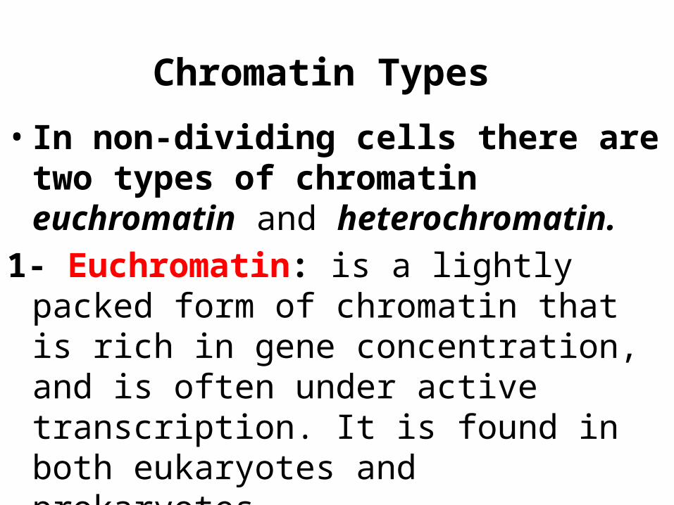
• In non-dividing cells there are two types of chromatin euchromatin and heterochromatin.
1- Euchromatin: is a lightly packed form of chromatin that is rich in gene concentration, and is often under active transcription. It is found in both eukaryotes and prokaryotes.
Chromatin Types

2- Hetrochromatin: • Heterochromatin is a tightly packed form of
DNA. Heterochromatin is inactive and remains compact during interphase.
• Heterochromatin plays a role in gene regulation and the protection of the integrity of chromosomes, attributed to the dense packing of DNA, which makes it less accessible to protein factors that bind DNA or its associated factors.

The compaction level of interphase chromosomes is not completely uniform Euchromatin
Less condensed regions of chromosomes Transcriptionally active Regions where 30 nm fiber forms radial loop domains
Heterochromatin Tightly compacted regions of chromosomes Transcriptionally inactive (in general) Radial loop domains compacted even further
Heterochromatin vs Euchromatin
Types of Heterocromatin
1- Constitutive heterochromatin: remains compact in all cells and at all times and occurs around the chromosome centromere and near telomeres.
• It represents the silenced part of DNA.
2- Facultative heterochromatin: • Is a chromatin that has been inactivated
in specific types of differentiated cells.
• An example of facultative heterochromatin is X- chromosome inactivation in female mammals: one X- chromosome is packaged in facultative heterochromatin and silenced, while the other X chromosome is packaged in euchromatin and expressed.
Chromatin Function1. Package DNA into a smaller volume to fit into
the cell. 2. Strengthen the DNA to allow mitosis and
meiosis.
3. Serve as a mechanism to control expression.
4. Changes in chromatin structure are affected mainly by methylation (DNA and proteins) and acetylation (proteins). Chromatin structure is also relevant to DNA replication and DNA repair.
5. Histones are the proteins closely associated with DNA molecules. They are responsible for the structure of chromatin and play important roles in the regulation of gene expression.

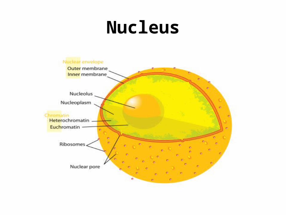
• The nucleoplasm is a highly viscous liquid that surrounds the chromosomes and nucleoli. Many substances such as nucleotides (necessary for purposes such as the replication of DNA) and enzymes (which direct activities that take place in the nucleus) are dissolved in the nucleoplasm.
NucleoplasmNucleoplasm
Nucleolus
• The prominent structure in the nucleus is the nucleolus.
• The nucleolus produces ribosomes, which move out of the nucleus and take positions on the rough endoplasmic reticulum where they are critical in protein synthesis.
Histone proteins are basic They contain many positively-charged amino acids
Lysine and arginine These bind with the phosphates along the DNA backbone
There are five types of histones:
H1, H2A, H2B, H3 and H4 H2A, H2B, H3 and H4 are the core histones
Two of each make up the octamer
H1 is the linker histone: Binds to linker DNA Also binds to nucleosomes
But not as tightly as the core histones
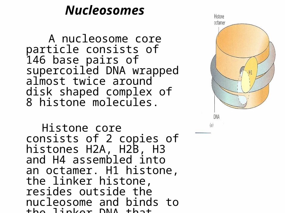
Nucleosomes
A nucleosome core particle consists of 146 base pairs of supercoiled DNA wrapped almost twice around disk shaped complex of 8 histone molecules.
Histone core consists of 2 copies of histones H2A, H2B, H3 and H4 assembled into an octamer. H1 histone, the linker histone, resides outside the nucleosome and binds to the linker DNA that connects one nucleosome to the next.

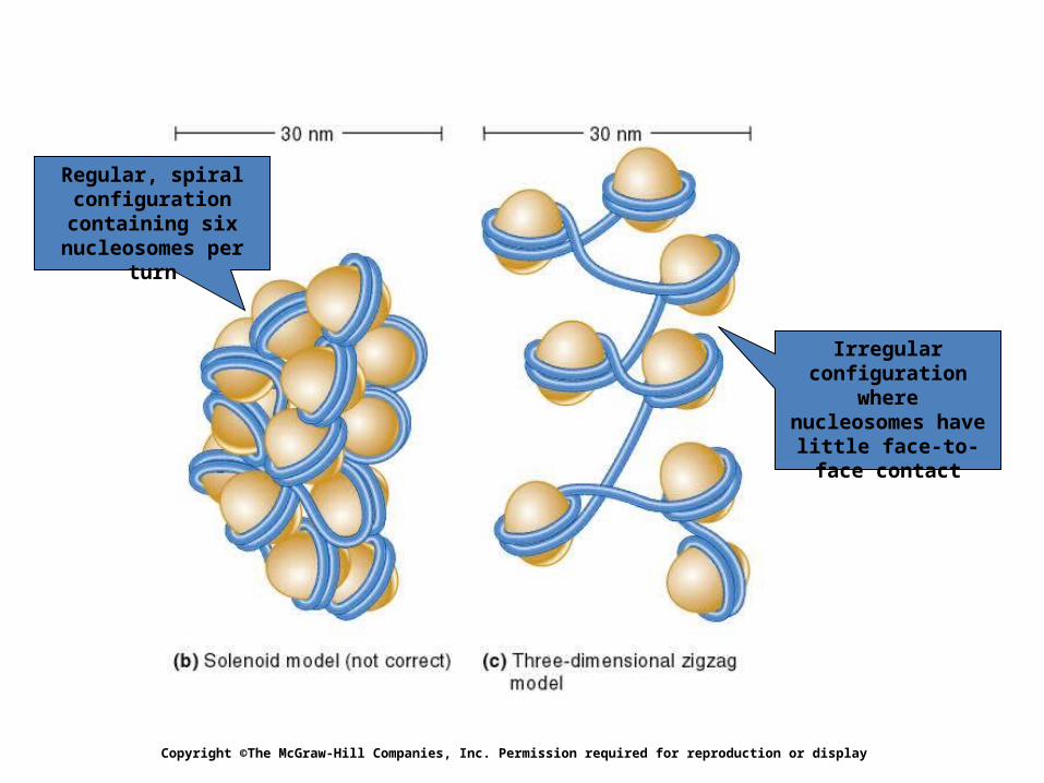
• The nucleosomes in turn form a helical solenoid; each turn of the solenoid includes about six nucleosomes.
• The solenoids themselves are organized into chromatin loops, which are attached to a protein scaffold. Each of these loops contains approximately 100,000 base pairs (bp), or 100 kilobases (kb), of DNA.
• The end result of this coiling and looping is that the DNA, is only about 1/10,000 as long as it would be if it were fully stretched out. ▪
Copyright ©The McGraw-Hill Companies, Inc. Permission required for reproduction or display
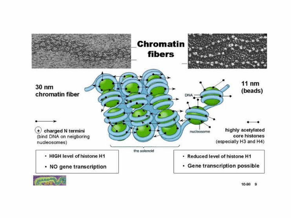
Regular, spiral configuration containing six
nucleosomes per turn
Irregular configuration where nucleosomes have little face-to-face
contact
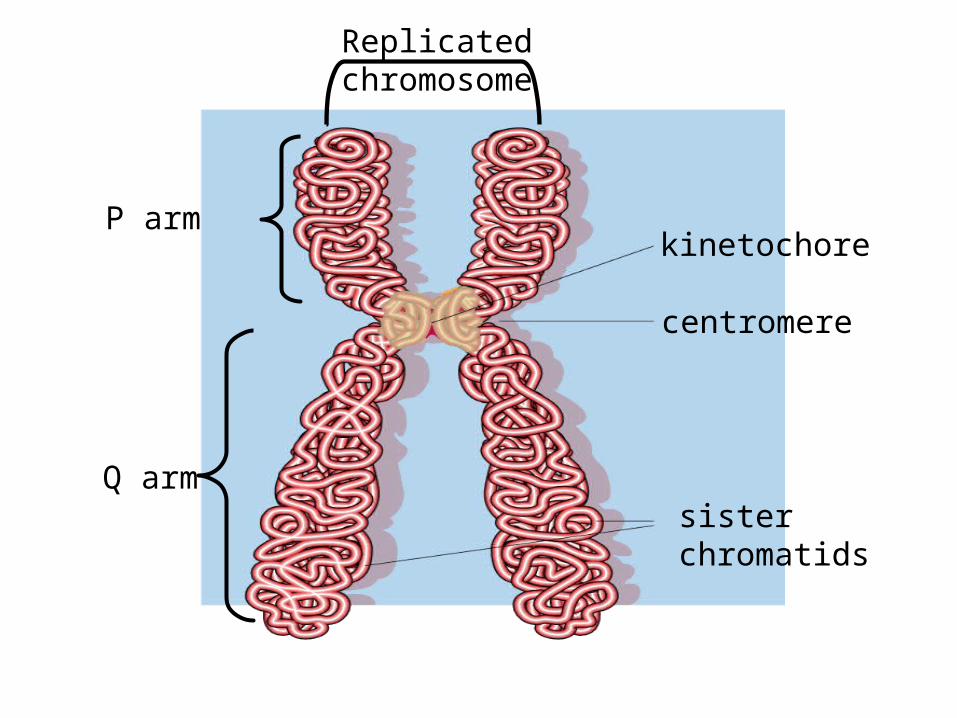
sister chromatids
centromere
kinetochore
Replicated chromosome
P arm
Q arm

Genome Organization • The metaphase chromosomes have been characterized into
different regions based on a specific banding patterns.
The principle bands are: G and R.• In G-bands, the dark regions tend to be heterochromatic, late-
replicating and AT rich. The bright regions tend to be euchromatic, early-replicating and GC rich.
• Bright field R-bands• The dark regions are euchromatic and the bright regions are
heterochromatic.• The R bands are enriched in acetylated histones and this
modification is conserved through mitosis suggesting that histone acetylation may serve as a marker for genome organization through the cell cycle.

Levels of Chromatin Organization• The structure of chromatin varies considerably as
the cell progresses through the cell cycle. The changes in structure are required to allow the DNA to be used and managed.
1- The lowest level of chromosome organisation (First degree of organization)
• DNA and histones are organized into repeating subunits called nucleosomes - the "beads on a string" structure.

• 2- Higher Levels of Chromatin Organization
Chromatin appears in interphase cells as tiny dots and fibers of 30 nm thickness.
The 30 nm fibers gather into larger
supercoiled loops of thick fibers which normally spread through the interphase nucleus.
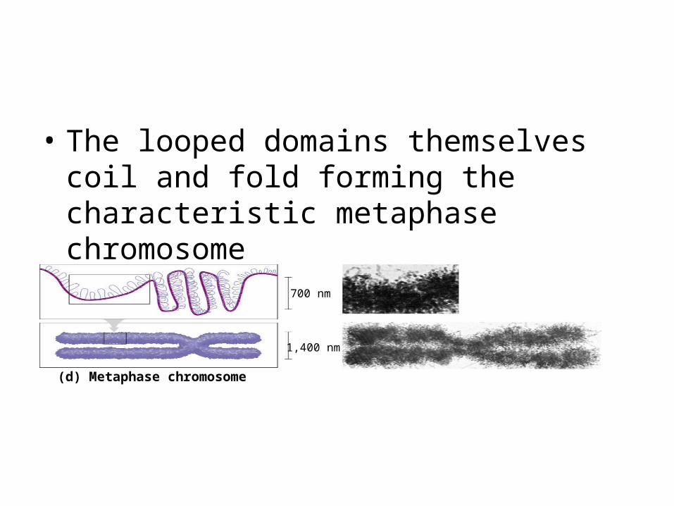
• The looped domains themselves coil and fold forming the characteristic metaphase chromosome
700 nm
1,400 nm
(d) Metaphase chromosome
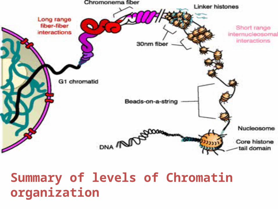
Summary of levels of Chromatin organization
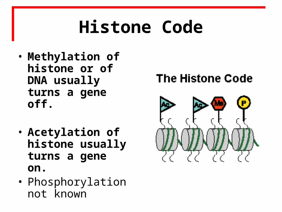
• Methylation of histone or of DNA usually turns a gene off.
• Acetylation of histone usually turns a gene on.
• Phosphorylation not known
Histone CodeHistone Code

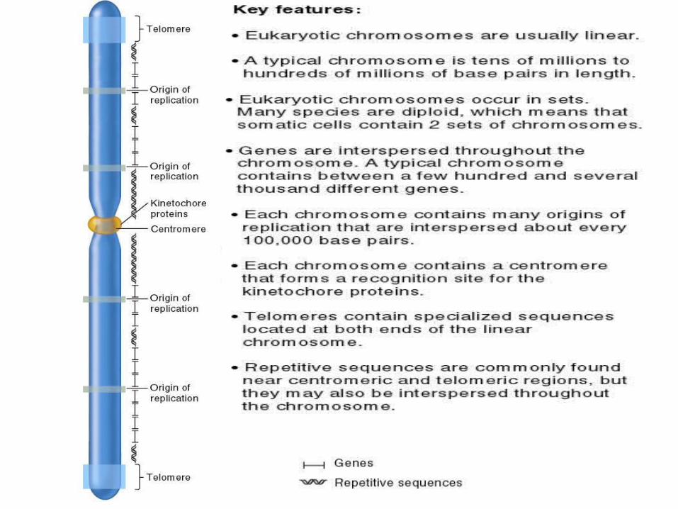
• Genes are located between the centromeric and telomeric regions along the entire chromosome– A single chromosome usually has a few hundred to
several thousand genes
• In lower eukaryotes (such as yeast)– Genes are relatively small
• They contain primarily the sequences encoding the polypeptides ie: Very few introns are present
• In higher eukaryotes (such as mammals)– Genes are long
• They tend to have many introns
• Sequence complexity refers to the number of times a particular base sequence appears in the genome.
• There are three main types of repetitive sequences– Unique or non-repetitive.– Moderately repetitive.– Highly repetitive.
Repetitive Sequences

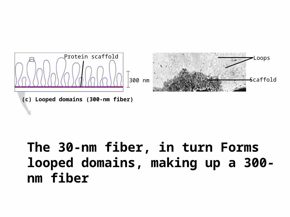
Protein scaffold
300 nm
(c) Looped domains (300-nm fiber)
Loops
Scaffold
The 30-nm fiber, in turn Forms looped domains, making up a 300-nm fiber