origin and proliferation of multiple- drug resistance in
TRANSCRIPT

Origin and Proliferation of Multiple-Drug Resistance in Bacterial Pathogens
CitationChang, Hsiao-Han, Ted Cohen, Yonatan H. Grad, William P. Hanage, Thomas F. O’Brien, and Marc Lipsitch. 2015. “Origin and Proliferation of Multiple-Drug Resistance in Bacterial Pathogens.” Microbiol. Mol. Biol. Rev. 79 (1) (February 4): 101–116. doi:10.1128/mmbr.00039-14.
Published Versiondoi:10.1128/MMBR.00039-14
Permanent linkhttp://nrs.harvard.edu/urn-3:HUL.InstRepos:26882169
Terms of UseThis article was downloaded from Harvard University’s DASH repository, and is made available under the terms and conditions applicable to Other Posted Material, as set forth at http://nrs.harvard.edu/urn-3:HUL.InstRepos:dash.current.terms-of-use#LAA
Share Your StoryThe Harvard community has made this article openly available.Please share how this access benefits you. Submit a story .
Accessibility

Origin and Proliferation of Multiple-Drug Resistance in BacterialPathogens
Hsiao-Han Chang,a Ted Cohen,a,b Yonatan H. Grad,a,c William P. Hanage,a Thomas F. O’Brien,d Marc Lipsitcha,e
Center for Communicable Disease Dynamics, Department of Epidemiology, Harvard School of Public Health, Boston, Massachusetts, USAa; Epidemiology of MicrobialDiseases, Yale School of Public Health, New Haven, Connecticut, USAb; Division of Infectious Diseases, Brigham and Women’s Hospital, Harvard Medical School, Boston,Massachusetts, USAc; The World Health Organization Collaborating Centre for Surveillance of Antimicrobial Resistance, Department of Medicine, Brigham and Women’sHospital and Harvard Medical School, Boston, Massachusetts, USAd; Department of Immunology and Infectious Diseases, Harvard School of Public Health, Boston,Massachusetts, USAe
SUMMARY . . . . . . . . . . . . . . . . . . . . . . . . . . . . . . . . . . . . . . . . . . . . . . . . . . . . . . . . . . . . . . . . . . . . . . . . . . . . . . . . . . . . . . . . . . . . . . . . . . . . . . . . . . . . . . . . . . . . . . . . . . . . . . . . . . . . . . . . . . . . . . . . . .102INTRODUCTION . . . . . . . . . . . . . . . . . . . . . . . . . . . . . . . . . . . . . . . . . . . . . . . . . . . . . . . . . . . . . . . . . . . . . . . . . . . . . . . . . . . . . . . . . . . . . . . . . . . . . . . . . . . . . . . . . . . . . . . . . . . . . . . . . . . . . . . . . . . .102THE EXCESS OF MDR IN BACTERIA. . . . . . . . . . . . . . . . . . . . . . . . . . . . . . . . . . . . . . . . . . . . . . . . . . . . . . . . . . . . . . . . . . . . . . . . . . . . . . . . . . . . . . . . . . . . . . . . . . . . . . . . . . . . . . . . . . . . . . . . .102
Streptococcus pneumoniae. . . . . . . . . . . . . . . . . . . . . . . . . . . . . . . . . . . . . . . . . . . . . . . . . . . . . . . . . . . . . . . . . . . . . . . . . . . . . . . . . . . . . . . . . . . . . . . . . . . . . . . . . . . . . . . . . . . . . . . . . . . . . . . .102Enterobacteriaceae . . . . . . . . . . . . . . . . . . . . . . . . . . . . . . . . . . . . . . . . . . . . . . . . . . . . . . . . . . . . . . . . . . . . . . . . . . . . . . . . . . . . . . . . . . . . . . . . . . . . . . . . . . . . . . . . . . . . . . . . . . . . . . . . . . . . . . . .102Neisseria gonorrhoeae . . . . . . . . . . . . . . . . . . . . . . . . . . . . . . . . . . . . . . . . . . . . . . . . . . . . . . . . . . . . . . . . . . . . . . . . . . . . . . . . . . . . . . . . . . . . . . . . . . . . . . . . . . . . . . . . . . . . . . . . . . . . . . . . . . . . .102Mycobacterium tuberculosis . . . . . . . . . . . . . . . . . . . . . . . . . . . . . . . . . . . . . . . . . . . . . . . . . . . . . . . . . . . . . . . . . . . . . . . . . . . . . . . . . . . . . . . . . . . . . . . . . . . . . . . . . . . . . . . . . . . . . . . . . . . . . . .103Staphylococcus aureus . . . . . . . . . . . . . . . . . . . . . . . . . . . . . . . . . . . . . . . . . . . . . . . . . . . . . . . . . . . . . . . . . . . . . . . . . . . . . . . . . . . . . . . . . . . . . . . . . . . . . . . . . . . . . . . . . . . . . . . . . . . . . . . . . . . .103
MECHANISMS LEADING TO EXCESS MDR . . . . . . . . . . . . . . . . . . . . . . . . . . . . . . . . . . . . . . . . . . . . . . . . . . . . . . . . . . . . . . . . . . . . . . . . . . . . . . . . . . . . . . . . . . . . . . . . . . . . . . . . . . . . . . . . .104Explanations for the Origin of MDR Strains . . . . . . . . . . . . . . . . . . . . . . . . . . . . . . . . . . . . . . . . . . . . . . . . . . . . . . . . . . . . . . . . . . . . . . . . . . . . . . . . . . . . . . . . . . . . . . . . . . . . . . . . . . . . . .104
Single biochemical mechanism conferring resistance to multiple drugs . . . . . . . . . . . . . . . . . . . . . . . . . . . . . . . . . . . . . . . . . . . . . . . . . . . . . . . . . . . . . . . . . . . . . . . . . . . . . .104(i) Testable predictions . . . . . . . . . . . . . . . . . . . . . . . . . . . . . . . . . . . . . . . . . . . . . . . . . . . . . . . . . . . . . . . . . . . . . . . . . . . . . . . . . . . . . . . . . . . . . . . . . . . . . . . . . . . . . . . . . . . . . . . . . . . . . .104(ii) Practical implications . . . . . . . . . . . . . . . . . . . . . . . . . . . . . . . . . . . . . . . . . . . . . . . . . . . . . . . . . . . . . . . . . . . . . . . . . . . . . . . . . . . . . . . . . . . . . . . . . . . . . . . . . . . . . . . . . . . . . . . . . . . .105
Genetic linkage . . . . . . . . . . . . . . . . . . . . . . . . . . . . . . . . . . . . . . . . . . . . . . . . . . . . . . . . . . . . . . . . . . . . . . . . . . . . . . . . . . . . . . . . . . . . . . . . . . . . . . . . . . . . . . . . . . . . . . . . . . . . . . . . . . . . . . . .105(i) Examples . . . . . . . . . . . . . . . . . . . . . . . . . . . . . . . . . . . . . . . . . . . . . . . . . . . . . . . . . . . . . . . . . . . . . . . . . . . . . . . . . . . . . . . . . . . . . . . . . . . . . . . . . . . . . . . . . . . . . . . . . . . . . . . . . . . . . . . . .105(ii) Testable predictions . . . . . . . . . . . . . . . . . . . . . . . . . . . . . . . . . . . . . . . . . . . . . . . . . . . . . . . . . . . . . . . . . . . . . . . . . . . . . . . . . . . . . . . . . . . . . . . . . . . . . . . . . . . . . . . . . . . . . . . . . . . . .105(iii) Practical implications . . . . . . . . . . . . . . . . . . . . . . . . . . . . . . . . . . . . . . . . . . . . . . . . . . . . . . . . . . . . . . . . . . . . . . . . . . . . . . . . . . . . . . . . . . . . . . . . . . . . . . . . . . . . . . . . . . . . . . . . . . . .105
Highly mutable or recombinogenic bacterial lineages . . . . . . . . . . . . . . . . . . . . . . . . . . . . . . . . . . . . . . . . . . . . . . . . . . . . . . . . . . . . . . . . . . . . . . . . . . . . . . . . . . . . . . . . . . . . . . . .106(i) Examples . . . . . . . . . . . . . . . . . . . . . . . . . . . . . . . . . . . . . . . . . . . . . . . . . . . . . . . . . . . . . . . . . . . . . . . . . . . . . . . . . . . . . . . . . . . . . . . . . . . . . . . . . . . . . . . . . . . . . . . . . . . . . . . . . . . . . . . . .107(ii) Complexities . . . . . . . . . . . . . . . . . . . . . . . . . . . . . . . . . . . . . . . . . . . . . . . . . . . . . . . . . . . . . . . . . . . . . . . . . . . . . . . . . . . . . . . . . . . . . . . . . . . . . . . . . . . . . . . . . . . . . . . . . . . . . . . . . . . . .107(iii) Testable predictions . . . . . . . . . . . . . . . . . . . . . . . . . . . . . . . . . . . . . . . . . . . . . . . . . . . . . . . . . . . . . . . . . . . . . . . . . . . . . . . . . . . . . . . . . . . . . . . . . . . . . . . . . . . . . . . . . . . . . . . . . . . . .107(iv) Practical implications . . . . . . . . . . . . . . . . . . . . . . . . . . . . . . . . . . . . . . . . . . . . . . . . . . . . . . . . . . . . . . . . . . . . . . . . . . . . . . . . . . . . . . . . . . . . . . . . . . . . . . . . . . . . . . . . . . . . . . . . . . . .107
Multidrug therapy with accelerated treatment failure in resistant infections . . . . . . . . . . . . . . . . . . . . . . . . . . . . . . . . . . . . . . . . . . . . . . . . . . . . . . . . . . . . . . . . . . . . . . . . . .107(i) Example . . . . . . . . . . . . . . . . . . . . . . . . . . . . . . . . . . . . . . . . . . . . . . . . . . . . . . . . . . . . . . . . . . . . . . . . . . . . . . . . . . . . . . . . . . . . . . . . . . . . . . . . . . . . . . . . . . . . . . . . . . . . . . . . . . . . . . . . . .108(ii) Testable predictions . . . . . . . . . . . . . . . . . . . . . . . . . . . . . . . . . . . . . . . . . . . . . . . . . . . . . . . . . . . . . . . . . . . . . . . . . . . . . . . . . . . . . . . . . . . . . . . . . . . . . . . . . . . . . . . . . . . . . . . . . . . . .108(iii) Practical implications . . . . . . . . . . . . . . . . . . . . . . . . . . . . . . . . . . . . . . . . . . . . . . . . . . . . . . . . . . . . . . . . . . . . . . . . . . . . . . . . . . . . . . . . . . . . . . . . . . . . . . . . . . . . . . . . . . . . . . . . . . . .108
Explanations for the Proliferation of MDR Strains . . . . . . . . . . . . . . . . . . . . . . . . . . . . . . . . . . . . . . . . . . . . . . . . . . . . . . . . . . . . . . . . . . . . . . . . . . . . . . . . . . . . . . . . . . . . . . . . . . . . . . . . .108Associated linkage selection . . . . . . . . . . . . . . . . . . . . . . . . . . . . . . . . . . . . . . . . . . . . . . . . . . . . . . . . . . . . . . . . . . . . . . . . . . . . . . . . . . . . . . . . . . . . . . . . . . . . . . . . . . . . . . . . . . . . . . . . . . .108
(i) Examples . . . . . . . . . . . . . . . . . . . . . . . . . . . . . . . . . . . . . . . . . . . . . . . . . . . . . . . . . . . . . . . . . . . . . . . . . . . . . . . . . . . . . . . . . . . . . . . . . . . . . . . . . . . . . . . . . . . . . . . . . . . . . . . . . . . . . . . . .108(ii) Testable predictions . . . . . . . . . . . . . . . . . . . . . . . . . . . . . . . . . . . . . . . . . . . . . . . . . . . . . . . . . . . . . . . . . . . . . . . . . . . . . . . . . . . . . . . . . . . . . . . . . . . . . . . . . . . . . . . . . . . . . . . . . . . . .109(iii) Practical implications . . . . . . . . . . . . . . . . . . . . . . . . . . . . . . . . . . . . . . . . . . . . . . . . . . . . . . . . . . . . . . . . . . . . . . . . . . . . . . . . . . . . . . . . . . . . . . . . . . . . . . . . . . . . . . . . . . . . . . . . . . . .109
Bystander selection . . . . . . . . . . . . . . . . . . . . . . . . . . . . . . . . . . . . . . . . . . . . . . . . . . . . . . . . . . . . . . . . . . . . . . . . . . . . . . . . . . . . . . . . . . . . . . . . . . . . . . . . . . . . . . . . . . . . . . . . . . . . . . . . . . . .109(i) Examples . . . . . . . . . . . . . . . . . . . . . . . . . . . . . . . . . . . . . . . . . . . . . . . . . . . . . . . . . . . . . . . . . . . . . . . . . . . . . . . . . . . . . . . . . . . . . . . . . . . . . . . . . . . . . . . . . . . . . . . . . . . . . . . . . . . . . . . . .109(ii) Testable predictions . . . . . . . . . . . . . . . . . . . . . . . . . . . . . . . . . . . . . . . . . . . . . . . . . . . . . . . . . . . . . . . . . . . . . . . . . . . . . . . . . . . . . . . . . . . . . . . . . . . . . . . . . . . . . . . . . . . . . . . . . . . . .110(iii) Practical implications . . . . . . . . . . . . . . . . . . . . . . . . . . . . . . . . . . . . . . . . . . . . . . . . . . . . . . . . . . . . . . . . . . . . . . . . . . . . . . . . . . . . . . . . . . . . . . . . . . . . . . . . . . . . . . . . . . . . . . . . . . . .110
Positive epistasis between drug resistance determinants or between resistance determinants and genetic background. . . . . . . . . . . . . . . . . . . . . . . . . . . .110(i) Examples . . . . . . . . . . . . . . . . . . . . . . . . . . . . . . . . . . . . . . . . . . . . . . . . . . . . . . . . . . . . . . . . . . . . . . . . . . . . . . . . . . . . . . . . . . . . . . . . . . . . . . . . . . . . . . . . . . . . . . . . . . . . . . . . . . . . . . . . .110(ii) Testable predictions . . . . . . . . . . . . . . . . . . . . . . . . . . . . . . . . . . . . . . . . . . . . . . . . . . . . . . . . . . . . . . . . . . . . . . . . . . . . . . . . . . . . . . . . . . . . . . . . . . . . . . . . . . . . . . . . . . . . . . . . . . . . .110(iii) Practical implications . . . . . . . . . . . . . . . . . . . . . . . . . . . . . . . . . . . . . . . . . . . . . . . . . . . . . . . . . . . . . . . . . . . . . . . . . . . . . . . . . . . . . . . . . . . . . . . . . . . . . . . . . . . . . . . . . . . . . . . . . . . .110
Niche differentiation: aggregation of multiple drug selection pressures within specific populations.. . . . . . . . . . . . . . . . . . . . . . . . . . . . . . . . . . . . . . . . . . . . . . . . .111(continued)
Published 4 February 2015
Citation Chang H-H, Cohen T, Grad YH, Hanage WP, O’Brien TF, Lipsitch M. 4February 2015. Origin and proliferation of multiple-drug resistance in bacterialpathogens. Microbiol Mol Biol Rev doi:10.1128/MMBR.00039-14.
Address correspondence to Marc Lipsitch, [email protected].
Copyright © 2015, American Society for Microbiology. All Rights Reserved.
doi:10.1128/MMBR.00039-14
crossmark
March 2015 Volume 79 Number 1 mmbr.asm.org 101Microbiology and Molecular Biology Reviews

(i) Examples . . . . . . . . . . . . . . . . . . . . . . . . . . . . . . . . . . . . . . . . . . . . . . . . . . . . . . . . . . . . . . . . . . . . . . . . . . . . . . . . . . . . . . . . . . . . . . . . . . . . . . . . . . . . . . . . . . . . . . . . . . . . . . . . . . . . . . . . .111(ii) Testable predictions . . . . . . . . . . . . . . . . . . . . . . . . . . . . . . . . . . . . . . . . . . . . . . . . . . . . . . . . . . . . . . . . . . . . . . . . . . . . . . . . . . . . . . . . . . . . . . . . . . . . . . . . . . . . . . . . . . . . . . . . . . . . .111(iii) Practical implications . . . . . . . . . . . . . . . . . . . . . . . . . . . . . . . . . . . . . . . . . . . . . . . . . . . . . . . . . . . . . . . . . . . . . . . . . . . . . . . . . . . . . . . . . . . . . . . . . . . . . . . . . . . . . . . . . . . . . . . . . . . .111
Importation of MDR strains and geographic source-sink dynamics . . . . . . . . . . . . . . . . . . . . . . . . . . . . . . . . . . . . . . . . . . . . . . . . . . . . . . . . . . . . . . . . . . . . . . . . . . . . . . . . . . .111(i) Example . . . . . . . . . . . . . . . . . . . . . . . . . . . . . . . . . . . . . . . . . . . . . . . . . . . . . . . . . . . . . . . . . . . . . . . . . . . . . . . . . . . . . . . . . . . . . . . . . . . . . . . . . . . . . . . . . . . . . . . . . . . . . . . . . . . . . . . . . .111(ii) Testable predictions . . . . . . . . . . . . . . . . . . . . . . . . . . . . . . . . . . . . . . . . . . . . . . . . . . . . . . . . . . . . . . . . . . . . . . . . . . . . . . . . . . . . . . . . . . . . . . . . . . . . . . . . . . . . . . . . . . . . . . . . . . . . .111(iii) Practical implications . . . . . . . . . . . . . . . . . . . . . . . . . . . . . . . . . . . . . . . . . . . . . . . . . . . . . . . . . . . . . . . . . . . . . . . . . . . . . . . . . . . . . . . . . . . . . . . . . . . . . . . . . . . . . . . . . . . . . . . . . . . .111
CONCLUSIONS . . . . . . . . . . . . . . . . . . . . . . . . . . . . . . . . . . . . . . . . . . . . . . . . . . . . . . . . . . . . . . . . . . . . . . . . . . . . . . . . . . . . . . . . . . . . . . . . . . . . . . . . . . . . . . . . . . . . . . . . . . . . . . . . . . . . . . . . . . . . .111ACKNOWLEDGMENTS. . . . . . . . . . . . . . . . . . . . . . . . . . . . . . . . . . . . . . . . . . . . . . . . . . . . . . . . . . . . . . . . . . . . . . . . . . . . . . . . . . . . . . . . . . . . . . . . . . . . . . . . . . . . . . . . . . . . . . . . . . . . . . . . . . . . . .113REFERENCES . . . . . . . . . . . . . . . . . . . . . . . . . . . . . . . . . . . . . . . . . . . . . . . . . . . . . . . . . . . . . . . . . . . . . . . . . . . . . . . . . . . . . . . . . . . . . . . . . . . . . . . . . . . . . . . . . . . . . . . . . . . . . . . . . . . . . . . . . . . . . . . .113
SUMMARY
Many studies report the high prevalence of multiply drug-resistant(MDR) strains. Because MDR infections are often significantlyharder and more expensive to treat, they represent a growing publichealth threat. However, for different pathogens, different underlyingmechanisms are traditionally used to explain these observations, andit is unclear whether each bacterial taxon has its own mechanism(s)for multidrug resistance or whether there are common mechanismsbetween distantly related pathogens. In this review, we provide a sys-tematic overview of the causes of the excess of MDR infections anddefine testable predictions made by each hypothetical mechanism,including experimental, epidemiological, population genomic, andother tests of these hypotheses. Better understanding the cause(s) ofthe excess of MDR is the first step to rational design of more effectiveinterventions to prevent the origin and/or proliferation of MDR.
INTRODUCTION
Bacterial pathogens that are resistant to multiple drugs repre-sent a growing public health threat, because multiply drug-
resistant (MDR) infections are challenging and expensive to treat(1–3), and few antimicrobial compounds, and still fewer antimi-crobial agents using novel mechanisms of action, are in clinicaldevelopment (4). Using both published and unpublished data (5–8), we show that there is often a positive correlation within abacterial population between resistance to one drug and resistanceto one or more other drugs. The high frequency of MDR isolatesamong resistant strains represents a scientific puzzle: why do re-sistance determinants aggregate in certain strains of bacteria?
Different underlying mechanisms are traditionally used to ex-plain these observations in different pathogens and are rarely crit-ically assessed or tested. It is unclear whether each bacterial taxonhas its own mechanism(s) or to what extent the pathways to theaccumulation of multiple resistances are shared among patho-gens. Understanding the causes of MDR is necessary for con-structing appropriate models to aid in the design and evaluation ofpotential interventions.
Here we describe the scope of the phenomenon of MDR inbacteria and enumerate possible mechanisms for the appearanceand proliferation of MDR bacteria. Importantly, we also proposeexperimental, epidemiological, population genomic, and otherstudy designs that can help to clarify the roles of these potentialmechanisms driving excess MDR.
THE EXCESS OF MDR IN BACTERIA
For many bacterial pathogens, the frequency of MDR pathogens ex-ceeds the product of the frequencies of individual resistance traits.While this phenomenon of excess MDR has rarely been the focus of
epidemiological studies, those studies which report the relevant datavery often find positive correlations between resistance phenotypes:isolates resistant to one drug are more likely to be resistant to others.Below we briefly review such findings for a range of pathogens, in-cluding novel analyses performed for this report.
Streptococcus pneumoniae
McCormick et al. (9) and Link-Gelles et al. (8) studied invasivepneumococcal disease isolates from population-based surveil-lance in different eras and reported that dual resistance to penicil-lin and erythromycin is more common than the product of theproportions of resistance to penicillin and erythromycin in theUnited States. We have expanded their analysis, using the samedata set from three epidemiologically distinct periods. We findsignificant positive correlations between resistance to drugs,including penicillin, erythromycin, tetracycline, clindamycin,trimethoprim-sulfamethoxazole, ceftazidime, and levofloxa-cin (Table 1).
Enterobacteriaceae
In the United Kingdom, sulfonamide-resistant strains of Esch-erichia coli were more likely to have resistance to antibiotics, in-cluding ampicillin, chloramphenicol, kanamycin, streptomycin,tetracycline, and trimethoprim, than sulfonamide-susceptiblestrains in both in 1991 and 1999 (6). The proportion of sulfon-amide-resistant strains that were resistant to at least two otherantibiotics of different chemical classes was also higher than thatof sulfonamide-susceptible strains (sulfonamide resistant versussulfonamide susceptible, 88.1% versus 17.8% in 1991 and 83.0%versus 28.4% in 1999) (6). In the United States, there was a signif-icant association between resistance to fluoroquinolones and plas-mid-mediated gentamicin resistance (10). Using data from a ter-tiary care hospital in the United States, we found that mostcorrelation coefficients of resistance to different drugs used totreat Klebsiella pneumoniae, E. coli, and Pseudomonas aeruginosainfections are significantly positive (Tables 2 to 4).
Neisseria gonorrhoeae
Fluoroquinolone resistance is associated with resistance to peni-cillin and tetracycline in N. gonorrhoeae in the United States dur-ing 2002 to 2007 (7). Ota et al. (11) also reported that in Ontario,Canada, in 2006, fluoroquinolone-resistant strains were morelikely to be resistant to penicillin, tetracycline, and erythromycinthan fluoroquinolone-sensitive strains (fluoroquinolone-resis-tant versus fluoroquinolone-sensitive, 98.4% versus 89.4% forpenicillin, 98.0% versus 81.1% for tetracycline, and 66.2% versus14.8% for erythromycin). We found that resistance to penicillin,tetracycline, and fluoroquinolones are positively correlated in the
Chang et al.
102 mmbr.asm.org March 2015 Volume 79 Number 1Microbiology and Molecular Biology Reviews

United States, using data from CDC’s Gonococcal Isolate Surveil-lance Project (GISP) 2011 Annual Report (12) (correlation coef-ficients of 0.371, 0.416, and 0.452 for penicillin and tetracycline,penicillin and ciprofloxacin, and tetracycline and ciprofloxacin,respectively [P values could not be calculated due to the lack ofsample size information]).
Mycobacterium tuberculosis
We calculated the correlation coefficients of resistance to differentdrugs used to treat tuberculosis (TB) by using the data from 114countries in Anti-Tuberculosis Drug Resistance in the World:
Fourth Global Report (13, 14). We found strong positive correla-tions between resistance to isoniazid, rifampin, ethambutol, andstreptomycin (Table 5).
Staphylococcus aureus
We found positive correlations between resistance to antibioticsused to treat S. aureus infection, including penicillin, erythromy-cin, clindamycin, tetracycline, levofloxacin, gentamicin, and tri-methoprim, using data from a tertiary care hospital in the UnitedStates (Table 6). The significant positive correlations between re-sistance to drugs used to treat S. aureus infection were also iden-
TABLE 1 Correlation coefficients of resistance to different drugs used to treat Streptococcus pneumoniae infections in the United Statesa
Period (n) Drug
Correlation coefficentb
ERY TET CLI SXT TAX CAZ LVX
Pre-PCV7 (7,571) PEN 0.610*** 0.370*** 0.266*** 0.700*** 0.454*** 0.037**ERY 0.427*** 0.399*** 0.574*** 0.311*** 0.015TET 0.574*** 0.338*** 0.173*** 0.009CLI 0.229*** 0.078*** 0.037**SXT 0.397*** 0.029*TAX 0.021
Intermediate PCV7 coverage (16,735) PEN 0.613*** 0.359*** 0.288*** 0.626*** 0.437*** 0.299*** 0.026***ERY 0.427*** 0.440*** 0.559*** 0.369*** 0.291*** 0.045***�-TET 0.668*** 0.321*** 0.230*** 0.291*** 0.052***CLI 0.234*** 0.198*** 0.301*** 0.040***SXT 0.389*** 0.277*** 0.035***TAX 0.727*** 0.026***CAZ �0.011
High PCV7 coverage (6,785) PEN 0.579*** 0.460*** 0.422*** 0.576*** 0.440*** 0.326*** 0.030*ERY 0.565*** 0.577*** 0.588*** 0.443*** 0.338*** 0.049***TET 0.819*** 0.399*** 0.516*** 0.415*** 0.026*CLI 0.343*** 0.516*** 0.393*** 0.035**SXT 0.454*** 0.336*** 0.045***TAX 0.727*** 0.014CAZ 0.019
a PCV7, 7-valent pneumococcal conjugate vaccine; PEN, penicillin; ERY, erythromycin; TET, tetracycline; CLI, clindamycin; SXT, trimethoprim-sulfamethoxazole; TAX,cefotaxime; CAZ, ceftazidime; LVX, levofloxacin.b ***, P � 0.001; **, P � 0.01; *, P � 0.1.
TABLE 2 Correlation coefficients of resistance to different drugs used to treat Klebsiella pneumoniae infection in a general hospital in the UnitedStatesa
Drug
Correlation coefficientb
AMP SAM FEP FOX CAZ CRO CIP GEN IPM LVX NIT SXT
AMK 0.031 0.304*** 0.266*** 0.218*** 0.451*** 0.442*** 0.425*** 0.174*** 0.247*** 0.432*** 0.098** 0.279***AMP 0.098** 0.042 0.064* 0.066* 0.067* 0.073* 0.042 0.027 0.072* 0.319*** 0.077*SAM 0.406*** 0.417*** 0.623*** 0.645*** 0.558*** 0.342*** 0.279*** 0.542*** 0.183*** 0.466***FEP 0.356*** 0.603*** 0.605*** 0.458*** 0.270*** 0.447*** 0.452*** 0.081** 0.366***FOX 0.410*** 0.400*** 0.436*** 0.227*** 0.379*** 0.446*** 0.180*** 0.213***CAZ 0.909*** 0.683*** 0.414*** 0.367*** 0.668*** 0.160*** 0.447***CRO 0.696*** 0.464*** 0.382*** 0.672*** 0.165*** 0.489***CIP 0.410*** 0.308*** 0.982*** 0.174*** 0.472***GEN 0.207*** 0.375*** 0.103*** 0.374***IPM 0.314*** 0.086** 0.232***LVX 0.169*** 0.483***NIT 0.127***a Data source, WHONET (n � 1,095). AMK, amikacin; AMP, ampicillin; SAM, ampicillin-sulbactam; FEP, cefepime; FOX, cefoxitin; CAZ, ceftazidime; CRO, ceftriaxone; CIP,ciprofloxacin; GEN, gentamicin; IPM, imipenem; LVX, levofloxacin; NIT, nitrofurantoin; SXT, trimethoprim-sulfamethoxazole.b ***, P � 0.001; **, P � 0.01; *, P � 0.1.
Mechanisms of Multiple-Drug Resistance in Bacteria
March 2015 Volume 79 Number 1 mmbr.asm.org 103Microbiology and Molecular Biology Reviews
tified in the province of British Columbia, Canada, in 2012 (15)(correlation coefficients of 0.229, 0.550, 0.054, and 0.052 formethicillin versus clindamycin, erythromycin, trimethoprim-sul-famethoxazole, and tetracycline, respectively [n � 5,214; P �0.001 for all cases]).
In summary, these data show positive correlation between re-sistance to different drugs and the higher-than-expected propor-tion of MDR for Gram-positive and Gram-negative bacteria andmycobacteria. The universality of excess MDR, involving resis-tance conferred by a range of genetic and biochemical mecha-nisms, raises the question of a shared mechanism(s) driving thesecommonly observed patterns.
It should be noted that the association between resistance to mul-tiple drugs and a higher-than-expected proportion of MDR shownhere is based purely on phenotype. Certainly, the genetic causes ofresistance to antibiotics or classes of antibiotics can vary even within aspecies (for example, different classes of extended-spectrum beta-lactamases in species of the Enterobacteriaceae [16] or the effluxpump [mef] and ribosomal methylase [erm] mechanisms of mac-rolide resistance in streptococcal species [17]). However, the ag-gregation of resistance phenotypes within certain subgroups of aspecies is a clinical problem and a scientific phenomenon worthyof understanding, even if the genetic causes may vary.
MECHANISMS LEADING TO EXCESS MDR
The excess of MDR could be caused by unexpectedly high rates oforigin, high rates of spread of MDR strains or determinants, or
both. A major complicating factor is the possibility of horizontalgene transfer, which can disseminate resistance to multiple anti-biotics in a single step. However, it is conceptually useful to sepa-rate the explanations for MDR bacteria into two phases: originand spread. It should be noted that the explanations we list herefor each phenomenon are not mutually exclusive.
Explanations for the Origin of MDR Strains
Single biochemical mechanism conferring resistance to multi-ple drugs. The simplest explanation for observing an excess ofMDR is that a single biochemical mechanism confers resistance tomore than one drug (Fig. 1A). An example is that of bacterialefflux pumps (18, 19), which extrude antibiotics out of cells suchthat the intracellular antibiotic concentration decreases and resis-tance to the antibiotics occurs. Some efflux systems are antibioticspecific, but others confer resistance to multiple drug classes (19).Typically, efflux pumps provide low-level drug resistance (20, 21).Another example is cell wall thickening in S. aureus that resulted inresistance to vancomycin and daptomycin, antibiotics with rela-tively large molecular sizes (22).
(i) Testable predictions. Standard bacterial genetics (knockoutand complementation or overexpression), combined with mea-surement of corresponding strains’ MICs (the lowest concentra-tion of an antibiotic that inhibits growth of a microorganism) forvarious drugs, can confirm or reject the hypothesis that a singlebiochemical mechanism confers multiple resistance. In the case
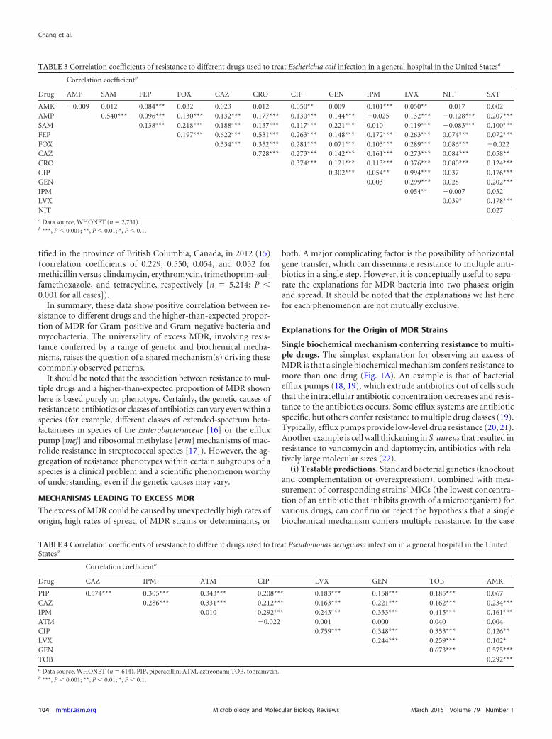
TABLE 3 Correlation coefficients of resistance to different drugs used to treat Escherichia coli infection in a general hospital in the United Statesa
Drug
Correlation coefficientb
AMP SAM FEP FOX CAZ CRO CIP GEN IPM LVX NIT SXT
AMK �0.009 0.012 0.084*** 0.032 0.023 0.012 0.050** 0.009 0.101*** 0.050** �0.017 0.002AMP 0.540*** 0.096*** 0.130*** 0.132*** 0.177*** 0.130*** 0.144*** �0.025 0.132*** �0.128*** 0.207***SAM 0.138*** 0.218*** 0.188*** 0.137*** 0.117*** 0.221*** 0.010 0.119*** �0.083*** 0.100***FEP 0.197*** 0.622*** 0.531*** 0.263*** 0.148*** 0.172*** 0.263*** 0.074*** 0.072***FOX 0.334*** 0.352*** 0.281*** 0.071*** 0.103*** 0.289*** 0.086*** �0.022CAZ 0.728*** 0.273*** 0.142*** 0.161*** 0.273*** 0.084*** 0.058**CRO 0.374*** 0.121*** 0.113*** 0.376*** 0.080*** 0.124***CIP 0.302*** 0.054** 0.994*** 0.037 0.176***GEN 0.003 0.299*** 0.028 0.202***IPM 0.054** �0.007 0.032LVX 0.039* 0.178***NIT 0.027a Data source, WHONET (n � 2,731).b ***, P � 0.001; **, P � 0.01; *, P � 0.1.
TABLE 4 Correlation coefficients of resistance to different drugs used to treat Pseudomonas aeruginosa infection in a general hospital in the UnitedStatesa
Drug
Correlation coefficientb
CAZ IPM ATM CIP LVX GEN TOB AMK
PIP 0.574*** 0.305*** 0.343*** 0.208*** 0.183*** 0.158*** 0.185*** 0.067CAZ 0.286*** 0.331*** 0.212*** 0.163*** 0.221*** 0.162*** 0.234***IPM 0.010 0.292*** 0.243*** 0.333*** 0.415*** 0.161***ATM �0.022 0.001 0.000 0.040 0.004CIP 0.759*** 0.348*** 0.353*** 0.126**LVX 0.244*** 0.259*** 0.102*GEN 0.673*** 0.575***TOB 0.292***a Data source, WHONET (n � 614). PIP, piperacillin; ATM, aztreonam; TOB, tobramycin.b ***, P � 0.001; **, P � 0.01; *, P � 0.1.
Chang et al.
104 mmbr.asm.org March 2015 Volume 79 Number 1Microbiology and Molecular Biology Reviews

where this is the only mechanism conferring resistance, the phe-notypes should be found together without exception. For exam-ple, the deletion of the efflux pump gene ifrA in Mycobacteriumsmegmatis decreased the MIC (that is, increased the susceptibility)to multiple drugs, and the overexpression of the same gene in-creased resistance to multiple drugs (23).
Another testable hypothesis is that the diversity and intensityof antimicrobial use in settings such as hospitals select for geneticchanges that expand the substrate specificity of resistance mecha-nisms. In vitro evolution of plasmid-borne TEM beta-lactamaseswith alternating exposure to a penicillin and a cephalosporin wasshown to select for dual specificity for these two related com-pounds; moreover, the sequence changes that evolved mimickedthose observed in human isolates, while exposure of plasmid-bearing bacteria to only one of the two antimicrobial compoundsproduced sequence changes in the beta-lactamase that were notpreviously observed in human isolates (24).
(ii) Practical implications. Development of efficient effluxpump inhibitors may lead to the renewed effectiveness of severaldrugs in treating infections for which this mechanism is important(25, 26). For example, verapamil, an efflux pump inhibitor, hasbeen shown to increase antituberculosis drug efficacy in mice(27). It should, however, be noted that efflux pump inhibitorsshould have limited effects on the host to have clinical utility.Those clinically available, such as verapamil and reserpine, havehost pharmacological effects that limit their utility as antimicro-bial therapies (25, 28).
Genetic linkage. MDR strains may arise because determinantsof resistance to multiple drug classes are genetically linked, be-cause they are either physically close on bacterial chromosomes(thus coinherited vertically and potentially cotransformed whenshared horizontally) or on the same horizontally transmitted ele-ment, such as a plasmid or conjugative transposon (Fig. 1B). In
such cases, when a strain acquires one resistance phenotype, itacquires many.
Horizontally transmitted elements, such as transposons, in-tegrons, and plasmids, are often modular and can incorporatenew elements over time (29, 30). Transposons contain genesencoding transposases that facilitate incorporation to and fromother genomic regions (18). Integrons accumulate gene cas-settes with specific recombination sites through site-specificrecombinases encoded by the integrons themselves. Trans-posons and integrons may appear on the chromosome or on aplasmid. These elements may be transmitted within and be-tween species through transformation, transduction, and/orconjugation (reviewed in reference 18).
The genetic mechanisms (cassette-based recombination, con-jugation, and transposition) underlying these mobile elements areincreasingly well understood (29, 30); however, the selective pres-sures that keep these resistance elements together (despite possi-ble fitness costs for some of the genes involved) are less clear.
(i) Examples. Conjugative transposon Tn1545 in S. pneumoniaecontains genes for resistance to multiple antibiotics, including ka-namycin, macrolide-lincosamide-streptogramin B-type antibiot-ics, and tetracycline, and is capable of transferring to a new bacte-rial host cell via conjugation or transposition to another genomicregion (31). Integrons in Enterobacteriaceae carry genes encodingresistance to unrelated antibiotics such as beta-lactams, aminogly-cosides, sulfonamides, and chloramphenicol (32). There are alsoexamples of plasmids without integrons in Enterobacteriaceae car-rying antibiotic resistance genes for multiple drug classes, such asaminoglycosides, beta-lactams, tetracycline, chloramphenicol,and sulfamethoxazole (33).
(ii) Testable predictions. In contrast with multiple resistancemechanisms encoded by a single gene or operon, these determi-nants are not necessarily always found in combination with oneanother but are encoded by genetic determinants that are adjacenton the chromosome or plasmid. This prediction can be tested byknockout, complementation, and/or sequencing of chromosomesor plasmids. Deleting each gene and comparing the MICs beforeand after deletion can test whether genes on the same chromo-some or plasmid confer resistance to different drugs. If genes con-ferring resistance to each drug are known, sequencing of chromo-somes or plasmids can confirm the genetic linkage of these geneson the same chromosome or plasmid.
(iii) Practical implications. The initial appearance of multiple-drug resistance on a single genetic element likely occurs when
TABLE 5 Correlation coefficients of resistance to different TB drugsamong pretreatment casesa
Drug
Correlation coefficientb
RMP EMB STM
INH 0.683 0.554 0.559RMP 0.621 0.481EMB 0.417a INH, isoniazid; RMP, rifampin; EMB, ethambutol; STM, streptomycin.b All of the cases have P values of � 0.001 (n � 18,619).
TABLE 6 Correlation coefficients of resistance to different drugs used to treat Staphylococcus aureus infection in a general hospital in the UnitedStatesa
Drug
Correlation coefficientb
OXA ERY CLI TCY GEN LVX SXT RIF
PEN 0.380*** 0.350*** 0.153*** 0.050* 0.046* 0.248*** 0.056** 0.027OXA 0.614*** 0.415*** 0.017 0.094*** 0.733*** 0.047* 0.123***ERY 0.420*** 0.052* 0.065** 0.603*** 0.054** 0.094***CLI 0.124*** 0.094*** 0.520*** 0.101*** 0.085***TCY 0.256*** 0.036 0.185*** 0.006GEN 0.111*** 0.304*** 0.109***LVX 0.109*** 0.140***SXT 0.064**a Data source, WHONET (n � 2,377). OXA, oxacillin; TCY, tetracycline; RIF, rifampin.b ***, P � 0.001; **, P � 0.01; *, P � 0.1.
Mechanisms of Multiple-Drug Resistance in Bacteria
March 2015 Volume 79 Number 1 mmbr.asm.org 105Microbiology and Molecular Biology Reviews

bacteria resistant to the component drugs come into proximity orcontact, such that horizontal transfer and recombination can oc-cur. In principle, an antibiotic management strategy might be de-signed to reduce the probability of such contacts. However, theo-retical studies of one such approach, known as antimicrobialcycling, have shown that cycling reduces such contacts only underrestricted conditions (34); in practice, cycling strategies are diffi-cult to implement and to study (35). Recently, an alternative strat-egy, “adjustable cycling,” in which treatment is changed when not
effective in patients, has been proposed and shown in a theoreticalmodel to suppress the emergence of MDR in most settings (36).
The clinical impact of these linked determinants depends ontheir persistence as a linked group. Improved understanding ofthe selective pressures that preserve and allow the proliferation ofthese multidrug resistance elements may improve our ability toreduce their frequency.
Highly mutable or recombinogenic bacterial lineages. Bacte-rial lineages vary in their rates of mutation. Such variation is often
FIG 1 The mechanisms for the origin and proliferation of MDR strains. Red and blue lightning bolts indicate treatments with drug A and drug B. Greenrectangles represent efflux pump genes. Red and blue rectangles represent drug A and drug B resistance determinants, respectively. (A) Efflux pump.Bacteria obtaining efflux pumps (green square) that extrude more than one antibiotic out of cells confers MDR. (B) Genetic linkage. If two resistancedeterminants are located in the same horizontally transferred element, when a strain acquires one resistance phenotype, it acquires both. (C) Differentialmutation rate. Highly mutable lineages have higher frequencies of acquiring multiple drug resistance determinants than those that are not highly mutable.(D) Multidrug therapy with accelerated treatment failure in resistant infections. If treatment fails and singly resistant strains emerge, they are likely toobtain second drug resistance and be replaced. The thickness of the arrows reflects the relative transition probabilities between states. (E) Associatedlinkage selection. Resistance to a new drug (blue) occurs on a background of resistance to an older drug (red) following a change in treatment practicesfrom drug A to drug B. The resistance to the older drug continues spreading because of the linkage to the resistance of the new drug and the selectivepressure from the usage of the new drug. (F) Bystander selection. Resistance in one bacterium is advantageous because it allows strains of that species tosurvive when the host is treated for another infection with a drug that also kills that species. For example, if drug A is used to treat this species and drugB is used to treat another infection in the same patients, MDR strains survive and strains resistant only to drug A are killed. (G) Positive epistasis. If thecost of MDR is smaller than the total cost of each resistance determinant on its own, MDR strains may outcompete strains with a limited number ofresistance elements and spread more quickly. (H) Niche differentiation. Multiple unrelated drug classes may be used more frequently in certainpopulation subgroups, resulting in an excess of MDR when the high- and low-use subgroups/settings are considered together. (I) Importation of MDRstrains. The MDR strains from a high-drug-use “source” population are introduced into a lower-drug-use “sink” population and are able to spread as aresult of competing successfully with pan-susceptible strains due to their resistance to the drug (blue) used in the sink population.
Chang et al.
106 mmbr.asm.org March 2015 Volume 79 Number 1Microbiology and Molecular Biology Reviews

due to variation in genes involved in DNA proofreading, such asthe mismatch repair system (37). Additionally, certain bacteriallineages vary in their ability to accept and integrate transformingDNA (38). Because mutation and recombination are the sourcesof drug resistance genes and alleles, more highly mutable or highly“recombinogenic” lineages should have higher frequencies ofmultiple-drug resistance determinants, so that in the populationas a whole, these lineages would likely contribute disproportion-ately to the frequency of multiple-drug resistance (Fig. 1C).
(i) Examples. Such a process has been proposed for the accu-mulation of MDR in highly mutable lineages, such as the Beijinglineage, in tuberculosis (39). Combining mathematical modeling(40) with experimental measurements of resistance mutationrates, Ford et al. (39) argued that higher mutation rates in somestrains could lead to the presence of multiply drug-resistant vari-ants within a patient at the start of treatment. Such preexistingresistant variants could increase the risk of treatment failure.
In S. pneumoniae, resistance to multiple drugs is more com-mon in lineages showing evidence of higher rates of recombina-tion (41), a finding that has been supported by populationgenomic studies (38, 42, 43).
(ii) Complexities. Antibiotic exposure at sublethal concentra-tions can induce elevated mutation rates in bacteria (44, 45).Therefore, antibiotic exposure, which is generally seen as a selec-tive force, may also play a role in the generation of mutations. Asa consequence, mutation rates during treatment (especially whendrug concentrations are low) may be elevated compared to thosemeasured in antibiotic-free settings. This mutation rate increase isnot specific to mutations conferring resistance to the drug admin-istered but rather is a general elevation due to DNA damagecaused by reactive oxygen species created by the bacteria underantibiotic stress (45).
Mutation and recombination rates are also traits under selec-tion in their own right. Selection in changing environments (46–48) and specifically for resistance to multiple antibiotics (49) mayprovide second-order selection for elevated mutation rates. Thus,high mutation rates may be both a cause and a consequence ofmultiple-drug resistance.
(iii) Testable predictions. A straightforward test of the hypoth-esis that higher rates of mutation and recombination lead to fasteraccumulation of multiple-drug resistance is to assess whetherpresent-day MDR strains show elevated rates of mutation or re-combination compared to those of non-MDR strains. It may bepossible to identify susceptible ancestral strains to currently ob-served MDR strains by phylogenetic analysis, and the mutationalor recombinational assays could then be performed on these an-cestral strains if historical samples are available.
If the variation in recombination rate contributes to the highlevel of MDR, strains with higher recombination rate are expectedto acquire MDR with a higher probability. Recombination eventscan be inferred in a bacterial lineage from the existence of tracts ofhigh densities of single-nucleotide polymorphisms (SNPs), indi-cating import of genetic material from a diverged source (43).Those sites at which recombination has not occurred can then beused to estimate a phylogeny and molecular clock by conventionalmeans, and inferred recombination events can be mapped ontothe tree topology for estimating the ratio of recombination tomutation rate (for an example, see reference 43). These analysesmay be performed using the software packages ClonalFrame andGubbins (43, 50, 51). It should be noted that this approach as-
sumes that the genomes under analysis are closely related, thatthere has not been time for extensive clustering of SNPs to havebeen produced in genes under selection, and that anomalous con-centrations of SNPs are therefore best explained by recombinationwith a divergent donor strain that is not in the data set.
If variation in mutation rate leads to the excess of MDR, theseMDR strains are expected to show higher substitution rates atsynonymous sites or other neutral sites such as pseudogenes, be-cause neutral theory predicts that the substitution rate of neutralsites equals the mutation rate (52). Population genetic tools, suchas the PAML package (53), can be used to estimate lineage-specificsubstitution rates when sequences from both an outgroup andstrains of interest are available. However, the amounts of homol-ogous recombination thought to occur in many bacterial speciespresents significant challenges to phylogenetic and population ge-netic analyses (54) and may produce considerable biases in esti-mates of mutation rate (55).
While elevated mutation or recombination rates in MDR lin-eages (39) are evidence in favor of this mechanism, the absence ofhigher rates in MDR strains does not disprove it. For example,recombination or mutation rates may have varied over the evolu-tionary history of the lineage, such that multiple resistance accu-mulated while the lineage was highly mutable/susceptible to re-combination, but this trait may have changed since the resistancedeterminants were acquired; this is a particular risk for highlychangeable lineages (56).
(iv) Practical implications. The practice of combination treat-ment with multiple drug classes, described a century ago by Eh-rlich (57), remains a standard approach for treatment of infec-tions, such as tuberculosis, in which resistance occurs primarily bymutation and mutants resistant to any single drug are expected tobe present in many infected hosts (58). If it were possible to assessthe genetic background, and hence the likely mutation rate, of apathogen infecting an individual, it is possible that the number ordose of drugs would be increased in treating highly mutable lin-eages to counteract the risk of preexisting MDR strains in a pre-dominantly drug-susceptible infection. While fanciful at the pres-ent, as rapid pathogen genome sequencing becomes morecommon in diagnostic microbiology, the practicality of this ap-proach may also increase (59, 60).
Multidrug therapy with accelerated treatment failure in resis-tant infections. As noted in the preceding section, multidrug ther-apy has long been recommended for treating infections in whichresistance is acquired by mutation to ensure that mutants that areresistant to one drug are killed by other drugs in the “cocktail”(57). This strategy is motivated by the idea that the probability ofobtaining mutants resistant to multiple drugs with differentmechanisms is much smaller than that of obtaining singly resis-tant mutants, and using drug combinations is more likely to kill allthe bacteria, preventing the emergence of drug resistance. In par-ticular, if the product of the expected frequency of bacteria resis-tant to all drugs being used (P) and the bacterial population size(N) is much less than one (NP �� 1), most hosts will harbor nobacteria which are resistant to every drug being taken. This calcu-lation is typically made on the assumptions that the infecting in-oculum is susceptible to all drugs in the regimen and that anyresistant mutants will arise during the course of replication of thebacterial population within the host, at a predictable frequency(40). If, however, the infecting inoculum is already composed ofbacteria resistant to one or more of the drugs used, the frequency
Mechanisms of Multiple-Drug Resistance in Bacteria
March 2015 Volume 79 Number 1 mmbr.asm.org 107Microbiology and Molecular Biology Reviews

of bacteria resistant to all the other drugs will be considerablyhigher than assumed and possibly high enough to make NP � 1,indicating that a mutant resistant to all drugs used may be presentand able to replicate. In simple terms, the emergence of multipleresistance is much more likely from a singly resistant precursorthan from a pan-susceptible precursor; the first resistance muta-tion may form a “slippery slope” facilitating the emergence andselection of further resistance mutations. In this mechanism, un-der multidrug therapy, the singly resistant state is unstable, andthose strains will be replaced by strains that have acquired muta-tions for additional drug resistance, leading to an excess of MDR(61, 62) (Fig. 1D).
(i) Example. As drug resistance in M. tuberculosis is generallycaused by spontaneous mutations (63) and the population of M.tuberculosis within a patient is large enough to generate drug-resistant mutants (64), multidrug chemotherapy is typically usedfor treating tuberculosis (58) in order to reduce the probability ofemerging drug resistance.
One problem that has been observed in treatment of tubercu-losis is the existence of mixed infections with a subpopulation ofresistant organisms, which may be undetected when treatment isinitiated but is rapidly selected by first-line treatment tailored tothe assumed full susceptibility of the infecting strain (61, 62).
Even starting from a susceptible inoculum, singly resistantstrains (or in rare cases, even doubly resistant strains [40]) couldbe present due to patient noncompliance with the treatment reg-imen, such as interrupted antibiotic treatment or insufficient dos-age (65). Directly observed therapy for tuberculosis improves pa-tient adherence and decreases the frequency of acquired drugresistance (66).
(ii) Testable predictions. Evidence for the importance of thismechanism would include a high frequency of MDR (but notsingly resistant strains) developing during multidrug therapy ofinfections for which the drug susceptibility of the initial infectionwas unknown or mismeasured, perhaps due to a mixed infectionor a reinfection (61, 62). If genetic mechanisms of drug resistanceare known, sequences of bacteria isolated at different time pointsfrom the same patients could be used to test this hypothesis. It isexpected under this mechanism that singly resistant strains werepresent at the beginning of treatment and MDR emerged later on,and thus a gene(s) or mutation(s) conferring single-drug resis-tance is expected to be found in sequences of isolates from thebeginning of treatment. Of course, if the singly resistant infectionwere known at the time of treatment initiation, it would be inap-propriate to treat it as if it were pan-susceptible. Thus, to knowabout this problem (in a timely fashion) is to try to prevent it, andonly retrospective investigations are likely to find evidence for thisproblem (61, 62).
(iii) Practical implications. Standard WHO-recommended re-treatment regimens for tuberculosis have historically called foraddition of a single drug to a failing four-drug regimen. Such anapproach resulted in many treatment failures due to the acquisi-tion of resistance to the single effective drug that was added(67, 68).
In settings where this mechanism plays a role, in practicemainly tuberculosis, there is a special value of rapid diagnosticsthat can assess drug susceptibility at baseline, such as GeneXpertMTB/Rif (69) or rapid pathogen genome sequencing, becausesuch diagnostics could confirm that the majority population ofthe infecting organism has susceptibility to enough drugs to pre-
vent the emergence of further resistance (perhaps requiring addi-tion or substitution of drugs in the standard regimen, when resis-tance to one or more drugs is found at baseline). This diagnosticinformation would not only improve patient outcomes but alsoreduce the rate at which new multiresistant strains are generated.In the absence of such baseline diagnostics, the use of extra drugsin a multidrug regimen might be warranted when the presence ofresistance to one or more drugs in the patient is suspected due topopulation history or patient risk factors. Because singly drug-resistant strains are more likely to arise due to patient noncompli-ance with the treatment regimen, directly observed therapy mayhelp to reduce the possibility of the excess of MDR due to treat-ment failure.
Explanations for the Proliferation of MDR Strains
Once MDR bacteria have emerged in one or more hosts, theirproliferation depends on their ability to survive and be transmit-ted to other hosts, and changes in their relative frequency reflectnatural selection—their differential survival and transmissioncompared to other lineages. In this section, we consider five mech-anisms that can provide a selective advantage to MDR strains,leading to increases in their frequency.
Associated linkage selection. The proliferation of a gene (andthe strain harboring it) need not be the consequence of directselection upon that gene and the trait it encodes but may resultfrom selection of others that are inherited along with it. This is thephenomenon of linkage, and it is especially hard to disentangle inpartially clonal organisms like most bacteria. If resistance to a newdrug occurs on a genetic background of resistance to older drugsfollowing a change in treatment practices, then resistance to theolder drugs can continue spreading because of the linkage to theresistance to the new drug, selected by use of the new drug (Fig.1E). Resistance to new drugs is likely to arise on the background ofresistance to the older drugs because the frequency of older drugresistance is likely to be high due to longstanding selection pres-sure imposed by usage of the older drugs (70). This can be exac-erbated if particular genetic backgrounds are more able to toleratethe fitness costs of resistance determinants, either because the fit-ness costs are lower in these backgrounds or because these back-grounds have higher fitness to begin with and hence can bettertolerate a given fitness cost (see also “Positive epistasis betweendrug resistance determinants or between resistance determinantsand genetic background” below).
(i) Examples. Regarding E. coli, although sulfonamide prescrip-tions in the United Kingdom decreased greatly, the proportion ofsulfonamide-resistant E. coli did not decline (6). One possible ex-planation is the close linkage between sulfonamide resistancegenes and other antibiotic resistance determinants and their con-tinued selection through the usage of other antibiotics. sulII genesencoding sulfonamide resistance in E. coli are located on plasmidscarrying several resistance determinants (6). A follow-up study 5years later reported the persistence of sulfonamide resistance andthe continued association between sulfonamide resistance and re-sistance to other drugs (71).
In S. pneumoniae infections, antibiotics other than penicillin,such as azithromycin (72), erythromycin, trimethoprim-sulfame-thoxazole (73, 74), and cephalosporins (75), may select more ef-ficiently for penicillin-resistant strains than penicillin itself. Whilethe MICs associated with resistance to azithromycin are fartherabove clinically achievable concentrations, penicillin dosing can
Chang et al.
108 mmbr.asm.org March 2015 Volume 79 Number 1Microbiology and Molecular Biology Reviews

be increased to combat less-susceptible strains and still have lim-ited effects on toxicity experienced by the patient. As clinicallyachievable levels of macrolides (such as azithromycin) kill suscep-tible but not resistant bacteria while clinically achievable levels ofpenicillin may have some inhibitory effect on resistant strains,macrolides are more selective killers of susceptible bacteria thanpenicillin. If most resistance is multidrug resistance, then penicil-lin resistance will be coselected with macrolide resistance. This isexacerbated in drugs with a long half-life (e.g., azithromycin),which allows them to sustain high concentrations for a longerperiod.
(ii) Testable predictions. Associated linkage selection shouldbe tested as a mechanism for persistence of resistance to previouslyused drugs after the use of such drugs is reduced in a population.If the persisting lineages are resistant to both the previously useddrug and the drug(s) that is used in its place, then this could beevidence of associated linkage selection. Within individuals, theselective role of one drug in promoting resistance to another, un-related drug may be assessed by comparing the prevalence of re-sistance to the second drug in individuals treated with the first, asin the S. pneumoniae examples described above.
In ecological studies of the relationship between antimicrobialuse and resistance, associations between use of one drug and pop-ulation-level resistance to another are expected when associatedlinkage selection is at play. However, given the positive correla-tions across populations between high use of some antimicrobialclasses and high use of other classes, such data may also reflectdirect effects of each drug class on resistance to itself. For thisreason, individual-level data, such as those described above for S.pneumoniae, are more informative.
Using sequence data from good-quality longitudinal samplesdocumenting the emergence of resistance, phylogenetic analysiscould also be used to test this hypothesis. If resistance to the newdrug arose in a background of resistance to older drugs, we wouldsee in phylogenetic trees that the strains with the new drug resis-tance would coalesce with the older drug-resistant strains morerecently than the coalescence between the older drug-resistant andolder drug-susceptible strains. Such inference may be more com-plicated when resistance is encoded by mobile genetic elements.
(iii) Practical implications. In principle, recommendations fordrug choice for treating a particular infection could be changedwhen resistance to a currently used drug is still low, so the newdrug resistance is less likely to happen in the genetic backgroundof resistance to older drugs. The practicality of this idea is limitedby the paucity of alternative classes of drugs available, and itshould be noted that such a policy (i.e., recommending a newfirst-line treatment when the frequency of resistance to the currenttreatment exceeds 5%) (76) has not prevented the success of MDRstrains of N. gonorrhoeae in the United States. (The effectivenessmay be reduced by importation of MDR strains; see “Importationof MDR strains and geographic source-sink dynamics” below.)The potential of this approach, i.e., changing drug choice recom-mendations when resistance to a currently used drug is low, likelydepends on factors such as the relative fitness advantages of dif-ferent resistance patterns. Modeling may help determine any cir-cumstances in which this strategy is advantageous.
Perhaps a more effective strategy, when drugs vary in theirtendency to select MDR strains within a host during treatment, isto reduce selection for resistance by choosing a drug regimen thatis less likely to select for MDR strains while maintaining treatment
efficacy. For example, it has been suggested that choosing amoxi-cillin-clavulanate over azithromycin can accomplish these twingoals in treatment of acute otitis media (77). Here, as describedabove, the explanation seems to be that MICs of strains resistant toamoxicillin-clavulanate are typically close to clinically achievableconcentrations, so there may be some effect of this combinationon even the “resistant” strains, whereas azithromycin-resistantstrains have MICs far above in vivo concentrations and hence arelittle affected by treatment.
There has been much discussion of the need for new diagnos-tics which would permit rapid assessment of the resistance phe-notype of an infection, permitting tailoring of the treatment to theindividual resistance phenotype, rather than the “conservative”approach of empirical therapy with a drug that is statistically likelyto be effective in the absence of knowledge of the susceptibilityprofile of a particular patient’s infection (78). Predictions of the-oretical models may help to assess the effects of such diagnosticsand treatment protocols on the selection of singly and multiplyresistant strains. One model of the impact of the GeneXpert MTB/RIF diagnostic for tuberculosis infection and rifampin resistanceindicated that the system’s use would reduce the absolute preva-lence of MDR TB, but because the projected effect on reducing theprevalence of drug-susceptible forms of TB was more dramatic,the relative proportion of MDR in the TB population would in-crease (2). This finding emphasizes the point that reducing diseaseburden overall, specifically MDR disease burden, is the major goalof public health interventions. While the proportion of diseaseburden that is MDR may be easier to measure, reducing this pro-portion should rarely if ever be a goal in itself, given that one canreduce MDR and non-MDR disease and have either an increase ordecrease in the proportion of MDR.
Bystander selection. When a drug is used to treat infection witha particular species, other species carried by that same host (“by-standers”) may be affected by the treatment, and multidrug resis-tant variants of these bystander species may have an advantageunder a varying regimen of treatments experienced by differenthosts (Fig. 1F).
(i) Examples. Selection for resistance to antimicrobials in com-mensal organisms, such as the normal flora of the digestive andupper respiratory tracts, must occur by this mechanism, sincecommensals by definition are not causing infections yet are sub-ject to selection by systemic antibiotics (79, 80). During broad-spectrum antimicrobial therapy, the proportion of resistantstrains of commensal organisms in the gut may increase (81). Theselective agent need not be even an antimicrobial used for treat-ment but may be another selective agent that favors a trait linkedto drug resistance in commensals. For example, exposure of com-mensal gut flora to mercury during installation and removal ofdental fillings selected for drug-resistant strains that were alsomercury resistant (82). Genes conferring resistance to environ-mental hazards such as heavy metals are often transferred betweenlineages together with antibiotic resistance genes on plasmids(83).
Penicillins and tetracyclines were recommended for treatinggonorrhea in the United States prior to 1993 and then were re-placed by fluoroquinolones and later by cephalosporins. Duringthe period of the rise in the proportion of fluoroquinolone-resis-tant N. gonorrhoeae strains, the MDR strains resistant to fluoro-quinolones, penicillin, and tetracycline increased faster than otherfluoroquinolone-resistant types even though penicillins and tet-
Mechanisms of Multiple-Drug Resistance in Bacteria
March 2015 Volume 79 Number 1 mmbr.asm.org 109Microbiology and Molecular Biology Reviews

racyclines were no longer recommended for treatment. It has beensuggested that during the same period, although penicillins andtetracyclines were not recommended for treating gonorrhea, pa-tients with asymptomatic gonorrhea might have been treated withpenicillins and/or tetracyclines for infections other than gonor-rhea, and therefore MDR N. gonorrhoeae strains were selected dueto their resistance to penicillin and tetracycline (7), a suggestionthat has also been proposed in the United Kingdom (84).
While bystander selection is most often considered for com-mensal or asymptomatically infecting organisms, it may also oc-cur when an infection is undiagnosed or misdiagnosed, and treat-ment is directed at the (nonexistent) infection the patient isthought to have. A major driver of fluoroquinolone resistance inM. tuberculosis may be the use of fluoroquinolone monotherapyamong individuals with unrecognized pulmonary tuberculosisbeing treated for presumptive community-acquired pneumonia(85). This can lead to multiple resistance if the M. tuberculosisstrains were already resistant to other drugs or later become soduring treatment.
(ii) Testable predictions. In assessing the relationship betweenantimicrobial use and resistance in a particular pathogen, it maybe more relevant to consider total prescriptions than to considerprescriptions for treatment of that pathogen, a practice that hasbeen common in studies of commensal colonizing bacteria (62,86, 87) but has not to our knowledge been examined, for example,in the setting of sexually transmitted diseases. If bacteria of speciesA resistant to drugs that are no longer used for treatment of speciesA infections are found to persist especially in settings where thesedrugs are used for treatment of other infections, and resistancemutations are deleterious, bystander selection would be a likelyhypothesis to explain their persistence. More generally, a predic-tion of this mechanism is that when comparing bacterial popula-tions across space and/or time, the prevalence of particular resis-tance phenotypes should be positively correlated across differentnamed bacterial species, a phenomenon that has been called core-sistance (88).
(iii) Practical implications. The existence of bystander selec-tion is cited as a reason to prefer narrow-spectrum antimicrobialsthat can target the infecting pathogens without affecting bystand-ers. For example, current U.S. guidelines for treatment of uncom-plicated urinary tract infections recommend narrow-spectrumagents for this reason, which is broadly termed avoidance of “col-lateral damage” (89).
Much of the clinical judgment around antimicrobial steward-ship and avoiding unnecessary use comes from a concern aboutthe bystander effects on the individual’s commensal flora (90).While they are correct for broad-spectrum antibiotics, such con-cerns are not evidence based in the case of narrow-spectrum anti-infective agents, including many antituberculosis drugs and anti-viral drugs. There are many reasons not to overuse such drugs,including side effects and cost. However, inadvertent oseltamivirtreatment of an individual not infected with influenza virus, forexample, is unlikely to have any effect on resistance: if the infec-tion is not there and the bystander flora are not affected by osel-tamivir, there is no species on which bystander selection can act.
Positive epistasis between drug resistance determinants orbetween resistance determinants and genetic background. Fit-ness is defined as the ability to survive and leave offspring in thepopulation. The presence of resistance has been shown in some(but not all) cases to lead to a “fitness cost,” reducing the growth
and survival of resistant strains in the absence of antibiotics (91). Afundamental factor determining the success of combinations ofthese resistance genes is how they interact in epistasis. Epistasisoccurs when the combined fitness effect of multiple alleles fromdifferent loci is different from the sum of the individual alleleeffects. Epistasis is widespread in eukaryotes (92, 93), bacteria(94), and viruses (95, 96). If the cost of MDR in the absence ofantimicrobial use is smaller than the total cost of each resistancedeterminant on its own (positive epistasis), MDR strains may out-compete strains with a limited number of resistance elements andspread more quickly (Fig. 1G). [In a continuous-time model, pos-itive epistasis is defined by comparing the fitness cost of MDR andthe “sum” of the fitness cost of each resistance determinant; in adiscrete-time model, assuming that the costs of resistance to twodrugs are c1 and c2, no epistasis means that the cost of MDR isequal to 1 � (1 � c1)(1 � c2).]
Alternatively, if drugs that are used have interactions with eachother (97) and the combined effect of multiple drugs is higherthan the total of the individual effects (“synergistic effects”), theselective pressure of MDR is greater than would be expected if theeffects of drugs were additive. This may create epistatic fitnessinteractions between resistance determinants in the setting ofmultiple-drug treatment if resistance to one drug is more fitnessenhancing in a strain which is already resistant to other drugs. Thisprocess, if it occurs, may lead to a disproportionate increase in thefrequency of MDR strains relative to that of singly resistant strains.
(i) Examples. Positive epistasis is pervasive among alleles con-ferring resistance to different antibiotics (quinolone, rifampin,and streptomycin) in E. coli and may explain the high level ofMDR in E. coli (98). Additionally, in P. aeruginosa the cost ofacquiring streptomycin resistance mutations is greater in a rifam-pin-sensitive background than in a rifampin-resistant back-ground (99), indicating positive epistasis and suggesting thatstrains resistant to both streptomycin and rifampin are selectivelymore favored than strains with only streptomycin resistance evenwhen rifampin is not used for treatment.
Moreover, epistasis may exist between the genetic backgroundand drug resistance determinants. As an example, the fitness costsof resistance mutations in the genetic background of Beijing lin-eage M. tuberculosis are smaller than those in other genetic back-grounds, or compensatory mutations are in easier reach for them,possibly explaining the association between Beijing TB genotypeand MDR (100). In a mouse model of gonococcal infection, thefitness cost of fluoroquinolone resistance mutations depends ongenetic background (101).
(ii) Testable predictions. Phylogenetic methods (95, 102) havebeen developed to test for the presence of epistasis in general,based on the idea that sites with epistatic interactions will tend toshow correlated substitutions within phylogenies. These ap-proaches can in principle be applied to the context of multiple-drug resistance, although this may be hampered by the presence ofhorizontal transfer, which makes it hard to know whether cooc-currence of mutations is genuinely independent or merelythrough introduction from the same source by recombination.Likewise, experimental measurements of the fitnesses of differentgenotypes (103) could also be used for detecting epistasis betweenresistance determinants. In such settings, it is important to distin-guish between epistatic interactions in the drug-exposed anddrug-free settings, which may not always be the same.
(iii) Practical implications. In principle, if positive epistasis be-
Chang et al.
110 mmbr.asm.org March 2015 Volume 79 Number 1Microbiology and Molecular Biology Reviews

tween resistance to different drugs could be detected experimen-tally before the emergence of MDR, we might avoid using combi-nations of drugs for which resistance determinants may havepositive epistasis and increase the spread of MDR. However, thedownside of such a choice would be to reduce the usage of highlysynergistic combinations of drugs, which may be valuable thera-peutically (97).
Niche differentiation: aggregation of multiple drug selectionpressures within specific populations. The use of multiple unre-lated drug classes is higher in certain population subgroups, suchas young children and the elderly (104) and sexually active personswith a high incidence of sexually transmitted infection (105). Useis also higher in certain settings, such as hospitals and long-term-care facilities, than in the general population. Antibiotic use alsovaries geographically, both within (5) and between (86) countries.The prevalence of resistance to each of these drugs may be higherin the high-use settings/subgroups, resulting in an excess of MDRwhen the high- and low-use subgroups/settings are consideredtogether (Fig. 1H). In population genetic terms, this is an exampleof the Wahlund effect, in which associations between allele fre-quencies are created when two partially or fully distinct popula-tions are considered together (106, 107).
(i) Examples. High levels of use of multiple antimicrobialclasses in hospitals are thought to account for the high prevalenceof multiresistant organisms in these settings, with particularlyhigh levels of use and resistance in intensive care units (108).
Antibiotic use, including the use of classes such as macrolides,penicillins, and cephalosporins, is higher in children under 5 thanin older children or adults in the United States (87, 104). Youngage is a risk factor for resistance to multiple drugs in commoncolonizing bacteria such as S. pneumoniae (109). A notable “ex-ception that proves the rule” in the case of S. pneumoniae is resis-tance to fluoroquinolones, which appears sporadically and is onlyvery weakly associated with resistance to other drug classes (Table1). Fluoroquinolones are not commonly used in children due toside effects and hence do not contribute to the common selectiveforce for multiple resistance in children, who appear to be a “coregroup” for S. pneumoniae transmission (110, 111). Thus, thismechanism would predict exactly the pattern observed: high levelsof correlated MDR to all drug classes except fluoroquinolones.
(ii) Testable predictions. A signal of this mechanism is popu-lation admixture, such that the multiresistant strains form a sub-population genetically distinct from the susceptible ones, evenafter excluding sites encoding resistance mechanisms. Such ad-mixture may be detected by using F-statistics (112, 113), princi-pal-component analysis (114), or clustering methods (115) ongenetic data after resistance-determining sites have been removed.
(iii) Practical implications. Identification of specific popula-tions at risk for multiple resistance may aid in the selection ofempirical antimicrobial regimens that maximize the probabilityof treatment success. In situations where the highly treated groupis also a “core group” that is a source of transmission to othergroups, the avoidance of a particular antimicrobial class in thecore group may preserve treatment options for the noncoregroup, as they are unlikely to be infected with a strain resistant tothat class. Such an argument has been suggested as an additionalreason to avoid fluoroquinolone use in children, in order to pre-serve the effectiveness of this class for treating adults (3).
Importation of MDR strains and geographic source-sink dy-namics. High levels of antimicrobial use in certain populations
may increase the prevalence of resistance to many different drugclasses. If some of the above-described mechanisms are operative,this may lead to an excess of MDR strains within the population,or perhaps MDR strains will simply be at a high frequency giventhe frequency of each resistance determinant in this population.Either way, the MDR strains in this “source” population may thenbe introduced into other, “sink” populations, where they spread,competing successfully with pan-susceptible strains because theyhave resistance to the drug(s) used in these recipient populations(Fig. 1I). The result is that most strains in the sink populations areeither pan-susceptible or multiply resistant. As in other cases ofpopulation admixture, the sink population reflects a mix of nativeand imported strains, and the imported strains create an associa-tion between resistance to one drug and resistance to others. Thisprocess is different from the one described above because it canhappen in a single, truly well-mixed population, as long as there issome importation of strains from another, largely independentpopulation.
(i) Example. With N. gonorrhoeae, East Asian strains arethought to have entered western North America and spread east-ward due to travelers bringing MDR strains (11, 116). The coun-tries that are highly connected to the Pacific Rim show similarepidemiological patterns (116).
(ii) Testable predictions. If MDR strains are imported, theyhave a genetic background different from that of native strains. Inthis case, similar to niche differentiation in the mechanism de-scribed above, genetic differentiation between multiply resistantstrains and native strains is expected to be high and could be ex-amined by the methods listed for that mechanism after excludingresistance determinant sites. If genetic data from parental andadmixed populations are available, the admixture proportionfrom each parental population can be estimated by maximum-likelihood methods (117) and Bayesian approaches (118, 119).Epidemiologically, populations that have similar exposure to thesource populations are expected to show similar patterns of drugresistance. It is also expected that the frequency of travel betweensource populations and sink populations is related to the appear-ance of MDR strains in sink populations.
From an individual risk factor perspective, studies can assessthe extent to which MDR strains are associated with migrationfrom (120, 121) or travel to high-resistance areas. This mechanismpredicts that when MDR is rare, such associations should bestrong, but as the MDR strains spread endemically within the“sink” population, the association may decline (7).
(iii) Practical implications. To the extent that multiple-drugresistance in many populations is a consequence of importationfrom the highest-use populations, international coordination ofantimicrobial control policies becomes increasingly important.Moreover, heightened surveillance for MDR strains among trav-elers may be appropriate, as a means of detecting and delayingfurther spread of such strains. Once the MDR strains becomewidespread in a new population, however, such measures may beof little value.
CONCLUSIONS
Excess MDR is observed in many bacterial species. We have con-sidered nine possible explanatory genetic and epidemiologicalmechanisms. We first considered mechanisms for the appearanceof MDR strains, including individual biochemical mechanismsresponsible for the MDR phenotype (e.g., efflux pumps), genetic
Mechanisms of Multiple-Drug Resistance in Bacteria
March 2015 Volume 79 Number 1 mmbr.asm.org 111Microbiology and Molecular Biology Reviews

linkage, differential mutation or recombination rate, and multi-drug therapy with accelerated treatment failure with multidrugresistance. Mechanisms for the proliferation of MDR strains in-clude associated linkage selection, bystander selection, positiveepistasis, niche differentiation, and importation of MDR from ahigh-use population, followed by spread in a “recipient” popula-tion.
We have documented the phenomenon of MDR frequenciesexceeding those expected from the product of individual resis-tance frequencies by counting individual patient isolates with eachphenotype. It is worth emphasizing that from an evolutionaryperspective, many bacterial isolates with a given phenotype, suchas MDR, may result from a small number of “origin” events (122).In this case, the origin of MDR may not occur at a particularly highrate, and the high prevalence of MDR must be attributed mainly tosuccessful spread of MDR strains. On the other hand, in S. pneu-moniae, even within what appeared to be a clonal lineage, therewere multiple events of gain and loss of resistance determinantsboth by point mutations and by horizontal gene transfer (43). Inthe extreme case, there may be very little clonal spread of a partic-ular resistance phenotype but rather repeated appearance in mul-tiple lineages, as appears to occur for fluoroquinolone resistancein S. pneumoniae (123, 124).
These mechanisms are not mutually exclusive; several mecha-nisms may contribute to the origin, and several to the dissemina-tion, of MDR bacteria. In our classification we have tried to sepa-rate mechanisms that reflect different kinds of events at the patientlevel: those mechanisms listed for “origin” generally refer to waysin which MDR organisms arise within a patient, while those listedfor “dissemination” generally refer to the process of spread be-tween patients. Seen at a higher level of generality, one could arguethat observing an excess of MDR reflects either a higher rate offormation of such strains or a higher fitness of such strains thanexpected from the observed rates of formation and proliferationof the individual drug resistance alleles. From a broad, populationgenetic perspective, several of these processes, which we have dis-tinguished, could be combined under the heading of epistasis(92). For example, we have described treatment failure leading tomultiple-drug resistance as a distinct process for the origin ofMDR strains. It could alternatively be described as enhanced fit-ness of resistance to a second drug in strains already resistant to afirst drug, leading to the survival of dual-resistant strains undercombination therapy. Similarly, one could recast bystander selec-tion as a form of epistasis. MDR strains have a fitness advantagehigher than the sum of fitness advantages of singly resistant strainsin the environment where individuals are treated with a range ofantibiotics. Nonetheless, because they involve different kinds ofevents at the patient level (such as multidrug therapy or treatmenttargeting another infection) and since the testable predictions andpractical implications of these mechanisms vary, we have chosento consider them separately. The unifying perspective of popula-tion genetics offers a complementary approach that notes thecommon features of these mechanisms.
If the genetic mechanism of resistance in a “bug-drug” combi-nation is point mutations, then a differential mutation rate andmultidrug therapy with treatment failure are most likely to befactors that lead to the excess of drug resistance. As mentionedabove, drug resistance in M. tuberculosis is from point mutations,and both a differential mutation rate and multidrug therapy withtreatment failure played roles in MDR in TB (39). On the other
hand, if the genetic mechanism of resistance is horizontal trans-mission of plasmids or mobile elements, then a differential recom-bination rate, genetic linkage, and associated linkage selectionmay play more important roles, because resistance determinantsthat are located in the same mobile elements or plasmids are likelyto be transmitted together. As described above, conjugative trans-posons in S. pneumoniae contain MDR determinants, and bothgenetic linkage and recombination rate play important roles in theexcess of MDR S. pneumoniae (31, 38).
The increasing ease and economy of genomic methods aremaking these tools increasingly important for studying MDRmechanisms. Hypotheses involving differential mutation/recom-bination rate (see “Highly mutable or recombinogenic bacteriallineages” above) can be tested by estimating historic mutation orrecombination rates for lineages with and without MDR fromsequencing data. If the genetic mechanisms of drug resistance areknown, sequencing of bacteria can be used to test for multiplemechanisms: the presence of a single element conferring MDR(see “Single biochemical mechanism conferring resistance to mul-tiple drugs”), genetic linkage between drug resistance determi-nants (see “Genetic linkage”), the presence of a single resistancedeterminant in strains at the beginning of treatment in longitudi-nal samples from a patient (see “Multidrug therapy with acceler-ated treatment failure in resistant infections”), the correlation inthe presence of drug resistance determinants in sequences of dif-ferent bacterial species across space or time (see “Bystander selec-tion”), and signs of population admixture (see “Niche differenti-ation: aggregation of multiple drug selection pressures withinspecific populations” and “Importation of MDR strains and geo-graphic source-sink dynamics”). Furthermore, phylogeneticmethods (125) can be used to test whether resistance to the newdrug arose in a background of resistance to older drugs; if this isthe case, strains resistant to the new drug should be more closelyrelated to those resistant to the old, compared with suitable sus-ceptible controls (see “Associated linkage selection”). Positiveepistasis could be detected by examining whether substitutionrates between different resistance mutations are correlated (see“Positive epistasis between drug resistance determinants or be-tween resistance determinants and genetic background”). Whilethe potential of these methods is clear, they are crucially depen-dent on adequate sampling and quality metadata (such as timeand place of isolation, reason for obtaining an isolate [carriagesurveillance, outbreak-driven surveillance, or clinical culture],patient characteristics, and disease manifestation). Such epidemi-ological context is invaluable in aiding the interpretation of con-clusions from phylogenies or other genomic analyses (126).
Through understanding these genetic and epidemiologicalmechanisms of MDR and using the tools we have suggestedhere to test each hypothetical mechanism, we will be able toidentify the cause(s) of the excess of MDR and design moreeffective interventions to prevent the origin and/or prolifera-tion of MDR. Although some of our practical suggestions arelimited by the availability of alternative drugs, developing andapplying quick and sensitive diagnostic tools and using nar-row-spectrum antibiotics (for example, fidaxomicin is a nar-row-spectrum antibiotic for Clostridium difficile [127]) may betwo general ways to reduce the frequency of MDR strains. Al-though development of pathogen-specific antibiotics is unlikely, itis increasingly of interest and will potentially provide more oppor-tunity to reduce the spread of MDR.
Chang et al.
112 mmbr.asm.org March 2015 Volume 79 Number 1Microbiology and Molecular Biology Reviews

ACKNOWLEDGMENTS
We are grateful to Lauren M. Childs for reading the manuscript and of-fering suggestions.
This work was supported by the National Institute of General Med-ical Sciences of the National Institutes of Health under award numberU54GM088558 (H.-H.C., T.C., Y.H.G., W.P.H. and M.L.), the Na-tional Institute of General Medical Sciences of the National Institutesof Health under award number R01GM103525 (T.F.O.), National In-stitutes of Health grant R01-AI106786-01 (W.P.H.), and National In-stitutes of Health grant K08-AI104767 (Y.H.G.).
The funders had no role in study design, data collection and analysis,decision to publish, or preparation of the manuscript, and the content issolely the responsibility of the authors and does not necessarily representthe official views of the funders.
REFERENCES1. Bolan GA, Sparling PF, Wasserheit JN. 2012. The emerging threat of
untreatable gonococcal infection. N Engl J Med 366:485– 487. http://dx.doi.org/10.1056/NEJMp1112456.
2. Menzies NA, Cohen T, Lin HH, Murray M, Salomon JA. 2012. Populationhealth impact and cost-effectiveness of tuberculosis diagnosis with XpertMTB/RIF: a dynamic simulation and economic evaluation. PLoS Med9:e1001347. http://dx.doi.org/10.1371/journal.pmed.1001347.
3. Pichichero ME, Casey JR. 2007. Emergence of a multiresistant serotype19A pneumococcal strain not included in the 7-valent conjugate vaccineas an otopathogen in children. JAMA 298:1772–1778. http://dx.doi.org/10.1001/jama.298.15.1772.
4. Coates AR, Halls G, Hu Y. 2011. Novel classes of antibiotics or more ofthe same? Br J Pharmacol 163:184 –194. http://dx.doi.org/10.1111/j.1476-5381.2011.01250.x.
5. Centers for Disease Control and Prevention. 2013. Antibiotic resis-tance threats in the United States, 2013. Centers for Disease Control andPrevention, Atlanta, GA.
6. Enne VI, Livermore DM, Stephens P, Hall LM. 2001. Persistence ofsulphonamide resistance in Escherichia coli in the UK despite national pre-scribing restriction. Lancet 357:1325–1328. http://dx.doi.org/10.1016/S0140-6736(00)04519-0.
7. Goldstein E, Kirkcaldy RD, Reshef D, Berman S, Weinstock H, SabetiP, Del Rio C, Hall G, Hook EW, Lipsitch M. 2012. Factors related toincreasing prevalence of resistance to ciprofloxacin and other antimicro-bial drugs in Neisseria gonorrhoeae, United States. Emerg Infect Dis18:1290 –1297. http://dx.doi.org/10.3201/eid1808.111202.
8. Link-Gelles R, Thomas A, Lynfield R, Petit S, Schaffner W, HarrisonL, Farley MM, Aragon D, Nicols M, Kirley PD, Zansky S, Jorgensen J,Juni BA, Jackson D, Moore MR, Lipsitch M. 2013. Geographic andtemporal trends in antimicrobial nonsusceptibility in Streptococcuspneumoniae in the post-vaccine era in the United States. J Infect Dis208:1266 –1273. http://dx.doi.org/10.1093/infdis/jit315.
9. McCormick AW, Whitney CG, Farley MM, Lynfield R, Harrison LH,Bennett NM, Schaffner W, Reingold A, Hadler J, Cieslak P, SamoreMH, Lipsitch M. 2003. Geographic diversity and temporal trends ofantimicrobial resistance in Streptococcus pneumoniae in the UnitedStates. Nat Med 9:424 – 430. http://dx.doi.org/10.1038/nm839.
10. Hwang TJ, Hooper DC. 2014. Association between fluoroquinoloneresistance and resistance to other antimicrobial agents among Esche-richia coli urinary isolates in the outpatient setting: a national cross-sectional study. J Antimicrob Chemother 69:1720 –1722. http://dx.doi.org/10.1093/jac/dku029.
11. Ota KV, Jamieson F, Fisman DN, Jones KE, Tamari IE, Ng LK, TownsL, Rawte P, Di Prima A, Wong T, Richardson SE. 2009. Prevalence ofand risk factors for quinolone-resistant Neisseria gonorrhoeae infectionin Ontario. Can Med Assoc J 180:287–290. http://dx.doi.org/10.1503/cmaj.080222.
12. Centers for Disease Control and Prevention. 2012. Gonococcal IsolateSurveillance Project (GISP) 2011 annual report. Centers for DiseaseControl and Prevention, Atlanta, GA.
13. Izu A, Cohen T, Degruttola V. 2013. Bayesian estimation of mixturemodels with prespecified elements to compare drug resistance in treat-ment-naive and experienced tuberculosis cases. PLoS Computat Biol9:e1002973. http://dx.doi.org/10.1371/journal.pcbi.1002973.
14. World Health Organization. 2008. Anti-tuberculosis drug resistance inthe world: fourth global report. World Health Organization, Geneva,Switzerland.
15. BC Centre for Disease Control. 2013. Antimicrobial resistance trends inthe province of British Columbia, 2012. BC Centre for Disease Control,Vancouver, Canada.
16. Bradford PA. 2001. Extended-spectrum beta-lactamases in the 21st cen-tury: characterization, epidemiology, and detection of this importantresistance threat. Clin Microbiol Rev 14:933–951. http://dx.doi.org/10.1128/CMR.14.4.933-951.2001.
17. Roberts MC, Sutcliffe J, Courvalin P, Jensen LB, Rood J, Seppala H.1999. Nomenclature for macrolide and macrolide-lincosamide-streptogramin B resistance determinants. Antimicrob Agents Che-mother 43:2823–2830.
18. Alekshun MN, Levy SB. 2007. Molecular mechanisms of antibacterial mul-tidrug resistance. Cell 128:1037–1050. http://dx.doi.org/10.1016/j.cell.2007.03.004.
19. Poole K. 2005. Efflux-mediated antimicrobial resistance. J AntimicrobChemother 56:20 –51. http://dx.doi.org/10.1093/jac/dki171.
20. Chang TM, Lu PL, Li HH, Chang CY, Chen TC, Chang LL. 2007.Characterization of fluoroquinolone resistance mechanisms and theircorrelation with the degree of resistance to clinically used fluoroquino-lones among Escherichia coli isolates. J Chemother 19:488 – 494. http://dx.doi.org/10.1179/joc.2007.19.5.488.
21. Pumbwe L, Chang A, Smith RL, Wexler HM. 2006. Clinical signifi-cance of overexpression of multiple RND-family efflux pumps in Bacte-roides fragilis isolates. J Antimicrob Chemother 58:543–548. http://dx.doi.org/10.1093/jac/dkl278.
22. Cui L, Tominaga E, Neoh HM, Hiramatsu K. 2006. Correlation betweenreduced daptomycin susceptibility and vancomycin resistance in vancomy-cin-intermediate Staphylococcus aureus. Antimicrob Agents Chemother 50:1079–1082. http://dx.doi.org/10.1128/AAC.50.3.1079-1082.2006.
23. Li XZ, Zhang L, Nikaido H. 2004. Efflux pump-mediated intrinsic drugresistance in Mycobacterium smegmatis. Antimicrob Agents Chemother48:2415–2423. http://dx.doi.org/10.1128/AAC.48.7.2415-2423.2004.
24. Blazquez J, Morosini MI, Negri MC, Baquero F. 2000. Selection ofnaturally occurring extended-spectrum TEM beta-lactamase variants byfluctuating beta-lactam pressure. Antimicrob Agents Chemother 44:2182–2184. http://dx.doi.org/10.1128/AAC.44.8.2182-2184.2000.
25. Neyfakh AA, Bidnenko VE, Chen LB. 1991. Efflux-mediated multidrugresistance in Bacillus subtilis: similarities and dissimilarities with themammalian system. Proc Natl Acad Sci U S A 88:4781– 4785. http://dx.doi.org/10.1073/pnas.88.11.4781.
26. Pages JM, Amaral L. 2009. Mechanisms of drug efflux and strategies tocombat them: challenging the efflux pump of Gram-negative bacteria.Biochim Biophys Acta 1794:826–833. http://dx.doi.org/10.1016/j.bbapap.2008.12.011.
27. Gupta S, Tyagi S, Almeida DV, Maiga MC, Ammerman NC, BishaiWR. 2013. Acceleration of tuberculosis treatment by adjunctive therapywith verapamil as an efflux inhibitor. Am J Respir Crit Care Med 188:600 – 607. http://dx.doi.org/10.1164/rccm.201304-0650OC.
28. Ughachukwu P, Unekwe P. 2012. Efflux pump-mediated resistance inchemotherapy. Ann Med Health Sci Res 2:191–198. http://dx.doi.org/10.4103/2141-9248.105671.
29. Hall RM. 2012. Integrons and gene cassettes: hotspots of diversity inbacterial genomes. Ann N Y Acad Sci 1267:71–78. http://dx.doi.org/10.1111/j.1749-6632.2012.06588.x.
30. Roberts AP, Mullany P. 2009. A modular master on the move: theTn916 family of mobile genetic elements. Trends Microbiol 17:251–258.http://dx.doi.org/10.1016/j.tim.2009.03.002.
31. Courvalin P, Carlier C. 1986. Transposable multiple antibiotic resis-tance in Streptococcus pneumoniae. Mol Gen Genet 205:291–297. http://dx.doi.org/10.1007/BF00430441.
32. Nordmann P, Dortet L, Poirel L. 2012. Carbapenem resistance inEnterobacteriaceae: here is the storm! Trends Mol Med 18:263–272. http://dx.doi.org/10.1016/j.molmed.2012.03.003.
33. Huang XZ, Frye JG, Chahine MA, Glenn LM, Ake JA, Su W, NikolichMP, Lesho EP. 2012. Characteristics of plasmids in multi-drug-resistantEnterobacteriaceae isolated during prospective surveillance of a newlyopened hospital in Iraq. PLoS One 7:e40360. http://dx.doi.org/10.1371/journal.pone.0040360.
34. Bergstrom CT, Lo M, Lipsitch M. 2004. Ecological theory suggests thatantimicrobial cycling will not reduce antimicrobial resistance in hospi-
Mechanisms of Multiple-Drug Resistance in Bacteria
March 2015 Volume 79 Number 1 mmbr.asm.org 113Microbiology and Molecular Biology Reviews

tals. Proc Natl Acad Sci U S A 101:13285–13290. http://dx.doi.org/10.1073/pnas.0402298101.
35. Fridkin SK. 2003. Routine cycling of antimicrobial agents as an infec-tion-control measure. Clin Infect Dis 36:1438 –1444. http://dx.doi.org/10.1086/375082.
36. Abel Zur Wiesch P, Kouyos R, Abel S, Viechtbauer W, Bonhoeffer S.2014. Cycling empirical antibiotic therapy in hospitals: meta-analysis andmodels. PLoS Pathog 10:e1004225. http://dx.doi.org/10.1371/journal.ppat.1004225.
37. LeClerc JE, Li B, Payne WL, Cebula TA. 1996. High mutation frequen-cies among Escherichia coli and Salmonella pathogens. Science 274:1208 –1211. http://dx.doi.org/10.1126/science.274.5290.1208.
38. Croucher NJ, Finkelstein JA, Pelton SI, Mitchell PK, Lee GM, ParkhillJ, Bentley SD, Hanage WP, Lipsitch M. 2013. Population genomics ofpost-vaccine changes in pneumococcal epidemiology. Nat Genet 45:656 – 663. http://dx.doi.org/10.1038/ng.2625.
39. Ford CB, Shah RR, Maeda MK, Gagneux S, Murray MB, Cohen T,Johnston JC, Gardy J, Lipsitch M, Fortune SM. 2013. Mycobacteriumtuberculosis mutation rate estimates from different lineages predict sub-stantial differences in the emergence of drug-resistant tuberculosis. NatGenet 45:784 –790. http://dx.doi.org/10.1038/ng.2656.
40. Colijn C, Cohen T, Ganesh A, Murray M. 2011. Spontaneous emergenceof multiple drug resistance in tuberculosis before and during therapy. PLoSOne 6:e18327. http://dx.doi.org/10.1371/journal.pone.0018327.
41. Hanage WP, Fraser C, Tang J, Connor TR, Corander J. 2009. Hyper-recombination, diversity, and antibiotic resistance in pneumococcus.Science 324:1454 –1457. http://dx.doi.org/10.1126/science.1171908.
42. Chewapreecha C, Harris SR, Croucher NJ, Turner C, Marttinen P,Cheng L, Pessia A, Aanensen DM, Mather AE, Page AJ, Salter SJ,Harris D, Nosten F, Goldblatt D, Corander J, Parkhill J, Turner P,Bentley SD. 2014. Dense genomic sampling identifies highways of pneu-mococcal recombination. Nat Genet 46:305–309. http://dx.doi.org/10.1038/ng.2895.
43. Croucher NJ, Harris SR, Fraser C, Quail MA, Burton J, van der LindenM, McGee L, von Gottberg A, Song JH, Ko KS, Pichon B, Baker S,Parry CM, Lambertsen LM, Shahinas D, Pillai DR, Mitchell TJ, Dou-gan G, Tomasz A, Klugman KP, Parkhill J, Hanage WP, Bentley SD.2011. Rapid pneumococcal evolution in response to clinical interven-tions. Science 331:430 – 434. http://dx.doi.org/10.1126/science.1198545.
44. Baquero F, Negri MC, Morosini MI, Blazquez J. 1998. Antibiotic-selective environments. Clin Infect Dis 27(Suppl 1):S5–S11. http://dx.doi.org/10.1086/514916.
45. Kohanski MA, DePristo MA, Collins JJ. 2010. Sublethal antibiotictreatment leads to multidrug resistance via radical-induced mutagenesis.Molecular cell 37:311–320. http://dx.doi.org/10.1016/j.molcel.2010.01.003.
46. Leigh EG, Jr. 1973. The evolution of mutation rates. Genetics 73(Suppl):1–18.
47. Palmer ME, Lipsitch M. 2006. The influence of hitchhiking and delete-rious mutation upon asexual mutation rates. Genetics 173:461– 472.http://dx.doi.org/10.1534/genetics.105.049445.
48. Tenaillon O, Taddei F, Radmian M, Matic I. 2001. Second-orderselection in bacterial evolution: selection acting on mutation and recom-bination rates in the course of adaptation. Res Microbiol 152:11–16. http://dx.doi.org/10.1016/S0923-2508(00)01163-3.
49. Mao EF, Lane L, Lee J, Miller JH. 1997. Proliferation of mutators in Acell population. J Bacteriol 179:417– 422.
50. Croucher NJ, Page AJ, Connor TR, Delaney AJ, Keane JA, Bentley SD,Parkhill J, Harris SR. 20 November 2014. Rapid phylogenetic analysis oflarge samples of recombinant bacterial whole genome sequences usingGubbins. Nucleic Acids Res http://dx.doi.org/10.1093/nar/gku1196.
51. Didelot X, Falush D. 2007. Inference of bacterial microevolution usingmultilocus sequence data. Genetics 175:1251–1266. http://dx.doi.org/10.1534/genetics.106.063305.
52. Kimura M. 1968. Evolutionary rate at the molecular level. Nature 217:624 – 626. http://dx.doi.org/10.1038/217624a0.
53. Yang Z. 2007. PAML 4: phylogenetic analysis by maximum likelihood. Mol BiolEvol 24:1586–1591. http://dx.doi.org/10.1093/molbev/msm088.
54. Feil EJ, Holmes EC, Bessen DE, Chan MS, Day NP, Enright MC,Goldstein R, Hood DW, Kalia A, Moore CE, Zhou J, Spratt BG. 2001.Recombination within natural populations of pathogenic bacteria:short-term empirical estimates and long-term phylogenetic conse-
quences. Proc Natl Acad Sci U S A 98:182–187. http://dx.doi.org/10.1073/pnas.98.1.182.
55. Castillo-Ramirez S, Harris SR, Holden MT, He M, Parkhill J, BentleySD, Feil EJ. 2011. The impact of recombination on dN/dS within re-cently emerged bacterial clones. PLoS Pathog 7:e1002129. http://dx.doi.org/10.1371/journal.ppat.1002129.
56. Giraud A, Radman M, Matic I, Taddei F. 2001. The rise and fall ofmutator bacteria. Curr Opin Microbiol 4:582–585. http://dx.doi.org/10.1016/S1369-5274(00)00254-X.
57. Ehrlich P. 1913. Chemotherapeutics: scientific principles, methods andresults. Lancet ii:445– 451.
58. Mitchison DA. 1992. The Garrod Lecture. Understanding the chemo-therapy of tuberculosis— current problems. J Antimicrob Chemother29:477– 493.
59. Koser CU, Ellington MJ, Cartwright EJ, Gillespie SH, Brown NM,Farrington M, Holden MT, Dougan G, Bentley SD, Parkhill J, PeacockSJ. 2012. Routine use of microbial whole genome sequencing in diagnos-tic and public health microbiology. PLoS Pathog 8:e1002824. http://dx.doi.org/10.1371/journal.ppat.1002824.
60. Koser CU, Fraser LJ, Ioannou A, Becq J, Ellington MJ, Holden MT,Reuter S, Torok ME, Bentley SD, Parkhill J, Gormley NA, Smith GP,Peacock SJ. 2014. Rapid single-colony whole-genome sequencing ofbacterial pathogens. J Antimicrob Chemother 69:1275–1281. http://dx.doi.org/10.1093/jac/dkt494.
61. Hingley-Wilson SM, Casey R, Connell D, Bremang S, Evans JT,Hawkey PM, Smith GE, Jepson A, Philip S, Kon OM, Lalvani A. 2013.Undetected multidrug-resistant tuberculosis amplified by first-line ther-apy in mixed infection. Emerg Infect Dis 19:1138 –1141. http://dx.doi.org/10.3201/eid1907.130313.
62. van Rie A, Victor TC, Richardson M, Johnson R, van der Spuy GD,Murray EJ, Beyers N, Gey van Pittius NC, van Helden PD, WarrenRM. 2005. Reinfection and mixed infection cause changing Mycobacte-rium tuberculosis drug-resistance patterns. Am J Respir Crit Care Med172:636 – 642. http://dx.doi.org/10.1164/rccm.200503-449OC.
63. Almeida Da Silva PE, Palomino JC. 2011. Molecular basis and mecha-nisms of drug resistance in Mycobacterium tuberculosis: classical andnew drugs. J Antimicrob Chemother 66:1417–1430. http://dx.doi.org/10.1093/jac/dkr173.
64. Shimao T. 1987. Drug resistance in tuberculosis control. Tubercle 68:5–18. http://dx.doi.org/10.1016/S0041-3879(87)80014-4.
65. Lipsitch M, Levin BR. 1998. Population dynamics of tuberculosis treat-ment: mathematical models of the roles of non-compliance and bacterialheterogeneity in the evolution of drug resistance. Int J Tuberc Lung Dis2:187–199.
66. Weis SE, Slocum PC, Blais FX, King B, Nunn M, Matney GB, GomezE, Foresman BH. 1994. The effect of directly observed therapy on therates of drug resistance and relapse in tuberculosis. N Engl J Med 330:1179 –1184. http://dx.doi.org/10.1056/NEJM199404283301702.
67. Furin J, Gegia M, Mitnick C, Rich M, Shin S, Becerra M, Drobac P,Farmer P, Hurtado R, Joseph JK, Keshavjee S, Kalandadze I. 2012.Eliminating the category II retreatment regimen from national tubercu-losis programme guidelines: the Georgian experience. Bull World HealthOrgan 90:63– 66. http://dx.doi.org/10.2471/BLT.11.092320.
68. Lew W, Pai M, Oxlade O, Martin D, Menzies D. 2008. Initial drugresistance and tuberculosis treatment outcomes: systematic review andmeta-analysis. Ann Intern Med 149:123–134. http://dx.doi.org/10.7326/0003-4819-149-2-200807150-00008.
69. Boehme CC, Nabeta P, Hillemann D, Nicol MP, Shenai S, Krapp F,Allen J, Tahirli R, Blakemore R, Rustomjee R, Milovic A, Jones M,O’Brien SM, Persing DH, Ruesch-Gerdes S, Gotuzzo E, Rodrigues C,Alland D, Perkins MD. 2010. Rapid molecular detection of tuberculosisand rifampin resistance. N Engl J Med 363:1005–1015. http://dx.doi.org/10.1056/NEJMoa0907847.
70. Leavis HL, Bonten MJ, Willems RJ. 2006. Identification of high-riskenterococcal clonal complexes: global dispersion and antibiotic resis-tance. Curr Opin Microbiol 9:454 – 460. http://dx.doi.org/10.1016/j.mib.2006.07.001.
71. Bean DC, Livermore DM, Papa I, Hall LM. 2005. Resistance amongEscherichia coli to sulphonamides and other antimicrobials now littleused in man. J Antimicrob Chemother 56:962–964. http://dx.doi.org/10.1093/jac/dki332.
72. Barkai G, Greenberg D, Givon-Lavi N, Dreifuss E, Vardy D, Dagan R.
Chang et al.
114 mmbr.asm.org March 2015 Volume 79 Number 1Microbiology and Molecular Biology Reviews

2005. Community prescribing and resistant Streptococcus pneumoniae.Emerg Infect Dis 11:829–837. http://dx.doi.org/10.3201/eid1106.050198.
73. Arason VA, Kristinsson KG, Sigurdsson JA, Stefansdottir G, MolstadS, Gudmundsson S. 1996. Do antimicrobials increase the carriage rate ofpenicillin resistant pneumococci in children? Cross sectional prevalencestudy. BMJ 313:387–391.
74. Goldstein FW. 1999. Penicillin-resistant Streptococcus pneumoniae: se-lection by both beta-lactam and non-beta-lactam antibiotics. J Antimi-crob Chemother 44:141–144. http://dx.doi.org/10.1093/jac/44.2.141.
75. Samore MH, Lipsitch M, Alder SC, Haddadin B, Stoddard G, Wil-liamson J, Sebastian K, Carroll K, Ergonul O, Carmeli Y, Sande MA.2006. Mechanisms by which antibiotics promote dissemination of resis-tant pneumococci in human populations. Am J Epidemiol 163:160 –170.http://dx.doi.org/10.1093/aje/kwj021.
76. Kirkcaldy RD, Kidd S, Weinstock HS, Papp JR, Bolan GA. 2013.Trends in antimicrobial resistance in Neisseria gonorrhoeae in the USA:the Gonococcal Isolate Surveillance Project (GISP), January 2006-June2012. Sex Transm Infect 89(Suppl 4):iv5–iv10. http://dx.doi.org/10.1136/sextrans-2013-051162.
77. Dagan R, Johnson CE, McLinn S, Abughali N, Feris J, Leibovitz E,Burch DJ, Jacobs MR. 2000. Bacteriologic and clinical efficacy of amoxi-cillin/clavulanate vs. azithromycin in acute otitis media. Pediatr InfectDis J 19:95–104. http://dx.doi.org/10.1097/00006454-200002000-00002.
78. Institute of Medicine Forum on Microbial Threats. 2010. Appendix A,contributed manuscripts. Antibiotic resistance: implications for globalhealth and novel intervention strategies: workshop summary. NationalAcademies Press, Washington, DC.
79. Fantin B, Duval X, Massias L, Alavoine L, Chau F, Retout S, Andre-mont A, Mentre F. 2009. Ciprofloxacin dosage and emergence of resis-tance in human commensal bacteria. J Infect Dis 200:390 –398. http://dx.doi.org/10.1086/600122.
80. Lacey RW, Lord VL, Howson GL, Luxton DE, Trotter IS. 1983.Double-blind study to compare the selection of antibiotic resistance byamoxycillin or cephradine in the commensal flora. Lancet ii:529 –532.
81. Carlet J. 2012. The gut is the epicentre of antibiotic resistance. AntimicrobResist Infect Control 1:39. http://dx.doi.org/10.1186/2047-2994-1-39.
82. Summers AO, Wireman J, Vimy MJ, Lorscheider FL, Marshall B, LevySB, Bennett S, Billard L. 1993. Mercury released from dental “silver”fillings provokes an increase in mercury- and antibiotic-resistant bacteriain oral and intestinal floras of primates. Antimicrob Agents Chemother37:825– 834. http://dx.doi.org/10.1128/AAC.37.4.825.
83. Silveira E, Freitas AR, Antunes P, Barros M, Campos J, Coque TM,Peixe L, Novais C. 2014. Co-transfer of resistance to high concentrationsof copper and first-line antibiotics among Enterococcus from differentorigins (humans, animals, the environment and foods) and clonal lin-eages. J Antimicrob Chemother 69:899 –906. http://dx.doi.org/10.1093/jac/dkt479.
84. Ison CA, Deal C, Unemo M. 2013. Current and future treatment op-tions for gonorrhoea. Sex Transm Infect 89(Suppl 4):iv52–iv56. http://dx.doi.org/10.1136/sextrans-2012-050913.
85. Chen TC, Lu PL, Lin CY, Lin WR, Chen YH. 2011. Fluoroquinolonesare associated with delayed treatment and resistance in tuberculosis: asystematic review and meta-analysis. Int J Infect Dis 15:e211–216. http://dx.doi.org/10.1016/j.ijid.2010.11.008.
86. Goossens H, Ferech M, Vander Stichele R, Elseviers M, ESAC ProjectGroup. 2005. Outpatient antibiotic use in Europe and association withresistance: a cross-national database study. Lancet 365:579 –587. http://dx.doi.org/10.1016/S0140-6736(05)70799-6.
87. Hicks LA, Chien YW, Taylor TH, Jr, Haber M, Klugman KP, ActiveBacterial Core Surveillance T. 2011. Outpatient antibiotic prescribingand nonsusceptible Streptococcus pneumoniae in the United States,1996 –2003. Clin Infect Dis 53:631– 639. http://dx.doi.org/10.1093/cid/cir443.
88. Perez-Trallero E, Garcia-de-la-Fuente C, Garcia-Rey C, Baquero F,Aguilar L, Dal-Re R, Garcia-de-Lomas J. 2005. Geographical and eco-logical analysis of resistance, coresistance, and coupled resistance to an-timicrobials in respiratory pathogenic bacteria in Spain. AntimicrobAgents Chemother 49:1965–1972. http://dx.doi.org/10.1128/AAC.49.5.1965-1972.2005.
89. Gupta K, Hooton TM, Naber KG, Wullt B, Colgan R, Miller LG,Moran GJ, Nicolle LE, Raz R, Schaeffer AJ, Soper DE, InfectiousDiseases Society of America, European Society for Microbiology andInfectious Diseases. 2011. International clinical practice guidelines for
the treatment of acute uncomplicated cystitis and pyelonephritis inwomen: a 2010 update by the Infectious Diseases Society of America andthe European Society for Microbiology and Infectious Diseases. ClinInfect Dis 52:e103– e120. http://dx.doi.org/10.1093/cid/ciq257.
90. Stewardson AJ, Huttner B, Harbarth S. 2011. At least it won’t hurt: thepersonal risks of antibiotic exposure. Curr Opin Pharmacol 11:446 – 452.http://dx.doi.org/10.1016/j.coph.2011.06.011.
91. Andersson DI, Hughes D. 2010. Antibiotic resistance and its cost: is itpossible to reverse resistance? Nat Rev Microbiol 8:260 –271. http://dx.doi.org/10.1038/nrmicro2319.
92. Corbett-Detig RB, Zhou J, Clark AG, Hartl DL, Ayroles JF. 2013.Genetic incompatibilities are widespread within species. Nature 504:135–137. http://dx.doi.org/10.1038/nature12678.
93. Tong AH, Lesage G, Bader GD, Ding H, Xu H, Xin X, Young J, BerrizGF, Brost RL, Chang M, Chen Y, Cheng X, Chua G, Friesen H,Goldberg DS, Haynes J, Humphries C, He G, Hussein S, Ke L, KroganN, Li Z, Levinson JN, Lu H, Menard P, Munyana C, Parsons AB, RyanO, Tonikian R, Roberts T, Sdicu AM, Shapiro J, Sheikh B, Suter B,Wong SL, Zhang LV, Zhu H, Burd CG, Munro S, Sander C, Rine J,Greenblatt J, Peter M, Bretscher A, Bell G, Roth FP, Brown GW,Andrews B, Bussey H, Boone C. 2004. Global mapping of the yeastgenetic interaction network. Science 303:808 – 813. http://dx.doi.org/10.1126/science.1091317.
94. Remold SK, Lenski RE. 2004. Pervasive joint influence of epistasis andplasticity on mutational effects in Escherichia coli. Nat Genet 36:423–426. http://dx.doi.org/10.1038/ng1324.
95. Kryazhimskiy S, Dushoff J, Bazykin GA, Plotkin JB. 2011. Prevalenceof epistasis in the evolution of influenza A surface proteins. PLoS genetics7:e1001301. http://dx.doi.org/10.1371/journal.pgen.1001301.
96. Sanjuan R, Moya A, Elena SF. 2004. The contribution of epistasis to thearchitecture of fitness in an RNA virus. Proc Natl Acad Sci U S A 101:15376 –15379. http://dx.doi.org/10.1073/pnas.0404125101.
97. Yeh P, Tschumi AI, Kishony R. 2006. Functional classification of drugsby properties of their pairwise interactions. Nat Genet 38:489 – 494. http://dx.doi.org/10.1038/ng1755.
98. Trindade S, Sousa A, Xavier KB, Dionisio F, Ferreira MG, Gordo I. 2009.Positive epistasis drives the acquisition of multidrug resistance. PLoS Genet5:e1000578. http://dx.doi.org/10.1371/journal.pgen.1000578.
99. Ward H, Perron GG, Maclean RC. 2009. The cost of multiple drugresistance in Pseudomonas aeruginosa. J Evol Biol 22:997–1003. http://dx.doi.org/10.1111/j.1420-9101.2009.01712.x.
100. Hanekom M, Gey van Pittius NC, McEvoy C, Victor TC, Van HeldenPD, Warren RM. 2011. Mycobacterium tuberculosis Beijing genotype: atemplate for success. Tuberculosis 91:510 –523. http://dx.doi.org/10.1016/j.tube.2011.07.005.
101. Kunz AN, Begum AA, Wu H, D’Ambrozio JA, Robinson JM, ShaferWM, Bash MC, Jerse AE. 2012. Impact of fluoroquinolone resistancemutations on gonococcal fitness and in vivo selection for compensatorymutations. J Infect Dis 205:1821–1829. http://dx.doi.org/10.1093/infdis/jis277.
102. Nasrallah CA, Huelsenbeck JP. 2013. A phylogenetic model for thedetection of epistatic interactions. Mol Biol Evol 30:2197–2208. http://dx.doi.org/10.1093/molbev/mst108.
103. Lenski RE. 1991. Quantifying fitness and gene stability in microorgan-isms. Biotechnology 15:173–192.
104. Grijalva CG, Nuorti JP, Griffin MR. 2009. Antibiotic prescription ratesfor acute respiratory tract infections in US ambulatory settings. JAMA302:758 –766. http://dx.doi.org/10.1001/jama.2009.1163.
105. Steen R, Chersich M, Gerbase A, Neilsen G, Wendland A, Ndowa F, Akl EA,Lo YR, de Vlas SJ. 2012. Periodic presumptive treatment of curable sexuallytransmitted infections among sex workers: a systematic review. AIDS 26:437–445. http://dx.doi.org/10.1097/QAD.0b013e32834ed991.
106. Hartl DL, Clark AG. 1997. Principles of population genetics, 3rd ed.Sinauer Associates, Sunderland, MA.
107. Wahlund S. 1928. Zusammensetzung von Populationen und Korrela-tionserscheinungen vom Standpunkt der Vererbungslehre aus betra-chtet. Hereditas 11:65–106.
108. Brusselaers N, Vogelaers D, Blot S. 2011. The rising problem of anti-microbial resistance in the intensive care unit. Ann Intens Care 1:47.http://dx.doi.org/10.1186/2110-5820-1-47.
109. Huang SS, Hinrichsen VL, Stevenson AE, Rifas-Shiman SL, KleinmanK, Pelton SI, Lipsitch M, Hanage WP, Lee GM, Finkelstein JA. 2009.Continued impact of pneumococcal conjugate vaccine on carriage in
Mechanisms of Multiple-Drug Resistance in Bacteria
March 2015 Volume 79 Number 1 mmbr.asm.org 115Microbiology and Molecular Biology Reviews

young children. Pediatrics 124:e1– e11. http://dx.doi.org/10.1542/peds.2008-3099.
110. Centers for Disease Control Prevention. 2005. Direct and indirecteffects of routine vaccination of children with 7-valent pneumococcalconjugate vaccine on incidence of invasive pneumococcal disease–United States, 1998 –2003. MMWR Morb Mortal Wkly Rep 54:893– 897.
111. Walter ND, Taylor TH, Jr, Dowell SF, Mathis S, Moore MR, ActiveBacterial Core Surveillance System Team. 2009. Holiday spikes inpneumococcal disease among older adults. N Engl J Med 361:2584 –2585. http://dx.doi.org/10.1056/NEJMc0904844.
112. Weir BS, Cockerham CC. 1984. Estimating F-statistics for the anal-ysis of population structure. Evolution 38:1358 –1370. http://dx.doi.org/10.2307/2408641.
113. Wright S. 1951. The genetical structure of populations. Ann Eugenics15:323–354.
114. Patterson N, Price AL, Reich D. 2006. Population structure and eige-nanalysis. PLoS Genet 2:e190. http://dx.doi.org/10.1371/journal.pgen.0020190.
115. Pritchard JK, Stephens M, Donnelly P. 2000. Inference of populationstructure using multilocus genotype data. Genetics 155:945–959.
116. Tapsall J. 2009. Multidrug-resistant Neisseria gonorrhoeae. Can MedAssoc J 180:268 –269. http://dx.doi.org/10.1503/cmaj.081721.
117. Wang J. 2003. Maximum-likelihood estimation of admixture propor-tions from genetic data. Genetics 164:747–765.
118. Corander J, Marttinen P. 2006. Bayesian identification of admixtureevents using multilocus molecular markers. Mol Ecol 15:2833–2843.http://dx.doi.org/10.1111/j.1365-294X.2006.02994.x.
119. Marttinen P, Baldwin A, Hanage WP, Dowson C, MahenthiralingamE, Corander J. 2008. Bayesian modeling of recombination events inbacterial populations. BMC Bioinformatics 9:421. http://dx.doi.org/10.1186/1471-2105-9-421.
120. Holzknecht BJ, Hardardottir H, Haraldsson G, Westh H, Valsdottir F,
Boye K, Karlsson S, Kristinsson KG, Gudlaugsson O. 2010. Changingepidemiology of methicillin-resistant Staphylococcus aureus in Icelandfrom 2000 to 2008: a challenge to current guidelines. J Clin Microbiol48:4221– 4227. http://dx.doi.org/10.1128/JCM.01382-10.
121. Moonan PK, Teeter LD, Salcedo K, Ghosh S, Ahuja SD, Flood J,Graviss EA. 2013. Transmission of multidrug-resistant tuberculosis inthe USA: a cross-sectional study. Lancet Infect Dis 13:777–784. http://dx.doi.org/10.1016/S1473-3099(13)70128-2.
122. Grad YH, Kirkcaldy RD, Trees D, Dordel J, Harris SR, Goldstein E,Weinstock H, Parkhill J, Hanage WP, Bentley S, Lipsitch M. 2014.Genomic epidemiology of Neisseria gonorrhoeae with reduced susceptibil-ity to cefixime in the USA: a retrospective observational study. Lancet InfectDis 14:220–226. http://dx.doi.org/10.1016/S1473-3099(13)70693-5.
123. Fuller JD, Low DE. 2005. A review of Streptococcus pneumoniae infec-tion treatment failures associated with fluoroquinolone resistance. ClinInfect Dis 41:118 –121. http://dx.doi.org/10.1086/430829.
124. Pletz MW, McGee L, Jorgensen J, Beall B, Facklam RR, Whitney CG,Klugman KP. 2004. Levofloxacin-resistant invasive Streptococcus pneu-moniae in the United States: evidence for clonal spread and the impact ofconjugate pneumococcal vaccine. Antimicrob Agents Chemother 48:3491–3497. http://dx.doi.org/10.1128/AAC.48.9.3491-3497.2004.
125. Stamatakis A. 2014. RAxML version 8: a tool for phylogenetic analysisand post-analysis of large phylogenies. Bioinformatics 30:1312–1313.http://dx.doi.org/10.1093/bioinformatics/btu033.
126. Grad YH, Lipsitch M. 2014. Epidemiologic data and pathogen genomesequences: a powerful synergy for public health. Genome Biol 15:538.http://dx.doi.org/10.1186/s13059-014-0538-4.
127. Sears P, Ichikawa Y, Ruiz N, Gorbach S. 2013. Advances in the treat-ment of Clostridium difficile with fidaxomicin: a narrow spectrum anti-biotic. Ann N Y Acad Sci 1291:33– 41. http://dx.doi.org/10.1111/nyas.12135.
Chang et al.
116 mmbr.asm.org March 2015 Volume 79 Number 1Microbiology and Molecular Biology Reviews