original article ambulatory treatment in the … · then described by bell in 1804.2 spontaneous...
TRANSCRIPT

ORIGINAL ARTICLE
Ambulatory treatment in the management ofpneumothorax: a systematic review of the literatureFraser John H Brims,1,2 Nick A Maskell3
▸ Additional material ispublished online only. To viewplease visit the journal online(http://dx.doi.org/10.1136/thoraxjnl-2012-202875).1Respiratory Department, SirCharles Gairdner Hospital,Perth, Western Australia,Australia2Division of Surgery andInterventional Sciences,University College London,London, UK3Academic Respiratory Unit,School of Clinical Sciences,University of Bristol, Bristol, UK
Correspondence toDr Fraser John H Brims,Respiratory Department,B block, Sir Charles GairdnerHospital, Hospital Avenue,Nedlands, Perth, WA 6009,Australia;[email protected]
Received 15 October 2012Revised 11 February 2013Accepted 24 February 2013Published Online First20 March 2013
To cite: Brims FJH,Maskell NA. Thorax2013;68:664–669.
ABSTRACTIntroduction Spontaneous pneumothorax (SP) isbroken down into primary (PSP: no known underlyinglung disease), secondary (SSP: known lung disease) andfrom trauma or iatrogenic pneumothorax (IP). Currenttreatments include a conservative approach, needleaspiration, chest drain, suction and surgery. A Heimlichvalve (HV) is a lightweight one-way valve designed forthe ambulatory treatment of pneumothorax (with anintercostal catheter).Methods We performed a systematic review across nineelectronic databases for studies reporting the use of HV foradults with pneumothorax. Randomised controlled trials(RCT), case control studies and case series were included,unrestricted by year of publication. Measures of interestincluded the use only of a HV to manage SP or IP, (ie,avoidance of further procedures), successful treatment asoutpatient (OP) and complications.Results Eighteen studies were included reporting on theuse of HV in 1235 patients, 992 cases of SP (of which413 were reported as PSP) and 243 IP. The overall qualityof the reports was moderate to poor with high risk of bias.Success with HV alone was 1060/1235 (85.8%) andtreatment as OP successful in 761/977 (77.9%). Seriouscomplications are rare. Long-term outcomes arecomparable with current treatments.Conclusions High-quality data to support the use of HVfor ambulatory treatment of pneumothorax is sparse. Theuse of HV in such circumstances may have benefits forpatient comfort, mobility and avoidance of hospitaladmission, with comparable outcomes to current practice.There is urgent need for a carefully designed RCT toanswer his question.
INTRODUCTIONPneumothorax is defined as the presence of air inthe pleural space.1 It was first described by Itard in1803, and treatment with needle aspiration (NA)then described by Bell in 1804.2 Spontaneouspneumothorax (SP) is broken down into primary(PSP: no known underlying lung disease), second-ary (SSP: known lung disease) and non-spontaneous from trauma or iatrogenic pneumo-thorax (IP: most commonly from subclavian veincatheterisation and transthoracic biopsy3). In theUSA, the incidence of PSP presenting to hospital is7.4/100 000 for men and 1.2/100 000 for womenper year, and for SSP 6.3/100 000 (men) and2.0/100 000 (women) per year.4 In the UK,between 1950 and 1997, the incidence of SP (PSPand SSP combined) in those presenting to hospitalwas 16.7/100 000 for men and 5.8/100 000 forwomen per year.5 When combined with new
presentations to primary care, the rates rise to 40.7(men) and 15.6 (women) per 100 000 per year.5 SPclassically affects men more than women (ratio2.5:1)5 6 and those with ‘ectomorphic’ bodyhabitus.1 PSP carries a very low mortality withmost cases of death from SP occurring above theage of 55 years,5 suggesting that the majority ofthese cases are likely to have SSP with underlyinglung disease. The underlying pathological cause ofSP is likely to be the rupture of small bullae orblebs (so-called ‘emphysema-like changes’) on thepleural surface, which allows egress of air from thelung into the pleural space.7
Despite recognition of pneumothorax for morethan 200 years, there still remains significant contro-versy and a wide variation in treatment both nation-ally and internationally.8–11 The poor consensus inrecommended management of SP is highlighted bythree international guidelines (the American Collegeof Chest Physicians Delphi consensus statement from2001,12 the British Thoracic Society guidelines20108 and the Belgian Society of Pulmonologyguidelines 200513) contrasting sharply in manyaspects of proposed treatment, and these inter-national bodies do not even agree on a definition of
Editor’s choiceScan to access more
free content
Key messages
What is the key question?▸ Controversy exists with the optimal
management of pneumothorax, and Heimlichvalves (HV) with an intercostal catheter mayoffer an alternative to current conventionaltherapy. We performed a systematic review toexamine the existing data for effectiveness andsafety for the use of HV in spontaneous andiatrogenic pneumothorax.
What is the bottom line?▸ Quality reliable data is sparse, but there is
enough to suggest that HV for pneumothoraxmay be effective and safe in the ambulatorytreatment of pneumothorax with avoidance offurther procedures in the majority of cases.
Why read on?▸ The ambulatory management of pneumothorax
is attractive as it is likely to improve comfortand mobility, and reduce or avoid hospitaladmission, with comparable outcomes tocurrent treatments.
664 Brims FJH, et al. Thorax 2013;68:664–669. doi:10.1136/thoraxjnl-2012-202875
Pleural disease
group.bmj.com on September 4, 2013 - Published by thorax.bmj.comDownloaded from

size of pneumothorax. Many experts increasingly argue that treat-ment options for PSP should concentrate more on patient-orientated aspects, such as symptoms, rather than chest x-ray(CXR) appearances.9 The approach of managing PSP based onclinical and symptomatic criteria as compared with CXR appear-ance, is currently being examined in a large Australasian rando-mised controlled trial.14 The lack of clear consensus in treatmentlikely contributes to both the poor adherence to guidelines andwide variations in practice that are observed worldwide.
Conventionally, the recognised treatment options for SPinclude a conservative approach (ie, observation alone) for smallSP, NA of air from the pleural cavity, or placement of an inter-costal chest tube (ICT) connected to an underwater seal.8 12
Persistent air leak can be managed with the use of an ICTwithunderwater seal connected to suction (a practice with little evi-dence base) and, after prolonged air leak, surgery to repair orresect the damaged lung followed often by pleurodesis (the iat-rogenic induction of pleural fibrosis) is advocated.8 12 NA alonehas been demonstrated to carry a highly variable success rate of30–80%8; after NA failure, with current accepted approaches,admission for inpatient treatment is required for persistentpneumothorax.
Ambulatory treatments for some diseases are desirable forhealthcare institutions not least for the potential financial implica-tions of inpatient bed-days saved. The treatment of SP, and in par-ticular PSP would lend itself well to outpatient (OP)-orientatedmanagement; patients are generally young, with few or nocomorbidities, and the condition itself caries a low morbidity andmortality.5 This is not a new concept, with reports in the literaturedating back to 197315 advocating the use of a Heimlich fluttervalve (HV: a lightweight one-way valve specifically designed forthe ambulatory treatment of pneumothorax16) attached to anintercostal catheter with patients managed out of hospital. Thisapproach is very attractive to patients as it does not involve con-nection to a drain bottle, and thus, encourages mobility and abilityto more comfortably perform common activities of dailyliving.17 18
This systematic review was designed to concisely assess thepublished literature to examine the evidence for the use ofHeimlich valves (HVs) in the management of adults withpneumothorax as compared with conventional approaches and,furthermore, to establish if such management can be safely andeffectively performed in an OP environment.
METHODSWe used a systematic review methodology based on thePRISMA19 approach and principles. As the authors were awarethat high-quality trials data is lacking in this subject field, wespecifically allowed consideration of case series within the sum-mation of the literature.
Eligibility criteriaStudies were considered eligible for inclusion with the followingcriteria: adult patients with spontaneous (primary and second-ary) and IP; interventions consisting of conservative approach,NA, ICT, catheter and HV; comparator with any one of theabove; outcome: an assessment of the efficacy or reportedsuccess of the treatment modality; randomised controlled trials(RCTs), case control study, case series. Exclusions consisted ofthe following: letters, editorials and studies examining pneumo-thorax post-thoracic surgery or traumatic pneumothorax.Studies involving postsurgery cases with a clear delineation ofoutcomes between SP and surgery cases were permitted.
Sources of informationThe search strategy included several data sources unrestricted byyears of publication although the full text of the study musthave been in English. The literature search included the follow-ing electronic (online) databases: Cochrane Library (includingthe Cochrane Central Register of Controlled Trials (CENTRAL),Cochrane Databases of Systematic Reviews (CDSR), Database ofAbstracts of Reviews of Effects (DARE), Heath TechnologyAssessment (HTA) database, National Health Service (NHS)Economic Evaluation database (EED)), Medline (throughPubmed interface), Embase, and Web of Science.
Searches were conducted between 5 April and 15 May 2012.We used the following search terms, adapted for each databaseas appropriate
▸ (Drainage OR thoracic drainage OR ambulatory care ORcatheters OR catheterisation OR aspiration OR needlesOR needle OR manual OR simple OR spontaneous(MeSH terms), with HV (all fields)) AND
▸ (pneumothorax (MeSH term) OR pneumothoraces (allfields)) AND
▸ (clinical trial OR randomised controlled trial OR compara-tive study OR evaluation OR case report (publication type)).
In addition to electronic database scrutiny, we hand-searchedtextbooks and reference lists of included studies and articles.Lead authors and subject experts were contacted to establishany unpublished grey literature. We included any studies fulfill-ing the above criteria, and then independently screenedand assessed each article identifying those potentially relevant.Studies were reviewed in three stages based on the title, abstract,and then full text with consensus sought at each stage ofreview. Two authors (FJB and NAM) independently performedthe literature search and assimilation of suitable reports. Theprotocol utilised for the study is available in the supplementarymaterial online.
Data collection processFor selected studies, data were extracted onto an electronicform (Microsoft Excel 2010, Microsoft Corp, USA). Extractedinformation included: authors, year, geographical area, samplesize, nature of pneumothorax (primary, secondary, iatrogenic,mix), intervention type(s), any control/comparator measures,outcomes reported—for each intervention, timescale of assess-ment, reported complications, study type, assumptions/simplifications.
Quality—risk of bias in individual studiesThe overall quality of each study was judged independently bythe two authors (FJB and NAM) including assessment of studytype, internal validity, generalisability, heterogeneity andprecision.
For comparative experimental studies we assessed theadequacy of sequence generation, allocation concealment, blind-ing, completeness of data, outcome reporting and baselinecomparability.
Measures of interestThe primary measure of interest was use only of a HV (withintercostal catheter) to manage the pneumothorax, that is,avoidance of larger ICT and/or surgery; this outcome forms thedefinition of ‘overall success’ within the presentation of results.
Additional measures of interest were as follows: where applic-able—use of a HV to facilitate only outpatient-based treatment;use of HV for different types of pneumothorax (PSP/SSP/IP)
Brims FJH, et al. Thorax 2013;68:664–669. doi:10.1136/thoraxjnl-2012-202875 665
Pleural disease
group.bmj.com on September 4, 2013 - Published by thorax.bmj.comDownloaded from

need for surgery; recurrence rate (more than 1 week after treat-ment); financial assessment/implications; reported complicationswith ‘serious’ complication defined by the following: death, lifethreatening or serious injury, need for hospital admission, orprolonged admission, persistent or significant disability or incap-acity. For financial considerations, due to variance in currencyand wide difference in dates of studies, a cost ratio was calcu-lated, rather than using original costs reported.
Synthesis of resultsWhere possible, estimates of effect were collated across theselected studies. Due to the wide heterogeneity and non-comparative nature of the studies, a simple proportion of eachoutcome of interest was calculated.
RESULTSEighteen studies from nine countries over a period of fourdecades reporting on the use of a HV in 1235 patients were eli-gible for review. Figure 1 presents a flow chart for full break-down in the identification of suitable studies. This included twoRCTs20 21 and three prospective series,17 18 22 the rest wereretrospective case series.15 23–34 There were 992 cases of SP (ofwhich 413 were reported as PSP) and 243 IP. Two studies
included reports on postsurgical patients, from which the resultswere clearly separated from SP and IP, allowing inclusion.18 24
Table 1 provides a summary of included reports.
Risk of bias assessmentAs all but two of the studies available were case series, theoverall quality assessment of the assimilated data was assessed asmoderate to poor, with a high risk of bias.
Primary and secondary outcomesData synthesis on outcomes was not possible. The two rando-mised controlled studies included had different comparatorswith use of HV against NA,20 and HV against ICT,21 prohibitingfurther evaluation. Therefore, we provide a narrative synthesis.Table 2 provides a summary of key outcomes. Reported overallsuccess (use of HV with no further intervention) was 85.8%(95% CI 83.7 to 87.7). Thirteen studies describe the use of aHV in an OP setting with a reported success rate of 77.9%(95% CI 75.2 to 80.4).18 20 22–30 33 34
Variance in management and approachThere was a wide variance on methodological approach withinthe reports. Seven studies clearly stated a conservative approach
Figure 1 PRISMA flow diagram ofevidence synthesis.
666 Brims FJH, et al. Thorax 2013;68:664–669. doi:10.1136/thoraxjnl-2012-202875
Pleural disease
group.bmj.com on September 4, 2013 - Published by thorax.bmj.comDownloaded from

to small PSP.17 20 27–31 Algorithms for active treatment variedfrom placement of a catheter with HV followed by NA,22 30
HV plus underwater seal,28 29 HV plus suction,25 or HV with
no further action.17 20 21 27 31 Several series did not dischargepatients from hospital unless there had been objective improve-ments in CXR appearances of the pneumothorax by whatevermeans.23 25 28 29 33 34
The size of intercostal catheter used varied at 5.5–20 F tubes,with the older reports favouring larger tubes; all the reports inthe last 10 years used catheters less than 12F. Anatomical place-ment of the tubes varied between the second intercostal space,midclavicular line and fifth intercostal space, anterior midaxil-lary line.
Need for surgeryAll but two studies18 21 presented outcome data for patientsrequiring surgery for persistent pneumothorax. For all patientstreated with HV, 119/1181 (10.1%) required surgical interven-tion, usually for persistent air leak. Protocol and methodologicalapproach as to the appropriate timing and indication for surgeryvaried widely. One study from Korea reported a remarkablyhigh requirement for surgery of 26/47 (55.3%)33 with littleexplanation, although the use of suction was not commentedupon.
Table 1 Summary and characteristics of studies included
Authors Year Study design OutcomeInterventionn=
Pneumothoraxtype Exclusions Setting Quality
Ho et al 2011 RCT Need for secondprocedure
25 (23controls)
PSP Tension pneumothorax, trauma,pleural effusions, SSP, bleedingdisorders
Single centre.Singapore
Verygood
Roegglaet al
1996 RCT Need for secondprocedure
19 (13controls)
SP—not defined None stated Single centre.Austria
Moderate
Vallee et al 1988 Prospective series Re-expansion 37 PSP (19), SSP (18) Need for mechanical ventilation,hydrothorax, tension pneumothorax
Single centre.USA
Good
Marquetteet al
2006 Prospectiveconsecutive cases
Re-expansion 41 PSP Previous pneumothorax Single centre.France
Good
Derneviket al
2003 Prospective series Treatment asoutpatient
55 PSP (35), SSP (20) None stated Single centre.Sweden
Moderate
Lai et al 2012 Retrospectivecase-note review
Need for secondprocedure
55 PSP Tension pneumothorax Single centre.Singapore
Poor
Ponn et al 1997 Retrospectiveseries
Treatment asoutpatient
240 PSP (96), SSP (80) Pleural effusion, pleural infection Single centre.USA
Poor
Hassaniet al
2009 Retrospective caseseries
Re-expansion 62 PSP SSP, IP, postsurgery, traumatic,tension pneumothorax, effusion
Single centre.Canada
Moderate
Campisiet al
1997 Retrospective caseseries
Treatment asoutpatient
14 PSP (13), SSP (1) None stated Single centre.USA
Poor
Cannonet al
1981 Retrospectiveseries
Treatment asoutpatient
41 PSP (34), IP (7) None stated Single centre.USA
Poor
Mercieret al
1976 Case series Treatment asoutpatient
226 PSP (174), SSP (52) None stated Single centre.Canada
Poor
Page et al 1975 Retrospective caseseries
Treatment asoutpatient
104 PSP None stated Single centre.Canada
Poor
Conces et al 1988 Retrospective caseseries
Re-expansion 84 PSP (14), IP (66) None stated Single centre.USA
Poor
Bernsteinet al
1973 Retrospective caseseries
Re-expansion 18 SP—not defined None stated Single centre.UK
Poor
Minamiet al
1992 Prospective caseseries
Re-expansion 71 SP—not defined IP Single centre.Japan
Moderate
Martin et al 1996 Retrospective caseseries
Re-expansion 84 PSP (11), SSP (21),IP (52)
hydropneumothorax, tensionpneumothorax, need for mechanicalventilation
Single centre.USA
Moderate
Choi et al 2007 Retrospective caseseries
Treatment asoutpatient
47 PSP (43), SSP (4) Trauma, hydropneumothorax, pleuralinfection
Single centre.Korea
Moderate
Gupta et al 2008 Retrospective caseseries
Treatment asoutpatient
191 IP None stated Single centre.USA
Moderate
Intervention, use of a HV for treatment of a pneumothorax; IP, iatrogenic pneumothorax; PSP, primary spontaneous pneumothorax; RCT, randomised controlled trial; SP, spontaneouspneumothorax; SSP, secondary spontaneous pneumothorax.
Table 2 Overall outcomes from all studies
Outcome measure: n/N= % 95% CI
Success with HV alone:All cases 1060/1235 85.8 83.7 to 87.7As outpatient 761/977 77.9 75.2 to 80.4PSP 344/413 83.3 79.4 to 86.6SSP 110 124 88.7 81.9 to 93.4Iatrogenic pneumothorax 237/243 97.5 94.7 to 98.9
Need for surgery (all HV cases) 119/1181 10.1 8.5 to 11.9Reoccurrence (all HV cases: 6–31months follow up)
40/266 15.0 11.2 to 19.8
‘Success’ is defined as the ‘use only of a HV (with intercostal catheter) to manage thepneumothorax, that is, avoidance of larger ICT and/or surgery’ with all studies havingvariable designs and management algorithms.HV, Heimlich valve; PSP, primary spontaneous pneumothorax; SSP, secondaryspontaneous pneumothorax.
Brims FJH, et al. Thorax 2013;68:664–669. doi:10.1136/thoraxjnl-2012-202875 667
Pleural disease
group.bmj.com on September 4, 2013 - Published by thorax.bmj.comDownloaded from

FinancialFour studies reported healthcare economic utilisation, with datafor three usable. One study22 compared the use of HV in inpati-ents as compared with standard inpatient ICT and reported acost ratio of 1:3. The same study examined the cost of NAversus inpatient ICT, and reported a cost ratio of 1:7. Twostudies27 33 compared the use of OP HV with inpatient ICTreporting cost ratios of 1:3.5 and 1:5.
RecurrenceData on long-term recurrence of pneumothorax after HV treat-ment was presented in five studies. Reported recurrence ratesvaried between 11% and 24% with follow-up periods between6 months and 31 months.15 17 25 33 One study reported a recur-rence rate after HV use of 7% with no follow-up periodstated.32
ComplicationsSerious complications were rare, and no deaths were reported asa consequence of HV treatment. Table 3 presents a summary ofdata on complications.
DISCUSSIONThis is the first systematic review to examine the evidence forthe use of HV in the treatment of pneumothorax. Despitenearly 40 years of reports in the literature, quality evidence tosupport the use of HV for pneumothorax remains sparse withjust one good-quality randomised controlled trial to accompanymore than a thousand other reported cases. Despite mixedmethodology and a high risk of reporting bias, there is enoughdata to support the notion that HV might be useful in the treat-ment of non-traumatic pneumothorax with reasonable treat-ment success on varied parameters in the studies assessed. Thistreatment has the potential for significant improvements in thetreatment of pneumothorax, pending the results of welldesigned and conducted comparative studies.
The use of a HV attached to a secure intercostal catheterwould potentially facilitate ambulatory treatment of pneumo-thorax and plausibly, in selected individuals’ OP-based care.Indeed this management option has been attempted in the vastmajority of cases we have identified, with reported success in761/977 (77.9%; 95% CI 75.2 to 80.4). Strategy varied widelyas to when a patient was discharged after initial placement ofthe ICT and HV. Nevertheless, given the young age group,minimal comorbidity and low mortality associated with PSP,5
there is now persuasive evidence to support further research asto the usability and safety of this approach.
In cases where there was clear delineation between PSP andSSP, there appears to be similar success rates with the use of HV(PSP 344/413 (83.3%; 95% CI 79.4 to 86.6) and SSP (110/124(88.7%; 95% CI 81.9 to 93.4)), although the likelihood ofselection bias in SSP cases in particular is high, with more severeor sick cases likely not to be selected for this innovative treat-ment. IP appears to have a good success rate with a HV(reported as 97.5%; 95% CI 94.7 to 98.9), which again may bebiased by selection, or that these patients usually improve well,anyway, as there is frequently no ongoing air leak.
ComplicationsIn considering the case for the use of HV in the management ofpneumothorax, it is important to consider the complicationsassociated with their use; table 3 lists the significant complica-tions reported from the studies. With consideration for likelymarked limitations with bias and under-reporting, there are nodeaths and no visceral punctures reported, with the mostcommon problem appearing to be tube blockage or dislodge-ment. Despite the frequent use of larger drains in the olderreports there were few reports of significant pain. These datashould be compared with known complications with insertionof chest drains where more serious harm and pain is well recog-nised,8 with a recent British Thoracic Society pleural proceduresaudit from the UK stating 25% of patients reported significantpain after insertion of a chest drain for pneumothorax.35
Recurrence and need for surgeryThe indications for, and timing of, surgery in the managementof SP remains controversial with little evidence base to supportpractice, and there was a wide spectrum of timing and indica-tions in the studies examined for this review. The rates of thosedeemed to require surgery in this report (10.1%) are compar-able with reports from randomised trials examining NA versustube drainage for SP.36–39 Similarly, long-term recurrence ratesreported in the HV studies (15.0%, range 7–24%) are alsosimilar to those reported elsewhere in the literature (22–29%).36–39 It is important to note that the use and timing ofsurgery for management of SP is controversial, with Chee et alreporting on 115 patients with SP where 97% of PSP and 79%of SSP with persistent air leak resolved spontaneously with tubedrainage alone, with no mortality in the groups.40 Currentguidelines suggest consideration of surgical referral with persist-ent air leak, or failure of lung re-expansion, at 3–5 days afterpresentation.8
Implications for healthcare resourcesThere is little reliable data from this review to confidently statea possible healthcare economic benefit from the use of HV toavoid hospital admission, although two studies suggest a benefitin favour of HV use compared with ICT as an inpatient.27 33 In2005/2006, hospital episode statistics report 5954 finished con-sultant episodes for PSP in England.41 If half the attempts attreatment with NA are successful8 this suggests that upwards of3000 patients with PSP will be admitted for ICTeach year, witha mean length of hospital stay of 5 days.36 39 Assuming HV issuccessful in the treatment of pneumothorax in approximately80% of cases, the adoption of this treatment could save nearly12 000 bed days per year in England alone. A detailed economicanalysis of healthcare utilisation of possible benefits should beintegral to future prospective studies.
Table 3 Reported complications from all studies (n=1235)
Complication n=
Death 0Visceral puncture/injury 0Haemothorax (all managed conservatively) 4Incorrect connection—tension pneumothorax 1Local cellulitis 1Tube blockage with exudate 2HV/catheter dislodged 8Pain after insertion 1Surgical emphysema 4
HV, Heimlich valve.
668 Brims FJH, et al. Thorax 2013;68:664–669. doi:10.1136/thoraxjnl-2012-202875
Pleural disease
group.bmj.com on September 4, 2013 - Published by thorax.bmj.comDownloaded from

LimitationsOverall, the data quality for this systematic review is fairly poor,with a high risk of reporting bias and, therefore, interpretationof these results in this study should be guarded. After directcommunication with the author seeking clarity with RCTdesign, just one report may be regarded as very good quality20
although a prospective consecutive case series of 42 patientsalso provides useful data, albeit with no control group.17 Boththese reports present comparable outcome and safety data tothe rest of the reports in this review.
SUMMARYAfter 40 years of reports using HVs in the ambulatory care ofSP, reliable, quality data are sparse. The use of HV in such cir-cumstances may have benefits for patient comfort, mobility andavoidance of hospital admission, with comparable outcomes tocurrent practice, although the current published literaturecannot reliably inform this. There is an unmet need to examinethe potential for ambulatory treatment of SP with high-qualityRCTs required to provide reliable data on outcomes,health-related quality of life, total days hospitalised and painscores to inform future management.
Contributors FJHB conceived the project, performed data collection, analysis,synthesis and manuscript preparation. NAM performed data collection, analysis andmanuscript preparation.
Funding FJHB received funding from the Department of Health’s National Institutefor Health Research Comprehensive Biomedical Research Centre funding scheme(University College London Hospitals) during the conduct of this research.
Competing interests FJHB has received reimbursement for travel expenses tomedical conferences from Rocket Medical, and has worked on the advisory board forCareFusion. NAM has received research funding from Novartis and CareFusion, andhas worked on the advisory board for CareFusion.
Provenance and peer review Not commissioned; externally peer reviewed.
REFERENCES1 Miller A. Spontaneous pneumothorax. In: Light R, Lee Y. Textbook of pleural
diseases. 2nd edn. London: Hodder Arnold, 2008: 515–32.2 Emerson C. Pneumothorax: a historical, clinical, and experimental study. John
Hopkins Hosp Rep 1903;11:1–450.3 Baumann M. Non-spontaneous pneumothorax. In: Light R, Lee Y. Textbook of
pleural diseases. 2nd edn. London: Hodder Arnold, 2008: 533–44.4 Melton LJ 3rd, Hepper NG, Offord KP. Incidence of spontaneous pneumothorax in
Olmsted County, Minnesota: 1950 to 1974. Am Rev Respir Dis1979;120:1379–82.
5 Gupta D, Hansell A, Nichols T, et al. Epidemiology of pneumothorax in England.Thorax 2000;55:666–71.
6 Ferraro P, Beauchamp G, Lord F, et al. Spontaneous primary and secondarypneumothorax: a 10-year study of management alternatives. Can J Surg1994;37:197–202.
7 Noppen M, Baumann MH. Pathogenesis and treatment of primary spontaneouspneumothorax: an overview. Respiration 2003;70:431–8.
8 MacDuff A, Arnold A, Harvey J. Management of spontaneous pneumothorax: BritishThoracic Society Pleural Disease Guideline 2010. Thorax 2010;65(Suppl 2):ii18–31.
9 Simpson G. Spontaneous pneumothorax: time for some fresh air. Intern Med J2010;40:231–4.
10 Packham S, Jaiswal P. Spontaneous pneumothorax: use of aspiration and outcomesof management by respiratory and general physicians. Postgrad Med J2003;79:345–7.
11 Medford AR, Pepperell JC. Management of spontaneous pneumothorax comparedto British Thoracic Society (BTS) 2003 guidelines: a district general hospital audit.Prim Care Respir J 2007;16:291–8.
12 Baumann MH, Strange C, Heffner JE, et al. Management of spontaneouspneumothorax: an American College of Chest Physicians Delphi consensusstatement. Chest 2001;119:590–602.
13 De Leyn P, Lismonde M, Ninane V, et al. Guidelines Belgian Society of Pneumology.Guidelines on the management of spontaneous pneumothorax. Acta Chir Belg2005;105:265–7.
14 Australia & New Zealand Clinical Trials Registry. Secondary Australia & New ZealandClinical Trials Registry 2013. http://www.anzctr.org.au/Trial/Registration/TrialReview.aspx?ID=336270. Trial ID: ACTRN12611000184976 (accessed 28 Jan 2013).
15 Bernstein A, Waqaruddin M, Shah M. Management of spontaneous pneumothoraxusing a Heimlich flutter valve. Thorax 1973;28:386–9.
16 Heimlich HJ. Valve drainage of the pleural cavity. Dis Chest 1968;53:282–7.17 Marquette CH, Marx A, Leroy S, et al. Simplified stepwise management of primary
spontaneous pneumothorax: a pilot study. Eur Respir J 2006;27:470–6.18 Dernevik L, Roberts D, Hamraz B, et al. Management of pneumothorax with a
mini-drain in ambulatory and hospitalized patients. Scand Cardiovasc J2003;37:172–6.
19 Moher D, Liberati A, Tetzlaff J, et al. Preferred reporting items for systematic reviewsand meta-analyses: the PRISMA statement. BMJ 2009;339:b2535.
20 Ho KK, Ong ME, Koh MS, et al. A randomized controlled trial comparing minichesttube and needle aspiration in outpatient management of primary spontaneouspneumothorax. Am J Emerg Med 2011;29:1152–7.
21 Roggla M, Wagner A, Brunner C, et al. The management of pneumothorax withthe thoracic vent versus conventional intercostal tube drainage. Wien KlinWochenschr 1996;108:330–3.
22 Vallee P, Sullivan M, Richardson H, et al. Sequential treatment of a simplepneumothorax. Ann Emerg Med 1988;17:936–42.
23 Lai SM, Tee AK. Outpatient treatment of primary spontaneous pneumothorax usinga small-bore chest drain with a Heimlich valve: the experience of a Singaporeemergency department. Eur J Emerg Med 2012;19:400–4.
24 Ponn RB, Silverman HJ, Federico JA. Outpatient chest tube management. AnnThorac Surg 1997;64:1437–40.
25 Hassani B, Foote J, Borgundvaag B. Outpatient management of primaryspontaneous pneumothorax in the emergency department of a community hospitalusing a small-bore catheter and a Heimlich valve. Acad Emerg Med2009;16:513–18.
26 Campisi P, Voitk AJ. Outpatient treatment of spontaneous pneumothorax in acommunity hospital using a Heimlich flutter valve: a case series. J Emerg Med1997;15:115–19.
27 Cannon WB, Mark JB, Jamplis RW. Pneumothorax: a therapeutic update. Am J Surg1981;142:26–9.
28 Mercier C, Page A, Verdant A, et al. Outpatient management of intercostal tubedrainage in spontaneous pneumothorax. Ann Thorac Surg 1976;22:163–5.
29 Page A, Cossette R, Dontigny L, et al. Spontaneous pneumothorax: outpatientmanagement with intercostal tube drainage. Can Med Assoc J 1975;112:707–9.
30 Conces DJ Jr., Tarver RD, Gray WC, et al. Treatment of pneumothoraces utilizingsmall caliber chest tubes. Chest 1988;94:55–7.
31 Minami H, Saka H, Senda K, et al. Small caliber catheter drainage for spontaneouspneumothorax. Am J Med Sci 1992;304:345–7.
32 Martin T, Fontana G, Olak J, et al. Use of pleural catheter for the management ofsimple pneumothorax. Chest 1996;110:1169–72.
33 Choi SH, Lee SW, Hong YS, et al. Can spontaneous pneumothorax patients betreated by ambulatory care management? Eur J Cardiothorac Surg 2007;31:491–5.
34 Gupta S, Hicks ME, Wallace MJ, et al. Outpatient management of postbiopsypneumothorax with small-caliber chest tubes: factors affecting the need forprolonged drainage and additional interventions. Cardiovasc Intervent Radiol2008;31:342–8.
35 Hooper C, Maskell N. British Thoracic Society national pleural procedures audit2010. Thorax 2011;66:636–7.
36 Noppen M, Alexander P, Driesen P, et al. Manual aspiration versus chest tubedrainage in first episodes of primary spontaneous pneumothorax: a multicenter,prospective, randomized pilot study. Am J Respir Crit Care Med 2002;165:1240–4.
37 Harvey J, Prescott RJ. Simple aspiration versus intercostal tube drainage forspontaneous pneumothorax in patients with normal lungs. British Thoracic SocietyResearch Committee. BMJ 1994;309:1338–9.
38 Ayed AK, Chandrasekaran C, Sukumar M. Aspiration versus tube drainage inprimary spontaneous pneumothorax: a randomised study. Eur Respir J2006;27:477–82.
39 Andrivet P, Djedaini K, Teboul JL, et al. Spontaneous pneumothorax. Comparison ofthoracic drainage vs immediate or delayed needle aspiration. Chest1995;108:335–9.
40 Chee CB, Abisheganaden J, Yeo JK, et al. Persistent air-leak in spontaneouspneumothorax—clinical course and outcome. Respir Med 1998;92:757–61.
41 HESonline. http://www.hesonline.nhs.uk Secondary http://www.hesonline.nhs.uk2012. (accessed 27 Jul 2012).
Brims FJH, et al. Thorax 2013;68:664–669. doi:10.1136/thoraxjnl-2012-202875 669
Pleural disease
group.bmj.com on September 4, 2013 - Published by thorax.bmj.comDownloaded from
doi: 10.1136/thoraxjnl-2012-202875 2013 68: 664-669 originally published online March 20, 2013Thorax
Fraser John H Brims and Nick A Maskell literaturepneumothorax: a systematic review of the Ambulatory treatment in the management of
http://thorax.bmj.com/content/68/7/664.full.htmlUpdated information and services can be found at:
These include:
Data Supplement http://thorax.bmj.com/content/suppl/2013/03/19/thoraxjnl-2012-202875.DC1.html
"Supplementary Data"
References http://thorax.bmj.com/content/68/7/664.full.html#ref-list-1
This article cites 37 articles, 11 of which can be accessed free at:
serviceEmail alerting
the box at the top right corner of the online article.Receive free email alerts when new articles cite this article. Sign up in
CollectionsTopic
(1260 articles)Epidemiologic studies (377 articles)Clinical trials (epidemiology)
(81 articles)Editor's choice Articles on similar topics can be found in the following collections
Notes
http://group.bmj.com/group/rights-licensing/permissionsTo request permissions go to:
http://journals.bmj.com/cgi/reprintformTo order reprints go to:
http://group.bmj.com/subscribe/To subscribe to BMJ go to:
group.bmj.com on September 4, 2013 - Published by thorax.bmj.comDownloaded from

www.thelancet.com/respiratory Vol 3 July 2015 563
Series
Lancet Respir Med 2015; 3: 563–77
This is the fi rst in a Series of two papers about pleural disease
See Editorial page 497
See Comment page 505
See Online for a discussion with Nick Maskell and Najib Rahman
Oxford Centre for Respiratory Medicine (J P Corcoran MRCP, J M Wrightson DPhil, N M Rahman DPhil) and Department of Cardiothoracic Surgery (E Belcher PhD), Oxford University Hospitals NHS Trust, Oxford, UK; University of Oxford Respiratory Trials Unit, Churchill Hospital, Oxford, UK (J P Corcoran, J M Wrightson, N M Rahman); NIHR Oxford Biomedical Research Centre, University of Oxford, Oxford, UK (J M Wrightson, N M Rahman); Division of Thoracic Surgery, Northwestern Memorial Hospital, Northwestern University Feinberg School of Medicine, Chicago, IL, USA (Prof M M DeCamp MD); and Division of Pulmonary and Critical Care Medicine, Johns
Pleural disease 1
Pleural infection: past, present, and future directionsJohn P Corcoran, John M Wrightson, Elizabeth Belcher, Malcolm M DeCamp, David Feller-Kopman, Najib M Rahman
Pleural space infections are increasing in incidence and continue to have high associated morbidity, mortality, and need for invasive treatments such as thoracic surgery. The mechanisms of progression from a non-infected, pneumonia-related eff usion to a confi rmed pleural infection have been well described in the scientifi c literature, but the route by which pathogenic organisms access the pleural space is poorly understood. Data suggests that not all pleural infections can be related to lung parenchymal infection. Studies examining the microbiological profi le of pleural infection inform antibiotic choice and can help to delineate the source and pathogenesis of infection. The development of radiological methods and use of clinical indices to predict which patients with pleural infection will have a poor outcome, as well as inform patient selection for more invasive treatments, is particularly important. Randomised clinical trial and case series data have shown that the combination of an intrapleural tissue plasminogen activator and deoxyribonuclease therapy can potentially improve outcomes, but the use of this treatment as compared with surgical options has not been precisely defi ned, particularly in terms of when and in which patients it should be used.
IntroductionDespite advances in medical diagnostic and therapeutic strategies, pleural infection (empyema or complex parapneumonic eff usion) is an important problem worldwide that continues to be associated with substantial morbidity and mortality. This disorder was reliably described by Hippocrates more than two millennia ago and has claimed many lives since that time, including those of medical luminaries such as Guillaume Dupuytren (1777–1835) and William Osler (1849–1919). The basic principles of treating pleural infection, which include adequate drainage of the infected fl uid collection, nutritional support, and an appropriate antibiotic therapy, have remained constant since the mid 20th century.
The incidence of pleural infection in both adult and paediatric populations continues to rise inexorably.1–5 Postulated reasons for this rise include an improvement in clinical awareness and diagnostics, a replacement phenomenon associated with widening use of multivalent pneumococcal vaccines,3,6,7 and a vulnerable ageing population living with chronic disease. One in fi ve patients will need surgical intervention to adequately treat their pleural infection,8,9 whereas the 1-year mortality from the disorder has remained steady at about 20% for more than two decades.5,8–10 Of particular concern is that the greatest increase in caseload is in patients aged older than 65 years1 and immunocompromised patients, whose mortality from pleural infection is above 30%,1,8,9,11 related to frail health and comorbidity. There are any number of potential reasons for the failure of treatments to have a substantial and lasting eff ect on key clinical outcomes. These reasons might include variability in clinical practice and disagreement about how these patients are best managed,12–17 despite the availability of consensus guidelines.5,18
This Series paper addresses our understanding of pleural infection, specifi cally its pathophysiology, diag-nosis, and treatment, together with developments in
clinical and laboratory research, and future areas of investigation for management of this disorder.
PathophysiologyParapneumonic eff usions occur in up to half of all cases of community-acquired pneumonia, with about 10% of these eff usions becoming complex due to co-infection of the pleural space.19,20 The initial formation of a para pneumonic eff usion is thought to be caused by increased permeability of the visceral pleural membranes and leakage of interstitial fl uid in response to infl ammation of the underlying lung parenchyma. The promotion of neutrophil migration together with the release of pro-infl ammatory cytokines, including interleukin-6, interleukin-8, and tumour necrosis
Key messages
• The incidence of pleural infection continues to rise and this disease remains associated with a poor clinical outcome, with up to 20% of patients requiring surgery or dying
• The process by which bacteria translocate the infected lung and multiply in the pleural space is incompletely understood, but there is an increasing understanding of the infl ammatory pathways associated with progression from simple to complex, fi brinous infected eff usion
• A score to predict clinical outcome at baseline in pleural infection has been derived and might be helpful in the future to plan treatment escalation and invasive interventions
• The microbiological profi le of pleural infection suggests a diff erent set of organisms to those seen in pneumonia, with oropharyngeal and microaspiration potential sources
• Conventional microbiological analysis is only slightly sensitive for the identifi cation of causative organism, and this can be improved by the inoculation of pleural fl uid into culture media bottles, and potentially in the future by the use of molecular microbiological techniques
• Intrapleural tPA and DNase has been shown to signifi cantly improve drainage and can have important eff ects on reducing surgical requirement and hospital stay
• Surgery remains a key treatment modality in selected cases, but the precise surgical method of choice, patient selection, and timing are not well defi ned

564 www.thelancet.com/respiratory Vol 3 July 2015
Series
Hopkins Hospital, Baltimore, MD, USA (D Feller-Kopman MD)
Correspondence to:Dr Najib M Rahman,
Oxford Centre for Respiratory Medicine, Churchill Hospital,
Oxford OX3 7LE, [email protected]
factor-α (TNFα), result in the development of intercellular gaps between pleural mesothelial cells21,22 that facilitate the accumulation of excess pleural fl uid. During this early exudative stage, the fl uid is uncomplicated and shows no microbiological or biochemical features of pleural infection. In most cases, the parapneumonic eff usion will simply resolve with appropriate antibiotic therapy for the underlying pneumonia.
The reasons why and means by which secondary bacterial invasion of the pleural space occurs are incompletely understood, a knowledge gap that is likely to be one of the barriers to therapeutic progress. Although studies in animals frequently rely on artifi cial infection of the pleural space via a percutaneous route (rather than due to co-infected lung parenchyma), practical and ethical limitations exist in clinical research, notably the need for repeated invasive sampling to study the evolution of pleural infection.23 An additional complication is that pleural infection can arise spontaneously without underlying lung consolidation,24–26 implying contamination of the pleural space by another route (eg, haematogenous seeding of bacteria). Nonetheless, a study using a murine in-vivo model together with in-vitro cell line studies27 has shown that Streptococcus pneumonia (S pneumonia), a common cause of pleural infection in both adults and children, is
capable of translocating through visceral mesothelial cells from the parenchyma to pleural space, thereby instigating the infl ammatory cell and cytokine responses associated with pleural infection.
As bacteria multiply, various changes occur within the pleural space (fi gure 1), resulting in the characteristic clinical and biochemical features associated with a complicated parapneumonic eff usion, so-called because of the adverse clinical outcomes seen unless the collection is drained. Bacterial metabolism and neutrophil phagocytic activity result in the production of lactic acid and carbon dioxide production, causing in turn a decrease in pleural fl uid pH and glucose concentration,28,29 both of which are clinically used as laboratory markers of pleural infection.5,30 The continued release of infl am matory cytokines such as interleukin-6, interleukin-8, TNFα, vascular endothelial growth factor (VEGF), and monocyte chemotactic protein (MCP), which are all linked to ongoing excess fl uid production, occurs together with rising levels of fi brinolysis inhibitors such as tissue plasminogen activator inhibitor (PAI).31 This depression of fi brinolytic activity is unique to infected eff usions,31 resulting in fi brin deposition that both coats the visceral and parietal pleural surfaces and divides the space into separate pockets. Finally, purulent fl uid (empyema) develops in the context of bacterial and leucocytic cell death and lysis.
Figure 1: Development of pleural infection—possible routes and mechanismsIL-6=interleukin-6. IL-8=interleukin-8. MCP=monocyte chemoattractant protein. TNFα=tumour necrosis factor-α. VEGF=vascular endothelial growth factor. TGFβ=tumour growth factor-β. PDGF=platelet-derived growth factor. LDG=lactate dehydrogenase. PAI=plasminogen activator inhibitor.
Potential routes of bacterial entry into the pleural space
Proposed mechanism of pleural infection development in association with pneumonia
Influx of neutrophils and monocytes into pleural space
Release of pleural proinflammatory cytokines (eg, IL-6, IL-8, MCP, TNFα, VEGF) within pleural space
Increased pleural permeability
Pleural fluid accumulation
Bacterial translocation into pleural space Bacterial replication
and neutrophil phagocytosis
Suppression of fibrinolysis (raised PAI)
Fibrin deposition—septations, pleural thickening
TGFβ, PDGF
Fibrous inelastic pleural peel
Development of pleural infection
Characteristic biochemistry of pleural infection (low pH, low glucose, high LDH)
Development of sterile simple parapneumonic effusion
1 Transpleural from adjacent consolidated lung • Pronounced role of oral anaerobes
2 Visceral pleural defects or fistulae • Lung cancer, post radiotherapy, postoperative (eg, broncial stump dehiscence) • Necrotising pneumonia, tuberculosis, fungal
3 Haematogenous spread (bacteraemia) • Uncertain whether more probable in presence of pre-existing fluid (eg, hepatic hydrothorax)
4 Penetrating injury across parietal ± visceral pleura • Trauma • Iatrogenic (eg, post chest tube insertion)
5 Spread from mediastinum • Oesophageal rupture or other
6 Transdiaphragmatic spread • Intra-abdominal infections • Transcolonic followed by transdiaphragmatic bacterial translocation (eg, alcoholic cirrhosis)
Pneumonia-associated inflammation

www.thelancet.com/respiratory Vol 3 July 2015 565
Series
As the infection progresses from an acute to a chronic state, fi broblast proliferation occurs along the established fi brin matrix. This proliferation creates dense inelastic septations and collagenous thickening within and around the pleural cavity, walling off residual infection but also restricting lung expansion and compliance. The rate at which this change occurs
varies greatly between individuals, with data from rabbit and mouse models of pleural infection or fi brosis suggesting a role for signalling proteins such as platelet-derived growth factor (PDGF) and transforming growth factor-β (TGFβ),32 which off ers the prospect of a novel therapeutic target for future investigation.33 Although surgical intervention is
Figure 2: Diagnostic and therapeutic pathway for the patient with pleural infectionBlack boxes, text & arrows represent established treatment pathway; red boxes, text & arrows represent potential future directions for clinical care and research. tPA=tissue plasminogen activator.
Clinical presentation consistent with pleural infection (eg, fever, pleurisy, malaise, dyspnoea)
Risk stratification using baseline physiological and biochemical parameters
Diagnostic imaging studies
Thoracic ultrasound Computed tomography
Risk stratification using thoracic ultrasound—eg, septations, pleural thickening
Risk stratification using CT—eg, loculation, pleural thickening/enhancement
Diagnostic thoracentesis with guidance of thoracic ultrasound
Confirmed pleural infection—eg, pleural fluid pH <7·2 or glucose <3 mmol/L; frankly purulent
Pleural fluid for diagnostic microbiology (plain and blood culture bottle culture)
Drainage of infected pleural collection
Broad spectrum antibiotics based on local practice and microbiology
Chest tube insertion with guidance of thoracic ultrasound
Modify antibiotics on the basis of positive culture results and clinical response
Monitor response to initial treatment during 48–72 h
Treatment success: ie, clinical, radiographicand biochemical recovery
Treatment failure: eg, significant residual collection, ongoing sepsis
Referral for surgical intervention
Surgical drainage and obliteration of infected pleural space (video-assisted thoracoscopic surgery or thoracotomy)
Escalation of antibiotic therapy
Combination intrapleural therapy (tPA + DNase) if surgery either inappropriate or delayed
Choice of surgical intervention (eg, local vs general anaesthetic; partial debridement vs full decortication)
Medical thoracoscopy as rescue therapy
Nucleic acid amplification techniques for rapid bacterial identification
Biopsies of parietal pleura to increase diagnostic yield
Risk stratification on basis of bacteriology
Intrapleural antibiotics
Intermittent therapeutic thoracentesis
Thoracoscopic (surgical/medical) drainage in selected (eg, stratified as high risk) cases
Early combination intrapleural therapy in selected cases
High volume pleural irrigation

566 www.thelancet.com/respiratory Vol 3 July 2015
Series
almost certainly needed by this point to ensure adequate clearance of infected material from the pleural space, clinical outcomes are unpredictable, with some patients having no long-term sequelae and others showing permanent impairment of lung function.
Diagnosis and outcome predictionThe diagnosis and treatment of pleural infection depends on the awareness of the clinician assessing the patient (fi gure 2). An absence of improvement despite ade quate antibiotic therapy for apparently uncomplicated (assumed by the clinician) pneumonia or presentation with a pleural eff usion alongside symptoms that vary from those specifi c for infection (fever, rigours) to those non-specifi c for infection (malaise, anorexia) should all prompt suspicion for a potential diagnosis of pleural infection. This situation is especially true in elderly and nursing-home patients who often present with an indolent course characterised by a so-called failure to thrive, anaemia, and weight loss.34 The delayed recognition and subsequent treatment of pleural infection inevitably negatively aff ects morbidity and mortality,10,12 inspiring the often repeated maxim that the sun should never set on a parapneumonic eff usion. In the absence of any reliable alternative means to determine which parapneumonic eff usions are either already infected or will probably become infected, pleural fl uid sampling is always indicated. Current guidelines5 strongly recommend the use of thoracic ultrasound to guide any intervention for pleural fl uid (fi gure 2). With evidence showing how thoracic ultrasound reduces the risk of iatrogenic complications,35,36 blind thoracentesis in this clinical scenario is almost impossible to justify. Ultrasound guidance is additionally useful in the context of suspected infection when collections might be septated, in small volume, or multi-loculated.
The diagnosis of pleural infection can be confi rmed if appropriate laboratory investigations are requested and correctly interpreted. If pus or micro biologically positive fl uid (by Gram staining or culture) is clearly noted, diagnosis confi rmation is straight forward. However, almost half of infected pleural eff usions turn out to be microbiologically negative.37 The potential delay in waiting for a positive culture result when infection is already suspected is clinically unacceptable. Therefore, in most cases, clinicians use pleural fl uid pH and glucose concentration as biochemical surrogates of bacterial infection to make a diagnosis.5,30 Pleural fl uid pH is most sensitive in isolation, but is also prone to instability and contamination depending on how and when it is analysed;38 if concerns regarding the accuracy of a pH result exist, then pleural fl uid glucose can be used as a more stable and reliable measure. Ultimately, any test should be interpreted by taking into account the clinical presentation.
Once the diagnosis of pleural infection has been confi rmed, standard treatment consists of drainage of the infected collection, usually via a percutaneous chest drain, and broad-spectrum antibiotics.5 A small but substantial proportion of patients will either not improve with this conservative approach and need surgical intervention, or will die within a year of their initial diagnosis.8,9 The identifi cation of individuals who are at increased risk of morbidity and mortality early in their treatment is of crucial importance so that appropriate resources can be used to improve clinical outcome. However, clinicians have no reliable means by which to risk stratify patients with pleural infection. Studies on this topic have implicated features including fl uid purulence, loculation of or septations within a collection, low pleural fl uid white cell count, pathogenic organism, and delayed presentation and drainage, as all having a potential eff ect on outcome.12,37,39–41 However, none of the studies were pros-pectively validated or provided an easily accessible, specifi c, and systematic approach to patient assessment.
In view of the increasingly widespread use of bedside thoracic ultrasound by respiratory clinicians, particular interest in sonographic surrogates of poor response to medical therapy in pleural infection exists.42,43 Two studies40,41 have directly addressed such surrogates and have suggested that the presence of septations is predictive of poor outcome in pleural infection. This probably supports the suggestion that the septations in pro-fi brotic infected pleural collections lead to diffi culty in percutaneous tube drainage, and therefore ineff ective-ness of medical therapy. However, results from these two studies are weak due to their unblinded design. Consequently, more prospective studies are needed to elucidate the clinical meaning and relevance of septations within the infected pleural space as identifi ed by thoracic ultrasound. Furthermore, ultrasound is operator-dependent, and the expertise of the individual clinician at the bedside is probably an additional con-founding factor when applying this technique on a wide basis to guide clinical care.
A prediction model,44 reported in 2014, derived from two large prospective randomised trials8,9 of patients with pleural infection, off ers promise in potentially allowing the risk stratifi cation of patients with pleural infection, and is being studied by a large multicentre observational study (ISRCTN 50236700) to ascertain its validity. Whether this or another outcome prediction model will have any infl uence on either morbidity or mortality from pleural infection is unclear. However, the availability of a validated risk stratifi cation score will certainly have an eff ect on the way patients with a pleural infection are managed as has been the case in other respiratory diseases.
Microbiological overview Development of pleural infectionThe means by which bacteria enter the pleural space is being investigated.27 In view of the association between

www.thelancet.com/respiratory Vol 3 July 2015 567
Series
pleural infection and pneumonia, bacterial spread across the visceral pleura from consolidated lung probably has a substantial role in pleural infection. Such a concept, however, might be a considerable oversimplifi cation since the two diseases have substantially diff erent bacteriological patterns.
Animal models of pleural infection provide useful insight into the development of infection. Experimental inoculation of bacteria directly into the pleural space of rabbits creates biochemically and histocytologically similar patterns of pleural infection to human disease, although substantial challenges remain in closely modelling human pleural infection. One challenge is that bacterial inoculation alone often results in either bacterial clearance or animal death from sepsis unless additional experimental steps are taken to allow a localised empyema to develop. For example, nutrient broth has been injected together with the bacterial inoculant (to encourage bacterial replication within the pleural space), parenteral antibiotics have been given (to prevent animal death from sepsis),45 and other techniques have been used to cause pleural infl ammation (thereby creating an initial exudative pleural eff usion, proposed to sustain initial bacterial replication).29,46 The second challenge is that these models of disease failed to reproduce the initial pneumonia often associated with pleural infection. However, one mouse model of pleural infection has successfully used intranasal inoculation with S pneumoniae to cause consolidation and pleural infection, with a pattern similar to human disease.27 Early fi brinous adhesions were noted, as were characteristic visceral pleural mesothelial cell changes (and eventual cell necrosis) and bacteria in close proximity to the submesothelial cell layer. Importantly, in-vitro studies using mesothelial cells and confocal microscopy suggested that S pneumoniae crosses the mesothelial cell layer using an intracellular route, rather than a paracellular route. These fi ndings, taken together, are highly suggestive of a transpleural spread of infection, at least for S pneumoniae in this mouse model of disease.
With increasing use of cross-sectional imaging, pleural infection without adjacent consolidation has become a recognised event, although it only occurs in a few cases (about 30% in an unreported analysis of the MIST2 cohort9). This pattern of disease suggests that other mechanisms are also responsible for bacterial entry into the pleural space, including haematogeneous spread, transdiaphragmatic spread, or spread from oesophageal or mediastinal disease (fi gure 1).
Animal models of pleural infection show that sustained pleural space bacterial replication is more likely in the presence of fl uid. Patients with pre-existing pleural eff usions might therefore be at higher risk of pleural infection than those patients without pre-existing pleural eff usions. Indeed, spontaneous bacterial empyema is increasingly recognised as a complication of hepatic hydrothorax in patients with cirrhosis, analogous to spontaneous bacterial peritonitis.24,26
Transient bacteraemia, together with the impaired reticuloendothelial phagocytic activity associated with cirrhosis, are proposed to cause bacterial seeding of the hepatic hydrothorax.Transcolonic translocation followed by trans diaphragmatic translocation of bacteria to the pleural space is another possible mechanism for pleural space entry.
More studies are needed to clarify the ability of diff erent bacteria to enter and cause disease within the pleural space, particularly in view of the diff erent bacteriological features of pleural infection and pneumonia. Whereas S pneumoniae and atypical organisms (ie, Mycoplasma and Legionella spp) account for most community-acquired pneumonia, the Streptococcus milleri (S milleri) group, Staphylococcus aureus (S aureus), and aerobic Gram-negative organisms have a much larger role in patients with pleural infection, especially in pleural infection acquired in hospital (see section on overall bacteriology). Vaccine studies in humans suggest that pneumococcal serotypes vary in their propensity to cause pleural infection.47 Additionally, experimental evidence suggests that host responses (including cytokine release profi le and mesothelial cell death) vary depending on bacterial species, after these have gained access to the pleural space.48,49
Overall bacteriologyLarge multicentre studies have characterised the bacteriological features of pleural infection and show key diff erences between community-acquired and hospital-acquired infections. Bacterial isolate data from the MIST1 study,8,37 the largest multicentre randomised trial of pleural infection in adults with 454 participants, showed that community-acquired infection in adults is most commonly streptococcal (52%), with 24% from the S milleri group (Streptococcus anginosus-constellatus-intermedius) and 21% from S pneumonia, 20% from anaerobic microbes, 10% from S aureus, and 8% from Enterobacteriaceae (including Escherichia coli and Proteus spp). Hospital-acquired infection in adults is most commonly caused by S aureus (35%), particularly methicillin-resistant S aureus, 18% from Entero bacteriaceae, 18% from Streptococcus spp (7% from the S milleri group, 5% from S pneumoniae), 12% from Enterococcus spp, and 8% from anaerobes. Similar patterns have been observed in other studies9,50 and highlight the importance of including methicillin-resistant S aureus and resistant Gram-negative coverage in empirical antibiotic choice for hospital-acquired infection. Pleural tuberculosis causes a type 4 hypersensitivity reaction within the pleural space, and is a common cause of pleural eff usion in high-prevalence settings, but is beyond the scope of this Series paper.
Age-dependent variation in the type of bacterial infection has also been shown, with a strikingly higher rate of S pneumoniae (up to 85%) and Streptococcus pyogenes in children.51,52 Although patterns of pleural infection in adults are similar in the UK,9,37 Scandinavia,53

568 www.thelancet.com/respiratory Vol 3 July 2015
Series
and Australia,50 substantial geographical variation occurs in Asia, where Klebsiella pneumoniae is often the most common pathogen, causing up to 25% of cases (table).54,55 Patient risk stratifi cation might be achieved by knowledge of bacterial causes, since specifi c mortality profi les are associated with each bacterial pattern. One study showed that one-year mortality values varied depending on bacterial subtype: 17% with Streptococcal spp, 20% with anaerobes, 45% with Gram-negative bacteria, 44% with S aureus, and 46% with mixed aerobic bacteria.37 These fi ndings appear to hold true beyond the confounding eff ects of whether infection was community or hospital acquired; however, they do not provide defi nitive evidence that the organisms are the cause of the variation in mortality.
Pneumococcal diseaseMost studies suggest that the incidence of pneumococcal infections have increased in the past 10–15 years.56,57 An emergence of virulent serotypes, including serotypes 1, 7F, and 19A, associated with pleural infection has also taken place.58,59 One study suggested a four-fold increase in serotype 19A,59 which is particularly associated with prolonged duration of fever, need for intensive care admission, and surgical treatment for pleural infection.60 The original seven-valent pneumococcal conjugate vaccine introduced in the USA in 2000 covered serotypes 4, 6B, 9V, 14, 18C, 19F, and 23F. Studies have suggested that widespread vaccination programmes might have caused a replacement phenomenon with non-vaccine serotypes becoming increasingly responsible for disease—in Utah, non-vaccine serotypes accounted for 62% of cases of paediatric pneumococcal empyema before introduction of the seven-valent pneumococcal conjugate vaccine, rising to 98% in 2007.61 The updated conjugate vaccine (13-valent pneumococcal conjugate
vaccine) has added six further serotypes (1, 3, 5, 6A, 7F, and 19A), and the consequent eff ects on pleural infection will be of interest for the health-care community, although preliminary data have not shown an eff ect on rates of pleural infection.62
Oropharyngeal commensalsThe role of oropharyngeal bacteria in pleural infection has long been recognised, particularly those bacteria reported in the gingival crevices. Studies from the 1920s investigated the polymicrobial anaerobic and facultatively anaerobic bacteria seen in lung abscesses and empyema. Noting these bacteria to be very similar to gingival crevice bacteria, Smith63 inoculated the trachea of animals with human periodontal material, successfully causing lung abscess and empyema. This suggested that aspiration of these bacteria probably has a role in disease development.
The S milleri group of bacteria are the most frequent cause of community-acquired pleural infection and are facultatively anaerobic commensals of the oropharynx. They are infrequent causes of pneumonia and their overrepresentation in pleural infection is therefore of interest. Nucleic acid amplifi cation techniques (NAAT) have reported co-localisation of S milleri and anaerobes in pleural infection,64 and experimental evidence suggests that they are synergistic.65 Routine laboratory culturing of pleural fl uid samples probably under-estimates anaerobes, given their fastidious nature and possible prior antibiotic use in the patient. In NAAT and enhanced culture studies, anaerobes were noted in 33–74% of cases.64,66 NAAT studies have also identifi ed substantial polymicrobiality associated with anaerobic infection, identifying many species previously not reported in the pleural space but almost all recognised as oropharyngeal commensals.64 Other oropharyngeal
Brims et al (2014)50
Meyer et al (2011)53
Meyer et al (2011)53
Lin et al (2010)55
Maskell et al (2006)37
Rahman et al (2011)9
Marks et al (2012)12
Country Australia Denmark Taiwan Taiwan UK UK UK
Total number of patients or isolates 713 patients 291 isolates 139 isolates 169 isolates 396 isolates 97 isolates 406 patients
Staphylococcus aureus 12 18 6 ·· 14 16 16
Viridans streptococci 9 25 27 18 ·· ·· ··
Streptococcus milleri group 7 ·· 19 ·· 21 21 4
Streptococcus pneumoniae ·· 7 4 ·· 19 25 10
Anaerobes ·· 17 27 ·· 18 7 6
Haemophilus infl uenzae ·· 1 4 ·· ·· ·· ··
Enterobacteriaceae ·· 12 34 ·· 9 9 6
Klebsiella pneumoniae 3 ·· 24 24 ·· ·· ··
Enterococcus spp ·· 4 1 ·· 3 ·· 3
Pseudomonas spp 5 2 2 ·· ·· ·· 4
Yeasts ·· 2 ·· ·· ·· ·· 2
Mycobacterium spp ·· ·· ·· ·· 1 3 9
Values shown are expressed as % of isolates (or patients). ··=not reported.
Table: Representative bacteriological analysis from large international studies of pleural infection

www.thelancet.com/respiratory Vol 3 July 2015 569
Series
bacteria previously isolated include Eikenella corrodens (a facultatively anaerobic Gram-negative bacillus), Gemella morbillorum (a microaerophilic Gram-positive coccus), Capnocytophaga spp (a carbon dioxide-dependent Gram-negative bacillus), and Mycoplasma salivarium.64,67–70 The frequent role and polymicrobiality of oropharyngeal bacteria in pleural infection adds to the evidence that aspiration plays a key part in the development of pleural infection. The defective mucociliary clearance and low oxygen tension associated with an atelectatic or consolidated lung could create the ideal conditions to allow oropharyngeal anaerobes to fl ourish in the lung and potentially spread into the pleural space.
Atypical pneumonia pathogens and other unusual pleural space pathogensDespite the high frequency with which atypical organisms cause pneumonia, these organisms are rarely identifi ed in pleural infection, suggesting an absence of tropism for the pleural space and also that routine atypical antibiotic coverage is not necessary for pleural infection.5,37,53 Other bacteria reported to rarely cause pleural infection (usually in immunosuppressed patients), include Pasteurella multocida (usually associated with animal bites or scratches), non-typhoidal salmonella, Nocardia spp, and non-tuberculous mycobacteria such as Mycobacterium abscessus, chelonae, and kansasii.
Cirrhosis-associated spontaneous bacterial empyemaData for pathogens noted with cirrhosis-associated spontaneous bacterial empyema are restricted to case series. However, the patterns of infection are clearly not typical of either community-acquired or hospital-acquired pleural infection. Bacteria reported in these cases are mostly associated with the gastrointestinal tract, in cluding Enterococcus spp, Salmonella enteritidis, Clostridium perfringens, Pasteurella multocida, and Aeromonas spp.24,26
Non-bacterial causesFungal pleural infection is associated with substantial mortality and is usually iatrogenic or associated with comorbidities or immunosuppression.71 Candidal pleural infection is particularly suggestive of oesophageal rupture (either spontaneous or malignant) with Candida albicans seen most frequently and with Candida glabrata or Candida tropicalis seen less frequently. Other fungi, mostly Aspergillus spp, are also occasionally isolated particularly in patients who have received lung transplants.71,72 Despite the ubiquity of Pneumocystis jirovecii in the upper and lower respiratory tract, one study73 reported no evidence of this fungus in pleural infection using highly sensitive quantitative NAAT.
Although bacterial pleural infection is associated with epidemics of infl uenza (eg, the 2009 H1N1 infl uenza A epidemic was associated with increased rates of pneumococcal and S pyogenes pleural infection),74 only a few small studies have addressed the direct role of
common respiratory viruses in community-acquired pleural infection. One study75 used NAAT to search for nine groups of viruses in forty-eight pleural fl uid samples (only twelve of which were parapneumonic), but showed no evidence of viral infection. Another study76 reported evidence of a novel torque teno mini virus in pleural infection, the relevance of which is unclear given the ubiquity of such viruses in human beings and the absence of a clear association with disease. Pleural eff usions are associated with adenovirus, hantavirus, cytomegalovirus, and herpes viruses.
Although outside the scope of this Series paper, many protozoa can cause pleural infection including Entamoeba histolytica, Toxoplasma gondii (particularly in immuno suppressed individuals), and Trichomonas spp. Trichomonas tenax is of particular interest, being an oropharyngeal commensal; it is unlikely to be seen as a lone pathogen since its reproduction is reliant on bacteria to provide nutrients.77 Other parasites, including hydatid disease (Echinococcus spp), fi lariasis (Wuchereria bancrofti), Paragonimus westermani, and Strongyloides stercoralis, have also been reported to cause pleural disease.
Microbiological diagnostic yield in pleural infectionIn view of positive culture tests in only 30–40% of cases of pleural infection,9,37 studies have addressed methods to improve bacterial aetiological diagnosis. One study showed that bedside inoculation of pleural fl uid into blood culture bottles (besides conventional aerobic and anaerobic culture) might increase sensitivity by about 20%.78 Diagnosis of pneumococcal disease can be improved by testing pleural fl uid using commercially-available immuno chromatographic pneumococcal anti-gen tests. Studies have shown these tests to have sensitivity greater than 84% and specifi city greater than 94%.79,80
NAAT, which can amplify and detect DNA (or RNA) present in clinical samples, has been studied in aetiological diagnosis. NAAT has signifi cant theoretical advantages. Unlike culture tests, organism detection is less susceptible to prior antibiotic use. Furthermore, these techniques will not suff er from the methodological diffi culties associated with culture of fastidious organisms. Particular success has been achieved using NAAT in paediatric pleural infection to amplify pneumococcal gene targets, such as the autolysin and pneumolysin genes.51,81,82 Besides techniques targeting single pathogens, multiplex polymerase chain reaction assays can test for many pathogens in a single NAAT experiment.83 Quantifi cation of bacterial load can likewise be achieved with quantitative NAAT assays. Polymerase chain reaction-based estimates of bacterial load are associated with conventional pleural fl uid parameters such as pH, glucose, lactate dehydrogenase, purulence, and culture status. Such estimates might be associated with key clinical outcomes such as length of hospital stay or duration of pleural drainage.84

570 www.thelancet.com/respiratory Vol 3 July 2015
Series
Technological advances and decreasing costs of nucleic acid sequencing have led to interest in the role of sequencing-based strategies for the diagnosis of infection. Nucleic acid sequencing, unlike other NAAT, provides a relatively assumption-free strategy for the identifi cation of pathogens, including the recognition of unknown or unsuspected pathogens. A common sequencing target used for bacterial identifi cation is the 16S ribosomal RNA gene, present in all bacteria. In the past, capillary-based sequencing of the 16S ribosomal RNA gene was methodologically restricted in being able to identify only one pathogen per clinical sample unless expensive cloning techniques were used.37,85 These limitations have been overcome with next-generation sequencing capable of identifying thousands of species in one sample.64
Intrapleural therapiesBesides appropriate antibiotic coverage based on local microbiological prevalence and resistance patterns, the treatment of pleural infection necessitates adequate drainage of the infected collection. Since most patients are initially managed with a percutaneous chest tube, great importance is placed on maximising the success of this approach and limiting treatment failure. Clinicians have been interested in the potential value of intrapleural fi brinolytic drugs for over half a century and how these drugs might prevent the progression of pleural infection to its more chronic fi brotic state.86 This interest has focused on the physiological changes that occur in the infected pleural space, notably the depression of intrinsic fi brinolytic activity and consequent increase in fi brin load.31 Reversal of this process has been assumed to facilitate both drainage of the collection by disrupting septations (further assumed to correlate with relevant clinical outcomes) and reduce the burden of fi brous thickening that might otherwise restrict the underlying lung.
Streptokinase and urokinase were the fi rst fi brinolytic drugs to be widely available and used in both adult and paediatric pleural infection, with several case series and trials showing promise but without being able to provide defi nitive proof of eff ect on patient morbidity or mortality.87 An exception to this was a single, small, but well-designed trial demonstrating reduced need for surgery and reduced mortality with intrapleural strepto-kinase.88 However, subsequent reporting of a large multi-centre randomised controlled trial8 of intrapleural streptokinase versus placebo (MIST1) with 454 participants showed no evidence of a signifi cant improvement in key outcomes including death, rate of surgical referral, length of hospital stay, or lung function. Postulated reasons for this include recruitment of patients who have already progressed to the late stages of their infection, and failure to stratify for the presence or absence of septations on ultrasound. Heterogeneity within the study population regarding the intrapleural activity of endogenous PAI-1
might be of relevance; this mediator not only directly inhibits streptokinase but has also been shown in an animal model of pleural injury to contribute to the severity of loculation and poor outcomes with intrapleural fi brinolytic therapy.89,90 This principle is reaffi rmed in a follow-up study by the same group in rabbits, which has shown that direct inhibition of PAI-1 signifi cantly increases the duration and effi cacy (as measured by breakdown of intrapleural septations) of streptokinase.91 Although this pathway merits further investigation as a potential future therapeutic target, this investigation is presently hindered by the absence of any widely available means of monitoring or understanding the baseline PAI-1 activity in human participants with pleural infection. Since streptokinase is dependent on the physiological availability of plasminogen to form its active complex, clinicians should consider whether it might be the wrong choice of fi brinolytic agent when used in isolation. Additionally, because streptokinase has no eff ect on fl uid viscosity, this disadvantage together with the development of intrapleural septations represents another barrier to successful drainage.
To achieve successful drainage, in-vitro studies eff ectively show the need to not only break down the physical barrier created by fi brinous septations, but also to modify the viscosity of pleural fl uid, which is frequently increased in infection as a consequence of cell degradation products. Results from a study of human-derived samples of purulent fl uid incubated with streptokinase, urokinase, combination streptokinase and strepdornase (streptococcal deoxyribonuclease, DNase), or saline showed that only the fl uid incubated with the combination streptokinase and strepdornase achieved liquefaction.92 Similar results were seen in a larger study93 of purulent pleural fl uid derived from an experimental rabbit model of empyema proving the importance of DNase specifi cally in reducing fl uid viscosity. The combination of intrapleural direct tissue plasminogen activator (tPA)—a fi brinolytic agent that avoids the plasminogen complex step needed by streptokinase—and DNase in another study23 that used the same animal model suggests additional promise in reducing the anatomical sequelae of pleural infection.
For the translation of in-vitro results to a human adult population, the MIST2 study9 was designed as a double-placebo randomised controlled trial in pleural infection, using tPA as a directly-acting fi brinolytic drug to disrupt septations and DNase with the aim of reducing fl uid viscosity within the pleural space and enhancing drainage. This four group study with 210 participants showed that combination tPA and DNase intrapleural therapy signifi cantly reduced chest radio graphic opacifi cation (the primary outcome), whereas tPA or DNase alone had no eff ect compared with placebo. In the combination therapy group, secondary outcome measures including surgical referral rate and length of hospital stay showed trends for a reduction but these were not statistically

www.thelancet.com/respiratory Vol 3 July 2015 571
Series
signifi cant.9 Intrapleural drugs given alone showed no benefi t and DNase alone was actually associated with an increased surgical referral rate. No signifi cant increases in mortality or adverse event rate in any experimental study group were shown when compared with placebo,9 implying a good safety profi le for the combination intrapleural treatment. This fi nding was further re-affi rmed in a later retrospective case series.94
The results of the MIST2 study9 were consistent with those of MIST18 in ruling out a role for single agent fi brinolytics in pleural infection. Therefore, the com-bination of DNase to reduce fl uid viscosity together with a fi brinolytic such as tPA to lyse septations is probably most effi cacious at maximising drainage. Randomisation of participants in the MIST2 study9 was minimised (to prevent imbalances between the treatments received by patients in specifi ed sub-groups) by purulence of pleural fl uid in view of a previous study that suggested that this characteristic might be associated with variation in key clinical outcomes from pleural infection.39 However, the presence of purulence was not reported to be directly associated with the effi cacy of combination intrapleural therapy in the MIST2 study. This fi nding implies that the mechanism of action of DNase is not associated with a crude macroscopic measure of the likely DNA load within the pleural space, assuming that purulent fl uid will contain more DNA than non-purulent fl uid. Therefore, small amounts of DNA within infected pleural fl uid are possibly of some clinical relevance, either by causing a degree of increased fl uid viscosity, or perhaps supporting the yet unproven hypothesis that biofi lm formation by bacteria within the pleural space aff ects outcome. This theory might also explain the increased surgical referral rate seen in those study participants who were randomised to intrapleural DNase alone. DNase might have lysed biofi lms and released bacteria that could not be drained in the absence of tPA, increasing local and systemic infl ammation and thereby prompting surgical referral.
Nonetheless, the means by which fi brinolytics improve clearance of infected material from the pleural space is almost certainly more complex than mere mechanical disruption. Data from animal23 and human94 studies show that the administration of intrapleural tPA is associated with up to a ten-fold increase in pleural fl uid output. A study using an in-vivo mouse model of pleural infection95 has shown this to be a class eff ect with streptokinase, urokinase, and tPA all stimulating excess pleural fl uid formation. Contemporaneous studies by the same group using cell lines in vitro and the same murine model suggest that this is mediated via MCP-1 expression and protein release by mesothelial cells, with pleural fl uid levels of this cytokine directly correlating with volume of fl uid produced. Furthermore, blockade of MCP-1 activity results in loss of the fl uid stimulating eff ect of tPA in mice.95 This potent stimulation of fl uid production might have additional benefi ts by causing a
therapeutic lavage of the pleural space and aiding the clearance of infected material—this would be consistent with a pilot study that has shown a potential benefi t from saline irrigation via the intercostal chest drains of people with pleural infection.96 MCP-1 is already known to have a potential role in mechanisms of repair following pleural injury, as well as inducing endothelial perm-eability and the co-activation of other infl ammatory pathways.22,97 Scientifi c research is needed to better defi ne the mechanisms of this pathway and identify potential translational uses for clinical benefi t.
Surgical managementCurrent guidelines5,18 advocate the use of surgery as a rescue therapy in cases of pleural infection that have either failed to respond to standard medical treatment (ie, percutaneous drainage and antibiotics) or if progression to an advanced fi brotic state is suspected with extensive pleural thickening requiring decortication. The timing of surgical intervention to ensure adequate clearance of infected material from the pleural space is crucial and has been shown to be potentially life-saving in this selected population of patients. Although randomised trial data8,9 have shown that most patients (around 80%) can be successfully managed medically, surgery is a fi rst-line treatment for pleural infection and empyema, particularly in the USA. This approach of early surgical intervention has been justifi ed on the basis of improved clinical outcome and shorter hospital stays for patients managed in this way.12,15,98–100
Although surgical treatment for pleural infection used to necessitate open thoracotomy, most cases are now managed using video-assisted thoracoscopic surgery (VATS). This less invasive approach potentially widens the population who might be suitable for surgical therapy,100 although large case series2,12 from both the USA and UK show a continued preference to operate on younger and less comorbid individuals than seen in an unselected population of patients with pleural infection.8,9 Nonetheless, a meta-analysis101 has suggested that VATS is superior to thoracotomy with respect to length of hospital stay, postoperative morbidity and complication rate, and similar from the perspective of disease resolution. Surgical clearance of potentially infected material from the pleural space need not be perfect, but rather suffi cient to allow the patient to recover. A more conservative debridement without full decortication might be adequate in selected cases to avoid compromising key long-term outcomes.102 For patients who are not fi t for general anaesthesia, thoracoscopic drainage can still be used with sedation and local anaesthesia. This approach has been applied successfully by thoracic surgeons103 and also physicians with expertise in medical thoracoscopy but only in small studies.104,105
Compared with larger studies that have assessed the effi cacy of intrapleural fi brinolytics with or without DNase, few data are available on the use of surgery and

572 www.thelancet.com/respiratory Vol 3 July 2015
Series
specifi cally VATS as a fi rst-line intervention. In a small randomised trial106 of 20 participants, which compared VATS with intercostal drainage plus intrapleural streptokinase, VATS was successful as a fi rst-line therapy in 91% of participants as compared with 44% in the streptokinase group, with patients not needing any additional invasive therapeutic intervention. VATS was also associated with a signifi cant reduction in the duration that a chest tube remained in situ, decreased lengths of intensive care unit and hospital stays, and a reduction in overall costs.106 Other small studies107,108 of early surgery (ie, on or very soon after admission to hospital) in adults with pleural infection have also suggested that this is associated with reduced hospital lengths of stay and cost. However, these studies and other reported studies relating to this approach recruited small participant numbers and were underpowered as a result. Nevertheless, delayed referral for surgical treatment does clearly result in more diffi cult procedures including a higher rate of conversion of VATS to thoracotomy.109
The surgical data in adults seem to contradict data from children with pleural infection, for whom a series of larger randomised studies110–112 have shown no clinical benefi t and increased cost of VATS compared with chest tube drainage in fi brinolytic therapy. A smaller study113 that randomised 18 paediatric patients to VATS or tube thoracostomy (with or without fi brinolytics as indicated) did fi nd that surgery was associated with a shorter hospital admission. This diff erence might be the result of diff ering microbiological organisms,109 with most paediatric pleural infection being caused by S pneumoniae as opposed to the many diff erent organisms seen in adult pleural infection. This diff erence raises the question of whether microbiological analysis might be one means by which patients could be stratifi ed as low risk or high risk for morbidity or mortality. The high-risk group, which potentially includes patients with Gram-negative organisms or hospital-acquired infections, is suitable for early aggressive therapy including surgery with the aim of improving relevant clinical outcomes. However, this paradigm of using less invasive treatment options for patients deemed to be of low risk according to their microbiological profi le might be changing with the increasing prevalence of empyema complicating pneumococcal pneumonia in both children and adults.114,115 This alarming trend has been seen despite an increasing uptake of the multivalent pneumococcal conjugate vaccine,3,57 with the rate of pneumococcal pneumonia admissions falling at the same time as empyema increases, potentially as a result of selection of more invasive serotypes.
For patients in whom decortication fails, empyema recurs, or who have a bronchopleural fi stula, thoracoplasty might be a good surgical option in a highly selected group of patients. Modern techniques using muscle fl aps to eliminate residual pleural space issues have reduced the
need for radical chest wall mobilisation and mutilation.116 Open window thoracotomy remains a potentially life-saving option for patients with persistent pleural space infection after thoracic surgical procedures—for example, pneumonectomy or lobectomy. Techniques centre on resection of two or more ribs with marsupialisation to facilitate open drainage and are associated with acceptable long-term outcomes in high-risk population.117 The use of vacuum-associated closure devices might enhance the care of these patients by accelerating recovery118 and this approach merits further assessment. The chronically infected pleural space unsuitable for additional surgical intervention—for example, in the setting of severe comorbidity or poor performance status—can also be managed using long-term thoracostomy. This can take the form of a wide-bore drain with one-way (Heimlich) valve or tunnelled catheter.119,120 In all these circumstances, long-term antibiotic suppression therapy is probably needed as an adjunct to achieve adequate sepsis control.
From a practical perspective, clinicians should consider both the available data as well as local resources (ie, the presence of a thoracic surgeon skilled in minimally invasive surgery) when devising a cogent treatment plan for patients with pleural infection. A short illness of less than 10 days without gross purulence together with the absence of septations on ultrasound examination or many locules detected by CT favours a conservative approach using chest tube drainage with or without intra-pleural fi brinolytics. However, a combination of features including symptom duration of more than 2 weeks, gross purulence on thoracentesis, pleural loculation, or a sign of split pleura with pleural enhancement on CT examination might suggest the presence of a visceral cortex and the need for surgical debridement to re-expand the aff ected lung and obliterate the infected pleural space. Regardless of this, although surgery remains a key intervention in the management of pleural infection, more large-scale randomised trials are needed to defi ne when and how it should be used in this population.
Future directionsAlthough avoidance of thoracotomy is clearly a desired outcome, avoidance of surgery might not be the outcome measure that should be examined in future studies of treatment in pleural infection. If VATS can be the defi nitive therapeutic procedure for empyema and permit discharge from hospital in less than a week (whereas the average hospital length of stay for patients receiving combination tPA and DNase in the MIST2 trial was 12 days),9 VATS might become the procedure of choice as a fi rst-line treatment advised immediately on admission to hospital. Additionally, since the drug cost alone of tPA and DNase intrapleural therapy is roughly £1000, early VATS might achieve substantial cost savings in the UK and also other countries where VAT is easily available. A cost analysis of the MIST2 trial is underway, although a multicentre trial comparing early VATS with combination tPA and DNase

www.thelancet.com/respiratory Vol 3 July 2015 573
Series
intrapleural therapy assessing key clinical outcomes in pleural infection is needed, and being planned by the authors to answer these questions.
In view of the therapeutic primacy of achieving successful drainage of infected material from the pleural space, most studies have focused on how this might be best achieved, for example, through chest tube drainage (with or without intrapleural fi brinolytics) or by surgical intervention. A subset of patients might exist, however, in whom outpatient management using therapeutic thoracentesis only whenever needed or for whom drainage is not even necessary, is an acceptable strategy.121
The development of a method for the identifi cation of these patients using a combination of clinical, radiological, biochemical, and microbiological data might help reduce treatment costs through avoidance of hospital admission. However, this will only be possible once our understanding of the pathogenesis and progression of pleural infection is improved.
The primary outcome in the MIST2 study9 was improvement in the chest radiograph (a surrogate marker for successful clearance of the infected collection) and the study was underpowered to assess for signifi cant improve-ments in mortality and morbidity from pleural infection. Future large multicentre trials should address this key question and other patient-centred endpoints. Since about 70% of patients can avoid surgery, future trials should try to identify patients in whom medical therapy is likely to fail to enable them to be sent for surgery early during their admission. Likewise, the identifi cation of patients who might do well with medical therapy would be useful so that surgery can be avoided in these individuals (regardless of the outcome of any cost-benefi t analysis comparing medical and surgical treatment pathways).
The development and validation of a risk stratifi cation model for patients with pleural infection based on biochemical and physiological parameters at initial presentation to hospital is already in progress.44 However, other easily accessible biomarkers could provide additional prognostic information. These biomarkers might include radiological features such as septation density on ultrasound, pleural thickening or loculation on CT, or microbiological cause. These biomarkers have all been shown to have potential value in retrospective studies10,37,122 and merit prospective assessment on a wider scale. Newer biomarkers including MCP-195 and PAI-191 are still being investigated in the laboratory setting and are unlikely to reach the bedside for several years—however, they off er promise as both therapeutic targets and a means of monitoring response to treatment. Future laboratory studies involving these and other biomarkers should also investigate the precise mechanisms by which intrapleural fi brinolytics act. An increased understanding of these pathways might help us stratify patients into those who are likely to respond to intrapleural fi brinolytics and those who might be better served by surgery in the event that initial medical treatment fails.
Another aspect of the MIST2 protocol that is under investigation is the dosing regimen. In the MIST2 study,9 tPA was given and the chest drain clamped for one hour, followed by administration of DNase and further clamping of the chest drain for another hour, with this pattern being repeated twice daily for three days. This labour-intensive practice has led some treatment centres to combine the drugs as part of an abridged administration protocol;94 however, we do not recommend this approach on the basis of evidence available. More studies are needed to ascertain the optimum doses of tPA and DNase. If a lower dosing regimen than that used in the MIST2 study can be shown to be equally effi cacious, the cost-benefi t analysis would substantially change and might make combination intrapleural therapy a more attractive and widely available option.
As with tuberculous eff usions, the pleural fl uid might not be the best type of sample to undertake culture tests. Parietal pleural biopsies used in culture tests might substantially increase the diagnostic microbiological yield in pleural infection (thereby reducing the number of culture negative cases that have to be treated with empirical or blind use of antibiotics), and a specifi c study to assess this possibility is planned. Diff erent antibiotics have the ability to penetrate the pleural space at widely diff erent rates, with some of them not achieving therapeutic levels as shown in rabbits,123–125 particularly for spaces that are multiloculated with pus and fi brin. Continued investigation in both the laboratory setting and with human participants addressing the precise pharmacokinetics of intravenous antibiotics and how this translates across to the infected pleural space is necessary to inform clinicians. These data might aff ect antibiotic choice, dosing regimens, and duration of therapy. Data are also scarce regarding the intra-pleural administration of antibiotics and whether this direct approach (as opposed to waiting for antibiotics administered intra-venously to gradually diff use into the pleural space) might accelerate sterilisation of the pleural space; further investigation is necessary focusing on experimental models together with translational studies at the bedside. Another potential therapeutic adjunct that has been reported and needs additional study is the use of pleural irrigation, either with saline96 or povidone-iodine.126
Search strategy and selection criteria
We searched Embase and Medline for peer-reviewed articles published in English from Jan 1, 2000 to Dec 31, 2014, using the search terms “pleural empyema”, “pleural eff usion”, “empyema”, “parapneumonic eff usion”, “pleural infection”, “pleural collection”, and “parapneumonic infection”. Reference lists of identifi ed articles deemed to be of relevance were searched. Other studies including conference abstracts that were known to the authors to be relevant but not identifi ed by this search strategy were also screened.

574 www.thelancet.com/respiratory Vol 3 July 2015
Series
Fundamental for the improvement of management of patients with pleural infection is an understanding of the progression from pneumonia to an infected pleural space, where parenchymal infection is present. Little robust data exists describing how this process might occur. We propose that the factors associated with the development of pleural infection are probably related to host infl ammatory and immune status or variation, and bacterial characteristics. Translational studies addressing bacterial translocation, fl uid formation, and the est-ablish ment of infectious niches within the pleural space in parallel to host assessments are necessary. The changing aspects of pneumococcal infection,3,6,7,57,115 as a result of serotype selection after the introduction of multivalent vaccines, might be one way to increase our understanding of how and why pleural infection develops in diff erent settings.
The prevalence of pleural infection continues to increase with substantial long-term mortality, rising as high as 30% in the most elderly patients with pronounced frailty or comorbidity. This high mortality is probably due not only to the eff ect of medical comorbidity but also to a chronic infl ammatory eff ect and alteration in immune response similar to that seen in other respiratory infections, which include exacerbations of chronic obstructive pulmonary disease and community-acquired pneumonia.127–131 The identifi cation of strategies by which this legacy eff ect can be modifi ed, for example, by optimising the management of comorbidities such as diabetes, ischaemic heart disease, and airways disease, is probably crucial in altering the long-term outlook after pleural infection.
ConclusionDespite pronounced advances in medical and surgical treatment, pleural infection remains an important and morbid clinical challenge. Our understanding of the pathophysiological process whereby organisms gain access to the pleural space and establish infection is incomplete. Future studies that address the progression of pleural infection might shed light on how interventions can reduce the substantial morbidity and mortality associated with this disease. Study on the microbiological profi le of pleural infection suggests a broad range of causative organisms that are distinct from the causes of lung parenchymal infection. Clinical scoring systems to predict poor outcome in patients with pleural infection are being assessed and might provide not only a method of risk stratifi cation at baseline, but also the potential to select patients early that need more invasive and expensive treatments. Although promising data exist regarding the use of intrapleural tPA and DNase treatment for pleural infection, the exact role of these treatments is uncertain, especially compared with early VATS. More studies are necessary to defi ne the role of these treatments more clearly.
ContributorsJPC and NMR conceived and designed the Series paper. JPC and JMW did the scientifi c literature search and contributed equally to the collection and analysis of the results. JPC wrote the sections on introduction, pathophysiology, diagnosis and outcome prediction, and intrapleural therapies. JMW wrote the section on microbiology. EB and MMDC wrote the section on surgical management. DF-K and NMR wrote the sections on future directions, conclusion, and revised the article. All authors approved the fi nal version of the article.
Declaration of interestsJPC is study coordinator for PILOT (ISRCTN 50236700), an observational study of pleural infection funded by the Medical Research Council, UK. DF-K has provided consultancy services to Spiration, Inc. NMR has provided consultancy services for Rocket Medical UK and was corresponding author for the MIST2 study, which was supported by an unrestricted educational grant from Roche UK to the University of Oxford. NMR is also chief investigator for the PILOT study and is director of the University of Oxford Respiratory Trials Unit that published the MIST1 and MIST2 studies. JMW, EB, and MMDC declare no competing interests.
AcknowledgmentsNo external funding was sought or needed for the production of this article. JMW and NMR are funded by the National Institute of Health Research Oxford Biomedical Research Centre.
References1 Grijalva CG, Zhu Y, Nuorti JP, Griffi n MR. Emergence of
parapneumonic empyema in the USA. Thorax 2011; 66: 663–68.2 Farjah F, Symons RG, Krishnadasan B, Wood DE, Flum DR.
Management of pleural space infections: a population-based analysis. J Thorac Cardiovasc Surg. 2007; 133: 346–51.
3 Li ST, Tancredi DJ. Empyema hospitalizations increased in US children despite pneumococcal conjugate vaccine. Pediatrics 2010; 125: 26–33.
4 Roxburgh CS, Youngson GG, Townend JA, Turner SW. Trends in pneumonia and empyema in Scottish children in the past 25 years. Arch Dis Child 2008; 93: 316–18.
5 Davies HE, Davies RJ, Davies CW, and the BTS Pleural Disease Guideline Group. Management of pleural infection in adults: British Thoracic Society Pleural Disease Guideline 2010. Thorax 2010; 65 (suppl 2): ii41–53.
6 Muñoz-Almagro C, Jordan I, Gene A, Latorre C, Garcia-Garcia JJ, Pallares R. Emergence of invasive pneumococcal disease caused by nonvaccine serotypes in the era of 7-valent conjugate vaccine. Clin Infect Dis 2008; 46: 174–82.
7 Koshy E, Murray J, Bottle A, Sharland M, Saxena S. Impact of the seven-valent pneumococcal conjugate vaccination (PCV7) programme on childhood hospital admissions for bacterial pneumonia and empyema in England: national time-trends study, 1997–2008. Thorax 2010; 65: 770–74.
8 Maskell NA, Davies CW, Nunn AJ, et al, and the First Multicenter Intrapleural Sepsis Trial (MIST1) Group. U.K. Controlled trial of intrapleural streptokinase for pleural infection. N Engl J Med 2005; 352: 865–74.
9 Rahman NM, Maskell NA, West A, et al. Intrapleural use of tissue plasminogen activator and DNase in pleural infection. N Engl J Med 2011; 365: 518–26.
10 Nielsen J, Meyer CN, Rosenlund S. Outcome and clinical characteristics in pleural empyema: a retrospective study. Scand J Infect Dis 2011; 43: 430–35.
11 Desai G, Amadi W. Three years’ experience of empyema thoracis in association with HIV infection. Trop Doct 2001; 31: 106–07.
12 Marks DJ, Fisk MD, Koo CY, et al. Thoracic empyema: a 12-year study from a UK tertiary cardiothoracic referral centre. PLoS One 2012; 7: e30074.
13 Davies HE, Rosenstengel A, Lee YC. The diminishing role of surgery in pleural disease. Curr Opin Pulm Med 2011; 17: 247–54.
14 Corcoran JP, Rahman NM. Point: should fi brinolytics be routinely administered intrapleurally for management of a complicated parapneumonic eff usion? Yes. Chest 2014; 145: 14–17.
15 Suchar AM, Zureikat AH, Glynn L, Statter MB, Lee J, Liu DC. Ready for the frontline: is early thoracoscopic decortication the new standard of care for advanced pneumonia with empyema? Am Surg 2006; 72: 688–92.

www.thelancet.com/respiratory Vol 3 July 2015 575
Series
16 Scarci M, Zahid I, Billé A, Routledge T. Is video-assisted thoracoscopic surgery the best treatment for paediatric pleural empyema? Interact Cardiovasc Thorac Surg 2011; 13: 70–76.
17 Lee SF, Lawrence D, Booth H, Morris-Jones S, Macrae B, Zumla A. Thoracic empyema: current opinions in medical and surgical management. Curr Opin Pulm Med 2010; 16: 194–200.
18 Colice GL, Curtis A, Deslauriers J, et al. Medical and surgical treatment of parapneumonic eff usions : an evidence-based guideline. Chest 2000; 118: 1158–71.
19 Strange C, Sahn SA. The defi nitions and epidemiology of pleural space infection. Semin Respir Infect 1999; 14: 3–8.
20 Chalmers JD, Singanayagam A, Murray MP, Scally C, Fawzi A, Hill AT. Risk factors for complicated parapneumonic eff usion and empyema on presentation to hospital with community-acquired pneumonia. Thorax 2009; 64: 592–97.
21 Nasreen N, Mohammed KA, Hardwick J, et al. Polar production of interleukin-8 by mesothelial cells promotes the transmesothelial migration of neutrophils: role of intercellular adhesion molecule-1. J Infect Dis 2001; 183: 1638–45.
22 Kroegel C, Antony VB. Immunobiology of pleural infl ammation: potential implications for pathogenesis, diagnosis and therapy. Eur Respir J 1997; 10: 2411–18.
23 Zhu Z, Hawthorne ML, Guo Y, et al. Tissue plasminogen activator combined with human recombinant deoxyribonuclease is eff ective therapy for empyema in a rabbit model. Chest 2006; 129: 1577–83.
24 Xiol X, Castellví JM, Guardiola J, et al. Spontaneous bacterial empyema in cirrhotic patients: a prospective study. Hepatology 1996; 23: 719–23.
25 Jaff e A, Calder AD, Owens CM, Stanojevic S, Sonnappa S. Role of routine computed tomography in paediatric pleural empyema. Thorax 2008; 63: 897–902.
26 Tu CY, Chen CH. Spontaneous bacterial empyema. Curr Opin Pulm Med 2012; 18: 355–58.
27 Wilkosz S, Edwards LA, Bielsa S, et al. Characterization of a new mouse model of empyema and the mechanisms of pleural invasion by Streptococcus pneumoniae. Am J Respir Cell Mol Biol 2012; 46: 180–87.
28 Light RW, MacGregor MI, Ball WC Jr, Luchsinger PC. Diagnostic signifi cance of pleural fl uid pH and PCO2. Chest 1973; 64: 591–96.
29 Sahn SA, Reller LB, Taryle DA, Antony VB, Good JT Jr. The contribution of leukocytes and bacteria to the low pH of empyema fl uid. Am Rev Respir Dis 1983; 128: 811–15.
30 Heff ner JE, Brown LK, Barbieri C, DeLeo JM. Pleural fl uid chemical analysis in parapneumonic eff usions. A meta-analysis. Am J Respir Crit Care Med 1995; 151: 1700–08.
31 Idell S, Girard W, Koenig KB, McLarty J, Fair DS. Abnormalities of pathways of fi brin turnover in the human pleural space. Am Rev Respir Dis 1991; 144: 187–94.
32 Mutsaers SE, Kalomenidis I, Wilson NA, Lee YC. Growth factors in pleural fi brosis. Curr Opin Pulm Med 2006; 12: 251–58.
33 Kunz CR, Jadus MR, Kukes GD, Kramer F, Nguyen VN, Sasse SA. Intrapleural injection of transforming growth factor-beta antibody inhibits pleural fi brosis in empyema. Chest 2004; 126: 1636–44.
34 El Solh AA, Alhajjhasan A, Ramadan FH, Pineda LA. A comparative study of community- and nursing home-acquired empyema thoracis. J Am Geriatr Soc 2007; 55: 1847–52.
35 Diacon AH, Brutsche MH, Solèr M. Accuracy of pleural puncture sites: a prospective comparison of clinical examination with ultrasound. Chest 2003; 123: 436–41.
36 Mercaldi CJ, Lanes SF. Ultrasound guidance decreases complications and improves the cost of care among patients undergoing thoracentesis and paracentesis. Chest 2013; 143: 532–38.
37 Maskell NA, Batt S, Hedley EL, Davies CW, Gillespie SH, Davies RJ. The bacteriology of pleural infection by genetic and standard methods and its mortality signifi cance. Am J Respir Crit Care Med 2006; 174: 817–23.
38 Rahman NM, Mishra EK, Davies HE, Davies RJ, Lee YC. Clinically important factors infl uencing the diagnostic measurement of pleural fl uid pH and glucose. Am J Respir Crit Care Med 2008; 178: 483–90.
39 Davies CW, Kearney SE, Gleeson FV, Davies RJ. Predictors of outcome and long-term survival in patients with pleural infection. Am J Respir Crit Care Med 1999; 160: 1682–87.
40 Huang HC, Chang HY, Chen CW, Lee CH, Hsiue TR. Predicting factors for outcome of tube thoracostomy in complicated parapneumonic eff usion for empyema. Chest 1999; 115: 751–56.
41 Chen CH, Chen W, Chen HJ, et al. Transthoracic ultrasonography in predicting the outcome of small-bore catheter drainage in empyemas or complicated parapneumonic eff usions. Ultrasound Med Biol 2009; 35: 1468–74.
42 Havelock T, Teoh R, Laws D, Gleeson F, and the BTS Pleural Disease Guideline Group. Pleural procedures and thoracic ultrasound: British Thoracic Society Pleural Disease Guideline 2010. Thorax 2010; 65 (suppl 2): ii61–76.
43 Rahman NM, Singanayagam A, Davies HE, et al. Diagnostic accuracy, safety and utilisation of respiratory physician-delivered thoracic ultrasound. Thorax 2010; 65: 449–53.
44 Rahman NM, Kahan BC, Miller RF, Gleeson FV, Nunn AJ, Maskell NA. A clinical score (RAPID) to identify those at risk for poor outcome at presentation in patients with pleural infection. Chest 2014; 145: 848–55.
45 Sasse SA, Causing LA, Mulligan ME, Light RW. Serial pleural fl uid analysis in a new experimental model of empyema. Chest 1996; 109: 1043–48.
46 Strange C, Allen ML, Harley R, Lazarchick J, Sahn SA. Intrapleural streptokinase in experimental empyema. Am Rev Respir Dis 1993; 147: 962–66.
47 Fletcher MA, Schmitt HJ, Syrochkina M, Sylvester G. Pneumococcal empyema and complicated pneumonias: global trends in incidence, prevalence, and serotype epidemiology. Eur J Clin Microbiol Infect Dis 2014; 33: 879–910.
48 Varano Della Vergiliana JF, Lansley SM, Porcel JM, et al. Bacterial infection elicits heat shock protein 72 release from pleural mesothelial cells. PLoS One 2013; 8: e63873.
49 Lee Y, Varnao J, Rashwan R, Waterer G, Townsend T, Kay I. Staphylococcus aureus, but not other bacterial empyema pathogens, induce the release of selected cytokines from mesothelial cells. Respirology 2014; 19 (suppl 2): 128.
50 Brims F, Rosenstengal A, Yogendran, et al. The bacteriology of pleural infection in Western Australia. Am J Respir Crit Care Med 2014; 189: A5472.
51 Blaschke AJ, Heyrend C, Byington CL, et al. Molecular analysis improves pathogen identifi cation and epidemiologic study of pediatric parapneumonic empyema. Pediatr Infect Dis J 2011; 30: 289–94.
52 Eastham KM, Freeman R, Kearns AM, et al. Clinical features, aetiology and outcome of empyema in children in the north east of England. Thorax 2004; 59: 522–25.
53 Meyer CN, Rosenlund S, Nielsen J, Friis-Møller A. Bacteriological aetiology and antimicrobial treatment of pleural empyema. Scand J Infect Dis 2011; 43: 165–69.
54 Chen KY, Hsueh PR, Liaw YS, Yang PC, Luh KT. A 10-year experience with bacteriology of acute thoracic empyema: emphasis on Klebsiella pneumoniae in patients with diabetes mellitus. Chest 2000; 117: 1685–89.
55 Lin YT, Chen TL, Siu LK, Hsu SF, Fung CP. Clinical and microbiological characteristics of community-acquired thoracic empyema or complicated parapneumonic eff usion caused by Klebsiella pneumoniae in Taiwan.Eur J Clin Microbiol Infect Dis 2010; 29: 1003–10.
56 Hendrickson DJ, Blumberg DA, Joad JP, Jhawar S, McDonald RJ. Five-fold increase in pediatric parapneumonic empyema since introduction of pneumococcal conjugate vaccine. Pediatr Infect Dis J 2008; 27: 1030–32.
57 Burgos J, Lujan M, Falcó V, et al. The spectrum of pneumococcal empyema in adults in the early 21st century. Clin Infect Dis 2011; 53: 254–61.
58 Grau I, Ardanuy C, Calatayud L, et al. Invasive pneumococcal disease in healthy adults: increase of empyema associated with the clonal-type Sweden(1)-ST306. PLoS One 2012; 7: e42595.
59 Thomas MF, Sheppard CL, Guiver M, et al. Emergence of pneumococcal 19A empyema in UK children. Arch Dis Child 2012; 97: 1070–72.
60 Lai CY, Huang LM, Lee PY, Lu CY, Shao PL, Chang LY. Comparison of invasive pneumococcal disease caused by serotype 19A and non-19A pneumococci in children: more empyema in serotype 19A invasive pneumococcal disease. J Microbiol Immunol Infect 2014; 47: 23–27.

576 www.thelancet.com/respiratory Vol 3 July 2015
Series
61 Byington CL, Hulten KG, Ampofo K, et al. Molecular epidemiology of pediatric pneumococcal empyema from 2001 to 2007 in Utah. J Clin Microbiol 2010; 48: 520–25.
62 Thomas MF, Sheppard C, Guiver M, et al. Paediatric pneumococcal empyema serotypes have not changed following introduction of the 13 valent pneumococcal vaccine. Thorax 2013; 68: A39.
63 Smith DT. Experimental aspiratory abscess. Arch Surg 1927; 14: 231–39.64 Wrightson JM, Wray JA, Street TL, Chapman SJ, Crook DW,
Rahman NM. S114: Previously unrecognised oral anaerobes in pleural infection. Thorax 2014; 69 (suppl S2): A61–62.
65 Shinzato T, Saito A. A mechanism of pathogenicity of “Streptococcus milleri group” in pulmonary infection: synergy with an anaerobe. J Med Microbiol 1994; 40: 118–23.
66 Boyanova L, Djambazov V, Gergova G, et al. Anaerobic microbiology in 198 cases of pleural empyema: a Bulgarian study. Anaerobe 2004; 10: 261–67.
67 Hourmont K, Klingler PJ, Wetscher G, Kafka R, Gadenstätter M, Bonatti H. Capnocytophaga pleural empyema following laparoscopic Nissen fundoplication. A rare complication, a rare pathogen. Surg Endosc 2000; 14: 866.
68 Valipour A, Koller H, Setinek U, Burghuber OC. Pleural empyema associated with Gemella morbillorum: report of a case and review of the literature. Scand J Infect Dis 2005; 37: 378–81.
69 Baracaldo R, Foltzer M, Patel R, Bourbeau P. Empyema caused by Mycoplasma salivarium. J Clin Microbiol 2012; 50: 1805–06.
70 Hoyler SL, Antony S. Eikenella corrodens: an unusual cause of severe parapneumonic infection and empyema in immunocompetent patients. J Natl Med Assoc 2001; 93: 224–29.
71 Ko SC, Chen KY, Hsueh PR, Luh KT, Yang PC. Fungal empyema thoracis: an emerging clinical entity. Chest 2000; 117: 1672–78.
72 Wahidi MM, Willner DA, Snyder LD, Hardison JL, Chia JY, Palmer SM. Diagnosis and outcome of early pleural space infection following lung transplantation. Chest 2009; 135: 484–91.
73 Wrightson JM, Rahman NM, Novak T, et al. Pneumocystis jirovecii in pleural infection: a nucleic acid amplifi cation study. Thorax 2011; 66: 450–51.
74 Ampofo K, Herbener A, Blaschke AJ, et al. Association of 2009 pandemic infl uenza A (H1N1) infection and increased hospitalization with parapneumonic empyema in children in Utah. Pediatr Infect Dis J 2010; 29: 905–09.
75 Sevin CM, Peng S, Skouras V, et al. Do viral infections cause pleural eff usions? Am J Respir Crit Care Med 2009; 179: A4459 (abstr).
76 Galmès J, Li Y, Rajoharison A, et al. Potential implication of new torque teno mini viruses in parapneumonic empyema in children. Eur Respir J 2013; 42: 470–79.
77 Lewis KL, Doherty DE, Ribes J, Seabolt JP, Bensadoun ES. Empyema caused by trichomonas. Chest 2003; 123: 291–92.
78 Menzies SM, Rahman NM, Wrightson JM, et al. Blood culture bottle culture of pleural fl uid in pleural infection. Thorax 2011; 66: 658–62.
79 Le Monnier A, Carbonnelle E, Zahar JR, et al. Microbiological diagnosis of empyema in children: comparative evaluations by culture, polymerase chain reaction, and pneumococcal antigen detection in pleural fl uids. Clin Infect Dis 2006; 42: 1135–40.
80 Strachan RE, Cornelius A, Gilbert GL, et al, and the Australian Research Network in Empyema (ARNiE). A bedside assay to detect Streptococcus pneumoniae in children with empyema. Pediatr Pulmonol 2011; 46: 179–83.
81 Strachan RE, Cornelius A, Gilbert GL, et al. Pleural fl uid nucleic acid testing enhances pneumococcal surveillance in children. Respirology 2012; 17: 114–19.
82 Falguera M, López A, Nogués A, Porcel JM, Rubio-Caballero M. Evaluation of the polymerase chain reaction method for detection of Streptococcus pneumoniae DNA in pleural fl uid samples. Chest 2002; 122: 2212–16.
83 Wrightson JM, Rahman NM, Crook DW, Wray JA. Improving pathogen identifi cation in pleural infection—application of molecular techniques. Am J Respir Crit Care Med 2012; 185: A5244 (abstr).
84 Muñoz-Almagro C, Gala S, Selva L, Jordan I, Tarragó D, Pallares R. DNA bacterial load in children and adolescents with pneumococcal pneumonia and empyema. Eur J Clin Microbiol Infect Dis 2011; 30: 327–35.
85 Saglani S, Harris KA, Wallis C, Hartley JC. Empyema: the use of broad range 16S rDNA PCR for pathogen detection. Arch Dis Child 2005; 90: 70–73.
86 Tillett WS, Sherry S. The eff ect in patients of streptococcal fi brinolysin (streptokinase) and streptococcal desoxyribonuclease on fi brinous, purulent, and sanguinous pleural exudations. J Clin Invest 1949; 28: 173–90.
87 Cameron R, Davies HR. Intra-pleural fi brinolytic therapy versus conservative management in the treatment of parapneumonic eff usions and empyema. Cochrane Database Syst Rev 2004: CD002312.
88 Diacon AH, Theron J, Schuurmans MM, Van de Wal BW, Bolliger CT. Intrapleural streptokinase for empyema and complicated parapneumonic eff usions. Am J Respir Crit Care Med 2004; 170: 49–53.
89 Komissarov AA, Florova G, Azghani A, Karandashova S, Kurdowska AK, Idell S. Active α-macroglobulin is a reservoir for urokinase after fi brinolytic therapy in rabbits with tetracycline-induced pleural injury and in human pleural fl uids. Am J Physiol Lung Cell Mol Physiol 2013; 305: L682–92.
90 Karandashova S, Florova G, Azghani AO, et al. Intrapleural adenoviral delivery of human plasminogen activator inhibitor-1 exacerbates tetracycline-induced pleural injury in rabbits. Am J Respir Cell Mol Biol 2013; 48: 44–52.
91 Florova G, Azghani A, Karandashova S, et al. Targeting of plasminogen activator inhibitor 1 improves fi brinolytic therapy for tetracycline-induced pleural injury in rabbits. Am J Respir Cell Mol Biol 2015; 52: 429–37.
92 Simpson G, Roomes D, Heron M. Eff ects of streptokinase and deoxyribonuclease on viscosity of human surgical and empyema pus. Chest 2000; 117: 1728–33.
93 Light RW, Nguyen T, Mulligan ME, Sasse SA. The in vitro effi cacy of varidase versus streptokinase or urokinase for liquefying thick purulent exudative material from loculated empyema. Lung 2000; 178: 13–18.
94 Piccolo F, Pitman N, Bhatnagar R, et al. Intrapleural tissue plasminogen activator and deoxyribonuclease for pleural infection. An eff ective and safe alternative to surgery. Ann Am Thorac Soc 2014; 11: 1419–25.
95 Lansley SM, Cheah HM, Varano Della Vergiliana JF, Chakera A, Lee YG. Tissue plasminogen activator potently stimulates pleural eff usion via an MCP-1 Dependent Mechanism. Am J Respir Cell Mol Biol 2014; published online Dec 4. DOI:10.1165/rcmb.2014-0017OC.
96 Hooper AJ, Wallis AJ, Clive AO et al. Pleural Irrigation Trial (PIT): standard care versus pleural irrigation, a randomised controlled trial in patients with pleural infection. Thorax 2012; 67: A11.
97 Nasreen N, Mohammed KA, Galff y G, Ward MJ, Antony VB. MCP-1 in pleural injury: CCR2 mediates haptotaxis of pleural mesothelial cells. Am J Physiol Lung Cell Mol Physiol 2000; 278: L591–98.
98 Solaini L, Prusciano F, Bagioni P. Video-assisted thoracic surgery in the treatment of pleural empyema. Surg Endosc 2007; 21: 280–84.
99 Chung JH, Lee SH, Kim KT, Jung JS, Son HS, Sun K. Optimal timing of thoracoscopic drainage and decortication for empyema. Ann Thorac Surg 2014; 97: 224–29.
100 Schweigert M, Solymosi N, Dubecz A, et al. Surgical management of pleural empyema in the very elderly. Ann R Coll Surg Engl 2012; 94: 331–35.
101 Chambers A, Routledge T, Dunning J, Scarci M. Is video-assisted thoracoscopic surgical decortication superior to open surgery in the management of adults with primary empyema? Interact Cardiovasc Thorac Surg 2010; 11: 171–77.
102 Kho P, Karunanantham J, Leung M, Lim E. Debridement alone without decortication can achieve lung re-expansion in patients with empyema: an observational study. Interact Cardiovasc Thorac Surg 2011; 12: 724–27.
103 Tacconi F, Pompeo E, Fabbi E, Mineo TC. Awake video-assisted pleural decortication for empyema thoracis. Eur J Cardiothorac Surg 2010; 37: 594–601.
104 Brutsche MH, Tassi GF, Györik S, et al. Treatment of sonographically stratifi ed multiloculated thoracic empyema by medical thoracoscopy. Chest 2005; 128: 3303–09.
105 Ravaglia C, Gurioli C, Tomassetti S, et al. Is medical thoracoscopy effi cient in the management of multiloculated and organized thoracic empyema? Respiration 2012; 84: 219–24.
106 Wait MA, Sharma S, Hohn J, Dal Nogare A. A randomized trial of empyema therapy. Chest 1997; 111: 1548–51.

www.thelancet.com/respiratory Vol 3 July 2015 577
Series
107 Bilgin M, Akcali Y, Oguzkaya F. Benefi ts of early aggressive management of empyema thoracis. ANZ J Surg 2006; 76: 120–22.
108 Lim TK, Chin NK. Empirical treatment with fi brinolysis and early surgery reduces the duration of hospitalization in pleural sepsis. Eur Respir J 1999; 13: 514–18.
109 Lardinois D, Gock M, Pezzetta E, et al. Delayed referral and gram-negative organisms increase the conversion thoracotomy rate in patients undergoing video-assisted thoracoscopic surgery for empyema. Ann Thorac Surg 2005; 79: 1851–56.
110 Sonnappa S, Cohen G, Owens CM, et al. Comparison of urokinase and video-assisted thoracoscopic surgery for treatment of childhood empyema. Am J Respir Crit Care Med 2006; 174: 221–27.
111 Marhuenda C, Barceló C, Fuentes I, et al. Urokinase versus VATS for treatment of empyema: a randomized multicenter clinical trial. Pediatrics 2014; 134: e1301–07.
112 St Peter SD, Tsao K, Spilde TL, et al. Thoracoscopic decortication vs tube thoracostomy with fi brinolysis for empyema in children: a prospective, randomized trial. J Pediatr Surg 2009; 44: 106–11.
113 Kurt BA, Winterhalter KM, Connors RH, Betz BW, Winters JW. Therapy of parapneumonic eff usions in children: video-assisted thoracoscopic surgery versus conventional thoracostomy drainage. Pediatrics 2006; 118: e547–53.
114 Grijalva CG, Nuorti JP, Zhu Y, Griffi n MR. Increasing incidence of empyema complicating childhood community-acquired pneumonia in the United States. Clin Infect Dis 2010; 50: 805–13.
115 Grabenstein JD, Musey LK. Diff erences in serious clinical outcomes of infection caused by specifi c pneumococcal serotypes among adults. Vaccine 2014; 32: 2399–405.
116 Botianu PV, Dobrica AC, Butiurca A, Botianu AM. Complex space-fi lling procedures for intrathoracic infections—personal experience with 76 consecutive cases. Eur J Cardiothorac Surg 2010; 37: 478–81.
117 Reyes KG, Mason DP, Murthy SC, Su JW, Rice TW. Open window thoracostomy: modern update of an ancient operation. Thorac Cardiovasc Surg 2010; 58: 220–24.
118 Palmen M, van Breugel HM, Geskes GG, et al. Open window thoracostomy treatment of empyema is accelerated by vacuum-assisted closure. Ann Thorac Surg 2009; 88: 1131–36.
119 Davies HE, Rahman NM, Parker RJ, Davies RJ. Use of indwelling pleural catheters for chronic pleural infection. Chest 2008; 133: 546–49.
120 Corcoran JP, Ahmad M, Mukherjee R, Redmond KC. Pleuro-pulmonary complications of rheumatoid arthritis. Respir Care 2014; 59: 55–59.
121 Sasse S, Nguyen T, Teixeira LR, Light R. The utility of daily therapeutic thoracentesis for the treatment of early empyema. Chest 1999; 116: 1703–08.
122 Chen KY, Liaw YS, Wang HC, Luh KT, Yang PC. Sonographic septation: a useful prognostic indicator of acute thoracic empyema. J Ultrasound Med 2000; 19: 837–43.
123 Teixeira LR, Sasse SA, Villarino MA, Nguyen T, Mulligan ME, Light RW. Antibiotic levels in empyemic pleural fl uid. Chest 2000; 117: 1734–39.
124 Saroglou M, Tryfon S, Ismailos G, et al. Pharmacokinetics of Linezolid and Ertapenem in experimental parapneumonic pleural eff usion. J Infl amm (Lond) 2010; 7: 22.
125 Liapakis IE, Kottakis I, Tzatzarakis MN, et al. Penetration of newer quinolones in the empyema fl uid. Eur Respir J 2004; 24: 466–70.
126 Mullins MM, Walker I, Standridge RD. Using povidone-iodine to treat empyema. J Wound Care 2001; 10: 155–56.
127 Suissa S, Dell’Aniello S, Ernst P. Long-term natural history of chronic obstructive pulmonary disease: severe exacerbations and mortality. Thorax 2012; 67: 957–63.
128 Schmidt SA, Johansen MB, Olsen M, et al. The impact of exacerbation frequency on mortality following acute exacerbations of COPD: a registry-based cohort study. BMJ Open 2014; 4: e006720.
129 Guertler C, Wirz B, Christ-Crain M, Zimmerli W, Mueller B, Schuetz P. Infl ammatory responses predict long-term mortality risk in community-acquired pneumonia. Eur Respir J 2011; 37: 1439–46.
130 Bruns AH, Oosterheert JJ, Cucciolillo MC, et al. Cause-specifi c long-term mortality rates in patients recovered from community-acquired pneumonia as compared with the general Dutch population. Clin Microbiol Infect 2011; 17: 763–68.
131 Restrepo MI, Faverio P, Anzueto A. Long-term prognosis in community-acquired pneumonia. Curr Opin Infect Dis 2013; 26: 151–58.

Comment
www.thelancet.com/respiratory Vol 3 July 2015 505
See Series pages 563 and 578
See Online for a discussion with Nick Maskell and Najib Rahman
Malignant pleural eff usion (MPE) is a potentially debilitating disorder in which cancer (commonly of the breast or lung) causes accumulation of fl uid in the pleural cavity. MPE often results in severe breathlessness, which can be improved by pleural drainage procedures. As a result of the increasing global cancer prevalence and more eff ective, better tolerated, systemic therapies, the burden of MPE is rising. An increasing number of high-quality, suitably powered randomised trials in MPE have begun to provide a robust evidence base for some of the treatment approaches available.1,2 Therefore, dedicated pleural services, providing a wider range of management strategies, including indwelling pleural catheters (IPC) and local anaesthetic thoracoscopy, are becoming widespread. Because of the expansion of these alternative treatment options, the traditional approach of admitting all patients with MPE for a chest drain and pleurodesis is now outdated. Ambulatory management can be a realistic and appealing treatment strategy for many patients.
Infl ammation seems to have a crucial role in MPE by contributing to both morbidity and mortality. Markers of both systemic infl ammation (eg, blood neutrophil-lymphocyte ratio)3–5 and localised pleural infl ammation (eg, pleural fl uid lactate dehydrogenase)4,6 are associated with a worse prognosis. These fi ndings have led to a need for more accurate prognostic methods to assist clinicians and patients in selecting the most appropriate treatment. Several prognostic scores exist for patients with MPE associated with pleural mesothelioma, which although complex to calculate, can help to predict an individual’s survival.5,7,8
International collaboration has led to the development of the LENT prognostic score for all cell types of malignant eff usions. This score combines markers of local and systemic infl ammation along with tumour type and Eastern Cooperative Oncology Group (ECOG) performance score and can help predict survival more accurately than performance status alone.4 In those patients with the highest scores, treatment could be best focused on symptom control and end-of-life care in the community rather than attempting to achieve a defi nitive pleurodesis. The LENT score requires more widespread validation and impact analysis before its routine clinical use. However, it has the potential to improve the information available to clinicians during
the early assessment of patients with MPE and assist appropriate recruitment to clinical trials.
Localised pleural infl ammation is also essential for successful pleurodesis through the formation of adhesions and fi brosis that obliterate the pleural cavity; however, pleural infl ammation could also contribute to side-eff ects such as pain and fever after administration of intrapleural sclerosants. Even within the context of large randomised trials, pleurodesis success rates remain lower than 80%,1,2 which has led to the search for more eff ective treatments and modes of delivery. The potential of harnessing the host immune response to induce pleural infl ammation has led to the study of intrapleural bacterial moieties including Corynebacterium parvum, Lipoteichoic Acid-T, and Streptococcus pyogenes (OK-432), with varying degrees of success.9–11
IPCs are increasing in popularity and off er a cost-eff ective long-term outpatient management strategy for patients with MPE. Findings from the Therapeutic Inter ventions in the Malignant Eff usions-2 trial1 showed IPCs conferred similar control of breathlessness and quality of life but signifi cantly shorter length of hospital stay than did inpatient talc pleurodesis. IPCs also allow long-term, out-patient access to the pleural cavity, making them an ideal potential portal for local drug delivery. Instillation of sclerosants through an IPC in those patients with complete lung re-expansion is an attractive proposition, which could harness the benefi ts of both techniques. Trial data regarding the effi cacy of this approach is awaited with interest.
There is also a role for IPCs in the context of a so-called trapped lung, whereby the lung fails to completely re-expand after drainage of an eff usion. This complication aff ects 10–20% of patients with MPE, and those with a high pleural tumour burden (resulting in a visceral pleural rind) or a heavily loculated eff usion are likely to be at highest risk. The absence of parietal and visceral pleural apposition greatly reduces the chances of pleurodesis success and potentiates the production of pleural fl uid to fi ll the space between the pleural layers (eff usion ex-vacuo), which can result in pain during pleural aspirations, rapid recurrence of breathlessness after drainages, and more limited long-term treatment options.
There is a common misconception that active eff usion management in trapped lung is futile. However, pleural eff usion drainage might relieve pressure on surrounding
Individualised management of malignant pleural eff usion
Dr P
Mar
azzi/
Scie
nce
Phot
o Li
brar
y

Comment
506 www.thelancet.com/respiratory Vol 3 July 2015
structures and improve diagphragmatic motion, thereby enhancing respiratory mechanics and improving symptoms of breathlessness. However, trapped lung often goes under-recognised in clinical practice. The diagnosis might be suspected if a patient shows marked symptoms (chest discomfort and pain) during fl uid drainage or if a post-aspiration chest radiograph shows a hydropneumothorax. Alternatively, pleural manometry, which measures the change in pleural pressure during a pleural aspiration, could be used to identify those with trapped lung and establish the extent of residual visceral pleural elasticity, although its routine clinical role is debated.12,13 Early work evaluating M-mode ultrasound to detect trapped lung has also shown some promise.14
In theory, the early identifi cation and management of malignant eff usions might help limit the formation of trapped lung by promoting complete lung re-expansion, maintaining visceral pleural elasticity, and minimising the formation of septations and loculations due to repeated invasive procedures. Specifi c studies assessing this concept, as well as examining improved identifi cation techniques and management options for patients with trapped lung, are much needed to provide robust data regarding this under-researched subgroup.
A proactive approach to MPE treatment in general has several potential benefi ts in terms of streamlining the patient pathway and avoiding recurrent and prolonged hospital admissions. Early observational data in those with known MPE suggests that the combination of thoracoscopic talc poudrage with an IPC might be an eff ective management strategy, although randomised data are needed to evaluate this approach in more detail.15 Taking this idea a step further, in those with suspected MPE, a diagnostic thoracoscopy in conjunction with talc poudrage or insertion of an IPC, or both, might be an attractive one-stop approach to diagnosis and management in the future.
As the treatment options for MPE become more complex, the outcome measures used by future clinical trials need to be carefully considered. Rigorously recorded, patient reported outcome measures, such as breathlessness scales, quality-of-life scores, and patient satisfaction, are essential to ensure clinically relevant conclusions are drawn regarding the relative effi cacy of the various management strategies.
Several questions remain unanswered regarding the management of MPE. In the future, more creative
management strategies that combine the benefi ts of a few established treatments might help improve care of patients with MPE and facilitate ambulatory management. Provision of a variety of treatment approaches according to an individual’s prognosis, clinical features, and personal preferences is necessary to ensure an individualised, patient-centred approach to care.
Amelia O Clive, Rahul Bhatnagar, Ioannis Psallidas, *Nick A MaskellAcademic Respiratory Unit, School of Clinical Sciences, University of Bristol, Southmead Hospital, Bristol BS10 5NB UK (AOC, RB, NAM); Oxford Respiratory Trials Unit, Oxford Centre for Respiratory Medicine, Oxford University Hospitals, Churchill site, Oxford, UK (IP)[email protected]
NAM reports grants from CareFusion to run the IPC plus study and personal fees to attend a CareFusion advisory board meeting, outside of the submitted work. The other authors declare no competing interests.
1 Davies HE, Mishra EK, Kahan BC, et al. Eff ect of an indwelling pleural catheter vs chest tube and talc pleurodesis for relieving dyspnea in patients with malignant pleural eff usion: the TIME2 randomized controlled trial. JAMA 2012; 307: 2383–89.
2 Dresler CM, Olak J, Herndon JE 2nd, et al. Phase III intergroup study of talc poudrage vs talc slurry sclerosis for malignant pleural eff usion. Chest 2005; 127: 909–15.
3 Anevlavis S, Kouliatsis G, Sotiriou I, et al. Prognostic factors in patients presenting with pleural eff usion revealing malignancy. Respiration 2014; 87: 311–16.
4 Clive AO, Kahan BC, Hooper CE, et al. Predicting survival in malignant pleural eff usion: development and validation of the LENT prognostic score. Thorax 2014; 69: 1098–104.
5 Linton A, Pavlakis N, O’Connell R, et al. Factors associated with survival in a large series of patients with malignant pleural mesothelioma in New South Wales. Br J Cancer 2014; 111: 1860–69.
6 Bielsa S, Salud A, Martinez M, et al. Prognostic signifi cance of pleural fl uid data in patients with malignant eff usion. Eur J Intern Med 2008; 19: 334–39.
7 Herndon JE 2nd, Green MR, Chahinian AP, Corson JM, Suzuki Y, Vogelzang NJ. Factors predictive of survival among 337 patients with mesothelioma treated between 1984 and 1994 by the Cancer and Leukemia Group B. Chest 1998; 113: 723–31.
8 Curran D, Sahmoud T, Therasse P, van Meerbeeck J, Postmus PE, Giaccone G. Prognostic factors in patients with pleural mesothelioma: the European Organization for Research and Treatment of Cancer experience. J Clin Oncol 1998; 16: 145–52.
9 Leahy BC, Honeybourne D, Brear SG, Carroll KB, Thatcher N, Stretton TB. Treatment of malignant pleural eff usions with intrapleural Corynebacterium parvum or tetracycline. Eur J Respir Dis 1985; 66: 50–54.
10 Rahman NM, Davies HE, Salzberg M, et al. Use of lipoteichoic acid-T for pleurodesis in malignant pleural eff usion: a phase I toxicity and dose-escalation study. Lancet Oncol 2008; 9: 946–52.
11 Yoshida K, Sugiura T, Takifuji N, et al. Randomized phase II trial of three intrapleural therapy regimens for the management of malignant pleural eff usion in previously untreated non-small cell lung cancer: JCOG 9515. Lung Cancer 2007; 58: 362–68.
12 Feller-Kopman D. Point: should pleural manometry be performed routinely during thoracentesis? Yes. Chest 2012; 141: 844–45.
13 Maldonado F, Mullon JJ. Counterpoint: should pleural manometry be performed routinely during thoracentesis? No. Chest 2012; 141: 846–48.
14 Salamonsen MR, Lo AK, Ng AC, Bashirzadeh F, Wang WY, Fielding DI. Novel use of pleural ultrasound can identify malignant entrapped lung prior to eff usion drainage. Chest 2014; 146: 1286–93.
15 Reddy C, Ernst A, Lamb C, Feller-Kopoman D. Rapid pleurodesis for malignant pleural eff usions: a pilot study. Chest 2011; 139: 1419–23.

578 www.thelancet.com/respiratory Vol 3 July 2015
Series
Lancet Respir Med 2015; 3: 578–88
This is the second in a Series of two papers about pleural disease
See Editorial page 497
See Comment page 505
See Online for a discussion with Nick Maskell and Najib Rahman
*These authors contributed equally
Academic Respiratory Unit, School of Clinical Sciences,
University of Bristol, Bristol, UK (O J Bintcliff e MBChB,
N A Maskell DM); Oxford Centre for Respiratory Medicine and
Oxford NIHR Biomedical Research Centre, Churchill
Hospital, Oxford, UK (R J Hallifax BMBCh,
N M Rahman DPhil); Department of Radiology,
North Bristol NHS Trust, Bristol, UK (A Edey MBBS);
Johns Hopkins Hospital, Baltimore, MD, USA
(Prof D Feller-Kopman MD); Centre for Asthma, Allergy and Respiratory Research, School of
Medicine and Pharmacology, University of Western
Australia, Perth, Australia (Prof Y C G Lee PhD); Service de
Pneumologie, Centre Hospitalier Universitaire de
Nice, France (Prof C H Marquette PhD);
Centre Valaisan de Pneumologie, Montana,
Switzerland (Prof J-M Tschopp MD); and
University Hospitals Bristol, Bristol, UK (D West MBChB)
Correspondence to:Dr Nick A Maskell, Academic
Respiratory Unit, School of Clinical Sciences, University of
Bristol, Learning and Research, Southmead Hospital, Bristol
BS10 5NB, [email protected]
Pleural disease 2
Spontaneous pneumothorax: time to rethink management?Oliver J Bintcliff e*, Rob J Hallifax*, Anthony Edey, David Feller-Kopman, Y C Gary Lee, Charles H Marquette, Jean-Marie Tschopp, Douglas West, Najib M Rahman, Nick A Maskell
There are substantial diff erences in international guidelines for the management of pneumothorax and much geographical variation in clinical practice. These discrepancies have, in part, been driven by a paucity of high-quality evidence. Advances in diagnostic techniques have increasingly allowed the identifi cation of lung abnormalities in patients previously labelled as having primary spontaneous pneumothorax, a group in whom recommended management diff ers from those with clinically apparent lung disease. Pathophysiological mechanisms underlying pneumothorax are now better understood and this may have implications for clinical management. Risk stratifi cation of patients at baseline could help to identify subgroups at higher risk of recurrent pneumothorax who would benefi t from early intervention to prevent recurrence. Further research into the roles of conservative management, Heimlich valves, digital air-leak monitoring, and pleurodesis at fi rst presentation might lead to an increase in their use in the future.
IntroductionSpontaneous pneumothorax is a common clinical prob-lem. However, the best management strategy is con-troversial, with substantial variation in practice, largely driven by a paucity of evidence. In this Series paper, we provide an overview of existing data and suggest that new approaches to defi nition, risk stratifi cation, and treatment of pneumothorax might be necessary. We challenge the traditional view of primary spontaneous pneumothorax occurring in patients with no underlying lung disease; it may be that such patients should be considered on a continuum with secondary spontaneous
pneumothorax. We explore the evidence behind current management guidelines, with emphasis on newer and controversial strategies such as conservative or ambulatory management, methods of risk stratifi cation in primary spontaneous pneumothorax (including lung density assessment and air-leak measurement), as well as medical and surgical approaches to treating prolonged air leak and preventing recurrence.
(Re)classifi cation of pneumothoraxThe classifi cation of pneumothorax as either primary or secondary dates back to the early 20th century; the fi rst description of pneumothorax in patients with no known underlying respiratory disease was published by Kjærgaard in 1932.1 This report acknowledged the distinction between “pneumothorax simple” (in patients with no underlying lung disease) and pneumothorax secondary to tuberculous disease. It was important to distinguish between tuberculosis and other causes of pneumothorax to avoid unnecessary confi nement of a patient in a sanatorium for a year.2 The classifi cation of pneumothorax as primary and secondary was, therefore, proposed when the relevant causes, spectrum of disease, and treatment options were markedly diff erent from those seen now. Our understanding of pneumothorax has advanced such that even in patients labelled with primary pneumothorax without known previous respiratory disease, detectable lung abnormalities are seen, including emphysema-like changes,3 subpleural blebs, and bullae.4 Additionally, smoking is the main risk factor for primary spontaneous pneumothorax; it increases the risk of pneumothorax because of damage to lung parenchyma. This association exposes the misconception that primary spontaneous pneumothorax occurs in normal lungs. The distinction between primary and secondary pneu mothorax has become artifi cial because of the frequent presence of lung abnormalities in all categories of patients with pneumothorax, although the nature and degree of under lying lung abnormality
Key messages
• There is increasing evidence of underlying lung abnormalities in patients traditionally labelled as having primary spontaneous pneumothorax
• Advances in our understanding could allow a reclassifi cation of pneumothorax that more clearly addresses the underlying cause
• Careful consideration of specifi c disorders associated with causes of pneumothorax might lead to improvements in management tailored to the individual patient
• If radiological, clinical, and demographic information can be shown to stratify patients according to risk of recurrence after an initial pneumothorax, this would allow the early targeted use of recurrence-prevention strategies
• Research into the use of Heimlich valves might lead to an increase in their use in the future, allowing ambulatory management in the patient’s home
• The safety and effi cacy of conservative management in patients with large primary spontaneous pneumothoraces is being assessed in a large randomised controlled trial that is currently recruiting participants
• Smoking cessation is associated with a reduction in the risk of recurrent pneumothorax and is strongly advised in all patients

www.thelancet.com/respiratory Vol 3 July 2015 579
Series
remains an important determinant of prognosis and recommended management.
The bimodal age distribution seen in pneumothorax, with one peak occurring in patients aged 15–34 years and another in those aged over 55 years,5 in addition to the diff erence in recurrence rates between primary and secondary spontaneous pneumothorax,6 supports the argument that these disorders have diff erent causal mechanisms. However, the diff erences in recommended management between groups have never been pros-pectively validated. There is probably a continuum between primary spontaneous pneumothorax (eg, in a tall, otherwise healthy 18-year-old man who has never smoked) and secondary spontaneous pneumothorax (eg, in a 65-year-old man with chronic obstructive pulmonary disease [COPD]), and the reality is a spectrum between these two extremes, somewhat similar to the distinction between chronic bronchitis and emphysema, which are now regarded under the umbrella term of COPD.
In view of our improved understanding of the pathological processes and causes of pneumothorax, are patients well served by the traditional distinctions between primary and secondary spontaneous pneu-mothorax? Does the high prevalence of respiratory bronchiolitis in patients with primary spontaneous pneumothorax point towards a poorly understood parenchymal process implicated in the development of pneumothorax?7,8 Should a more robust assessment of idiopathic pneumothorax be recommended to exclude the presence of underlying parenchymal disease? A more comprehensive categorisation, taking into account the degree of lung abnormality, might allow more eff ective tailoring of treatment and management priorities and allow a distinction to be made between individuals with genuinely idiopathic pneumothorax and those with detectable lung abnormalities. This approach could potentially provide a more accurate assessment of risk of pneumothorax recurrence (table) and hence improve management.
Specifi c causes of pneumothoraxAlthough height and male sex are risk factors for primary spontaneous pneumothorax,9 smoking is the most important risk factor contributing to development of the disease. Large observational studies of primary spontaneous pneumothorax have shown that most patients are smokers and detected a dose-response relation between number of cigarettes smoked and risk of pneumothorax.10 Smoking cessation is associated with a substantial reduction in the risk of recurrence.11
Cannabis smoking is associated with severe emph-ysema, mimicking the process seen with tobacco smoke, but can produce marked lung destruction and extensive bullous disease.12,13 The pattern of lung injury and development of pneumothorax might be attributed to the deeper inhalation often seen in cannabis smokers and valsalva-like manoeuvres associated with it.14 The
accelerated lung damage seen in some cannabis smokers would suggest that this disease process might be more akin to secondary spontaneous pneumothorax, even in the absence of previous clinically apparent lung disease.
Several important inherited disorders predispose to pneumothorax, such as Marfan’s syndrome,15 Birt-Hogg-Dubé syndrome,16 other mutations of the folliculin gene (FLCN),17 α1-antitrypsin defi ciency,18 and homo-cysteinuria.19 Although most of the individual inherited disorders are rare, taken together they make up a substantial minority. The identifi cation of these inherited disorders may have implications for management of the initial pneumothorax or other multisystem aspects of patient management; it could also indicate the need for screening of relatives. The identifi cation of Birt-Hogg-Dubé syndrome, for instance, which may be present in 5–10% of patients with primary spontaneous pneumothorax,20 could be important to ensure targeted screening for renal tumours.
The association between anorexia nervosa and pneumothorax, a process likely explained by the eff ects of malnutrition on pulmonary parenchyma, may call for an individualised management plan in view of the apparent tendency towards prolonged air leak and an increased incidence of contralateral recurrence in patients with a body-mass index (BMI) less than 18·5 kg/m².21,22 A French study reported that thoracic endometriosis was present on histopathological exam-ination in seven of 32 women referred for surgery for pneumothorax.23 Although it is unclear whether these rates are representative of the total population of women with apparent primary spontaneous pneumothorax, or indeed if the abnormalities are causally related to their pneumothoraces, the recognition of this process could allow tailored treatment, including hormone therapy, for
Management issues
Iatrogenic pneumothorax Likely benign course in which conservative management may be appropriate
Traumatic pneumothorax Management of coincident trauma or parenchymal injury
Pneumothorax associated with endometriosis Potential role for hormone treatment; high risk of recurrence
Pneumothorax with a genetic predisposition (eg, Marfan’s syndrome, Birt-Hogg-Dubé syndrome)
Related multisystem pathology; familial screening
Idiopathic pneumothorax Likely low risk of recurrence; conservative or ambulatory management may be appropriate
Pneumothorax with previously unrecognised abnormal parenchyma (eg, respiratory bronchiolitis or bullous disease)
Smoking cessation; consideration of early surgical intervention in selected cases
Pneumothorax associated with infection or immunocompromise
Identifi cation of underlying immunocompromise; targeted antimicrobial treatment
Pneumothorax with abnormal parenchyma in context of known lung disease (eg, COPD, cystic fi brosis, lung cancer, interstitial lung disease)
High risk of ongoing air leak and recurrence; suitability for surgical intervention; optimising management of existing lung disease
COPD=chronic obstructive pulmonary disease.
Table: A possible alternative classifi cation system to categorise pneumothorax
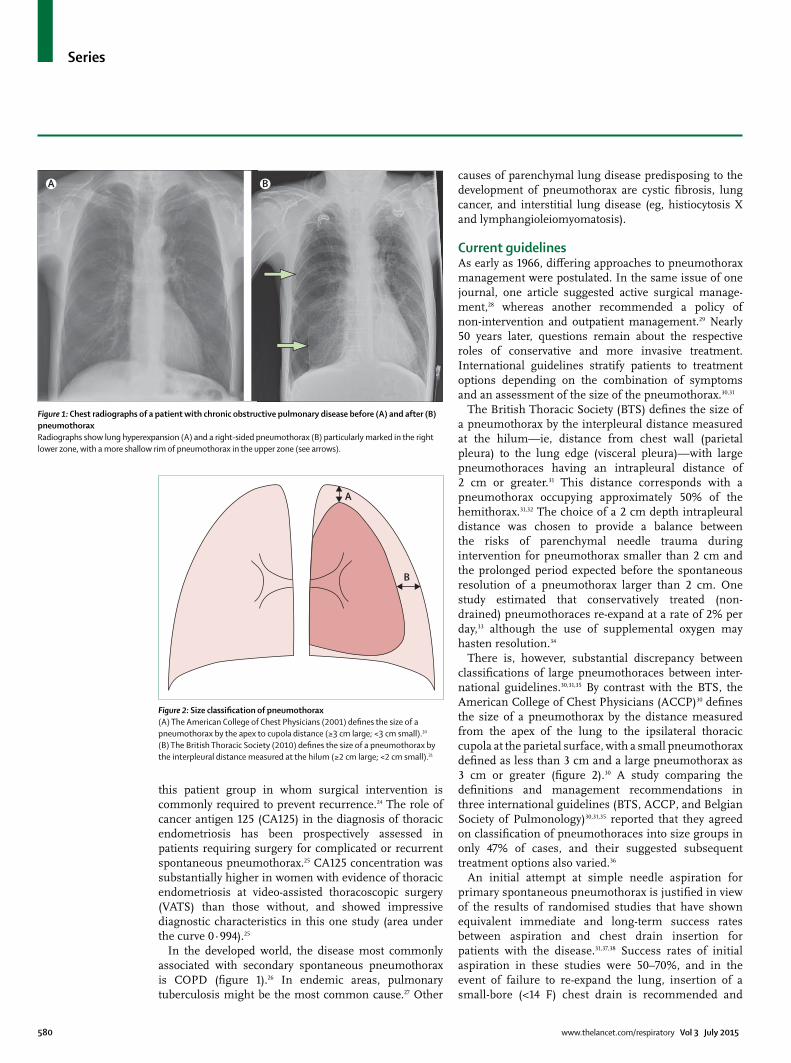
580 www.thelancet.com/respiratory Vol 3 July 2015
Series
this patient group in whom surgical inter vention is commonly required to prevent recurrence.24 The role of cancer antigen 125 (CA125) in the diagnosis of thoracic endometriosis has been prospectively assessed in patients requiring surgery for complicated or recurrent spontaneous pneumothorax.25 CA125 concentration was substantially higher in women with evidence of thoracic endometriosis at video-assisted thoracoscopic surgery (VATS) than those without, and showed impressive diagnostic characteristics in this one study (area under the curve 0·994).25
In the developed world, the disease most commonly associated with secondary spontaneous pneumothorax is COPD (fi gure 1).26 In endemic areas, pulmonary tuberculosis might be the most common cause.27 Other
causes of parenchymal lung disease predisposing to the develop ment of pneumothorax are cystic fi brosis, lung cancer, and interstitial lung disease (eg, histiocytosis X and lymphangioleiomyomatosis).
Current guidelinesAs early as 1966, diff ering approaches to pneumothorax management were postulated. In the same issue of one journal, one article suggested active surgical manage-ment,28 whereas another recommended a policy of non-intervention and outpatient management.29 Nearly 50 years later, questions remain about the respective roles of conservative and more invasive treatment. International guidelines stratify patients to treatment options depending on the combination of symptoms and an assessment of the size of the pneumothorax.30,31
The British Thoracic Society (BTS) defi nes the size of a pneumothorax by the interpleural distance measured at the hilum—ie, distance from chest wall (parietal pleura) to the lung edge (visceral pleura)—with large pneumothoraces having an intrapleural distance of 2 cm or greater.31 This distance corresponds with a pneumothorax occupying approxi mately 50% of the hemithorax.31,32 The choice of a 2 cm depth intrapleural distance was chosen to provide a balance between the risks of parenchymal needle trauma during intervention for pneumothorax smaller than 2 cm and the prolonged period expected before the spontaneous resolution of a pneumothorax larger than 2 cm. One study estimated that conservatively treated (non-drained) pneumothoraces re-expand at a rate of 2% per day,33 although the use of supplemental oxygen may hasten resolution.34
There is, however, substantial discrepancy between classifi cations of large pneumothoraces between inter-national guidelines.30,31,35 By contrast with the BTS, the American College of Chest Physicians (ACCP)30 defi nes the size of a pneumothorax by the distance measured from the apex of the lung to the ipsilateral thoracic cupola at the parietal surface, with a small pneumothorax defi ned as less than 3 cm and a large pneumothorax as 3 cm or greater (fi gure 2).30 A study comparing the defi nitions and management recommendations in three international guidelines (BTS, ACCP, and Belgian Society of Pulmonology)30,31,35 reported that they agreed on classifi cation of pneumothoraces into size groups in only 47% of cases, and their suggested subsequent treatment options also varied.36
An initial attempt at simple needle aspiration for primary spontaneous pneumothorax is justifi ed in view of the results of randomised studies that have shown equivalent immediate and long-term success rates between aspiration and chest drain insertion for patients with the disease.31,37,38 Success rates of initial aspiration in these studies were 50–70%, and in the event of failure to re-expand the lung, insertion of a small-bore (<14 F) chest drain is recommended and
Figure 1: Chest radiographs of a patient with chronic obstructive pulmonary disease before (A) and after (B) pneumothoraxRadiographs show lung hyperexpansion (A) and a right-sided pneumothorax (B) particularly marked in the right lower zone, with a more shallow rim of pneumothorax in the upper zone (see arrows).
A B
Figure 2: Size classifi cation of pneumothorax(A) The American College of Chest Physicians (2001) defi nes the size of a pneumothorax by the apex to cupola distance (≥3 cm large; <3 cm small).30 (B) The British Thoracic Society (2010) defi nes the size of a pneumothorax by the interpleural distance measured at the hilum (≥2 cm large; <2 cm small).31
A
B

www.thelancet.com/respiratory Vol 3 July 2015 581
Series
admission of the patient to hospital. International guidelines suggest the use of smaller bore drains (with a Seldinger technique) rather than large-bore surgical drains in uncomplicated pneumothorax.30,31 Smaller drains have a similar success rate to larger drains and lower levels of discomfort associated with their use.30,31,39,40
Patients with secondary spontaneous pneumothorax tend to have more severe symptoms, greater co-morbidity, and higher mortality rates than do those with primary spontaneous pneumothorax;41,42 treatment recom mendations therefore diff er. The BTS and ACCP guidelines suggest that all patients with secondary spontaneous pneumothoraces are admitted to hospital, with most patients requiring chest drain insertion,30,31 due in part to the reduced likelihood of spontaneously resolving an air leak.43,44 BTS guidelines suggest that an air leak in secondary spontaneous pneumothorax beyond 48 h is an indication for surgical referral.31 Most of the ACCP consensus panel recommended surgical intervention after an initial secondary spontaneous pneumothorax,30 and both guidelines comment on medical pleurodesis as an option in patients unfi t for surgery and the use of Heimlich valves in selected patients.30,31
Evidently, guidelines cannot be wholly prescriptive in managing all patients with pneumothorax, and as a result there is extensive variation in practice, both between individual clinicians and geographically. In some countries there is a lower threshold for off ering surgical intervention at the fi rst episode of pneumothorax, and in other countries a much more conservative approach is favoured. This variation is shown by the recommendation in some countries that all patients are admitted to hospital, global variability in the uptake of needle aspiration versus intercostal drain insertion, and the relative willingness in some countries to observe patients with pneumothoraces that have few or no symptoms.45–47
Lung apposition and pneumothorax resolutionFundamental to the lack of progress in management of primary spontaneous pneumothorax is a poor under-standing of its precise causal mechanisms. For decades, clinicians believed that primary spontaneous pneumo-thorax resulted from the leakage of air from the lung into the pleural cavity via a single breach site (eg, bleb) in the visceral pleura. A major revelation the past decade is that the “one-airway-one-bleb-one-leak” concept is over-simplistic and likely to be incorrect. Although blebs can be a source of air leak, many patients do not have detectable blebs.48,49 Noppen and colleagues50,51 have described diff use areas of weakness in the visceral pleura, which may be responsible for air leak, reiterating an old suggestion that the visceral pleura is lined with pores that permit air passage into the pleural cavity.
The importance of collateral ventilation within, and even between, lobes of the lung (from incomplete fi ssures) is now well established, in part from lessons learned from endobronchial lung volume reduction strategies. This mechanism also applies in pneumothorax; on average three endobronchial valves are needed to stop an air leak, suggesting the existence of multiple feeding pathways.52 Unravelling the mechanism of how air moves from the lung to the pleural cavity will have major eff ects on how primary spontaneous pneumothorax should be managed.
Two crucial questions arise when presented with a patient with primary spontaneous pneumothorax: has the air leak stopped and what is the risk of recurrence? Conventional maxims on the approach to both questions have recently been challenged, making the management of primary spontaneous pneumothorax an exciting area of research.
Removal of the pleural air in an attempt to re-expand the lung has been the standard approach passed on from one generation to the next. Stradling and Poole29 fi rst proposed that visceral leak sites are more likely to heal if the lung is collapsed, allowing apposition of the visceral wound. This concept has been downplayed as clinicians became more concerned about improving the appearances of radio graphs. Large drains, apical placement of tubes, and application of suction were all tried and are still routinely used to restore full lung expansion during ongoing air leaks. Although striving for lung re-expansion may, in some cases, be needed to improve patient symptoms, it might not be necessary in patients with primary spontaneous pneumothorax, in whom symptoms usually subside after 24 h.53 Clinicians have rarely questioned how expansion of the lung aids healing of the leak. Bringing normal visceral and parietal pleura surfaces together would not usually facilitate a spontaneous pleurodesis. Half a century since the report by Stradling and Poole,29 the non-intervention approach is fi nally being tested in a randomised trial in Australasia that is recruiting clinically stable patients with large primary spontaneous pneumo thoraces (ACTRN12611000184976).
If the Stradling and Poole hypothesis is true, and that healing of the visceral wound is the key to management, then recent data on the resurgence of the role of blood patch in persistent air leak (especially in secondary spontaneous pneumothorax) should be further explored.54 Blood instillation could promote clotting over the wound as a mechanism of healing the air leak rather than a means of achieving symphysis of the pleural surfaces.
Lung density and risk stratifi cationAn accurate assessment of the risk of recurrence is crucial to improving care in spontaneous pneu-mothorax. Most centres (and international guidelines) recommend reserving defi nitive recurrence prevention

582 www.thelancet.com/respiratory Vol 3 July 2015
Series
approaches until the second or third presentation of primary spontaneous pneumothorax. This recom-mendation is based on the commonly quoted fi gure of a 20–30% risk of recurrence after an initial primary spontaneous pneumothorax; however, high-quality studies have quoted recurrence rates in excess of this.55,56 Two of the largest randomised controlled trials (RCTs) to date have shown high recurrence rates of 49% (in 214 patients with primary spontaneous pneumothorax)55 and 41% (in 229 patients with primary or secondary spontaneous pneumothorax)56 in control groups who received simple drainage only. The discrepancy between the recurrence rates recorded in these trials and those from smaller cohort studies could have two explanations. First, most of the smaller studies were retrospective and thus vulnerable to selection bias. Second, the two large RCTs had an intervention group and required, as an entry criterion, a pneumothorax of adequate size for drainage. If the recurrence rate for patients with a pneumothorax of an adequate size for drainage is in fact in excess of 40%, this would favour the selected early use of strategies to prevent recurrence.
All international guidelines have so far treated all primary spontaneous pneumothoraces in the same way with regards to recurrence prevention, irrespective of established risk factors such as height, family history, smoking status, and the size of the initial pneumothorax. It is possible that a patient with Marfan’s syndrome with multiple blebs on CT, presenting with a complete lung collapse, will have a higher risk of recurrence than a patient with a small rim of primary spontaneous pneumothorax and no other risk factors. Thus, there is a need for stratifi cation of primary spontaneous pneumothoraces based on phenotype, demographic and radiological features, or biomarkers, which will require a longitudinal observational database on a multinational scale.
Studies have previously explored whether the risk of recurrence of pneumothorax depends on the presence of blebs, bullae, or parenchymal abnormalities. The diff erence in recurrence rates for pneumothorax between primary and secondary disease types suggests that the presence of diff use lung disease predisposes to recurrent episodes.6 Patients with primary spontaneous pneumothorax who have emphysema-like changes might represent a group at a higher risk of recurrence than patients with no such lung abnormalities because the natural history of their pneumothorax may more closely resemble secondary spontaneous pneumothorax than it does in those with genuinely idiopathic pneumothorax. However, advanced techniques such as fl uorescein-enhanced autofl uorescence thoracoscopy, which is mainly a research tool at present, might be needed to identify these emphysema-like changes (fi gure 3).50
Low-radiation dose CT may, in the future, allow a risk stratifi cation of patients at baseline through identifi cation of lung abnormalities, either ipsilaterally or in the contralateral lung. Previous studies with diff erent designs have investigated the presence of bullae on CT scans, with contradictory results. Early small studies had suggested that presence of bullae might be predictive of high recurrence rate and hence justify early surgical intervention.57,58
Sihoe and colleagues58 reported that contralateral recurrence was substantially more common in patients with blebs and bullae on the contralateral lung at the time of initial surgery for unilateral primary spontaneous pneumothorax than it was in patients without contra-lateral blebs and bullae. However, the statistical signifi cance calculations in this study were later called into question.59 Other studies have shown no diff erence in recurrence rates, irrespective of presence or absence of blebs or bullae.4,60,61 There were no diff erences in the thoracoscopic features of blebs and bullae between patients with fi rst and recurrent pneumothoraces in a study of 82 patients, suggesting that recurrence of pneumothorax cannot be predicted by thoracoscopic features.62 Ouanes-Besbes and colleagues63 assessed a bullae scoring system in a prospective cohort of 80 patients and recorded no diff erence in recurrence rates between patients with and without dystrophic lesions seen on CT. However, a subsequent study of 176 patients, which used the same dystrophic severity score as Ounes-Besbes and colleagues, did suggest that blebs and bullae were predictive of recurrence.64 Therefore, the usefulness of CT in predicting recurrence of pneumothorax has not been fi rmly established and needs prospective validation.
An alternative radiological predictor could incorporate the objective quantifi cation of low-density areas within the lung (rather than bullae), which correspond to emphysema-like change and may predispose to recurrent episodes. Smit and colleagues65 reported that
Figure 3: Fluorescein-enhanced autofl uorescence thoracoscopy after pneumothoraxThe lung (on the right of the image) has a green-yellow hue, indicating subpleural fl uorescein, which suggests the presence of a region of parenchymal abnormality. Image courtesy of Marc Noppen.

www.thelancet.com/respiratory Vol 3 July 2015 583
Series
lung density measurements on CT done during expiration were lower in patients with primary spontaneous pneumothorax (21 with, 20 without bullae) in both the aff ected lung and the contralateral lung, compared with 41 healthy volunteers, suggesting the presence of air trapping in patients with pneumothorax. These changes seemed to be independent of smoking behaviour and presence or absence of bullae.65 Un-published pilot work from the Bristol pleural research group lends support to these fi ndings by showing that volumetric apical low lung density on CT (less than –950 Hounsfi eld units is substantially greater in patients with primary pneumothorax than in controls who are smokers or non-smokers (Maskell NA, unpublished). Whether lung density in patients with primary spontaneous pneumothorax is correlated with risk of recurrence is yet to be established, but if so, this association could allow a more accurate estimate of the risk of recurrence.
If physiological, demographic, or radiological infor-mation is shown to act as a predictor for risk of recurrence, this will allow more informed discussions with patients about their individual risk. If this approach proves possible, the early identifi cation of patients at the highest risk of recurrence could be of great value in establishing a subgroup of patients who would benefi t from being off ered defi nitive intervention after an initial pneumothorax.
Ambulatory care: the role of Heimlich valvesThe clinical value of observing stable patients, especially those with primary spontaneous pneumothorax, for days in hospital can be questioned. Patients requiring chest drain insertion have historically been admitted to hospital and the drain connected to a bulky underwater seal or suction device. This approach has remained unchanged despite decades of documented use of Heimlich valves (one-way valves connected to the end of the chest drain), which allow greater mobility and potentially allow patients to be discharged home, thus letting the lung re-expand over time at home.
As early as 1976, small studies have shown the feasibility of this outpatient management of primary spontaneous pneumothorax. A case series of 226 patients with primary spontaneous pneumothorax managed by observation or fl utter valve concluded that outpatient management was “safe, effi cient, and economical”.66 A randomised trial of 30 patients with primary spontaneous pneumothorax (17 assigned to thoracic vent, 13 to standard chest drain) showed no signifi cant diff erence between groups in complications or re-expansion rates, but 70% of patients assigned to thoracic vent were managed as outpatients and needed fewer analgesics, with patients in the control group staying in hospital for a mean of 8 days.67 A systematic review of 18 studies of ambulatory management with Heimlich valves reported an overall success rate of 86% and
successful outpatient management in 78% of cases, with few complications.68 However, the evidence was of poor quality with a high risk of bias, consisting mainly of two small randomised trials with the remainder being case series. Despite further obser vational studies reporting eff ectiveness and a cost saving,69,70 the paucity of robust data is probably the reason for low levels of uptake of Heimlich valves into standard clinical practice.
The BTS guidelines mention Heimlich valves briefl y with respect to “facilitating mobilisation and outpatient care”, but the guidance is not more prescriptive in its recommendation.31 The ACCP consensus statement, however, provides the physician with the option to discharge “reliable” patients home with a small-bore catheter attached to a Heimlich valve if the lung has re-expanded after the removal of pleural air.30 Appropriately powered and robust RCTs will help to identify whether there are advantages associated with the use of Heimlich valves and to ascertain the patient population in whom their use is benefi cial. A grant to undertake such a trial has recently been supported in the UK (National Institute of Health Research grant: PB-PG-0213–30098).
Digital air-leak measurementWhen standard management does not suffi ciently resolve the air leak, surgical referral is recommended.31 However, the optimum timing of defi nitive intervention is unknown. Current guidelines suggest that in patients with a persistent air leak or failure of lung re-expansion, an early (3–5 days) thoracic surgical opinion should be sought, but there are no published data on prediction of persistent air leak or requirement for inpatient surgical intervention.
If persistent air leak could be predicted early (ie, within 48 h), this would allow triage of patients to new management pathways with informed patient-physician discussions. Stable patients predicted to have low probability of long-term leak could be discharged home with an ambulatory drain to allow resolution at home; those likely to have signifi cant persistent air leak could be triaged early for assessment for more defi nitive intervention, rather than waiting for daily assessment and referral after four or fi ve nights in hospital.
Digital measurement of air leak is possible with commercially available systems capable of providing regulated suction (via rotary pump and diaphragm) and validated measurements of air leak (through a revolutions per minute counter or thermodilution principles).71 There are, so far, no published studies assessing air leak in medical patients with either primary or secondary spontaneous pneumothoraces; however, postoperative data from patients who have undergone thoracic surgery suggest that digital measurement of air leak might be a useful strategy.72
A case series of 142 patients who had undergone thoracic surgery reported postoperative air leak of more

584 www.thelancet.com/respiratory Vol 3 July 2015
Series
than 180 mL/min on day 2 after surgery to be predictive of prolonged air leak (>5 days).73 Five RCTs, a case-control study, and one large observational study (total of 956 postsurgical patients) showed that digital suction devices measured air leak more accurately than the traditional “bubbles in a chamber” method (currently used in medical management of pneumothorax), and reduced length of drainage and hospital stay after surgery.74–76
Although digital air-leak measurement has not been robustly assessed in the medical management of pneumothorax, and is not considered standard manage-ment, these studies of surgical patients suggest that it may well be a surrogate marker for persistent air leak, and hence non-resolving pneumothorax, and therefore allow early identifi cation of patients who require more defi nitive management. However, caution is required in the application of postsurgical data to patients with medical spontaneous pneumothorax.
Conservative managementInternational guidelines suggest a role for conservative management (observation alone) of clinically stable patients with primary spontaneous pneumothorax with close radiological follow-up to ensure resolution.30,31 The previously mentioned study in Australasia (ACTRN12611000184976) is randomly assigning clinically stable patients with large primary spontaneous pneumothoraces to either observation without pleural intervention or standard care with needle aspiration and intercostal drain insertion. If leaving primary spontaneous pneumothorax undrained is shown to support healing of air leaks, it will profoundly alter management approaches worldwide.
Historically, studies of pneumothorax have focused on radiological evidence of lung re-expansion, rather than patient-centred outcomes such as degree of breath lessness and the need for further intervention. This approach has extended into international guidelines, in which achieving lung re-expansion has often been seen as the primary objective, rather than a means to reduce patient symptoms and ensure haemodynamic stability.
Prevention of pneumothorax recurrenceMedical or surgical pleurodesis is advised for second ipsilateral primary spontaneous pneumothorax.30,31,35 However, because of the high rates of recurrence reported in the fi rst year, the argument could be made to off er pleurodesis at the fi rst episode.
Chen and colleagues55 provide an important insight into the feasibility of pleurodesis after simple aspiration for primary spontaneous pneumothorax and attempt to redefi ne a treatment algorithm for the fi rst episode of the disease. There are, however, important limitations to consider. The diff erence between the typically quoted recurrence rates for primary spontaneous
pneumothorax (around 30%),37 and the rate recorded in their control group (49%) calls into question the relevance of the statistically signifi cant diff erence between the minocycline and control groups (29% vs 49%). Tetracycline and its derivatives are no longer recommended sclerosing agents in the UK;31 however, ACCP guidelines suggest use of either doxycycline or talc slurry. Although there are no directly comparative controlled trials for pneumothorax, graded talc seems more eff ective than tetracyclines and has been shown to be safe.77–80 The high success rate of talc poudrage during thoracoscopy might be explained by a diff use distribution of talc particles within the pleural cavity under direct vision or the brief interval between talc application and lung re-expansion within the same procedure.78
When invasive procedures are inappropriate (either patient suitability or preference), medical pleurodesis is a suitable alternative. However, the case for instillation of minocycline as fi rst-line treatment for a fi rst episode of pneumothorax to achieve a recurrence rate similar to that provided by simple aspiration or drainage in other studies is not suffi ciently convincing to prompt a decisive change in standard patient management.
Targeted surgical managementElective surgery is commonly undertaken to reduce the risk of recurrent pneumothorax after a second episode, but surgical intervention is also recommended when simple medical management does not resolve an acute air leak.31 However, the best possible timing for surgery has not been established. UK guidelines suggest 5–7 days from the onset of air leak but evidence is limited.31 Indeed, one study including patients with both primary and secondary spontaneous pneumothoraces reported that after 14 days of drainage, air leaks had stopped in all cases of primary spontaneous pneumothorax and in 79% of those with secondary spontaneous pneumothorax, although many of these patients also received chemical pleurodesis.44 Another study suggests that most primary spontaneous pneumothoraces resolve by 9 days, thereby advocating referral at 7–9 days.81 By contrast, a UK series reported inferior outcomes when surgery was delayed beyond 21 days from acute presentation.82 The chosen timescales might refl ect the degree of acceptability to patients, the clinician’s patience, and available health-care resources, rather than high-quality evidence.
Current indications for more invasive treatment to prevent recurrence are second (ie, recurrent) ipsilateral pneumothorax, bilateral pneumothorax, and professions at risk (eg, pilots and divers).31 Surgical bullectomy alone is associated with a high rate of pneumothorax recurrence (6–14%),83–85 suggesting that resection of bullae alone is not suffi cient, unless there is a proven air leak at this site. Surgical series showing poorer rates of recurrence prevention with simple bullectomy compared

www.thelancet.com/respiratory Vol 3 July 2015 585
Series
with bullectomy and pleurectomy or pleurodesis confi rm that it is necessary to also undertake diff use treatment of the visceral pleura.83–85
There are two approaches for pneumothorax surgery: thoracotomy or VATS. Irrespective of the approach, visible blebs and bullae are usually resected and then partial pleurectomy, pleural abrasion, or instillation of a sclerosing agent (ie, talc) is undertaken. VATS is better tolerated than open thoracotomy.86 VATS has grown in popularity, and accounted for over 80% of pneumothorax surgery in the UK in 2010.87 However, some studies have reported higher rates of pneu-mothorax recurrence in patients assigned to VATS than in those assigned to open thoracotomy, with respective recurrence rates of 5% versus 1% in a meta-analysis88 and 3·8% versus 1·8% in a study that used propensity score analysis.89 Another study of minithoracotomy compared with VATS showed similar rates of recurrence (2·7% and 3%, respectively) and postoperative pain.90 Patients assigned to VATS had higher patient satisfaction level (assessed by use of the ipsilateral arm postoperatively) and return to activity than patients assigned to minithoracotomy.90 After bullectomy at VATS for primary spontaneous pneumothorax, coverage of the staple line with a cellulose mesh and fi brin glue has been shown to be no worse than mechanical pleurodesis in terms of recurrence of pneumothorax in a large RCT.91 New approaches including single port or awake VATS have also been described.92,93 Reductions in morbidity may lead to a re-evaluation of the role and timing of surgery for primary spontaneous pneumothorax.
An earlier RCT assessing the effi cacy of talc pleurodesis by medical thoracoscopy under local anaesthesia compared with chest tube drainage in cases of recurrent or complicated primary spontaneous pneumothorax, showed a very low long-term recurrence rate (around 5%) after talc poudrage with no diff erence in costs or complication rates between these two approaches.78 There has been a trend towards the use of less invasive surgical approaches, such as VATS, driven by strong evidence of their effi cacy in treating patients with recurrent pneumothorax.90 However, the case could be made for referral of higher risk patients on fi rst presentation.
Important questions for future researchThe identifi cation of factors predicting both persistent air leak and recurrence of pneumothorax would be of great value in early stratifi cation of patients to the appropriate management strategy. Digital measure-ment of air leak and radiological features, respectively, could hold promise in this area. Modern resources potentially allow a more detailed work-up of patients with primary spontaneous pneumothorax than was possible historically; however, the extent to which this will alter management and the role, for instance, of
measuring CA125 concentration in female patients with primary spontaneous pneumothorax warrants further exploration.
The role of conservative management in pneumothorax is being assessed in a large RCT that is currently recruiting participants (ACTRN12611000184976), and funding has been made available from the National Institute of Health Research—Research for Patient Benefi t Grant (RfPB) for a randomised, controlled trial assessing length of stay for ambulatory management (using valve device) against standard BTS guidelines for primary spontaneous pneumothorax, to begin recruiting in UK in 2015. These two trials will help to map out the roles of conservative and ambulatory management.
There is evidence from an RCT suggesting a role for bedside (chemical) pleurodesis in reducing the risk of recurrence in primary spontaneous pneumothorax.55 However, for this strategy to be taken up more widely, further work will be necessary because of the high rates of pneumothorax recurrence seen in this study.
ConclusionsPneumothorax has been an under-researched area, and available evidence has been of fairly low quality, giving rise to international guidelines largely based on consensus and observational evidence, with few areas of agreement between them. Future high-quality studies may allow development of tailored management strategies, in creasingly personalised care, a move towards outpatient-based treatment, and more con-servative management. A risk stratifi cation system at fi rst presentation could identify patients who will benefi t from intervention to prevent recurrence at the fi rst presentation, rather than, as has historically been the case, simply waiting for a recurrence to occur. Further studies are needed to redefi ne the treatment
Search strategy and selection criteria
We identifi ed relevant studies for inclusion in this Series paper by searches of Medline, between Jan 1, 1980 to Jan 13, 2015, without language restrictions, with the search term “pneumothorax” appearing within the title and abstract of relevant study types (all clinical trials, guidelines, reviews, systematic reviews, and meta-analyses). Studies were restricted to those on adult patients and recent studies were prioritised. Additionally, the most up-to-date guidelines from international societies (American College of Chest Physicians, British Thoracic Society, and the Belgian Society of Pneumonology) were reviewed. Titles and abstracts were screened for relevance. We excluded studies related to traumatic pneumothorax or management of complications after thoracic surgery. Where relevant, further studies were identifi ed through reference lists of reviewed papers. For foreign language studies, translations were used.

586 www.thelancet.com/respiratory Vol 3 July 2015
Series
framework that would provide early triage to patients who can be treated conservatively, those who are suitable for ambulatory management as outpatients, and those who require early intervention. Such a framework would allow availability of a tailored spectrum of options and ensure that invasive treatments are off ered promptly to those at high risk of recurrence.ContributorsOJB and RJH contributed equally to the search and appraisal of relevant studies for inclusion and the initial drafting of the manuscript. AE, DF-K, YCGL, CHM, J-MT, DW, NMR, and NAM reviewed the manuscript and proposed amendments, corrections, and clarifi cations. All authors approved the fi nal version for submission.
Declaration of interestsDF-K reports personal fees from Spiration, outside of the submitted work. DW reports grants from Covidien, outside of the submitted work. NAM reports grants from CareFusion to run the IPC plus study and personal fees to attend a CareFusion advisory board meeting, outside of the submitted work. NMR reports grants and personal fees from Rocket Medical UK, outside of the submitted work. All other authors declare no competing interests.
References1 Kjæergaard H. Spontaneous pneumothorax in the apparently
healthy. Acta Med Scand Suppl 1932; 43: 1–159.2 Hyde B, Hyde L. Spontaneous pneumothorax—contrast of the
benign idiopathic and the tuberculous types. Ann Intern Med 1950; 33: 1373–77.
3 Bense L, Lewander R, Eklund G, et al. Nonsmoking, non-alpha 1-antitrypsin defi ciency-induced emphysema in nonsmokers with healed spontaneous pneumothorax, identifi ed by computed tomography of the lungs. Chest 1993; 103: 433–38.
4 Mitlehner W, Friedrich M, Dissmann W. Value of computer tomography in the detection of bullae and blebs in patients with primary spontaneous pneumothorax. Respiration 1992; 59: 221–27.
5 Gupta D, Hansell A, Nichols T, et al. Epidemiology of pneumothorax in England. Thorax 2000; 55: 666–71.
6 Guo Y, Xie C, Rodriguez RM, et al. Factors related to recurrence of spontaneous pneumothorax. Respirology 2005; 10: 378–84.
7 Cottin V, Streichenberger N, Gamondès JP, et al. Respiratory bronchiolitis in smokers with spontaneous pneumothorax. Eur Respir J 1998; 12: 702–04.
8 Cheng YL, Huang TW, Lin CK, et al. The impact of smoking in primary spontaneous pneumothorax. J Thorac Cardiovasc Surg 2009; 138: 192–95.
9 Melton LJ 3rd, Hepper NG, Off ord KP. Infl uence of height on the risk of spontaneous pneumothorax. Mayo Clin Proc 1981; 56: 678–82.
10 Bense L, Eklund G, Wiman LG. Smoking and the increased risk of contracting spontaneous pneumothorax. Chest 1987; 92: 1009–12.
11 Sadikot RT, Greene T, Meadows K, et al. Recurrence of primary spontaneous pneumothorax. Thorax 1997; 52: 805–09.
12 Johnson MK, Smith RP, Morrison D, et al. Large lung bullae in marijuana smokers. Thorax 2000; 55: 340–42.
13 Gill A. Bong lung: regular smokers of cannabis show relatively distinctive histologic changes that predispose to pneumothorax. Am J Surg Pathol 2005; 29: 980–82.
14 Feldman AL, Sullivan JT, Passero MA, et al. Pneumothorax in polysubstance-abusing marijuana and tobacco smokers: three cases. J Subst Abuse 1993; 5: 183–86.
15 Dyhdalo K, Farver C. Pulmonary histologic changes in Marfan syndrome: a case series and literature review. Am J Clin Pathol 2011; 136: 857–63.
16 Toro JR, Pautler SE, Stewart L, et al. Lung cysts, spontaneous pneumothorax, and genetic associations in 89 families with Birt-Hogg-Dubé syndrome. Am J Respir Crit Care Med 2007; 175: 1044.
17 Ren HZ, Zhu CC, Yang C, et al. Mutation analysis of the FLCN gene in Chinese patients with sporadic and familial isolated primary spontaneous pneumothorax Clin Genet 2008; 74: 178–83.
18 Daniel R, Teba L. Spontaneous pneumothorax and alpha 1-antitrypsin defi ciency. Respir Care 2000; 45: 327–29.
19 Bass HN, LaGrave D, Mardach R, et al. Spontaneous pneumothorax in association with pyridoxine-responsive homocystinuria. J Inherit Metab Dis 1999; 20: 831–32.
20 Johannesma PC, Reinhard R, Kon Y, et al. Prevalence of Birt-Hogg-Dubé syndrome in patients with apparently primary spontaneous pneumothorax. Eur Respir J 2015; 45: 1191–94.
21 Biffl WL, Narayanan V, Gaudiani JL, et al. The management of pneumothorax in patients with anorexia nervosa: a case report and review of the literature. Patient Saf Surg 2010; 4: 1.
22 Huang TW, Lee SC, Cheng YL, et al. Contralateral recurrence of primary spontaneous pneumothorax. Chest 2007; 132: 1146–50.
23 Alifano M, Roth T, Broët SC, et al. Catamenial pneumothorax: a prospective study. Chest 2003; 124: 1004–08.
24 Joseph J, Sahn SA. Thoracic endometriosis syndrome: new observations from an analysis of 110 cases. Am J Med 1996; 100: 164–70.
25 Bagan P, Berna P, Assouad J, Hupertan V, Le Pimpec Barthes F, Riquet M. Value of cancer antigen 125 for diagnosis of pleural endometriosis in females with recurrent pneumothorax. Eur Respir J 2008; 31: 140–42.
26 Weissberg D, Refaely Y. Pneumothorax: experience with 1,199 patients. Chest 2000; 117: 1279–85.
27 Shamaei M, Tabarsi P, Pojhan, et al. Tuberculosis-associated secondary pneumothorax: a retrospective study of 53 patients. Respir Care 2011; 56: 298–302.
28 Ruckley CV, McCormack RJ. The management of spontaneous pneumothorax. Thorax 1966; 21: 139–44.
29 Stradling P, Poole G. Conservative management of spontaneous pneumothorax. Thorax 1966; 21: 145–49.
30 Baumann MH, Strange C, Heff ner JE, et al, and the AACP Pneumothorax Consensus Group. Management of spontaneous pneumothorax: an American College of Chest Physicians Delphi consensus statement. Chest 2001; 119: 590–602.
31 MacDuff A, Arnold A, Harvey J, and the BTS Pleural Disease Guideline Group. Management of spontaneous pneumothorax: British Thoracic Society Pleural Disease Guideline 2010. Thorax 2010; 65 (suppl 2): ii18–31.
32 Henry M, Arnold T, Harvey J, and the Pleural Diseases Group, Standards of Care Committee, British Thoracic Society. BTS guidelines for the management of spontaneous pneumothorax. Thorax 2003; 58 (suppl 2): ii39–52.
33 Kelly AM, Weldon D, Tsang AY, et al. Comparison between two methods for estimating pneumothorax size from chest x-rays. Respir Med 2006; 100: 1356–59.
34 Northfi eld TC. Oxygen therapy for spontaneous pneumothorax. BMJ 1971; 4: 86–88.
35 De Leyn P, Lismonde M, Ninane V, et al. Belgian Society of Pneumology. Guidelines on the management of spontaneous pneumothorax. Acta Chir Belg 2005; 105: 265–67.
36 Kelly A-M, Druda D. Comparison of size classifi cation of primary spontaneous pneumothorax by three international guidelines: a case for international consensus? Respir Med 2008; 102: 1830–32.
37 Noppen M, Alexander P, Driesen P, et al. Manual aspiration versus chest tube drainage in fi rst episodes of primary spontaneous pneumothorax: a multicenter, prospective, randomized pilot study. Am J Respir Crit Care Med 2002; 165: 1240–44.
38 Wakai A, O’Sullivan RG, McCabe G. Simple aspiration versus intercostal tube drainage for primary spontaneous pneumothorax in adults. Cochrane Database Syst Rev 2007; 1: CD004479.
39 Vedam H, Barnes DJ. Comparison of large- and small-bore intercostal catheters in the management of spontaneous pneumothorax. Intern Med J 2003; 33: 495–99.
40 Clementsen P, Evald T, Grode G, et al. Treatment of malignant pleural eff usion: pleurodesis using a small percutaneous catheter. A prospective randomized study. Respir Med 1998; 92: 593–96.
41 Spector ML, Stern RC. Pneumothorax in cystic fi brosis: a 26-year experience. Ann Thorac Surg 1989; 47: 204–07.
42 Wait MA, Estrera A. Changing clinical spectrum of spontaneous pneumothorax. Am J Surg 1992; 164: 528–31.

www.thelancet.com/respiratory Vol 3 July 2015 587
Series
43 Schoenenberger RA, Haefeli WE, Weiss P, et al. Timing of invasive procedures in therapy for primary and secondary spontaneous pneumothorax. Arch Surg 1991; 126: 764–66.
44 Chee CB, Abisheganaden J, Yeo JK, et al. Persistent air-leak in spontaneous pneumothorax–clinical course and outcome. Respir Med 1998; 92: 757–61.
45 Baumann MH, Strange C. The clinician’s perspective on pneumothorax management. Chest 1997; 112: 822–28.
46 Janssen JP, Cuesta MA, Postmus PE. Treatment of spontaneous pneumothorax: survey among Dutch pneumonologists. Ned Tijdschr Geneeskd 1994; 138: 661–64 (in Dutch).
47 Ong ME, Chan YH, Kee TY, et al. Spontaneous pneumothorax outcome study (SPOT phase I): a 2-year review. Eur J Emerg Med 2004; 11: 89–94.
48 Cardillo G, Carleo F, Giunti R, et al. Videothoracoscopic talc poudrage in primary spontaneous pneumothorax: a single-institution experience in 861 cases. J Thorac Cardiovasc Surg 2006; 131: 322–28.
49 Khan OA, Tsang GM, Barlow CW, et al. Routine histological analysis of resected lung tissue in primary spontaneous pneumothorax–is it justifi ed? Heart Lung Circ 2006; 15: 137–38.
50 Noppen M, Dekeukeleire T, Hanon S, et al. Fluorescein-enhanced autofl uorescence thoracoscopy in patients with primary spontaneous pneumothorax and normal subjects. Am J Respir Crit Care Med 2006; 174: 26–30.
51 Noppen M. Do blebs cause primary spontaneous pneumothorax? Con: Blebs do not cause primary spontaneous pneumothorax. J Bronchol 2002; 9: 319–23.
52 Travaline JM, McKenna RJ Jr, De Giacomo T, et al, and the Endobronchial Valve for Persistent Air Leak Group. Treatment of persistent pulmonary air leaks using endobronchial valves. Chest 2009; 136: 355–60.
53 Noppen M, Schramel F. Pneumothorax. Eur Respir Mon 2002; 22: 279–96.
54 Cao G, Kang J, Wang F, Wang H. Intrapleural instillation of autologous blood for persistent air leak in spontaneous pneumothorax in patients with advanced chronic obstructive pulmonary disease. Ann Thorac Surg 2012; 93: 1652–57.
55 Chen JS, Chan WK, Tsai KT, et al. Simple aspiration and drainage and intrapleural minocycline pleurodesis versus simple aspiration and drainage for the initial treatment of primary spontaneous pneumothorax: an open-label, parallel-group, prospective, randomised, controlled trial. Lancet 2013; 381: 1277–82.
56 Light RW, O’Hara VS, Moritz TE, et al. Intrapleural tetracycline for the prevention of recurrent spontaneous pneumothorax. Results of a Department of Veterans Aff airs cooperative study. JAMA 1990; 264: 2224–30.
57 Warner BW, Bailey WW, Shipley RT. Value of computed tomography of the lung in the management of primary spontaneous pneumothorax. Am J Surg 1991; 162: 39–42.
58 Sihoe AD, Yim AP, Lee TW, et al. Can CT scanning be used to select patients with unilateral primary spontaneous pneumothorax for bilateral surgery? Chest 2000; 118: 380–83.
59 Noppen Ml. CT scanning and bilateral surgery for unilateral primary pneumothorax? Chest 2001; 119: 1293–94.
60 Amjadi K, Alvarez GG, Vanderhelst E, et al. The prevalence of blebs or bullae among young healthy adults: a thoracoscopic investigation. Chest 2007; 132: 1140–45.
61 Martínez-Ramos D, Angel-Yepes V, Escrig-Sos J, et al. Usefulness of computed tomography in determining risk of recurrence after a fi rst episode of primary spontaneous pneumothorax: therapeutic implications. Arch Bronconeumol 2007; 43: 304–08 (in Spanish).
62 Janssen JP, Schramel FM, Sutedja TG, et al. Videothoracoscopic appearance of fi rst and recurrent pneumothorax. Chest 1995; 108: 330–34.
63 Ouanes-Besbes L, Golli M, Knani J, et al. Prediction of recurrent spontaneous pneumothorax: CT scan fi ndings versus management features. Respir Med 2007; 101: 230–36.
64 Casali C, Stefani A, Ligabue G, et al. Role of blebs and bullae detected by high-resolution computed tomography and recurrent spontaneous pneumothorax. Ann Thorac Surg 2013; 95: 249–55.
65 Smit HJ, Golding RP, Schramel FM, et al. Lung density measurements in spontaneous pneumothorax demonstrate airtrapping. Chest 2004; 125: 2083–90.
66 Mercier C, Page A, Verdant A, et al. Outpatient management of intercostal tube drainage in spontaneous pneumothorax. Ann Thorac Surg 1976; 22: 163–65.
67 Röggla M, Wagner A, Brunner C, et al. The management of pneumothorax with the thoracic vent versus conventional intercostal tube drainage. Wien Klin Wochenschr 1996; 108: 330–33.
68 Brims FJ, Maskell NA. Ambulatory treatment in the management of pneumothorax: a systematic review of the literature. Thorax 2013; 68: 664–69.
69 Massongo M, Leroy S, Scherpereel A, et al. Outpatient management of primary spontaneous pneumothorax: a prospective study. Eur Respir J 2014; 43: 582–90.
70 Voisin F, Sohier L, Rochas Y, et al. Ambulatory management of large spontaneous pneumothorax with pigtail catheters. Ann Emerg Med 2014; 64: 222–28.
71 Cerfolio RJ, Bryant AS. The quantifi cation of postoperative air leaks. Multimed Man Cardiothorac Surg 2009; 2009: 003129.
72 Pompili C, Detterbeck F, Papagiannopoulos K, et al. Multicenter international randomized comparison of objective and subjective outcomes between electronic and traditional chest drainage systems. Ann Thorac Surg 2014; 98: 490–96, discussion 496–97.
73 Billé A, Borasio P, Gisabella M, et al. Air leaks following pulmonary resection for malignancy: risk factors, qualitative and quantitative analysis. Interact Cardiovasc Thorac Surg 2011; 13: 11–15.
74 Cerfolio RJ, Varela G, Brunelli A. Digital and smart chest drainage systems to monitor air leaks: the birth of a new era? Thorac Surg Clin 2010; 20: 413–20.
75 Pompili C, Brunelli A, Salati M, et al. Impact of the learning curve in the use of a novel electronic chest drainage system after pulmonary lobectomy: a case-matched analysis on the duration of chest tube usage. Interact Cardiovasc Thorac Surg 2011; 13: 490–93, discussion 493.
76 Mier JM, Molins L, Fibla JJ. The benefi ts of digital air leak assessment after pulmonary resection: prospective and comparative study. Cir Esp 2010; 87: 385–89 (in Spanish).
77 Bridevaux PO, Tschopp JM, Cardillo G, et al. Short-term safety of thoracoscopic talc pleurodesis for recurrent primary spontaneous pneumothorax: a prospective European multicentre study. Eur Respir J 2011; 38: 770–73.
78 Tschopp JM, Boutin C, Astoul P, et al, and the ESMEVAT team (European Study on Medical Video-Assisted Thoracoscopy). Talcage by medical thoracoscopy for primary spontaneous pneumothorax is more cost-eff ective than drainage: a randomised study. Eur Respir J 2002; 20: 1003–09.
79 el Khawand C, Marchandise FX, Mayne A, et al. Spontaneous pneumothorax. Results of pleural talc therapy using thoracoscopy. Rev Mal Respir 1995; 12: 275–81 (in French).
80 Györik S, Erni S, Studler U, et al. Long-term follow-up of thoracoscopic talc pleurodesis for primary spontaneous pneumothorax. Eur Respir J 2007; 29: 757–60.
81 Mathur R, Cullen J, Kinnear WJ, et al. Time course of resolution of persistent air leak in spontaneous pneumothorax. Respir Med 1995; 89: 129–32.
82 Waller DA, McConnell SA, Rajesh PB. Delayed referral reduces the success of video-assisted thoracoscopic surgery for spontaneous pneumothorax. Respir Med 1998; 92: 246–49.
83 Horio H, Nomori H, Kobayashi R, et al. Impact of additional pleurodesis in video-assisted thoracoscopic bullectomy for primary spontaneous pneumothorax. Surg Endosc 2002; 16: 630–34.
84 Hatz RA, Kaps MF, Meimarakis G, et al. Long-term results after video-assisted thoracoscopic surgery for fi rst-time and recurrent spontaneous pneumothorax. Ann Thorac Surg 2000; 70: 253–57.
85 Loubani M, Lynch V. Video assisted thoracoscopic bullectomy and acromycin pleurodesis: an eff ective treatment for spontaneous pneumothorax. Respir Med 2000; 94: 888–90.
86 Vohra HA, Adamson L, Weeden DF. Does video-assisted thoracoscopic pleurectomy result in better outcomes than open pleurectomy for primary spontaneous pneumothorax? Interact Cardiovasc Thorac Surg 2008; 7: 673–77.
87 SCTS. Second national thoracic surgery activity and outcomes report 2011. Oxfordshire, UK: Society for Cardiothoracic Surgery in Great Britain and Ireland, 2011.

588 www.thelancet.com/respiratory Vol 3 July 2015
Series
88 Bille A, Barker A, Maratos EC, Edmonds L, Lim E. Surgical access rather than method of pleurodesis (pleurectomy or pleural abrasion) infl uences recurrence rates for pneumothorax surgery: systematic review and meta-analysis. Gen Thorac Cardiovasc Surg 2012; 60: 321–25.
89 Pagès PB, Delpy JP, Falcoz PE, et al, and the Epithor Project (French Society of Thoracic and Cardiovascular Surgery). Videothoracoscopy versus thoracotomy for the treatment of spontaneous pneumothorax: a propensity score analysis. Ann Thorac Surg 2015; 99: 258–63.
90 Foroulis CN, Anastasiadis K, Charokopos N, et al. A modifi ed two-port thoracoscopic technique versus axillary minithoracotomy for the treatment of recurrent spontaneous pneumothorax: a prospective randomized study. Surg Endosc 2012; 26: 607–14.
91 Lee S, Kim HR, Cho S, et al, and the Korean Pneumothorax Study Group. Staple line coverage after bullectomy for primary spontaneous pneumothorax: a randomized trial. Ann Thorac Surg 2014; 98: 2005–11.
92 Yang HC, Cho S, Jheon S. Single-incision thoracoscopic surgery for primary spontaneous pneumothorax using the SILS port compared with conventional three-port surgery. Surg Endosc 2013; 27: 139–45.
93 Rocco G, La Rocca A, Martucci N, Accardo R. Awake single-access (uniportal) video-assisted thoracoscopic surgery for spontaneous pneumothorax. J Thorac Cardiovasc Surg 2011; 142: 944–45.